TopBP1 activates ATR through ATRIP and a PIKK regulatory domain
13
TopBP1 activates ATR through ATRIP and a PIKK regulatory domain Daniel A. Mordes, Gloria G. Glick, Runxiang Zhao, and David Cortez 1 Department of Biochemistry, Vanderbilt University School of Medicine, Nashville, Tennessee 37232, USA The ATR (ATM and Rad3-related) kinase and its regulatory partner ATRIP (ATR-interacting protein) coordinate checkpoint responses to DNA damage and replication stress. TopBP1 functions as a general activator of ATR. However, the mechanism by which TopBP1 activates ATR is unknown. Here, we show that ATRIP contains a TopBP1-interacting region that is necessary for the association of TopBP1 and ATR, for TopBP1-mediated activation of ATR, and for cells to survive and recover DNA synthesis following replication stress. We demonstrate that this region is functionally conserved in the Saccharomyces cerevisiae ATRIP ortholog Ddc2, suggesting a conserved mechanism of regulation. In addition, we identify a domain of ATR that is critical for its activation by TopBP1. Mutations of the ATR PRD (PIKK [phosphoinositide 3-kinase related kinase] Regulatory Domain) do not affect the basal kinase activity of ATR but prevent its activation. Cellular complementation experiments demonstrate that TopBP1-mediated ATR activation is required for checkpoint signaling and cellular viability. The PRDs of ATM and mTOR (mammalian target of rapamycin) were shown previously to regulate the activities of these kinases, and our data indicate that the DNA-PKcs (DNA-dependent protein kinase catalytic subunit) PRD is important for DNA-PKcs regulation. Therefore, divergent amino acid sequences within the PRD and a unique protein partner allow each of these PIK kinases to respond to distinct cellular events. [Keywords: ATR; ATRIP; TopBP1; checkpoint; PIKK; Ddc2] Supplemental material is available at http://www.genesdev.org. Received February 22, 2008; revised version accepted April 7, 2008. The DNA damage response maintains genome integrity through the coordination of DNA replication, cell cycle progression, transcription, apoptosis, senescence, and DNA repair. At the apex of this pathway are two closely related kinases—ATM (ataxia-telangiectasia mutated) and ATR (ATM and Rad3-related)—that phosphorylate hundreds of proteins, including the tumor suppressor proteins BRCA1 and p53 (Kastan and Bartek 2004). ATM primarily responds to DNA double-strand breaks, while ATR is activated by replication stress and ssDNA gaps. ATR and ATM are members of the PIKK (phospho- inositide 3-kinase related kinases) family of protein ki- nases, which regulate diverse biological activities. Other members of this family include mTOR (mammalian tar- get of rapamycin), which coordinates protein synthesis and cell growth; DNA-PKcs (DNA-dependent protein ki- nase catalytic subunit), which promotes DNA double- strand break repair by nonhomologous end-joining; and SMG1, which regulates nonsense-mediated mRNA de- cay (Abraham 2004). PIKKs are large proteins (2549–4128 amino acids) with a common domain architecture. They contain dozens of N-terminal HEAT repeats that may mediate protein–protein interactions and a highly con- served C-terminal kinase domain flanked by the FAT (FRAP, ATM, TRRAP) and FATC (FAT C terminus) do- mains of unknown function. Homozygous mutations in ATM cause ataxia-telangi- ectasia, which is characterized by progressive neurode- generation and severe cancer predisposition, and people with a heterozygous ATM mutation have an increased risk of breast cancer (Shiloh 2003). ATR mutations are infrequent because ATR is essential for cell viability and embryonic development (Brown and Baltimore 2000; Cortez et al. 2001). However, hypomorphic mutations in ATR have been linked to rare cases of Seckel syndrome (O’Driscoll et al. 2003). This syndrome is characterized by microcephaly and developmental defects. ATR forms a stable complex with ATRIP (ATR-inter- acting protein), which regulates the localization of ATR and is essential for ATR signaling in response to DNA damage and replication stress (Cortez et al. 2001; Zou and Elledge 2003; Ball et al. 2005). Many proteins in ad- dition to ATRIP participate in the ATR pathway includ- ing TopBP1, which functions in both the initiation of DNA replication and checkpoint signaling (Garcia et al. 2005). Depletion of TopBP1 in mammalian cells does not affect the localization of ATR–ATRIP to sites of DNA damage but inhibits the damage-inducible phosphoryla- 1 Corresponding author. E-MAIL [email protected]; FAX (615) 343-0704. Article is online at http://www.genesdev.org/cgi/doi/10.1101/gad.1666208. 1478 GENES & DEVELOPMENT 22:1478–1489 © 2008 by Cold Spring Harbor Laboratory Press ISSN 0890-9369/08; www.genesdev.org Cold Spring Harbor Laboratory Press on July 28, 2016 - Published by genesdev.cshlp.org Downloaded from
-
Upload
independent -
Category
Documents
-
view
0 -
download
0
Transcript of TopBP1 activates ATR through ATRIP and a PIKK regulatory domain
TopBP1 activates ATR through ATRIPand a PIKK regulatory domainDaniel A. Mordes, Gloria G. Glick, Runxiang Zhao, and David Cortez1
Department of Biochemistry, Vanderbilt University School of Medicine, Nashville, Tennessee 37232, USA
The ATR (ATM and Rad3-related) kinase and its regulatory partner ATRIP (ATR-interacting protein)coordinate checkpoint responses to DNA damage and replication stress. TopBP1 functions as a generalactivator of ATR. However, the mechanism by which TopBP1 activates ATR is unknown. Here, we show thatATRIP contains a TopBP1-interacting region that is necessary for the association of TopBP1 and ATR, forTopBP1-mediated activation of ATR, and for cells to survive and recover DNA synthesis following replicationstress. We demonstrate that this region is functionally conserved in the Saccharomyces cerevisiae ATRIPortholog Ddc2, suggesting a conserved mechanism of regulation. In addition, we identify a domain of ATRthat is critical for its activation by TopBP1. Mutations of the ATR PRD (PIKK [phosphoinositide 3-kinaserelated kinase] Regulatory Domain) do not affect the basal kinase activity of ATR but prevent its activation.Cellular complementation experiments demonstrate that TopBP1-mediated ATR activation is required forcheckpoint signaling and cellular viability. The PRDs of ATM and mTOR (mammalian target of rapamycin)were shown previously to regulate the activities of these kinases, and our data indicate that the DNA-PKcs(DNA-dependent protein kinase catalytic subunit) PRD is important for DNA-PKcs regulation. Therefore,divergent amino acid sequences within the PRD and a unique protein partner allow each of these PIK kinasesto respond to distinct cellular events.
[Keywords: ATR; ATRIP; TopBP1; checkpoint; PIKK; Ddc2]
Supplemental material is available at http://www.genesdev.org.
Received February 22, 2008; revised version accepted April 7, 2008.
The DNA damage response maintains genome integritythrough the coordination of DNA replication, cell cycleprogression, transcription, apoptosis, senescence, andDNA repair. At the apex of this pathway are two closelyrelated kinases—ATM (ataxia-telangiectasia mutated)and ATR (ATM and Rad3-related)—that phosphorylatehundreds of proteins, including the tumor suppressorproteins BRCA1 and p53 (Kastan and Bartek 2004). ATMprimarily responds to DNA double-strand breaks, whileATR is activated by replication stress and ssDNA gaps.
ATR and ATM are members of the PIKK (phospho-inositide 3-kinase related kinases) family of protein ki-nases, which regulate diverse biological activities. Othermembers of this family include mTOR (mammalian tar-get of rapamycin), which coordinates protein synthesisand cell growth; DNA-PKcs (DNA-dependent protein ki-nase catalytic subunit), which promotes DNA double-strand break repair by nonhomologous end-joining; andSMG1, which regulates nonsense-mediated mRNA de-cay (Abraham 2004). PIKKs are large proteins (2549–4128amino acids) with a common domain architecture. Theycontain dozens of N-terminal HEAT repeats that may
mediate protein–protein interactions and a highly con-served C-terminal kinase domain flanked by the FAT(FRAP, ATM, TRRAP) and FATC (FAT C terminus) do-mains of unknown function.
Homozygous mutations in ATM cause ataxia-telangi-ectasia, which is characterized by progressive neurode-generation and severe cancer predisposition, and peoplewith a heterozygous ATM mutation have an increasedrisk of breast cancer (Shiloh 2003). ATR mutations areinfrequent because ATR is essential for cell viability andembryonic development (Brown and Baltimore 2000;Cortez et al. 2001). However, hypomorphic mutations inATR have been linked to rare cases of Seckel syndrome(O’Driscoll et al. 2003). This syndrome is characterizedby microcephaly and developmental defects.
ATR forms a stable complex with ATRIP (ATR-inter-acting protein), which regulates the localization of ATRand is essential for ATR signaling in response to DNAdamage and replication stress (Cortez et al. 2001; Zouand Elledge 2003; Ball et al. 2005). Many proteins in ad-dition to ATRIP participate in the ATR pathway includ-ing TopBP1, which functions in both the initiation ofDNA replication and checkpoint signaling (Garcia et al.2005). Depletion of TopBP1 in mammalian cells does notaffect the localization of ATR–ATRIP to sites of DNAdamage but inhibits the damage-inducible phosphoryla-
1Corresponding author.E-MAIL [email protected]; FAX (615) 343-0704.Article is online at http://www.genesdev.org/cgi/doi/10.1101/gad.1666208.
1478 GENES & DEVELOPMENT 22:1478–1489 © 2008 by Cold Spring Harbor Laboratory Press ISSN 0890-9369/08; www.genesdev.org
Cold Spring Harbor Laboratory Press on July 28, 2016 - Published by genesdev.cshlp.orgDownloaded from

tion of ATR substrates (Liu et al. 2006). As first discov-ered by Dunphy and colleagues, the TopBP1 protein di-rectly stimulates the kinase activity of the ATR–ATRIPcomplex in vitro and in cells (Kumagai et al. 2006). Aregion between the sixth and seventh BRCT domainsof TopBP1 called the ATR Activation Domain (AAD)is sufficient to activate ATR (Kumagai et al. 2006).The mechanisms that regulate TopBP1 access to ATR–ATRIP are likely to be complex but include post-trans-lational modifications and recruitment of TopBP1 inde-pendently of ATR–ATRIP to sites of replication stress orDNA damage (Delacroix et al. 2007; Lee et al. 2007; Yooet al. 2007). TopBP1 binds to the phosphorylated C-ter-minal tail of the Rad9 protein, which is recruited tossDNA gaps as part of a checkpoint clamp complex.
Most checkpoint proteins and activities are conservedin all eukaryotic cells. In Saccharomyces cerevisiae, theorthologs of ATR and ATRIP are Mec1 and Ddc2, whichform a stable complex required for checkpoint signalingin response to replication stress and DNA damage. Ddc2-deficient yeast exhibit hypersensitivity to DNA damage,and Ddc2 is required for phosphorylation of Mec1 sub-strates in response to DNA damage (Paciotti et al. 2000;Rouse and Jackson 2000). The homolog of TopBP1 in S.cerevisiae is thought to be Dpb11. However, Dpb11lacks sequence homology with the AAD of TopBP1, andit is unclear whether it is a direct activator of Mec1. In atleast some circumstances, the ortholog of hRad9 (Ddc1)is capable of activating Mec1–Ddc2 complexes in vitro(Majka et al. 2006).
To gain insight into how TopBP1 activates ATR–ATRIP complexes, we examined the interaction betweenthese proteins. We identified a conserved region ofATRIP that is necessary for the interaction of ATR–ATRIP with TopBP1 and TopBP1-dependent ATR acti-vation. We also defined a regulatory domain within ATRthat mediates TopBP1-dependent ATR kinase activa-tion. The ATR regulatory domain maps to the regionbetween the kinase and FATC domains. This region isimportant for regulation of multiple PIK kinases includ-ing mTOR, ATM, and DNA-PKcs suggesting that diver-gent sequences within this region provide unique regu-latory opportunities for each of these kinases.
Results
ATRIP promotes the association of ATR and TopBP1
To understand how TopBP1 activates the ATR–ATRIPcomplex, we sought to examine the interaction betweenthese proteins. Nuclear extracts from mammalian cellswere incubated with recombinant GST-tagged TopBP1fragments. Endogenous ATR and ATRIP associated withTopBP1 fragments containing the AAD, but not with aC-terminal fragment of TopBP1 lacking the AAD or withGST alone (Fig. 1A). The amount of ATR and ATRIPassociated with TopBP1 was not reproducibly affected bytreating cells with ionizing radiation. An interaction be-tween xATM and recombinant xTopBP1 has been dem-onstrated in Xenopus egg extracts and is potentiated by
DNA templates that mimic DNA damage (Yoo et al.2007). However, we were unable to detect an associationbetween human ATM and TopBP1 even in the presenceof DNA damage (Fig. 1A). Thus, the observed associationwith the TopBP1 AAD is specific to the ATR kinase.
Since TopBP1 stimulation of ATR kinase activity re-quires ATRIP (Kumagai et al. 2006; Ball et al. 2007),TopBP1 may interact with ATRIP, or ATRIP may stabi-lize an interaction between TopBP1 and ATR. Indeed,when ATR was transiently overexpressed in mammaliancells, the association of ATR with TopBP1 was signifi-cantly increased by simultaneous overexpression ofATRIP (Fig. 1B). This observation is consistent withstudies performed in Xenopus egg extracts that indicate
Figure 1. ATRIP promotes the association of ATR andTopBP1. (A) Nuclear extracts from 293T cells treated with 8 Gyof IR or mock-treated were incubated with equal amounts ofrecombinant GST-tagged fragments of TopBP1 fragments([AAD] amino acids 978–1286 [Kumagai et al. 2006]; [7&8]BRCT repeats 7 and 8, amino acids 1182–1522; [AAD + 7&8]amino acids 978–1522) bound to glutathione beads. Proteinsbound to the beads were eluted, separated by SDS-PAGE, andimmunoblotted with antibodies to ATR, ATRIP, or ATM (WB).A duplicate gel was stained with Coomassie blue to verify equalamounts of GST-tagged TopBP1 proteins (CB). (B) Nuclear ex-tracts from 293T cells transfected with a vector encoding ATRor vectors encoding ATR and ATRIP were incubated with re-combinant fragments of TopBP1 bound to glutathione beads.Bound proteins were eluted, separated by SDS-PAGE, and im-munoblotted with an antibody to ATR. (C) Nuclear extractsfrom U2OS cells stably expressing HA-tagged wild-type (wt)ATRIP or HA-tagged ATRIP lacking the C-terminal 32 aminoacids (�C) were incubated with recombinant GST-tagged frag-ments of TopBP1 bound to glutathione beads. Proteins bound tothe beads were eluted, separated by SDS-PAGE, and blottedwith an anti-HA antibody. Input in all experiments is 5% of theextract added to the binding reaction.
Regulation of ATR activation
GENES & DEVELOPMENT 1479
Cold Spring Harbor Laboratory Press on July 28, 2016 - Published by genesdev.cshlp.orgDownloaded from
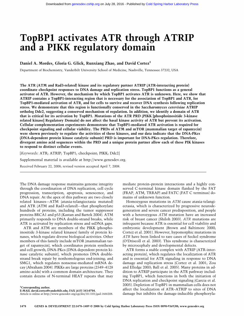
that xTopBP1 depends on the presence of xATRIP to as-sociate with xATR–xATRIP (Kumagai et al. 2006). Totest if ATRIP can associate with TopBP1 independentlyof ATR, nuclear extracts from U2OS cells expressingwild-type ATRIP or mutant ATRIP lacking the C-termi-nal ATR-interacting domain (Ball et al. 2005; Falck et al.2005) were incubated with recombinant TopBP1 frag-ments. The binding of this ATRIP�C mutant to TopBP1is severely reduced compared with wild-type ATRIP (Fig.1C). Taken together, these data suggest that the associa-tion of the ATR–ATRIP complex with TopBP1 mightinvolve binding surfaces on both ATR and ATRIP.
Identification of a TopBP1-interacting regionof ATRIP
To search for TopBP1-binding surfaces on the ATRIP andATR proteins, we used a yeast two-hybrid approach.Given that the TopBP1 AAD is sufficient to associatewith the ATR–ATRIP complex, the TopBP1 AAD fusedto the GAL4 DNA-binding domain was used as a bait toscreen a library containing thousands of random frag-ments of ATRIP fused to the activation domain of GAL4.Sequencing of ATRIP fragments selected in the screen-ing procedure revealed a minimal interacting region ofATRIP consisting of amino acids 203–348 (Fig. 2A). Thisregion is adjacent to the predicted ATRIP coiled-coildomain (amino acids 108–217), which mediates homo-oligomerization (Ball and Cortez 2005; Itakura et al.2005). Sequence conservation of this region amongATRIP homologs is low except for a section of ∼30 aminoacids starting at amino acid 308 (Supplemental Fig. S1A).Deletion of amino acids 301–338 of ATRIP eliminatedactivation of ATR by TopBP1 in vitro, but also causedreduced binding to ATR (data not shown). An aminoacid substitution mutation in this region of ATRIP(LLSS332AAAA) nearly abolished the association ofATRIP with TopBP1 (Fig. 2B), yet preserved the ability ofATRIP to bind ATR (Fig. 2C) and localize to damage-induced foci (data not shown). Furthermore, the associa-tion of ATR with TopBP1 was also significantly reducedin the presence of this ATRIP-top mutant compared withwild-type ATRIP (Fig. 2B), which supports the idea thatATR association with TopBP1 is dependent on ATRIP.
Since the ATR–ATRIP complex containing theATRIP-top mutant had decreased association withTopBP1, we expected that it would have a decreased abil-ity to be activated by TopBP1 as well. To test this hy-pothesis, wild-type ATR–ATRIP and mutant ATR–ATRIP-top complexes were immunopurified from mam-malian nuclear extracts. Addition of the TopBP1 AAD towild-type ATR–ATRIP complexes caused a robust stimu-lation of ATR kinase activity toward a substrate of ATR,MCM2 (Cortez et al. 2004; Yoo et al. 2004). The ATRIP-top mutant severely attenuated TopBP1-dependentstimulation of ATR (Fig. 2C). Thus, a previously unchar-acterized domain of ATRIP is important for an interac-tion between TopBP1 and the ATR–ATRIP complex andfor ATR activation by TopBP1.
The interaction between TopBP1 and ATRIP isessential for checkpoint responses to replication stress
A critical function of the ATR-dependent checkpointsignaling pathway in preserving genomic stability is topromote cell cycle recovery after replication fork arrest(Casper et al. 2002; Zachos et al. 2003). Since the ATRIP-top mutation selectively impairs TopBP1 associationwith and activation of ATR–ATRIP without impairingcomplex stability or localization to sites of DNA dam-age, this separation-of-function mutant provides amethod to specifically assess the functional importanceof TopBP1-dependent ATR activation. This is especiallyimportant given the possibility that RNAi depletion ofTopBP1 likely interferes with many cellular processes inaddition to checkpoints, such as replication control,making it difficult to unambiguously interpret pheno-typic effects. To assess whether TopBP1 binding toATRIP and activation of ATR is necessary for cells todisplay a normal checkpoint response to stalled replica-tion forks, we created U2OS cell lines stably expressingsiRNA-resistant wild-type ATRIP, the ATRIP-top mu-
Figure 2. Identification of an ATRIP region necessary forTopBP1 association. (A) Schematic diagram showing the frag-ments of ATRIP that interacted with TopBP1 in a yeast two-hybrid assay (black lines). (CRD) Checkpoint Recruitment Do-main (Ball et al. 2007). (B) Nuclear extracts from 293T cellstransfected with vectors encoding ATR and wild-type ATRIP(wt) or ATR and ATRIP-top were incubated with recombinantGST-tagged fragments of TopBP1 bound to glutathione beads.Proteins bound to the beads were eluted, separated by SDS-PAGE, and blotted with antibodies to ATR or ATRIP. (C) Wild-type ATR–ATRIP (wt) or ATR–ATRIP-top complexes were iso-lated from transfected 293T cells and incubated with recombi-nant TopBP1 AAD, MCM2 substrate, and �-32PATP. Kinasereactions were separated by SDS-PAGE, stained with Coo-massie blue (CB), and exposed to film (autorad). A duplicate gelwas blotted and probed with anti-ATRIP and anti-ATR antibod-ies (WB).
Mordes et al.
1480 GENES & DEVELOPMENT
Cold Spring Harbor Laboratory Press on July 28, 2016 - Published by genesdev.cshlp.orgDownloaded from
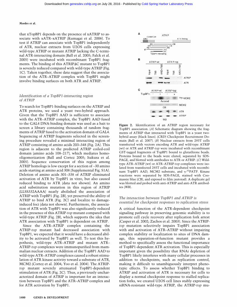
tant, or empty vector as a control. Three days after trans-fection with siRNA targeting endogenous ATRIP, thelevels of wild-type ATRIP and ATRIP-top were compa-rable (Fig. 3B). The cell cycle profile of asynchronouslydividing cells expressing ATRIP-top is similar to cellsexpressing wild-type ATRIP (Fig. 3A), indicating thatATRIP-top did not affect normal cell cycle progression.These cell lines were treated with hydroxyurea (HU),which causes replication forks to stall and arrests cells inearly S phase. After 24 h, the HU was removed, and cellswere released into media containing nocodazole to pre-vent cell division. The ability of the ATRIP-top mutantto support recovery and completion of DNA replicationwas compared with wild-type ATRIP. Sixteen hours afterrelease from HU, most of the cells expressing wild-typeATRIP had completed S phase (Fig. 3A). Yet, the majorityof ATRIP-depleted cells expressing the ATRIP-top mu-tant or lacking any exogenous ATRIP were unable toresume DNA replication and complete S phase. Consis-tent with the reduced ability of ATRIP-top cells to re-cover from replication stress, these cells also displayedsignificantly reduced viability compared with wild-typeATRIP-expressing cells after treatment with HU (Fig.3C). The ATRIP-top cells also exhibited a defect in theG2/M checkpoint after treatment with ionizing radia-tion (Fig. 3D). Therefore, the ability of ATR–ATRIP com-plexes to promote recovery from stalled replication forksand cell cycle arrest after DNA damage is dependent onan interaction between TopBP1 and ATRIP that leads toATR kinase activation.
Conservation of the TopBP1-dependent ATRIPregulatory domain in S. cerevisiae
It is unclear whether the mechanism of activation ofMec1–Ddc2 complexes in S. cerevisiae is the same asATR–ATRIP since no yeast protein has obvious se-
quence similarity to the AAD of TopBP1. To addresswhether the mechanism of regulation for Mec1–Ddc2complexes is likely to be similar to ATR–ATRIP, weintroduced a mutation into Ddc2 that is analogous to theATRIP-top mutant. Despite little sequence similarity,Ddc2 and ATRIP have a common functional domain ar-chitecture: an N-terminal checkpoint recruitment do-main, a predicted coiled-coil domain, and a C-terminaldomain essential for interaction with Mec1 or ATR, re-spectively (Wakayama et al. 2001; Ball et al. 2005, 2007;Falck et al. 2005). To generate a ddc2-top mutation thatcorresponds to the ATRIP-top mutation, we used posi-tion and secondary structure predictions as a guide (Fig.4A). Specifically, we mutated leucine residues that wereapproximately the same number (115) of amino acidsC-terminal from the end of the predicted coiled-coil do-main and within the third predicted �-helix (-top:LLLR257AAAA). For comparison, we made two addi-tional ddc2 alleles with mutations in the fourth pre-dicted �-helix after the coiled-coil domain (-A1:LLED274AAAA and -A2: LIKE281AAAA). Mutant orwild-type Ddc2 were expressed on a low-copy plasmidunder the endogenous DDC2 promoter in a ddc2� strain(Paciotti et al. 2000; Rouse and Jackson 2002). The top,A1, and A2 mutants were expressed at comparable levelsas wild-type Ddc2 and are able to interact with Mec1equivalently to wild-type Ddc2 (Fig. 4B; data not shown).The top mutation but not the A1 or A2 mutation causedmarked sensitivity to HU and the alkalyating agentmethyl methanesulfonate (MMS) (Fig. 4C). Furthermore,the sensitivity to DNA damage of the ddc2-top straincorrelated with reduced phosphorylation of the Mec1substrate Rad53 following a challenge with either HU orMMS, suggesting that disruption of this Ddc2 domaincauses a defect in Mec1 activation (Fig. 4D). These datasuggest that although the protein equivalent to the
Figure 3. ATRIP association with TopBP1 is essentialfor cellular recovery from replication stress. (A–C)U2OS cells stably expressing siRNA-resistant wild-typeATRIP (wt), ATRIP-top, or an empty vector (vector)were transfected with siRNA targeting ATRIP to de-plete endogenous ATRIP. Three days later, cells wereexposed to 1 mM HU for 24 h. (A) Cells were collectedimmediately (0 hr) or rinsed and released into mediacontaining 1 µg/mL nocodazole for either 8 h (8 hr) or 16h (16 hr). Cells were fixed and stained with propidiumiodine and processed for FACS analysis. (Asynch) Asyn-chronous cells that were not exposed to HU. (B) Immu-noblot showing ATRIP levels from U2OS cells stablyexpressing wild-type ATRIP (wt), ATRIP-top (top), orempty vector (vt). (C) Twenty-four hours after releasefrom HU, cellular viability was measured using a col-orimetric assay. Viability was normalized to cells ex-pressing exogenous wild-type ATRIP. Error bars indi-cate standard error, n = 6. (D) Three days after siRNAtransfection, cells were treated with 4 Gy of IR, and 1µg/mL nocodazole was added to the media. Sixteenhours later, the percentage of mitotic cells were deter-mined by propidium iodine and antiphosphohistone H3staining followed by flow cytometry.
Regulation of ATR activation
GENES & DEVELOPMENT 1481
Cold Spring Harbor Laboratory Press on July 28, 2016 - Published by genesdev.cshlp.orgDownloaded from

TopBP1 ATR activator in budding yeast is unclear, acommon mechanism of regulation dependent on theATRIP (or Ddc2) protein is likely to exist for vertebrateand yeast ATR kinases.
Regulation of ATR activation via a PIKK regulatorydomain
Since the ATR-binding-defective ATRIP�C protein doesnot bind as well to TopBP1 as wild-type ATRIP (Fig. 1),we hypothesized that regions of ATR contribute to theinteraction between TopBP1 and the ATR–ATRIP com-plex. ATR has similar domain architecture to othermembers of the PIKK protein kinase family. It containsdozens of N-terminal HEAT repeats and a kinase domainflanked by FAT and FATC domains (Fig. 5A). The N-terminal heat repeats provide a binding surface forATRIP (Ball et al. 2005; Chen et al. 2007). Aside fromthis, little is known about the structure of ATR. Weagain used the TopBP1 AAD as bait in a yeast two-hybridapproach to screen a library of thousands of ATR frag-ments. All of the interacting fragments recovered con-tained amino acids 2483–2597 of ATR (Fig. 5A). Theseresidues span the C-terminal end of the kinase domainand an uncharacterized region of ATR between the ki-nase and FATC domains, which we named the PIKKRegulatory Domain (PRD) for reasons discussed below.
Although the kinase and FATC domains share a highdegree of sequence similarity among all PIKK familymembers, the sequence of the PRD is highly divergent inthese paralogs (Supplemental Fig. S1B). However, thePRD has a high degree of sequence identity within or-thologous ATR proteins from different organisms. Dele-tion of the entire ATR PRD abolished all kinase activity(data not shown), probably as a result of disrupting thefolding of the adjacent kinase domain. Scanning muta-genesis of the ATR PRD identified two mutations(K2589E and HVL2591AAA) that largely eliminatedTopBP1-activation of ATR kinase activity withoutchanging the basal activity of the kinase in the absenceof TopBP1 (Fig. 5B; Supplemental Fig. S2). Several othermutations in this region including a small deletion�2569–2576 and a charge reversal of a lysine one aminoacid separated from K2589 (K2587E) had no significanteffect on either the basal or TopBP1-activated ATR ki-nase activity (Fig. 5B,C; Supplemental Fig. S2). None ofthese mutations had any effect on the ability of ATR tobind ATRIP. Mutation of K2589 to alanine did not im-pair ATR activation (Supplemental Fig. S2A), indicatingthat a post-translation modification at this site is notnecessary for ATR activation by TopBP1.
Since ATR K2589E has equivalent basal kinase activ-ity toward itself, ATRIP, and a substrate as wild-typeATR (Fig. 5D), the mutation does not alter the catalyticactivity of ATR in the unactivated state but specificallyaffects the formation of the active ATR protein. Addi-tionally, this mutation does not affect the ability of ATRto form homo-oligomeric complexes (Supplemental Fig.S3).
Next, we tested the ability of ATR–ATRIP complexescontaining ATR K2589E to associate with TopBP1. TheATR K2589E mutant modestly decreased the associationwith TopBP1 (approximately twofold) in pull-down as-says compared with wild-type ATR–ATRIP complexes(Fig. 5E). The remaining association is likely mediated bythe ATRIP–TopBP1-binding interface.
Regulation of PIK kinases via the PIK regulatoryand FATC domains
A recent study suggested that the region of ATM equiva-lent to the ATR PRD is targeted for acetylation by theTip60 histone acetylase (Sun et al. 2007). The DNA-dam-aged inducible acetylation of a specific lysine in theATM PRD is necessary for the increased kinase activityof ATM after DNA damage. To test whether acetylationof the ATR PRD might regulate its activation byTopBP1, we made lysine-to-arginine mutations in thePRD. None of these mutations, even when combinedinto a single ATR protein, had any effect on TopBP1-mediated activation of ATR in vitro (Supplemental Fig.S2B). Also, siRNA depletion of Tip60 did not affect theability of cells to recover from HU (data not shown).Hence, the PRD of ATR is likely not regulated throughacetylation in the same manner as the PRD of ATM.
To determine if the PRD is necessary for the regula-tion of other PIKK family members besides ATR and
Figure 4. An S. cerevisiae ddc2-top mutant is defective incheckpoint signaling. (A) Secondary structure prediction of theTopBP1-interacting region of ATRIP and the equivalent regionof S. cerevisiae Ddc2. Hashed boxes denote the C-terminal endsof the coiled-coil domains. Solid boxes indicate predicted �-helices. The asterisk denotes the location of the ATRIP-top mu-tation or the ddc2-top (LLLR257AAAA) mutation. The adjacentdots in Ddc2 denote the location of the A1 (LLED274AAAA)and A2 (LIKE281AAAA) mutations. (B) Immunoblot showingDdc2 levels in yeast strains expressing Ddc2 mutants, wild-typeDdc2, or empty vector. (C) Serial dilutions of the indicated yeaststrains grown on YPD with no drug (mock), 150 mM HU (+HU),or 0.008% MMS (+MMS). (D) Exponentially growing yeast weretreated with no drug (mock), 150 mM HU (+HU), or 0.015%MMS (+MMS) for 90 min. Extracts were prepared, separated bySDS-PAGE, and immunoblotted with an antibody againstRad53. The top bands are phosphorylated forms of Rad53.
Mordes et al.
1482 GENES & DEVELOPMENT
Cold Spring Harbor Laboratory Press on July 28, 2016 - Published by genesdev.cshlp.orgDownloaded from
ATM, we examined the PRD of DNA-PKcs. Activationof DNA-PKcs requires the Ku70/80 heterodimer andDNA ends (Smith and Jackson 1999). Electron micros-copy structural studies of the DNA-PK complex demon-strated an interaction between the Ku70/80 heterodimerand a region immediately C-terminal to the kinase do-main, suggesting that Ku70/80 binding could be medi-ated in part by the DNA-PKcs PRD (Spagnolo et al.2006). Therefore, we tested if the PRD is necessary forthe activation of DNA-PKcs. In response to dsDNAbreaks, DNA-PKcs is autophosphorylated at Ser 2056, anevent that is required for nonhomologous end-joining-mediated DNA double-strand break repair (Chen et al.2005). Wild-type DNA-PKcs and DNA-PKcs PRD mu-tants (M1: K4043E/K4048E/R4049E/K4050E, M2: D4062K/E4063K/E4069K, M3: K4075E/R4082E/R4085E/R4090E)were expressed in DNA-PKcs-defective cells. Cells wereexposed to ionizing radiation and the phosphorylationstatus of Ser 2056 was assessed. Although wild-typeDNA-PKcs and the M2 DNA-PKcs PRD mutant exhib-ited Ser 2056 phosphorylation, the M1 and M3 DNA-PKcs PRD mutants did not (Supplemental Fig. S4). Thus,specific residues within the PRD of DNA-PKcs are re-quired for DNA-PKcs autophosphorylation, suggestingthat the PRD is important for the regulation of DNA-PKcs as well.
Since the FATC domain amino acid sequence is highlyconserved among PIK kinases and adjacent to the PRD,
we tested if it is also important for TopBP1 to activateATR. Deletion of part of or the entire FATC domainabolished even the basal kinase activity of ATR (Supple-mental Fig. S5). Since the FATC domain of ATR cansubstitute for the FATC domain of ATM (Jiang et al.2006), we performed the reciprocal experiment. Replace-ment of the FATC domain of ATR with that of ATM alsoresulted in a kinase-dead mutant, indicating that it can-not substitute (Supplemental Fig. S5). Thus, the FATCdomain of ATR is essential for even basal ATR kinaseactivity.
ATR regulation through the PRD is essentialfor checkpoint signaling and cell viability
The ATR-PRD mutation provides a second separation offunction mutant useful for examining the functional im-portance of TopBP1-mediated ATR activation. To deter-mine if regulation of ATR by TopBP1 through the PRD isnecessary for cellular responses to replication stress, wecreated ATRflox/− cell lines expressing a Tet-inducibleform of either wild-type ATR or mutant K2589E ATR.The cell lines were treated with tetracycline to induceexpression of the exogenous ATR prior to deletion of thefloxed ATR allele with the Cre recombinase. Expressionlevels of the wild-type or K2589E ATR protein weresimilar and near the endogenous amount of ATR expres-sion (Fig. 6A). Phosphorylation of the essential kinase
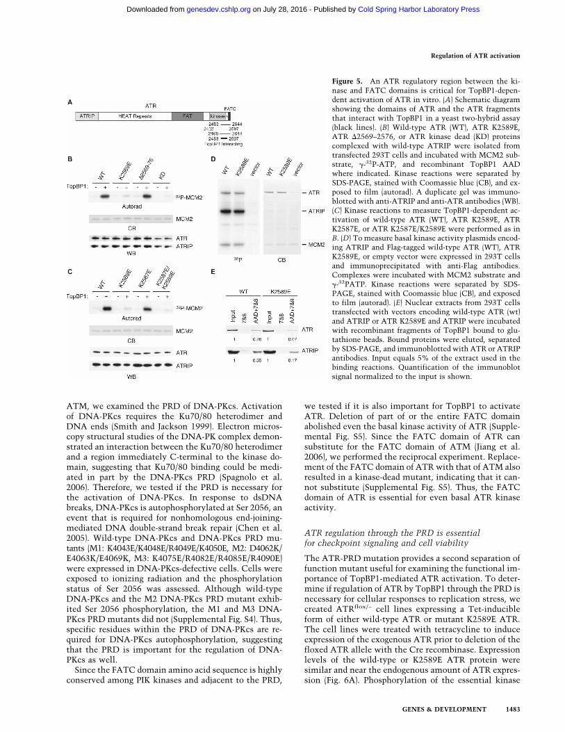
Figure 5. An ATR regulatory region between the ki-nase and FATC domains is critical for TopBP1-depen-dent activation of ATR in vitro. (A) Schematic diagramshowing the domains of ATR and the ATR fragmentsthat interact with TopBP1 in a yeast two-hybrid assay(black lines). (B) Wild-type ATR (WT), ATR K2589E,ATR �2569–2576, or ATR kinase dead (KD) proteinscomplexed with wild-type ATRIP were isolated fromtransfected 293T cells and incubated with MCM2 sub-strate, �-32P-ATP, and recombinant TopBP1 AADwhere indicated. Kinase reactions were separated bySDS-PAGE, stained with Coomassie blue (CB), and ex-posed to film (autorad). A duplicate gel was immuno-blotted with anti-ATRIP and anti-ATR antibodies (WB).(C) Kinase reactions to measure TopBP1-dependent ac-tivation of wild-type ATR (WT), ATR K2589E, ATRK2587E, or ATR K2587E/K2589E were performed as inB. (D) To measure basal kinase activity plasmids encod-ing ATRIP and Flag-tagged wild-type ATR (WT), ATRK2589E, or empty vector were expressed in 293T cellsand immunoprecipitated with anti-Flag antibodies.Complexes were incubated with MCM2 substrate and�-32PATP. Kinase reactions were separated by SDS-PAGE, stained with Coomassie blue (CB), and exposedto film (autorad). (E) Nuclear extracts from 293T cellstransfected with vectors encoding wild-type ATR (wt)and ATRIP or ATR K2589E and ATRIP were incubatedwith recombinant fragments of TopBP1 bound to glu-tathione beads. Bound proteins were eluted, separatedby SDS-PAGE, and immunoblotted with ATR or ATRIPantibodies. Input equals 5% of the extract used in thebinding reactions. Quantification of the immunoblotsignal normalized to the input is shown.
Regulation of ATR activation
GENES & DEVELOPMENT 1483
Cold Spring Harbor Laboratory Press on July 28, 2016 - Published by genesdev.cshlp.orgDownloaded from
Chk1, an ATR substrate (Liu et al. 2000), was used toassess ATR signaling in cells. Robust Chk1 phosphory-lation following HU treatment is observed in all the celllines prior to deletion of the endogenous ATR allele (Fig.6A). However, the K2589E ATR protein failed to supportChk1 phosphorylation after HU treatment, yieldingphospho-Chk1 levels comparable with cells not express-ing any exogenous ATR. In contrast, wild-type, exog-enous ATR could support Chk1 phosphorylation in cellslacking endogenous ATR (Fig. 6A, last lane). TwoK2589E ATR-expressing clonal cell lines yielded identi-cal results. Thus, ATR K2589E is defective in checkpointsignaling after replication stress.
ATR is essential for the viability of proliferating cells,but its essential function is not clear (Brown and Balti-more 2000; Cortez et al. 2001). Given that the K2589E
mutant ATR could not support checkpoint signaling, wewanted to assay whether this mutant would be capableof supporting cellular viability even in the absence ofexogenously added genotoxic agents. The ATRflox/− cellslines stably expressing wild-type or mutant K2589E ATRwere treated with adenovirus expressing Cre recombi-nase and plated at low density. After 17 d, surviving cellcolonies were stained. The colony formation assay re-vealed a dramatic difference in the number of coloniesbetween the two cell lines (Fig. 6B). Only a few coloniesgrew from the mutant cell line. PCR genotyping of thesecolonies from a duplicate sample indicated that all thesurviving colonies expressing the K2589E ATR had notundergone Cre-mediated recombination to delete the en-dogenous ATR allele; whereas, all of the colonies ex-pressing wild-type ATR underwent Cre-mediated recom-bination and lacked the endogenous ATR allele (Supple-mental Fig. S6). Thus, although we could obtain anATR−/− cell line rescued by a wild-type ATR cDNA, wewere not able to obtain an ATR−/− cell line expressingonly the K2589E ATR mutant. This suggests thatTopBP1-mediated activation of ATR is essential for theviability of, at least, this human cell type in culture.
Discussion
The PIK kinases regulate many cellular responses in-cluding nutrient sensing and the DNA damage response.Therefore, their activities impact many human diseases.Unfortunately, their large size and atypical kinase do-mains have made understanding their activation mecha-nisms difficult. In this study, we define how the TopBP1activator protein binds to the ATR–ATRIP complex andidentify critical regulatory regions within both theATRIP and ATR proteins. Importantly, we provide evi-dence that the ATRIP regulatory region is conservedfunctionally in the yeast ATRIP protein Ddc2, and theATR regulatory domain is a common site for regulationof most, if not all, of the PIK kinases.
Our data support the ATR activation model shown inFigure 6C. In the absence of DNA damage or replicationstress, ATR has basal kinase activity. Following a chal-lenge to the genome that exposes ssDNA gaps, ATRIPand the 9–1–1 checkpoint clamp are recruited indepen-dently (Melo et al. 2001; Zou et al. 2002). ATRIP bringsATR and Rad9 brings TopBP1. The assembly and con-centration of these components at sites of DNA damagefacilitates an interaction between TopBP1 and interact-ing surfaces on both ATRIP and ATR. This interactionthen promotes ATR activation.
Additional regulatory steps are clearly important. Forexample, the ATR–ATRIP complex, 9–1–1 checkpointclamp, and TopBP1 are phosphorylated, suggesting thatpost-translational modifications may fine-tune ATR ac-tivation (Roos-Mattjus et al. 2003; Myers et al. 2007;Venere et al. 2007; Yoo et al. 2007). Furthermore, specificATR substrates (such as Chk1) require additional proteincofactors (such as Claspin) to be efficiently phosphory-lated (Kumagai and Dunphy 2000; Liu et al. 2006). Ex-actly how TopBP1 binding increases ATR kinase activity
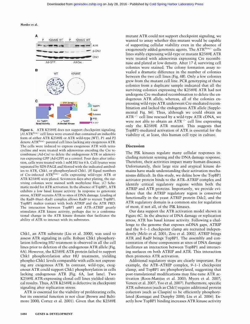
Figure 6. ATR K2589E does not support checkpoint signaling.(A) ATRflox/− cell lines were created that contained an inducibleform of either ATR K2589E or ATR wild-type (WT). P1 and P2denote ATRflox/− parental cell lines lacking any exogenous ATR.The cells were induced to express exogenous ATR with tetra-cycline and were treated with adenovirus encoding the Cre re-combinase (Ad-Cre) to delete the endogenous ATR or adenovi-rus expressing GFP (Ad-GFP) as a control. Four days after infec-tion, cells were treated with 1 mM HU for 6 h. Cell lysates wereseparated by SDS-PAGE and blotted with the indicated antibod-ies to ATR, Chk1, or phosphorylated Chk1. (B) Equal numbersof Cre-infected ATRflox/− cells expressing wild-type ATR orATR K2589E were plated. Seventeen days after plating, the sur-viving colonies were stained with methylene blue. (C) Sche-matic model for ATR activation. In the absence of TopBP1, ATRexhibits a low basal kinase activity. In response to genotoxicstress, ATRIP recruits ATR to sites of DNA damage. Loading ofthe Rad9–Hus1–Rad1 complex allows Rad9 to recruit TopBP1.TopBP1 makes contact with both ATRIP and the ATR PRD.The interaction between TopBP1 and ATR–ATRIP greatlystimulates ATR kinase activity, perhaps due to a conforma-tional change in the ATR kinase domain that facilitates theability of ATR to interact with its substrates.
Mordes et al.
1484 GENES & DEVELOPMENT
Cold Spring Harbor Laboratory Press on July 28, 2016 - Published by genesdev.cshlp.orgDownloaded from

will require a structural description of these proteins.One possibility is that it alters the conformation of theATR kinase domain such that substrates can access ATRmore easily. Consistent with this interpretation, wefound that TopBP1 binding to ATR decreases the appar-ent Km of ATR for substrates (D.A. Mordes, unpubl.).
ATRIP provides several functions to regulate ATRactivation
ATR function is dependent on its binding partnerATRIP. ATRIP provides at least four activities that pro-mote ATR signaling. First, it stabilizes ATR (Cortezet al. 2001). Second, it promotes the localization ofATR to sites of DNA damage or replication stress (Zouand Elledge 2003; Ball et al. 2005, 2007). Third, ATRIPpost-translational modifications regulate ATR signaling(Myers et al. 2007; Venere et al. 2007). Finally, as dem-onstrated in this report, ATRIP binds directly to TopBP1,and this binding is essential for ATR activation.
Mutations of the TopBP1-interacting region of ATRIPimpair the ability of cells to recover from replicationstress and arrest the cell cycle after DNA damage. Mu-tations of the same region in S. cerevisiae ATRIP (Ddc2)cause sensitivity to replication stress and impair activa-tion of Mec1 in response to DNA damage. This suggeststhat the mechanism of activation for ATR may be con-served throughout evolution. Although Mec1 and Ddc2are clear orthologs of ATR and ATRIP, respectively, noobvious S. cerevisiae ortholog exists for TopBP1. Twocheckpoint proteins, Dpb11 and Ddc1, are candidates forprotein activators of Mec1, but neither share sequenceidentity to the TopBP1 AAD. Like TopBP1, Dpb11 is aBRCT-repeat-containing protein that has essential rolesin replication and checkpoint signaling. Yet, Dpb11 hasnot been reported to activate Mec1 directly. In verte-brates, TopBP1 is recruited to gapped ssDNA regionsthrough an interaction with the Rad9 subunit of thecheckpoint clamp (Delacroix et al. 2007; Lee et al. 2007).Ddc1 is the yeast ortholog of Rad9 and also binds toDpb11 (Wang and Elledge 2002). The C-terminal tail ofDdc1 stimulates the kinase activity of the Mec1–Ddc2complex in vitro under low-salt conditions (Majka et al.2006). However, overexpression of this portion of Ddc1is not sufficient to cause activation of Mec1. In contrast,overexpression of the TopBP1 AAD is sufficient to causepan-nuclear activation of ATR in mammalian cells(Kumagai et al. 2006; Ball et al. 2007). It is possible thatboth proteins may act as activators of Mec1 in responseto different types of DNA damage or they may form anactivator complex. In any case, our results predict that aMec1 activator is likely to bind to a surface on Ddc2encoded just C-terminal of the coiled-coil domain con-taining amino acids 257–260.
PIK kinase regulation
PIKKs are key regulators of many critical signaling path-ways, including cell growth, cell cycle checkpoints, and
DNA damage. Despite their diverse functions, emergingdata suggest many similarities among them. PIKKs asso-ciate with an interacting partner (ATRIP for ATR, NBS1for ATM, Ku70/80 for DNA-PKcs, and Rictor and Raptorfor mTOR) that is critical for their function and, in thecase of ATRIP, NBS1, and Ku80, their localization tosites of DNA damage (Cortez et al. 2001; Hara et al.2002; Sarbassov et al. 2004; Falck et al. 2005). PIKKs alsorequire an interaction with another regulatory protein orprotein complex for their activation. G�L (mLST8) bindsto the kinase domain of mTOR and stimulates its cata-lytic activity (Kim et al. 2003; Wullschleger et al. 2005).Rheb also activates mTOR–Raptor complexes (Long etal. 2005). The Mre11/Rad50 complex makes multiplecontacts with ATM and is sufficient to stimulate its ki-nase activity especially in the presence of DNA (Lee andPaull 2004). The Ku70/Ku80 complex stimulates DNA-PKcs in the presence of DNA ends (Smith and Jackson1999). Finally, TopBP1 stimulates the kinase activity ofATR in an ATRIP-dependent manner (Kumagai et al.2006). There are apparently even common regulators ofmost, if not all, the PIK kinases. Recently, Tel2 has beendemonstrated to interact with all PIKKs and regulatetheir stability (Takai et al. 2007).
Given the similarity of sequence and structure of thePIK kinases, it might be expected that the mechanismscontrolling their activation would be similar (althoughresponsive to different inputs). Indeed, the FATC domainis highly conserved in sequence among PIKKs and isfunctionally interchangeable in some instances (Jiang etal. 2006). The FATC domain is required for the kinaseactivity of ATM, DNA-PKcs, mTOR, and SMG-1 (Baninet al. 1998; Priestley et al. 1998; Takahashi et al. 2000;Morita et al. 2007). Our data indicate that the FATCdomain of ATR is essential for its kinase activity as well.We noticed that mutation of the FATC domain of ATRcaused a reduction in expression levels, and a similarobservation has been made for ATM (Jiang et al. 2006).Since all kinase activity is lost when the FATC domainis mutated, we suspect that the FATC domain may beimportant for the stability of PIKKs or for proper foldingof the kinase domain. One possibility is that the FATCdomain makes critical contacts within the kinase orFAT domains that stabilize the kinase domain.
In contrast to FATC domain alterations, mutations inthe PRD (the region between the kinase and FATC do-mains) of ATM, ATR (this study), mTOR, and SMG-1 donot abolish PIK kinase activity (Sekulic et al. 2000;Morita et al. 2007; Sun et al. 2007). However, these mu-tations do impair kinase regulation. This region does notexhibit sequence similarity among PIKKs, suggestingthat it could be sensitive to different regulatory inputsfor each of the kinases. The PRD domain of mTOR con-tains an Akt phosphorylation site that regulates its ac-tivity (Sekulic et al. 2000). The Tip60 histone acetyl-transferase complex acetylates ATM within the ATMPRD, and this acetylation is important for the activationof ATM kinase activity after DNA damage (Sun et al.2005, 2007). We now show that the ATR PRD is criticalfor TopBP1-dependent activation of ATR and the PRD of
Regulation of ATR activation
GENES & DEVELOPMENT 1485
Cold Spring Harbor Laboratory Press on July 28, 2016 - Published by genesdev.cshlp.orgDownloaded from
DNA-PKcs is necessary for its autophosphorylation. De-spite the similarity of ATM and ATR, we have no evi-dence that the ATR PRD is acetylated. In fact, mutationsof all the lysines in this region to arginines did not im-pair TopBP1-dependent activation.
We propose that the PRD has evolved to allow distinctregulation of the kinase activity of the PIKK familymembers. Differences in the PRD and the PIK-bindingpartner provide specificity for the types of cellular eventsthat can activate each PIKK. It will be interesting to de-termine whether the ATM PRD or mTOR PRD is im-portant for Mre11/Rad50-dependent or G�L/Rheb-de-pendent ATM or mTOR activation, respectively. Also, itwill be important to examine the impact of ATM PRDacetylation on Mre11/Rad50-dependent regulation.
Defects in cellular responses to replication stressin cells lacking TopBP1-dependent ATR activation
Previous studies have demonstrated critical roles forATR signaling and the TopBP1 protein for replicationstress-induced checkpoints. Furthermore, the C-termi-nal half of TopBP1 was shown to be essential for check-point signaling in Xenopus laevis egg extracts (Hashi-moto et al. 2006; Kumagai et al. 2006; Yan et al. 2006).However, the interpretation of genetic experiments onTopBP1 in human cells is problematic given that itforms complexes with many other proteins and regulatesboth DNA replication and checkpoints. The analysis ofthe separation-of-function mutants that we created inboth ATR and ATRIP allows us to unambiguously definethe cellular requirements for TopBP1-dependent ATRactivation. Our analysis reveals that not only is TopBP1-dependent ATR activation essential for checkpoint re-sponses to replication stress, but it is also required forthe essential function of ATR in promoting cellular vi-ability.
Conclusions
Our data suggest both similarities and differences amongthe regulation of the PIKK family of protein kinases. Theability of TopBP1 to uniquely stimulate ATR is ex-plained, in part, by the requirement of an ATRIP surfacefor the binding of TopBP1 to the ATR–ATRIP complex.The PRD of ATR and the other PIKKs is a second im-portant regulatory determinant. The exact mechanismby which the PRD functions in PIKK activation remainsto be determined, but given its position between the ki-nase and FATC domains, it seems likely that it mediatesa conformational change that may allow greater kinaseactivity.
Inhibition of ATR sensitizes cancer cells to multipleDNA-damaging agents (Wilsker and Bunz 2007). Thusfar, specific inhibitors of ATM and DNA-PKcs kinaseshave been developed and are being studied as potentialtherapeutic agents (Lord et al. 2006); however, inhibitorsof the ATR kinase have not been isolated. Targeting theinteraction between ATRIP and TopBP1 or the ATR
PRD may provide a novel means for developing an agentto disrupt ATR signaling.
Materials and methods
Cell lines
HEK 293T and U2OS cell lines were maintained inDMEM + 7.5% fetal bovine serum. The V3 CHO cell line wasmaintained in �MEM supplemented with 10% fetal bovine se-rum, 10 µg/mL ciprofloxacin (Mediatech), 50 U/mL penicillin,and 50 µg/mL streptomycin (Invitrogen). Construction of ATRIPstable cell lines and use of ATRIP siRNA were as describedpreviously (Ball et al. 2005), except transfections of siRNAs at 10nM were performed with HiPerFect (Qiagen). Plasmid transfec-tions of HEK 293T cells were performed with Lipofectamine 2000(Invitrogen). Plasmid transfections of V3 CHO cells were per-formed with TransIT-CHO (Mirus Bio). HCT116 ATRflox/− cellswere maintained in McCoy’s medium + 7.5% fetal bovine se-rum. Clonal ATR stable lines were made by transfecting HA-tagged ATR tetracycline-inducible vectors into ATRflox/−-TetRcells, and selecting with 300 µg/mL Hygromycin B (Invitrogen)for single colonies. ATR expression was induced with 1 µg/mLtetracycline (Invitrogen). Deletion of the ATR gene and PCRgenotype analysis for the ATR allele was performed as describedpreviously (Cortez et al. 2001). For the colony formation assays,equal numbers of cells were plated onto 60 mM tissue cul-ture dishes and incubated for 17 d in the presence of 300 µg/mLHygromycin B and 1 µg/mL tetracycline. Media was changedevery 3 d. Colonies were stained with methylene blue(Sigma).
Yeast
Ddc2 mutations were made in the pNML1 centromeric plasmidencoding myc-Ddc2 under the endogeonous Ddc2 promoter(Rouse and Jackson 2002) and were expressed in strainDMP2995/1B: MATa ade2-1 can1-100 his3-11,15 leu2-3,112trp1-1 ura3 sml1��KanMX4 ddc2��KanMX4 (Paciotti et al.2000). Yeast protein samples were prepared using TCA precipi-tation as described (Longhese et al. 1997).
DNA constructs
PCR site-directed mutagenesis was performed using the Quick-Change method and PfuUltra DNA polymerase (Stratagene). Allconstructs generated using PCR were confirmed by sequencing.The DNA-PKcs expression vector was kindly provided by Dr.Kathryn Meek. Cloning details for any construct are availableupon request.
Protein interactions
TopBP1-binding assay: Recombinant GST-tagged TopBP1 frag-ments were purified from Escherichia coli with glutathioneSepharose 4B beads (GE Healthcare). Nuclear extracts were pre-pared as described (Kumagai et al. 2006) and incubated with theindicated GST-tagged proteins bound to glutathione Sepharosebeads overnight at 4°C. Beads were washed three times in low-salt buffer (20 mM HEPES-KOH at pH 7.9, 175 mM NaCl, 20%glycerol, 0.05% Tween 20), and proteins were eluted and sepa-rated by SDS-PAGE prior to immunoblotting.
Two-hybrid experiments were performed using the TopBP1AAD (amino acids 978–1286) cloned into pDAB1 containing theDNA-binding domain of Gal4. This bait was used to screen the
Mordes et al.
1486 GENES & DEVELOPMENT
Cold Spring Harbor Laboratory Press on July 28, 2016 - Published by genesdev.cshlp.orgDownloaded from
pACT-ATRIP and pACT-ATR cDNA fragment libraries that wedescribed previously (Ball et al. 2005) using the PJ694A yeaststrain (James et al. 1996).
Kinase assays were performed largely as described previously(Cortez et al. 2001; Ball et al. 2007). Assays measuring the basalATR activity were performed by immunoprecipitating Flag-ATR protein using anti-Flag M2 agarose beads (Sigma) fromTGN buffer (50 mM Tris, 150 mM NaCl, 10% glycerol,1% Tween 20, 5 µg/mL aprotinin, 5 µg/mL leupeptin, 1 mMNaF, 50 mM �-glycerol phosphate, 1 mM sodium vanadate,1 mM dithiothreitol, 1 mM phenylmethylsulfonyl fluoride) celllysates. Immunoprecipitates were washed three times in TGNbuffer, once in TGN + 500 mM LiCl, and twice in kinase buffer(10 mM HEPES at pH 7.5, 50 mM NaCl, 50 mM �-glycerolphosphate, 10 mM MgCl2, 1 mM diothiothreitol) prior to per-forming kinase reactions. Assays measuring TopBP1 stimula-tion of ATR activity were performed by immunoprecipitatingFlag-ATR/HA-ATRIP complexes using anti-HA agarose beads(Sigma). Immunoprecipitates were washed three times in TGNbuffer, once in TGN + 500 mM LiCl, and twice in kinase bufferprior to performing kinase reactions. GST-TopBP1 AAD wasadded to the reactions prior to adding ATP and substrate. Allreactions were stopped within the linear range of the assay andanalyzed by SDS-PAGE and autoradiography.
For experiments to assess ATR oligomerization, 293T cellstransiently expressing the indicated plasmid were lysed inCHAPS lysis buffer as described (Ball and Cortez 2005).
Antibodies
The ATRIP-N antibody has been described previously (Cortez etal. 2001). The following antibodies were purchased: ATM (No-vus), ATR and Chk1 (Santa Cruz Biotechnologies), Chk1 P317(Cell Signaling), DNA-PKcs (Serotec), DNA-PKcs pS2056 (Ab-cam), HA.11 and Myc9E10 (Covance), and Flag M2 (Sigma). TheRad53 antibody was a gift from Stephen Elledge.
Bioinformatics
Protein sequence alignments were performed using ClustalW,Boxshade, and Coils (http://www.ch.embnet.org). Protein sec-ondary structure predictions were generated by PsiPred (http://bioinf.cs.ucl.ac.uk/psipred).
HU recovery, viability, and checkpoint assays
Three days after siRNA transfection, cells were incubated inmedia with or without HU for 24 h. Cells were released intofresh media for 24 h, and cell viability was assessed using theWST-1 reagent assay (Roche), which is a colorimetric assaybased on the cleavage of the tetrazolium salt WST-1 by mito-chondrial dehydrogenases. The percentage viability was calcu-lated as the ratio of the 450 nM absorbance of the HU-treatedcells to the untreated cells. The HU recovery assays were per-formed using the same protocol with the following differences:Nocodazole was added to the media when the HU was removed,and cells were harvested at 0, 8, or 16 h after release. Harvestedcells were fixed in ethanol, stained with propidium iodide, andanalyzed on a BD Biosciences FACSCalibur. A stock solution ofHU (Sigma) was prepared in water at 1 M and stored frozen.Nocodazole (Acros) was dissolved in dimethylsulfoxide at 10mg/mL. For the G2/M checkpoint assay, harvested cells werefixed in ethanol, permeabilized, stained with an antibodyagainst H3 phospho-S10 antibody and an anti-FITC secondaryantibody, propidium iodide, and then analyzed by FACS. Cells
with 4n DNA content and H3 phospho-S10-positive werecounted as mitotic cells.
Acknowledgments
We thank William Dunphy, Stephen Elledge, Steve Jackson,Maria Longhese, and Kathyrn Meek for reagents. This work wassupported by a National Cancer Institute grant (R01CA102729),the Ingram Charitable Fund, and the Pew Scholars Program inthe Biological Sciences, sponsored by the Pew CharitableTrusts. D.A.M. was supported by a Public Health Service award(T32GM07347) from the National Institute of General MedicalStudies for the Vanderbilt Medical Scientist Training Programand a Department of Defense Breast Cancer Research Programpredoctoral fellowship.
References
Abraham, R.T. 2004. PI 3-kinase related kinases: ‘Big’ players instress-induced signaling pathways. DNA Repair (Amst.) 3:883–887.
Ball, H.L. and Cortez, D. 2005. ATRIP oligomerization is re-quired for ATR-dependent checkpoint signaling. J. Biol.Chem. 280: 31390–31396.
Ball, H.L., Myers, J.S., and Cortez, D. 2005. ATRIP binding toreplication protein A-single-stranded DNA promotes ATR–ATRIP localization but is dispensable for Chk1 phosphory-lation. Mol. Biol. Cell 16: 2372–2381.
Ball, H.L., Ehrhardt, M.R., Mordes, D.A., Glick, G.G., Chazin,W.J., and Cortez, D. 2007. Function of a conserved check-point recruitment domain in ATRIP proteins. Mol. Cell.Biol. 27: 3367–3377.
Banin, S., Moyal, L., Shieh, S., Taya, Y., Anderson, C.W.,Chessa, L., Smorodinsky, N.I., Prives, C., Reiss, Y., Shiloh,Y., et al. 1998. Enhanced phosphorylation of p53 by ATM inresponse to DNA damage. Science 281: 1674–1677.
Brown, E.J. and Baltimore, D. 2000. ATR disruption leads tochromosomal fragmentation and early embryonic lethality.Genes & Dev. 14: 397–402.
Casper, A.M., Nghiem, P., Arlt, M.F., and Glover, T.W. 2002.ATR regulates fragile site stability. Cell 111: 779–789.
Chen, B.P., Chan, D.W., Kobayashi, J., Burma, S., Asaithamby,A., Morotomi-Yano, K., Botvinick, E., Qin, J., and Chen, D.J.2005. Cell cycle dependence of DNA-dependent protein ki-nase phosphorylation in response to DNA double strandbreaks. J. Biol. Chem. 280: 14709–14715.
Chen, X., Zhao, R., Glick, G.G., and Cortez, D. 2007. Functionof the ATR N-terminal domain revealed by an ATM/ATRchimera. Exp. Cell Res. 313: 1667–1674.
Cortez, D., Guntuku, S., Qin, J., and Elledge, S.J. 2001. ATR andATRIP: Partners in checkpoint signaling. Science 294: 1713–1716.
Cortez, D., Glick, G., and Elledge, S.J. 2004. Minichromosomemaintenance proteins are direct targets of the ATM and ATRcheckpoint kinases. Proc. Natl. Acad. Sci. 101: 10078–10083.
Delacroix, S., Wagner, J.M., Kobayashi, M., Yamamoto, K., andKarnitz, L.M. 2007. The Rad9–Hus1–Rad1 (9–1–1) clamp ac-tivates checkpoint signaling via TopBP1. Genes & Dev. 21:1472–1477.
Falck, J., Coates, J., and Jackson, S.P. 2005. Conserved modes ofrecruitment of ATM, ATR and DNA-PKcs to sites of DNAdamage. Nature 434: 605–611.
Garcia, V., Furuya, K., and Carr, A.M. 2005. Identification and
Regulation of ATR activation
GENES & DEVELOPMENT 1487
Cold Spring Harbor Laboratory Press on July 28, 2016 - Published by genesdev.cshlp.orgDownloaded from
functional analysis of TopBP1 and its homologs. DNA Re-pair (Amst.) 4: 1227–1239.
Hara, K., Maruki, Y., Long, X., Yoshino, K., Oshiro, N., Hidayat,S., Tokunaga, C., Avruch, J., and Yonezawa, K. 2002. Raptor,a binding partner of target of rapamycin (TOR), mediatesTOR action. Cell 110: 177–189.
Hashimoto, Y., Tsujimura, T., Sugino, A., and Takisawa, H.2006. The phosphorylated C-terminal domain of XenopusCut5 directly mediates ATR-dependent activation of Chk1.Genes Cells 11: 993–1007.
Itakura, E., Sawada, I., and Matsuura, A. 2005. Dimerization ofthe ATRIP protein through the coiled-coil motif and its im-plication to the maintenance of stalled replication forks.Mol. Biol. Cell 16: 5551–5562.
James, P., Halladay, J., and Craig, E.A. 1996. Genomic librariesand a host strain designed for highly efficient two-hybridselection in yeast. Genetics 144: 1425–1436.
Jiang, X., Sun, Y., Chen, S., Roy, K., and Price, B.D. 2006. TheFATC domains of PIKK proteins are functionally equivalentand participate in the Tip60-dependent activation of DNA-PKcs and ATM. J. Biol. Chem. 281: 15741–15746.
Kastan, M.B. and Bartek, J. 2004. Cell-cycle checkpoints andcancer. Nature 432: 316–323.
Kim, D.H., Sarbassov, D.D., Ali, S.M., Latek, R.R., Guntur,K.V., Erdjument-Bromage, H., Tempst, P., and Sabatini,D.M. 2003. G�L, a positive regulator of the rapamycin-sen-sitive pathway required for the nutrient-sensitive interac-tion between raptor and mTOR. Mol. Cell 11: 895–904.
Kumagai, A. and Dunphy, W.G. 2000. Claspin, a novel proteinrequired for the activation of Chk1 during a DNA replicationcheckpoint response in Xenopus egg extracts. Mol. Cell 6:839–849.
Kumagai, A., Lee, J., Yoo, H.Y., and Dunphy, W.G. 2006.TopBP1 activates the ATR–ATRIP complex. Cell 124: 943–955.
Lee, J.H. and Paull, T.T. 2004. Direct activation of the ATMprotein kinase by the Mre11/Rad50/Nbs1 complex. Science304: 93–96.
Lee, J., Kumagai, A., and Dunphy, W.G. 2007. The Rad9–Hus1–Rad1 checkpoint clamp regulates interaction of TopBP1with ATR. J. Biol. Chem. 282: 28036–28044.
Liu, Q., Guntuku, S., Cui, X.S., Matsuoka, S., Cortez, D., Tamai,K., Luo, G., Carattini-Rivera, S., DeMayo, F., Bradley, A., etal. 2000. Chk1 is an essential kinase that is regulated by Atrand required for the G(2)/M DNA damage checkpoint. Genes& Dev. 14: 1448–1459.
Liu, S., Bekker-Jensen, S., Mailand, N., Lukas, C., Bartek, J., andLukas, J. 2006. Claspin operates downstream of TopBP1 todirect ATR signaling towards Chk1 activation. Mol. Cell.Biol. 26: 6056–6064.
Long, X., Lin, Y., Ortiz-Vega, S., Yonezawa, K., and Avruch, J.2005. Rheb binds and regulates the mTOR kinase. Curr.Biol. 15: 702–713.
Longhese, M.P., Paciotti, V., Fraschini, R., Zaccarini, R., Plev-ani, P., and Lucchini, G. 1997. The novel DNA damagecheckpoint protein ddc1p is phosphorylated periodicallyduring the cell cycle and in response to DNA damage inbudding yeast. EMBO J. 16: 5216–5226.
Lord, C.J., Garrett, M.D., and Ashworth, A. 2006. Targeting thedouble-strand DNA break repair pathway as a therapeuticstrategy. Clin. Cancer Res. 12: 4463–4468.
Majka, J., Niedziela-Majka, A., and Burgers, P.M. 2006. Thecheckpoint clamp activates Mec1 kinase during initiation ofthe DNA damage checkpoint. Mol. Cell 24: 891–901.
Melo, J.A., Cohen, J., and Toczyski, D.P. 2001. Two checkpointcomplexes are independently recruited to sites of DNA dam-
age in vivo. Genes & Dev. 15: 2809–2821.Morita, T., Yamashita, A., Kashima, I., Ogata, K., Ishiura, S., and
Ohno, S. 2007. Distant N- and C-terminal domains are re-quired for intrinsic kinase activity of SMG-1, a critical com-ponent of nonsense-mediated mRNA decay. J. Biol. Chem.282: 7799–7808.
Myers, J.S., Zhao, R., Xu, X., Ham, A.J., and Cortez, D. 2007.Cyclin-dependent kinase 2 dependent phosphorylation ofATRIP regulates the G2–M checkpoint response to DNAdamage. Cancer Res. 67: 6685–6690.
O’Driscoll, M., Ruiz-Perez, V.L., Woods, C.G., Jeggo, P.A., andGoodship, J.A. 2003. A splicing mutation affecting expres-sion of ataxia-telangiectasia and Rad3-related protein (ATR)results in Seckel syndrome. Nat. Genet. 33: 497–501.
Paciotti, V., Clerici, M., Lucchini, G., and Longhese, M.P. 2000.The checkpoint protein Ddc2, functionally related to S.pombe Rad26, interacts with Mec1 and is regulated byMec1-dependent phosphorylation in budding yeast. Genes &Dev. 14: 2046–2059.
Priestley, A., Beamish, H.J., Gell, D., Amatucci, A.G., Muhl-mann-Diaz, M.C., Singleton, B.K., Smith, G.C., Blunt, T.,Schalkwyk, L.C., Bedford, J.S., et al. 1998. Molecular andbiochemical characterisation of DNA-dependent protein ki-nase-defective rodent mutant irs-20. Nucleic Acids Res. 26:1965–1973.
Roos-Mattjus, P., Hopkins, K.M., Oestreich, A.J., Vroman, B.T.,Johnson, K.L., Naylor, S., Lieberman, H.B., and Karnitz, L.M.2003. Phosphorylation of human Rad9 is required for geno-toxin-activated checkpoint signaling. J. Biol. Chem. 278:24428–24437.
Rouse, J. and Jackson, S.P. 2000. LCD1: An essential gene in-volved in checkpoint control and regulation of the MEC1signalling pathway in Saccharomyces cerevisiae. EMBO J.19: 5801–5812.
Rouse, J. and Jackson, S.P. 2002. Lcd1p recruits Mec1p to DNAlesions in vitro and in vivo. Mol. Cell 9: 857–869.
Sarbassov, D.D., Ali, S.M., Kim, D.H., Guertin, D.A., Latek,R.R., Erdjument-Bromage, H., Tempst, P., and Sabatini,D.M. 2004. Rictor, a novel binding partner of mTOR, definesa rapamycin-insensitive and raptor-independent pathwaythat regulates the cytoskeleton. Curr. Biol. 14: 1296–1302.
Sekulic, A., Hudson, C.C., Homme, J.L., Yin, P., Otterness,D.M., Karnitz, L.M., and Abraham, R.T. 2000. A direct link-age between the phosphoinositide 3-kinase-AKT signalingpathway and the mammalian target of rapamycin in mito-gen-stimulated and transformed cells. Cancer Res. 60: 3504–3513.
Shiloh, Y. 2003. ATM and related protein kinases: Safeguardinggenome integrity. Nat. Rev. Cancer 3: 155–168.
Smith, G.C. and Jackson, S.P. 1999. The DNA-dependent pro-tein kinase. Genes & Dev. 13: 916–934.
Spagnolo, L., Rivera-Calzada, A., Pearl, L.H., and Llorca, O.2006. Three-dimensional structure of the human DNA-PKcs/Ku70/Ku80 complex assembled on DNA and its impli-cations for DNA DSB repair. Mol. Cell 22: 511–519.
Sun, Y., Jiang, X., Chen, S., Fernandes, N., and Price, B.D. 2005.A role for the Tip60 histone acetyltransferase in the acety-lation and activation of ATM. Proc. Natl. Acad. Sci. 102:13182–13187.
Sun, Y., Xu, Y., Roy, K., and Price, B.D. 2007. DNA damage-induced acetylation of lysine 3016 of ATM activates ATMkinase activity. Mol. Cell. Biol. 27: 8502–8509.
Takahashi, T., Hara, K., Inoue, H., Kawa, Y., Tokunaga, C.,Hidayat, S., Yoshino, K., Kuroda, Y., and Yonezawa, K. 2000.Carboxyl-terminal region conserved among phosphoinosit-ide-kinase-related kinases is indispensable for mTOR func-
Mordes et al.
1488 GENES & DEVELOPMENT
Cold Spring Harbor Laboratory Press on July 28, 2016 - Published by genesdev.cshlp.orgDownloaded from
tion in vivo and in vitro. Genes Cells 5: 765–775.Takai, H., Wang, R.C., Takai, K.K., Yang, H., and de Lange, T.
2007. Tel2 regulates the stability of PI3K-related protein ki-nases. Cell 131: 1248–1259.
Venere, M., Snyder, A., Zgheib, O., and Halazonetis, T.D. 2007.Phosphorylation of ATR-interacting protein on Ser239 me-diates an interaction with breast-ovarian cancer susceptibil-ity 1 and checkpoint function. Cancer Res. 67: 6100–6105.
Wakayama, T., Kondo, T., Ando, S., Matsumoto, K., and Sugi-moto, K. 2001. Pie1, a protein interacting with Mec1, con-trols cell growth and checkpoint responses in Saccharomy-ces cerevisiae. Mol. Cell. Biol. 21: 755–764.
Wang, H. and Elledge, S.J. 2002. Genetic and physical interac-tions between DPB11 and DDC1 in the yeast DNA damageresponse pathway. Genetics 160: 1295–1304.
Wilsker, D. and Bunz, F. 2007. Loss of ataxia telangiectasia mu-tated- and Rad3-related function potentiates the effects ofchemotherapeutic drugs on cancer cell survival. Mol. CancerTher. 6: 1406–1413.
Wullschleger, S., Loewith, R., Oppliger, W., and Hall, M.N.2005. Molecular organization of target of rapamycin com-plex 2. J. Biol. Chem. 280: 30697–30704.
Yan, S., Lindsay, H.D., and Michael, W.M. 2006. Direct require-ment for Xmus101 in ATR-mediated phosphorylation ofClaspin bound Chk1 during checkpoint signaling. J. CellBiol. 173: 181–186.
Yoo, H.Y., Shevchenko, A., and Dunphy, W.G. 2004. Mcm2 is adirect substrate of ATM and ATR during DNA damage andDNA replication checkpoint responses. J. Biol. Chem. 279:53353–53364.
Yoo, H.Y., Kumagai, A., Shevchenko, A., and Dunphy, W.G.2007. Ataxia-telangiectasia mutated (ATM)-dependent acti-vation of ATR occurs through phosphorylation of TopBP1 byATM. J. Biol. Chem. 282: 17501–17506.
Zachos, G., Rainey, M.D., and Gillespie, D.A. 2003. Chk1-defi-cient tumour cells are viable but exhibit multiple check-point and survival defects. EMBO J. 22: 713–723.
Zou, L. and Elledge, S.J. 2003. Sensing DNA damage throughATRIP recognition of RPA–ssDNA complexes. Science 300:1542–1548.
Zou, L., Cortez, D., and Elledge, S.J. 2002. Regulation of ATRsubstrate selection by Rad17-dependent loading of Rad9complexes onto chromatin. Genes & Dev. 16: 198–208.
Regulation of ATR activation
GENES & DEVELOPMENT 1489
Cold Spring Harbor Laboratory Press on July 28, 2016 - Published by genesdev.cshlp.orgDownloaded from
10.1101/gad.1666208Access the most recent version at doi: 2008 22: 1478-1489 Genes Dev.
Daniel A. Mordes, Gloria G. Glick, Runxiang Zhao, et al. TopBP1 activates ATR through ATRIP and a PIKK regulatory domain
Material
Supplemental
http://genesdev.cshlp.org/content/suppl/2008/05/21/22.11.1478.DC1.html
References
http://genesdev.cshlp.org/content/22/11/1478.full.html#related-urls
Articles cited in:
http://genesdev.cshlp.org/content/22/11/1478.full.html#ref-list-1This article cites 60 articles, 40 of which can be accessed free at:
Related Content
Genes Dev. June 1, 2008 22: 1416-1421Anna E. Burrows and Stephen J. ElledgeHow ATR turns on: TopBP1 goes on ATRIP with ATR
ServiceEmail Alerting
click here.right corner of the article orReceive free email alerts when new articles cite this article - sign up in the box at the top
http://genesdev.cshlp.org/subscriptionsgo to: Genes & Development To subscribe to
Copyright © 2008, Cold Spring Harbor Laboratory Press
Cold Spring Harbor Laboratory Press on July 28, 2016 - Published by genesdev.cshlp.orgDownloaded from