Influence of tolrestat on the defective leukocyte–endothelial interaction in experimental diabetes
12
Ž . European Journal of Pharmacology 391 2000 163–174 www.elsevier.nlrlocaterejphar Influence of tolrestat on the defective leukocyte–endothelial interaction in experimental diabetes Jose Walber M.C. Cruz a , Maria A. Oliveira a , Thomas C. Hohman b,1 , Zuleica B. Fortes a, ) ´ a Department of Pharmacology, Institute of Biomedical Sciences, UniÕersity of Sao Paulo, AÕ. Prof. Lineu Prestes, 1524, Cidade UniÕersitaria, ˜ ´ 05508-900 Sao Paulo, SP, Brazil ˜ b Wyeth-Ayerst Research, Princeton, NJ, USA Received 13 January 2000; accepted 18 January 2000 Abstract One of the most devastating secondary complications of diabetes is the blunted inflammatory response that becomes evident even in the very early stages of poorly controlled diabetes mellitus. While the etiology of this diminished response is not clearly understood, it has been linked to a decrease in the respiratory burst of neutrophils, as well as a decrease in microvessel response to inflammatory mediators and defective leukocyte–endothelial interactions. Using video microscopy to visualize vessels of the internal spermatic fascia, we have characterized leukocyte–endothelial interactions in alloxan-induced diabetic and in galactosemic rats by quantitating the number of leukocytes rolling along the venular endothelium and the number of leukocytes sticking to the vascular wall after topical application of Ž . Ž . zymosan-activated plasma or leukotriene B 1 ngrml , as well as after the application of a local irritant stimulus carrageenan, 100 mg. 4 We observed that while 33 days of alloxan-induced diabetes or 7 days of galactosemia had no effect on total or differential leukocyte Ž . counts and on the wall shear rate, both treatments significantly P -0.001 reduced the number of leukocytes rolling along the venular endothelium by about 70% and the number of adhered leukocytes in postcapillary venules by 60%. These effects were not observed in diabetic and galactosemic animals treated with an aldose reductase inhibitor. The results suggest that impaired leukocyte–endothelial cell interactions are a consequence of an enhanced flux through the polyol pathway. q 2000 Elsevier Science B.V. All rights reserved. Keywords: Tolrestat; Leukocyte–endothelial interaction; Diabetes mellitus; Polyol pathway 1. Introduction The long-term hyperglycemia associated with both type Ž . Ž . 1 insulin-dependent and type 2 non-insulin dependent diabetes mellitus results in the slow development of multi- ple secondary complications, which include a reduced inflammatory response, as well as neuropathy, nephropa- thy, retinopathy and cataract formation. The mechanisms through which altered glucose metabolism leads to the development of secondary complications are incompletely understood. Increased polyol pathway activity, myo-in- Ž ositol depletion and protein glycation Frank, 1991; Cameron and Cotter, 1993; Tomlinson, 1993; Bucala et al., . 1995; Stevens et al., 1995 have all been implicated. ) Corresponding author. Tel.: q 55-11-818-7317; fax: q 55-11-818- 7317. Ž . E-mail address: [email protected] Z.B. Fortes . 1 Present address: Pharmacia and Upjohn, 100 route 206 North Pea- pack, New Jersey 07977, USA. Quantitative alterations of inflammatory events are no- ticeable even in the early stages of poorly controlled Ž diabetes mellitus Garcia-Leme, 1989; Garcia-Leme and . Farsky, 1993 . Neutrophils are an essential and integral part of the inflammatory response and their function is Ž reduced in diabetic patients and animals Movat and Baum, 1971; Bagdade et al., 1974; Mohandes et al., 1982; Pereira et al., 1987; Wilson et al., 1987; Sannomiya et al., 1990; . Fortes et al., 1991 . Using an in vitro assay of neutrophil Ž . killing of Escherichia coli , Boland et al. 1993 reported that this neutrophil function was restored in diabetic pa- tients treated with the aldose reductase inhibitor ponalre- stat. Similar observations have been reported for diabetic Ž animals Wilson et al., 1987, 1989; Lockington et al., . 1987 , supporting the suggestion that excess glucose metabolism by aldose or aldehyde reductase may decrease the availability of the reduced form of the enzyme cofactor Ž . nicotinamide adenine dinucleotide phosphate NADPH Ž and block the activity of NADPH oxidase Kamura et al., . 1995; Fukase et al., 1996 , limiting the respiratory burst 0014-2999r00r$ - see front matter q 2000 Elsevier Science B.V. All rights reserved. Ž . PII: S0014-2999 00 00057-1
Transcript of Influence of tolrestat on the defective leukocyte–endothelial interaction in experimental diabetes

Ž .European Journal of Pharmacology 391 2000 163–174www.elsevier.nlrlocaterejphar
Influence of tolrestat on the defective leukocyte–endothelial interactionin experimental diabetes
Jose Walber M.C. Cruz a, Maria A. Oliveira a, Thomas C. Hohman b,1, Zuleica B. Fortes a,)´a Department of Pharmacology, Institute of Biomedical Sciences, UniÕersity of Sao Paulo, AÕ. Prof. Lineu Prestes, 1524, Cidade UniÕersitaria,˜ ´
05508-900 Sao Paulo, SP, Brazil˜b Wyeth-Ayerst Research, Princeton, NJ, USA
Received 13 January 2000; accepted 18 January 2000
Abstract
One of the most devastating secondary complications of diabetes is the blunted inflammatory response that becomes evident even inthe very early stages of poorly controlled diabetes mellitus. While the etiology of this diminished response is not clearly understood, it hasbeen linked to a decrease in the respiratory burst of neutrophils, as well as a decrease in microvessel response to inflammatory mediatorsand defective leukocyte–endothelial interactions. Using video microscopy to visualize vessels of the internal spermatic fascia, we havecharacterized leukocyte–endothelial interactions in alloxan-induced diabetic and in galactosemic rats by quantitating the number ofleukocytes rolling along the venular endothelium and the number of leukocytes sticking to the vascular wall after topical application of
Ž . Ž .zymosan-activated plasma or leukotriene B 1 ngrml , as well as after the application of a local irritant stimulus carrageenan, 100 mg .4
We observed that while 33 days of alloxan-induced diabetes or 7 days of galactosemia had no effect on total or differential leukocyteŽ .counts and on the wall shear rate, both treatments significantly P-0.001 reduced the number of leukocytes rolling along the venular
endothelium by about 70% and the number of adhered leukocytes in postcapillary venules by 60%. These effects were not observed indiabetic and galactosemic animals treated with an aldose reductase inhibitor. The results suggest that impaired leukocyte–endothelial cellinteractions are a consequence of an enhanced flux through the polyol pathway. q 2000 Elsevier Science B.V. All rights reserved.
Keywords: Tolrestat; Leukocyte–endothelial interaction; Diabetes mellitus; Polyol pathway
1. Introduction
The long-term hyperglycemia associated with both typeŽ . Ž .1 insulin-dependent and type 2 non-insulin dependent
diabetes mellitus results in the slow development of multi-ple secondary complications, which include a reducedinflammatory response, as well as neuropathy, nephropa-thy, retinopathy and cataract formation. The mechanismsthrough which altered glucose metabolism leads to thedevelopment of secondary complications are incompletelyunderstood. Increased polyol pathway activity, myo-in-
Žositol depletion and protein glycation Frank, 1991;Cameron and Cotter, 1993; Tomlinson, 1993; Bucala et al.,
.1995; Stevens et al., 1995 have all been implicated.
) Corresponding author. Tel.: q55-11-818-7317; fax: q55-11-818-7317.
Ž .E-mail address: [email protected] Z.B. Fortes .1 Present address: Pharmacia and Upjohn, 100 route 206 North Pea-
pack, New Jersey 07977, USA.
Quantitative alterations of inflammatory events are no-ticeable even in the early stages of poorly controlled
Ždiabetes mellitus Garcia-Leme, 1989; Garcia-Leme and.Farsky, 1993 . Neutrophils are an essential and integral
part of the inflammatory response and their function isŽreduced in diabetic patients and animals Movat and Baum,
1971; Bagdade et al., 1974; Mohandes et al., 1982; Pereiraet al., 1987; Wilson et al., 1987; Sannomiya et al., 1990;
.Fortes et al., 1991 . Using an in vitro assay of neutrophilŽ .killing of Escherichia coli, Boland et al. 1993 reported
that this neutrophil function was restored in diabetic pa-tients treated with the aldose reductase inhibitor ponalre-stat. Similar observations have been reported for diabetic
Žanimals Wilson et al., 1987, 1989; Lockington et al.,.1987 , supporting the suggestion that excess glucose
metabolism by aldose or aldehyde reductase may decreasethe availability of the reduced form of the enzyme cofactor
Ž .nicotinamide adenine dinucleotide phosphate NADPHŽand block the activity of NADPH oxidase Kamura et al.,
.1995; Fukase et al., 1996 , limiting the respiratory burst
0014-2999r00r$ - see front matter q 2000 Elsevier Science B.V. All rights reserved.Ž .PII: S0014-2999 00 00057-1

( )J.W.M.C. Cruz et al.rEuropean Journal of Pharmacology 391 2000 163–174164
and bacterial killing. Defects in the inflammatory responseare not limited to reduced respiratory bursts and bacterialkilling but also include an impaired microvascular re-sponse to inflammatory mediators such as histamine and
Ž .bradykinin Fortes et al., 1983a,b, 1984, 1989 , as well asimpaired chemotactile responses between endothelial cells
Ž .and leukocytes Fortes et al., 1991 . Utilizing videomi-croscopy, we have previously demonstrated that treatmentof diabetic animals with the aldose reductase inhibitortolrestat corrected the reduced vasodilation of mesentericterminal arteriole and the venule paired with it to theinflammatory mediators histamine and bradykinin topically
Ž .applied Fortes et al., 1996 . All of these studies togetherprovide evidence that the increased flux of glucose throughthe polyol pathway in diabetic animal may interfere withthe inflammatory response at several different levels. Sinceleukocytes are an essential part of the inflammatory re-sponse, a defective leukocyte–endothelial interaction mayinterfere with the response to injury.
We have now extended our previous observations of theinternal spermatic fascia and report that endothelial–leukocyte interactions are also reduced in diabetic andgalactosemic animals and that these effects are normalizedby aldose reductase inhibitor treatment.
2. Methods
2.1. Animals
Male Wistar rats weighing between 150 and 180 gobtained from our breeding colony at the Institute wererandomized into six groups that were age- and weight-matched, with at least eight animals per group. The groups
Ž . Ž .consisted of the following: i alloxan-diabetic rats; iiŽ .non-diabetic controls; iii alloxan-diabetic rats treated with
Ž . Ž .tolrestat 5 mgrkg, daily for 30 days; iv non-diabeticŽ .animals treated with tolrestat for 30 days; v galactosemic
Ž . Žrats; and vi galactosemic rats treated with tolrestat 5.mgrkg, daily for 7 days. In all of the treated groups,
tolrestat was suspended in saline with 2% Tween 80 and
Žwas administered by gavage at a daily dose of 5 mgrkg.body weight . Diabetic and non-diabetic control groups
received the same volume of vehicle alone, about 1 ml.The experimental protocols were approved and performedin accordance with the guidelines of the Institute ofBiomedical Sciences Committee.
2.2. Induction of diabetes and galactosemia
Diabetes mellitus was induced with an i.v. injection ofalloxan, 40 mgrkg, dissolved in physiological saline. Con-trol rats were injected with physiological saline alone.After administration of alloxan, animals were allowed freeaccess to food and water. After 33 days the presence ofdiabetes was verified by blood glucose concentrations)11.2 mM determined with a blood glucose monitorŽ .Biobras type TA80GL in samples obtained from the cut
Ž .tip of the tail according to Jarrett et al. 1970 . Tolrestattreatment was initiated 3 days after the alloxan injection.
Galactosemia was induced by feeding animals with adiet containing 50% galactose for 7 days. Tolrestat treat-
Ž .ment 5 mgrkg per day for 7 days of galactosemic ratsstarted concomitantly with the induction of galactosemia.
2.3. Leukocyte counts
Leukocyte counts were performed on blood samplescollected at the time of sacrifice. Total leukocyte countswere made in a Neubauer chamber. Stained blood films
Ž .were used for differential leukocyte counts Kaplow, 1965 .Blood samples for these measurements were collectedfrom the abdominal aorta, collected while the rats wereunder anesthesia. The presence of ketone bodies was quali-tatively assessed in plasma with the aid of reagent strips
Žused according to the manufacturer’s instructions Miles.Int., Cali, Colombia .
2.4. Surgical preparation
The animals were anesthetized with an i.p. injection ofsodium pentobarbital, 40 mgrkg, additional anesthetic was
Table 1Ž .Characteristics of saline- and tolrestat-treated alloxan 33 days diabetic rats and their respective controls
Values are means"S.E.M.; 10 animals were used in each group. q, Increase; y, decrease.
Ž . Ž .Group Body weight change g Plasma glucose mmolrl Blood leukocyte counts3Ž .cellsrmm
Saline-treatedControl q63.4"9.5 5.7"0.1 18,430"632
a aDiabetic y12.9"1.6 33.7"2.6 18,895"567
Tolrestat-treatedControl q63.7"6.1 5.6"0.2 18,255"727
a aDiabetic y13.6"2.6 32.0"1.8 18,105"585
aP-0.05 vs. controls.

( )J.W.M.C. Cruz et al.rEuropean Journal of Pharmacology 391 2000 163–174 165
Table 2Ž .Blood leukocyte counts in saline- and tolrestat-treated alloxan 33 days diabetic rats and their respective controls
Values are means"S.E.M.; 10 animals were used in each group.3Group Cellsrmm
Lymphocytes Monocytes Neutrophils Eosinophils
Saline-treatedControl 11,058"488 2820"246 4188"364 368"49Diabetic 11,054"470 2513"155 4912"247 416"57
Tolrestat-treatedControl 11,354"574 2734"177 3882"212 255"72Diabetic 10,519"435 2770"195 4508"466 308"70
given as required to maintain the same depth of anesthesia,usually not more than 10% of the required dose. Theinternal spermatic fascia of the wall of the scrotal chamberwas exteriorized for microscopic examination in situ. Thiswas done through a longitudinal incision of the skin anddartos muscle in the midline over the ventral aspect of thescrotum and opening of the cremaster muscle to expose theinternal fascia. This procedure does not require extensivesurgical manipulation for the observation of the vascularnetwork and provides a valuable means for transilluminat-ing a tissue for quantitative studies of the microcirculation.In addition, the preparation is not affected by respiratorymovements of the animals, and its microcirculatory charac-teristics remain basically invariant throughout the courseof the experiment. The animals were maintained on aspecial board thermostatically controlled at 378C, whichincluded a transparent platform on which the tissue to betransilluminated was placed. The preparation was keptmoist and warmed by irrigating the tissue with warmedŽ .378C Ringer Locke’s solution, pH 7.20–7.40, containing
Ž .1% gelatin. The composition of the solution was in mM154 NaCl; 5.6 KCl; 2 CaCl .2H O; 6 NaHCO and 52 2 3
glucose. The rate of outflow of the solution onto theexposed tissue was controlled to maintain the preparationin continuous contact with a film of the liquid. A 500-linetelevision camera was incorporated onto a triocular Zeissmicroscope to facilitate observation of the enlarged imageŽ .=3400 on the video screen. Images were recorded on a
video recorder with a =40 long distance objective with a0.65 numerical aperture. An image-splitting micrometerwas adjusted to the phototube of the microscope as de-
Ž .scribed by Baez 1969 . The image splitter sheared theoptical image into two separate images and displaced onewith respect to the other. By rotating the image splitter inthe phototube, the shearing was maintained in a directionat right angles to the axis of the vessel. The displacementof one image from the other allowed measurement of thevessel diameter. Vessels selected for study were third-ordervenules, defined according to their branch-order location
Žwithin the microvascular network Gore and Bohlen, 1977;.Rhodin, 1986 . These vessels corresponded to postcapil-
lary venules, with diameters ranging from 12 to 16 mm.ŽThe left carotid artery of each anesthetized sodium
.pentobarbital, 40 mgrkg, i.p. rat was catheterized andmean arterial blood pressure and heart rate were measured.
Ž .The catheter was filled with heparinized saline 20 IUrml .Direct blood pressure recordings were obtained by con-
Žnecting the arterial cannula to a physiograph MK-III,.Narco Bio System, Houston, TX . Indirect heart rate
recordings were obtained by counting waveforms gener-ated on the physiograph tracings. Centerline red blood cellvelocity was measured using an optical Doppler velocime-
Žter Microcirculation Research Institute, Texas A&M Uni-.versity, College Station, USA that was calibrated against
a rotating glass disk coated with red blood cells. Venularblood flow was calculated from the product of mean red
Table 3Characteristics of saline-treated and tolrestat-treated 7 days galactosemic rats and their respective controlsValues are mean"S.E.M.; eight animals were used in each group. q, Increase; y, decrease.
Ž . Ž .Group Body weight change g Plasma glucose mmolrl Blood leukocyte counts3Ž .cellsrmm
Saline-treatedControl q27.6"2.2 6.8"0.09 18,636"644
aGalactosemic y7.2"0.8 4.0"0.23 18,715"571
Tolrestat-treatedControl q26.1"1.8 5.2"0.05 18,106"595
aGalactosemic y6.6"0.9 4.1"0.10 18,100"712
aP-0.05 vs. controls.

( )J.W.M.C. Cruz et al.rEuropean Journal of Pharmacology 391 2000 163–174166
Table 4Blood leukocyte counts in saline- and tolrestat-treated galactosemic rats and their respective controlsValues are means"S.E.M.; eight animals were used in each group.
3Group Cellsrmm
Lymphocytes Monocytes Neutrophils Eosinophils
Saline-treatedControl 11,275"564 2492"259 4519"309 350"101Galactosemic 11,463"530 2339"242 4608"258 305"91
Tolrestat-treatedControl 11,565"596 1870"90 4503"376 159"62Galactosemic 10,475"588 2715"415 4480"294 430"77
Ž .blood cell velocity V scenterline velocityr1.6 andmean
microvascular cross-sectional area, with cylindrical geome-Ž .try assumed. Venular wall shear rate g was calculatedŽfrom the Newtonian definition: gs8 V rD wheremean v,
. Ž .D svessel diameter Davis,1987; Panes et al., 1996 .´v
2.5. Experimental protocols
2.5.1. Leukocyte rollingIn a series of experiments, interaction of leukocytes
with the luminal surface of the venular endothelium wasstudied in a segment of the vessel. Rolling leukocytesŽ .‘‘rollers’’ were defined as those white blood cells thatmoved at a velocity less than that of erythrocytes in the
Ž .same stream. The number of rolling leukocytes ‘‘rollers’’was determined in 10-min periods. These leukocytes movedsufficiently slowly to be individually visible and were
Žcounted as they rolled past a 100-mm length venule Fortes.et al., 1991 .
2.5.2. Chemoattractant-induced leukocyte adhesionLeukocytes adhering to the endothelium were quantified
following the application of irritant stimuli such asleukotriene B or zymosan-activated plasma. A leukocyte4
was considered to be adherent to the venular endotheliumŽ .if it remained stationary for )30 s Granger et al., 1989 .
Ž .Adherent cells ‘‘stickers’’ were expressed as the numberper 100-mm length of venule. Adhesion was investigatedunder two conditions. In one, the internal spermatic fascia,after a suitable control period of normal circulation, was
exposed to 0.1 ml of a solution containing 10% zymosan-activated homologous plasma in physiological saline. Toobtain activated plasma, zymosan a from Saccharomices
Ž .cereÕisae was incubated 1 mgrml with plasma fromnormal animals for 1 h at 378C. Following centrifugationat 1600=g for 10 min, the supernatant fraction, thezymosan-activated plasma, was collected and diluted 1:10with physiological saline and topically added to the prepa-ration. Adhesion of leukocytes was assessed after 10 minof addition of zymosan. Plasma treated identically, exceptfor the addition of zymosan, was used as a control. Leuko-cyte adhesion was also quantitated using the same protocol
Žin animals given a local application of leukotriene B 14.ngrml–0.1 ml . Each section of the vascular bed was
tested only once, and no more than two determinationswere performed on a single animal. The two measurementswere averaged for each animal.
2.5.3. Carrageenan-induced leukocyte transmigrationIn another series of experiments, the number of leuko-
cytes that accumulated in a 2000 mm2 standard area ofconnective tissue adjacent to a postcapillary venule wasdetermined after the induction of a local inflammatoryresponse. Cells were counted on the recorded image. Fivedifferent fields were evaluated for each animal to avoidvariability based on sampling. Data were then averaged foreach animal. The inflammatory reaction was evoked byinjecting 100 mg of carrageenan in 0.1 ml of saline intothe scrotum of the animals and the number of migratedcells was counted after 2 h of carrageenan injection.
Table 5Ž .The effect of tolrestat treatment on carbohydrate and polyol levels in nmolrmg protein in the internal spermatic fascia of diabetic rats
Values are means"S.E.M. nsnumber of animals used.
Group Glucose myo-Inositol Sorbitol Fructose
Untreated control, ns10 51.5"5.1 4.1"0.4 0.18"0.04 0.25"0.03a b bDiabetic, ns14 232.2"18.2 8.1"0.9 0.40"0.04 1.76"0.24
Treated control, ns11 38.3"3.7 5.9"1.2 0.12"0.05 0.28"0.03a aDiabetic, ns11 153.7"9.9 8.8"1.8 0.22"0.01 0.87"0.1
aP-0.001 vs. controls.bP-0.01 vs. controls.

( )J.W.M.C. Cruz et al.rEuropean Journal of Pharmacology 391 2000 163–174 167
Table 6Ž .The effect of tolrestat treatment on carbohydrate and polyol levels in nmolrmg protein in the internal spermatic fascia of galactosemic rats
Values are mean"S.E.M. of eight animals in each group and are expressed as nmolrmg protein.
Group Glucose myo-Inositol Sorbitol Fructose Galactose Galactitol
UntreatedControl 49.0"6.4 8.9"1.3 0.21"0.04 0.56"0.08 6.5"2.9 0.7"0.2
a bGalactosemic 42.5"4.6 16.4"4.2 0.18"0.05 0.76"0.15 41.3"11.7 53.1"20.2
TreatedControl 45.7"5.4 7.1"1.1 0.12"0.03 0.50"0.06 4.4"1.4 0.45"0.12
aGalactosemic 41.2"3.9 10.0"2.0 0.13"0.02 0.65"0.08 57.6"19.4 5.0"1.2
aP-0.05 vs. controls.bP-0.001 vs. controls.
2.6. Tissue collection and biochemical analyses
After anesthesia of the animals, the left internal sper-matic fascia was rapidly removed, frozen and stored aty808C for subsequent carbohydrate analysis. Tissue galac-tose, galactitol, glucose, sorbitol, fructose and myo-inositollevels were quantitated using gas chromatography. For thisprocedure the carbohydrates were derivatized as previously
Ž .described Guerrant and Mass, 1984 . In brief, this proce-dure involves the conversion of aldoses to their corre-sponding aldonitrile acetates and polyols to their corre-sponding acetates. These derivatives were separated andquantitated with a Hewlett Packard gas chromatographŽ .Model 5890, Piscataway, NJ , equipped with a capillarycolumn with a cross-linked methyl silicone phase and aflame ionization detector. This procedure results in a linearinstrument response for polyol concentrations ranging from0.5 to 300 mg and has a limit of detection of 0.04nmolrmg tissue. Values were expressed as nmolrmg ofprotein.
2.7. Reagents
The following reagents were used: alloxan hydrate,Žgalactose, leukotriene B , zymosan all from Sigma, MO,4
.USA . Carrageenan sodium salt, a 60,000- to 100,000-Mrpolysaccharide composed of sulphated galactose units, was
Ž . Žfrom Marine Colloids Springfield, NJ and tolrestat kindly.supplied by Wyeth, Sao Paulo, Brazil .˜
2.8. Statistical analysis
Data are given as mean"S.E.M. One-way analysis ofvariance followed by Tukey–Kramer multiple comparisonstest and Student’s t-test were used, when appropriate. Theminimum acceptable level of significance was P at a valueless than or equal to 0.05.
3. Results
3.1. Effects of diabetes, galactosemia and tolrestat-treat-ment on body weight, plasma glucose, blood leukocytecounts and tissue polyol leÕels
Body weight gain in the alloxan-treated animals 33 daysŽafter the induction of diabetes was significantly less P-
Table 7Ž . Ž . Ž . ŽDiameter, mean arterial blood pressure levels Map , heart rate HR and venular flow velocity, obtained in diabetic rats 33 days , galactosemic rats 7
.days and their respective controlsValues are mean"S.E.M.; nsnumber of animals used in each group.
y1Ž . Ž . Ž . Ž . Ž .Group Diameter mm Map mm Hg HR bpm Flow velocity mmrs Shear rate s n
UntreatedControl 15.5"0.6 101.1"5.4 360.0"15.5 2.1"0.1 1086.0"64.2 6Diabetic 14.8"0.4 89.3"9.5 337.5"15.7 1.9"0.2 958.4"114.1 7Control 16.2"0.3 110.6"3.1 370.0"10.0 1.8"0.0 867.3"30.6 6Galactosemic 16.1"0.4 107.8"3.5 367.5"13.6 1.8"0.1 892.1"57.4 8
TreatedControl 15.5"0.6 97.5"4.6 360.0"15.5 2.0"0.1 958.3"69.0 6Diabetic 15.7"0.4 96.0"5.6 360.0"13.0 1.9"0.1 977.9"60.8 7Control 16.3"0.4 100.3"6.7 360.0"15.5 1.8"0.1 915.2"44.4 6Galactosemic 16.1"0.5 100.0"4.5 352.5"7.5 1.7"0.1 846.6"41.0 8

( )J.W.M.C. Cruz et al.rEuropean Journal of Pharmacology 391 2000 163–174168
ŽFig. 1. Bar graphs showing number of rolling leukocytes ‘‘rollers’’r10.minr100 mm venule in postcapillary venules of internal spermatic fascia
Ž . Ž .studied using in vivo video microscopy. A Saline-treated D andŽ .tolrestat-treated alloxan diabetic DqT and their respective control rats
Ž . Ž .C and CqT, respectively . Ten animals were used in each group. BŽ . Ž .Saline-treated G and tolrestat-treated galactosemic GqT and their
Ž .respective control rats C and CqT, respectively . Eight animals wereused in each group.
USignificantly different from controls treated with
saline or tolrestat and tolrestat-treated diabetic rats, P -0.001.
.0.05 than that of the control animals. Blood glucoseŽ .concentrations were significantly elevated P-0.05 about
6-fold in samples from diabetic animals collected at theterminal time point. Ketone bodies were present in samples
Žcollected from 80% of the diabetic animals data not.shown . None of these parameters were altered by tolrestat
Ž .treatment Table 1 . Total and differential leukocyte countsŽwere unaltered by diabetes or tolrestat treatment Tables 1
.and 2 .Body weight gain in the animals fed a 50% galactose
Ž .diet for 7 days was significantly less P-0.05 than thatof the control animals. Neither galactose feeding nor tolre-stat treatment of galactosemic rats had an effect on plasma
Ž .glucose levels or leukocyte counts Tables 3 and 4 .Glucose levels were increased more than 4-fold and
sorbitol levels were increased 2-fold in the internal sper-matic fascia of diabetic rats. While tolrestat treatmentprevented the accumulation of sorbitol, fructose levels inthe diabetic animals were only reduced by 60% with
Ž .tolrestat treatment Table 5 . Galactose feeding produced a76-fold increase in tissue galactitol levels that were re-
Ž .duced by about 90% with tolrestat treatment Table 6 .Tissue myo-inositol levels were not significantly altered by
Ždiabetes, galactosemia or tolrestat treatment Tables 5 and.6 .
3.2. Influence of diabetes and tolrestat treatment on bloodpressure, heart rate, flow Õelocity and wall shear rate
Under baseline conditions, diabetic rats had similarmean arterial blood pressure and heart rate relative tocontrol rats. Tolrestat treatment did not interfere with these
Ž .parameters Table 7 . Mesenteric venules of diabetic ratshad a similar centerline red blood cell velocity and wallshear rate relative to control rats. Tolrestat treatment did
Ž .not interfere with these parameters Table 7 . Similarly tothat observed in diabetic rats, there were no differences inmean arterial blood pressure, heart rate, flow velocity andwall shear rate in galactosemic and control rats. Tolrestat
Ž .treatment did not interfere with these parameters Table 7 .
Fig. 2. Bar graphs showing the number of adhered leukocytesŽ .‘‘stickers’’r100 mm venule in postcapillary venules of internal sper-matic fascia 10 min after application of zymosan-activated plasma using
Ž . Ž .in vivo video microscopy. A Saline-treated D and tolrestat-treatedŽ . Žalloxan diabetic DqT and their respective control rats C and CqT,
. Ž .respectively . Ten animals were used in each groups. B Saline-treatedŽ . Ž .G and tolrestat-treated galactosemic GqT and their respective control
Ž .rats C and CqT, respectively . Eight animals were used in each group.U
Significantly different from controls treated with saline or tolrestat andtolrestat-treated diabetic rats, P -0.001.

( )J.W.M.C. Cruz et al.rEuropean Journal of Pharmacology 391 2000 163–174 169
Fig. 3. Bar graphs showing the number of adhered leukocytesŽ .‘‘stickers’’r100 mm venule in postcapillary venules of internal sper-matic fascia 10 min after application of leukotriene B studied using in4
Ž . Ž .vivo video microscopy. A Saline-treated D and tolrestat-treated al-Ž . Žloxan diabetic DqT and their respective control rats C and CqT,
. Ž .respectively . Ten animals were used in each group. B Saline-treatedŽ . Ž .G and tolrestat-treated galactosemic GqT and their respective control
Ž .rats C and CqT, respectively . Eight animals were used in each group.U
Significantly different from controls treated with saline or tolrestat andtolrestat-treated diabetic rats, P -0.001.
3.3. Influence of diabetes mellitus and galactosemia onleukocyte–endothelial interaction and the effect of tolrestat
3.3.1. Leukocyte rollingPostcapillary venules of the internal spermatic fascia
chosen for microscopic observations had resting diametersranging between 12 and 16 mm. Leukocytes that havebeen displaced from the axial zone of the cell column rollalong the vessel wall and eventually bind to and interactwith endothelial cells during an inflammatory response.Relative to non-diabetic animals, we observed a 70%
Ž .decrease in the number of rolling cells ‘‘rollers’’ inŽ .diabetic animals Fig. 1A . Comparable results were ob-
Žtained in ketotic and non-ketotic diabetic animals data not.shown . The decrease in the number of rolling cells was
not dependent on the number of circulating leukocytessince total and differential leukocyte counts in the periph-eral blood were not different between non-diabetic and
Ž .diabetic animals Tables 1 and 2 . Treatment of diabeticrats with tolrestat prevented the decrease in the number of
rolling leukocytes. In contrast, treatment of control ratsŽ .with tolrestat was without effect Fig. 1A . Similar results
Ž .were observed in galactosemic animals Fig. 1B .
3.3.2. Chemoattractant-induced leukocyte firm adhesionIn diabetic and galactosemic animals, following the
topical administration of zymosan activated plasma orleukotriene B , the number of leukocytes adhering to the4
Ž . Ž .vessel ‘‘stickers’’ was significantly reduced P-0.001Ž .compared to non-diabetic controls Figs. 2 and 3 . Chronic
administration of tolrestat prevented this impaired responsein diabetic and galactosemic rats, but produced no effect in
Ž .non-diabetic or non-galactosemic controls Figs. 2 and 3 .No leukocyte adhesion was observed following application
Žof non-activated plasma to the preparations data not.shown .
3.3.3. Carrageenan-induced leukocyte transmigrationŽWhen the animals were injected with an irritant 100
.mg carrageenan into the scrotum to induce a local inflam-
ŽFig. 4. Bar graphs showing the number of migrated leukocytes migrated2 .cellsr2000 mm in postcapillary venules of internal spermatic fascia 2 h
Ž . Ž .after local stimuli of carrageenan. A Saline-treated D and tolrestat-Ž . Žtreated alloxan diabetic DqT and their respective control rats C and
. Ž .CqT, respectively . Ten animals were used in each group. B Saline-Ž . Ž .treated G and tolrestat-treated galactosemic GqT and their respective
Ž .control rats C and CqT, respectively . Eight animals were used in eachgroups.
USignificantly different from controls treated with saline or
tolrestat and tolrestat-treated diabetic rats, P -0.001.

( )J.W.M.C. Cruz et al.rEuropean Journal of Pharmacology 391 2000 163–174170
matory response, marked differences between the numberof adherent cells in control, diabetic and galactosemic ratswere observed. In the former group, the number of cells
Ž .rolling along the vessel was reduced data not shown , andleukocytes accumulated in the connective tissue adjacentto the venule in a pattern characteristic of the inflamma-tory reaction. In contrast, in diabetic and galactosemicanimals, the number of rolling leukocytes remained practi-cally unaltered by the irritant, and only a few cells were
Žfound in an equivalent area in the perivascular tissue Fig..4 . Chronic administration of tolrestat prevented these
impaired responses in both diabetic and galactosemic ratsŽ .Fig. 4 .
4. Discussion
The precise mechanism by which chronic hyper-glycemia leads to the development of the long-term com-
Žplications of diabetes Reichard et al., 1993; The Diabetes.Control and Complication Trial Research Group, 1993 is
not completely understood. Accumulating evidence hassuggested that the hyperglycemia-induced acceleration ofpolyol pathway activity mediates a number of metabolicderangements that result in the cellular dysfunction thatcharacterizes diabetic complications. We now suggest thatthis pathway may also be involved in leukocyte dysfunc-tion in diabetes mellitus.
For the present study, postcapillary venules were cho-sen for observations on leukocyte–endothelial interactionsin particular because they are considered to be the majorsite for leukocyte adhesion to the vascular wall in response
Ž .to noxious stimuli Garcia-Leme, 1989 .The initial low affinity interaction between leukocyte
and venular endothelium is manifested as a rolling behav-ior. Rolling leukocytes can then become firmly adherentŽ .stationary on the vessel wall where the process oftransendothelial migration can occur if a chemotactic sig-
Žnal is generated in the perivascular compartment Panes et´.al., 1999 . In the present study we studied rolling be-
haviour without any stimulus except the ‘‘exposuretrauma’’. Although some adhesion might occur under theseconditions, it is not readily apparent.
A large number of mediators have been implicated inthe initiation of leukocyte–endothelial cells adhesion dur-ing inflammation. The experimental strategy we employedto assess the contribution of specific mediators to this facetof the inflammatory response involved exposure of non-in-flamed venules to exogenous sources of mediators such asLTB and zymosan-activated plasma that generates LTB ,4 4
C and C . These mediators are highly chemotactic for3a 5aŽneutrophils Bjork et al., 1982; McMillan and Foster,¨
.1988; Fretland et al., 1991 . Therefore, in this case, neu-trophils are the most likely candidates responsible for theleukocyte response seen in our study.
To evaluate the inflammatory response as a whole, weused an irritant agent widely used to study the inflamma-
Ž .tory process, carrageenan tested 2 h after applicationŽPereira et al., 1987; Fortes et al., 1991; Tomlinson et al.,
.1994 . After 1–2 h of carrageenan stimulus we and othersŽhave described an influx of neutrophils Petrone et al.,.1980; Pereira et al., 1987; Fortes et al., 1991 . The absence
of this response in diabetic and galactosemic animalssuggests that neutrophils are the main leukocyte subtype
Ž .altered by diabetes mellitus Fortes et al., 1991 . Corrobo-rating this, histological studies have established that thephenotype of adhering and migrating cells are neutrophils
Ž .in the early event first 8 h and monocytes and lympho-Ž . Žcytes at later times longer than 8 h Kubes and Granger,
.1996 . As pointed out by these authors, although it istempting to conclude that the rolling cells follow the sametime frame, it remains impossible to determine the pheno-type of the rolling population of leukocytes. Therefore, wehave used the rather vague term ‘‘leukocyte’’ to describe
Ž .the rolling cells as suggested by Kubes and Granger 1996in a recent review.
It has been well established that leukocyte infiltration isa multi-step mechanism that requires that leukocytes mov-ing at very high speeds in the mainstream of blood makeinitial transient contact with endothelial cells lining thevessel wall and roll along at a greatly reduced velocityrelative to red blood cells. Once cells begin to roll, theycan then firmly adhere and finally migrate out of thevasculature. It should be noted that this is an interdepen-dent series of events, in as much as inhibiting leukocyterolling prevents subsequent leukocyte adhesion and ulti-
Žmately leukocyte emigration out of the vasculature Kubes,.1997 . Our observations demonstrate that leukocytes rolling
along the venular endothelium and sticking to the venularwall, as well as migrating into the perivascular tissue underthe influence of irritant stimuli, are impaired in the diabeticstate. While it is conceivable that all the stages of leuko-cyte recruitment area are affected by the diabetic state,decreased rolling behaviour alone might explain decreasedadhesion and migration since the initial rolling interactionis a precondition for firm adhesion to occur in vivo at
Ž .physiological blood flow Lindbom et al., 1992 . Thereduced number of rollers, adhered and migrated cells indiabetic rats was not observed in diabetic animals treatedwith tolrestat. In contrast, no quantitative changes wereobserved in control animals treated with tolrestat. Theseobservations demonstrate a positive association betweenpolyol pathway activation and leukocyte dysfunction inexperimental diabetes mellitus.
Besides increased polyol pathway flux, the majormetabolic changes caused by hyperglycemia are elevatedoxygen free radical formation, decreased resistance tooxidative stress and advanced glycosylation. All of thesefactors could trigger the development of diabetic complica-tions.
Evidence suggests that the diabetic condition is associ-ated with increased free radical damage. In diabetic pa-tients, serum thiobarbituric acid-reactive material, which is
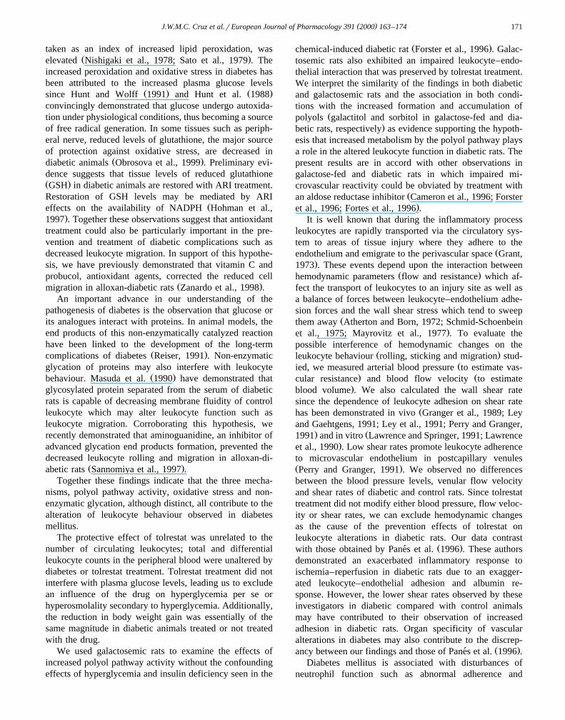
( )J.W.M.C. Cruz et al.rEuropean Journal of Pharmacology 391 2000 163–174 171
taken as an index of increased lipid peroxidation, wasŽ .elevated Nishigaki et al., 1978; Sato et al., 1979 . The
increased peroxidation and oxidative stress in diabetes hasbeen attributed to the increased plasma glucose levels
Ž . Ž .since Hunt and Wolff 1991 and Hunt et al. 1988convincingly demonstrated that glucose undergo autoxida-tion under physiological conditions, thus becoming a sourceof free radical generation. In some tissues such as periph-eral nerve, reduced levels of glutathione, the major sourceof protection against oxidative stress, are decreased in
Ž .diabetic animals Obrosova et al., 1999 . Preliminary evi-dence suggests that tissue levels of reduced glutathioneŽ .GSH in diabetic animals are restored with ARI treatment.Restoration of GSH levels may be mediated by ARI
Žeffects on the availability of NADPH Hohman et al.,.1997 . Together these observations suggest that antioxidant
treatment could also be particularly important in the pre-vention and treatment of diabetic complications such asdecreased leukocyte migration. In support of this hypothe-sis, we have previously demonstrated that vitamin C andprobucol, antioxidant agents, corrected the reduced cell
Ž .migration in alloxan-diabetic rats Zanardo et al., 1998 .An important advance in our understanding of the
pathogenesis of diabetes is the observation that glucose orits analogues interact with proteins. In animal models, theend products of this non-enzymatically catalyzed reactionhave been linked to the development of the long-term
Ž .complications of diabetes Reiser, 1991 . Non-enzymaticglycation of proteins may also interfere with leukocyte
Ž .behaviour. Masuda et al. 1990 have demonstrated thatglycosylated protein separated from the serum of diabeticrats is capable of decreasing membrane fluidity of controlleukocyte which may alter leukocyte function such asleukocyte migration. Corroborating this hypothesis, werecently demonstrated that aminoguanidine, an inhibitor ofadvanced glycation end products formation, prevented thedecreased leukocyte rolling and migration in alloxan-di-
Ž .abetic rats Sannomiya et al., 1997 .Together these findings indicate that the three mecha-
nisms, polyol pathway activity, oxidative stress and non-enzymatic glycation, although distinct, all contribute to thealteration of leukocyte behaviour observed in diabetesmellitus.
The protective effect of tolrestat was unrelated to thenumber of circulating leukocytes; total and differentialleukocyte counts in the peripheral blood were unaltered bydiabetes or tolrestat treatment. Tolrestat treatment did notinterfere with plasma glucose levels, leading us to excludean influence of the drug on hyperglycemia per se orhyperosmolality secondary to hyperglycemia. Additionally,the reduction in body weight gain was essentially of thesame magnitude in diabetic animals treated or not treatedwith the drug.
We used galactosemic rats to examine the effects ofincreased polyol pathway activity without the confoundingeffects of hyperglycemia and insulin deficiency seen in the
Ž .chemical-induced diabetic rat Forster et al., 1996 . Galac-tosemic rats also exhibited an impaired leukocyte–endo-thelial interaction that was preserved by tolrestat treatment.We interpret the similarity of the findings in both diabeticand galactosemic rats and the association in both condi-tions with the increased formation and accumulation of
Žpolyols galactitol and sorbitol in galactose-fed and dia-.betic rats, respectively as evidence supporting the hypoth-
esis that increased metabolism by the polyol pathway playsa role in the altered leukocyte function in diabetic rats. Thepresent results are in accord with other observations ingalactose-fed and diabetic rats in which impaired mi-crovascular reactivity could be obviated by treatment with
Žan aldose reductase inhibitor Cameron et al., 1996; Forster.et al., 1996; Fortes et al., 1996 .
It is well known that during the inflammatory processleukocytes are rapidly transported via the circulatory sys-tem to areas of tissue injury where they adhere to the
Žendothelium and emigrate to the perivascular space Grant,.1973 . These events depend upon the interaction between
Ž .hemodynamic parameters flow and resistance which af-fect the transport of leukocytes to an injury site as well asa balance of forces between leukocyte–endothelium adhe-sion forces and the wall shear stress which tend to sweep
Žthem away Atherton and Born, 1972; Schmid-Schoenbein.et al., 1975; Mayrovitz et al., 1977 . To evaluate the
possible interference of hemodynamic changes on theŽ .leukocyte behaviour rolling, sticking and migration stud-
Žied, we measured arterial blood pressure to estimate vas-. Žcular resistance and blood flow velocity to estimate
.blood volume . We also calculated the wall shear ratesince the dependence of leukocyte adhesion on shear rate
Žhas been demonstrated in vivo Granger et al., 1989; Leyand Gaehtgens, 1991; Ley et al., 1991; Perry and Granger,
. Ž1991 and in vitro Lawrence and Springer, 1991; Lawrence.et al., 1990 . Low shear rates promote leukocyte adherence
to microvascular endothelium in postcapillary venulesŽ .Perry and Granger, 1991 . We observed no differencesbetween the blood pressure levels, venular flow velocityand shear rates of diabetic and control rats. Since tolrestattreatment did not modify either blood pressure, flow veloc-ity or shear rates, we can exclude hemodynamic changesas the cause of the prevention effects of tolrestat onleukocyte alterations in diabetic rats. Our data contrast
Ž .with those obtained by Panes et al. 1996 . These authors´demonstrated an exacerbated inflammatory response toischemia–reperfusion in diabetic rats due to an exagger-ated leukocyte–endothelial adhesion and albumin re-sponse. However, the lower shear rates observed by theseinvestigators in diabetic compared with control animalsmay have contributed to their observation of increasedadhesion in diabetic rats. Organ specificity of vascularalterations in diabetes may also contribute to the discrep-
Ž .ancy between our findings and those of Panes et al. 1996 .´Diabetes mellitus is associated with disturbances of
neutrophil function such as abnormal adherence and

( )J.W.M.C. Cruz et al.rEuropean Journal of Pharmacology 391 2000 163–174172
Žchemotaxis Rayfield et al., 1982; Wilson, 1986; Pereira et.al., 1987 . The mechanisms underlying leukocyte accumu-
lation in a tissue depend on the interaction between thecells and the vascular endothelium. During the develop-ment of inflammatory responses, leukocytes roll along thelining endothelium of postcapillary venules and eventuallybecome firmly attached to the vascular wall before migrat-ing into tissues. The expression of adhesion moleculesmight be reduced in diabetes mellitus and this could beresponsible for the decreased rolling behaviour. Specificadhesion glycoproteins expressed on the surface of leuko-cytes and endothelial cells play a relevant role in the
Žadhesion phenomenon Perry and Granger, 1991; Albeldaet al., 1994; Granger and Kubes, 1994; Malik and Lo,
.1996 . Leukocyte rolling is dependent on the selectinŽfamily of adhesion molecules. P-selectin induced in min-
. Ž .utes and E-selectin 4–6 h for maximum induction ex-pressed on activated endothelium each contribute signifi-
Žcantly to the rolling events Bevilacqua et al., 1987; Geng.et al., 1990; Abassi et al., 1993; Jones et al., 1993 . Since
neither adhesion molecule is thought to be expressed con-stitutively, a stimulus such as surgical manipulation associ-ated with intravital microscopy is required for the expres-
Ž .sion of either adhesion molecule Kubes, 1997 .ŽGlycoproteins of the CD11rCD18 complex b inte-2
.grins expressed on leukocytes interact with ligands suchŽ .as intercellular adhesion molecule-1 ICAM-1 on endothe-
lial cells to mediate leukocyte adhesion and emigrationŽ .Von Adrian et al., 1991, 1992 . Blockade of cell adhesionmolecules either on leukocytes or endothelial cells or bothcan effectively inhibit leukocyte adhesion. One possibleexplanation for the abnormal leukocyte function in dia-betes mellitus might be a down regulation of adhesionmolecules involved in leukocyte recruitment such as se-lectin or ICAM-1. This latter adhesion molecule is consti-tutively expressed on vascular endothelium and showsmarked upregulation on most tissues during the develop-
Žment of acute and chronic inflammatory diseases Dustinet al., 1986; Adams et al., 1989; Cosimi et al., 1990;Wegner et al., 1990; Norris et al., 1991; Crockett-Torabi,
.1998 . Further, a reduced expression of ICAM-1 has beenlinked to the appearance of a protein plasma factor in
Ž .alloxan-diabetic rats Sannomiya et al., 1996 . Recently,we have observed a low level of immunoreactivity toanti-ICAM-1 in venules of the internal spermatic fascia
Ž .from diabetic rats unpublished observations . Though oxi-dants are critical for ICAM-1 transcription in endothelialcells in response to some proinflammatory mediators, like
Ž .TNF-a and interferon g Rahman et al., 1998 , and en-hanced free radical generation has been demonstrated indiabetes, we could not find an increased expression of this
Žadhesion molecule in our diabetic rats Sannomiya et al.,.1996, unpublished observations . Alteration of the expres-
sion of other adhesion molecules on leukocytes androrendothelial cells might occur in diabetes. Studies are inprogress to address this issue.
Based on our present findings, we hypothesize that theaccelerated formation of sorbitol in diabetic animalsmay increase the intracellular osmolarity or decrease theavailability of the enzyme cofactor NADPH leading todisturbance of endothelial cell functions that might alterleukocyte–endothelial interactions. Regardless of themechanism, inhibition of the polyol pathway corrected thedefective leukocyte–endothelial interaction found in exper-imental diabetes and may have a similar effect in diabeticpatients.
Acknowledgements
This work was supported in part by a grant fromFundacao de Amparo a Pesquisa do Estado de Sao Paulo˜ ` ˜Ž .FAPESP .
References
Abassi, O., Kishimoto, J.K., McIntire, L.V., Anderson, D.C., Smith,C.W., 1993. E-selectin supports neutrophil rolling in vitro underconditions of flow. J. Clin. Invest. 92, 2719–2730.
Adams, D.H., Hubscher, S.G., Shaw, J., Rothlein, R., Neuberger, J.M.,1989. Intercellular adhesion molecule-1 on liver allografts duringrejection. Lancet 2, 1122–1125.
Albelda, S.M., Smith, C.W., Ward, P.A., 1994. Adhesion molecules andinflammatory injury. FASEB J. 8, 504–512.
Atherton, A., Born, G.V.R., 1972. Quantitative investigation of theadhesiveness of circulating polymorphonuclear leucocytes to bloodvessels walls. J. Physiol. 222, 447–474.
Baez, S., 1969. Simultaneous measurements of radii and wall thickness ofmicrovessels in the anesthetized rat. Circ. Res. 25, 315–329.
Bagdade, J.D., Root, R.K., Bulger, R.J., 1974. Impaired leukocyte func-tion in patients with poorly controlled diabetes. Diabetes 23, 9–15.
Bevilacqua, M.P., Pober, J.S., Mendrick, D.L., Cotran, R.S. Jr., Gim-brone, A., 1987. Identification of an inducible endothelial–leukocyteadhesion molecule. Proc. Natl. Acad. Sci. U. S. A. 84, 9238–9242.
Bjork, J., Hedqvist, P., Arfors, K.-E., 1982. Increase in vascular perme-¨ability induced by leukotriene B4 and the role of polymorphonuclearleukocytes. Inflammation 6, 189–200.
Boland, O.M., Blackwell, C.C., Clarke, B.F., Ewing, D.J., 1993. Effectsof ponalrestat, an aldose reductase inhibitor, on neutrophil killing ofEscherichia coli and autonomic function in patients with diabetesmellitus. Diabetes 42, 336–340.
Bucala, R., Cerami, A., Vlassara, H., 1995. Advanced glycosylation endproducts in diabetic complications. Biochemical and prospects fortherapeutic interventions. Diabetes Rev. 3, 258–268.
Cameron, N.E., Cotter, M.A., 1993. Potential therapeutics approaches tothe treatment or prevention of diabetic neuropathy: evidence from
Ž .experimental studies. Diabetic Med. 10 7 , 593–605.Cameron, N.E., Cotter, M.A., Dines, K.C., Hohman, T.C., 1996. Reversal
of defective peripheral nerve conduction velocity, nutritive en-doneurial blood flow, and oxygenation by a novel aldose reductaseinhibitor, WAY-121–509, in streptozotocin-induced diabetic rat. J.
Ž .Diabetes Complications 10 1 , 43–53.Cosimi, A.B., Conti, D., Delmonico, F.L., Preffer, F.I., Wee, S.L.,
Rothlein, R., Faanes, R., Colvin, R.B., 1990. In vivo effects ofŽ .monoclonal antibody to ICAM-1 CD54 in nonhuman primates with
renal allografts. J. Immunol. 144, 4606–4612.Crockett-Torabi, E., 1998. Selectins and mechanisms of signal transduc-
tion. J. Leukocyte Biol. 63, 1–14.

( )J.W.M.C. Cruz et al.rEuropean Journal of Pharmacology 391 2000 163–174 173
Davis, M.J., 1987. Determination of volumetric flow in capillary tubesusing an optical Doppler velocimeter. Microvasc. Res. 34, 223–230.
Dustin, M.L., Rothlein, R., Bhan, A.K., Dinarello, C.A., Springer, T.A.,1986. Induction by IL-1 and interferon g: tissue distribution, bio-
Ž .chemistry, and function of a natural adherence molecule ICAM-1 . J.Immunol. 137, 245–254.
Forster, H.G., Wee, P.M., Hohman, T.C., Epstein, M., 1996. Impairmentof afferent arteriolar myogenic responsiveness in the galactose-fed ratis prevented by tolrestat. Diabetologia 39, 907–914.
Fortes, Z.B., Becker, C., Oliveira, M.A., Scivoletto, R., 1996. Influenceof aldose reductase inhibition on the microvascular reactivity inexperimental diabetes. Gen. Pharmacol. 27, 917–921.
Fortes, Z.B., Farsky, S.P., Oliveira, M.A., Garcia-Leme, J., 1991. Directvital microscopic study of defective leukocyte–endothelial interactionin diabetes mellitus. Diabetes 40, 1267–1273.
Fortes, Z.B., Garcia-Leme, J., Scivoletto, R., 1983a. Influence of diabeteson the reactivity of mesenteric microvessels to histamine, bradykininand acetylcholine. Br. J. Pharmacol. 78, 39–48.
Fortes, Z.B., Garcia-Leme, J., Scivoletto, R., 1983b. Vascular reactivityin diabetes mellitus: role of the endothelial cell. Br. J. Pharmacol. 79,771–781.
Fortes, Z.B., Garcia-Leme, J., Scivoletto, R., 1984. Vascular reactivity indiabetes mellitus: possible role of insulin on the endothelial cell. Br. J.Pharmacol. 83, 635–643.
Fortes, Z.B., Scivoletto, R., Garcia-Leme, R., 1989. Functional changesin the microcirculation of alloxan-induced diabetic rats. Gen. Pharma-col. 20, 615–643.
Frank, R.N., 1991. On the pathogenesis of diabetic retinopathy: a 1990update. Ophthalmology 98, 586–593.
Fretland, D.J., Widomski, D.L., Anglin, C.P., Levin, S., Gaginella, T.S.,1991. Modulation of the chemotactic properties of complement frag-ments C and C by the anti-inflammatory agent SC-41930. Agents5a 3
Actions 34, 5–7.Fukase, S., Sato, S., Mori, K., Secchi, E.F., Kador, P.F., 1996. Polyol
pathway and NADPH-dependent reductase in dog leukocytes. J.Ž .Diabetes Complication 10 6 , 304–313.
Garcia-Leme, J., 1989. Hormones and Inflammation. CRC Press, BocaRaton, FL.
Garcia-Leme, J., Farsky, S.P., 1993. Hormonal control of inflammatoryresponses. Mediators Inflammation 2, 181–198.
Geng, J.-G., Bevilacqua, M.P., Moore, K.L., McIntyre, T.M., Prescott,S.M., Kim, J.M., Bliss, G.A., Zimmerman, G.A, McEver, R.P., 1990.Rapid neutrophil adhesion to activated endothelium mediated byGMP-140. Nature 343, 757.
Gore, R.W., Bohlen, H.G., 1977. Microvascular pressures in rat intestinalmuscle and mucosal villii. Am. J. Phys. 233, H685–H693.
Granger, D.N., Benoit, J.N., Suzyki, M., Grisham, M.B., 1989. Leuko-cyte adherence to venular endothelium during ischemia–reperfusion.Am. J. Physiol. 257, 683–688.
Granger, D.N., Kubes, P., 1994. The microcirculation and inflammation:modulation of leukocyte–endothelial cell adhesion. J. Leukocyte Biol.55, 662–675.
Grant, L., 1973. The stickining and emigration of white blood cells inŽ .inflammation. In: Zweifach, B.W., Grant, L., McCluskey, L. Eds. ,
The Inflammatory Process 2 Academic Press, Orlando, FL, pp. 205–249.
Guerrant, G.O., Mass, C.W., 1984. Determination of monosaccharides asaldonitrile O-methyloxamine, alditol and cyclito acetate derivativesby gas chromatography. Anal. Chem. 56, 633–638.
Hohman, T.C., Banas, D., Basso, M., 1997. Increased oxidative stress inexperimental diabetic neuropathy may be linked to increased competi-tion for NADPH. Diabetes 46, 31A.
Hunt, J.V., Dean, R.T., Wolff, S.P., 1988. Hydroxyl radical productionand autoxidative glycosylation. Biochem. J. 256, 205–212.
Hunt, J.V., Wolff, S.P., 1991. The role of histidine residues in thenon-enzymic covalent attachment of glucose and ascorbic acid toprotein. Free Radical Res. Commun. 14, 279–287.
Jarrett, R.J., Keen, H., Hardwick, C., 1970. Instant blood sugar measure-ment using Dextrostix and a reflectance meter. Diabetes 19, 724.
Jones, D.A., Abassi, O., McIntyre, L.V., McEver, R.P., Cotran, R.S.,Gimbrone, M.A., 1993. P-Selectin mediates neutrophil rolling onhistamine-stimulated endothelial cells. Biophys. J. 65, 1560.
Kamura, T., Suzuki, K., Matsumae, H., Sano, T., Sakamoto, N., Hotta,N., 1995. Effects of glucose and SNK-860, an aldose reductaseinhibitor, on the polyol pathway and chemiluminescence response of
Ž .human neutrophils in vitro. Diabetic Med. 12 5 , 392–396.Kaplow, L.S., 1965. A simplified myeloperoxidase stain using benzidine
dihydrochloride. Blood 26, 215–219.Kubes, P., 1997. Mast cell activation and leukocyte rolling responses. In:
Ž .Bochner, B.S. Ed. , Adhesion Molecules in Allergic Disease. MarcelDekker, NY, pp. 279–296.
Kubes, P., Granger, D.N., 1996. Leukocyte–endothelial cell interactionsevoked by mast cells. Cardiovasc. Res. 32, 699–708.
Lawrence, M.B., Smith, C.W., Eskin, S.G., McIntire, L.V., 1990. Effectof venous shear stress on CD18-mediated neutrophil adhesion tocultured endothelium. Blood 75, 227–237.
Lawrence, M.B., Springer, T.A., 1991. Leukocytes roll on a selectin atphysiologic flow rates: distinction from and prerequisite for adhesionthrough integrins. Cell 65, 859–873.
Ley, K., Gaehtgens, P., 1991. Endothelial, not hemodynamic, differencesare responsible for preferential leukocyte rolling in rat mesentericvenules. Circ. Res. 69, 1034–1041.
Ley, K., Gaehtgens, P., Fennie, C., Singer, M.S., Lasky, L.A., Rosen,S.D., 1991. Lectin-like cell adhesion molecule 1 mediates leukocyterolling in mesenteric venules in vivo. Blood 77, 2553–2555.
Lindbom, L., Xie, X., Raud, J., Hedqvist, P., 1992. Chemoattractant-in-duced firm adhesion of leukocytes to vascular endothelium in vivo iscritically dependent on initial leukocyte rolling. Acta Physiol. Scand.146, 415–421.
Lockington, T.L., Partridge, T.A., Barrett, M.C., Wise, P.H., 1987.Sorbinil protects neutrophils from a glucose-related impairment in
Ž .chemotaxis. Diabetic Med. 4, 381, Abstract .Malik, A.B., Lo, S.K., 1996. Vascular endothelial adhesion molecules
and tissue inflammation. Pharmacol. Rev. 48, 213–229.Masuda, M., Murakami, T., Egawa, H., Murata, K., 1990. Decreased
fluidity of polymorphonuclear leukocyte membrane in streptozotocin-induced diabetic rats. Diabetes 39, 466–470.
Mayrovitz, H.N., Wiedeman, M.P., Tuma, R.F., 1977. Experimental andclinical-thrombosis: factors influencing leukocyte adherence in mi-crovessels. Thromb. Haemostasis 38, 823–830.
McMillan, R.M., Foster, S.J., 1988. Leukotriene B and inflammatory4
disease. Agents Actions 24, 114–119.Mohandes, E., Touraine, J.L., Osman, M., Salle, B., 1982. Neutrophil
chemotaxis in infants of diabetic mothers and in preterms at birth. J.Clin. Lab. Immunol. 8, 117–120.
Movat, A.G., Baum, J., 1971. Chemotaxis of polymorphonuclear leuko-cyte from patients with diabetes mellitus. N. Engl. J. Med. 284,621–623.
Nishigaki, I., Hagihara, H., Tsunekawa, M., Maseki, M., Yagi, K., 1978.Lipid peroxide levels of serum lipoprotein fractions of diabetic pa-tients. Biochem. Med. 25, 373–378.
Norris, P., Poston, R.N., Thomas, D.S., Thornhill, M., Hawk, J., Haskard,D.O., 1991. The expression of endothelial leukocyte adhesion
Ž . Ž .molecule-1 ELAM-1 , intercellular adhesion molecule-1 ICAM-1Ž .and vascular cell adhesion molecule-1 VCAM-1 in experimental
cutaneous inflammation: a comparison of ultraviolet erythema anddelayed hypersensitivity. J. Invest. Dermatol. 96, 763–770.
Obrosova, I.G., Fathallah, L., Lang, H.J., Greene, D.A., 1999. Evaluationof a sorbitol dehydrogenase inhibitor on diabetic peripheral nervemetabolism: a prevention study. Diabetologia 42, 1187–1194.
Panes, J., Kurose, I., Rodriguez-Vaca, M.D., Anderson, D.C., Miyasaka,´M., Tso, P., Granger, D.N., 1996. Diabetes exarcebates inflammatoryresponses to ischemia–reperfusion. Circulation 93, 161–167.
Panes, J., Perry, M., Granger, D.N., 1999. Leukocyte–endothelial cell´

( )J.W.M.C. Cruz et al.rEuropean Journal of Pharmacology 391 2000 163–174174
adhesion: avenues for therapeutic intervention. Br. J. Pharmacol. 126,537–550.
Pereira, M.A.A., Sannomiya, P., Garcia-Leme, J., 1987. Inhibition ofleukocyte chemotaxis by factor in alloxan-induced diabetic rat plasma.Diabetes 36, 1307–1314.
Perry, M.A., Granger, D.N., 1991. Role of CD11rCD18 in shear rate-de-pendent leukocyte–endothelial cell interactions in cat mesentericvenules. J. Clin. Invest. 87, 1798–1804.
Petrone, W.I., English, D., Wong, K., McCord, J.M., 1980. Free radicalsand inflammation: superoxide-dependent activation of a neutrophilchemotactic factor in plasma. Proc. Natl. Acad. Sci. 77, 1159–1163.
Rahman, A., Kefer, J., Bando, M., Niles, W.D., Malik, A.B., 1998.E-selectin expression in human endothelial cells by TNF-a-inducedoxidant generation and NF-xB activation. Am. J. Phys. 275, L533–544.
Rayfield, E.J., Ault, M.J., Keusch, G.T., Brothers, M.J., Nechmias, C.,Smith, H., 1982. Infection and diabetes: the case for glucose control.Am. J. Med. 72, 439–450.
Reichard, H.G., Wilson, B., Rosenquist, U., 1993. The effect of long-termintensified insulin treatment on the development of microvascularcomplications of diabetes mellitus. N. Engl. J. Med. 329, 304–309.
Reiser, K., 1991. Nonenzymatic glycation of collagen in aging anddiabetes. Proc. Soc. Exp. Biol. Med. 37, 17–29.
Rhodin, J.A.G., 1986. Architecture of the vessel wall. In: Bohr, D.F.,Ž .Somlyo, A.P., Sparks, H.V. Eds. , Handbook of Physiology. The
Cardiovascular System Sect. 2 Vol. IIpp. 1–31, Bethesda, MD.Sannomiya, P., Oliveira, M.A., Anjos-Valotta, E., Fortes, Z.B., 1996.
Inhibition of leukocyte–endothelial interactions in diabetes mellitus:Ž .role of intercelular adhesion molecule-1 ICAM-1 . J. Vasc. Res. 33,
6.Sannomiya, P., Oliveira, M.A., Fortes, Z.B., 1997. Aminoguanidine and
the prevention of leukocyte dysfunction in diabetes mellitus: a directmicroscopy study. Br. J. Pharmacol. 122, 894–898.
Sannomiya, P., Pereira, M.A.A., Garcia-Leme, J., 1990. Inhibition ofleukocyte chemotaxis by serum factor in diabetes mellitus: selectivedepression of cell responses mediated by complement-derivedchemoattractants. Agents Action 30, 369–376.
Sato, Y., Hotta, N., Sakamoto, N., Matsuoka, S., Oshishi, N., Yagi, K.,1979. Lipid peroxide levels of plasma of diabetic patients. Biochem.Med. 21, 104–107.
Schmid-Schoenbein, G.W., Fung, Y.C., Zweifach, B.W., 1975. Vascularendothelium–leukocyte interaction, sticking shear force in venules.Circ. Res. 36, 173–184.
Stevens, M.J., Feldman, E.L., Greene, D.A., 1995. The aetiology ofdiabetic neuropathy: the combined roles of metabolic and vascular
Ž .defects. Diabetic Med. 12 7 , 566–579.The Diabetes Control And Complication Trial Research Group, 1993.
The effect of intensive treatment of diabetes on development andprogression of long-term complications in insulin-dependent diabetes.N. Engl. J. Med. 329, 977–986.
Tomlinson, D.R., 1993. Aldose reductase inhibitors and complications ofŽ .diabetes mellitus. Diabetic Med. 10 3 , 214–230.
Tomlinson, A., Appleton, I., Moore, A.R., Gilroy, D.W., Willis, D.,Mitchell, J.A., Willoughby, D.A., 1994. Cyclo-oxygenase and nitricoxide synthase isoforms in rat carrageenin-induced pleurisy. Br. J.Pharmacol. 113, 693–698.
Von Adrian, U.H., Chambers, J.D., Mcevoy, L.M., Bargatze, R.F.,Arfors, K., Butcher, E.C., 1991. Two-step model of leukocyte–endo-thelial cell interaction in inflammation: distinct roles for LECAM-1and the leukocyte b integrins in vivo. Proc. Natl. Acad. Sci. U. S. A.2
88, 7538–7542.Von Adrian, U.H., Hansell, P., Chambers, J.D., Berger, E.M., Filho, I.T.,
Butcher, E.C., Arfors, K.E., 1992. L-selectin function is required forbeta-2 integrin-mediated neutrophil adhesion at physiologic shearrates in vivo. Am. J. Physiol. 263, H1034–H1044.
Wegner, C.D., Gundel, R.H., Reilly, P., Haynes, N., Letts, L.G., Roth-Ž .lein, R., 1990. Intercellular adhesion molecule-1 ICAM-1 in the
pathogenesis of asthma. Science 247, 456–459.Wilson, R.M., 1986. Neutrophil function in diabetes. Diabetic Med. 3,
509–512.Wilson, R.M., Tebbs, S., Gonzales, A.M., 1989. Aldose reductase inhibi-
tion restores neutrophil killing of Candida albicans to normal. Dia-Ž . Ž .betic Med. 6 Suppl. 1 , 24, Abstract .
Wilson, R.M., Tomlinson, D.R., Reeves, W.G., 1987. Neutrophil sorbitolproduction impairs oxidative killing in diabetes. Diabetic Med. 4,37–40.
Zanardo, R.O., Oliveira, M.A., Fortes, Z.B., 1998. Endothelial–leukocyteinteraction in diabetes: antioxidant treatment. Hypertension 33, 1095.