Experimental demonstration of the growth rate-lifespan trade-off
Upload
independentCategory
view
0download
0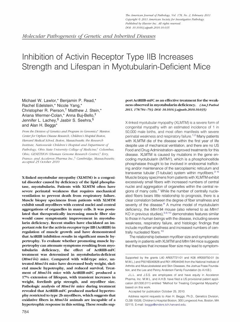
The American Journal of Pathology, Vol. 178, No. 2, February 2011
Copyright © 2011 American Society for Investigative Pathology.
Published by Elsevier Inc. All rights reserved.
DOI: 10.1016/j.ajpath.2010.10.035
Molecular Pathogenesis of Genetic and Inherited Diseases
Inhibition of Activin Receptor Type IIB Increases
Strength and Lifespan in Myotubularin-Deficient MiceMichael W. Lawlor,* Benjamin P. Read,*Rachel Edelstein,* Nicole Yang,*Christopher R. Pierson,† Matthew J. Stein,*Ariana Wermer-Colan,* Anna Buj-Bello,‡
Jennifer L. Lachey,§ Jasbir S. Seehra,§
and Alan H. Beggs*From the Division of Genetics and Program in Genomics,* Manton
Center for Orphan Disease Research, Children’s Hospital Boston,
Harvard Medical School, Boston, Massachusetts; the Research
Institute, Nationwide Children’s Hospital and Department of
Pathology, Ohio State University College of Medicine,† Columbus,
Ohio; GENETHON (Human Genome Research Centre),‡ Evry,
France; and Acceleron Pharma Inc.,§ Cambridge, Massachusetts;accepted 25 October 2010
X-linked myotubular myopathy (XLMTM) is a congeni-tal disorder caused by deficiency of the lipid phospha-tase, myotubularin. Patients with XLMTM often havesevere perinatal weakness that requires mechanicalventilation to prevent death from respiratory failure.Muscle biopsy specimens from patients with XLMTMexhibit small myofibers with central nuclei and centralaggregations of organelles in many cells. It was postu-lated that therapeutically increasing muscle fiber sizewould cause symptomatic improvement in myotubu-larin deficiency. Recent studies have elucidated an im-portant role for the activin-receptor type IIB (ActRIIB) inregulation of muscle growth and have demonstratedthat ActRIIB inhibition results in significant muscle hy-pertrophy. To evaluate whether promoting muscle hy-pertrophy can attenuate symptoms resulting from myo-tubularin deficiency, the effect of ActRIIB-mFCtreatment was determined in myotubularin-deficient(Mtm1�4) mice. Compared with wild-type mice, un-treated Mtm1�4 mice have decreased body weight, skel-etal muscle hypotrophy, and reduced survival. Treat-ment of Mtm1�4 mice with ActRIIB-mFC produced a17% extension of lifespan, with transient increases inweight, forelimb grip strength, and myofiber size.Pathologic analysis of Mtm1�4 mice during treatmentrevealed that ActRIIB-mFC produced marked hypertro-phy restricted to type 2b myofibers, which suggests thatoxidative fibers in Mtm1�4 animals are incapable of a
hypertrophic response in this setting. These results sup-784
port ActRIIB-mFC as an effective treatment for the weak-ness observed in myotubularin deficiency. (Am J Pathol
2011, 178:784–793; DOI: 10.1016/j.ajpath.2010.10.035)
X-linked myotubular myopathy (XLMTM) is a severe form ofcongenital myopathy with an estimated incidence of 1 in50,000 male births, and most often manifests with severeperinatal weakness and respiratory failure.1,2 Many patientswith XLMTM die of the disease within the first year of lifedespite use of mechanical ventilation, and there are no USFood and Drug Administration–approved treatments for thisdisease. XLMTM is caused by mutations in the gene en-coding myotubularin (MTM1), which is a phosphoinositidephosphatase thought to be involved in endosomal traffick-ing and/or maintenance of the sarcoplasmic reticulum andtransverse tubular (T-tubular) system within myofibers.3–6
Muscle biopsy specimens from patients with XLMTM exhibitexcessively small fibers with increased numbers of centralnuclei and aggregation of organelles within the central re-gions of many cells.7 While the number of centrally nucle-ated fibers bears little relationship to prognosis, there is aclear correlation between the degree of fiber smallness andseverity of the disease.8 A murine model of myotubularindeficiency, the Mtm1�4 mouse (also referred to as Mtm1KO in previous studies),3,9,10 demonstrates features similarto those in human beings with the disease, including severeweakness, respiratory failure, and histologic findings thatinclude myofiber smallness and increased numbers of cen-trally nucleated fibers.10
The relationship between myofiber size and symptomaticseverity in patients with XLMTM and Mtm1�4 mice suggeststhat therapies that increase fiber size may lead to symptom-
Supported by the grants L40 AR057721-01 and K08 AR059750-01 (toM.W.L.) and P50 NS040828 and R01 AR044345 from the National Institute ofArthritis and Musculoskeletal and Skin Diseases, the Joshua Frase Founda-tion, and the Lee and Penny Anderson Family Foundation (to A.H.B.).
J.L.L. and J.S.S. are employees of and have equity in AcceleronPharma, Inc. M.W.L. and A.H.B. have filed a US provisional patent appli-cation (61/330,011) entitled “Method for Treating Congenital Myopathy”based on this work.
Accepted for publication October 25, 2010.
Address reprint requests to Alan H. Beggs, Ph.D., Genetics Division,CLSB 15026, Children’s Hospital Boston, 300 Longwood Ave, Boston, MA
02115. E-mail: [email protected].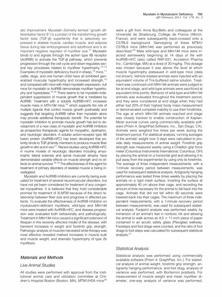
Myostatin in Myotubularin Deficiency 785AJP February 2011, Vol. 178, No. 2
atic improvement. Myostatin (formerly termed “growth dif-ferentiation factor 8”) is a protein of the transforming growthfactor beta (TGF-�) superfamily that is selectively ex-pressed in skeletal muscle, cardiac muscle, and adiposetissue during late embryogenesis and adulthood and is animportant negative regulator of myofiber size.11 Myostatinbinds to and signals through the activin type IIB receptor(ActRIIB) to activate the TGF-� pathway, which preventsprogression through the cell cycle and down-regulates sev-eral key processes related to myofiber hypertrophy.12,13
Examples of myostatin deficiency found in sheep,14 mice,11
cattle, dogs, and one human child have all exhibited gen-eralized muscular hypertrophy and increased strength,13
and compared with mice with intact myostatin expression, nullmice for myostatin or ActRIIB demonstrate myofiber hypertro-phy and hyperplasia.15,16 There seems to be myostatin-inde-pendent suppression of muscle growth that is mediated byActRIIB. Treatment with a soluble ActRIIB-mFC increasesmuscle mass in MTm1�4 mice,16 which supports the role ofmultiple ligands that control muscle growth postnatally andsuggests that targeting ActRIIB rather than myostatin alonemay provide additional therapeutic benefit. The potential formyostatin inhibition to promote muscle growth has led to de-velopment of a new class of myostatin and ActRIIB inhibitorsas prospective therapeutic agents for myopathic, dystrophic,and neurologic disorders. A soluble activin-receptor type IIBfusion protein (ActRIIB-mFC) has been developed that po-tently binds to TGF-� family members to produce muscle fibergrowth in vitro and in vivo17. Recent studies using ActRIIB-mFCin murine models of neurologic disorders including amyo-trophic lateral sclerosis and spinal muscular atrophy havedemonstrated variable effects on muscle strength and no ef-fects on animal survival.18,19 The effectiveness of this agent fortreatment of primary disorders of skeletal muscle is being in-vestigated.
Myostatin and ActRIIB inhibitors are currently being eval-uated for treatment of several neuromuscular disorders, buthave not yet been considered for treatment of any congen-ital myopathies. It is believed that they hold considerablepromise for treatment of XLMTM because of the direct re-lationship between fiber size and prognosis in affected pa-tients. To evaluate the effectiveness of ActRIIB inhibition onmyotubularin-deficient myofibers, wild-type and Mtm1�4mice were treated with ActRIIB-mFC, and disease progres-sion was evaluated both behaviorally and pathologically.Treatment in Mtm1�4 mice caused a significant extension oflifespan in this severely affected model of the disease, withtransient increases in weight and forelimb grip strength.Pathologic analysis of muscles harvested while therapy wasmost effective revealed marked increases in muscle bulkand muscle weight, and dramatic hypertrophy of type 2bmyofibers.
Materials and Methods
Live Animal Studies
All studies were performed with approval from the insti-tutional animal care and utilization committee at Chil-
dren’s Hospital Boston (Boston, MA). MTM1/HSA mice20were a gift from Anna Buj-Bello and colleagues at theUniversité de Strasbourg, Collège de France (Illkirch,France), and were subsequently back-crossed onto aC57BL6 background. Genotyping of these MTM1/C57BL6 mice (Mtm1�4) was performed as previouslydescribed.20 Male wild-type and Mtm1�4 mice were in-jected semiweekly beginning at 14 days of life withActRIIB-mFC (also called RAP-031; Acceleron PharmaInc., Cambridge, MA) at a dose of 20 mg/kg. This dosagewas chosen because it was above the level at whichmuscle hypertrophy plateaued in wild-type mice (datanot shown). Vehicle-treated animals were injected with anequivalent volume of Tris-buffered saline solution. Treat-ment was continued until Mtm1�4 animals were judged tobe at end stage, and wild-type animals were sacrificed atequivalent time points. Behavior of wild-type and Mtm1�4animals was evaluated throughout the treatment period,and they were considered at end stage when they hadeither lost 20% of their highest body mass measurementor demonstrated complete inability to use their hindlimbs.The age at which Mtm1�4 animals reached end stagewas closely tracked to enable construction of Kaplan-Meier survival curves using commercially available soft-ware (Prism 4; GraphPad Software, Inc., San Diego, CA).Animals were weighed five times per week during thetreatment period. For statistical analysis, running averagesof the animals’ weight over 3 days were calculated to pro-vide daily measurements of animal weight. Forelimb gripstrength was measured weekly using a Chatillon grip forcemeter (Columbus Instruments International, Columbus, OH)by placing the animal on a horizontal grid and allowing it topull away from the experimenter by using only its forelimbs.The average of three independent measurements, with a1-minute recovery period between measurements, wasused for subsequent statistical analysis. Antigravity hangingperformance was tested three times weekly by placing theanimals on a rigid mesh surface, inverting the surface atapproximately 40 cm above their cage, and recording theamount of time necessary for the animal to fall back into thecage. Animals that did not fall within 60 seconds werelowered back into their cages. The maximum of three inde-pendent measurements, with a 1-minute recovery periodbetween measurements, was used for subsequent statisti-cal analysis. Footprint analysis was performed weekly byimmersion of an animal’s feet in nontoxic ink and allowingthe animal to walk across an 8.5 � 11-inch piece of papercontained in an acrylic safety glass (Plexiglas) walkway.Footsteps and foot drags were counted, and the ratio of footdrags to foot steps was calculated for subsequent statisticalanalysis.21
Statistical Analysis
Statistical analysis was performed using commerciallyavailable software (Prism 4; GraphPad, Inc.). For statisti-cal analysis of animal weight, forelimb grip strength, an-tigravity hanging performance, and foot drag, analysis ofvariance was performed, with Bonferroni posttests. Formeasurement of muscle weight and mean myofiber di-
ameter, one-way analysis of variance was performed,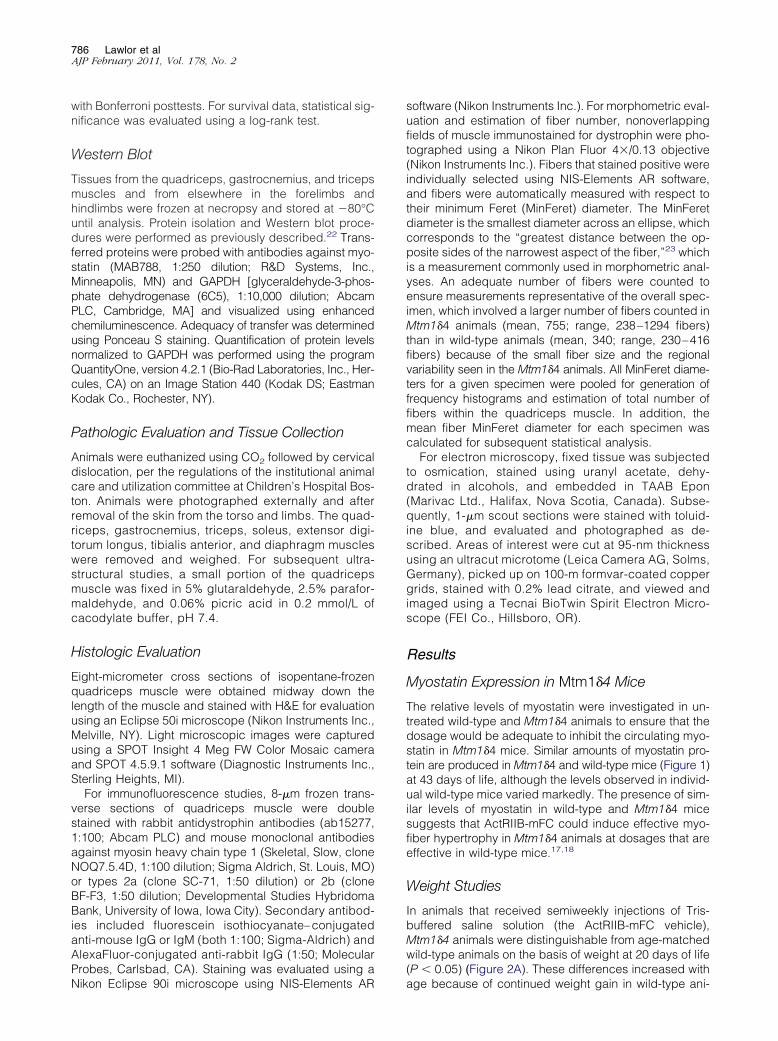
786 Lawlor et alAJP February 2011, Vol. 178, No. 2
with Bonferroni posttests. For survival data, statistical sig-nificance was evaluated using a log-rank test.
Western Blot
Tissues from the quadriceps, gastrocnemius, and tricepsmuscles and from elsewhere in the forelimbs andhindlimbs were frozen at necropsy and stored at �80°Cuntil analysis. Protein isolation and Western blot proce-dures were performed as previously described.22 Trans-ferred proteins were probed with antibodies against myo-statin (MAB788, 1:250 dilution; R&D Systems, Inc.,Minneapolis, MN) and GAPDH [glyceraldehyde-3-phos-phate dehydrogenase (6C5), 1:10,000 dilution; AbcamPLC, Cambridge, MA] and visualized using enhancedchemiluminescence. Adequacy of transfer was determinedusing Ponceau S staining. Quantification of protein levelsnormalized to GAPDH was performed using the programQuantityOne, version 4.2.1 (Bio-Rad Laboratories, Inc., Her-cules, CA) on an Image Station 440 (Kodak DS; EastmanKodak Co., Rochester, NY).
Pathologic Evaluation and Tissue Collection
Animals were euthanized using CO2 followed by cervicaldislocation, per the regulations of the institutional animalcare and utilization committee at Children’s Hospital Bos-ton. Animals were photographed externally and afterremoval of the skin from the torso and limbs. The quad-riceps, gastrocnemius, triceps, soleus, extensor digi-torum longus, tibialis anterior, and diaphragm muscleswere removed and weighed. For subsequent ultra-structural studies, a small portion of the quadricepsmuscle was fixed in 5% glutaraldehyde, 2.5% parafor-maldehyde, and 0.06% picric acid in 0.2 mmol/L ofcacodylate buffer, pH 7.4.
Histologic Evaluation
Eight-micrometer cross sections of isopentane-frozenquadriceps muscle were obtained midway down thelength of the muscle and stained with H&E for evaluationusing an Eclipse 50i microscope (Nikon Instruments Inc.,Melville, NY). Light microscopic images were capturedusing a SPOT Insight 4 Meg FW Color Mosaic cameraand SPOT 4.5.9.1 software (Diagnostic Instruments Inc.,Sterling Heights, MI).
For immunofluorescence studies, 8-�m frozen trans-verse sections of quadriceps muscle were doublestained with rabbit antidystrophin antibodies (ab15277,1:100; Abcam PLC) and mouse monoclonal antibodiesagainst myosin heavy chain type 1 (Skeletal, Slow, cloneNOQ7.5.4D, 1:100 dilution; Sigma Aldrich, St. Louis, MO)or types 2a (clone SC-71, 1:50 dilution) or 2b (cloneBF-F3, 1:50 dilution; Developmental Studies HybridomaBank, University of Iowa, Iowa City). Secondary antibod-ies included fluorescein isothiocyanate– conjugatedanti-mouse IgG or IgM (both 1:100; Sigma-Aldrich) andAlexaFluor-conjugated anti-rabbit IgG (1:50; MolecularProbes, Carlsbad, CA). Staining was evaluated using a
Nikon Eclipse 90i microscope using NIS-Elements ARsoftware (Nikon Instruments Inc.). For morphometric eval-uation and estimation of fiber number, nonoverlappingfields of muscle immunostained for dystrophin were pho-tographed using a Nikon Plan Fluor 4�/0.13 objective(Nikon Instruments Inc.). Fibers that stained positive wereindividually selected using NIS-Elements AR software,and fibers were automatically measured with respect totheir minimum Feret (MinFeret) diameter. The MinFeretdiameter is the smallest diameter across an ellipse, whichcorresponds to the “greatest distance between the op-posite sides of the narrowest aspect of the fiber,”23 whichis a measurement commonly used in morphometric anal-yses. An adequate number of fibers were counted toensure measurements representative of the overall spec-imen, which involved a larger number of fibers counted inMtm1�4 animals (mean, 755; range, 238–1294 fibers)than in wild-type animals (mean, 340; range, 230–416fibers) because of the small fiber size and the regionalvariability seen in the Mtm1�4 animals. All MinFeret diame-ters for a given specimen were pooled for generation offrequency histograms and estimation of total number offibers within the quadriceps muscle. In addition, themean fiber MinFeret diameter for each specimen wascalculated for subsequent statistical analysis.
For electron microscopy, fixed tissue was subjectedto osmication, stained using uranyl acetate, dehy-drated in alcohols, and embedded in TAAB Epon(Marivac Ltd., Halifax, Nova Scotia, Canada). Subse-quently, 1-�m scout sections were stained with toluid-ine blue, and evaluated and photographed as de-scribed. Areas of interest were cut at 95-nm thicknessusing an ultracut microtome (Leica Camera AG, Solms,Germany), picked up on 100-m formvar-coated coppergrids, stained with 0.2% lead citrate, and viewed andimaged using a Tecnai BioTwin Spirit Electron Micro-scope (FEI Co., Hillsboro, OR).
Results
Myostatin Expression in Mtm1�4 Mice
The relative levels of myostatin were investigated in un-treated wild-type and Mtm1�4 animals to ensure that thedosage would be adequate to inhibit the circulating myo-statin in Mtm1�4 mice. Similar amounts of myostatin pro-tein are produced in Mtm1�4 and wild-type mice (Figure 1)at 43 days of life, although the levels observed in individ-ual wild-type mice varied markedly. The presence of sim-ilar levels of myostatin in wild-type and Mtm1�4 micesuggests that ActRIIB-mFC could induce effective myo-fiber hypertrophy in Mtm1�4 animals at dosages that areeffective in wild-type mice.17,18
Weight Studies
In animals that received semiweekly injections of Tris-buffered saline solution (the ActRIIB-mFC vehicle),Mtm1�4 animals were distinguishable from age-matchedwild-type animals on the basis of weight at 20 days of life(P � 0.05) (Figure 2A). These differences increased with
age because of continued weight gain in wild-type ani-
Myostatin in Myotubularin Deficiency 787AJP February 2011, Vol. 178, No. 2
mals in comparison with the plateau observed after 34days of life in Mtm1�4 animals. Compared with vehicle-treated mice, wild-type animals treated with semiweeklyinjections of ActRIIB-mFC, 20 mg/kg beginning at 14days of life, showed significant weight gain after 36 daysof life (P � 0.05), and continued to gain weight with age.ActRIIB-mFC treated Mtm1�4 animals initially exhibited amodest sustained weight gain, reaching a maximum of124% at 53 days of life; however, the weight of these
Figure 1. Myostatin expression in wild-type and Mtm1�4 mice. A: Westernblot analysis performed on the triceps muscle of 43 day-old animals demon-strates similar levels of myostatin expression in Mtm1�4 mice and theirwild-type littermates. These are nonadjacent lanes from the same immunoblotand using the same exposure time, as denoted by the vertical line. B: Expressionof myostatin normalized to GAPDH expression in vehicle-treated wild-typeand Mtm1�4 mice at 43 days of life. Numbers are shown in reference to themean value for the wild-type animals, and reflect the mean (SEM) value of 3animals for each genotype.
animals plateaued quickly and did not increase as ob-served in wild-type animals.
Antigravity Hanging Performance
At antigravity hanging assay, in which animals are sus-pended from a mesh grid until they either drop into thecage or have been hanging for 60 seconds, wild-typemice were able to hang for up to 60 seconds starting from3 weeks of life (Figure 2B). Treatment of wild-type micewith ActRIIB-mFC led to a slight decrease in antigravityhanging performance, which was statistically significantonly at 26 to 27 days of life (P � 0.05). In contrast,vehicle-treated Mtm1�4 animals exhibited impairedhanging performance at 2 weeks of life, which subse-quently degenerated into nearly complete inability to re-main suspended against gravity by 28 to 29 days of life.Treatment with ActRIIB-mFC did not measurably improvethe antigravity hanging performance of Mtm1�4 mice.
Forelimb Grip Strength
Mtm1�4 animals exhibited consistently lower forelimbgrip strength measurements at all ages tested (P �0.001) (Figure 2C). Forelimb grip force measurements invehicle-treated wild-type animals showed consistentgains in grip strength as the animals aged, whereas thegrip force of Mtm1�4 animals was greatest at 5 weeks oflife and then decreased as the disease progressed.Compared with their vehicle-treated counterparts, Ac-tRIIB-mFC–treated wild-type animals also demonstratedincreased grip strength as the treatment period pro-gressed, with grip force up to 135% greater at 9 weeks oflife, the latest time point tested. In ActRIIB-mFC–treatedMtm1�4 animals, grip strength was transiently improved
Figure 2. Behavioral findings in vehicle- andActRIIB-mFC–treated mice. A: Body weight ofvehicle- and ActRIIB-mFC–treated mice. Mea-surements are the running mean (SEM) of (n)animals in each treatment group. B: Antigravityhanging performance of vehicle- and ActRIIB-mFC–treated mice. C: Forelimb grip force of ve-hicle- and ActRIIB-mFC–treated mice. D: Mea-surements are mean (SEM) values of (n) animalsin each treatment group. E: Kaplan-Meier sur-vival curves for vehicle- (Veh) and ActRIIB-mFC–treated Mtm1�4 mice. *P � 0.05.
788 Lawlor et alAJP February 2011, Vol. 178, No. 2
by 116% at 5 weeks of life (P � 0.05), after which gripstrength declined in both vehicle- and ActRIIB-mFC–treated Mtm1�4 animals.
Footprint Analysis
Abnormalities of gait can be used to differentiate wild-type from Mtm1�4 animals. Mtm1�4 mice experiencehindlimb weakness with disease progression, which canbe visualized and quantified as foot drags on a footprint-ing assay. While foot drags are extremely uncommon inwild-type mice, the number of drags observed in Mtm1�4mice steadily increased after 3 weeks of life (P � 0.05)(Figure 2D). Treatment with ActRIIB-mFC did not haveany effect on the number of foot drags observed at foot-print analysis.
Survival
Similar to the first published reports using Mtm1�4 miceon the HSA background, which stated that the survival ofMtm1�4 animals ranged from 6 to 12 weeks (mean, 59days), in the present study, untreated and vehicle-treatedMtm1�4 animals demonstrated a maximum lifespan ofapproximately 8 to 9 weeks (mean, 56.1 days; range,32–65 days). In comparisons of vehicle- and ActRIIB-mFC–treated Mtm1�4 mice, treatment with ActRIIB-mFCsignificantly lengthened this lifespan by 17% (mean, 67.6days; range, 61–74 days), with a shift in median survivalfrom 58 days to 68 days (P � 0.05) (Figure 2E). Thissurvival benefit was due to both a decrease in the num-ber of early deaths and delayed death in the oldesttreated animals. This survival benefit also seems to bedose-dependent; reducing the dose to 5 mg/kg of Ac-tRIIB-mFC in a pilot study of six animals resulted in anincrease in median survival to 65 days (data not shown),which is still a significant improvement over that in thevehicle-treated animals (P � 0.05).
Gross Evaluation
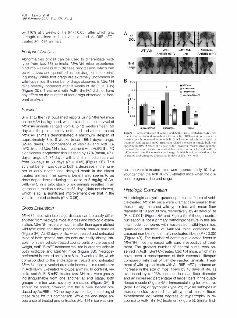
Mtm1�4 mice with late-stage disease can be easily differ-entiated from wild-type mice at gross and histologic exam-ination. Mtm1�4 mice are much smaller than age-matchedwild-type mice and have proportionately smaller muscles(Figure 3A). At 43 days of life, when treated and untreatedmice of both genetic backgrounds are easily distinguish-able from their vehicle-treated counterparts on the basis ofweight, ActRIIB-mFC treatment resulted in larger muscles inboth wild-type and Mtm1�4 mice (Figure 3B). Necropsyperformed in treated animals at 8 to 10 weeks of life, whichcorresponded to the end-stage in treated and untreatedMtm1�4 mice, revealed dramatic increases in muscle sizein ActRIIB-mFC–treated wild-type animals. In contrast, ve-hicle- and ActRIIB-mFC–treated Mtm1�4 mice were grosslyindistinguishable from one another at end stage; bothgroups of mice were severely emaciated (Figure 3A). Itshould be noted, however, that the survival benefit pro-duced by ActRIIB-mFC therapy prevented age-matching ofthese mice for this comparison. While the end-stage ap-
pearance of treated and untreated Mtm1�4 mice was sim-ilar, the vehicle-treated mice were approximately 10 daysyounger than the ActRIIB-mFC–treated mice when the dis-ease progressed to end stage.
Histologic Examination
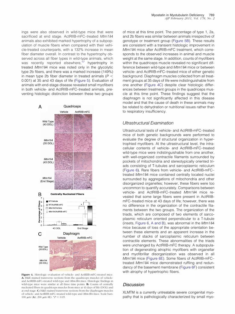
At histologic analysis, quadriceps muscle fibers of vehi-cle-treated Mtm1�4 mice were dramatically smaller thanthose of age-matched wild-type mice, with mean fiberdiameter of 19 and 30 mm, respectively, by 43 days of life(P � 0.001) (Figure 4A and Figure 5). Although centralnucleation is not a primary pathologic feature in this an-imal model, compared with muscles from wild-type mice,quadriceps muscles of Mtm1�4 mice contained in-creased numbers of centrally nucleated fibers (P � 0.05)(Figure 4B). The number of centrally nucleated fibers inMtm1�4 mice increased with age, irrespective of treat-ment. The greatest number of central nuclei was ob-served in ActRIIB-mFC–treated Mtm1�4 mice, which mayhave been a consequence of their extended lifespancompared with that of vehicle-injected animals. Treat-ment of wild-type animals with ActRIIB-mFC produced anincrease in the size of most fibers by 43 days of life, asevidenced by a 123% increase in mean fiber diameterand an increased percentage of large fibers in the quad-riceps muscle (Figure 4A). Immunostaining for oxidative(type 1 or 2a) or glycolytic (type 2b) myosin subtypes inthese muscles revealed that all types of muscle fibersexperienced equivalent degrees of hypertrophy in re-
Figure 3. Gross evaluation of vehicle- and ActRIIB-mFC–treated mice. A: Grossexamination of skinned animals at 43 days of life (DOL) or at end stage (�9weeks) reveals increased muscle bulk in wild-type animals as a result oftreatment with ActRIIB-mFC. Treatment-related increase in muscle bulk wasapparent in Mtm1�4 mice at 43 days of life; however, muscle atrophy in theterminal phase of disease prevents differentiation of vehicle- and ActRIIB-mFC–treated Mtm1�4 animals at end stage. B: Weights of individual musclesin treated and untreated animals at 43 days of life. *P � 0.05.
sponse to ActRIIB-mFC treatment (Figure 5). Similar find-
Myostatin in Myotubularin Deficiency 789AJP February 2011, Vol. 178, No. 2
ings were also observed in wild-type mice that weresacrificed at end stage. ActRIIB-mFC–treated Mtm1�4animals also exhibited marked hypertrophy of a subpop-ulation of muscle fibers when compared with their vehi-cle-treated counterparts, with a 132% increase in meanfiber diameter overall. In contrast to the hypertrophy ob-served across all fiber types in wild-type animals, whichwas recently reported elsewhere,17 hypertrophy intreated Mtm1�4 mice was noted only in the glycolytictype 2b fibers, and there was a marked increase (149%)in mean type 2b fiber diameter in treated animals (P �0.001) at 35 and 43 days of life (Figure 5). Evaluation ofanimals with end-stage disease revealed small myofibersin both vehicle- and ActRIIB-mFC–treated animals, pre-venting histologic distinction between these two groups
Figure 4. Histologic evaluation of vehicle- and ActRIIB-mFC–treated mice.A: H&E-stained transverse sections from the quadriceps muscles of vehicle-and ActRIIB-mFC–treated wild-type and Mtm1�4 mice. Histologic findings inwild-type mice were similar at all three time points. B: Counts of centrallynucleated fibers in quadriceps muscles from mice at 43 days of life (DOL) andat end stage. C: H&E-stained transverse sections from the diaphragm muscles
of vehicle- and ActRIIB-mFC–treated wild-type and Mtm1�4 mice. Scale bars:100 �m (A); 200 �m (C). *P � 0.05.of mice at this time point. The percentage of type 1, 2a,and 2b fibers was similar between animals irrespective ofgenotype or treatment group (Figure 5B). These resultsare consistent with a transient histologic improvement inMtm1�4 mice after ActRIIB-mFC treatment, which corre-sponds to the observed increases in animal and muscleweight at the same stage. In addition, counts of myofiberswithin the quadriceps muscle revealed no significant dif-ference between wild-type and Mtm1�4 mice or betweenvehicle- and ActRIIB-mFC–treated mice of either geneticbackground. Diaphragm muscles collected from all treat-ment groups at 35 days of life were indistinguishable fromone another (Figure 4C) despite clear histologic differ-ences between treatment groups in the quadriceps mus-cle at this time point. These findings suggest that thediaphragm is not significantly affected in this diseasemodel and that the cause of death in these animals maybe related to dehydration or nutritional issues rather thanto respiratory insufficiency.
Ultrastructural Examination
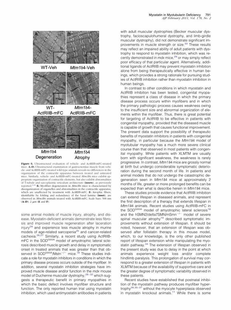
Ultrastructural tests of vehicle- and ActRIIB-mFC–treatedmice of both genetic backgrounds were performed toevaluate the degree of structural organization in hyper-trophied myofibers. At the ultrastructural level, the intra-cellular contents of vehicle- and ActRIIB-mFC–treatedwild-type mice were indistinguishable from one another,with well-organized contractile filaments surrounded bypockets of mitochondria and stereotypically oriented tri-ads consisting of T-tubules and sarcoplasmic reticulum(Figure 6). Rare fibers from vehicle- and ActRIIB-mFC–treated Mtm1�4 mice contained centrally located nucleisurrounded by aggregations of mitochondria and otherdisorganized organelles; however, these fibers were toouncommon to quantify accurately. Comparisons betweenvehicle- and ActRIIB-mFC–treated Mtm1�4 mice re-vealed that some large fibers were present in ActRIIB-mFC–treated mice at 43 days of life; however, there wasno difference in the organization of the contractile fila-ments between the two groups. The organization of thetriads, which are composed of two elements of sarco-plasmic reticulum oriented perpendicular to a T-tubule(insets, Figure 6, A and B), was abnormal in the Mtm1�4mice because of loss of the appropriate orientation be-tween these elements and an apparent increase in thenumber of stacks of sarcoplasmic reticulum betweencontractile elements. These abnormalities of the triadswere unchanged by ActRIIB-mFC therapy. A subpopula-tion of degenerating atrophic myofibers with organellarand myofibrillar disorganization was observed in allMtm1�4 mice (Figure 6E). Some fibers of ActRIIB-mFC–treated Mtm1�4 mice demonstrated ruffling and redun-dancy of the basement membrane (Figure 6F) consistentwith atrophy of hypertrophic fibers.
Discussion
XLMTM is a currently untreatable severe congenital myo-
pathy that is pathologically characterized by small myo-
demons0.0001.
790 Lawlor et alAJP February 2011, Vol. 178, No. 2
fibers and increased numbers of centrally nucleatedmyofibers. Based on the relationship between small fibersize and poor prognosis, the present study evaluated thetherapeutic efficacy of ActRIIB-mFC, an agent that in-duces myofiber hypertrophy through inhibition of myosta-
Figure 5. Histologic analysis according to fiber type. A: Immunostaining of tor Mtm1�4 mouse at 43 days of life for dystrophin (red) and either myosin hwas performed in all treatment groups at 35 and 43 days of life and at endand myofiber size was quantitated according to fiber type. B: Percentages of43 days of life, and end stage were analyzed. Mean MinFeret diameter measfor panel C also applies to panels D and E. F and G: Frequency histogramsActRIIB-mFC–treated wild-type (F) and Mtm1�4 (G) mice. *P � 0.01. **P �
tin, in the Mtm1�4 mouse. After treatment with ActRIIB-
mFC, Mtm1�4 mice exhibited increases in weight,muscle weight, and myofiber size, which behaviorallycorresponded to a transient increase in forelimb gripstrength and a 17% increase in lifespan.
Preclinical trials using myostatin loss or inhibition have
e sections from the quadriceps muscles of an ActRIIB-mFC–treated wild-typeain type 1, type 2a, or type 2b fibers (green). Scale bar � 100 �m. Stainingetermined by complete hindlimb paralysis or a �20% loss in body weight),a, and 2b fibers were counted. C–E: Muscles from animals at 35 days of life,
ts for type 1 (C), type 2a (D), and type 2b (E) fibers are shown. The legendtrate the distribution of fiber sizes in the quadriceps muscles of vehicle- and
ransverseavy ch
stage (dtype 1, 2uremen
been effective in ameliorating symptoms of weakness in
Myostatin in Myotubularin Deficiency 791AJP February 2011, Vol. 178, No. 2
some animal models of muscle injury, atrophy, and dis-ease. Myostatin-deficient animals demonstrate less fibro-sis and improved muscle regeneration after lacerationinjury24 and experience less muscle atrophy in murinemodels of age-related sarcopenia25 and cancer-relatedcachexia.26,27 Similarly, a recent study using ActRIIB-mFC in the SODG93A model of amyotrophic lateral scle-rosis described muscle growth and delay in symptomaticonset in treated animals that was greater than that ob-served in SODG93A/Mstn�/� mice.18 These studies indi-cate a role for myostatin inhibitors in conditions in which theprimary disease process occurs outside of the myofiber. Inaddition, several myostatin inhibition strategies have im-proved muscle disease and/or function in the mdx mousemodel of Duchenne muscular dystrophy,28–32 which sug-gests a therapeutic benefit in primary myopathies inwhich the basic defect involves myofiber structure andfunction. The only reported human trial using myostatin
Figure 6. Ultrastructural evaluation of vehicle- and ActRIIB-mFC–treatedmice. A–D: Ultrastructural examination of gastrocnemius muscle from vehi-cle- and ActRIIB-mFC–treated wild-type animals reveals no differences in theorganization of the contractile apparatus between treated and untreatedmice. Similarly, vehicle- and ActRIIB-mFC–treated Mtm1�4 mice exhibit ap-propriate organization of contractile elements, but also exhibit abnormalitiesof T-tubule and sarcoplasmic reticulum architecture (insets), as previouslyreported.4,41 E: Myofiber degeneration in Mtm1�4 mice is characterized bydisorganization of organelles and abnormalities in the contractile apparatus,which are unaffected by treatment with ActRIIB-mFC. F: Atrophic fibers,identifiable by folding and redundancy of the basal lamina (inset), areobserved in Mtm1�4 animals treated with ActRIIB-mFC. Scale bars: 500 nm(A–D); 2 �m (E and F).
inhibition, which used antimyostatin antibodies in patients
with adult muscular dystrophies (Becker muscular dys-trophy, facioscapulohumeral dystrophy, and limb-girdlemuscular dystrophy), did not demonstrate significant im-provements in muscle strength or size.33 These resultsmay reflect an impaired ability of adult patients with dys-trophy to respond to myostatin inhibition, which was re-cently demonstrated in mdx mice,29 or may simply reflectpoor efficacy of that particular agent. Alternatively, addi-tional ligands of ActRIIB may prevent myostatin inhibitionalone from being therapeutically effective in human be-ings, which provides a strong rationale for pursuing stud-ies of ActRIIB inhibition rather than myostatin inhibition inhuman beings.
In contrast to other conditions in which myostatin andActRIIB inhibition has been tested, congenital myopa-thies represent a class of disease in which the primarydisease process occurs within myofibers and in whichthe primary pathologic process causes weakness owingto the insufficient size and abnormal organization of ele-ments within the myofiber. Thus, there is great potentialfor targeting of ActRIIB to be effective in patients withcongenital myopathy, provided that the diseased muscleis capable of growth that causes functional improvement.The present data support the possibility of therapeuticbenefits of myostatin inhibitors in patients with congenitalmyopathy, in particular because the Mtm1�4 model ofmyotubular myopathy has a much more severe clinicalcourse than that observed in most patients with congen-ital myopathy. While patients with XLMTM are usuallyborn with significant weakness, the weakness is rarelyprogressive. In contrast, Mtm1�4 mice are grossly normalat birth but undergo considerable symptomatic deterio-ration during the second month of life. In patients andanimal models that do not undergo the catastrophic de-generation seen in Mtm1�4 mice at approximately 2months of life, greater or more prolonged benefits can beexpected than what is describe herein in Mtm1�4 mice.
These studies provide evidence that ActRIIB inhibitioncan extend lifespan in diseased animals, and representthe first description of a therapy that extends lifespan inMtm1�4 animals. Recent studies using ActRIIB-mFC inthe SODG93A model of amyotrophic lateral sclerosis18
and the hSMN2/delta7SMN/mSmn�/� model of severespinal muscular atrophy19 described symptomatic im-provements without extension of lifespan. It should benoted, however, that an extension of lifespan was ob-served after follistatin therapy in this mouse model,which, to our knowledge, is the only other publishedreport of lifespan extension while manipulating the myo-statin pathway.34 The extension of lifespan observed inthe present study was due to delay in the point at whichanimals experience weight loss and/or completehindlimb paralysis. This prolongation of survival may cor-respond to a greater extension of lifespan in patients withXLMTM because of the availability of supportive care andthe greater degree of symptomatic variability observed inthese patients.
Recent studies have established that postnatal inhibi-tion of the myostatin pathway produces myofiber hyper-trophy28,35–37 without the myocyte hyperplasia observed
in myostatin knockout animals.11 While there is some
792 Lawlor et alAJP February 2011, Vol. 178, No. 2
evidence that myostatin inhibition during developmentcan produce increased numbers of type 2b fibers inskeletal muscle,38 treatment of wild-type and Mtm1�4mice with ActRIIB-mFC did not change the number orpercentage of type 1, 2a, and 2b fibers in the quadricepsmuscles. These findings, which are consistent with arecent report of the effects of ActRIIB-mFC treatment inwild-type mouse muscle,17 support the theory that myo-cyte hyperplasia or changes in fiber type distribution donot occur with postnatal inhibition of myostatin or ActRIIB.In addition, while satellite cell behavior in response totreatment was not specifically examined, no evidence ofsatellite cell fusion or myoblast proliferation was ob-served at histologic evaluation of ActRIIB-mFC–treatedmuscles.
In contrast to the hypertrophy observed in all fibertypes in treated wild-type animals,17 ActRIIB-mFC–treated Mtm1�4 animals did not demonstrate hypertro-phy in the oxidative type 1 and 2a myofibers at 43 days oflife. The degree to which hypertrophy was observed intype 2b fibers, however, markedly exceeded the degreeof hypertrophy observed in treated wild-type animals.These results suggest that myotubularin deficiency inter-feres with the hypertrophic response to myostatin inhibi-tion in oxidative fibers. The growth pathways in type 1and 2a myofibers are calcium-dependent, whereas hy-pertrophy in type 2b fibers occurs through the relativelycalcium-independent Akt pathway (reviewed in 39, 40).Recent studies of myotubularin-deficient mice,9 fish,5
and dogs41 have described abnormalities in T-tubule andsarcoplasmic reticulum architecture and abnormalities inintracellular calcium handling.9 These abnormalities inintracellular calcium may interfere with activation of hy-pertrophic pathways in oxidative fibers and, thereby, pre-vent ActRIIB-mFC–induced hypertrophy in these fibers.The Akt pathway may not be affected by abnormalities ofcalcium homeostasis in myotubularin-deficient myofibers,which could explain the marked fiber type-specific hyper-trophy observed in Mtm1�4 animals. However, myotubu-larin seems to have some critical role in type 2b myofibersbecause these fibers eventually become atrophic between43 days of life and disease end stage.
In conclusion, the present study provides evidencethat myostatin inhibition via treatment with ActRIIB-mFC istherapeutically beneficial to Mtm1�4 mice according tofindings of behavioral and pathologic studies. Further-more, the selective hypertrophy of type 2b myofibers inMtm1�4 animals may provide important informationabout the biological processes in which myotubularin isinvolved. Although these findings may provide the firstpotential therapy for XLMTM in children, the selectivenature of the response to treatment provides interestingpossibilities for improving therapeutic responses oncethe role of myotubularin in oxidative myofibers is morecompletely understood.
Acknowledgments
We thank Drs. Vandana Gupta, Ph.D., Pankaj Agrawal,
M.D., and Behzad Moghadaszadeh, Ph.D., for scien-tific advice, and Ms. Samantha Lundeen for technicalassistance with the Western blot analyses.
This study is dedicated to the memory of Joshua MilesFrase, who provided tremendous inspiration to childrenwith congenital myopathy, their families, and researchersaround the world.
References
1. Heckmatt JZ, Sewry CA, Hodes D, Dubowitz V: Congenital centronu-clear (myotubular) myopathy: a clinical, pathological and geneticstudy in eight children. Brain 1985, 108 (Pt 4):941–964
2. Jungbluth H, Wallgren-Pettersson C, Laporte J: Centronuclear (myo-tubular) myopathy. Orphanet J Rare Dis 2008, 3:26
3. Buj-Bello A, Fougerousse F, Schwab Y, Messaddeq N, Spehner D,Pierson CR, Durand M, Kretz C, Danos O, Douar AM, Beggs AH,Schultz P, Montus M, Denefle P, Mandel JL: AAV-mediated intramus-cular delivery of myotubularin corrects the myotubular myopathy phe-notype in targeted murine muscle and suggests a function in plasmamembrane homeostasis. Hum Mol Genet 2008, 17:2132–2143
4. Cao C, Backer JM, Laporte J, Bedrick EJ, Wandinger-Ness A: Se-quential actions of myotubularin lipid phosphatases regulate endo-somal PI(3)P and growth factor receptor trafficking. Mol Biol Cell2008, 19:3334–3346
5. Dowling JJ, Vreede AP, Low SE, Gibbs EM, Kuwada JY, BonnemannCG, Feldman EL: Loss of myotubularin function results in T-tubuledisorganization in zebrafish and human myotubular myopathy. PLoSGenet 2009, 5:e1000372
6. Laporte J, Hu LJ, Kretz C, Mandel JL, Kioschis P, Coy JF, Klauck SM,Poustka A, Dahl N: A gene mutated in X-linked myotubular myopathydefines a new putative tyrosine phosphatase family conserved inyeast. Nat Genet 1996, 13:175–182
7. Pierson CR, Tomczak K, Agrawal P, Moghadaszadeh B, Beggs AH:X-linked myotubular and centronuclear myopathies. J NeuropatholExp Neurol 2005, 64:555–564
8. Pierson CR, Agrawal PB, Blasko J, Beggs AH: Myofiber size corre-lates with MTM1 mutation type and outcome in X-linked myotubularmyopathy. Neuromuscul Disord 2007, 17:562–568
9. Al-Qusairi L, Weiss N, Toussaint A, Berbey C, Messaddeq N, Kretz C,Sanoudou D, Beggs AH, Allard B, Mandel JL, Laporte J, JacquemondV, Buj-Bello A: T-tubule disorganization and defective excitation-con-traction coupling in muscle fibers lacking myotubularin lipid phospha-tase. Proc Natl Acad Sci USA 2009, 106:18763–18768
10. Buj-Bello A, Laugel V, Messaddeq N, Zahreddine H, Laporte J,Pellissier JF, Mandel JL: The lipid phosphatase myotubularin is es-sential for skeletal muscle maintenance but not for myogenesis inmice. Proc Natl Acad Sci USA 2002, 99:15060–15065
11. McPherron AC, Lawler AM, Lee SJ: Regulation of skeletal musclemass in mice by a new TGF-beta superfamily member. Nature 1997,387:83–90
12. McCroskery S, Thomas M, Maxwell L, Sharma M, Kambadur R:Myostatin negatively regulates satellite cell activation and self-re-newal. J Cell Biol 2003, 162:1135–1147
13. Joulia-Ekaza D, Cabello G: Myostatin regulation of muscledevelopment: molecular basis, natural mutations, physiopathologicalaspects. Exp Cell Res 2006, 312:2401–2414
14. Clop A, Marcq F, Takeda H, Pirottin D, Tordoir X, Bibe B, Bouix J,Caiment F, Elsen JM, Eychenne F, Larzul C, Laville E, Meish F,Milenkovic D, Tobin J, Charlier C, Georges M: A mutation creating apotential illegitimate microRNA target site in the myostatin gene af-fects muscularity in sheep. Nat Genet 2006, 38:813–818
15. Lee SJ, McPherron AC: Regulation of myostatin activity and musclegrowth. Proc Natl Acad Sci USA 2001, 98:9306–9311
16. Lee SJ, Reed LA, Davies MV, Girgenrath S, Goad ME, Tomkinson KN,Wright JF, Barker C, Ehrmantraut G, Holmstrom J, Trowell B, Gertz B,Jiang MS, Sebald SM, Matzuk M, Li E, Liang LF, Quattlebaum E,Stotish RL, Wolfman NM: Regulation of muscle growth by multipleligands signaling through activin type II receptors. Proc Natl Acad SciUSA 2005, 102:18117–18122
17. Cadena SM, Tomkinson KN, Monnell TE, Spaits MS, Kumar R, Un-derwood KW, Pearsall RS, Lachey JL: Administration of a soluble
Myostatin in Myotubularin Deficiency 793AJP February 2011, Vol. 178, No. 2
activin type IIB receptor promotes skeletal muscle growthindependent of fiber type [published online ahead of print May 13,2010]. J Appl Physiol 2010, 109:635–642
18. Morrison BM, Lachey JL, Warsing LC, Ting BL, Pullen AE, UnderwoodKW, Kumar R, Sako D, Grinberg A, Wong V, Colantuoni E, Seehra JS,Wagner KR: A soluble activin type IIB receptor improves function in amouse model of amyotrophic lateral sclerosis. Exp Neurol 2009,217:258–268
19. Sumner CJ, Wee CD, Warsing LC, Choe DW, Ng AS, Lutz C, WagnerKR: Inhibition of myostatin does not ameliorate disease features ofsevere SMA mice. Hum Mol Genet 2009, 18:3145–3152
20. Buj-Bello A, Furling D, Tronchere H, Laporte J, Lerouge T, Butler-Browne GS, Mandel JL: Muscle-specific alternative splicing of myo-tubularin-related 1 gene is impaired in DM1 muscle cells. Hum MolGenet 2002, 11:2297–2307
21. Crawley JN: What’s Wrong with My Mouse? Behavioral Phenotypingof Transgenic and Knockout Mice. 2nd ed. Hoboken, NJ: Wiley-Liss;2007, p 368
22. Wattanasirichaigoon D, Swoboda KJ, Takada F, Tong HQ, Lip V,Iannaccone ST, Wallgren-Pettersson C, Laing NG, Beggs AH: Muta-tions of the slow muscle alpha-tropomyosin gene, TPM3, are a rarecause of nemaline myopathy. Neurology 2002, 59:613–617
23. Brooke MH, Engel WK: The histographic analysis of human musclebiopsies with regard to fiber types. 4. Children’s biopsies, Neurology1969, 19:591–605
24. Zhu J, Li Y, Shen W, Qiao C, Ambrosio F, Lavasani M, Nozaki M,Branca MF, Huard J: Relationships between transforming growthfactor-beta1, myostatin, and decorin: implications for skeletal musclefibrosis. J Biol Chem 2007, 282:25852–25863
25. Siriett V, Salerno MS, Berry C, Nicholas G, Bower R, Kambadur R,Sharma M: Antagonism of myostatin enhances muscle regenerationduring sarcopenia. Mol Ther 2007, 15:1463–1470
26. Liu CM, Yang Z, Liu CW, Wang R, Tien P, Dale R, Sun LQ: Myostatinantisense RNA-mediated muscle growth in normal and cancer ca-chexia mice. Gene Ther 2008, 15:155–160
27. Benny Klimek ME, Aydogdu T, Link MJ, Pons M, Koniaris LG, Zim-mers TA: Acute inhibition of myostatin-family proteins preserves skel-etal muscle in mouse models of cancer cachexia. Biochem BiophysRes Commun 391:1548–1554
28. Bogdanovich S, Krag TO, Barton ER, Morris LD, Whittemore LA,Ahima RS, Khurana TS: Functional improvement of dystrophic muscleby myostatin blockade. Nature 2002, 420:418–421
29. Murphy KT, Ryall JGM, Snell SM, Nair L, Koopman R, Krasney PA,Ibebunjo C, Holden KS, Loria PM, Salatto CT, Lynch GS: Antibody-
directed myostatin inhibition improves diaphragm pathology in youngbut not adult dystrophic mdx mice. Am J Pathol 176:2425–243430. Qiao C, Li J, Zheng H, Bogan J, Yuan Z, Zhang C, Bogan D, KornegayJ, Xiao X: Hydrodynamic limb vein injection of adeno-associated virusserotype 8 vector carrying canine myostatin propeptide gene intonormal dogs enhances muscle growth. Hum Gene Ther 2009, 20:1–10
31. Tsuchida K: Myostatin inhibition by a follistatin-derived peptide ame-liorates the pathophysiology of muscular dystrophy model mice. ActaMyol 2008, 27:14–18
32. Wagner KR, McPherron AC, Winik N, Lee SJ: Loss of myostatinattenuates severity of muscular dystrophy in mdx mice. Ann Neurol2002, 52:832–836
33. Wagner KR, Fleckenstein JL, Amato AA, Barohn RJ, Bushby K, Es-colar DM, Flanigan KM, Pestronk A, Tawil R, Wolfe GI, Eagle M,Florence JM, King WM, Pandya S, Straub V, Juneau P, Meyers K,Csimma C, Araujo T, Allen R, Parsons SA, Wozney JM, Lavallie ER,Mendell JR: A phase I/II trial of MYO-029 in adult subjects withmuscular dystrophy. Ann Neurol 2008, 63:561–571
34. Rose FF, Jr., Mattis VB, Rindt H, Lorson CL: Delivery of recombinantfollistatin lessens disease severity in a mouse model of spinal mus-cular atrophy. Hum Mol Genet 2009, 18:997–1005
35. Grobet L, Poncelet D, Royo LJ, Brouwers B, Pirottin D, Michaux C,Menissier F, Zanotti M, Dunner S, Georges M: Molecular definition ofan allelic series of mutations disrupting the myostatin function andcausing double-muscling in cattle. Mamm Genome 1998, 9:210–213
36. Whittemore LA, Song K, Li X, Aghajanian J, Davies M, Girgenrath S,Hill JJ, Jalenak M, Kelley P, Knight A, Maylor R, O’Hara D, Pearson A,Quazi A, Ryerson S, Tan XY, Tomkinson KN, Veldman GM, Widom A,Wright JF, Wudyka S, Zhao L, Wolfman NM: Inhibition of myostatin inadult mice increases skeletal muscle mass and strength. BiochemBiophys Res Commun 2003, 300:965–971
37. Zhu X, Hadhazy M, Wehling M, Tidball JG, McNally EM: Dominantnegative myostatin produces hypertrophy without hyperplasia inmuscle. FEBS Lett 2000, 474:71–75
38. Girgenrath S, Song K, Whittemore LA: Loss of myostatin expressionalters fiber-type distribution and expression of myosin heavy chainisoforms in slow- and fast-type skeletal muscle. Muscle Nerve 2005,31:34–40
39. Bassel-Duby R, Olson EN: Signaling pathways in skeletal muscleremodeling. Annu Rev Biochem 2006, 75:19–37
40. Sandri M: Signaling in muscle atrophy and hypertrophy. Physiology(Bethesda) 2008, 23:160–170
41. Beggs AH, Bohm J, Snead E, Kozlowski M, Maurer M, Minor K,Childers MK, Taylor SM, Hitte C, Mickelson JR, Guo LT, Mizisin AP,Buj-Bello A, Tiret L, Laporte J, Shelton GD: MTM1 Mutation associ-
ated with X-linked myotubular myopathy in Labrador retrievers. ProcNatl Acad Sci USA 2010, 107:14697–14702.Copyright © 2022 FDOKUMEN