Lack of the Long Pentraxin PTX3 Promotes Autoimmune Lung Disease but not Glomerulonephritis in...
13
Lack of the Long Pentraxin PTX3 Promotes Autoimmune Lung Disease but not Glomerulonephritis in Murine Systemic Lupus Erythematosus Maciej Lech 1. , Christoph Ro ¨ mmele 1. , Onkar P. Kulkarni 1 , Heni Eka Susanti 1 , Adriana Migliorini 1 , Cecilia Garlanda 2 , Alberto Mantovani 2,3 , Hans-Joachim Anders 1 * 1 Medizinische Poliklinik, University of Munich, Munich, Germany, 2 Istituto Clinico Humanitas, IRCCS, Rozzano, Italy, 3 Department of Translational Medicine, University of Milan, Rozzano, Italy Abstract The long pentraxin PTX3 has multiple roles in innate immunity. For example, PTX3 regulates C1q binding to pathogens and dead cells and regulates their uptake by phagocytes. It also inhibits P-selectin-mediated recruitment of leukocytes. Both of these mechanisms are known to be involved in autoimmunity and autoimmune tissue injury, e.g. in systemic lupus erythematosus, but a contribution of PTX3 is hypothetical. To evaluate a potential immunoregulatory role of PTX3 in autoimmunity we crossed Ptx3-deficient mice with Fas-deficient (lpr) C57BL/6 (B6) mice with mild lupus-like autoimmunity. PTX3 was found to be increasingly expressed in kidneys and lungs of B6lpr along disease progression. Lack of PTX3 impaired the phagocytic uptake of apoptotic T cells into peritoneal macrophages and selectively expanded CD4/CD8 double negative T cells while other immune cell subsets and lupus autoantibody production remained unaffected. Lack of PTX3 also aggravated autoimmune lung disease, i.e. peribronchial and perivascular CD3+ T cell and macrophage infiltrates of B6lpr mice. In contrast, histomorphological and functional parameters of lupus nephritis remained unaffected by the Ptx3 genotype. Together, PTX3 specifically suppresses autoimmune lung disease that is associated with systemic lupus erythematosus. Vice versa, loss-of-function mutations in the Ptx3 gene might represent a genetic risk factor for pulmonary (but not renal) manifestations of systemic lupus or other autoimmune diseases. Citation: Lech M, Ro ¨ mmele C, Kulkarni OP, Susanti HE, Migliorini A, et al. (2011) Lack of the Long Pentraxin PTX3 Promotes Autoimmune Lung Disease but not Glomerulonephritis in Murine Systemic Lupus Erythematosus. PLoS ONE 6(5): e20118. doi:10.1371/journal.pone.0020118 Editor: Mehrdad Matloubian, University of California, San Francisco, United States of America Received December 28, 2010; Accepted April 25, 2011; Published May 27, 2011 Copyright: ß 2011 Lech et al. This is an open-access article distributed under the terms of the Creative Commons Attribution License, which permits unrestricted use, distribution, and reproduction in any medium, provided the original author and source are credited. Funding: The work was supported by a grant from the Deutsche Forschungsgemeinschaft (AN372/11-1 and GRK 1202) to H.J.A. and European Commission (ERC project HIIS to A. Montovani). The funders had no role in study design, data collection and analysis, decision to publish, or preparation of the manuscript. Competing Interests: The authors have declared that no competing interests exist. * E-mail: [email protected] . These authors contributed equally to this work. Introduction Systemic lupus erythematosus (SLE) involves polyclonal auto- immunity against multiple nuclear autoantigens and presents clinically in a broad spectrum of manifestations ranging from mild fever, skin rashes, and arthralgia to severe inflammation of kidney, lungs, or brain [1]. It has become evident that SLE is not a single disease with a uniform trigger but rather a syndrome that can develop from many different causes [2]. The pathogenesis of SLE is largely based on variable combinations of genetic variants that promote loss-of-tolerance or tissue inflammation [2,3]. For example, some gene affect apoptosis, opsonization of dying cells, phagocytosis or the digestion of self-DNA which increase the exposure of nuclear particles to the immune system [4]. Another set of risk genes enhance the immune recognition of self nucleic acids by Toll-like receptors (TLR) in dendritic cells which increases the production of type I interferon [5,6] and eventually the expansion of autoreactive lymphocytes [7]. A third class of genetic lupus risk factors affects tissue inflammation [4]. Pentraxins belong to the first (and the third) group of molecules. The short pentraxins, C-reactive protein (CRP) and serum amyloid P (SAP), are acute phase proteins that are strongly induced in hepatocytes in response to IL-6 [8]. CRP and SAP bind to all types of microorganisms, dead cells, and other particles and facilitate complement–mediated killing as well as uptake of the particle into phagocytes [8]. As such the short pentraxins foster the rapid clearance of pathogens and dead cells from the extracellular space [9]. The latter is particularly important in order to prevent an exposure of nuclear particles to the immune system [10]. In analogy to complement deficiency genetic lack of CRP or SAP is associated with impaired clearance of apoptotic cells and the onset of lupus [11,12,13]. Pentraxin gene polymorphisms are unlikely to account broadly for human SLE but, interestingly, serum CRP and SAP levels are usually low in the majority of lupus patients in the absence of infection despite significant SLE activity [14,15]. This has been attributed to anti-pentraxin antibodies as well as to the suppressive effect of IFN-a on the promotor activity of the short-pentraxins [16,17]. A recent study also examined serum levels of the long pentraxin PTX3 which were high in patients with all kinds of rheumatic diseases but remain low in patients affected by SLE [18,19]. Anti-PTX3 antibodies behaved the opposite way [18,19]. The long pentraxin PTX3, in contrast to the short pentraxins, is produced outside the liver by neutrophils, macrophages, myeloid PLoS ONE | www.plosone.org 1 May 2011 | Volume 6 | Issue 5 | e20118
-
Upload
independent -
Category
Documents
-
view
1 -
download
0
Transcript of Lack of the Long Pentraxin PTX3 Promotes Autoimmune Lung Disease but not Glomerulonephritis in...

Lack of the Long Pentraxin PTX3 Promotes AutoimmuneLung Disease but not Glomerulonephritis in MurineSystemic Lupus ErythematosusMaciej Lech1., Christoph Rommele1., Onkar P. Kulkarni1, Heni Eka Susanti1, Adriana Migliorini1, Cecilia
Garlanda2, Alberto Mantovani2,3, Hans-Joachim Anders1*
1 Medizinische Poliklinik, University of Munich, Munich, Germany, 2 Istituto Clinico Humanitas, IRCCS, Rozzano, Italy, 3 Department of Translational Medicine, University of
Milan, Rozzano, Italy
Abstract
The long pentraxin PTX3 has multiple roles in innate immunity. For example, PTX3 regulates C1q binding to pathogens anddead cells and regulates their uptake by phagocytes. It also inhibits P-selectin-mediated recruitment of leukocytes. Both ofthese mechanisms are known to be involved in autoimmunity and autoimmune tissue injury, e.g. in systemic lupuserythematosus, but a contribution of PTX3 is hypothetical. To evaluate a potential immunoregulatory role of PTX3 inautoimmunity we crossed Ptx3-deficient mice with Fas-deficient (lpr) C57BL/6 (B6) mice with mild lupus-like autoimmunity.PTX3 was found to be increasingly expressed in kidneys and lungs of B6lpr along disease progression. Lack of PTX3 impairedthe phagocytic uptake of apoptotic T cells into peritoneal macrophages and selectively expanded CD4/CD8 doublenegative T cells while other immune cell subsets and lupus autoantibody production remained unaffected. Lack of PTX3also aggravated autoimmune lung disease, i.e. peribronchial and perivascular CD3+ T cell and macrophage infiltrates ofB6lpr mice. In contrast, histomorphological and functional parameters of lupus nephritis remained unaffected by the Ptx3genotype. Together, PTX3 specifically suppresses autoimmune lung disease that is associated with systemic lupuserythematosus. Vice versa, loss-of-function mutations in the Ptx3 gene might represent a genetic risk factor for pulmonary(but not renal) manifestations of systemic lupus or other autoimmune diseases.
Citation: Lech M, Rommele C, Kulkarni OP, Susanti HE, Migliorini A, et al. (2011) Lack of the Long Pentraxin PTX3 Promotes Autoimmune Lung Disease but notGlomerulonephritis in Murine Systemic Lupus Erythematosus. PLoS ONE 6(5): e20118. doi:10.1371/journal.pone.0020118
Editor: Mehrdad Matloubian, University of California, San Francisco, United States of America
Received December 28, 2010; Accepted April 25, 2011; Published May 27, 2011
Copyright: � 2011 Lech et al. This is an open-access article distributed under the terms of the Creative Commons Attribution License, which permits unrestricteduse, distribution, and reproduction in any medium, provided the original author and source are credited.
Funding: The work was supported by a grant from the Deutsche Forschungsgemeinschaft (AN372/11-1 and GRK 1202) to H.J.A. and European Commission (ERCproject HIIS to A. Montovani). The funders had no role in study design, data collection and analysis, decision to publish, or preparation of the manuscript.
Competing Interests: The authors have declared that no competing interests exist.
* E-mail: [email protected]
. These authors contributed equally to this work.
Introduction
Systemic lupus erythematosus (SLE) involves polyclonal auto-
immunity against multiple nuclear autoantigens and presents
clinically in a broad spectrum of manifestations ranging from mild
fever, skin rashes, and arthralgia to severe inflammation of kidney,
lungs, or brain [1]. It has become evident that SLE is not a single
disease with a uniform trigger but rather a syndrome that can
develop from many different causes [2]. The pathogenesis of SLE
is largely based on variable combinations of genetic variants that
promote loss-of-tolerance or tissue inflammation [2,3]. For
example, some gene affect apoptosis, opsonization of dying cells,
phagocytosis or the digestion of self-DNA which increase the
exposure of nuclear particles to the immune system [4]. Another
set of risk genes enhance the immune recognition of self nucleic
acids by Toll-like receptors (TLR) in dendritic cells which
increases the production of type I interferon [5,6] and eventually
the expansion of autoreactive lymphocytes [7]. A third class of
genetic lupus risk factors affects tissue inflammation [4].
Pentraxins belong to the first (and the third) group of molecules.
The short pentraxins, C-reactive protein (CRP) and serum
amyloid P (SAP), are acute phase proteins that are strongly
induced in hepatocytes in response to IL-6 [8]. CRP and SAP bind
to all types of microorganisms, dead cells, and other particles and
facilitate complement–mediated killing as well as uptake of the
particle into phagocytes [8]. As such the short pentraxins foster the
rapid clearance of pathogens and dead cells from the extracellular
space [9]. The latter is particularly important in order to prevent
an exposure of nuclear particles to the immune system [10]. In
analogy to complement deficiency genetic lack of CRP or SAP is
associated with impaired clearance of apoptotic cells and the onset
of lupus [11,12,13]. Pentraxin gene polymorphisms are unlikely to
account broadly for human SLE but, interestingly, serum CRP
and SAP levels are usually low in the majority of lupus patients in
the absence of infection despite significant SLE activity [14,15].
This has been attributed to anti-pentraxin antibodies as well as to
the suppressive effect of IFN-a on the promotor activity of the
short-pentraxins [16,17]. A recent study also examined serum
levels of the long pentraxin PTX3 which were high in patients with
all kinds of rheumatic diseases but remain low in patients affected
by SLE [18,19]. Anti-PTX3 antibodies behaved the opposite way
[18,19].
The long pentraxin PTX3, in contrast to the short pentraxins, is
produced outside the liver by neutrophils, macrophages, myeloid
PLoS ONE | www.plosone.org 1 May 2011 | Volume 6 | Issue 5 | e20118

dendritic cells, as well as a number of non-immune cells in
response to IL-1, TNF-a, and TLR agonists [20]. PTX3 shares
some immunomodulatory functions with the short pentraxins such
as binding to C1q and activation of the classical complement
pathway [21], and inhibiting the amplification loop of the
alternative complement pathway [22], and accelerating host
defense to pathogens [8]. However, PTX3 seems to also have
unique immunoregulatory functions such as modulating the
phagocytic uptake of apoptotic cells by macrophages and dendritic
cells [23,24,25,26], and interacting with P-selectin which inhibits
leukocyte recruitment [27]. Although all of the aforementioned
mechanisms might be involved in the pathogenesis of autoimmune
diseases, a contribution of PTX3 to SLE is speculative to date.
PTX3 might promote SLE via modulating the clearance of
apoptotic cells or by driving complement-mediated tissue pathol-
ogy. PTX3 might also protect from SLE manifestations by
suppressing P-selectin-mediated leukocyte recruitment to affected
organs.
In order to test whether Ptx3 serves as a modifier gene on
established SLE we generated Ptx3-deficient C57BL/6lpr/lpr (B6lpr)
mice and compared the phenotype with wild type B6lpr mice, an
autoimmune mouse strain that develop lupus autoantibodies but
only mild SLE manifestations in kidneys and lung [28].
Results
Lack of PTX3 impairs the clearance of apoptotic cellsPTX3 was reported to regulate the C1q-mediated phagocytosis
of apoptotic cells in vitro [21]. As impaired clearance of apoptotic
cells is a well established pathomechanism of SLE [29] we first
tested the ability of Ptx3-deficient mice to clear apoptotic cells by
phagocytosis in vivo. PHrodo-labeled apoptotic cells were injected
into the peritoneal cavities of Ptx3-deficient and wild type B6lpr
mice. After 45 minutes peritoneal lavage fluids were analyzed by
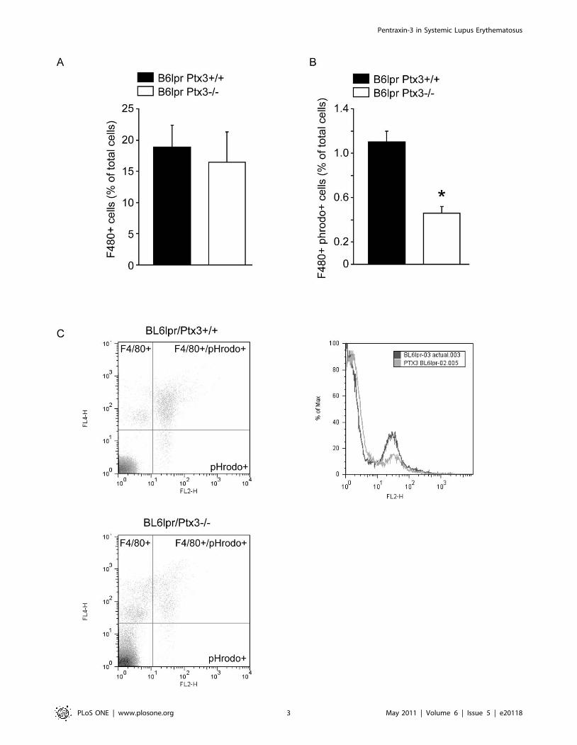
flow cytometry for pHrodo+ F4/80 macrophages. F4/80+peritoneal macrophages of Ptx3-deficient mice displayed a
significantly reduced capacity to take up apoptotic cells as
compared to wild type B6lpr mice (Figure 1). Thus, lack of
PTX3 is associated with a reduced clearance of apoptotic cells.
PTX3 expression in autoimmune B6lpr miceNext we characterized the expression of PTX3 mRNA in solid
organs of 6 week old B6lpr mice. PTX3 mRNA was highly
expressed in bone marrow (Figure 2A). Among the solid organs
PTX3 mRNA levels were much higher heart and lungs as
compared to the kidneys and the urinary bladder. PTX3 protein
expression was confirmed by Western blot in spleens, kidneys and
lungs of B6lpr mice. Spleen and kidney PTX3 expression
decreased over time when autoimmunity progresses in B6lpr mice
(Figure 2B). By contrast, PTX3 protein expression increased in
lungs at 6 months of age in B6lpr mice indicating local production
of PTX3 in lungs which are usually affected by autoimmune tissue
injury in B6lpr mice. Thus, PTX3 is increasingly expressed in lungs
during the progression of autoimmunity of B6lpr mice.
PTX3 suppresses lymphoproliferation in B6lpr miceNext we generated Ptx3-deficient B6lpr mice. The autoimmune
phenotype of homozygous B6lpr mice is introduced only by
mutation of a single lupus susceptibility gene (lpr) which impairs
Fas-induced apoptosis of autoreactive B and T cells [28]. Because
B6lpr mice develop only mild autoimmune syndrome, litters of
B6lpr/Ptx32/2 mice could be bred along Mendelian ratios from
B6lpr/Ptx3+/2 mice and revealed no differences in body weight gain
between the two genotypes (Figure 3A). For SLE phenotype
analysis we first evaluated the size of spleens and lymph nodes in 6
months old B6lpr and B6lpr/Ptx32/2 mice. Spleens and lymph nodes
were significantly enlarged in B6lpr/Ptx32/2 mice as compared to
B6lpr mice. This was evident from spleen and cervical lymph node
weights (Figure 3B).
PTX3 specifically suppresses CD4/CD8 double negative Tcells in B6lpr mice
Because we had found Ptx3-deficiency impaired the rapid
clearance of apoptotic cells we hypothesized that sustained
exposure to dead cells would modulate the activation of dendritic
cells, cells that handle lupus autoantigens and drive the expansion
of autoreactive lymphocytes in SLE, like in E8-Mag or DNAse 1-
deficient mice [30,31]. We performed flow cytometry to quantify
and characterize the activation state of CD11c+ dendritic cells
without additional stimuli directly after the spleen harvest at 6
months of age. However, the numbers of CD11c+/CD40+ or the
CD11b/MHCII+ cells were identical as well as the total numbers
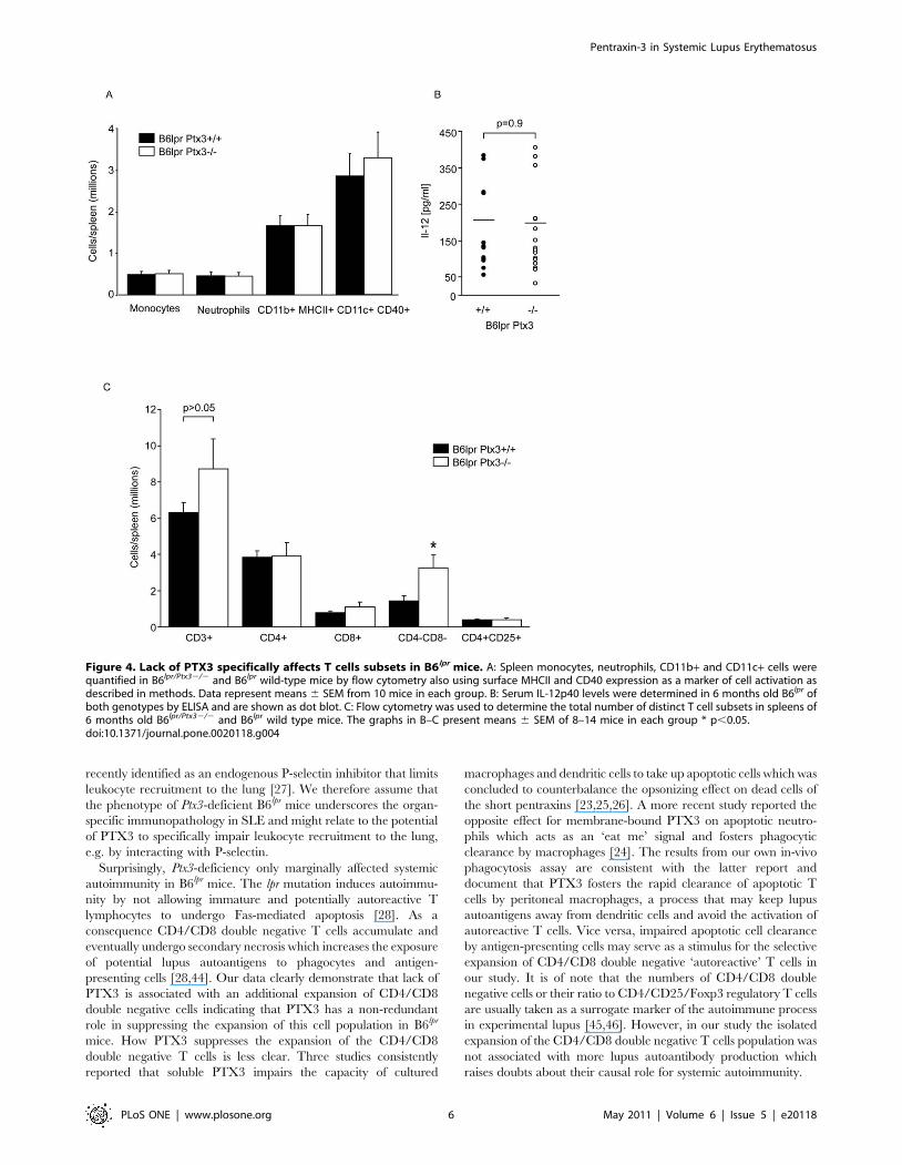
of monocytes or neutrophils in both genotypes (Figure 4A).
Consistent with this finding serum levels of IL-12 were unaffected
by the PTX3 genotype (Figure 4B). Does lack of PTX3 affect T
cell populations in B6lpr mice? The numbers of CD4/CD8 double
negative ‘autoreactive’ T cells were increased in Ptx3-deficient
B6lpr mice (Figure 4C). In contrast, the numbers of all other T cell
subsets, i.e. CD4+, CD8+, and CD4+/CD25+ T cells were
comparable between the two genotypes (Figure 4C). Thus, PTX3-
deficiency is associated with a selective expansion of CD4/CD8
double negative T cells in B6lpr mice.
PTX3 does not regulate B cell expansion andautoantibody production in B6lpr mice
Flow cytometry did not reveal any difference in numbers of
mature B cells, follicular B cells, marginal zone B cells, and plasma
cells in spleens (Figure 5A) which was consistent with the
comparable size of the IgM+ plasma cell areas in spleens of B6lpr
and B6lpr/Ptx32/2 mice (Figure 5B). Consistent with the numbers of
B cells, Ptx3-deficient B6lpr mice displayed similar serum IgG levels
as compared to 6 months old B6lpr wild-type mice (Figure 6A). At
that time lack of PTX3 did also had no effect on serum dsDNA
autoantibody levels from total IgG, IgG1, IgG2a/c, and IgG3.
The specificity of dsDNA autoantibodies was confirmed by using
the Critidiae luciliae assay. Diluted serum from B6lpr/Ptx32/2 mice
showed comparable binding to the dsDNA of the flagellate’s
kinetoplast as serum of B6lpr mice (not shown). In addition, lack of
PTX3 did not affect the levels of anti-Sm IgG, anti-U1snRNP
IgG, and rheumatoid factor as compared to B6lpr mice (Figure 6B).
In addition, Ptx3-deficient B6 mice did not reveal any sign of
spontaneous autoimmunity, e.g. autoantibodies against ANA,
dsDNA or rheumatoid factor up to 12 months of age. Thus, PTX3
is redundant for the expansion of B cells and plasma cells as well as
for the production of numerous autoantibodies against nuclear
autoantigens in B6lpr mice lack of PTX3 alone does not induce
autoimmunity against DNA.
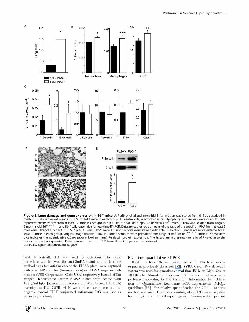
PTX3 suppresses autoimmune lung but not kidneydisease in B6lpr mice
SLE may be associated with little or severe autoimmune tissue
injury [32]. B6lpr mice do not develop major autoimmune tissue
injuries although mild glomerulonephritis develops from 6 months
of age [28]. Ptx3-deficient B6lpr mice revealed significant
peribronchial and perivascular neutrophils, CD3+ T cell infiltrates
accompanied by Mac2 + macrophages while significant pulmo-
nary pathology was absent in age-matched B6lpr mice (Figure 7,
Pentraxin-3 in Systemic Lupus Erythematosus
PLoS ONE | www.plosone.org 2 May 2011 | Volume 6 | Issue 5 | e20118
Pentraxin-3 in Systemic Lupus Erythematosus
PLoS ONE | www.plosone.org 3 May 2011 | Volume 6 | Issue 5 | e20118

8A and 8B). Given the known role of PTX3 for P-selectin-
mediated lung leukocyte recruitment we next determined the
mRNA expression levels of P-, E-, and L-selectin, PECAM, IP10
and Cxcr3 in lungs of both mouse strains. Lack of PTX3 was
associated with increased mRNA expression of P-selectin and E-
selectin (but not L-selectin, PECAM, IP10 or Cxcr3) in lungs of 6
months old B6lpr mice (Figure 8C). Consistent with mRNA data,
lack of PTX3 was associated with increased P-selectin expression
on the protein level (Figures 8D and 8E). In contrast, the Ptx3
Genotype had no effect on the activity of lupus nephritis,
proteinuria, renal P-selectin and E-selectin mRNA expression,
complement immunostaining, and renal leukocyte numbers
(Figure 9). Together, PTX3 protects B6lpr mice from autoimmune
lung disease but not from lupus nephritis
Figure 2. PTX3 expression in mice. A: RNA was isolated from organs of 6 week old B6lpr mice for real-time RT-PCR. Data are expressed as means ofthe ratio of the specific mRNA versus that of 18S rRNA 6 SEM. B: Protein samples were prepared from spleens, kidneys, and lungs of B6lpr mice at 1, 3,and 6 months of age. PTX3 Western blot indicates the quantitiative (20 mg protein load per lane) PTX3 protein expression in each organ over time.The histogram represents the ratio of PTX3 expression to the expression of the b-actin loading control. Data represent means 6 SEM from threeindependent experiments.doi:10.1371/journal.pone.0020118.g002
Figure 1. PTX3 fosters the phagocytic uptake of apoptotic cells. A: 26105 pHrodo-labeled apoptotic cells were injected intraperitoneally intoB6lprPtx3-deficient and B6lpr wildtype mice and 45 min later peritoneal lavage fluids where prepared for flow cytometry. Figure A displays thepercentage of F4/80+ peritoneal macrophages of all cells in lavage fluids which where not different in B6lpr (black bar) from B6lpr Ptx3-deficient mice(white bar). Figure B displays the number of F4/80+ cells also positive for pHrodo, a dye which gets only activated in the acidic environment ofphagolysosomes. Data represent means 6 SEM from three independent experiments; * p,0.05 versus wild type. Figure C displays representative dotplots from this experiment. C: Representative density plots of F4/80+Phrodo+ cells in B6lprPtx3-deficient and B6lpr wildtype mice.doi:10.1371/journal.pone.0020118.g001
Pentraxin-3 in Systemic Lupus Erythematosus
PLoS ONE | www.plosone.org 4 May 2011 | Volume 6 | Issue 5 | e20118

Discussion
The long pentraxin PTX3, like the short pentraxins, has
multiple regulatory roles on innate immunity. It modulates
opsonization (including dead cell clearance), complement activa-
tion, and leukocyte recruitment, all processes that affect autoim-
munity and autoimmune tissue injury [8]. The experimental
strategy of generating Ptx3-deficient autoimmune B6lpr mice
intended to test the role of Ptx3 as a potential modifier gene for
established autoimmunity. Our data now demonstrate that the
evolution of autoimmunity and lupus autoantibodies in B6lpr mice
seems to be independent of PTX3 but that PTX3 has a non-
redundant role in surpressing autoimmune lung injury.
Interstitial or alveolar lung disease is rare in humans and usually
absent in 6 months old B6lpr mice [33] but is well described in
MRLlpr mice which suffer from more advanced lupus-like
autoimmune tissue injuries at this age [34]. The peribronchial
and perivascular lymphocyte infiltrates that we observed in B6lpr/
Ptx32/2 mice were similar to those reported from MRLlpr mice and
to those that we had previously observed in B6lpr mice with
accelerated SLE [35]. Hence, lack of PTX3 specifically acceler-
ated the evolution of autoimmune lung disease, albeit not kidney
disease, in B6lpr mice. The knowledge about organ-specific
pathomechanisms in SLE is limited. Kidney disease in autoim-
mune mice (and lupus patients) mainly develops from immune
complex disease and depends on glomerular complement
activation and macrophage recruitment [36]. By contrast, lung
disease in MRLlpr mice involves the recruitment of CXCR3
positive T cells via local secretion of CXCL10 [37], and the
endothelial expression of selectins [38] and intercellular adhesion
molecule (ICAM)-1 [39]. In addition, TNF-a is a crucial mediator
of lung injury in experimental lupus [40,41]. Unspecific immuno-
suppressants like cyclophosphamide or dihydroorotate dehydro-
genase inhibitors suppress pulmonary and renal manifestations in
experimental lupus [42,43]. However, the aforementioned molec-
ular and cellular pathomechanisms of autoimmune lung injury
differ, at least in part, from those of lupus nephritis because Icam-1-
deficiency as well as TNF-a antagonism protects MRLlpr mice
from lung but not from kidney disease [39,41]. Furthermore, P-
selectin is only induced in lungs but not in kidneys upon immune
stimulation with LPS [38] or, as we found here, in experimental
SLE. The latter finding is of particular interest because PTX3 was
Figure 3. PTX3 and lymphoproliferation in B6lpr mice. A: Body weight increased similarly over time in Ptx3-deficient or wild type B6lpr mice.Data are means 6 SEM from at least 18 mice in each group. B: At 6 months of age Ptx3-deficient B6lpr mice revealed splenomegaly and hyperplasia ofcervical lymph nodes as compared to age-matched B6lpr control mice. Quantitative data on spleen and lymphnode weights are means 6 SEM from atleast 18 mice in each group, * p,0.05 versus B6lpr mice.doi:10.1371/journal.pone.0020118.g003
Pentraxin-3 in Systemic Lupus Erythematosus
PLoS ONE | www.plosone.org 5 May 2011 | Volume 6 | Issue 5 | e20118
recently identified as an endogenous P-selectin inhibitor that limits
leukocyte recruitment to the lung [27]. We therefore assume that
the phenotype of Ptx3-deficient B6lpr mice underscores the organ-
specific immunopathology in SLE and might relate to the potential
of PTX3 to specifically impair leukocyte recruitment to the lung,
e.g. by interacting with P-selectin.
Surprisingly, Ptx3-deficiency only marginally affected systemic
autoimmunity in B6lpr mice. The lpr mutation induces autoimmu-
nity by not allowing immature and potentially autoreactive T
lymphocytes to undergo Fas-mediated apoptosis [28]. As a
consequence CD4/CD8 double negative T cells accumulate and
eventually undergo secondary necrosis which increases the exposure
of potential lupus autoantigens to phagocytes and antigen-
presenting cells [28,44]. Our data clearly demonstrate that lack of
PTX3 is associated with an additional expansion of CD4/CD8
double negative cells indicating that PTX3 has a non-redundant
role in suppressing the expansion of this cell population in B6lpr
mice. How PTX3 suppresses the expansion of the CD4/CD8
double negative T cells is less clear. Three studies consistently
reported that soluble PTX3 impairs the capacity of cultured
macrophages and dendritic cells to take up apoptotic cells which was
concluded to counterbalance the opsonizing effect on dead cells of
the short pentraxins [23,25,26]. A more recent study reported the
opposite effect for membrane-bound PTX3 on apoptotic neutro-
phils which acts as an ‘eat me’ signal and fosters phagocytic
clearance by macrophages [24]. The results from our own in-vivo
phagocytosis assay are consistent with the latter report and
document that PTX3 fosters the rapid clearance of apoptotic T
cells by peritoneal macrophages, a process that may keep lupus
autoantigens away from dendritic cells and avoid the activation of
autoreactive T cells. Vice versa, impaired apoptotic cell clearance
by antigen-presenting cells may serve as a stimulus for the selective
expansion of CD4/CD8 double negative ‘autoreactive’ T cells in
our study. It is of note that the numbers of CD4/CD8 double
negative cells or their ratio to CD4/CD25/Foxp3 regulatory T cells
are usually taken as a surrogate marker of the autoimmune process
in experimental lupus [45,46]. However, in our study the isolated
expansion of the CD4/CD8 double negative T cells population was
not associated with more lupus autoantibody production which
raises doubts about their causal role for systemic autoimmunity.
Figure 4. Lack of PTX3 specifically affects T cells subsets in B6lpr mice. A: Spleen monocytes, neutrophils, CD11b+ and CD11c+ cells werequantified in B6lpr/Ptx32/2 and B6lpr wild-type mice by flow cytometry also using surface MHCII and CD40 expression as a marker of cell activation asdescribed in methods. Data represent means 6 SEM from 10 mice in each group. B: Serum IL-12p40 levels were determined in 6 months old B6lpr ofboth genotypes by ELISA and are shown as dot blot. C: Flow cytometry was used to determine the total number of distinct T cell subsets in spleens of6 months old B6lpr/Ptx32/2 and B6lpr wild type mice. The graphs in B–C present means 6 SEM of 8–14 mice in each group * p,0.05.doi:10.1371/journal.pone.0020118.g004
Pentraxin-3 in Systemic Lupus Erythematosus
PLoS ONE | www.plosone.org 6 May 2011 | Volume 6 | Issue 5 | e20118

Together, the long pentraxin PTX3 is required to suppress lung
disease in systemic autoimmunity. PTX3 does not regulate lupus
nephritis of B6lpr mice. Furthermore, PTX3 is redundant for the
production of lupus autoantibodies in these mice, albeit it fosters
the clearance of apoptotic cells and (thereby) inhibits the
expansion of CD4/CD8 double negative T cells. These results
add on to previous data that have documented organ-specific
pathomechanisms for lupus manifestations. In addition, it is now
intriguing to speculate that loss-of-function mutations in the Ptx3
gene might represent a genetic risk factor for pulmonary
manifestations of human SLE or that recombinant PTX3 or
other PTX3 agonists might have the potential to specifically
suppress autoimmune lung disease.
Materials and Methods
Animal studiesPtx3-deficient mice were generated as previously described [47]
and backcrossed to the C57BL/6 strain (B6, CharlesRiver
Laboratories, Calco, Italy) to the N11 generation. B6Ptx32/2 and
B6lpr mice (Charles River) were mated to generate B6lpr/Ptx32/+
mice which were then mated among each other to generate B6lpr/
Ptx3+/+ and B6lpr/Ptx32/2 mice as described [35]. Littermates
female were used for all experimental procedures. In each
individual mouse the genotype was assured by PCR. Mice were
housed in groups of 5 mice in sterile filter top cages with a 12 hour
dark/light cycle and unlimited access to autoclaved food and
water. One cohort of mice was sacrificed by cervical dislocation at
24 weeks of age. All experimental procedures were performed
according to the German animal care and ethics legislation and
had been approved by the local government authorities (Regier-
ung von Oberbayern, Az 55.2-1-54-2531-11-10).
Phagocytose assay and generation of apoptotic cellsEL4 cells (16106/ml) were incubated with 90 mM of the DNA
topoisomerase I inhibitor Camptothecin (Calbiochem, San
Diego, USA) for 4 h at 37uC to induce apoptosis. Apoptosis
was confirmed by flow cytometry after staining with FITC
Annexin V Apoptosis Detection Kit 1 (BD Biosciences Pharmi-
gen, San Diego, CA). After 4 h the apoptotic cells (16106/ml)
were stained with 125 ng/ml pHrodo (Invitrogen, Eugene, OR).
Mice were injected intraperitoneally with 500 ml of 4%
thioglycollate medium (BD, Franklin Lakes, USA). After 73 h
of incubation the mice were injected intraperitoneally with 200 ml
of the apoptotic pHrodo-stained apoptotic EL4 cells. 40 min later
peritoneal fluid were centrifuges and resuspended in HBSS (PAN-
Biotech GmbH, Aldenbach, Germany) +4 mM EDTA (Bio-
chrom KG, Berlin, Germany). The cells were stained with anti-
Figure 5. PTX3 and B cell subsets in B6lpr mice. A: Flow cytometry was used to determine the total number of distinct B cell subsets in spleensof 6 months old B6lpr/Ptx32/2 and B6lpr wild type mice. The histogram presents means 6 SEM of 8–14 mice in each group. B: Spleens from the samemice were stained for IgM to localize plasma cell areas. The images are representative for 6 mice in each group. Original magnification 6100.doi:10.1371/journal.pone.0020118.g005
Pentraxin-3 in Systemic Lupus Erythematosus
PLoS ONE | www.plosone.org 7 May 2011 | Volume 6 | Issue 5 | e20118

F4/80 IgG (BD Pharmingen, Heidelberg, Germany) for flow
cytometry analysis.
Flow cytometryAnti-mouse CD3, CD4, CD8 and CD25 antibodies (BD
Pharmingen, Heidelberg, Germany) were used to detect
CD3+CD42CD82 double negative T cells and CD4+CD25+regulatory T cells populations in spleens. Anti-CD11c was used to
identify dendritic cells and their activation was assed by co-staining
for CD40 (BD Pharmingen, Heidelberg, Germany). Anti-mouse
B220, CD21, CD23, IgD, IgM antibodies (BD Pharmingen,
Heidelberg, Germany) were used to detect mature B cells
(B220+IgD+IgM+), marginal zone B cells (B220+CD21highC-
D23low) and follicular B cells (B220+CD21lowCD23high). Plasma
cells were identified by using anti-mouse antibodies for Ig k light
chain and CD138 (BD Pharmingen, Heidelberg, Germany).
Respective isotype antibodies were used to demonstrate specific
staining of cell subpopulations [35]. Quantification of cell number
was done using counting beads for FACS (Invitrogen).
Evaluation of autoimmune tissue injuryLungs, spleens and kidneys from all mice were fixed in 10%
buffered formalin, processed, and embedded in paraffin. Two mm
sections for periodic acid-Schiff (PAS) stains were prepared
following routine protocols [48]. The severity of the renal lesions
was graded using the activity and chronicity indices for human
lupus nephritis as described [49]. Autoimmune lung injury was
scored semiquantitatively (0–4) by assessing the extent of
peribronchial, perivascular or interstitial lymphocyte infiltrates as
described [34]. For lung immunostaining: anti-mCD3e (clone
500A2, 1:50), Mac2 (macrophages, Cederlane, Ontario, Canada,
1:5000), rat anti-mouse neutrophils (Serotec, Oxford, UK, 1:50)
were used. For IgM staining of the spleen sections, anti-mouse
IgM-mu-chain specific antibodies (Vector, Burlingame, CA) were
used. The slides were scanned using an Olympus BX 61
microscope and recorded via CellP software.
Serum IL-12 levels were determined by ELISA following the
manufacturer’s protocols (OptEiA, BD, Biosciences, Heidelberg,
Germany).
Ptx3 protein expression analysisWestern blot analysis were performed from kidney, lung and
spleen protein extracts, which were incubated in two times
loading buffer for 5 min at 95uC, resolved by 10% SDS-PAGE,
and transferred to an Immobilon-P membrane (Millipore,
Eschborn, Germany). After blocking with 1% Western Blocking
Reagent (Roche, Germany), the filter was incubated with Rat
anti mouse Ptx3 antibody (1:1000, Alpha Diagnostic Interna-
tional, San Antonio USA) overnight in TBS; b-actin (1:1000,
Cell Signaling Technology, Beverly, MA) for 1 hour), anti-P-
selectin monoclonal antibody (mAb) RB40 (anti-P-selectin were
prepared from hybridoma cells). Immune complexes were
Figure 6. PTX3 and humoral immunity in lupus of B6lpr mice. B6lpr/Ptx32/2 (%) and B6lpr wild-type mice (&) were bled at monthly intervals todetermine serum levels of IgG (A). Data represent means 6 SEM from at least 12 mice at each time point and genotype. B: DsDNA autoantibodies andvarious lupus autoantibodies were analysed by ELISA at 6 months only and are shown as dot blots. 6 mice at each time point and genotype. Nosignificant differences were detected between the two genotypes for any of the parameters.doi:10.1371/journal.pone.0020118.g006
Pentraxin-3 in Systemic Lupus Erythematosus
PLoS ONE | www.plosone.org 8 May 2011 | Volume 6 | Issue 5 | e20118

visualized using a peroxidase-conjugated anti-rat IgG Ab
(1:10000, Cell Signaling Technology, Beverly, MA) for 1 h and
processed for detection by ECL (Amersham Pharmacia Biotech
Europe, Freiburg, Germany).
Autoantibody analysisSerum antibody levels were determined by ELISA as described
[50,51]. Anti-dsDNA antibodies: NUNC maxisorp ELISA plates
were coated with poly-L-lysine (Trevigen, Gaithersburg, MD,
USA) and mouse embryonic stem cell dsDNA. After incubation
with mouse serum, dsDNA-specific IgG, IgG1, IgG2a/c, IgG2b,
IgG3 and serum IgG levels were detected by ELISA (Bethyl Labs,
Montgomery, TX, USA). Critidiae luciliae assay: 1:50 diluted serum
was applied to fixed C. luciliae slides (BioRad Laboratories,
Redmond USA.). Binding to C. luciliae kinetoplast was detected
with FITC-conjugated goat anti–mIgG (1:1000, Invitrogen,
Oregon USA). DAPI staining (Vector Laboratories, Burlingame
CA) allowed colocalization with kinetoplast dsDNA. For quanti-
tation of kinetoplast staining intensity a semiquantitative score
from 0–3 was used. Anti-Sm: NUNC maxisorp ELISA plates were
coated with Smith (Sm) antigen (Immunovision, Springdale, AR).
A horseradish peroxidase-conjugated goat anti-mouse IgG (Rock-
Figure 7. PTX3 and lung injury in B6lpr mice. Lung sections were stained either with PAS or with specific antibodies for neutrophils,macrophages or T lymphocytes. Images are representative for at least 12 mice in each group. Original magnification 6100 (PAS) and 6200(immunostaining).doi:10.1371/journal.pone.0020118.g007
Pentraxin-3 in Systemic Lupus Erythematosus
PLoS ONE | www.plosone.org 9 May 2011 | Volume 6 | Issue 5 | e20118
land, Gilbertsville, PA) was used for detection. The same
procedure was followed for anti-SmRNP and anti-nucleosome
antibodies as for anti-Sm except the ELISA plates were captured
with Sm-RNP complex (Immunovision) or dsDNA together with
histones (USB Corporation, Ohio USA) respectively instead of Sm
antigen. Rheumatoid factor: ELISA plates were coated with
10 mg/ml IgG (Jackson Immunoresearch, West Grove, PA, USA)
overnight at 4uC. C57BL/6 10 week mouse serum was used as
negative control. HRP conjugated anti-mouse IgG was used as
secondary antibody.
Real-time quantitative RT-PCRReal time RT-PCR was performed on mRNA from mouse
organs as previously described [52]. SYBR Green Dye detection
system was used for quantitative real-time PCR on Light Cycler
480 (Roche, Mannheim, Germany). All the technical steps were
performed according to The Minimum Information for Publica-
tion of Quantitative Real-Time PCR Experiments (MIQE)
guidelines [53]. For relative quantification the 22DDCt analysis
method was used. Controls consisting of ddH2O were negative
for target and housekeeper genes. Gene-specific primers
Figure 8. Lung damage and gene expression in B6lpr mice. A: Peribronchial and interstitial inflammation was scored from 0–4 as described inmethods. Data represent means 6 SEM of 8–12 mice in each group. B: Neutrophils, macrophages or T lymphocytes numbers were quantify, datarepresent means 6 SEM from at least 12 mice in each group, * p,0.05, **p,0.005, ***p,0.0005 versus B6lpr mice. C: RNA was isolated from lungs of6 months old B6lpr/Ptx32/2 and B6lpr wild-type mice for real-time RT-PCR. Data are expressed as means of the ratio of the specific mRNA from at least 5mice versus that of 18S rRNA 6 SEM. * p,0.05 versus B6lpr mice. D: Lung sections were stained with anti- P-selectin P. Images are representative for atleast 12 mice in each group. Original magnification 6100. E: Protein samples were prepared from lungs of B6lpr or B6Ptx32/2lpr mice. PTX3 Westernblot indicates the quantitiative (20 mg protein load per lane) P-selectin protein expression. The histogram represents the ratio of P-selectin to therespective b-actin expression. Data represent means 6 SEM from three independent experiments.doi:10.1371/journal.pone.0020118.g008
Pentraxin-3 in Systemic Lupus Erythematosus
PLoS ONE | www.plosone.org 10 May 2011 | Volume 6 | Issue 5 | e20118

(300 nM, Metabion, Martinsried, Germany) were designed to be
cDNA specific and to target possibly all known transcripts of gene
of interest. In silico specificity screen (BLAST) was performed.
PTX3: ID NM_008987 right: 59-CCTGCTTTGTGCTCTC
TGGT-39, left: 59-TCTCCAGCATGATGAACAGC-39; E-Se-
lectin: ID NM_011345 right: 59-TCTATTTCCCACGATG-
CATTT-39, left: 59-CTGCCAAAGCCTTCAATCAT-39; L-
Selectin: ID NM_011346 right: 59-TTCATGGCTTTCCTTT-
CACA–39; left: 59-CTGGCATTTCTCATT TGGCT-39; P-
Selectin: ID NM_011347 right: 59-GGACACTTGATGGCTT-
CACA-39, left: 59-CAGTTCATGTGCGATGAAGG-39; PE-
CAM-1: ID NM_001032378 right: 59-TCCTTCCTGCTTC-
TTGCTAGCT-39, left: 59-GAGCCCAATCACGTTTCAGT-
TT-39; IP-10: ID NM_021274 right: 59-GGCTGGTC-
ACCTTTCAGAAG-39, left: 59-ATGGATGGACAGCAGA-
GAGC-39; Cxcr3: ID NM_009910, right: 59- TCTCGT-
TTTCCCCATAATCG-39, left: 59-AGCCAAGCCATGTACC-
TTGA-39.
Figure 9. PTX3 and lupus nephritis in B6lpr mice. A: Kidney sections were stained with PAS. Images are representative for at least 12 mice ineach group, original magnification 6400. B: The composite activity index of lupus nephritis (0–24) was assessed as described in methods. Datarepresent means 6 SEM of 8–12 mice in each group. C: Urinary albumin/creatinine ratios were determined at 6 months as a functional marker ofglomerular damage. D: Renal P-selectin and E-selectin mRNA levels were determined by real-time RT-PCR and are expressed as means of the ratio ofthe specific mRNA versus that of 18S rRNA 6 SEM (n = 12 in each group). E: Neutrophils, macrophages or T lymphocytes numbers were quantify, datarepresent means 6 SEM from at least 12 mice in each group. F: C9 complement staining was performed on kidney sections. No significant differenceswere detected between the two genotypes.doi:10.1371/journal.pone.0020118.g009
Pentraxin-3 in Systemic Lupus Erythematosus
PLoS ONE | www.plosone.org 11 May 2011 | Volume 6 | Issue 5 | e20118

Statistical analysisOne-way ANOVA followed by post-hoc Bonferroni’s test was
used for multiple comparisons using GraphPad Prism 4.03 version.
Single groups were compared by unpaired two-tailed Students t-
test or non-parametric Mann-Whitney test. Data were expressed
as mean 6 SEM. Statistical significance was assumed at a p value
of ,0.05.
Acknowledgments
The expert technical assistance of Dan Draganovic and Janina Mandel-
baum is gratefully acknowledged. Parts of this work were performed as a
medical thesis project by C.R. at the Medical Faculty of the University of
Munich.
Author Contributions
Conceived and designed the experiments: ML CR H-JA. Performed the
experiments: ML CR OPK HES A. Migliorini. Analyzed the data: ML CR
OPK H-JA. Contributed reagents/materials/analysis tools: CG A.
Montovani. Wrote the paper: ML CR H-JA.
References
1. Kotzin BL (1996) Systemic lupus erythematosus. Cell 85: 303–306.
2. Goodnow CC (2007) Multistep pathogenesis of autoimmune disease. Cell 130:
25–35.
3. Gregersen PK, Olsson LM (2009) Recent advances in the genetics of
autoimmune disease. Annu Rev Immunol 27: 363–391.
4. Kanta H, Mohan C (2009) Three checkpoints in lupus development: central
tolerance in adaptive immunity, peripheral amplification by innate immunity
and end-organ inflammation. Genes Immun 10: 390–396.
5. Theofilopoulos AN, Baccala R, Beutler B, Kono DH (2005) Type I interferons
(alpha/beta) in immunity and autoimmunity. Annu Rev Immunol 23: 307–
336.
6. Hom G, Graham RR, Modrek B, Taylor KE, Ortmann W, et al. (2008)
Association of systemic lupus erythematosus with C8orf13-BLK and ITGAM-
ITGAX. N Engl J Med 358: 900–909.
7. Marshak-Rothstein A, Rifkin IR (2007) Immunologically active autoantigens:
the role of toll-like receptors in the development of chronic inflammatory
disease. Annu Rev Immunol 25: 419–441.
8. Bottazzi B, Doni A, Garlanda C, Mantovani A (2010) An integrated view of
humoral innate immunity: pentraxins as a paradigm. Annu Rev Immunol 28:
157–183.
9. Bijl M, Horst G, Bijzet J, Bootsma H, Limburg PC, et al. (2003) Serum amyloid
P component binds to late apoptotic cells and mediates their uptake by
monocyte-derived macrophages. Arthritis Rheum 48: 248–254.
10. Anders HJ (2009) Pseudoviral immunity - a novel concept for lupus. Trends Mol
Med 15: 553–561.
11. Bickerstaff MC, Botto M, Hutchinson WL, Herbert J, Tennent GA, et al. (1999)
Serum amyloid P component controls chromatin degradation and prevents
antinuclear autoimmunity. Nat Med 5: 694–697.
12. Russell AI, Cunninghame Graham DS, Shepherd C, Roberton CA, Whittaker J,
et al. (2004) Polymorphism at the C-reactive protein locus influences gene
expression and predisposes to systemic lupus erythematosus. Hum Mol Genet
13: 137–147.
13. Truedsson L, Bengtsson AA, Sturfelt G (2007) Complement deficiencies and
systemic lupus erythematosus. Autoimmunity 40: 560–566.
14. Voss A, Nielsen EH, Svehag SE, Junker P (2008) Serum amyloid P component-
DNA complexes are decreased in systemic lupus erythematosus. inverse
association with anti-dsDNA antibodies. J Rheumatol 35: 625–630.
15. Becker GJ, Waldburger M, Hughes GR, Pepys MB (1980) Value of serum C-
reactive protein measurement in the investigation of fever in systemic lupus
erythematosus. Ann Rheum Dis 39: 50–52.
16. Shoenfeld Y, Szyper-Kravitz M, Witte T, Doria A, Tsutsumi A, et al. (2007)
Autoantibodies against protective molecules–C1q, C-reactive protein, serum
amyloid P, mannose-binding lectin, and apolipoprotein A1: prevalence in
systemic lupus erythematosus. Ann N Y Acad Sci 1108: 227–239.
17. Enocsson H, Sjowall C, Skogh T, Eloranta ML, Ronnblom L, et al. (2009)
Interferon-alpha mediates suppression of C-reactive protein: explanation for
muted C-reactive protein response in lupus flares? Arthritis Rheum 60:
3755–3760.
18. Hollan I, Bottazzi B, Cuccovillo I, Forre OT, Mikkelsen K, et al. (2010)
Increased levels of serum pentraxin 3, a novel cardiovascular biomarker, in
patients with inflammatory rheumatic disease. Arthritis Care Res (Hoboken) 62:
378–385.
19. Bassi N, Ghirardello A, Blank M, Zampieri S, Sarzi-Puttini P, et al. (2010) IgG
anti-pentraxin 3 antibodies in systemic lupus erythematosus. Ann Rheum Dis
69: 1704–1710.
20. Garlanda C, Bottazzi B, Bastone A, Mantovani A (2005) Pentraxins at the
crossroads between innate immunity, inflammation, matrix deposition, and
female fertility. Annu Rev Immunol 23: 337–366.
21. Nauta AJ, Bottazzi B, Mantovani A, Salvatori G, Kishore U, et al. (2003)
Biochemical and functional characterization of the interaction between
pentraxin 3 and C1q. Eur J Immunol 33: 465–473.
22. Deban L, Jarva H, Lehtinen MJ, Bottazzi B, Bastone A, et al. (2008) Binding of
the long pentraxin PTX3 to factor H: interacting domains and function in the
regulation of complement activation. J Immunol 181: 8433–8440.
23. Baruah P, Dumitriu IE, Peri G, Russo V, Mantovani A, et al. (2006) The tissue
pentraxin PTX3 limits C1q-mediated complement activation and phagocytosis
of apoptotic cells by dendritic cells. J Leukoc Biol 80: 87–95.
24. Jaillon S, Jeannin P, Hamon Y, Fremaux I, Doni A, et al. (2009) Endogenous
PTX3 translocates at the membrane of late apoptotic human neutrophils and is
involved in their engulfment by macrophages. Cell Death Differ 16: 465–474.
25. Rovere P, Peri G, Fazzini F, Bottazzi B, Doni A, et al. (2000) The long pentraxin
PTX3 binds to apoptotic cells and regulates their clearance by antigen-
presenting dendritic cells. Blood 96: 4300–4306.
26. van Rossum AP, Fazzini F, Limburg PC, Manfredi AA, Rovere-Querini P, et al.
(2004) The prototypic tissue pentraxin PTX3, in contrast to the short pentraxin
serum amyloid P, inhibits phagocytosis of late apoptotic neutrophils by
macrophages. Arthritis Rheum 50: 2667–2674.
27. Deban L, Russo RC, Sironi M, Moalli F, Scanziani M, et al. (2010) Regulation
of leukocyte recruitment by the long pentraxin PTX3. Nat Immunol 11:
328–334.
28. Cohen PL, Eisenberg RA (1991) Lpr and gld: single gene models of systemic
autoimmunity and lymphoproliferative disease. Annu Rev Immunol 9: 243–269.
29. Munoz LE, Lauber K, Schiller M, Manfredi AA, Herrmann M (2010) The role
of defective clearance of apoptotic cells in systemic autoimmunity. Nat Rev
Rheumatol 6: 280–289.
30. Hanayama R, Tanaka M, Miyasaka K, Aozasa K, Koike M, et al. (2004)
Autoimmune disease and impaired uptake of apoptotic cells in MFG-E8-
deficient mice. Science 304: 1147–1150.
31. Napirei M, Karsunky H, Zevnik B, Stephan H, Mannherz HG, et al. (2000)
Features of systemic lupus erythematosus in Dnase1-deficient mice. Nat Genet
25: 177–181.
32. Rahman A, Isenberg DA (2008) Systemic lupus erythematosus. N Engl J Med
358: 929–939.
33. Pego-Reigosa JM, Medeiros DA, Isenberg DA (2009) Respiratory manifestations
of systemic lupus erythematosus: old and new concepts. Best Pract Res Clin
Rheumatol 23: 469–480.
34. Sunderrajan EV, McKenzie WN, Lieske TR, Kavanaugh JL, Braun SR, et al.
(1986) Pulmonary inflammation in autoimmune MRL/Mp-lpr/lpr mice.
Histopathology and bronchoalveolar lavage evaluation. Am J Pathol 124:
353–362.
35. Lech M, Kulkarni OP, Pfeiffer S, Savarese E, Krug A, et al. (2008) Tir8/Sigirr
prevents murine lupus by suppressing the immunostimulatory effects of lupus
autoantigens. J Exp Med 205: 1879–1888.
36. Kulkarni O, Anders HJ (2008) Chemokines in lupus nephritis. Front Biosci 13:
3312–3320.
37. Shiozawa F, Kasama T, Yajima N, Odai T, Isozaki T, et al. (2004) Enhanced
expression of interferon-inducible protein 10 associated with Th1 profiles of
chemokine receptor in autoimmune pulmonary inflammation of MRL/lpr mice.
Arthritis Res Ther 6: R78–R86.
38. Harari OA, Marshall D, McHale JF, Ahmed S, Haskard DO (2001) Limited
endothelial E- and P-selectin expression in MRL/lpr lupus-prone mice.
Rheumatology (Oxford) 40: 889–895.
39. Lloyd CM, Gonzalo JA, Salant DJ, Just J, Gutierrez-Ramos JC (1997)
Intercellular adhesion molecule-1 deficiency prolongs survival and protects
against the development of pulmonary inflammation during murine lupus. J Clin
Invest 100: 963–971.
40. Deguchi Y, Kishimoto S (1991) Tumour necrosis factor/cachectin plays a key
role in autoimmune pulmonary inflammation in lupus-prone mice. Clin Exp
Immunol 85: 392–395.
41. Kim N, Ussin L, Cheng X, Murali R, Sullivan KE (2002) TNFalpha inhibition
in MRL/lpr mice ameliorates pulmonary but not renal disease. J Autoimmun
19: 215–222.
42. Kulkarni OP, Sayyed SG, Kantner C, Ryu M, Schnurr M, et al. (2010) 4SC-
101, a novel small molecule dihydroorotate dehydrogenase inhibitor, suppresses
systemic lupus erythematosus in MRL-(Fas)lpr mice. Am J Pathol 176:
2840–2847.
43. Kulkarni O, Eulberg D, Selve N, Zollner S, Allam R, et al. (2009) Anti-Ccl2
Spiegelmer permits 75% dose reduction of cyclophosphamide to control diffuse
Pentraxin-3 in Systemic Lupus Erythematosus
PLoS ONE | www.plosone.org 12 May 2011 | Volume 6 | Issue 5 | e20118

proliferative lupus nephritis and pneumonitis in MRL-Fas(lpr) mice. J Pharmacol
Exp Ther 328: 371–377.44. Kakkanaiah VN, Pyle RH, Nagarkatti M, Nagarkatti PS (1990) Evidence for
major alterations in the thymocyte subpopulations in murine models of
autoimmune diseases. J Autoimmun 3: 271–288.45. Gutierrez-Ramos JC, Andreu JL, Revilla Y, Vinuela E, Martinez C (1990)
Recovery from autoimmunity of MRL/lpr mice after infection with aninterleukin-2/vaccinia recombinant virus. Nature 346: 271–274.
46. Singer PA, Theofilopoulos AN (1990) Novel origin of lpr and gld cells and
possible implications in autoimmunity. J Autoimmun 3: 123–135.47. Garlanda C, Hirsch E, Bozza S, Salustri A, De Acetis M, et al. (2002) Non-
redundant role of the long pentraxin PTX3 in anti-fungal innate immuneresponse. Nature 420: 182–186.
48. Kulkarni O, Pawar RD, Purschke W, Eulberg D, Selve N, et al. (2007)Spiegelmer inhibition of CCL2/MCP-1 ameliorates lupus nephritis in MRL-
(Fas)lpr mice. J Am Soc Nephrol 18: 2350–2358.
49. Allam R, Pawar RD, Kulkarni OP, Hornung V, Hartmann G, et al. (2008) Viral
59-triphosphate RNA and non-CpG DNA aggravate autoimmunity and lupusnephritis via distinct TLR-independent immune responses. Eur J Immunol 38:
3487–3498.
50. Patole PS, Pawar RD, Lichtnekert J, Lech M, Kulkarni OP, et al. (2007)Coactivation of Toll-like receptor-3 and -7 in immune complex glomerulone-
phritis. J Autoimmun 29: 52–59.51. Lech M, Skuginna V, Kulkarni OP, Gong J, Wei T, et al. (2010) Lack of
SIGIRR/TIR8 aggravates hydrocarbon oil-induced lupus nephritis. J Pathol
220: 596–607.52. Lech M, Avila-Ferrufino A, Skuginna V, Susanti HE, Anders HJ (2010)
Quantitative expression of RIG-like helicase, NOD-like receptor and inflamma-some-related mRNAs in humans and mice. Int Immunol 22: 717–728.
53. Bustin SA, Benes V, Garson JA, Hellemans J, Huggett J, et al. (2009) The MIQEguidelines: minimum information for publication of quantitative real-time PCR
experiments. Clin Chem 55: 611–622.
Pentraxin-3 in Systemic Lupus Erythematosus
PLoS ONE | www.plosone.org 13 May 2011 | Volume 6 | Issue 5 | e20118