![[Proliferative lupus nephritis treatment: practice survey in nephrology and internal medicine in France]](https://static.fdokumen.com/doc/165x107/6336db7d20d9c9602f0b0be8/proliferative-lupus-nephritis-treatment-practice-survey-in-nephrology-and-internal.jpg)
Autoreactive IgE Is Prevalent in Systemic Lupus Erythematosus and Is Associated with Increased...
11
Autoreactive IgE Is Prevalent in Systemic Lupus Erythematosus and Is Associated with Increased Disease Activity and Nephritis Barbara Dema 1 , Christophe Pellefigues 2 , Sarfaraz Hasni 3 , Nathalie Gault 6 , Chao Jiang 1 , Tiffany K. Ricks 1 , Michael M. Bonelli 8 , Jo ¨ rg Scheffel 1 , Karim Sacre ´ 2,4 , Mathieu Jablonski 5 , Delphine Gobert 4 , Thomas Papo 2,4 , Eric Daugas 2,5 , Gabor Illei 7 , Nicolas Charles 2 * . , Juan Rivera 1 * . 1 Laboratory of Molecular Immunogenetics, National Institute of Arthritis and Musculoskeletal and Skin Diseases, National Institutes of Health, Bethesda, Maryland, United States of America, 2 Institut National de la Sante ´ et de la Recherche Me ´ dicale U699, Universite ´ Paris Diderot, Paris, France, 3 Office of the Clinical Director, National Institute of Arthritis and Musculoskeletal and Skin Diseases, National Institutes of Health, Bethesda, Maryland, United States of America, 4 Department of Internal Medicine, Ho ˆ pital Bichat, Assistance Publique-Ho ˆ pitaux de Paris, Universite ´ Paris Diderot, Faculte ´ de Me ´decine site Bichat, Paris, France, 5 Department of Nephrology, Ho ˆ pital Bichat, Assistance Publique-Ho ˆ pitaux de Paris, Universite ´ Paris Diderot, Faculte ´ de Me ´ decine site Bichat, Paris, France, 6 Clinical Research Unit, Ho ˆ pital Bichat, Assistance Publique- Ho ˆ pitaux de Paris, Universite ´ Paris Diderot, Faculte ´ de Me ´ decine site Bichat, Paris, France, 7 Sjogren’s Syndrome Clinic, National Institute of Dental and Craniofacial Research, National Institutes of Health, Bethesda, Maryland, United States of America, 8 Lymphocyte Biology Section, National Institute of Arthritis and Musculoskeletal and Skin Diseases, National Institutes of Health, Bethesda, Maryland, United States of America Abstract The presence of autoantibodies in systemic lupus erythematosus, particularly those of the IgG subclass, have long been associated with disease onset and activity. Here we explored the prevalence of autoreactive IgE in SLE and its relevance to disease in French (n = 79) and United States (US) (n = 117) cohorts with a mean age of 41.5612.7 and 43.6615.3 years and disease duration of 13.568.5 and 16.6611.9 years, respectively. Our findings show that approximately 65% of all SLE subjects studied produced IgE antibodies to the seven autoantigens tested. This positivity was increased to almost 83% when only those subjects with active disease were considered. SLE subjects who were positive for anti-dsDNA, -Sm, and - SSB/La -specific IgE showed a highly significant association in the levels of these antibodies with disease activity similar to that of the corresponding IgG’s. A strong association of IgE autoantibodies with active nephritis was also found in the combined cohort analysis. A test of the predictive value of autoreactive IgE’s and IgGs for disease activity (SLE Disease Activity Index (SLEDAI) $4) revealed that the best predictors were dsDNA-specific IgE and IgG, and that the age of an SLE subject influenced this predictive model. The finding argue that the overall levels of IgE autoantibodies, independently or in combination with IgG autoantibodies, may serve as indicators of active disease. Citation: Dema B, Pellefigues C, Hasni S, Gault N, Jiang C, et al. (2014) Autoreactive IgE Is Prevalent in Systemic Lupus Erythematosus and Is Associated with Increased Disease Activity and Nephritis. PLoS ONE 9(2): e90424. doi:10.1371/journal.pone.0090424 Editor: Pierre Bobe ´, INSERM-Universite ´ Paris-Sud, France Received October 16, 2013; Accepted January 29, 2014; Published February 28, 2014 This is an open-access article, free of all copyright, and may be freely reproduced, distributed, transmitted, modified, built upon, or otherwise used by anyone for any lawful purpose. The work is made available under the Creative Commons CC0 public domain dedication. Funding: This work was supported by the Intramural Program of the National Institute of Arthritis and Musculoskeletal and Skin Diseases (NIAMS) of the National Institutes of Health (NIH), by the French Institut National de la Sante ´ Et de la Recherche Me ´ dicale (INSERM), by the Assistance Publique – Ho ˆ pitaux de Paris (AP-HP) and by grants from the Mairie de Paris (Emergences 2010), ANR JCJC SVSE1 2011 BASILE and the French Kidney Foundation (Fondation du Rein) to NC. NC is under a translational research contract with the AP-HP (CHRT). The authors wish to acknowledge the invaluable support of the Office of the Clinical Director, NIAMS. The funders had no role in study design, data collection and analysis, decision to publish, or preparation of the manuscript. Competing Interests: The authors have declared that no competing interests exist. * E-mail: [email protected] (JR); [email protected] (NC) . These authors contributed equally to this work. Introduction Systemic Lupus Erythematosus (SLE) is a chronic systemic autoimmune disease characterized by the loss of immune tolerance to self-antigens, dysregulated autoantibody production, and multiple clinical manifestations [1]. A hallmark of the disease is the overproduction of antibodies to nuclear antigens (ANA), dsDNA, Sm, RNP, Ro (SSA), La (SSB), and some phospholipids [2]. The detection of ANA, anti-Ro, anti-La and anti-phospholipid antibodies can occur years in advance of the clinical manifestation and diagnosis of SLE, and these antibodies accumulate with time. In contrast, the presence of anti-dsDNA and anti-Sm antibodies usually precedes by a few months the clinical manifestations of SLE and thus its diagnosis [3]. While the appearance of the different autoantibodies has been associated with diverse clinical manifestations [4], the pathogenesis of disease is most closely linked to the presence of dsDNA-specific IgG antibodies [5] and kidney biopsies reveal that a large proportion of SLE patients, that develop glomerular nephritis, have kidney deposits of dsDNA IgG antibodies [6]. However, not all patients produce dsDNA IgG antibodies and not all of these autoantibodies are pathogenic even though their presence is one criterion for SLE diagnosis [7], thus what other isotypes of autoantibodies might be linked to disease activity and kidney pathology is still a topic of considerable interest. IgE is the rarest of immunoglobulins in the blood with serum levels in a healthy human at approximately 150 ng/ml whereas PLOS ONE | www.plosone.org 1 February 2014 | Volume 9 | Issue 2 | e90424
-
Upload
independent -
Category
Documents
-
view
0 -
download
0
Transcript of Autoreactive IgE Is Prevalent in Systemic Lupus Erythematosus and Is Associated with Increased...
Autoreactive IgE Is Prevalent in Systemic LupusErythematosus and Is Associated with Increased DiseaseActivity and NephritisBarbara Dema1, Christophe Pellefigues2, Sarfaraz Hasni3, Nathalie Gault6, Chao Jiang1, Tiffany K. Ricks1,
Michael M. Bonelli8, Jorg Scheffel1, Karim Sacre2,4, Mathieu Jablonski5, Delphine Gobert4,
Thomas Papo2,4, Eric Daugas2,5, Gabor Illei7, Nicolas Charles2*., Juan Rivera1*.
1 Laboratory of Molecular Immunogenetics, National Institute of Arthritis and Musculoskeletal and Skin Diseases, National Institutes of Health, Bethesda, Maryland, United
States of America, 2 Institut National de la Sante et de la Recherche Medicale U699, Universite Paris Diderot, Paris, France, 3Office of the Clinical Director, National Institute
of Arthritis and Musculoskeletal and Skin Diseases, National Institutes of Health, Bethesda, Maryland, United States of America, 4Department of Internal Medicine, Hopital
Bichat, Assistance Publique-Hopitaux de Paris, Universite Paris Diderot, Faculte de Medecine site Bichat, Paris, France, 5Department of Nephrology, Hopital Bichat,
Assistance Publique-Hopitaux de Paris, Universite Paris Diderot, Faculte de Medecine site Bichat, Paris, France, 6Clinical Research Unit, Hopital Bichat, Assistance Publique-
Hopitaux de Paris, Universite Paris Diderot, Faculte de Medecine site Bichat, Paris, France, 7 Sjogren’s Syndrome Clinic, National Institute of Dental and Craniofacial
Research, National Institutes of Health, Bethesda, Maryland, United States of America, 8 Lymphocyte Biology Section, National Institute of Arthritis and Musculoskeletal
and Skin Diseases, National Institutes of Health, Bethesda, Maryland, United States of America
Abstract
The presence of autoantibodies in systemic lupus erythematosus, particularly those of the IgG subclass, have long beenassociated with disease onset and activity. Here we explored the prevalence of autoreactive IgE in SLE and its relevance todisease in French (n = 79) and United States (US) (n = 117) cohorts with a mean age of 41.5612.7 and 43.6615.3 years anddisease duration of 13.568.5 and 16.6611.9 years, respectively. Our findings show that approximately 65% of all SLEsubjects studied produced IgE antibodies to the seven autoantigens tested. This positivity was increased to almost 83%when only those subjects with active disease were considered. SLE subjects who were positive for anti-dsDNA, -Sm, and -SSB/La -specific IgE showed a highly significant association in the levels of these antibodies with disease activity similar tothat of the corresponding IgG’s. A strong association of IgE autoantibodies with active nephritis was also found in thecombined cohort analysis. A test of the predictive value of autoreactive IgE’s and IgGs for disease activity (SLE DiseaseActivity Index (SLEDAI) $4) revealed that the best predictors were dsDNA-specific IgE and IgG, and that the age of an SLEsubject influenced this predictive model. The finding argue that the overall levels of IgE autoantibodies, independently or incombination with IgG autoantibodies, may serve as indicators of active disease.
Citation: Dema B, Pellefigues C, Hasni S, Gault N, Jiang C, et al. (2014) Autoreactive IgE Is Prevalent in Systemic Lupus Erythematosus and Is Associated withIncreased Disease Activity and Nephritis. PLoS ONE 9(2): e90424. doi:10.1371/journal.pone.0090424
Editor: Pierre Bobe, INSERM-Universite Paris-Sud, France
Received October 16, 2013; Accepted January 29, 2014; Published February 28, 2014
This is an open-access article, free of all copyright, and may be freely reproduced, distributed, transmitted, modified, built upon, or otherwise used by anyone forany lawful purpose. The work is made available under the Creative Commons CC0 public domain dedication.
Funding: This work was supported by the Intramural Program of the National Institute of Arthritis and Musculoskeletal and Skin Diseases (NIAMS) of the NationalInstitutes of Health (NIH), by the French Institut National de la Sante Et de la Recherche Medicale (INSERM), by the Assistance Publique – Hopitaux de Paris (AP-HP)and by grants from the Mairie de Paris (Emergences 2010), ANR JCJC SVSE1 2011 BASILE and the French Kidney Foundation (Fondation du Rein) to NC. NC isunder a translational research contract with the AP-HP (CHRT). The authors wish to acknowledge the invaluable support of the Office of the Clinical Director,NIAMS. The funders had no role in study design, data collection and analysis, decision to publish, or preparation of the manuscript.
Competing Interests: The authors have declared that no competing interests exist.
* E-mail: [email protected] (JR); [email protected] (NC)
. These authors contributed equally to this work.
Introduction
Systemic Lupus Erythematosus (SLE) is a chronic systemic
autoimmune disease characterized by the loss of immune tolerance
to self-antigens, dysregulated autoantibody production, and
multiple clinical manifestations [1]. A hallmark of the disease is
the overproduction of antibodies to nuclear antigens (ANA),
dsDNA, Sm, RNP, Ro (SSA), La (SSB), and some phospholipids
[2]. The detection of ANA, anti-Ro, anti-La and anti-phospholipid
antibodies can occur years in advance of the clinical manifestation
and diagnosis of SLE, and these antibodies accumulate with time.
In contrast, the presence of anti-dsDNA and anti-Sm antibodies
usually precedes by a few months the clinical manifestations of
SLE and thus its diagnosis [3]. While the appearance of the
different autoantibodies has been associated with diverse clinical
manifestations [4], the pathogenesis of disease is most closely
linked to the presence of dsDNA-specific IgG antibodies [5] and
kidney biopsies reveal that a large proportion of SLE patients, that
develop glomerular nephritis, have kidney deposits of dsDNA IgG
antibodies [6]. However, not all patients produce dsDNA IgG
antibodies and not all of these autoantibodies are pathogenic even
though their presence is one criterion for SLE diagnosis [7], thus
what other isotypes of autoantibodies might be linked to disease
activity and kidney pathology is still a topic of considerable
interest.
IgE is the rarest of immunoglobulins in the blood with serum
levels in a healthy human at approximately 150 ng/ml whereas
PLOS ONE | www.plosone.org 1 February 2014 | Volume 9 | Issue 2 | e90424
that of IgG is 10 mg/ml. Normally, IgE functions to protect
against various parasitic infections [8], but it is best known to
mediate type I hypersensitivity since it binds the high affinity
receptor for IgE (FceRI) on mast cells and basophils (key cells in
allergy) and upon allergen recognition causes the activation of
these cells and the release of allergic mediators [9]. The presence
of IgE autoantibodies has been described in some autoimmune
diseases more than three decades ago [10], albeit there has been
little advance towards understanding the role of IgE in autoim-
munity. Generally, measurement of autoreactive IgE in diseases
like SLE has been performed on small patient cohorts using
methodologies with poor sensitivity for IgE detection, potentially
providing a false perception of the prevalence of these autoanti-
bodies. While some studies suggest that autoreactive IgE’s may
contribute to autoimmune diseases like SLE and to the underlying
pathologies [11,12], the general importance of IgE autoantibodies
in SLE has also been questioned [13] based on studies where a
small proportion of SLE subjects showed detectable levels of
autoreactive IgE’s [11]. Nonetheless, the prevalence of autoreac-
tive IgE in SLE and its importance to disease activity and
pathologies is underappreciated.
Recently, we reported that the presence of autoreactive
(dsDNA) IgE is linked to activation of basophils, which function
to amplify autoantibody production in SLE through support of
plasma cell survival [12]. In a mouse model of spontaneous lupus-
like disease (Lyn-deficient mice), we found that the presence of IgE
in circulating immune complexes caused the activation of
circulating blood basophils which, upon activation, home to the
secondary lymphoid organs by expressing CD62L in their cell
surface. In the lymph nodes and the spleen, basophils secreted
cytokines (IL-4 and IL-6) and expressed MHC-II and BAFF,
enhancing plasma cell survival and autoantibody production
[12,14]. Crossing of Lyn-deficient mice to IL-4-deficient or IgE-
deficient mice resulted in a marked reduction in Ig autoantibody
production and a failure of these double-deficient mice to develop
the lupus-like phenotype. Thus, the findings argue for a functional
role of autoreactive IgE in amplifying loss of self-tolerance through
basophil-mediated support of plasma cell autoantibody produc-
tion. Analysis of human disease also demonstrated the presence of
dsDNA-specific IgE antibodies and activated basophils in a small
cohort of SLE subjects [12]. dsDNA-specific IgE autoantibodies
were associated with active disease and with lupus nephritis,
suggesting that the observed role for autoreactive IgE and
basophils in amplifying autoantibody production, in mouse
models, might be a plausible immunological mechanism in the
progression of human disease.
To further advance the understanding of IgE in SLE we
measured the levels of IgE’s with specificity to the most common
SLE antigens, dsDNA, Sm, SSA (Ro), and SSB (La) in a sample of
196 SLE subjects in France or the US with a mean age of
41.5612.7 and 43.6615.3 years and disease duration of 13.568.5
and 16.6611.9 years, respectively (Table 1). The healthy controls
used in this study had a mean age 33.168.8 and 47.3612.7 in
France and the US, respectively (Table 1). The findings show that
autoreactive IgE’s are highly prevalent in SLE and that the
presence of high levels of dsDNA-specific IgE are tightly linked to
disease activity and to parameters of abnormal kidney function
similar to dsDNA-specific IgG. We also uncovered autoreactive
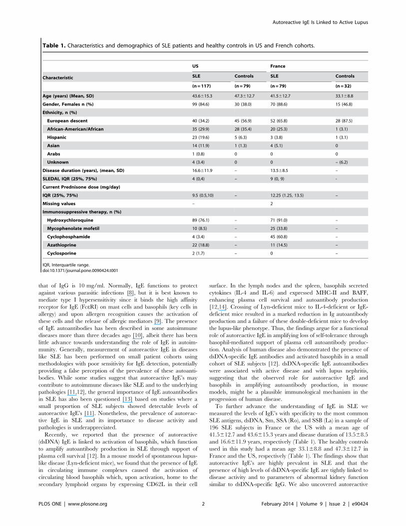
Table 1. Characteristics and demographics of SLE patients and healthy controls in US and French cohorts.
US France
Characteristic SLE Controls SLE Controls
(n=117) (n=79) (n=79) (n =32)
Age (years) (Mean, SD) 43.6615.3 47.3612.7 41.5612.7 33.168.8
Gender, Females n (%) 99 (84.6) 30 (38.0) 70 (88.6) 15 (46.8)
Ethnicity, n (%)
European descent 40 (34.2) 45 (56.9) 52 (65.8) 28 (87.5)
African-American/African 35 (29.9) 28 (35.4) 20 (25.3) 1 (3.1)
Hispanic 23 (19.6) 5 (6.3) 3 (3.8) 1 (3.1)
Asian 14 (11.9) 1 (1.3) 4 (5.1) 0
Arabs 1 (0.8) 0 0 0
Unknown 4 (3.4) 0 0 – (6.2)
Disease duration (years), (mean, SD) 16.6611.9 – 13.568.5 –
SLEDAI, IQR (25%, 75%) 4 (0,4) – 9 (0, 9) -
Current Prednisone dose (mg/day)
IQR (25%, 75%) 9.5 (0.5,10) – 12.25 (1.25, 13.5) –
Missing values – 2
Immunosuppressive therapy, n (%)
Hydroxychloroquine 89 (76.1) – 71 (91.0) –
Mycophenolate mofetil 10 (8.5) – 25 (33.8) –
Cyclophosphamide 4 (3.4) – 45 (60.8) –
Azathioprine 22 (18.8) – 11 (14.5) –
Cyclosporine 2 (1.7) – 0 –
IQR, Interquartile range.doi:10.1371/journal.pone.0090424.t001
Autoreactive IgE Is Linked to Active Lupus
PLOS ONE | www.plosone.org 2 February 2014 | Volume 9 | Issue 2 | e90424
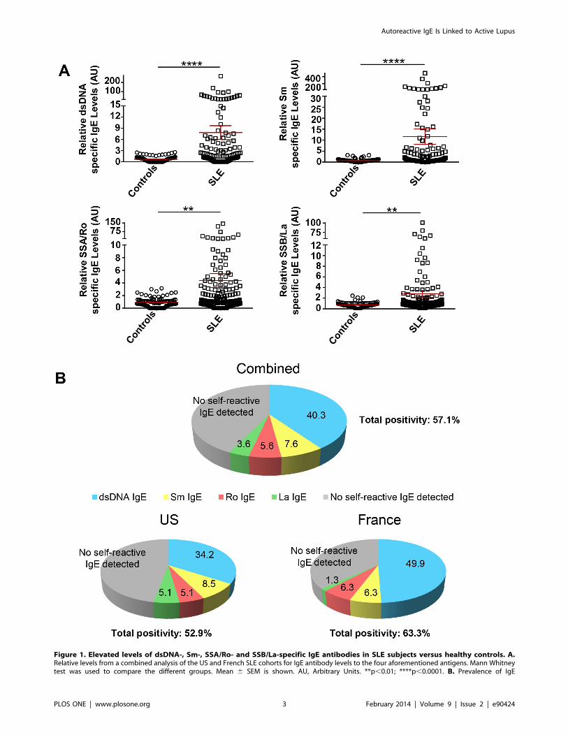
Figure 1. Elevated levels of dsDNA-, Sm-, SSA/Ro- and SSB/La-specific IgE antibodies in SLE subjects versus healthy controls. A.Relative levels from a combined analysis of the US and French SLE cohorts for IgE antibody levels to the four aforementioned antigens. Mann Whitneytest was used to compare the different groups. Mean 6 SEM is shown. AU, Arbitrary Units. **p,0.01; ****p,0.0001. B. Prevalence of IgE
Autoreactive IgE Is Linked to Active Lupus
PLOS ONE | www.plosone.org 3 February 2014 | Volume 9 | Issue 2 | e90424
IgE’s, but not IgG’s, to novel self-antigens that are linked with
hypocomplementemia, a serological marker of disease activity.
The findings demonstrate that autoreactive IgE is prevalent in
SLE and suggest that its detection together with anti-dsDNA IgG,
may serve to better predict increased disease activity and/or poor
kidney function.
Materials and Methods
Studied CohortsThis collaborative study was approved by the Institutional
Review Board of NIAMS (NIH, Bethesda, USA) and by the
Comite Regional de Protection des Personnes (CRPP, Paris,
France). A written consent was obtained from all healthy donors
and SLE patients. In the US, samples were obtained from SLE
patients enrolled in a long-term natural history study of SLE. In
France SLE samples were obtained from in- and out- patients
enrolled in a long term prospective study of SLE, and clinical data
were used after approval by the Comission Nationale de
l’Informatique et des Libertes (CNIL). All patients fulfilled the
American College of Rheumatology classification criteria for SLE
[7,15]. Samples from ‘‘healthy’’ volunteers were obtained from the
blood bank at the NIH Clinical Center and from the Universite
Paris Diderot, Faculte de Medecine site Bichat. The demograph-
ics and characteristics of all SLE patients and healthy controls are
summarized in Table 1. The data included herein is from one
sample provided by each individual SLE subject.
Sample CollectionWhole blood from SLE and healthy donors was collected in
4 ml SST (serum-separating tube) tube for serum. Serum samples
were aliquoted and kept at 280uC.
Enzyme-linked Immunosorbent Assays (ELISAs)The level of antibodies against the four common nuclear
antigens (dsDNA, Sm, Ro/SSA, La/SSB) was measured by an
ELISA. Horseradish peroxidase-conjugated antibodies to human
IgE (Mouse monoclonal anti-human IgE, Immunology Consul-
tants Laboratory) or IgG (Donkey anti-human IgG, Fc fragment
specific, Jackson Immunoresearch Laboratory) were used for
colorimetric detection was with tetramethylbenzidine substrate
(TMB, Invitrogen) and optical density (OD) was measured at
450 nm. Positive values were considered as greater than two
standard deviations over the mean absorption units obtained from
samples of healthy individuals. dsDNA-specific IgG levels were
quantified using standard laboratory procedures by the NIH
Clinical Center and by the INSERM U699 Total levels of IgE
were determined by ELISA (Calbiotech, for US; Bethyl Labora-
tories for France) following the manufacturers’ instructions.
To determine the level of IgG and IgE autoantibodies to
CLIP4, MPG and APEX nuclease 1, we used purified recombi-
nant human proteins, CLIP4 (RSNL2 protein, Abnova), MPG
(DNA-3 methyladenine glycosylase, Mybiosource), and APEX
nuclease 1 (DNA apurinic or apyrimidinic site lyase, Mybiosource)
in ELISA. Detection of reactive autoantibodies was as described
above.
Protein MicroarrayA Protoarray Human Protein Microarray v5.0 (Invitrogen Life
Technologies) with over 9000 purified human proteins was used
for novel antigen discovery. The purified recombinant proteins
spotted on nitrocellulose-coated glass slides were reacted with the
serum of SLE subjects or healthy controls and subsequently
incubated with biotinylated goat anti-human IgE antibody (5 mg/ml) (Vector Laboratories) to detect IgE antibody binding. Then
either an AlexaFluor 647-conjugated streptavidin (Invitrogen,
1 mg/ml) or an AlexaFluor 647-conjugated anti-V5 antibody
(Invitrogen, 0.1 mg/ml) was added for detection and normaliza-
tion, respectively. Arrays were scanned using fluorescent micro-
array GenePix 4000B Scanner and GenePix 6.0 software was used
for data acquisition. Data analysis was done using Invitrogens
proprietary Protoarray Prospector software. For each protein
spotted in every array, Z-Score, Z-factor, CI-P value (Chebyshevs
Inequality p-Value) was measured. M-Statistic is used to identify
those proteins which show a significant differential signal between
two populations. The ‘‘cutoff’’ value used corresponded to the
value of 200 RFU above the M-Statistic signal threshold
established for a specific protein.
StatisticsStudent t test or a non-parametric Mann-Whitney U-test (when
necessary) was used to compare SLE and control samples. A Chi2
assessment of the non-parametric Fisher exact test was used to
determine the association of autoreactive IgE with active nephritis
as well as the association of autoreactive IgE with active disease.
One-way ANOVA with Bonferroni correction test or Kruskal-
Wallis with Dunns correction test was applied when more than
two groups were compared. Mean and SEM is shown in the
scatter plot graphs. The analysis was performed using Graphpad
Prism 5.0. Grubbs’ outlier test was used to assess the outliers. In
some instances some outliers detected in the healthy control group
were excluded from the analysis because of the identification of
allergic disease or a urine infection and no clinical indications of
these conditions in the studied SLE subjects. For forest plot
analysis, a fixed effects model was applied and Hedges g statistics
was used to calculate the standardized mean difference (SMD).
Stepwise Logistic Regression offered by IBM SPSS Statistics 20
software was used to determine a predictive model of disease
activity (Not active, SLEDAI,4 vs Active, SLEDAI$4). Proba-
bility to enter a predictor in the model p,0.1 and to remove it p.
0.05. Model 1 included as predictors: positive levels of dsDNA IgE,
Sm IgE, SSA/Ro IgE, SSB/La IgE, dsDNA IgG, Sm IgG, SSA/
Ro IgG and SSB/La IgG; as confounders age, gender and
ethnicity (African American/African, European descendent, His-
panic and others). Model 2 included as predictors: positive levels of
autoreactive IgEs, autoreactive IgGs (positive for at least one of the
autoantibodies against the four common antigens); as confounders
age, gender and ethnicity. Factors not affecting the models were
eliminated from consideration. ROC Curve with AUC (area
under the curve) analysis and Youden’s J Index was calculated by
IBM SPSS in order to determine the optimal predictive model
selection.
autoantibodies to the aforementioned four common SLE autoantigens. Percentage of patients positive for IgE autoantibodies (2SD over the mean ofhealthy controls) to at least one of the four autoantigens tested. dsDNA-specific IgE (as the most prevalent autoreactive IgE) was used as thereference. The percent of anti-Sm IgE positive individuals are those without dsDNA-IgE, percent of anti-SSA/Ro positive individuals are withoutdsDNA- and Sm-IgE’s, and anti-SSB/La positive individuals are those without dsDNA, Sm and SSA/Ro reactivties. Therefore, patients in the group ofthose positive for anti-dsDNA IgE were also positive for the rest of IgE autoantibodies tested, but none of the subjects in the anti-SSB/La IgE positivegroup had detectable IgE autoantibodies for dsDNA, Sm, or SSA/Ro.doi:10.1371/journal.pone.0090424.g001
Autoreactive IgE Is Linked to Active Lupus
PLOS ONE | www.plosone.org 4 February 2014 | Volume 9 | Issue 2 | e90424
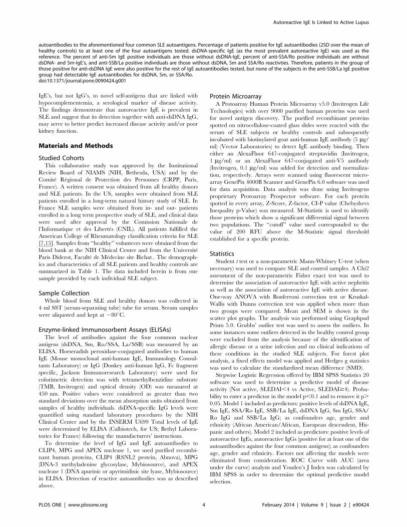
Figure 2. Comparison of the levels of dsDNA-, Sm-, SSA/Ro- and SSB/La-specific IgEs with disease activity. A. Combined analysis of thelevels of autoreactive IgE’s in the US and French SLE cohorts with disease activity as defined by SLEDAI score. Inactive, SLEDAI = 0; Mild, SLEDAI.0 to
Autoreactive IgE Is Linked to Active Lupus
PLOS ONE | www.plosone.org 5 February 2014 | Volume 9 | Issue 2 | e90424
Results
IgE Auto-antibodies to Common SLE Antigens arePrevalentThe characteristics and demographics of both French and US
SLE subjects studied herein are described in Table 1. French SLE
subjects were recruited through a long term prospective study of
SLE from a primary care center whereas US SLE subjects were
part of a long term natural history study of SLE in a secondary
care center. Thus, while the mean age of each cohort was similar,
the mean years of disease duration was somewhat less in the
French subjects (13.568.5) versus US subjects (16.6611.9) and
French subjects had a higher SLEDAI score (.9) than US subjects
(.4). As might be expected, given that the US subjects were
recruited from a secondary care center, only 13 of the 117 US
subjects (,11%) had active nephritis whereas 30 of the 79 French
subjects (,38%) were active. Treatment regimens were similar in
the two cohorts with the exception that a greater number of
French subjects (45) were on cyclophosphamide than US subjects
(4). The two cohorts also differed in their ethnic composition with
fewer Hispanics and Asians present among the French subjects.
It was not known if IgE autoantibodies are generated to all of
the antigens most frequently detected in the serologic profile of
SLE. Thus, we assayed for the presence of dsDNA, Sm, SSA/Ro
and SSB/La in both US and French SLE cohorts. IgE
autoantibodies to all four self-antigens were present with anti-
dsDNA and anti-Sm IgE antibodies having the most significant
association with SLE relative to healthy controls (Figure 1A).
Analysis of the overall frequency of all SLE patients with
autoreactive IgE positivity (a minimum of one auto-antigen
specific IgE) demonstrated a frequency of over 57% (Figure 1B)
with the US SLE cohort approaching 53% and the French cohort
at 63%. Thus, 47% of the US cohort and 37% of the French
cohort did not show IgE reactivity to one of the four auto-antigens
tested. Of the four autoantigen specificities, dsDNA specific-IgE
was the most frequent with 34.2% and 49.4% of the US and
French cohorts, respectively, having this autoantibody (Figure 1B).
To test whether the two cohorts had similar characteristics with
regards to autoreactive IgE’s we performed a forest plot (Figure S1
in File S1) and found no heterogeneity (I2 = 0%) in the levels of IgE
autoantibodies among the cohorts and no significant difference
was observed in the effect of the autoreactive IgE on the two
cohorts relative to healthy controls as shown by the overall
standardized mean difference (SMD) for each individual autore-
active IgE. This argues that the two cohorts showed similar
differences in levels of autoreactive IgE relative to healthy controls
and that the distribution of the detected autoreactive IgE is similar
in these two cohorts. No meaningful differences in the levels of IgE
autoantibodies were detected when either gender or ethnicity were
considered and no correlation was found between the total levels
,4; Active, SLEDAI $4. B. Combined US and French SLE cohort analysis of IgE autoantibodies with complement levels in serum. Low levels ofcomplement were considered as C3,80 mg/dl or C4,15 mg/dl. Kruskal-Wallis with Dunn’s multiple comparisons test was used to compare thedifferent groups. Mean 6 SEM is shown. AU, Arbitrary Units. *p,0.05; **p,0.01; ***p,0.001; ****p,0.0001.doi:10.1371/journal.pone.0090424.g002
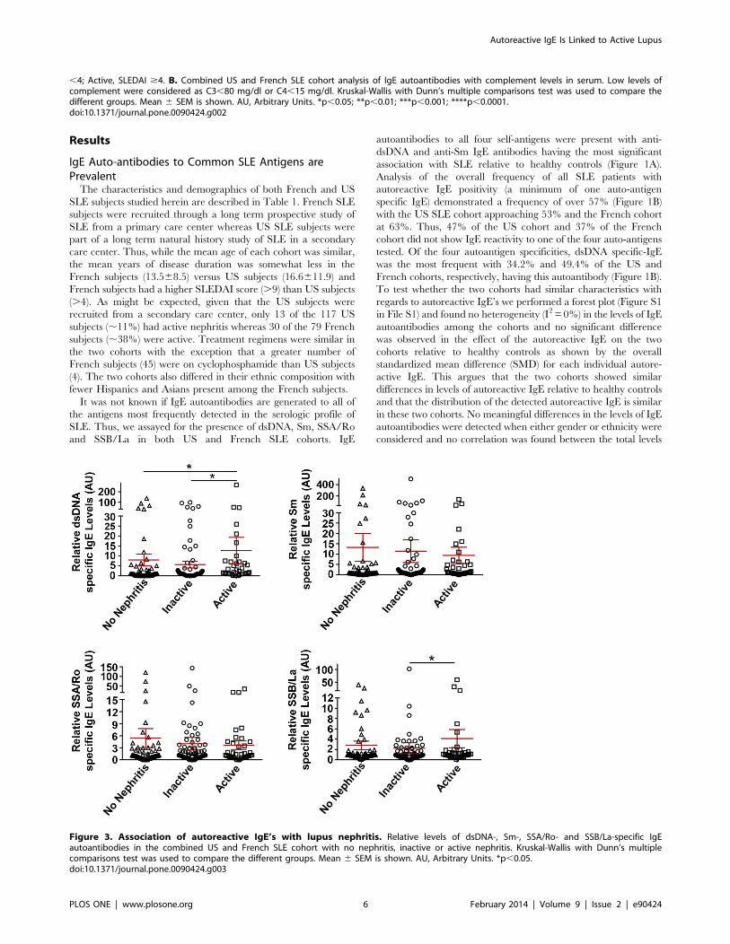
Figure 3. Association of autoreactive IgE’s with lupus nephritis. Relative levels of dsDNA-, Sm-, SSA/Ro- and SSB/La-specific IgEautoantibodies in the combined US and French SLE cohort with no nephritis, inactive or active nephritis. Kruskal-Wallis with Dunn’s multiplecomparisons test was used to compare the different groups. Mean 6 SEM is shown. AU, Arbitrary Units. *p,0.05.doi:10.1371/journal.pone.0090424.g003
Autoreactive IgE Is Linked to Active Lupus
PLOS ONE | www.plosone.org 6 February 2014 | Volume 9 | Issue 2 | e90424
of IgE and the levels of IgE autoantibodies in these cohorts (data
not shown).
IgE Autoantibodies are Associated with IncreasedDisease ActivityThe relationship between disease activity and the presence of
any one of the four auto-antigen specific IgE’s was explored.
Disease activity was based on a SLEDAI score of $4 as active
disease, .0 to ,4 for mild disease, and 0 for inactive. As shown in
Figure 2A, SLE subjects with autoreactive IgE’s were most
frequently found in the active disease group. SLE subjects with
mild disease also had significantly elevated levels of IgE
autoantibodies to dsDNA (Figure 2A).
To further verify the association of autoreactive IgE’s with
disease activity, we analyzed its relationship to hypocomplemen-
temia, a commonly used serological marker of increased disease
activity or disease relapses [16,17]. The levels of IgE autoanti-
bodies to the tested autoantigens (dsDNA, Sm, SSA/Ro, SSB/La)
were significantly elevated in SLE subjects with hypocomplemen-
temia (C3,80 mg/dl, C4,15 mg/dl) (Figure 2B). However,
dsDNA and Sm-specific IgE’s were also significantly elevated in
SLE subjects with normal complement levels. When the cohorts
were analyzed individually, only ds-DNA-specific IgE showed a
significant association with hypocomplementemia in both the
French and US cohorts (data not shown), demonstrating that this
autoreactive IgE is most significantly associated with hypocom-
plementemia.
While more than 50% of SLE subjects had autoreactive IgE to
one or more of the four common SLE auto-antigens, the
prevalence of these IgE’s in active SLE subjects (SLEDAI $4)
was 73.7% and 74.1% for the French and US subjects,
respectively, with dsDNA specific-IgE being the most prevalent
(US= 62.8%, French= 63.2%) (Figure S2A in File S1). When we
analyzed the prevalence of these autoreactive IgE’s in SLE subjects
with hypocomplementemia, it was higher in the French cohort
(90%) than in the US cohort (72%) with dsDNA-specific IgE being
found in 60% of the US and 80% of the French SLE subjects with
hypocomplementemia (Figure S2B in File S1). These findings
argue that the presence of autoreactive IgE’s (and in particular
dsDNA-specific IgE) in SLE may be a reasonable clinical indicator
of increased disease activity.
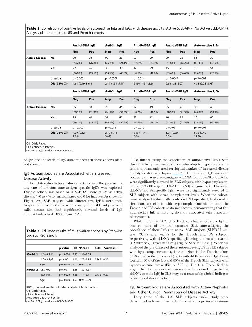
IgE Autoantibodies are Associated with Active Nephritisand Other Clinical Parameters of Disease ActivityForty three of the 196 SLE subjects under study were
determined to have active nephritis based on a protein/creatinine
Table 2. Correlation of positive levels of autoreactive IgEs and IgGs with disease activity (Active SLEDAI$4, No Active SLEDAI,4).Analysis of the combined US and French cohorts.
Anti-dsDNA IgE Anti-Sm IgE Anti-Ro/SSA IgE Anti-La/SSB IgE Autoreactive IgEs
Neg Pos Neg Pos Neg Pos Neg Pos Neg Pos
Active Disease No 90 33 93 28 92 29 99 22 51 32
(73.2%) (26.8%) (76.8%) (23.1%) (76.1%) (23.9%) (81.8%) (18.2%) (61.4%) (38.5%)
Yes 27 46 38 33 42 29 45 26 19 54
(36.9%) (63.1%) (53.5%) (46.5%) (59.2%) (40.8%) (63.4%) (36.6%) (26.0%) (73.9%)
p value p,0.0001 p= 0.0008 p= 0.014 p = 0.0044 p,0.0001
OR (95% CI) 4.64 (2.49–8.64) 2.84 (1.54–5.41) 2.19 (1.16–4.12) 2.6 (1.33–5.07) 4.53 (2.28–8.98)
Anti-dsDNA IgG Anti-Sm IgG Anti-Ro/SSA IgG Anti-La/SSB IgG Autoreactive IgGs
Neg Pos Neg Pos Neg Pos Neg Pos Neg Pos
Active Disease No 85 38 75 46 72 49 95 26 38 45
(69.1%) (31.2%) (61.9%) (38.0%) (59.5%) (40.5%) (78.5%) (21.5%) (45.8%) (54.2%)
Yes 25 48 31 40 29 42 48 23 10 63
(34.2%) (65.7%) (43.7%) (56.3%) (40.8%) (59.1%) (67.6%) (32.3%) (13.7%) (86.3%)
p value p,0.0001 p= 0.013 p= 0.012 p = 0.09 p,0.0001
OR (95% CI) 4.29 (2.32–7.95)
2.10 (1.16–3.82)
2.13 (1.17–3.86)
1.75 (0.90–3.38)
5.32 (2.40–11.78)
OR, Odds Ratio.CI, Confidence Interval.doi:10.1371/journal.pone.0090424.t002
Table 3. Adjusted results of Multivariate analysis by StepwiseLogistic Regression.
p value OR 95% CI AUC Youdens J
Model 1 dsDNA IgE p= 0.004 2.77 1.38–5.55
dsDNA IgG p,0.001 3.43 1.72–6.85 0.769 0.37
Age p= 0.008 0.97 0.94–0.99
Model 2 IgEs Pos p = 0.011 2.39 1.22–4.67
IgGs Pos p = 0.022 2.58 1.14–5.81 0.735 0.32
Age p= 0.003 0.97 0.94–0.99
ROC curve and Youden’s J Index analysis of both models.OR, Odds Ratio.CI, Confidence Interval.AUC, Area under the curve.doi:10.1371/journal.pone.0090424.t003
Autoreactive IgE Is Linked to Active Lupus
PLOS ONE | www.plosone.org 7 February 2014 | Volume 9 | Issue 2 | e90424
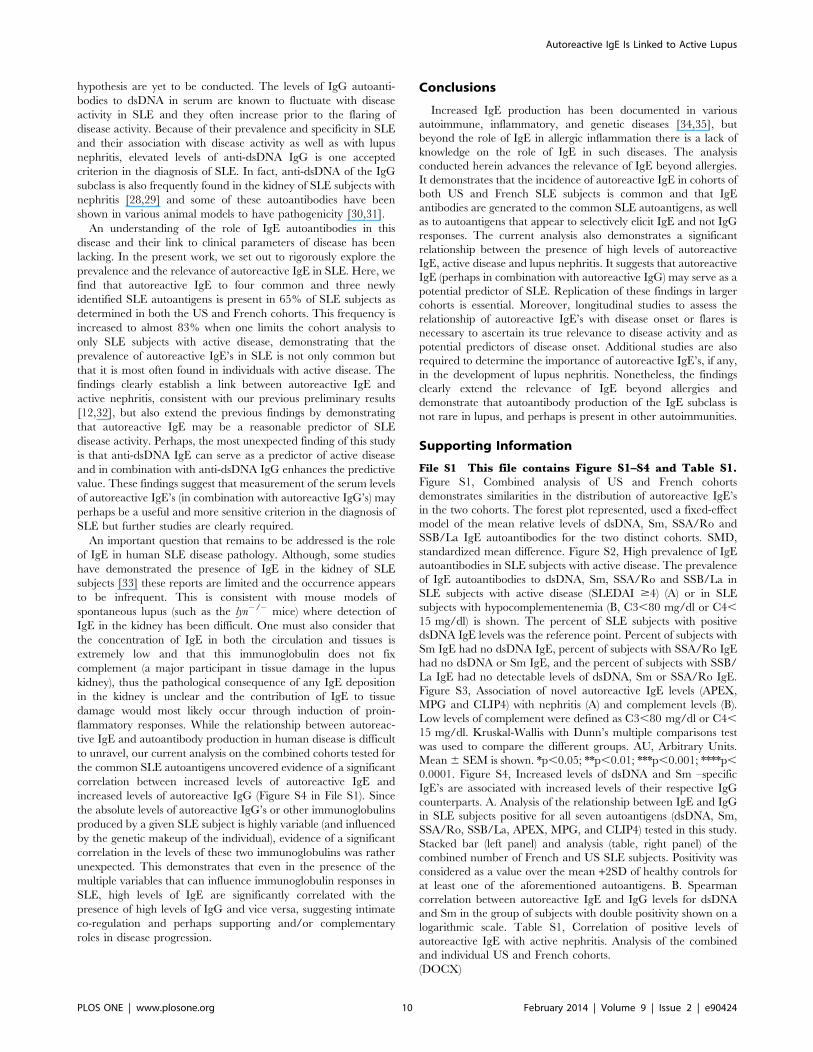
Figure 4. Identification of novel IgE autoantigens in SLE, overall prevalence, and association of all tested autoreactive IgE’s withSLE disease activity. A. Relative levels of APEX, MPG and CLIP4 specific IgEs and IgGs in US and French SLE cohorts. B. Overall prevalence of IgEautoantibodies in SLE for seven tested autoantigens tested (dsDNA, Sm, SSA/Ro and SSB/La, APEX, MPG and CLIP4). C. Comparison of the levels of all
Autoreactive IgE Is Linked to Active Lupus
PLOS ONE | www.plosone.org 8 February 2014 | Volume 9 | Issue 2 | e90424

ratio of $0.5, positivity for dsDNA IgG, and hypocomplemente-
mia. As shown in Figure 3, a significant association of active
nephritis with increased levels of dsDNA–specific IgE’s (no
nephritis vs active nephritis, p = 0.02) was observed. If the analysis
is limited to those individuals in the combined cohort who were
positive for increased levels of autoreactive IgE’s (Table S1 in File
S1), the association with dsDNA-IgE was highly significant.
Univariate analysis of the individual cohorts (Table S1 in File
S1) revealed no significant association in the US SLE subjects,
even for dsDNA-specific IgE (p = 0.20). In contrast, in French
cohort where 30 of 79 SLE subjects (,38%) had active nephritis,
both dsDNA and Sm- specific IgE’s showed a significant
association (p= 0.017 and 0.021, respectively) with this clinical
diagnosis (Table S1 in File S1).
Since the titer of circulating IgG autoantibodies (in particular
dsDNA-specific IgG) is known to be associated with SLE disease
activity, we set out to compare the relationship of autoreactive IgG
and IgE with disease activity. Analysis of SLE subjects, with active
disease, having positivity for autoreactive IgE or IgG to the four
common SLE autoantigens showed a similar significance (p,
0.0001) in the association of anti-dsDNA IgE or IgG with active
disease (Table 2). However, anti-Sm IgE showed a more
significant association with active disease than anti-Sm IgG
(p= 0.0008 vs. p= 0.013) and anti-SSB/La IgE also showed a
significant (p = 0.0044) association whereas the association of its
IgG counterpart did not achieve significance (Table 2).
Analysis of the predictive value of autoreactive IgE’s or IgG’s for
active disease in SLE subjects demonstrated that anti-dsDNA IgE
and anti-dsDNA IgG were significant in predicting SLE subjects
with active disease (Table 3, AUC=0.769, Youden’s J = 0.37),
compared to the individual analysis of each of the predictors
(dsDNA-IgE: AUC=0.72, Youden’s J = 0.36; dsDNA-IgG:
AUC=0.737, Youden’s J = 0.347). However, anti-dsDNA IgG
was a modestly stronger predictor (OR adjusted = 3.43 versus OR
2.77 for anti-dsDNA IgE) perhaps due to inclusion of dsDNA-
IgG’s as a determinant of SLEDAI. Inclusion of autoreactive IgG’s
and IgE’s with other specificities (Sm, SSA/Ro, SSB/La) reduced
the overall sensitivity of the model indicating that these were not
strong indicators of disease activity (data not shown). Introduction
of ethnicity, gender, and age as confounding factors revealed that
only age influenced the predictive model (Table 3). Thus, the
findings show that, like anti-dsDNA IgG, anti-dsDNA IgE is useful
in predicting disease activity and the use of both of these
autoantibodies increases the predictive ability (Table 3).
Discovery of Novel Auto-antigens for IgE in SLE and theirRelationship with Disease ActivityRoughly, 40–50% of SLE subjects tested did not have IgE
autoantibodies to the four common SLE autoantigens (Figure 1B).
To explore whether IgE antibodies to other autoantigens might be
present in these subjects, we used Invitrogen’s ProtoArray to
screen the sera of subjects from both the US and French cohort.
As shown in Figure 4A, high levels of IgE autoantibodies to APEX
nuclease 1 (APEX), N-methylpurine-DNA glycosylase (MPG), and
CAP-GLY domain containing linker protein family member 4
(CLIP4) were found in some SLE subjects but were minimally
detected in healthy controls. The difference in the relative
amounts of these IgE autoantibodies between healthy controls
and SLE subjects was significant. Minimal levels of IgG antibodies
to these autoantigens were detected and no changes in their levels
were observed in SLE subjects (Figure 4A). Inclusion of SLE
subjects with these novel IgE reactivities with those having at least
one of the four common SLE autoantibodies increased the
prevalence of autoreactive IgE’s from approximately 57%
(Figure 1B) to 65% (Figure 4B). Analysis of the association
between these new IgE autoantibodies with disease activity
revealed a highly significant association of APEX-, MPG-, and
CLIP4–specific IgE’s with active disease (Figure 4C). Approxi-
mately 74% of SLE subjects who had active disease also had IgE’s
that reacted to the four common SLE autoantigens (Figure S2A in
File S1). The inclusion of IgE reactivity to at least one or more of
the three novel autoantigens increased the proportion of active
SLE subjects with these autoantibodies to almost 83% (Figure 4D).
Thus, a large majority of SLE subjects with active disease have the
presence of a circulating autoreactive IgE. Autoreactive IgE’s to
APEX, MPG, and CLIP4 also showed a highly significant
association with hypocomplementemia (Figure S3A in File S1)
and with active nephritis (Figure S3B in File S1).
Of the 146 SLE subjects analyzed from the US and French
cohort who were positive for autoreactive IgE’s to all seven
autoantigens tested, 94 were positive for increased levels of
autoreactive IgE whereas 107 were found to have increased levels
of autoreactive IgG’s. Of this latter group, 76 subjects (71.1%)
were positive for both (Figure S4A in File S1). Of the autoantigens
tested, dsDNA-specific IgG and IgE and Sm-specific IgG and IgE
showed a significant (p = 0.046) or highly significant (p,0.0001)
correlation, respectively (Figure S4B in File S1).
Discussion
It is well known that the presence of circulating autoantibodies
of the IgA, IgM, and IgG isotypes are found in SLE. Moreover,
the presence of circulating IgE autoantibodies has also been
reported [10]. The presence of this diverse immunoglobulin
response is not surprising given that SLE is characterized by
abnormalities that involve hyperactive B and T cells (as well as
other cell types like monocytes) resulting in polyclonal B-cell
activation, increased numbers of plasma cells and autoantibody
production. The mechanistic role of the different immunoglobulin
isotypes in the onset and progression of SLE is, for the most part,
still unclear. However, clues are emerging from studies in both
mouse models and human disease. For example, IgA autoanti-
bodies to phospholipids (anti-cardiolipin) have been shown to be a
risk factor for thrombosis in SLE [18,19] and in some SLE patients
dsDNA-specific IgA is linked to active disease [20,21,22].
However, there are also reports that incidence of IgA-deficiency
(characterized by the lack of IgA-producing B cells) is also higher
in SLE and in such cases there appears to be no association to
clinical parameters of disease [23,24]. IgM autoantibodies (and in
particular those to dsDNA) are also frequent in lupus. However,
unlike their IgG counterpart, anti-dsDNA IgM appears to be
inversely linked to the development of nephritis [25] and in mouse
models there is evidence that such autoantibodies may have a
protective role [26]. It has been proposed that the ratio of IgG/
IgM anti-dsDNA antibodies might be a useful prognostic marker
for lupus nephritis [27], although rigorous studies to test this
tested autoreactive IgE antibodies (as in B) in US and French cohorts with disease activity as measured by SLEDAI score. Inactive, SLEDAI = 0; Mild,SLEDAI .0 to ,4; Active, SLEDAI $4. D. Prevalence of all tested autoreactive IgE’s in US and French SLE subjects with active disease. Mann Whitneytest or Kruskal-Wallis with Dunn’s multiple comparisons test (more than two groups) was used to compare the different groups. Mean 6 SEM isshown. **p,0.01; ***p,0.001, ****p,0.0001.doi:10.1371/journal.pone.0090424.g004
Autoreactive IgE Is Linked to Active Lupus
PLOS ONE | www.plosone.org 9 February 2014 | Volume 9 | Issue 2 | e90424
hypothesis are yet to be conducted. The levels of IgG autoanti-
bodies to dsDNA in serum are known to fluctuate with disease
activity in SLE and they often increase prior to the flaring of
disease activity. Because of their prevalence and specificity in SLE
and their association with disease activity as well as with lupus
nephritis, elevated levels of anti-dsDNA IgG is one accepted
criterion in the diagnosis of SLE. In fact, anti-dsDNA of the IgG
subclass is also frequently found in the kidney of SLE subjects with
nephritis [28,29] and some of these autoantibodies have been
shown in various animal models to have pathogenicity [30,31].
An understanding of the role of IgE autoantibodies in this
disease and their link to clinical parameters of disease has been
lacking. In the present work, we set out to rigorously explore the
prevalence and the relevance of autoreactive IgE in SLE. Here, we
find that autoreactive IgE to four common and three newly
identified SLE autoantigens is present in 65% of SLE subjects as
determined in both the US and French cohorts. This frequency is
increased to almost 83% when one limits the cohort analysis to
only SLE subjects with active disease, demonstrating that the
prevalence of autoreactive IgE’s in SLE is not only common but
that it is most often found in individuals with active disease. The
findings clearly establish a link between autoreactive IgE and
active nephritis, consistent with our previous preliminary results
[12,32], but also extend the previous findings by demonstrating
that autoreactive IgE may be a reasonable predictor of SLE
disease activity. Perhaps, the most unexpected finding of this study
is that anti-dsDNA IgE can serve as a predictor of active disease
and in combination with anti-dsDNA IgG enhances the predictive
value. These findings suggest that measurement of the serum levels
of autoreactive IgE’s (in combination with autoreactive IgG’s) may
perhaps be a useful and more sensitive criterion in the diagnosis of
SLE but further studies are clearly required.
An important question that remains to be addressed is the role
of IgE in human SLE disease pathology. Although, some studies
have demonstrated the presence of IgE in the kidney of SLE
subjects [33] these reports are limited and the occurrence appears
to be infrequent. This is consistent with mouse models of
spontaneous lupus (such as the lyn2/2 mice) where detection of
IgE in the kidney has been difficult. One must also consider that
the concentration of IgE in both the circulation and tissues is
extremely low and that this immunoglobulin does not fix
complement (a major participant in tissue damage in the lupus
kidney), thus the pathological consequence of any IgE deposition
in the kidney is unclear and the contribution of IgE to tissue
damage would most likely occur through induction of proin-
flammatory responses. While the relationship between autoreac-
tive IgE and autoantibody production in human disease is difficult
to unravel, our current analysis on the combined cohorts tested for
the common SLE autoantigens uncovered evidence of a significant
correlation between increased levels of autoreactive IgE and
increased levels of autoreactive IgG (Figure S4 in File S1). Since
the absolute levels of autoreactive IgG’s or other immunoglobulins
produced by a given SLE subject is highly variable (and influenced
by the genetic makeup of the individual), evidence of a significant
correlation in the levels of these two immunoglobulins was rather
unexpected. This demonstrates that even in the presence of the
multiple variables that can influence immunoglobulin responses in
SLE, high levels of IgE are significantly correlated with the
presence of high levels of IgG and vice versa, suggesting intimate
co-regulation and perhaps supporting and/or complementary
roles in disease progression.
Conclusions
Increased IgE production has been documented in various
autoimmune, inflammatory, and genetic diseases [34,35], but
beyond the role of IgE in allergic inflammation there is a lack of
knowledge on the role of IgE in such diseases. The analysis
conducted herein advances the relevance of IgE beyond allergies.
It demonstrates that the incidence of autoreactive IgE in cohorts of
both US and French SLE subjects is common and that IgE
antibodies are generated to the common SLE autoantigens, as well
as to autoantigens that appear to selectively elicit IgE and not IgG
responses. The current analysis also demonstrates a significant
relationship between the presence of high levels of autoreactive
IgE, active disease and lupus nephritis. It suggests that autoreactive
IgE (perhaps in combination with autoreactive IgG) may serve as a
potential predictor of SLE. Replication of these findings in larger
cohorts is essential. Moreover, longitudinal studies to assess the
relationship of autoreactive IgE’s with disease onset or flares is
necessary to ascertain its true relevance to disease activity and as
potential predictors of disease onset. Additional studies are also
required to determine the importance of autoreactive IgE’s, if any,
in the development of lupus nephritis. Nonetheless, the findings
clearly extend the relevance of IgE beyond allergies and
demonstrate that autoantibody production of the IgE subclass is
not rare in lupus, and perhaps is present in other autoimmunities.
Supporting Information
File S1 This file contains Figure S1–S4 and Table S1.Figure S1, Combined analysis of US and French cohorts
demonstrates similarities in the distribution of autoreactive IgE’s
in the two cohorts. The forest plot represented, used a fixed-effect
model of the mean relative levels of dsDNA, Sm, SSA/Ro and
SSB/La IgE autoantibodies for the two distinct cohorts. SMD,
standardized mean difference. Figure S2, High prevalence of IgE
autoantibodies in SLE subjects with active disease. The prevalence
of IgE autoantibodies to dsDNA, Sm, SSA/Ro and SSB/La in
SLE subjects with active disease (SLEDAI $4) (A) or in SLE
subjects with hypocomplementenemia (B, C3,80 mg/dl or C4,
15 mg/dl) is shown. The percent of SLE subjects with positive
dsDNA IgE levels was the reference point. Percent of subjects with
Sm IgE had no dsDNA IgE, percent of subjects with SSA/Ro IgE
had no dsDNA or Sm IgE, and the percent of subjects with SSB/
La IgE had no detectable levels of dsDNA, Sm or SSA/Ro IgE.
Figure S3, Association of novel autoreactive IgE levels (APEX,
MPG and CLIP4) with nephritis (A) and complement levels (B).
Low levels of complement were defined as C3,80 mg/dl or C4,
15 mg/dl. Kruskal-Wallis with Dunn’s multiple comparisons test
was used to compare the different groups. AU, Arbitrary Units.
Mean6 SEM is shown. *p,0.05; **p,0.01; ***p,0.001; ****p,
0.0001. Figure S4, Increased levels of dsDNA and Sm –specific
IgE’s are associated with increased levels of their respective IgG
counterparts. A. Analysis of the relationship between IgE and IgG
in SLE subjects positive for all seven autoantigens (dsDNA, Sm,
SSA/Ro, SSB/La, APEX, MPG, and CLIP4) tested in this study.
Stacked bar (left panel) and analysis (table, right panel) of the
combined number of French and US SLE subjects. Positivity was
considered as a value over the mean +2SD of healthy controls for
at least one of the aforementioned autoantigens. B. Spearman
correlation between autoreactive IgE and IgG levels for dsDNA
and Sm in the group of subjects with double positivity shown on a
logarithmic scale. Table S1, Correlation of positive levels of
autoreactive IgE with active nephritis. Analysis of the combined
and individual US and French cohorts.
(DOCX)
Autoreactive IgE Is Linked to Active Lupus
PLOS ONE | www.plosone.org 10 February 2014 | Volume 9 | Issue 2 | e90424

Acknowledgments
The authors wish to acknowledge the invaluable support of the Office of
the Clinical Director, National Institute of Arthritis and Musculoskeletal
and Skin Diseases, National Institutes of Health.
Author Contributions
Conceived and designed the experiments: BD NC SH ED GI JR.
Performed the experiments: BD CP MMB JS CJ TKR. Analyzed the data:
BD NG MMB SH GI NC JR. Contributed reagents/materials/analysis
tools: SH KS MJ DG TP ED. Wrote the paper: BD SH NG MJ DG ED
GI NC JR.
References
1. Frieri M (2013) Mechanisms of disease for the clinician: systemic lupus
erythematosus. Ann Allergy Asthma Immunol 110: 228–232.
2. Sawalha AH, Harley JB (2004) Antinuclear autoantibodies in systemic lupus
erythematosus. Curr Opin Rheumatol 16: 534–540.
3. Arbuckle MR, McClain MT, Rubertone MV, Scofield RH, Dennis GJ, et al.
(2003) Development of autoantibodies before the clinical onset of systemic lupus
erythematosus. N Engl J Med 349: 1526–1533.
4. Rahman A, Isenberg DA (2008) Systemic lupus erythematosus. N Engl J Med
358: 929–939.
5. Schroeder K, Herrmann M, Winkler TH (2012) The role of somatic
hypermutation in the generation of pathogenic antibodies in SLE. Autoimmu-
nity 46: 121–127.
6. Kalaaji M, Fenton KA, Mortensen ES, Olsen R, Sturfelt G, et al. (2007)
Glomerular apoptotic nucleosomes are central target structures for nephrito-
genic antibodies in human SLE nephritis. Kidney Int 71: 664–672.
7. Hochberg MC (1997) Updating the American College of Rheumatology revised
criteria for the classification of systemic lupus erythematosus. Arthritis Rheum
40: 1725.
8. Capron A, Dessaint JP (1977) IgE and cells in protective immunity. Pathol Biol
(Paris) 25: 287–290.
9. Blank U, Rivera J (2004) The ins and outs of IgE-dependent mast-cell exocytosis.
Trends Immunol 25: 266–273.
10. Permin H, Wiik A (1978) The prevalence of IgE antinuclear antibodies in
rheumatoid arthritis and systemic lupus erythematosus. Acta Pathol Microbiol
Scand C 86C: 245–249.
11. Atta AM, Santiago MB, Guerra FG, Pereira MM, Sousa Atta ML (2010)
Autoimmune response of IgE antibodies to cellular self-antigens in systemic
Lupus Erythematosus. Int Arch Allergy Immunol 152: 401–406.
12. Charles N, Hardwick D, Daugas E, Illei GG, Rivera J (2010) Basophils and the
T helper 2 environment can promote the development of lupus nephritis. Nat
Med 16: 701–707.
13. Bosch X, Lozano F, Cervera R, Ramos-Casals M, Min B (2011) Basophils, IgE,
and autoantibody-mediated kidney disease. J Immunol 186: 6083–6090.
14. Rodriguez Gomez M, Talke Y, Goebel N, Hermann F, Reich B, et al. (2010)
Basophils support the survival of plasma cells in mice. J Immunol 185: 7180–
7185.
15. Tan EM, Cohen AS, Fries JF, Masi AT, McShane DJ, et al. (1982) The 1982
revised criteria for the classification of systemic lupus erythematosus. Arthritis
Rheum 25: 1271–1277.
16. Kallenberg CG, Stegeman CA, Bootsma H, Bijl M, Limburg PC (2006)
Quantitation of autoantibodies in systemic autoimmune diseases: clinically
useful? Lupus 15: 397–402.
17. Livingston B, Bonner A, Pope J (2012) Differences in autoantibody profiles and
disease activity and damage scores between childhood- and adult-onset systemic
lupus erythematosus: a meta-analysis. Semin Arthritis Rheum 42: 271–280.
18. Sweiss NJ, Bo R, Kapadia R, Manst D, Mahmood F, et al. (2010) IgA anti-
beta2-glycoprotein I autoantibodies are associated with an increased risk of
thromboembolic events in patients with systemic lupus erythematosus. PLoS
One 5: e12280.
19. Mehrani T, Petri M (2011) Association of IgA Anti-beta2 glycoprotein I withclinical and laboratory manifestations of systemic lupus erythematosus.
J Rheumatol 38: 64–68.
20. Miltenburg AM, Roos A, Slegtenhorst L, Daha MR, Breedveld FC (1993) IgAanti-dsDNA antibodies in systemic lupus erythematosus: occurrence, incidence
and association with clinical and laboratory variables of disease activity.J Rheumatol 20: 53–58.
21. Witte T, Hartung K, Matthias T, Sachse C, Fricke M, et al. (1998) Associationof IgA anti-dsDNA antibodies with vasculitis and disease activity in systemic
lupus erythematosus. SLE Study Group. Rheumatol Int 18: 63–69.
22. Villalta D, Bizzaro N, Bassi N, Zen M, Gatto M, et al. (2013) Anti-dsDNAantibody isotypes in systemic lupus erythematosus: IgA in addition to IgG anti-
dsDNA help to identify glomerulonephritis and active disease. PLoS One 8:e71458.
23. Latiff AH, Kerr MA (2007) The clinical significance of immunoglobulin A
deficiency. Ann Clin Biochem 44: 131–139.24. Mantovani AP, Monclaro MP, Skare TL (2010) Prevalence of IgA deficiency in
adult systemic lupus erythematosus and the study of the association with itsclinical and autoantibody profiles. Rev Bras Rheumatol 50: 273–282.
25. Witte T, Hartung K, Sachse C, Matthias T, Fricke M, et al. (1998) IgM anti-dsDNA antibodies in systemic lupus erythematosus: negative association with
nephritis. SLE Study Group. Rheumatol Int 18: 85–91.
26. Werwitzke S, Trick D, Kamino K, Matthias T, Kniesch K, et al. (2005)Inhibition of lupus disease by anti-double-stranded DNA antibodies of the IgM
isotype in the (NZB x NZW)F1 mouse. Arthritis Rheum 52: 3629–3638.27. Forger F, Matthias T, Oppermann M, Becker H, Helmke K (2004) Clinical
significance of anti-dsDNA antibody isotypes: IgG/IgM ratio of anti-dsDNA
antibodies as a prognostic marker for lupus nephritis. Lupus 13: 36–44.28. Kenderov A, Minkova V, Mihailova D, Giltiay N, Kyurkchiev S, et al. (2002)
Lupus-specific kidney deposits of HSP90 are associated with altered IgGidiotypic interactions of anti-HSP90 autoantibodies. Clin Exp Immunol 129:
169–176.
29. Bijl M, Dijstelbloem HM, Oost WW, Bootsma H, Derksen RH, et al. (2002) IgGsubclass distribution of autoantibodies differs between renal and extra-renal
relapses in patients with systemic lupus erythematosus. Rheumatology (Oxford)41: 62–67.
30. Madaio MP, Carlson J, Cataldo J, Ucci A, Migliorini P, et al. (1987) Murinemonoclonal anti-DNA antibodies bind directly to glomerular antigens and form
immune deposits. J Immunol 138: 2883–2889.
31. Pankewycz OG, Migliorini P, Madaio MP (1987) Polyreactive autoantibodiesare nephritogenic in murine lupus nephritis. J Immunol 139: 3287–3294.
32. Charles N, Rivera J (2011) Basophils and autoreactive IgE in the pathogenesis ofsystemic lupus erythematosus. Current allergy and asthma reports 11: 378–387.
33. Tuma SN, Llach F, Sostrin S, Dubois EL, Massry SG (1981) Glomerular IgE
deposits in patients with lupus nephritis. Am J Nephrol 1: 31–36.34. Altin J, Shen C, Liston A (2010) Understanding the genetic regulation of IgE
production. Blood Rev 24: 163–169.35. Pate MB, Smith JK, Chi DS, Krishnaswamy G (2010) Regulation and
dysregulation of immunoglobulin E: a molecular and clinical perspective. ClinMol Allergy 8: 3.
Autoreactive IgE Is Linked to Active Lupus
PLOS ONE | www.plosone.org 11 February 2014 | Volume 9 | Issue 2 | e90424