The syndecan-1 heparan sulfate proteoglycan is a viable target for myeloma therapy
Upload
independentCategory
view
2download
0
Clin Exp Immunol 1995; 101:414-421
Antigen-specific B cells present cartilage proteoglycan (aggrecan) to an
autoreactive T cell hybridoma derived from a mouse with proteoglycan-induced arthritis
F. R. BRENNAN*, K. MIKECZt*, E. I. BUZAS$, D. RAGASA*, G. CS-SZABO*, G. NEGROIU*& T. T. GLANT*t Departments of *Biochemistry and tOrthopaedic Surgery, Rush Medical University at Rush-Presbyterian-St. Luke's Medical Centre, Chicago, IL, USA, and tDepartment of Anatomy, Histology and Embryology, University of Medicine,
Debrecen, Hungary
(Acceptedfor publication 3 May 1995)
SUMMARY
Cartilage proteoglycan (aggrecan)-induced polyarthritis in BALB/c mice is characterized bychronic inflammation and destruction of joint tissues similar to that observed in humanrheumatoid arthritis. The immunization of mice with fetal human proteoglycan (PG) elicitsspecific antibodies to the immunizing antigen of which a population cross-reacts with nativemouse PG. This (auto)antibody production is immediately followed by an explosive proliferationof autoreactive T cells, suggesting that PG-specific B cells may participate in antigen presentationof PG to autoreactive T cells. We therefore isolated B cells from the spleens and lymph nodes ofPG-immunized mice and examined their ability to present PG to a PG-specific T cell hybridoma.The antigen-specific T cell responses elicited by B cells from PG-immunized mice (both arthriticand clinically asymptomatic) were markedly higher than those of non-immune mice and keyholelimpet haemocyanin (KLH)-immunized mice, and these B cells could present low PG concentra-tions. Levels of B cell presentation corresponded with the serum levels of PG-specific antibodies,implying that these B cells were presenting the PG specifically via their surface immunoglobulin.This B cell-T cell interaction was strongly dependent on MHC class II/T cell receptor (TCR),LFA-1/intercellular adhesion molecule-i (ICAM-1) and CD28/B7 interactions, as antibodies toIa, ICAM-1 and B7-2 (but not to B7-1) markedly reduced presentation. These data indicate thatPG-specific B cells may play an essential role in governing the development of PG-induced arthritis.
Keywords proteoglycan arthritis animal model B cells T hybridoma antigen presentation
INTRODUCTION
Proteoglycan (aggrecan)-induced progressive polyarthritis inBALB/c mice shows many similarities to human rheumatoidarthritis (RA) [1-4], as indicated by clinical assessments,immunological tests and histopathology of joints. The devel-opment of arthritis in BALB/c mice, initiated by i.p. injectionwith glycosaminoglycan-depleted human fetal cartilage proteo-glycan (HFPG), is associated with the expression of both cell-mediated immunity and autoantibody production to the mouse'self' cartilage PG [1,2]. This is most probably due to cross-reactive epitopes which are present in both mouse and humanfetal PG.
We have generated PG-specific T cell hybridomas from micewith PG-induced arthritis [5,6] and used them as tools for
Correspondence: Professor T.T. Glant, Department of Biochemis-try, Rush-Presbyterian-St. Luke's Medical Centre, 1653 West CongressParkway, Chicago, IL 60612, USA.
studying how PG are presented to T cells by antigen-presentingcells (APC). It was shown previously that macrophages, synovialcells and chondrocytes were effective presenters of PG to these Tcell hybridomas, and hence these APC may play important rolesin the generation of autoreactive T cells in PG-immunized mice[7]. As the autoantibodies to mouse PG appear before thegeneration of mouse PG-specific T cell responses [8], and theproliferation of CD45R+ B cells appeared before an increase inCD4+ T helper cell numbers [8,9], we hypothesized that perhapsPG-specific B cells were also involved in the priming of auto-reactive T cells [10] and the subsequent development of arthritis.T cell-B cell cooperation has been shown to be important for thedevelopment of a number of autoimmune diseases [11,12].
To test this hypothesis, we examined the ability of B cellsfrom arthritic, PG-immunized and non-arthritic and normalmice to present PG to the PG-specific T cell hybridoma, 5/4E8,which induces arthritis when injected into irradiated BALB/cmice [5]. It was shown that only B cells from immunized mice
44 1995 Blackwell Science414
Proteoglycan presentation by B cells
(both arthritic and non-arthritic) but not from normal micecould present PG to the T cell hybridoma. Furthermore, this Tcell-B cell interaction was inhibited by antibodies to both B7-2and intercellular adhesion molecule-l (ICAM-1), whenceimmunotherapy of arthritic mice with these antibodies may
provide a useful therapeutic approach in preventing jointdestruction in mice with PG-induced arthritis.
MATERIALS AND METHODS
Preparation ofPG antigensPG from human adult cartilage (HAC) (42-52 years age
range), from femoral condyles of fetal human (34-40 weeksof gestation), bovine nasal cartilage (BNC), chick sternalcartilage (CSC), rat chondrosarcoma, dog articular cartilageand femoral condyles of newborn BALB/c mice were preparedas described [13]. Cartilage pieces were frozen immediately and50-tim cryostat sections were extracted at 4°C with 4 M
guanidium chloride in 50 mm sodium acetate pH 5 8, contain-ing a cocktail of protease inhibitors as described [13]. Highbuoyant density PG monomers (aggrecan) were prepared bydissociative caesium chloride gradient centrifugation. PG was
digested with protease-free chondroitinase ABC (SeikagakuAmerica, Inc., Rockville, MD) as described [13]. Cyanogenbromide (CNBr) digestion of HFPG was carried out as
described previously [7]. Samples were dialysed against dis-tilled water, lyophilized and sterilized by gamma-irradiation(250 Gy, caesium source).
Immunization of BALB/c miceFemale BALB/c mice (Charles River Colony) weighing 17-18 g
were used in all experiments and were injected intraperitoneallywith 100 ,ug of chondroitinase ABC-digested (i) arthritogenicHFPG, or (ii) non-arthritogenic CSC- or BNC-PG (measuredas protein) in Freund's complete adjuvant (FCA; Difco Labs,Detroit, MI) followed by three identical booster injections inFreund's incomplete adjuvant (FIA; Difco) on days 7, 28 and49 [1,2]. In addition, mice injected with adjuvant withoutantigen, with adjuvant and keyhole limpet haemocyanin(KLH) or non-immunized mice served as controls.
Culture mediaThe complete medium for culture of all cell types was Dulbec-co's modified Eagle's medium (DMEM; GIBCO, Grand Island,NY) supplemented (sDMEM) with 10 mm HEPES (BoehringerMannheim Biochemicals, Indianapolis, IN), 0-5 mm 2-mercap-toethanol (2-ME; Sigma Chemical Co., St Louis, MO), 1%non-essential amino acids (GIBCO), 1% sodium pyruvate(GIBCO), 100 pg/ml gentamicin (Sigma) and 10% heat-inactivated (56°C, 30 min) fetal calf serum (FCS; Hyclone,Logan, UT). Conditioned medium, as a source of IL-2,consisted of supernatants of rat spleen cells that had beenstimulated, for 48 h with 5 ,ug/ml concanavalin A (Con A)(ICN, Costa Mesa, CA) as we described [7]. All tissue cultureexperiments, unless indicated, were performed in a humidifiedatmosphere of 5% CO2 in air.
Cell lines and PG-specific T cell hybridomaThe cell lines described (CTLL-2 and BW5147) were obtainedfrom American Type Culture Collection (ATCC; Rockville,MD). The PG-specific T cell hybridoma 5/4E8 was obtained
following fusion of in vitro stimulated lymphocytes fromarthritic mice with BW5147 thymoma cells [14] as previouslydescribed [5,6]. Hybridoma 5/4E8 expressed T cell receptor(TCR) a,3+ (Vf34), CD4+ and CD8- phenotypes and secretedIL-2 but not IL-4 following stimulation with cartilage PG [5,7].
Measurement of anti-PG antibodiesSerum antibodies were measured by solution radioimmunoas-say (RIA) as described elsewhere [1,13]. Serial dilutions of sera
were incubated with 125I-labelled HAPG, HFPG, BNCPG, rat
chondrosarcoma or mouse PG and the antigen-antibodycomplex pelleted either with protein A bearing Staphylococcusaureus (Zysorbin; Zymed Labs, San Francisco, CA) for IgG or
with pig anti-mouse IgM followed by Zysorbin [15]. Radio-activity of pellets was measured using a gamma counter(Beckman, Model Gamma 5500B).
Detection of PG-specific IL-2 productionMouse spleen and lymph node cells (2 x 10⁵/well) were culturedwith PG in triplicate wells in 96-well microtitration plates for 24h and the supernatants were then harvested and assayed in a
CTLL-2 proliferation assay as described [8]. The result for PG-stimulated cells was expressed as Act/min (net counts/minshown as mean ct/min in figures), calculated by subtractingthe counts obtained with unstimulated cells.
T cell hybridoma 5/4E8 (1 x 10⁵ cells/well) was tested withPG or PG fragments in 96-well microtitration plates in thepresence of syngeneic APC (either 2 x 10⁵/well unseparatedmouse spleen cells, purified B cells or purified T cells) intriplicate wells at 37°C for 18 h and the supernatants were
then tested for IL-2 content as described above.
Isolation ofB cells using nylon woolScrubbed nylon fibre (-600 mg) (three denier, 3-81 cm; Poly-sciences, Inc., Warrington, PA) was packed into a 10-mlsyringe. The column was washed through and filled with 50 mlof sDMEM and incubated at 37°C for 2 h in air with 5% CO2.After incubation, 107 spleen mononuclear cells purified on
Lympholyte M (Cedarlane Laboratories, Westbury, NY) or
lymph node cells (plastic-adherent cells were removed in bothcases) in 2 ml of sDMEM at 37°C were layered gently onto thecolumn, followed by 1 ml warm medium. The column was
sealed and returned to the incubator for 45-60 min. The T cellswere then eluted with 20 ml medium, spun down, counted andresuspended to a concentration of 2 x 10⁶ cells/ml in sDMEM.The column was then washed through with 50 ml of warmmedium to remove the non-adherent 'null' cells. B cells were
eluted by adding a further 10 ml warm medium, rapidlyinserting the plunger, and compressing the wool as hard as
possible. This process was repeated twice; the collected B cellswere washed twice in DMEM and resuspended to a concentra-tion of 2 x 10⁶/ml in sDMEM. The enriched B cell populationsfrom both spleen and lymph nodes were shown to be 185%CD45R+, < 10% CD3+, and a mean of 7 4% mac-l + by flowcytometry (described below; Table 1, spleen cells shown).
Magnetic separation ofB cellsTo try to reduce the percentage of mac-I + cells in the B cellpopulation, splenic B cells were also isolated using miniMACSmagnetic separation columns (Miltenyi Biotech Inc, Sunnyvale,CA). Briefly, lymphocytes, after removing plastic-adherent cells
1995 Blackwell Science Ltd, Clinical and Experimental Immunology, 101:414-421
415

F. R. Brennan et al.
Table 1. Phenotypes of separated splenic B cell populations
Nylon wool Magnetic beads
B cells Non-B cells B cells Non-B cells
CD45R 81 1 + 40 17-2 ±2-3 97 ± 1 9 1-8 ± 0-23CD3 95 ± 1-2 72-5 +41 09 ± 02 872 ± 34mac-I 7-4± 03 16-4 ±24 1-8 ± 03 109 ± 11
IL-2 (ct/min) 58 000 ± 63 243 421 546 ± 21 394 563 ± 256 438 560 ± 27 351
B cells were isolated from mononuclear spleen cells (previously purified by Lympholyte M-sedimentation andplastic adherence for 1 h at 370C) either by adherence to nylon wool or by magnetic separation using an anti-CD45R MoAb. Cell surface phenotypes of B cell populations were determined by flow cytometry. Numbersindicate the mean percentage of cells positive for CD45R, CD3 or mac-I from six mice. Levels of IL-2 are shownas mean ct/min of CTLL-2 cells cultured with a conditioned media from cells stimulated with 25 pg/mlconcanavalin A.
from Lympholyte M-separated spleen cells, were incubatedwith a biotinylated MoAb to CD45R (clone RA3-6B2;GIBCO) for 30 min on ice. This MoAb recognizes an isoformof CD45R present only in murine B cells [17]. After threewashes in PBS, the cells were then incubated with MACScolloidal superparamagnetic microbeads (Miltenyi) conju-gated with streptavidin for 20 min and then with streptavidin-R-PE (GIBCO) for 30 min on ice. After a further two washes, thecells were separated on a type A2 steel wool column (Miltenyi)in the presence of a high gradient magnetic field. Cells recov-
ered from the column were shown to be 197% CD45R+, < 1%CD3 +, and < 2% mac-I + (Table 1). Cells that passed throughthe column were < 2% CD45R +, 1 1% mac- I +, and > 87%CD3 +.
Magnetic separation of B cells yielded the most pure B cellpopulation with no T cell contamination, as stimulation with2-5 ig/ml Con A resulted in no IL-2 production (Table 1).Nylon wool-separated B cells, however, produced IL-2 to ConA stimulation, suggesting the presence of small numbers of Tcells (Table 1).
Flow cytometryThe purified B cells were examined for CD45R, CD3, mac-i,
MHC class II, ICAM-1, B7-1 and B7-2 expression by flowcytometry as previously described [8]. Rat MoAbs specific forthe mouse Ia region (I-A I-Ed~k-reactive) were purifiedfrom the ascitic fluid of nude mice intraperitoneally injectedwith hybridoma M5/114.15.2 by affinity chromatography on
protein-A columns (Pierce, Rockford, IL). The biotinylatedgoat anti-rat IgG was obtained from Kirkegaard and PerryLaboratories, Inc. (Gaithersburg, MD). The murine ICAM-l-,B7-1- and B7-2-specific biotinylated MoAbs were obtainedfrom Pharmingen (San Diego, CA). The biotinylated MoAbsto CD45R, CD3 and mac-1 were obtained from GIBCO.
Effect ofspecific monoclonal antibodies on B cell presentation ofPGThe purified mouse B cells (from magnetic separation) were
cultured with the 5/4E8 hybridoma cells in the presence of 50
pg/ml HAPG and MoAbs for 18 h at 37°C. Non-biotinylatedMoAbs specific for ICAM-1, B7-1 and B7-2 were obtainedfrom Pharmingen. The anti-Ia MoAb was the same as the one
described above. The MoAbs were added to the B cells andhybridomas at a concentration of 50 ,g/ml and the effect on IL-2 production was measured as described above. The ICAM-1,B7-1 and B7-2-specific MoAbs (all IgG2a) were immobilized tothe wells of a 96-well plate by incubation for 18 h at 37°C insterile sodium carbonate buffer, pH 9 6. Immobilized rat IgG2a(Pharmingen) was used as an isotype control in all experiments.
Statistical analysisStudent's unpaired t-test was used to detect significant differ-ences (P < 0-05) between groups.
RESULTS
PG-specific antibodies and T cells in immunized miceIn HFPG-immunized mice, PG-specific antibodies to immuniz-ing antigen appeared after 2 weeks and autoreactive antibodiesto native mouse PG became detectable in sera -21-28 daysafter the first injection of PG (Fig. 1). Antibodies that cross-
reacted with HACPG, BNCPG and rat chondrosarcoma PGcould also be detected at this time (Table 2). In immunized non-
arthritic (BNCPG- or CSCPG-injected mice) the same anti-body profiles were obtained, but antibodies to mouse PG were
not detected, or were detected at very low level, in these animals(Table 2). No PG-specific antibodies were detected in KLH-immunized mice, in mice given adjuvant alone or in unimmu-nized mice (Table 2). Levels of PG-specific antibodies inHFPG- (Fig. 1) and BNCPG- or CSCPG-immunized mice(not shown) continued to increase over the next 2-3 weeks,plateauing at weeks 9-11 of immunization. PG-specific T cellresponses in the lymph nodes and spleen appeared in bothHFPG- (Fig. 1) and BNCPG- or CSCPG-immunized mice (notshown) after the appearance of PG-specific antibodies (usuallyaround the onset of arthritis in HFPG-immunized mice) andincreased as joint inflammation progressed (Fig. 1). T cells fromlymph nodes of all immunized mice generally responded more
strongly than splenic T cells.
Purified B cells from the spleen and lymph nodes of immunizedmice present PG (aggrecan) to a PG-specific T cell hybridomaLympholyte M-purified spleen and lymph node mononuclearcells (from either immunized or non-immune mice) were
1995 Blackwell Science Ltd, Clinical and Experimental Immunology, 101:414-421
416

a
E
0
I
E A
CU
C
0* c.r .,
0
0
E
-s
Fig. 1. N
humanIproliferz(b). Antand 1:2.immune
(U) and
responsition ofIresponsi
Proteoglycan presentation by B cells 417
DOO - effective presenters of HACPG to the T cell hybridoma (Fig.(a)
2a,b). However, nylon wool-adhered B cells from the spleens)OO and lymph nodes of immunized mice (either arthritic or non-
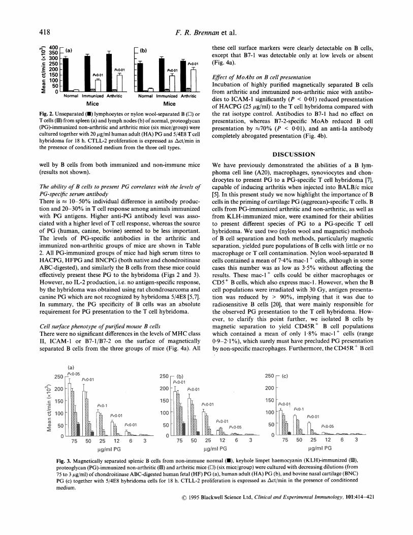
IOO 1 1 I t arthritic) could present HACPG to the T cell hybridoma at a
000 _ S 1 l relatively low (12.5 pg/ml) concentration (Fig. 2a,b). In most
D00 - Arthritis cases the responses of arthritic mice were slightly higher thanthose of immunized but non-arthritic mice, but these differ-DOO ences were not statistically significant. B cells from lymph nodes
were better at presenting the PG than splenic B cells. Presenta-ooo tion of HACPG by B cells from non-immune normal mice was
significantly (P < 0 01) lower than that of PG-immunized mice| l~a_-- ,I | ,go | (Fig. 2a,b).
0 2 4 8 12 16 20 24 28 32 Magnetically separated splenic B cells from immunized
mice could also present HFPG, HAPG and BNCPG very
16 strongly to the hybridoma over a wide range of antigen(b) FArthritis concentrations (2-5-50 ,ug/ml) (Fig. 3a-c). Responses to
14 human PG were always significantly higher (P < 0-05) than
12 those to BNCPG, independent of whether the B cells derived
10 T from HFPG, HACPG or BNC-immunized mice. Again, theresponses of immunized mice were significantly higher (P <
8 0-01) than those of both non-immune and KLH-immunized
6 + I I ^ F | | control all antigen concentrations. Unimmunized
and KLH-immunized animals could present the PG only at4 higher concentrations of antigen, suggesting that they were
2 - presenting the PG non-specifically, probably by phagocytosis[18-21]. B cells from KLH-immunized mice presented PG
0 2 4 8 12 16 20 24 28 32 significantly (P < 0 05) better than normal mice, perhapsWeek of immunization representative of an increased activation state of these B cells,
as activated B cells have a higher affinity for antigen andAeasurement of anti-proteoglycan (PG) antibodies in the sera of increased rates of phagocytosis over resting cells [18]. Thefetal PG (HFPG)-immunized BALB/c mice (a) and lymphocyte ability of B cells from PG-immunized mice, but not fromation in the presence of chondroitinase ABC-digested HFPG KLH-immunized or normal animals, to present the PG attibody titres (lines) were measured at 1:62 500 (E for HFPG) low concentrations suggests that these B cells are presenting500 (0 for mouse PG) serum dilutions using solution radio- the antigen specifically via their surface immunoglobulin)assay (RIA) [2,13]. Proliferation of T lymphocytes from spleen receptor.I joint draining lymph nodes (U) are shown as Act/min in We have previously shown that CNBr peptides of PG (but
e to chondroitin sulphate-depleted HFPG (columns). Prolifera-not native PG) can be presented by fixed APC in the absence of
to chondroitin sulphate-depleted HFPG (columns). immune processing, implying that these peptides bind directlyto MHC class II molecules on the surface of APC [7]. Insupport of this, CNBr peptides of PG were presented equally
Table 2. Proteoglycan (PG)-specific serum antibody titres in PG-immunized and control mice
Arthritic Immunized(HFPG- non-arthritic KLH- Adjuvant Normal
Groups immunized) (BNCPG-immunized) immunized only (non-immunized)
Antibodies toMouse PG 1116 ± 137 260 ± 245 ND ND NDHFPG 3862 ± 1041 2993 ±849 ND ND NDHACPG 3842 ± 977 3686 ± 566 ND ND NDBNCPG 2284 ± 266 2668 ± 442 ND ND NDCSCPG 1228 ± 228 1116 ± 327 ND ND ND
Mean serum antibody titres (Avct/min ± s.d.) measured in sera of five animals of each group are shown at the timeof antigen presentation experiments. 125I-labelled chondroitinase ABC-digested PG were calibrated to have a specificactivity of 105- 12 pCi/Mg PG. Serum dilutions of immunized mice (both arthritic and non-arthritic) were 1: 2500 formeasuring antibodies to mouse PG (first line) and 1: 62 500 in all other cases. No antibody to PG was detected (ND) inkeyhole limpet haemocyanin (KLH)-immunized, adjuvant-injected or normal mice up to 1: 100 serum concentrations.HFPG, Human fetal PG; HACPG, human adult cartilage PG; BNCPG, bovine nasal cartilage PG; CSCPG, chicksternal cartilage PG.
© 1995 Blackwell Science Ltd, Clinical and Experimental Immunology, 101:414-421
6C
50
40
30
20
10
F. R. Brennan et al.
- 350 _(a) (b).x 300 j f lr- 250 - fl P<o-0iE 200 - l Et NO1 NOo1ic100
Normal Immunized Arthritic Normal Immunized Arthritic
Mice Mice
Fig. 2. Unseparated (O) lymphocytes or nylon wool-separated B (O) or
T cells (1) from spleen (a) and lymph nodes (b) of normal, proteoglycan(PG)-immunized non-arthritic and arthritic mice (six mice/group) werecultured together with 20 jtg/ml human adult (HA) PG and 5/4E8 T cellhybridoma for 18 h. CTLL-2 proliferation is expressed as Act/min inthe presence of conditioned medium from the three cell types.
well by B cells from both immunized and non-immune mice(results not shown).
The ability ofB cells to present PG correlates with the levels ofPG-specific serum antibodyThere is I 10-50% individual difference in antibody produc-tion and 20-30% in T cell response among animals immunizedwith PG antigens. Higher anti-PG antibody level was asso-ciated with a higher level ofT cell response, whereas the sourceof PG (human, canine, bovine) seemed to be less important.The levels of PG-specific antibodies in the arthritic andimmunized non-arthritic groups of mice are shown in Table2. All PG-immunized groups of mice had high serum titres toHACPG, HFPG and BNCPG (both native and chondroitinaseABC-digested), and similarly the B cells from these mice couldeffectively present these PG to the hybridoma (Figs 2 and 3).However, no IL-2 production, i.e. no antigen-specific response,by the hybridoma was obtained using rat chondrosarcoma andcanine PG which are not recognized by hybridoma 5/4E8 [5,7].In summary, the PG specificity of B cells was an absoluterequirement for PG presentation to the T cell hybridoma.
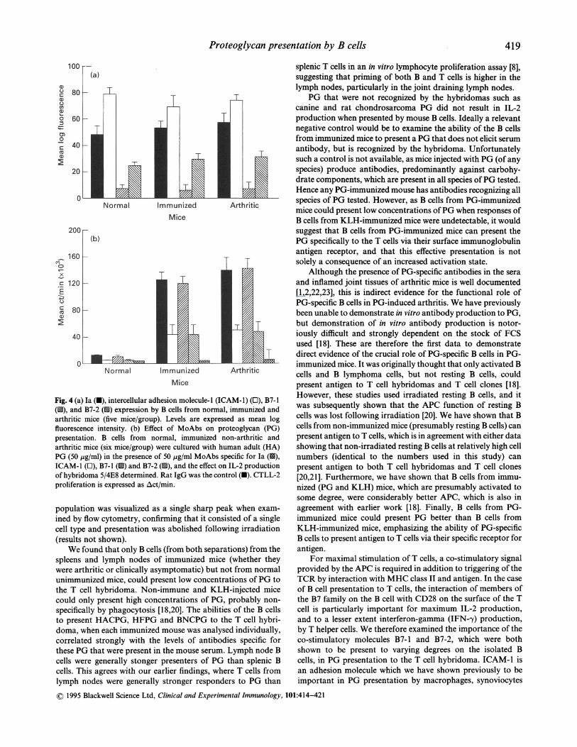
Cell surface phenotype ofpurified mouse B cellsThere were no significant differences in the levels of MHC classII, ICAM-1 or B7-1/B7-2 on the surface of magneticallyseparated B cells from the three groups of mice (Fig. 4a). All
these cell surface markers were clearly detectable on B cells,except that B7-1 was detectable only at low levels or absent(Fig. 4a).
Effect ofMoAbs on B cell presentationIncubation of highly purified magnetically separated B cellsfrom arthritic and immunized non-arthritic mice with antibo-dies to ICAM-1 significantly (P < 0-01) reduced presentationof HACPG (25 yg/ml) to the T cell hybridoma compared withthe rat isotype control. Antibodies to B7-1 had no effect onpresentation, whereas B7-2-specific MoAb reduced B cellpresentation by -70% (P < 0 01), and an anti-Ia antibodycompletely abrogated presentation (Fig. 4b).
DISCUSSION
We have previously demonstrated the abilities of a B lym-phoma cell line (A20), macrophages, synoviocytes and chon-drocytes to present PG to a PG-specific T cell hybridoma [7],capable of inducing arthritis when injected into BALB/c mice[5]. In this present study we now highlight the importance of Bcells in the priming of cartilage PG (aggrecan)-specific T cells. Bcells from PG-immunized arthritic and non-arthritic, as well asfrom KLH-immunized mice, were examined for their abilitiesto present different species of PG to a PG-specific T cellhybridoma. We used two (nylon wool and magnetic) methodsof B cell separation and both methods, particularly magneticseparation, yielded pure populations of B cells with little or nomacrophage or T cell contamination. Nylon wool-separated Bcells contained a mean of 7 4% mac-l + cells, although in somecases this number was as low as 3 5% without affecting theresults. These mac- 1 + cells could be either macrophages orCD5 + B cells, which also express mac- 1. However, when the Bcell populations were irradiated with 30 Gy, antigen presenta-tion was reduced by > 90%, implying that it was due toradiosensitive B cells [20], that were mainly responsible forthe observed PG presentation to the T cell hybridoma. How-ever, to clarify this point further, we isolated B cells bymagnetic separation to yield CD45R+ B cell populationswhich contained a mean of only 1 8% mac-l + cells (range0 9-2 1%), which surely must have precluded PG presentationby non-specific macrophages. Furthermore, the CD45R + B cell
(a)Pc-05
gn0x
c0._c
0
PcO4l
NO1
NO.o1
75 50 25 12 6 3
g/ml PG
250 -(b)P<0.01
150MO
100
50-O0
0~~~~~~~PO0
75 50 25 12 6 3
ttg/ml PG
250 r (c)
200 F
150
100
50
Fig. 3. Magnetically separated splenic B cells from non-immune normal (O), keyhole limpet haemocyanin (KLH)-immunized (O),proteoglycan (PG)-immunized non-arthritic (O) and arthritic mice (O) (six mice/group) were cultured with decreasing dilutions (from75 to 3 ,4g/ml) of chondroitinase ABC-digested human fetal (HF) PG (a), human adult (HA) PG (b), and bovine nasal cartilage (BNC)PG (c) together with 5/4E8 hybridoma cells for 18 h. CTLL-2 proliferation is expressed as Act/min in the presence of conditioned
medium.
1995 Blackwell Science Ltd, Clinical and Experimental Immunology, 101:414-421
75 50 25 . 12 6Iigtml PG
311 I I dob-L
418
Proteoglycan presentation by B cells
100
0u
c
0
S0
0,0
0
80
60
40 -
20
0
200-
160160x
c120
'800
40
ImmunizedMice
Normal ImmunizedMice
Arthritic
Fig. 4 (a) Ia (U), intercellular adhesion molecule-I (ICAM-1) (E), B7-1(u9), and B7-2 (1S) expression by B cells from normal, immunized andarthritic mice (five mice/group). Levels are expressed as mean logfluorescence intensity. (b) Effect of MoAbs on proteoglycan (PG)presentation. B cells from normal, immunized non-arthritic andarthritic mice (six mice/group) were cultured with human adult (HA)PG (50 Mig/ml) in the presence of 50 jig/ml MoAbs specific for Ia (1s),ICAM-1 (l), B7-1 (1S) and B7-2 (u), and the effect on IL-2 productionof hybridoma 5/4E8 determined. Rat IgG was the control (U). CTLL-2proliferation is expressed as Act/min.
population was visualized as a single sharp peak when exam-ined by flow cytometry, confirming that it consisted of a singlecell type and presentation was abolished following irradiation(results not shown).
We found that only B cells (from both separations) from thespleens and lymph nodes of immunized mice (whether theywere arthritic or clinically asymptomatic) but not from normalunimmunized mice, could present low concentrations of PG tothe T cell hybridoma. Non-immune and KLH-injected micecould only present high concentrations of PG, probably non-specifically by phagocytosis [18,20]. The abilities of the B cellsto present HACPG, HFPG and BNCPG to the T cell hybri-doma, when each immunized mouse was analysed individually,correlated strongly with the levels of antibodies specific forthese PG that were present in the mouse serum. Lymph node Bcells were generally stonger presenters of PG than splenic Bcells. This agrees with our earlier findings, where T cells fromlymph nodes were generally stronger responders to PG than
splenic T cells in an in vitro lymphocyte proliferation assay [8],suggesting that priming of both B and T cells is higher in thelymph nodes, particularly in the joint draining lymph nodes.
PG that were not recognized by the hybridomas such ascanine and rat chondrosarcoma PG did not result in IL-2production when presented by mouse B cells. Ideally a relevantnegative control would be to examine the ability of the B cellsfrom immunized mice to present a PG that does not elicit serumantibody, but is recognized by the hybridoma. Unfortunatelysuch a control is not available, as mice injected with PG (of anyspecies) produce antibodies, predominantly against carbohy-drate components, which are present in all species ofPG tested.Hence any PG-immunized mouse has antibodies recognizing allspecies of PG tested. However, as B cells from PG-immunizedmice could present low concentrations ofPG when responses ofB cells from KLH-immunized mice were undetectable, it wouldsuggest that B cells from PG-immunized mice can present thePG specifically to the T cells via their surface immunoglobulinantigen receptor, and that this effective presentation is notsolely a consequence of an increased activation state.
Although the presence of PG-specific antibodies in the seraand inflamed joint tissues of arthritic mice is well documented[1,2,22,23], this is indirect evidence for the functional role ofPG-specific B cells in PG-induced arthritis. We have previouslybeen unable to demonstrate in vitro antibody production to PG,but demonstration of in vitro antibody production is notor-iously difficult and strongly dependent on the stock of FCSused [18]. These are therefore the first data to demonstratedirect evidence of the crucial role of PG-specific B cells in PG-immunized mice. It was originally thought that only activated Bcells and B lymphoma cells, but not resting B cells, couldpresent antigen to T cell hybridomas and T cell clones [18].However, these studies used irradiated resting B cells, and itwas subsequently shown that the APC function of resting Bcells was lost following irradiation [20]. We have shown that Bcells from non-immunized mice (presumably resting B cells) canpresent antigen to T cells, which is in agreement with either datashowing that non-irradiated resting B cells at relatively high cellnumbers (identical to the numbers used in this study) canpresent antigen to both T cell hybridomas and T cell clones[20,21]. Furthermore, we have shown that B cells from immu-nized (PG and KLH) mice, which are presumably activated tosome degree, were considerably better APC, which is also inagreement with earlier work [18]. Finally, B cells from PG-immunized mice could present PG better than B cells fromKLH-immunized mice, emphasizing the ability of PG-specificB cells to present antigen to T cells via their specific receptor forantigen.
For maximal stimulation of T cells, a co-stimulatory signalprovided by the APC is required in addition to triggering of theTCR by interaction with MHC class II and antigen. In the caseof B cell presentation to T cells, the interaction of members ofthe B7 family on the B cell with CD28 on the surface of the Tcell is particularly important for maximum IL-2 production,and to a lesser extent interferon-gamma (IFN--y) production,by T helper cells. We therefore examined the importance of theco-stimulatory molecules B7-4 and B7-2, which were bothshown to be present to varying degrees on the isolated Bcells, in PG presentation to the T cell hybridoma. ICAM-1 isan adhesion molecule which we have shown previously to beimportant in PG presentation by macrophages, synoviocytes
© 1995 Blackwell Science Ltd, Clinical and Experimental Immunology, 101:414-421
419

420 F R. Brennan et al.
and chondrocytes [6], and has also been touted as a potentialco-stimulatory molecule. Antibodies to both ICAM- I and B7-2were strongly inhibitory, whereas B7-1-specific MoAbs had noeffect. This supports earlier data which showed that B7-2antibodies were more efficient inhibitors of T cell co-stimula-tion by activated B cells, splenocytes and dendritic cells thananti-B7-1 antibodies. These differences may simply be related tothe fact that B7-2 is expressed at higher levels on mouse B cells(both immunized and normal) than B7-1, or that B7-2 has ahigher affinity for CD28 than B7-1. We have used T cellhybridomas in our experiments, which may have co-stimula-tory requirements different from uiltransformed T cells. Indeed,there are reports of T cell hybridomas which could recognizepeptides presented by MHC molecules incorporated intoartificial membranes where no co-stimulation by APC wasrequired. However, we have shown that hybridoma 5/4E8does have co-stimulatory requirements, since antibodies toB7-2 reduce antigen presentation by mouse B cells (shown inthis study) and the mouse macrophage cell line J774 A. 1. Thus, ifthe interaction between the B cells and T cells in vivo is compar-able to that between the B cell and our hybridoma in vitro,antibodies to B7-2 may provide a useful immunotherapy toreduce B-T cooperation and therefore joint destruction in PG-induced arthritis. Indeed, inhibition of the CD28/B7 interactionin vivo prevents rejection ofxenogeneic pancreatic islets and heartgrafts, probably by reducing allogeneic T cell co-stimulation.
Although T cells probably play a critical role in theamplification of cross-reactive (auto)antibody production, thedata presented here also suggest that B cells elicited to foreignPG may initiate an autoimmune T cell response in vivo. It islikely that these PG-specific B cells in vivo take up theautoantigen (mouse PG) via their surface immunoglobulinreceptor or non-specifically by phagocytosis in the lymphnodes and spleen, process the large PG molecule and presentself-peptides to T cells. Autoreactive T cells subsequentlymigrate to and proliferate in the synovium and joint-draininglymph nodes [33], where self-peptides are present in relativelyhigh concentrations as a result of the normal turnover of thecartilage matrix and, even more, as a consequence of increasedPG degradation in inflammatory joint diseases [34]. Activatedautoreactive B and T cells in the synovium then release variouscytokines which promote proliferation of autopathogenic Tcells and provoke the release of proteolytic enzymes to degradecartilage matrix components [35-39]. Activated T cells mayalso produce IFN--y, which induces the expression of celladhesion molecules [40], hence facilitates further the invasionofinflammatory cells and up-regulates MHC class II expressionon synoviocytes [7] and promotes further generation of auto-reactive T cells.
ACKNOWLEDGMENTS
This work was supported by the NIH (AR 40310), the National andIllinois Arthritis Foundations (USA) and by the National ScienceFoundation of Research (OTKA F12912), Hungary.
REFERENCES
1 Giant TT, Mikecz K, Arzoumanian A, Poole AR. Protoglycan-induced arthritis in BALB/c mice. Clinical features and histopathol-ogy. Arthritis Rheum 1987; 30:201-12.
2 Mikecz K, Giant TT, Poole AR. Immunity to cartilage proteogly-cans in BALB/c mice with progressive polyarthritis and ankylosingspondylitis induced by injection of human cartilage proteoglycan.Arthritis Rheum 1987; 30:306-18.
3 Mikecz K, Giant TT, Buzis E, Poole AR. Cartilage proteoglycansas potential autoantigens in humans and in experimental animals.Agents Actions 1988; 23:63-66.
4 Glant TT, Mikecz K, Thonar EJA, Kuettner KE. Immuneresponses to cartilage proteoglycans in inflammatory animalmodels and human diseases. In: Woessner JF, Howell DS, eds.Cartilage degradation: basic and clinical aspects. New York: MarcelDekker, Inc. 1992: 435-73.
5 BuzAs El, Brennan FR, Negroiu G, Holl6 K, Mikecz K, Glant TT.Characterization of proteoglycan (aggrecan)-specific T cell hybri-domas. Arthritis Rheum 1994; 37:S358.
6 Brennan FR, Mikecz K, Buzis E, Glant TT. Interferon-gamma butnot granulocyte/macrophage colony-stimulating factor augmentsproteoglycan presentation by synovial cells and chondrocytes to anautopathogenic T cell hybridoma. Immunol Letters 1995; 45:87-91.
7 Brennan FR, Negroiu G, Buzas El et al. Presentation of cartilageproteoglycan to a T cell hybridoma derived from a mouse withproteoglycan-induced arthritis. Clin Exp Immunol 1995; 100:104-10.
8 Buzas E, Mikecz K, Brennan FR, Glant TT. Mediators of autop-athogenic effector cells in proteoglycan-induced arthritic and clini-cally asymptomatic BALB/c mice. Cell Immunol 1994; 158:292-304.
9 Brennan FR, Mikecz K, Buzas El, Giant TT. Humoral immuneresponses to self-proteoglycan (PG) triggers the activation ofautoreactive T cells in PG-induced arthritis in BALB/c mice.Arthritis Rheum 1994; 37:S396.
10 Lin R-H, Mamula MJ, Hardin JA, Janeway CA Jr. Induction ofautoreactive B cells allows priming of autoreactive T cells. J ExpMed 1991; 173:1433-9.
11 Male DK, Pryce G, Cooke A, Hutchings P, Marshall-Clarke S,Roitt IM. T and B cell connections in experimentally inducedautoimmunity. Ann NY Acad Sci 1986; 475:94-105.
12 Ando DG, Sercarz EE, Hahn BH. Mechanisms of T and B cellcollaboration in the in vitro production of anti-DNA antibodies inthe NZB/NZW F1 murine SLE model. J Immunol 1987; 138:3185-90.
13 Glant TT, Mikecz K, Poole AR. Monoclonal antibodies to differentprotein-related epitopes ofhuman articular cartilage proteoglycans.Biochem J 1986; 234:31-41.
14 Kappler JW, Skidmore B; White J, Marrack P. Antigen-inducible,H-2-restricted, interleukin-2-producing T cell hybridomas. J ExpMed 1981; 153:1198-214.
15 Glant TT, Mikecz K, Roughley PJ, Buzas E, Poole AR. Age-relatedchanges in protein-related epitopes of human articular-cartilageproteoglycans. Biochem J 1986; 236:71-75.
16 Miltenyi S, Muller W, Weichel W, Radbruch A. High gradientmagnetic cell separation with MACS. Cytometry 1990; 11:231-8.
17 Coffman RL, Weissman IL. B220: a B cell-specific member of theT200 glycoprotein family. Nature 1981; 289:681-3.
18 Chesnut RW, Colon SM, Grey HM. Requirements for the proces-sing of antigens by antigen-presenting B cells. I. Functionalcomparison of B cell tumors and macrophages. J Immunol 1982;129:2382-8.
19 Glimcher LH, Kim K-J, Green I, Paul WE. Ia antigen-bearing B celltumor lines can present protein antigen and alloantigen in a majorhistocompatibility complex-restricted fashion to antigen-reactive Tcells. J Exp Med 1982; 155:445-59.
20 Ashwell JD, DeFranco AL, Paul WE, Schwartz RH. Antigenpresentation by resting B cells. Radiosensitivity of the antigen-presenting function and two distinct pathways of T cell activation.J Exp Med 1984; 159:881-905.
21 Frohman M, Cowing C. Presentation of antigen by B cells:
© 1995 Blackwell Science Ltd. Clinical and Experimental Immunology, 101:414-421
Proteoglycan presentation by B cells 421
functional dependence on radiation dose, interleukins, cellularactivation, and differential glycosylation. J Immunol 1985;134:2269-75.
22 Dayer E, Mathai L, Glant TT, Mikecz K, Poole AR. Cartilageproteoglycan-induced arthritis in BALB/c mice: antibodies recog-nize human and mouse cartilage proteoglycan and can causedepletion of cartilage proteoglycan with little or no synovitis.Arthritis Rheum 1990; 33:1394-405.
23 Glant TT, Fulop C, Mikecz K, Buzds E, Molndr G, Erhardt P.Proteoglycan-specific autoreactive antibodies and T-lymphocytes inexperimental arthritis and human rheumatoid joint diseases. Bio-chem Soc Trans 1990; 18:796-9.
24 Mondino A, Jenkins MK. Surface proteins involved in T cellcostimulation. J Leuk Biol 1994; 55:805-15.
25 Jenkins MK, Pardoll DM, Mizuguchi J, Chused TM, Schwartz RH.Molecular events in the induction of a nonresponsive state in the IL-2 producing helper T lymphocyte clones. Proc Natl Acad Sci USA1987; 84:5409-13.
26 Van Seventer GA, Shimizu Y, Shaw S. Roles of multiple accessorymolecules in T-cell activation: bilateral interplay of adhesion andcostimulation. Curr Opin Immunol 1995; 3:294-303.
27 Lenschow DJ, Su H-T, Zuckerman LA et al. Expression andfunctional significance of an additional ligand for CTLA-4. ProcNatl Acad Sci USA 1993; 90:11054-8.
28 Boussiotis VA, Freeman GJ, Gribben JG, Daley J, Gray G, NadlerLM. Activated human B lymphocytes express three CTLA-4counterreceptors that costimulate T-cell activation. Proc NatlAcad Sci USA 1993; 90:11059-63.
29 Azuma M, Ito D, Yagita H et al. B70 is a second ligand for CTLA-4and CD28. Nature 1993; 366:76-79.
30 Brennan FR, Finnegan A, Mikecz K, Buzas El, Glant TT. IL-10inhibits antigen presentation by a mouse macrophage line to a T cellhybridoma by mechanisms independent of the downregulation ofB7-1. Eur J Immunol 1995; submitted.
31 Lenshow DJ, Zeng Y, Thistlewaite JR et al. Long term survival ofxenogeneic pancreatic islet grafts induced by CTLA-4 Ig. Science1992; 257:789-91.
32 Turka LA, Linsley PS, Lin H et al. T cell activation by CD28 ligandB7 is required for cardiac allograft rejection in vivo. Proc Nati AcadSci USA 1992; 89:11102-5.
33 Mikecz K, Glant TT. Migration and homing of lymphocytes tolymphoid and synovial tissues in proteoglycan-induced murinearthritis. Arthritis Rheum 1994; 37:1395-403.
34 Pettipher ER, Higgs GA, Henderson B. Interleukin 1 inducesleukocyte infiltration and cartilage proteoglycan degradation inthe synovial joint. Proc Natl Acad Sci USA 1986; 83:8749-53.
35 Kresina TF, Yoo JU, Goldberg VM. Evidence that a humoralimmune response to autologous cartilage proteoglycan can partici-pate in the induction of cartilage pathology. Arthritis Rheum 1988;31:248-57.
36 DiPasquale G. Interleukins and metalloproteinases in arthritis. In:Kresina TF, ed. Monoclonal antibodies, cytokines, and arthritis.Mediators of inflammation and therapy. New York: MarcellDekker, Inc. 1991:173-98.
37 Greenwald RA. Cartilage degradation in animal models of inflam-matory joint disease. In: Woessner JF, Howell DS, eds. Jointcartilage degradation. New York: Marcel Dekker, Inc. 1993:385-408.
38 Nietfeld JJ, Wilbrink B, Helle M et al. Interleukin-1-inducedinterleukin-6 is required for the inhibition of proteoglycan synth-esis by interleukin- 1 in human articular cartilage. Arthritis Rheum1990; 33:1695-701.
39 Shinmei M, Masuda K, Kikuchi T, Shimomura Y. Interleukin 1,tumor necrosis factor, and interleukin 6 as mediators of cartilagedestruction. Semin Arthritis Rheum 1989; 18 (Suppl. 3):27-32.
40 Mikecz K, Brennan FR, Kim JH, Glant TT. The role of adhesionmolecules in the development of autoimmune arthritis. Scand JRheumatol 1995; 24 (Suppl. 101): 91-98.
© 1995 Blackwell Science Ltd. Clinical and Experimental Immunology, 101:414-421
Copyright © 2022 FDOKUMEN