The syndecan-1 heparan sulfate proteoglycan is a viable target for myeloma therapy
35
doi:10.1182/blood-2007-04-082495 Prepublished online May 29, 2007; Sanderson Larry J Suva, Joshua Epstein, Shmuel Yaccoby, John D Shaughnessy, Jr, Bart Barlogie and Ralph D Vlodavsky, Venkataraman, Ram Sasisekharan, Annamaria Naggi, Giangiacomo Torri, Benito Casu, Israel Yang Yang, Veronica MacLeod, Yuemeng Dai, Yekaterina Khotskaya-Sample, Zachary Shriver, Ganesh myeloma therapy The syndecan-1 heparan sulfate proteoglycan is a viable target for (4217 articles) Neoplasia (790 articles) Cell Adhesion and Motility Articles on similar topics can be found in the following Blood collections http://bloodjournal.hematologylibrary.org/site/misc/rights.xhtml#repub_requests Information about reproducing this article in parts or in its entirety may be found online at: http://bloodjournal.hematologylibrary.org/site/misc/rights.xhtml#reprints Information about ordering reprints may be found online at: http://bloodjournal.hematologylibrary.org/site/subscriptions/index.xhtml Information about subscriptions and ASH membership may be found online at: digital object identifier (DOIs) and date of initial publication. the indexed by PubMed from initial publication. Citations to Advance online articles must include final publication). Advance online articles are citable and establish publication priority; they are appeared in the paper journal (edited, typeset versions may be posted when available prior to Advance online articles have been peer reviewed and accepted for publication but have not yet Copyright 2011 by The American Society of Hematology; all rights reserved. 20036. the American Society of Hematology, 2021 L St, NW, Suite 900, Washington DC Blood (print ISSN 0006-4971, online ISSN 1528-0020), is published weekly by For personal use only. by guest on June 9, 2013. bloodjournal.hematologylibrary.org From
-
Upload
independent -
Category
Documents
-
view
2 -
download
0
Transcript of The syndecan-1 heparan sulfate proteoglycan is a viable target for myeloma therapy
doi:10.1182/blood-2007-04-082495Prepublished online May 29, 2007;
SandersonLarry J Suva, Joshua Epstein, Shmuel Yaccoby, John D Shaughnessy, Jr, Bart Barlogie and Ralph D
Vlodavsky,Venkataraman, Ram Sasisekharan, Annamaria Naggi, Giangiacomo Torri, Benito Casu, Israel Yang Yang, Veronica MacLeod, Yuemeng Dai, Yekaterina Khotskaya-Sample, Zachary Shriver, Ganesh myeloma therapyThe syndecan-1 heparan sulfate proteoglycan is a viable target for
(4217 articles)Neoplasia � (790 articles)Cell Adhesion and Motility �
Articles on similar topics can be found in the following Blood collections
http://bloodjournal.hematologylibrary.org/site/misc/rights.xhtml#repub_requestsInformation about reproducing this article in parts or in its entirety may be found online at:
http://bloodjournal.hematologylibrary.org/site/misc/rights.xhtml#reprintsInformation about ordering reprints may be found online at:
http://bloodjournal.hematologylibrary.org/site/subscriptions/index.xhtmlInformation about subscriptions and ASH membership may be found online at:
digital object identifier (DOIs) and date of initial publication. theindexed by PubMed from initial publication. Citations to Advance online articles must include
final publication). Advance online articles are citable and establish publication priority; they areappeared in the paper journal (edited, typeset versions may be posted when available prior to Advance online articles have been peer reviewed and accepted for publication but have not yet
Copyright 2011 by The American Society of Hematology; all rights reserved.20036.the American Society of Hematology, 2021 L St, NW, Suite 900, Washington DC Blood (print ISSN 0006-4971, online ISSN 1528-0020), is published weekly by
For personal use only. by guest on June 9, 2013. bloodjournal.hematologylibrary.orgFrom

1
The syndecan-1 heparan sulfate proteoglycan is a viable target
for myeloma therapy
Yang Yang,1 Veronica MacLeod,2 Yuemeng Dai,2 Yekaterina Khotskaya-Sample,1
Zachary Shriver,3 Ganesh Venkataraman,3 Ram Sasisekharan,4 Annamaria Naggi,5
Giangiacomo Torri,5 Benito Casu,5 Israel Vlodavsky,6 Larry J. Suva,7 Joshua Epstein,8
Shmuel Yaccoby,8 John D. Shaughnessy, Jr.,8 Bart Barlogie8 and Ralph D. Sanderson1,8
1Department of Pathology, University of Alabama at Birmingham, Birmingham, AL,
2Department of Pathology, University of Arkansas for Medical Sciences, Little Rock,
AR, 3Momenta Pharmaceuticals, Inc., Cambridge, MA, 4Division of Bioengineering and
Environmental Health, Massachusetts Institute of Technology, Cambridge, MA, 5G.
Ronzoni Institute for Chemical and Biochemical Research, Milan, Italy, 6Cancer and
Vascular Biology Research Center, The Bruce Rappaport Faculty of Medicine, Technion,
Haifa, Israel, 7Center for Orthopedic Research, Department of Orthopedic Surgery,
University of Arkansas for Medical Sciences, 8Myeloma Institute for Research and
Therapy, University of Arkansas for Medical Sciences, Little Rock, AR
Running title: Targeting syndecan-1 (CD138) for myeloma therapy
Correspondence: Ralph D. Sanderson, Department of Pathology, University of Alabama
at Birmingham, 814 SHEL, 1530 3rd Ave. So., Birmingham, AL 35294; email:
Blood First Edition Paper, prepublished online May 29, 2007; DOI 10.1182/blood-2007-04-082495
Copyright © 2007 American Society of Hematology
For personal use only. by guest on June 9, 2013. bloodjournal.hematologylibrary.orgFrom
2
Abstract
The heparan sulfate proteoglycan syndecan-1 is expressed by myeloma cells and shed
into the myeloma microenvironment. High levels of shed syndecan-1 in myeloma patient
sera correlate with poor prognosis and studies in animal models indicate that shed
syndecan-1 is a potent stimulator of myeloma tumor growth and metastasis.
Overexpression of extracellular endosulfatases, enzymes which remove 6-O sulfate
groups from heparan sulfate chains, diminishes myeloma tumor growth in vivo. Together
these findings identify syndecan-1 as a potential target for myeloma therapy. Here three
different strategies were tested in animal models of myeloma with the following results:
(i) treatment with bacterial heparinase III, an enzyme that degrades heparan sulfate
chains, dramatically inhibited the growth of primary tumors in the SCID-hu model of
myeloma, (ii) treatment with an inhibitor of human heparanase, an enzyme that
synergizes with syndecan-1 in promoting myeloma progression, blocked the growth of
myeloma in vivo, and (iii) knockdown of syndecan-1 expression by RNAi diminished and
delayed myeloma tumor development in vivo. These results confirm the importance of
syndecan-1 in myeloma pathobiology and provide strong evidence that disruption of the
normal function or amount of syndecan-1 or its heparan sulfate chains is a valid
therapeutic approach for this cancer.
Introduction
Syndecan-1 (CD138) is a cell surface heparan sulfate-bearing proteoglycan that
was first detected by polymerase chain reaction (PCR) in mRNA from human myeloma
patients and later confirmed by monoclonal antibody staining to be present on all
For personal use only. by guest on June 9, 2013. bloodjournal.hematologylibrary.orgFrom
3
myeloma tumors.1,2 Syndecan-1 is shed from the myeloma tumor cell surface and
accumulates in the bone marrow and serum of patients. When present at high levels in
the serum, syndecan-1 is an independent indicator of poor prognosis.3-5 However, high
serum syndecan-1 is more than simply an indicator of poor prognosis. Studies in animal
models have shown that high levels of soluble syndecan-1 enhance both the growth and
metastasis of tumors.6 Syndecan-1 exerts its growth promoting effects by regulating the
activity of many effector molecules important for myeloma growth and survival
including hepatocyte growth factor (HGF) and heparin binding epidermal growth factor
(HB-EGF) family members among others.7,8 The high levels of heparan sulfate in the
tumor microenvironment resulting from syndecan-1 shedding also act as positive
regulators that condition the microenvironment for robust tumor growth. For example,
heparan sulfate binds to and promotes the activity of important angiogenic growth factors
such as fibroblast growth factor-2 (FGF-2) and vascular endothelial growth factor
(VEGF). This activity can occur in trans indicating that shed syndecan-1 can contribute
to the high level of angiogenesis seen in many myeloma patients.9,10 In addition, the
syndecan-1 that becomes embedded within the myeloma tumor stroma can serve as
reservoir for storage and concentration of heparan sulfate-binding growth factors that can
later be mobilized by cleavage of the heparan sulfate by heparanase.3,11 This
mobilization of heparan sulfate-retained growth factors in the bone marrow likely
contributes to the high rate of relapse among myeloma patients.
Because of its high level of expression on myeloma tumors, syndecan-1 has been
explored as a candidate antigen for antibody targeting of toxins to the tumor cell
surface.12-14 In addition, antibodies to syndecan-1 show promise as facilitators of
For personal use only. by guest on June 9, 2013. bloodjournal.hematologylibrary.orgFrom
4
myeloma immunotherapeutic approaches.15 However, specific strategies for disrupting
the function of syndecan-1 or its heparan sulfate chains as a potential therapy for
myeloma have not been reported. In general, targeting heparan sulfate proteoglycans as
an approach to cancer therapy has been a topic of interest among cancer researchers, but
testing of this approach in vivo has been limited.16 In a recent study, we demonstrated
that high levels of expression of the extracellular endosulfatases HSulf-1 or HSulf-2 by a
myeloma cell line attenuated tumor growth in vivo.17 These enzymes specifically remove
6-O sulfate groups from heparan sulfate chains thereby altering signaling pathways of
growth factors such as bone morphogenic protein (BMP), Wnt and FGF-2.18-20 Our
finding that enhanced HSulf expression alters heparan sulfate structure in vivo with a
resulting impact on tumor growth provided the first evidence that heparan sulfate might
be a target for myeloma therapy.
To test this idea further we utilized three distinct approaches to target heparan
sulfate or syndecan-1 in myeloma. These include: directly targeting heparan sulfate for
degradation with the enzyme bacterial heparinase III (HepIII), blocking the activity of
human heparanase, an enzyme that activates heparan sulfate and stimulates myeloma
growth and metastasis, and finally, by using shRNA to knockdown syndecan-1
expression by myeloma cells. All of these approaches, although distinct in their
mechanism of action against heparan sulfate or syndecan-1, resulted in diminished
myeloma growth in vivo. These studies confirm the importance of syndecan-1 in the
pathobiology of myeloma and provide strong evidence that targeting syndecan-1 is a
viable strategy for myeloma therapy.
For personal use only. by guest on June 9, 2013. bloodjournal.hematologylibrary.orgFrom
5
Materials and methods
Myeloma cells
Myeloma cells were obtained from bone marrow aspirates of 8 patients of the Myeloma
Institute for Research and Therapy, Little Rock, Arkansas. This work was approved by
the institutional review board of the University of Arkansas for Medical Sciences, Little
Rock, AR. Signed Institutional Review Board-approved consent forms are kept on
record. Informed consent was obtained in accordance with the Declaration of Helsinki.
The bone marrow samples were separated by Ficoll-Paque density centrifugation and the
proportion of myeloma cells present was determined by CD38/CD45 flow cytometry.
Only samples having a minimum of 25% myeloma tumor cells present were used for the
in vivo studies in SCID-hu mice. The CAG myeloma cell line was established from a
bone marrow aspirate taken from a myeloma patient as previously described.21 CAG cells
were transfected with empty vector only or vector containing the cDNA for human
heparanase to generate the CAG heparanase-low and CAG heparanase-high cells,
respectively. The methods for transfection and characterization of these cells have been
previously published.22
In vivo models of myeloma
The SCID-hu model was constructed as previously described.6,23,24 Briefly, 5-6 week old
male CB.17 scid/scid mice were obtained from Harlan Sprague Dawley, Inc.
(Indianapolis, IN) and were housed and monitored in the animal facility at the University
of Arkansas for Medical Sciences or the University of Alabama at Birmingham. All
experimental procedures and protocols were approved by the Institutional Animal Care
For personal use only. by guest on June 9, 2013. bloodjournal.hematologylibrary.orgFrom

6
and Use Committee’s of the respective institutions. Human femora were cut into halves
(approximately 5 × 5 × 10 mm) and implanted subcutaneously into each SCID mouse. Six
to eight weeks after implantation of bone, 1 x 10⁷ cells of a bone marrow aspirate from a
myeloma patient were injected directly into the marrow cavity of the bone implanted in
the SCID-hu host. Murine sera were collected weekly and the levels of human
immunoglobulin light chain (either kappa or lambda depending on the patient) were
assessed as previously described as an indicator of tumor burden.6 Treatment was begun
when human light chain present in the mouse serum reached a level, on average, of 36
µg/ml. Treatment with HepIII (15 mg/kg/day) or HepIII-generated fragments of heparan
sulfate (0.8 mg/kg/day) was accomplished by twice daily injections, one intraperitoneal
and one subcutaneous in the proximity of the human bone implant. For the subcutaneous
model, 1 x 10⁶ CAG cells were injected subcutaneously into the left flank of each mouse.
For animals being treated with the modified heparin 100NA,RO-H,25 ten days after
injection of tumor cells, Alzet osmotic pumps were implanted on the right flank of each
mouse. Pumps contained either the 100NA,RO-H at the indicated concentration or PBS as
a control and the solution was delivered continuously for 28 days. At the completion of
the treatment period, animals were killed and the wet weight of the subcutaneous tumors
and the mean sera kappa level among the experimental groups compared by the log-rank
test (P ≤ .05 was considered statistically significant). For experiments with the syndecan-
1 knockdown cells, 1 x 10⁶ CAG control shRNA or syndecan-1 shRNA infected cells
were injected subcutaneously and monitored for levels of kappa light chain present in the
mouse serum. The experiment was terminated eight weeks after injection of the tumor
cells.
For personal use only. by guest on June 9, 2013. bloodjournal.hematologylibrary.orgFrom

7
Preparation of recombinant HepIII, heparan sulfate fragments and
100NA,RO-H
Recombinant HepIII was expressed, purified to homogeneity and cleared of endotoxins
as described.26,27 Fragments of heparan sulfate generated by HepIII were collected by
treating partially purified syndecan-1 that was isolated from the culture media of CAG
cells overexpressing a form of syndecan-1 lacking its cytoplasmic and transmembrane
domain, thus mimicking shed syndecan-1.28 Syndecan-1 (200 µg) in PBS was treated
with HepIII (20 µg) at 370C for 1h followed by boiling for 15 min. Following sterile
filtration, the concentration of heparan sulfate was determined by disaccharide analysis.
Preparation and characterization of the heparanase inhibitor, 100NA,RO-H (100% N-
acetylated and 25% glycol-split heparin), was previously described in detail.25
Knockdown of syndecan-1 expression with shRNA
We utilized a sequence previously shown effective in siRNA knockout of syndecan-1
(1162AGGAGGAATTCTATGCCTGA1181)29 to design a synthetic double-stranded
oligonucleotide sequence for shRNA knockdown studies; (5’-
CGCGTCCCCGGAGGAATTCTATGCCTGATTCAAGAGATCAGGCATAGAATTC
CTCCTTTTTGGAAAT- 3’). A control oligonucleotide sequence not matching any
sequence in the human genome (5’ – AATTCTCCGAACGTGTCACGT – 3’; Qiagen,
Inc., Valencia, CA)30 was used to design a control shRNA sequence (5’-
CGCGTCCCCGTCTCCGAACGTGTCACGTTTCAAGAGAACGTGACACG
TTCGGAGACTTTTTGGAAAT-3’). Both double-stranded shRNA sequences were
obtained from Integrated DNA Technologies (Coralville, IA). The double-stranded
For personal use only. by guest on June 9, 2013. bloodjournal.hematologylibrary.orgFrom
8
oligonucleotides were cloned into pLVTHM, and virus generated by co-transfection of
293FT cells with the pLVTHM vector and helper plasmids pMD2G and pCMV-dR8.91
(all kindly provided by Dr. Didier Trono, Univ. of Geneva, Switzerland). The crude
lentivirus was filtered (0.45µ) and viral titers determined by measuring the percent of
green fluorescence protein (GFP) positive cells present 48 h after infection of CAG cells.
Stable cell lines expressing shRNA were achieved by infection at 50 MOI. GFP positive
CAG cells were sorted two times after which cells were over 99% positive. The
reduction of syndecan-1 expression was confirmed by RT-PCR, flow cytometry and by
Western blotting. For PCR the forward and reverse primers were 5'-
CTTCACACTCCCCACACAGA-3' (forward) and 5'-TCCTGTTTGGTGGGCTTCTG-3'
(reverse) for syndecan-1 and 5'-ACCACAGTCCATGCCCATCAC-3' (forward) and 5'-
TCCACCACCCTGTTGCTGTA-3' (reverse) for glyceraldehyde-3-phosphate
dehydrogenase (GAPDH). After initial denaturation at 95°C for 2 minutes, PCR was
carried out for 32 cycles (95 °C for 1 min; 58 °C for 1 min; and 72 °C for 1 min). PCR
products were separated by 1.2% agarose gel electrophoresis and visualized by ethidium
bromide staining. The size of PCR product was 396 bp for syndecan-1 and 452 bp for
GAPDH, respectively. For flow cytometry, cells were stained with antibody B-A38
(Serotec, Raleigh, NC) followed by anti-mouse conjugated to Alexa 647 (Invitrogen,
Carlsbad, CA). For Western blotting, cell extracts were separated on 4-15% gradient
SDS-PAGE, transferred to Nytran+ filters (Whatman/Schleicher & Schuell, Florham
Park, NJ) probed with antibody B-A38 followed by a horseradish peroxidase-conjugated
secondary anti-mouse antibody and visualized by chemiluminescence (GE Healthcare,
Pittsburgh, PA).
For personal use only. by guest on June 9, 2013. bloodjournal.hematologylibrary.orgFrom
9
Micro-computed tomography (MicroCT) analysis of bones
Upon termination of the experiment, the implanted human bones were removed from
SCID-hu mice and fixed in phosphate-buffered 10% formalin, pH 7.4 for a minimum of
24 h. Bones were then dehydrated successively in 70%, 95%, and 100% ethanol and
analyzed using a microCT 40 (Scanco Medical, Bassersdorf, Switzerland) using a voxel
size of 12 µm in all dimensions, as described previously31,32 Briefly, the region of
interest selected for analysis was the entire excised specimen and comprised
approximately 1000 transverse CT slices. Three dimensional reconstructions were
created by stacking the regions of interest from each 2-dimensional slice then applying a
grey-scale threshold and Gaussian noise filter specifically optimized for human bone.
Results
Treatment with HepIII inhibits growth of primary myeloma tumors in vivo
When administered in vivo, the heparan sulfate degrading enzyme bacterial
heparinase III has been shown to inhibit the growth and metastasis of both melanoma and
lung carcinoma tumors in animal models.26 This effect is thought to be due to HepIII-
mediated release of heparan sulfate fragments that possess anti-tumor activity. Because
of the abundant heparan sulfate in the myeloma microenvironment due to the presence of
both cell surface and shed syndecan-1, we tested the effects of this enzyme on established
myeloma tumors. Daily injections of HepIII into the animals bearing subcutaneous
For personal use only. by guest on June 9, 2013. bloodjournal.hematologylibrary.orgFrom
10
myeloma tumors substantially inhibited tumor growth (data not shown). To extend these
studies we established tumors by injecting cells from the bone marrow of myeloma
patients into human bones implanted in SCID mice (SCID-hu model).23 Once
engraftment of the human tumor was confirmed by detection of human light chain in the
serum of the mice, animals were treated by injection of HepIII (15 mg/kg/day). At the
end of the treatment period light chain levels, as a measure of tumor burden, were
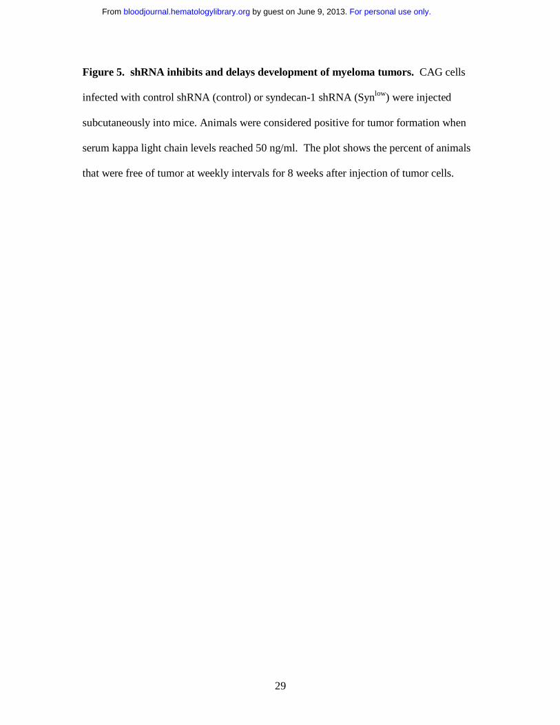
assessed. As expected, among the 8 patient tumors tested there is considerable variability
in rates of tumor growth (Fig. 1A). However, in all cases, tumors growth was attenuated
in animals treated with HepIII. In the case of three tumors, levels of light chain were
lower than at the start of treatment, suggesting that tumor growth was arrested by the
HepIII treatment (patients 3, 4 and 5). Even the most aggressive tumor which exhibited a
700% increase in light chain level was substantially retarded in its growth by treatment
with HepIII (patient 2). The most dramatic response was seen in the tumor established
from patient 5, where light chain levels increased almost 500% in the control animal but
were decreased by 58% in the HepIII treated animal (Fig. 1B).
As an alternative to treating tumors with the HepIII enzyme, we prepared fragments of
heparan sulfate ex vivo by treating syndecan-1 isolated from CAG myeloma cells with
HepIII.26 The fragments released by the enzyme were harvested and then injected into
SCID-hu animals bearing myeloma patient tumor. Although not as potent as the enzyme
itself, these heparan sulfate fragments exhibited significant anti-tumor activity (Fig. 1A).
When the increases in tumor burden from all eight patients were combined, treatment
with either HepIII or the HepIII-generated heparan sulfate fragments significantly
inhibited the growth of the primary myeloma tumors as compared to controls (Fig. 1C).
For personal use only. by guest on June 9, 2013. bloodjournal.hematologylibrary.orgFrom

11
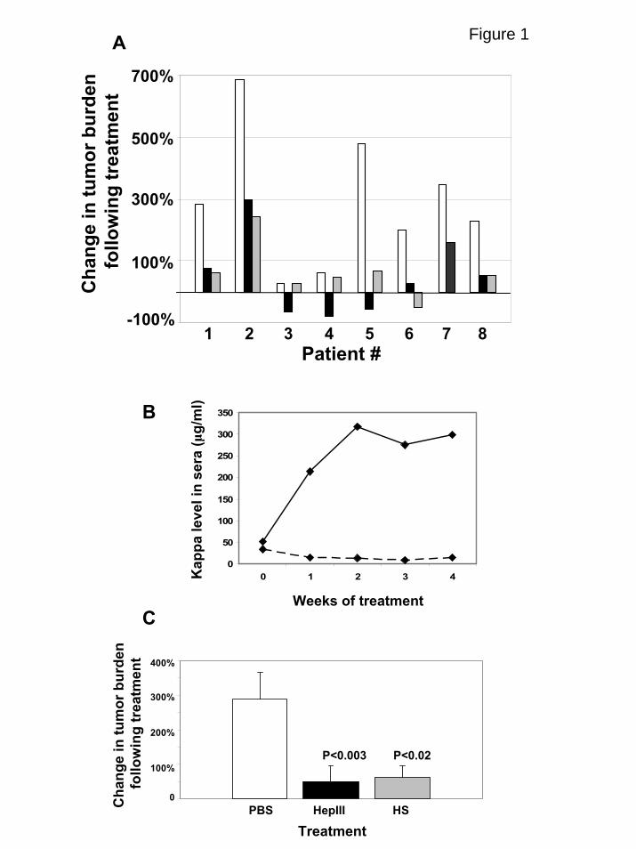
At termination of the experiment, microCT scanning of the human bones
harvested from animals reveals protection by HepIII from the bone destruction associated
with myeloma tumor growth (Fig. 2). Not all patient tumors induced the same extent of
osteolysis as the one shown in Fig. 2, but in several cases when there was significant
osteolysis in contols, in comparison, degradation was diminished in bones harvested from
the HepIII-treated animals.
Inhibition of myeloma growth in vivo by an inhibitor of heparanase
As a second approach to test targeting of heparan sulfate in myeloma tumors we
used an inhibitor of human heparanase. Heparanase is distinct in its action from that of
bacterial HepIII and in contrast to the anti-tumor activity of HepIII, human heparanase
has been shown to promote the growth and metastasis of many types of human tumors.11
In myeloma patients, enzymatically active heparanase is present in the bone marrow and
is associated with a high microvessel density and poor prognosis.33,34 Overexpression of
heparanase in CAG myeloma cells substantially enhances their growth and spontaneous
metastasis to bone.22 In addition, heparanase increases the synthesis and shedding of
syndecan-1 by myeloma cells thereby contributing to myeloma progression by elevating
levels of syndecan-1 in the tumor microenvironment.34,35 Thus, we reasoned that
inhibitors of heparanase activity may have a dramatic impact on the growth of myeloma
tumors in vivo. Recently, a series of chemically modified heparins were shown to be
potent inhibitors of heparanase in vitro.25 An important property of these modified
heparins is that they lack anti-coagulant activity and thus can be administered in vivo at
relatively high doses. We chose to use a modified heparin designated 100NA,RO-H that is
For personal use only. by guest on June 9, 2013. bloodjournal.hematologylibrary.orgFrom
12
100% N-acetylated and 25% glycol-split. At a concentration of 0.2 µg/ml this compound
inhibits 93.8% of heparanase activity but does not displace extracellular matrix-bound
FGF-2 or potentiate its growth promoting activity.25
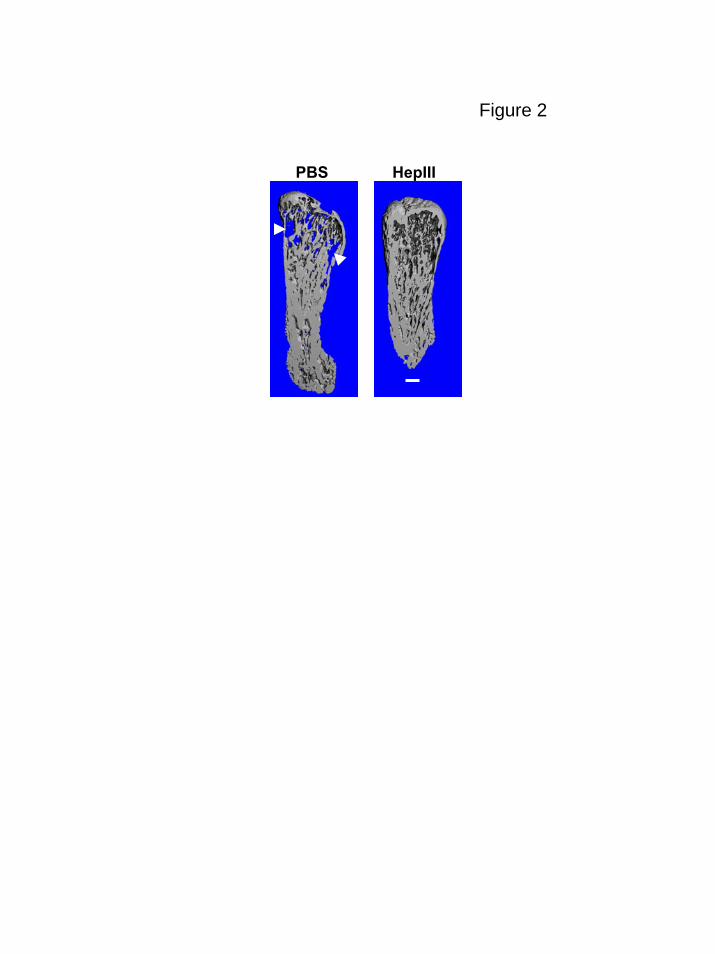
Two experiments testing the effectiveness of this compound were performed, one
utilizing CAG myeloma cells expressing high levels of heparanase and one utilizing CAG
myeloma cells expressing low levels of heparanase. We have previously demonstrated
that the CAG cells expressing high levels of heparanase exhibit very aggressive growth
and metastatic behavior in vivo as compared to control cells that express low levels of the
enzyme.22 For the present studies we used a subcutaneous tumor model similar to those
that have been used extensively for testing new therapeutics for their efficacy against
myeloma.36-38 Tumor cells were injected subcutaneously at a single site in each mouse.
After allowing ten days for tumors to establish, Alzet osmotic pumps were inserted into
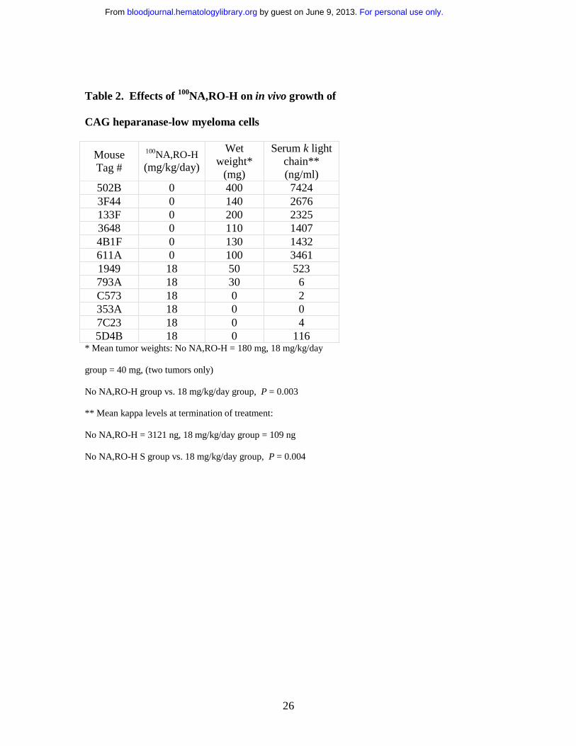
animals to deliver either PBS or the 100NA,RO-H for 28 days. Results demonstrate a
concentration dependent anti-tumor effect of the modified heparin on both the
heparanase-high and heparanase-low cells (Fig. 3 and Tables 1 and 2). At the 36
mg/kg/day concentration tested in the very aggressive heparanase-high cells, tumor was
found in only one of the six animals. The levels of serum kappa light chain demonstrated
that in addition to the absence of tumor at the subcutaneous site, there was not
widespread dissemination of tumor to other sites.
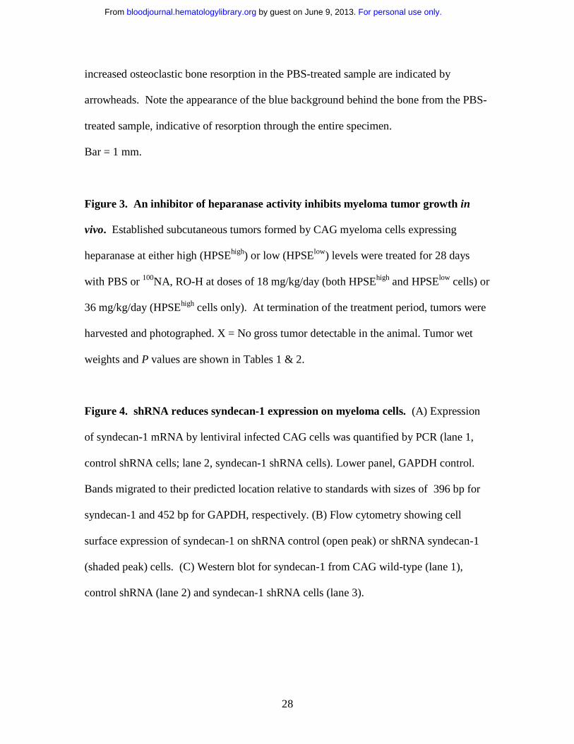
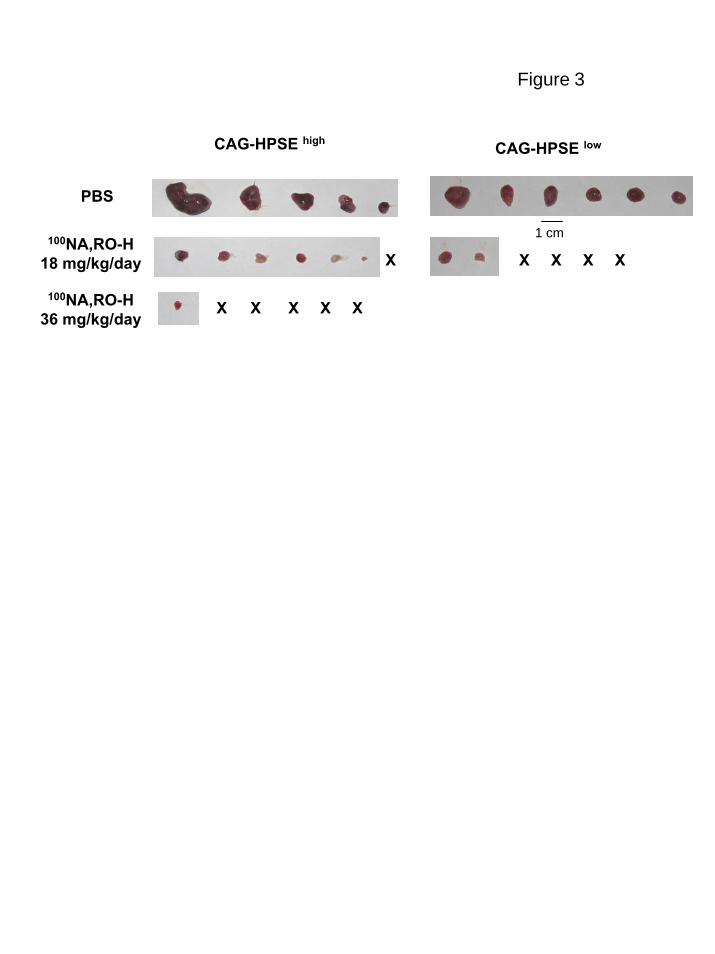
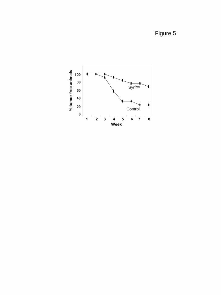
Myeloma cells with reduced expression of syndecan-1 grow poorly in vivo
As final confirmation that syndecan-1 is a viable target for myeloma therapy, we
used an shRNA to knockdown expression of syndecan-1 in the CAG myeloma cell line.
For personal use only. by guest on June 9, 2013. bloodjournal.hematologylibrary.orgFrom

13
We hypothesized that a reduction in syndecan-1 expression would diminish the level of
heparan sulfate at the cell surface and in the tumor microenvironment. A ten-fold
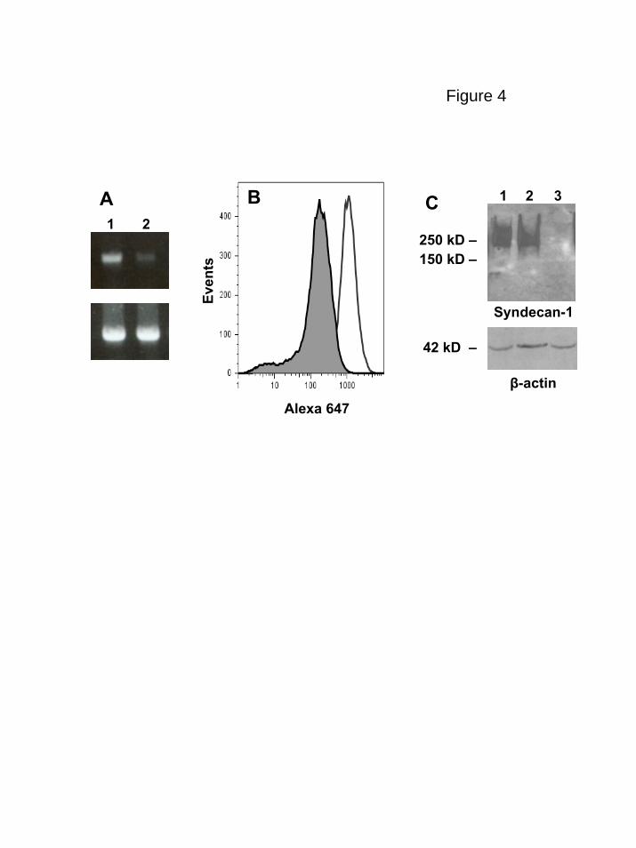
reduction in the level of cell surface syndecan-1 was demonstrated by flow cytometric
analysis in the knockdown cells as compared to controls and on Western blots syndecan-
1 from the knockdown cells was below the level of detection (Fig. 4). When injected
subcutaneously into SCID mice, cells having diminished levels of syndecan grew poorly.
In the first experiment, eight weeks after injection of cells having low syndecan-1
expression none of the animals formed tumors at the site of inoculation (0/6 animals
developed tumor, mean kappa light chain level = 0). In contrast, controls expressing
normal levels of syndecan-1 formed tumors (4/6 animals developed tumor, mean kappa
light chain level = 2533 ng/ml ). In a second experiment, four of the seven animals
bearing cells with syndecan-1 knockdown did develop tumors but the onset of tumor
development was delayed as compared to controls (data not shown). Figure 5 include
data from both experiments and reflects the overall decrease and delay in tumor
development in the cells expressing low levels of syndecan-1 as compared to controls.
Interestingly, at termination of the second experiment, when tumors were removed from
animals and immunostained for syndecan-1 expression, those formed from cells in which
syndecan-1 expression had been knocked down exhibited strong expression of syndecan-
1 (data not shown). Because the antibody used for staining syndecan-1 is human specific,
this result indicates that the level of syndecan-1 expression eventually increased in the
knockdown cells to relatively high levels.
For personal use only. by guest on June 9, 2013. bloodjournal.hematologylibrary.orgFrom
14
Discussion
The data presented here demonstrate that myeloma growth is substantially
inhibited in vivo by approaches that target either heparan sulfate or syndecan-1. In
addition, our previously published work shows that modulation of the sulfation pattern
within the heparan sulfate chains expressed by myeloma cells can also inhibit tumor
growth.17 Collectively, these results provide strong proof of principle supporting the idea
that targeting of heparan sulfate or syndecan-1 within the tumor microenvironment is a
viable therapeutic approach for myeloma.
HepIII is an enzyme whose activity generates heparan sulfate fragments with anti-
tumor activity.26 Myeloma may be a particularly good target for this enzyme because of
the high levels of syndecan-1 heparan sulfate at the cell surface and shed into the tumor
microenvironment. The activity of the enzyme on tumor-associated heparan sulfate
would likely impact the tumor in two ways. First, as mentioned above, anti-tumor
heparan sulfate fragments would be generated. Second, because syndecan-1 with its
heparan sulfate chains is a promoter of tumor growth and metastasis in vivo6, its
degradation by HepIII would diminish the positive growth effects of the proteoglycan.
Importantly, our studies here employed primary myeloma tumor cells growing in human
bone suggesting that HepIII can exert its anti-myeloma effect even on an established
tumor growing in a microenvironment where the tumor normally thrives. Growth
inhibition was also accomplished by treating animals with heparan sulfate fragments
generated by HepIII indicating that in this myeloma model the fragments have direct anti-
For personal use only. by guest on June 9, 2013. bloodjournal.hematologylibrary.orgFrom
15
tumor activity or that they are competing with the endogenous tumor heparan sulfate
thereby blocking their ability to promote tumor growth. Whatever the mode of action,
these findings indicate that the ex vivo identification and characterization of the specific
fragments of heparan sulfate having anti-tumor activity could provide a powerful new
therapeutic tool and circumvent the problems associated with utilizing the HepIII enzyme
itself as a therapy.
The finding that 100NA,RO-H, an engineered heparin-based compound having no
anti-coagulant activity can dramatically inhibit heparanase activity and myeloma growth
in vivo provides yet another proteoglycan-based approach to treat this cancer. It is
noteworthy that in these studies the highly aggressive CAG cell line was utilized. Like
many other myeloma cell lines, CAG cells cluster with the most aggressive of myeloma
tumors as assessed by gene profiling.39 In addition, these cells are capable of growing
outside the bone marrow microenvironment, a characteristic similar to many highly
aggressive late stage clinical myeloma tumors. In addition, to challenge the effectiveness
of the anti-heparanase compound, we utilized CAG cells overexpressing heparanase.
These cells exhibit an enhanced ability to grow and spontaneously metastasize to bone.22
In spite of this overexpression of heparanase, 100NA,RO-H was a potent inhibitor of CAG
tumor growth and metastasis and significantly inhibited tumor growth even at the lower
concentration tested (18 mg/kg/day). At the higher concentration tested (36 mg/kg/day),
tumor was found in only one animal of the six that were treated. Interestingly, in animals
bearing tumors formed by the heparanase-low CAG cells, the 18 mg/kg/day
concentration of 100NA,RO-H had a greater anti-tumor effect than it did on the
heparanase-high cells (Fig.3, Tables 1 and 2). This suggests that the effect of 100NA,RO-
For personal use only. by guest on June 9, 2013. bloodjournal.hematologylibrary.orgFrom

16
H is related to its anti-heparanase activity. Moreover, it indicates that this compound can
be effective against tumors that express relatively low levels of the enzyme. Importantly,
the treated animals showed no sign of adverse side effects even though the modified
heparin was delivered constantly and at high concentrations for 28 days.
Due to the correlation of heparanase expression with aggressive behavior in many
tumor types, it has been widely speculated that heparanase represents an important
therapeutic target.11,40-42 Numerous compounds with anti-heparanase activity have been
identified, and one, PI-88 is currently in clinical trials.43-45 The inhibitory action of
100NA, RO-H is thought to occur due to its tight binding to heparanase facilitated by the
enhanced flexibility of the heparin afforded by the glycol-splitting which opens the sugar
ring. In addition, once bound, the heparanase cannot cleave these heparins. The modified
heparins such as the one used here provide several advantages as potential therapeutics.
Heparin is readily available and there is a considerable amount of information regarding
its safety in humans. Chemical modifications can be controlled and analytical techniques
to verify its composition are well established thereby making scale-up production for
human therapeutics feasible.25
Knockdown of syndecan-1 expression using shRNA dramatically diminished the
ability of CAG cells to form subcutaneous tumors in vivo. This is particularly important
because the ability of cell lines to grow outside of the bone marrow microenvironment
reflects a robust propensity for growth and survival. Thus, the poor growth of these cells
in vivo following a 10-fold reduction of syndecan-1 expression suggests that even very
aggressive myeloma tumors need syndecan-1 for their growth in vivo. Interestingly, and
in contrast to our findings in vivo, the in vitro growth rate of the CAG cells was not
For personal use only. by guest on June 9, 2013. bloodjournal.hematologylibrary.orgFrom
17
affected by diminished syndecan-1 expression (data not shown). These data suggest that
the poor growth of low syndecan-1 expressers in vivo is related to the inability of the cells
to condition the tumor microenvironment in a manner that supports tumor survival and
subsequent growth. Due to the role of heparan sulfate as a co-receptor for pro-angiogenic
growth factors such as VEGF and FGF-2, the poor growth of tumors with reduced
syndecan-1 expression could reflect a poor angiogenic response of the host to the tumor.
The observation that syndecan-1 levels are relatively high in tumors that
eventually do grow out from the syndecan-1 knockdown cells is also important. This
suggests that after injection of the tumor cells, they either lose expression of the shRNA
or that a subpopulation of the knockdown cells that retained relatively high levels of
syndecan-1 expression are able to grow preferentially and eventually form tumors. It has
been demonstrated that a small population of syndecan-1 negative cells is present in
myeloma cell lines and clinical material and that these negative cells have greater
clonogenic potential than syndecan-1 positive cells.46 In light of that finding, it is
interesting that in our model, the syndecan-1 negative population within the CAG
knockdown population does not emerge as the predominant cell type in the tumors that
do form. One possibility is that the syndecan-1 negative cells do seed the tumor and
eventually shift to a syndecan-1 positive phenotype. However, the fact that many of the
animals injected with syndecan-1 knockdown cells do not form tumors at all suggests that
in the CAG cell line the syndecan-1 negative cell population does not have the potential
to form tumors in vivo.
Another possibility that would explain the delayed growth of the tumors forming
from syndecan-1 knockdown cells is that over time enough shed syndecan-1 accumulates
For personal use only. by guest on June 9, 2013. bloodjournal.hematologylibrary.orgFrom
18
in the tumor microenvironment to promote tumor growth. Although a reduction in
heparan sulfate levels within the tumor due to diminished syndecan-1 expression likely
has important effects, we can not rule out the possibility that the syndecan-1 core protein
also plays a role in promoting tumor growth. Interestingly, the core protein of syndecan-
1 was recently shown to regulate activation of alpha v beta 3 and alpha v beta 5 integrins
which act as regulators of myeloma cell invasion and angiogenesis.29,47-49
Why does targeting syndecan-1 or enzymes that modify syndecan-1 heparan
sulfate have such a dramatic impact on myeloma tumor growth and progression? Our
working hypothesis is that syndecan-1 acts as a master regulator that promotes the
activity of many signaling molecules within the tumor microenvironment. Via these
multiple regulatory activities, syndecan-1 drives robust tumor growth and progression.
For example, hepatocyte growth factor (HGF), a growth factor known to be upregulated
in many myeloma tumors,39 binds to the heparan sulfate of syndecan-1 and helps
potentiate signaling via the cMET receptor with resulting cell proliferation.7 In addition
to its activity on the myeloma cell surface, syndecan-1 is shed from myeloma tumor cells
and can accumulate in the tumor microenvironment.3 High levels of shed syndecan-1
correlate with poor patient prognosis and animal studies have demonstrated that myeloma
tumor growth and dissemination are enhanced when levels of shed syndecan-1 are
increased. 4-6 The growth stimulatory effects of shed syndecan-1 are likely due to
numerous interactions of the proteoglycan within the microenvironment. Heparan sulfate
is known to regulate the activity of angiogenic growth factors such as FGF-2 and VEGF
and we have found that myeloma cells engineered to express high levels of soluble
syndecan-1 form tumors having an elevated microvessel density as compared to controls
For personal use only. by guest on June 9, 2013. bloodjournal.hematologylibrary.orgFrom
19
(unpublished data). This is consistent with published work showing a positive correlation
between high levels of syndecan-1 in the serum of myeloma patients and elevated tumor
microvessel density.50 The heparan sulfate chains of syndecan-1 may also regulate bone
turnover in myeloma due to their interaction with OPG and DKK, two heparin-binding
molecules that are important modulators of myeloma bone disease.51-53 Thus, the heparan
sulfate proteoglycans may aid in molecular events that trigger increased bone turnover
which further stimulates tumor growth. Collectively, these and other pathways regulated
by syndecan-1 and/or heparan sulfate point to a major role for syndecan-1 in regulating
myeloma tumor growth and progression.
Although syndecan-1 is the most abundant source of heparan sulfate in the
myeloma tumor microenvironment, there are other heparan sulfate proteoglycans present.
For example, endothelial cells have been shown to express multiple heparan sulfate
proteoglycans that participate in regulating the angiogenic response.54,55 Using an
antisense RNA approach, specific targeting of perlecan, the basement membrane heparan
sulfate proteoglycan, was effective in blocking growth and angiogenesis of both colon
carcinoma and melanoma.56 An advantage of the approach we use here with both HepIII
and anti-heparanase strategies is that they would target not only syndecan-1, but other
heparan sulfates or heparan sulfate proteoglycans present in the myeloma
microenvironment.
The finding that multiple strategies targeting the syndecan-1/heparan
sulfate/heparanase axis are effective against myeloma provides strong proof of principle
for the approach. Because there is a high degree of genetic chaos within most myeloma
tumors, agents that target a single signaling pathway may have limited therapeutic value.
For personal use only. by guest on June 9, 2013. bloodjournal.hematologylibrary.orgFrom

20
The concept of targeting a master regulator of multiple signaling pathways such as
syndecan-1 and its heparan sulfate chains may be especially effective against myeloma
and thus provides a new therapeutic avenue for attacking this cancer.
Acknowledgements
The authors thank Dr. Didier Trono, University of Geneva, Switzerland, for plasmids
used to construct lentiviral vectors. This work was supported in part by grant P01
CA55819 (BB, JE, JS, RS) and CA 103054 (RDS) from the National Cancer Institute.
Authorship
Contribution: YY designed and performed the research and analyzed and interpreted the
data; VM prepared constructs, transfected cell lines and assisted with animal studies; YD
prepared shRNA constructs, infected cells and performed shRNA animal studies; YS
performed shRNA animal studies; ZS, GV, RSasisekharan provided recombinant HepIII
and assisted in design of HepIII-related experiments; AN, GT, BC provided 100NA,RO-H
and assisted in design of related experiments; IV characterized the anti-heparanase
activity of 100NA,RO-H; LJS performed micoCT analysis of bones; JE provided clinical
material; SY provided assistance and advice related to the SCID-hu model; JDS provided
clinical material; BB provided clinical material and clinical data; RSanderson
conceptualized and designed the research, analyzed and interpreted the data and wrote the
paper.
For personal use only. by guest on June 9, 2013. bloodjournal.hematologylibrary.orgFrom
21
Conflict of interest disclosure:
Two of the authors (ZS, GV) are employed by a company whose potential product was
studied in the present work. One of the authors (RSasisekharan) has declared a financial
interest in a company whose potential product was studied in the present work.
References
1. Ridley RC, Xiao HQ, Hata H, Woodliff J, Epstein J, Sanderson RD. Expression of syndecan regulates human myeloma plasma cell adhesion to type I collagen. Blood. 1993;81:767-774.
2. Wijdenes J, Vooijs WC, Clement C, et al. A Plasmocyte Selective Monoclonal Antibody (B-B4) Recognizes Syndecan-1. Br J Haematol. 1996;94:318-323.
3. Bayer-Garner IB, Sanderson RD, Dhodapkar MV, Owens RB, Wilson CS. Syndecan-1 (CD138) immunoreactivity in bone marrow biopsies of multiple myeloma: shed syndecan-1 accumulates in fibrotic regions. Mod Pathol. 2001;14:1052-1058.
4. Dhodapkar MV, Kelly T, Theus A, Athota AB, Barlogie B, Sanderson RD. Elevated levels of shed syndecan-1 correlate with tumour mass and decreased matrix metalloproteinase-9 activity in the serum of patients with multiple myeloma. Br J Haematol. 1997;99:368-371.
5. Seidel C, Sundan A, Hjorth M, et al. Serum syndecan-1: a new independent prognostic marker in multiple myeloma. Blood. 2000;95:388-392.
6. Yang Y, Yaccoby S, Liu W, et al. Soluble syndecan-1 promotes growth of myeloma tumors in vivo. Blood. 2002;100:610-617.
7. Derksen PW, Keehnen RM, Evers LM, van Oers MH, Spaargaren M, Pals ST. Cell surface proteoglycan syndecan-1 mediates hepatocyte growth factor binding and promotes Met signaling in multiple myeloma. Blood. 2002;99:1405-1410.
8. Mahtouk K, Cremer FW, Reme T, et al. Heparan sulphate proteoglycans are essential for the myeloma cell growth activity of EGF-family ligands in multiple myeloma. Oncogene. 2006;25:7180-7191.
9. Filla MS, Dam P, Rapraeger AC. The cell surface proteoglycan syndecan-1 mediates fibroblast growth factor-2 binding and activity. J Cell Physiol. 1998;174:310-321.
10. Jakobsson L, Kreuger J, Holmborn K, et al. Heparan sulfate in trans potentiates VEGFR-mediated angiogenesis. Dev Cell. 2006;10:625-634.
11. Ilan N, Elkin M, Vlodavsky I. Regulation, function and clinical significance of heparanase in cancer metastasis and angiogenesis. Int J Biochem Cell Biol. 2006;38:2018-2039.
12. Post J, Vooijs WC, Bast BJ, De Gast GC. Efficacy of an anti-CD138 immunotoxin and doxorubicin on drug-resistant and drug-sensitive myeloma cells. Int J Cancer. 1999;83:571-576.
For personal use only. by guest on June 9, 2013. bloodjournal.hematologylibrary.orgFrom
22
13. Ragnarsson L, Stromberg T, Wijdenes J, Totterman TH, Weigelt C. Multiple myeloma cells are killed by syndecan-1-directed superantigen-activated T cells. Cancer Immunol Immunother. 2001;50:382-390.
14. Tassone P, Goldmacher VS, Neri P, et al. Cytotoxic activity of the maytansinoid immunoconjugate B-B4-DM1 against CD138+ multiple myeloma cells. Blood. 2004;104:3688-3696.
15. Dhodapkar KM, Krasovsky J, Williamson B, Dhodapkar MV. Antitumor monoclonal antibodies enhance cross-presentation of cellular antigens and the generation of myeloma-specific killer T cells by dendritic cells. J Exp Med. 2002;195:125-133.
16. Fjeldstad K, Kolset SO. Decreasing the metastatic potential in cancers--targeting the heparan sulfate proteoglycans. Curr Drug Targets. 2005;6:665-682.
17. Dai Y, Yang Y, MacLeod V, et al. HSulf-1 and HSulf-2 are potent inhibitors of myeloma tumor growth in vivo. J Biol Chem. 2005;280:40066-40073.
18. Ai X, Do AT, Lozynska O, Kusche-Gullberg M, Lindahl U, Emerson CP, Jr. QSulf1 remodels the 6-O sulfation states of cell surface heparan sulfate proteoglycans to promote Wnt signaling. J Cell Biol. 2003;162:341-351.
19. Wang S, Ai X, Freeman SD, et al. QSulf1, a heparan sulfate 6-O-endosulfatase, inhibits fibroblast growth factor signaling in mesoderm induction and angiogenesis. Proc Natl Acad Sci U S A. 2004;101:4833-4838.
20. Viviano BL, Paine-Saunders S, Gasiunas N, Gallagher J, Saunders S. Domain-specific modification of heparan sulfate by Qsulf1 modulates the binding of the bone morphogenetic protein antagonist Noggin. J Biol Chem. 2004;279:5604-5611.
21. Borset M, Hjertner O, Yaccoby S, Epstein J, Sanderson RD. Syndecan-1 is targeted to the uropods of polarized myeloma cells where it promotes adhesion and sequesters heparin-binding proteins. Blood. 2000;96:2528-2536.
22. Yang Y, Macleod V, Bendre M, et al. Heparanase promotes the spontaneous metastasis of myeloma cells to bone. Blood. 2005;105:1303-1309.
23. Yaccoby S, Barlogie B, Epstein J. Primary myeloma cells growing in SCID-hu mice: a model for studying the biology and treatment of myeloma and its manifestations. Blood. 1998;92:2908-2913.
24. Namikawa R, Shtivelman E. SCID-hu mice for the study of human cancer metastasis. Cancer Chemother Pharmacol. 1999;43 Suppl:S37-41.
25. Naggi A, Casu B, Perez M, et al. Modulation of the heparanase-inhibiting activity of heparin through selective desulfation, graded N-acetylation, and glycol splitting. J Biol Chem. 2005;280:12103-12113.
26. Liu D, Shriver Z, Venkataraman G, El Shabrawi Y, Sasisekharan R. Tumor cell surface heparan sulfate as cryptic promoters or inhibitors of tumor growth and metastasis. Proc Natl Acad Sci U S A. 2002;99:568-573.
27. Pojasek K, Shriver Z, Hu Y, Sasisekharan R. Histidine 295 and histidine 510 are crucial for the enzymatic degradation of heparan sulfate by heparinase III. Biochemistry. 2000;39:4012-4019.
28. Langford JK, Stanley MJ, Cao D, Sanderson RD. Multiple heparan sulfate chains are required for optimal syndecan-1 function. J Biol Chem. 1998;273:29965-29971.
For personal use only. by guest on June 9, 2013. bloodjournal.hematologylibrary.orgFrom
23
29. Beauvais DM, Burbach BJ, Rapraeger AC. The syndecan-1 ectodomain regulates alpha v beta 3 integrin activity in human mammary carcinoma cells. J Cell Biol. 2004;167:171-181.
30. Yamawaki H, Pan S, Lee RT, Berk BC. Fluid shear stress inhibits vascular inflammation by decreasing thioredoxin-interacting protein in endothelial cells. J Clin Invest. 2005;115:733-738.
31. Rzonca SO, Suva LJ, Gaddy D, Montague DC, Lecka-Czernik B. Bone is a target for the antidiabetic compound rosiglitazone. Endocrinology. 2004;145:401-406.
32. Lazarenko OP, Rzonca SO, Suva LJ, Lecka-Czernik B. Netoglitazone is a PPAR-gamma ligand with selective effects on bone and fat. Bone. 2006;38:74-84.
33. Kelly T, Miao HQ, Yang Y, et al. High heparanase activity in multiple myeloma is associated with elevated microvessel density. Cancer Res. 2003;63:8749-8756.
34. Mahtouk K, Hose D, Raynaud P, et al. Heparanase influences expression and shedding of syndecan-1, and its expression by the bone marrow environment is a bad prognostic factor in multiple myeloma. Blood 2007;109:4914-4923.
35. Yang Y, Macleod V, Miao HQ, et al. Heparanase enhances syndecan-1 shedding: A novel mechanism for stimulation of tumor growth and metastasis. J Biol Chem. 2007; 282:13326-13333.
36. Podar K, Anderson KC. Inhibition of VEGF signaling pathways in multiple myeloma and other malignancies. Cell Cycle. 2007;6:538-542.
37. Podar K, Tonon G, Sattler M, et al. The small-molecule VEGF receptor inhibitor pazopanib (GW786034B) targets both tumor and endothelial cells in multiple myeloma. Proc Natl Acad Sci U S A. 2006;103:19478-19483.
38. Dalton W, Anderson KC. Synopsis of a roundtable on validating novel therapeutics for multiple myeloma. Clin Cancer Res. 2006;12:6603-6610.
39. Zhan F, Hardin J, Kordsmeier B, et al. Global gene expression profiling of multiple myeloma, monoclonal gammopathy of undetermined significance, and normal bone marrow plasma cells. Blood. 2002;99:1745-1757.
40. Kragh M, Loechel F. Non-anti-coagulant heparins: a promising approach for prevention of tumor metastasis. Int J Oncol. 2005;27:1159-1167.
41. Miao HQ, Liu H, Navarro E, Kussie P, Zhu Z. Development of heparanase inhibitors for anti-cancer therapy. Curr Med Chem. 2006;13:2101-2111.
42. Sanderson RD, Yang Y, Kelly T, MacLeod V, Dai Y, Theus A. Enzymatic remodeling of heparan sulfate proteoglycans within the tumor microenvironment: growth regulation and the prospect of new cancer therapies. J Cell Biochem. 2005;96:897-905.
43. Khachigian LM, Parish CR. Phosphomannopentaose sulfate (PI-88): heparan sulfate mimetic with clinical potential in multiple vascular pathologies. Cardiovasc Drug Rev. 2004;22:1-6.
44. Basche M, Gustafson DL, Holden SN, et al. A phase I biological and pharmacologic study of the heparanase inhibitor PI-88 in patients with advanced solid tumors. Clin Cancer Res. 2006;12:5471-5480.
45. Joyce JA, Freeman C, Meyer-Morse N, Parish CR, Hanahan D. A functional heparan sulfate mimetic implicates both heparanase and heparan sulfate in tumor angiogenesis and invasion in a mouse model of multistage cancer. Oncogene. 2005;24:4037-4051.
For personal use only. by guest on June 9, 2013. bloodjournal.hematologylibrary.orgFrom
24
46. Matsui W, Huff CA, Wang Q, et al. Characterization of clonogenic multiple myeloma cells. Blood. 2004;103:2332-2336.
47. Brooks PC, Clark RA, Cheresh DA. Requirement of vascular integrin alpha v beta 3 for angiogenesis. Science. 1994;264:569-571.
48. Ria R, Vacca A, Ribatti D, Di Raimondo F, Merchionne F, Dammacco F. Alpha(v)beta(3) integrin engagement enhances cell invasiveness in human multiple myeloma. Haematologica. 2002;87:836-845.
49. McQuade KJ, Beauvais DM, Burbach BJ, Rapraeger AC. Syndecan-1 regulates alphavbeta5 integrin activity in B82L fibroblasts. J Cell Sci. 2006;119:2445-2456.
50. Andersen NF, Standal T, Nielsen JL, et al. Syndecan-1 and angiogenic cytokines in multiple myeloma: correlation with bone marrow angiogenesis and survival. Br J Haematol. 2005;128:210-217.
51. Standal T, Seidel C, Hjertner O, et al. Osteoprotegerin is bound, internalized, and degraded by multiple myeloma cells. Blood. 2002;100:3002-3007.
52. Croucher PI, Shipman CM, Lippitt J, et al. Osteoprotegerin inhibits the development of osteolytic bone disease in multiple myeloma. Blood. 2001;98:3534-3540.
53. Tian E, Zhan F, Walker R, et al. The role of the Wnt-signaling antagonist DKK1 in the development of osteolytic lesions in multiple myeloma. N Engl J Med. 2003;349:2483-2494.
54. Stringer SE. The role of heparan sulphate proteoglycans in angiogenesis. Biochem Soc Trans. 2006;34:451-453.
55. Iozzo RV, San Antonio JD. Heparan sulfate proteoglycans: heavy hitters in the angiogenesis arena. J Clin Invest. 2001;108:349-355.
56. Sharma B, Handler M, Eichstetter I, Whitelock JM, Nugent MA, Iozzo RV. Antisense targeting of perlecan blocks tumor growth and angiogenesis in vivo. J Clin Invest. 1998;102:1599-1608.
For personal use only. by guest on June 9, 2013. bloodjournal.hematologylibrary.orgFrom

25
Table 1. Effects of 100NA,RO-H on in vivo growth of
CAG heparanase-high myeloma cells
Mouse Tag #
100NA,RO-H (mg/kg/day)
Wet weight*
(mg)
Serum k light chain** (ng/ml)
OBOB 0 1390 23271 OA14 0 350 4796 1310 0 240 2419 176D 0 170 2265 2A0C 0 100 3956 532B 18 80 582 3D4A 18 50 482 7003 18 30 34 956 18 20 101
283C 18 10 26 6464 18 1 5 4972 18 0 0 6D6B 36 5 197 5A67 36 0 0 737 36 0 2 7253 36 0 0 7670 36 0 5 7732 36 0 0
* Mean tumor weights: No NA,RO-H = 450 mg,
18 mg/kg/day group = 30 mg, 36 mg/kg/day (one tumor only) = 5 mg
No NA,RO-H group vs. 18 mg/kg/day group, P = 0.029
No NA,RO-H group vs. 36 mg/kg/day group, P = 0.033
** Mean kappa levels at termination of treatment: No NA,RO-H = 7341 ng,
18 mg/kg/day group = 176 ng, 36 mg/kg/day (one tumor only) = 34 ng
No NA,RO-H group vs. 18 mg/kg/day group, P = 0.028
No NA,RO-H group vs. 36 mg/kg/day group, P = 0.037
For personal use only. by guest on June 9, 2013. bloodjournal.hematologylibrary.orgFrom
26
Table 2. Effects of 100NA,RO-H on in vivo growth of
CAG heparanase-low myeloma cells
Mouse Tag #
100NA,RO-H (mg/kg/day)
Wet weight*
(mg)
Serum k light chain** (ng/ml)
502B 0 400 7424 3F44 0 140 2676 133F 0 200 2325 3648 0 110 1407 4B1F 0 130 1432 611A 0 100 3461 1949 18 50 523 793A 18 30 6 C573 18 0 2 353A 18 0 0 7C23 18 0 4 5D4B 18 0 116
* Mean tumor weights: No NA,RO-H = 180 mg, 18 mg/kg/day
group = 40 mg, (two tumors only)
No NA,RO-H group vs. 18 mg/kg/day group, P = 0.003
** Mean kappa levels at termination of treatment:
No NA,RO-H = 3121 ng, 18 mg/kg/day group = 109 ng
No NA,RO-H S group vs. 18 mg/kg/day group, P = 0.004
For personal use only. by guest on June 9, 2013. bloodjournal.hematologylibrary.orgFrom

27
Figure legends:
Figure 1. HepIII inhibits growth of primary myeloma tumors in vivo. (A) Tumors
formed by cells from myeloma patients were established in the SCID-hu host and then
treated for 28 days (patients 1,2,4,5,), 21 days (patients 6,7,8) or 14 days (patient 3) by
daily injection of either PBS (white bars), active recombinant HepIII enzyme (black bars)
or HepIII-generated heparan sulfate fragments (gray bars). At the end of the treatment
period, human light chain levels in the serum of the mice were analyzed and plotted as
the percentage increase or decrease over the light chain level present at the time treatment
was initiated. (B) Levels of kappa light chain measured at weekly intervals in animals
bearing tumor from patient #5 during treatment with PBS (solid line) or HepIII (dashed
line). (C) Percent change in tumor burden following treatment as measured by levels of
human light chain in the serum of mice. Bars show the combined mean percent change
of all 8 patients ± SEM following treatment with PBS, HepIII or heparan sulfate (HS)
fragments generated ex vivo fragments generated by HepIII.
Figure 2. Treatment with HepIII protects implanted bones from osteolysis. At the
termination of the experiment, implanted human bones were excised and imaged by
microCT. Shown are the 3 dimensional reconstructions of the bones (sliced
longitudinally through the mid point of each specimen) injected with cells from patient
#4 followed by treatment of the animal with either PBS (excessive bone resorption is
seen) or HepIII (no bone resorption is observed, trabecular bone is intact). The areas of
For personal use only. by guest on June 9, 2013. bloodjournal.hematologylibrary.orgFrom
28
increased osteoclastic bone resorption in the PBS-treated sample are indicated by
arrowheads. Note the appearance of the blue background behind the bone from the PBS-
treated sample, indicative of resorption through the entire specimen.
Bar = 1 mm.
Figure 3. An inhibitor of heparanase activity inhibits myeloma tumor growth in
vivo. Established subcutaneous tumors formed by CAG myeloma cells expressing
heparanase at either high (HPSEhigh) or low (HPSElow) levels were treated for 28 days
with PBS or 100NA, RO-H at doses of 18 mg/kg/day (both HPSEhigh and HPSElow cells) or
36 mg/kg/day (HPSEhigh cells only). At termination of the treatment period, tumors were
harvested and photographed. X = No gross tumor detectable in the animal. Tumor wet
weights and P values are shown in Tables 1 & 2.
Figure 4. shRNA reduces syndecan-1 expression on myeloma cells. (A) Expression
of syndecan-1 mRNA by lentiviral infected CAG cells was quantified by PCR (lane 1,
control shRNA cells; lane 2, syndecan-1 shRNA cells). Lower panel, GAPDH control.
Bands migrated to their predicted location relative to standards with sizes of 396 bp for
syndecan-1 and 452 bp for GAPDH, respectively. (B) Flow cytometry showing cell
surface expression of syndecan-1 on shRNA control (open peak) or shRNA syndecan-1
(shaded peak) cells. (C) Western blot for syndecan-1 from CAG wild-type (lane 1),
control shRNA (lane 2) and syndecan-1 shRNA cells (lane 3).
For personal use only. by guest on June 9, 2013. bloodjournal.hematologylibrary.orgFrom
29
Figure 5. shRNA inhibits and delays development of myeloma tumors. CAG cells
infected with control shRNA (control) or syndecan-1 shRNA (Synlow) were injected
subcutaneously into mice. Animals were considered positive for tumor formation when
serum kappa light chain levels reached 50 ng/ml. The plot shows the percent of animals
that were free of tumor at weekly intervals for 8 weeks after injection of tumor cells.
For personal use only. by guest on June 9, 2013. bloodjournal.hematologylibrary.orgFrom
Cha
nge
in tu
mor
bur
den
follo
win
g tr
eatm
ent
Patient #1 32 8764 5
-100%
100%
300%
500%
700%
Figure 1
0
50
100
150
200
250
300
350
0 1 2 3 4
Weeks of treatment
Kap
pa le
vel i
n se
ra (μ
g/m
l)B
A
C
Cha
nge
in tu
mor
bur
den
follo
win
g tr
eatm
ent
0
100%
300%
200%
400%
PBS HSHepIII
Treatment
P<0.003 P<0.02
For personal use only. by guest on June 9, 2013. bloodjournal.hematologylibrary.orgFrom
HepIIIPBS
Figure 2
For personal use only. by guest on June 9, 2013. bloodjournal.hematologylibrary.orgFrom
PBS
100NA,RO-H36 mg/kg/day
100NA,RO-H18 mg/kg/day
PumpTumor
Figure 3
X X XX X
1 cm
CAG-HPSE high CAG-HPSE low
X X X XX
For personal use only. by guest on June 9, 2013. bloodjournal.hematologylibrary.orgFrom
Alexa 647
Even
tsB
Figure 4
250 kD –150 kD –
42 kD –
Syndecan-1
1 2 3
β-actin
CA1 2
For personal use only. by guest on June 9, 2013. bloodjournal.hematologylibrary.orgFrom
Week
% tu
mor
free
ani
mal
s
Figure 5
Control
Synlow
100
80
60
40
20
01 32 4 5 6 7 8
For personal use only. by guest on June 9, 2013. bloodjournal.hematologylibrary.orgFrom