Methods collecting Axiidea and Gebiidea (Decapoda): a review

JOURNAL OF CRUSTACEAN BIOLOGY, 32(5), 802-815, 2012
PROTEOGLYCAN OCCURRENCE IN GASTROLITH OF THE CRAYFISH CHERAXQUADRICARINATUS (MALACOSTRACA: DECAPODA)
María S. Fernández 1,∗, Cristián Bustos 1, Gilles Luquet 2, Daniel Saez 1, Andrónico Neira-Carrillo 1,Marion Corneillat 3, Gérard Alcaraz 3, and José L. Arias 1
1 Faculty of Veterinary Science, University of Chile, Santiago, Chile2 Biogéosciences, UMR CNRS 6282, Université de Bourgogne, 6 Bd. Gabriel, 21000 Dijon, France
3 UPSP PROXISS, Département Agronomie Environnement, AgroSupDijon, Dijon, France
A B S T R A C T
Biomineralized structures are hybrid composites formed and stabilized by the close interaction of the organic and the inorganicphases. Crayfish are good models for studying biomineralization because they develop, in a molting-mineralization cycle, semi-sphericalmineralized structures referred to as gastroliths. The organic matrix of these structures consists of proteins, polysaccharides, and lipids.Chitin is the main polysaccharide and is concentrically arranged as fibrous chitin-protein lamellar structures. Although several proteinsand low-molecular weight phosphorylated components have been reported to be involved in gastrolith mineralization, the occurrenceand role of proteoglycans have not been fully documented. We have immunologically analyzed the proteoglycans in gastrolith matrixextracts and histological cross-sections of the gastrolith, and the forming epithelium during premolt and postmolt stages. The resultsindicate that gastroliths contain proteoglycans that have dermatan-, chondroitin-4- and 6-, and keratan sulfate glycosaminoglycans. Thesemacromolecules are closely associated with the mineral phase of the gastrolith and are easily removed by decalcification procedures. Thereis also evidence to indicate that epithelial secretion of some of these molecules is temporally regulated during the molting cycle. However,the precise role of these macromolecules in the calcification and stabilization of the amorphous calcium carbonate phase of the gastrolithremains to be established.
KEY WORDS: amorphous calcium carbonate, biomineralization, calcium storage, Cherax, gastrolith,glycosaminoglycans, proteoglycans
DOI: 10.1163/193724012X649804
INTRODUCTION
Biomineralization is a widespread phenomenon in natureleading to the formation of a variety of solid biominerals byliving organisms, such as intracellular crystals in prokary-otes, exoskeletons in protozoa, algae, and invertebrates,spicules, lenses, bones, teeth, statoliths, otoliths, eggshells,plant mineral structures, and also pathological biomineralssuch as gallstones, kidney stones, and oyster pearls (Lowen-stam and Weiner, 1989; Mann et al., 1989, 2007; Simkissand Wilbur, 1989; Bauerlein, 2000, 2007; Mann, 2001; Ariasand Fernández, 2007; Arias et al., 2007).
These biologically produced biominerals are inorganic-organic hybrid composites formed by self-assembled bottomup processes under mild conditions, where the small amountof organic component not only reinforces the mechanicalproperties of the resulting composite but exerts a crucialcontrol on the mineralization process by contributing tothe determination of the size, crystal morphology, specificcrystallographic orientation, and remarkable properties ofthe particles formed (Belcher et al., 1996; Falini et al., 1996;Weiner and Addadi, 1997; Nys et al., 1999; Smith et al.,1999). Therefore, understanding biological routes for thestructuring of biomaterials is becoming a valuable approachfor the synthesis of novel materials (Arias et al., 2004).
∗ Corresponding author; e-mail: [email protected]
Because of the significant calcium turnover that occurs intheir metabolism, Crustacea represents one of the most re-markable phyla in which to study such a biomineralizationprocess. Among crustaceans some species cyclically elab-orate calcium storage structures (Luquet and Marin, 2004).For example, crayfish develop a pair of semi-spherical min-eralized structures referred to as gastroliths. They are formedin the anterior lateral wall of the cardiac stomach betweenthe epidermis and the cuticle in a place known as the gas-trolith disc. Gastrolith development occurs during the pre-molt stage when the innermost part of the exoskeleton is re-sorbed. During gastrolith formation, histomorphological andhistochemical changes are observed in the gastrolith disc tis-sue (Travis, 1963). Notably the epidermal cells show a highconcentration of acid mucopolysaccharides followed by adecrease upon completion of the gastrolith. Gastroliths areresorbed during postmolt stage (Luquet and Marin, 2004).
The organic matrix of these structures consists of proteins,polysaccharides, and lipids (Travis, 1963). Most of the crus-tacean skeleton and calcified structures (gastrolith included)polysaccharides are in the form of alpha chitin, while barna-cle and mollusc shells contain beta chitin (Lowenstam andWeiner, 1989). Gastrolith chitin is concentrically arranged
© The Crustacean Society, 2012. Published by Brill NV, Leiden DOI:10.1163/193724012X649804

FERNÁNDEZ ET AL.: PROTEOGLYCAN OCCURRENCE IN CHERAX QUADRICARINATUS 803
as fibrous chitin-protein lamellae (Travis, 1960; Luquet andMarin, 2004; Shechter et al., 2008a).
Although chitin occurs in many calcium carbonate-basedbiominerals, its role is almost passive in the process ofbiomineralization (Erhlich, 2010). In fact, it has been demon-strated that neither chitin nor chitosan per se modify in vitrocalcium carbonate nucleation and growth (Neira-Carrillo etal., 2005; Diaz-Dosque et al., 2008). However, in manycalcium carbonate biomineralized structures, chitin is notpresent as a single component, but exists together withchitin-associated proteins and possibly polyanionic polysac-charides to form an insoluble three-dimensional scaffold-ing wherein crystallization is controlled in a confined space(Arias and Fernández, 2003, 2007, 2008; Arias et al., 2004).
Proteoglycans are glycoconjugates consisting of a proteincore to which linear polysaccharide side chains are cova-lently attached. These polysaccharides, referred to as gly-cosaminoglycans, are of variable length and are highly neg-atively charged. Glycosaminoglycans are alternating copoly-mers of hexosamine and either galactose or alduronic acid.Individual glycosaminoglycans differ from each other by thetype of hexose or alduronic acid, the position and configu-ration of the glycosidic linkages, and the degree and patternof sulfation (Casu, 1985; Iozzo, 1998; Huckerby, 2002; Sug-ahara and Kitagawa, 2002; Bülow and Hobert, 2006; Ariasand Fernández, 2008). The major sulfated glycosaminogly-cans are keratan sulfate, chondroitin-4- or 6-sulfate, der-matan sulfate, and heparan sulfate (Arias et al., 2004).
The occurrence of proteoglycans in crustacean calcifi-cations has not been fully documented. However, it hasbeen shown that the barnacle shell has a precise distri-bution of specific proteoglycans (Fernández et al., 2002;Arias and Fernández, 2003) Barnacle shell is built as con-centric mineralized layers where a keratan sulfate proteo-glycan is located in close association with an inert chitinlamina. Growth of calcite crystals occurs in a gel phase,in which chondroitin-6-sulfate and dermatan sulfate proteo-glycans are found (Rodríguez-Navarro et al., 2006). On theother hand, it is to notice that in some calcified biominer-als such as crustacean calcium storage structures, the ma-jor mineral is amorphous calcium carbonate, the stabiliza-tion of which remains unclear (Addadi et al., 2003; Luquetand Marin, 2004; Shechter et al., 2008a).
Proteoglycans are underestimated components of biomin-eralization (Arias and Fernández, 2008), and their role in theformation of the gastrolith remains unexplored.
In this study we have immunologically analyzed the oc-currence of some proteoglycans in gastrolith matrix extractsas well as their immunohistochemical localization in cross-sections of the epithelium that forms the gastrolith and in thegastrolith itself during premolt and postmolt stages.
MATERIALS AND METHODS
Animal and Sample Preparation
The freshwater crayfish, Cherax quadricarinatus von Mar-tens, 1868, were reared in a farm of the Faculty of Veterinaryand Animal Sciences, University of Chile, Santiago (ProjectFIA COO-1-DA. 13). Dorso-ventral radiographs of crayfishspecimens were obtained with a General Electric Mobile225 equipment, operating at 44 kV, 100 mA and 0.08 sec.
Fig. 1. Cherax quadricarinatus head with a fully developed gastrolithinside the stomach. g: gastrolith.
All radiographs were made using Kodak TMG Hyper SpeedGreen film placed in a 24 × 30 cassette with automaticdeveloping.
For scanning electron microscopy (SEM), sugar analysisand detection of proteoglycans from matrix extracts, gas-troliths were extracted at the ecdysis period when the ani-mal sheds its old exoskeleton (Fig. 1). After extraction, gas-troliths were incubated in 10% (v/v) NaOCl overnight to re-move superficial organic contaminants and then thoroughlyrinsed with distilled water. After drying, they were gentlyground into powder using an agate mortar and pestle.
For microscopic immunodetection experiments, speci-mens were first selected at different periods of their moltingcycle after radiographic analysis to assess the presence andlevel of development of the gastroliths according to Nakat-suji et al. (2000). After dissection, three molt stages wereselected, early premolt, premolt, and postmolt. Stomachswith corresponding gastrolith from the selected specimenswere extracted and treated for histological and immunohis-tochemical observations.
SEM Analysis
For SEM observation, fully developed gastroliths wereslightly broken in a mortar and pestle. While some pieceswere observed without any further treatment, others weredecalcified with 1 M EDTA for 2 min for a slight surfacedecalcification. For a more intensive decalcification, pieceswere incubated 30 min in 10% acetic acid. Then, afterrinsing with distilled water and air-drying, the gastrolithsamples were carbon covered and examined with a JEOL6400 F scanning electron microscope.
Sugar Analysis
Fully developed gastrolith powder was decalcified during48 hours in cold 10% acetic acid at 4°C. The solutionwas then centrifuged 30 min at 3893 g at 4°C. The pellet,corresponding to the acetic acid-insoluble matrix (AIM),was rinsed 10× with 50 ml MilliQ water (Millipore®),freeze-dried and weighed. The supernatant containing theacetic acid-soluble matrix (ASM) was filtered (5 μm cut-off) and concentrated with an Amicon ultrafiltration systemon a Millipore® membrane (YM10; 10-kDa cut-off). Thesolution (around 5 mL) was extensively dialyzed against

804 JOURNAL OF CRUSTACEAN BIOLOGY, VOL. 32, NO. 5, 2012
MilliQ water (3 days, several water changes) before beingfreeze-dried and weighed.
Lyophilized matrix samples were hydrolyzed in 2 M tri-fluoroacetic acid (TFA) at 105°C for 4 hours. Sampleswere evaporated to dryness before being resuspended withNaOH (20 mM). The acidic, neutral, and amino sugarcontents of the hydrolysates were determined by high-performance anion exchange-pulsed amperometric detection(HPAE-PAD) by using a CarboPac PA100 column (DionexP/N043055), according to the Dionex instructions. Non-hydrolyzed samples were analyzed similarly, in order to de-tect free monosaccharides in the samples. Blank experimentswere performed using BSA diluted in 10% acetic acid andsubmitted to all the steps of the extraction procedure. Notethat this technique does not allow the quantification of sialicacids, which are destroyed during hydrolysis with TFA.
Primary Antibodies for Immunohistochemistry andDot Blot
The monoclonal antibodies used for this study were pro-duced and characterized by Bruce Caterson and co-workersagainst specific epitopes present in proteoglycan substruc-tures located in the sulfated glycosaminoglycans, either insaturated disaccharides or in the unsaturated disaccharidesthat are produced after digestion with specific endoglyco-sidic enzymes (Caterson et al., 1987). These antibodies donot cross-react with proteins or other saccharide epitopes.These antibodies are defined as follows:
2B6 (IgG, ICN Biomedical Inc.): This mouse monoclonalantibody specifically binds to the unsaturated disaccharideproduced after digestion of dermatan sulfate or chondroitin-4-sulfate by chondroitinase ABC (EC 4.2.2.4) or afterdigestion of chondroitin-4-sulfate by chondroitinase ACII(EC 4.2.2.5) (Caterson et al., 1985, 1987).
3B3 (IgM, ICN Biomedical Inc.): This mouse monoclonalantibody recognizes chondroitin-6-sulfate after digestionwith chondroitinase ABC (EC 4.2.2.4) or chondroitinaseACII (EC 4.2.2.5) (Caterson et al., 1985).
5D4 (IgG, ICN Biomedical Inc.): This mouse monoclonalantibody recognizes a hypersulfated hexasaccharide of ker-atan sulfate (Mehmet et al., 1986).
Histological and Immunohistochemical Preparations
Stomachs from specimens at different molting stages werefixed in 2% paraformaldehyde, 0.2% glutaraldehyde in200 mM phosphate buffered saline solution (PBS), pH 7.4for 48 hours. Then samples were washed in PBS, decalcifiedfor 48 hours in Ana Morse decalcification solution contain-ing 10% formic acid and 5% citric acid (Morse, 1945) mixed1:1 with fixative solution, then changed to a 2:1 proportionfor another 48 hours and another change to a 4:1 proportionfor an additional 48 hours. Finally, samples were washedthree times with PBS, dehydrated in ethanol, embedded inparaffin, and processed using routine histological techniquesin order to obtain serial sections of the stomachs.
Paraffin sections were processed for hematoxylin-eosin(H-E) staining and for immunohistochemistry using mono-clonal antibodies against specific glycosaminoglycans (Ca-terson et al., 1985, 1987). Sections were enzymaticallytreated with chondroitinase ABC or ACII when needed.Horseradish peroxidase (HRP)-conjugated rabbit anti-mouse
Ig (Mouse/Rabbit Polyscan Kit, HRP-DAB Detection Sys-tem, Cell Marque) was used as secondary antibody.
As primary antibodies, we used 2B6 to detect bothdermatan sulphate, and chondroitin-4-sulfate, 3B3 to detectchondroitin-6-sulfate, and 5D4 to detect keratan sulfate. Allof the primary antibodies were diluted 1:100 in 1% BSA inPBS (BSA/PBS). For 2B6 and 3B3, the sections were treatedwith chondroitinase ABC prior to the primary antibodyincubation. Chondroitinase ABC treatment generates theunsaturated structure in chondroitin sulfate and dermatansulfate that is necessary for 3B3 and 2B6 reactivity.
Briefly, sections were hydrated in PBS for 10 minutes andthen, for 2B6 or 3B3, sections were treated with 0.1 IU/mLchondroitinase ABC for one hour at 37°C. All the sectionswere treated with 3% hydrogen peroxide in methanol for15 minutes to block endogenous peroxidase activity andthen incubated in 3% BSA/PBS for 15 minutes to blocknonspecific sites. Sections were incubated overnight at 4°Cin the respective primary antibodies. After three washes inPBS, sections were incubated with the secondary antibodyat room temperature for 45 minutes, washed with PBS,and finally incubated with diaminobenzidine chromogenfor 10 minutes at room temperature. Sections were thenwashed with distilled water and covered using a hydrophilicmounting medium. The intensity of the reaction is differentfor each primary antibody under equivalent conditions.Therefore, comparison of reaction intensity only can bedone for the same antibody in different samples. In eachexperiment, negative controls were done by incubatingsections only with the secondary antibody. As an additionalnegative control, sections not treated with chondroitinasesand then incubated only with the secondary antibody weredone. Images at two different magnifications were includedto better show the relationship between the gastrolith andthe epithelium that synthesizes it. The same samples wereobserved under fluorescence microscopy to detect chitinautofluorescence.
Proteoglycan Detection by Dot Blot
Powdered fully developed gastroliths were decalcified in10% formic acid at 4°C. The solution obtained, contain-ing the whole organic matrix, was then extensively dialyzedagainst bidistilled water in 3500 MWCO membrane tubing(Spectra/Por, Spectrum Lab., CA). Two fractions were ob-tained by centrifugation (1500× g for 20 minutes at 4°C):the supernatant corresponding to the soluble fraction and thepellet which represents the insoluble fraction. Both fractionswere separately lyophilized and separately processed to pre-pare two proteoglycan-enriched fractions (Yanagishita andHascall, 1984; Arias et al., 1992; Carrino et al., 1994, 1997).
Briefly, each of the lyophilized samples was solubilizedin a solution of 4 M guanidinium chloride, 0.05 M sodiumacetate, pH 5.8. The samples were chromatographed onSephadex G-50 (Pharmacia) columns eluted with 8 M urea,0.15 M sodium chloride, 0.5% CHAPS, 0.05 M sodium ac-etate, pH 6.0, and the V0 material was loaded onto a DEAE-Sephacel column that was equilibrated in the same solution.Proteoglycans are negatively charged molecules, which ad-sorb to DEAE-Sephacel. After the column was rinsed withloading buffer to remove the tissue proteins, the proteogly-cans were eluted in a stepwise fashion with 0.25 M and then

FERNÁNDEZ ET AL.: PROTEOGLYCAN OCCURRENCE IN CHERAX QUADRICARINATUS 805
Fig. 2. Radiographic view of early premolt (A), premolt (B) and early postmolt stages (C) crayfish cephalothorax. A, absence of radiopaque structure atlevel of stomach; B, presence of radiopaque structures corresponding to forming gastroliths; C, well-defined radiopaque structures corresponding to fullydeveloped gastroliths.
1.0 M sodium chloride, both in 8 M urea, 0.5% CHAPS,0.05 M sodium acetate, pH 6.0. The concentration of pro-teoglycans in the DEAE pools of the gastrolith soluble orinsoluble fractions was determined by the Safranin O assay(Carrino et al., 1991). The isolated proteoglycans were an-alyzed by dot blot with monoclonal antibodies (Carrino etal., 1991). For the dot blots, equal amounts of proteoglycanswere loaded for the proteoglycans isolated from the solubleand insoluble fractions. The samples were dot blotted witha Bio-Dot microfiltration apparatus (Bio-Rad, Richmond,CA). Samples in TBS were blotted onto nitrocellulose mem-brane (Bio-Rad), and the membranes blocked with 3% BSAin TBS-Tween (TBS with 0.05% Tween 20). Membraneswere then enzymatically treated with chondroitinase ABC orACII when needed, i.e., for 2B6 and 3B3. Membranes wereincubated with a primary antibody diluted 1:1000 in 3%bovine serum albumin (BSA) in TBS-Tween. After wash-ing 3 times in TBS-Tween, the membranes were incubatedwith a secondary antibody, alkaline phosphatase-conjugatedgoat anti-mouse (Promega), which was diluted 1:7500 inTBS-Tween. After washing again in TBS-Tween, the mem-branes were then incubated in alkaline phosphatase sub-strate solution, nitrotetrazolium blue and 5-bromo-4-chloro-3-indolyl phosphate in alkaline phosphatase buffer (0.1 Msodium chloride, 5 mM magnesium chloride, 0.1 M Tris,pH 9.5). Rinsing the membranes with distilled water stoppedthe color development. The membranes were then air-dried.TBS was used as a negative control (−C) and cartilage pro-teoglycans from bovine nasal septum as a positive control(+C).
RESULTS
Radiographic Analysis
By radiographic observation, three molt stages were selecteddepending on the presence or absence in the stomach of ra-diopaque structures corresponding to gastroliths. The stagethat we could designate intermolt due to the absence of a
radiopaque structure (Fig. 2A) according to the determina-tion of Nakatsuji et al. (2000) better corresponds, after ob-serving serial sections of the stomach, to an early premoltstage because of the presence of a gastrolith disc but with-out appreciable calcification. In premolt stage, well definedradiopaque structures were found (Fig. 2B) correspondingto gastroliths in formation, and in animals selected as beingin early postmolt by direct observation of a previous ecdy-sis, huge well defined radiopaque structures, correspondingto fully developed gastroliths, were observed in the stomachlumen (Fig. 2C).
SEM Analysis of Gastrolith Structure
Fully developed gastroliths, at ecdysis, are cup-shaped struc-tures showing an outer convex side in contact with the stom-ach epithelium, which synthesizes the gastrolith, and an in-ner concave side facing the stomach lumen (Fig. 3A). Incross-section, after slight decalcification they show a densestructure of successive concentric layers, as well as crossstriations (Fig. 3B). It is possible to sequentially decalcifythe gastroliths, and after 72 hours in formic acid or aceticacid, only organic fibers remain. Figure 3C shows large-diameter fibers probably corresponding to chitin-proteinfibers forming a basic 3-dimensional framework of organicmatrix. The pocket-like structures thus elaborated, withinwhich calcium carbonate is precipitated, are probably them-selves subdivided by fibers of nanometric diameters as ob-served by SEM at higher magnification (Fig. 3D).
Histological Analysis
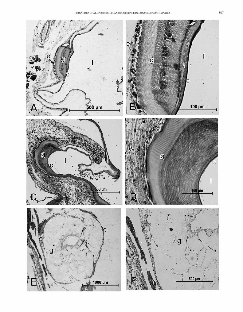
Sections of early premolt stage showed the initial formationof a gastrolith (Fig. 4A and B). At this stage the gastrolithdisc epithelium, the cuticle, and the initial gastrolith can bedistinguished. The gastrolith is in close association with thegastrolith disc formed by cuboidal cells of the epithelium.The cuticle covers the luminal side of the gastrolith in forma-tion, and the newest and oldest parts of the gastrolith can be

806 JOURNAL OF CRUSTACEAN BIOLOGY, VOL. 32, NO. 5, 2012
Fig. 3. Structure of a gastrolith. A, general aspect of paired gastroliths ofCherax quadricarinatus; B, gastrolith cross-section after partial decalcifi-cation with EDTA showing layered pattern; C, SEM view of organic matrixlayers remaining after complete decalcification; D, organic matrix fibers ofnanometric diameter after 30 min decalcification with acetic acid.
distinguished by their differential staining by hematoxylin-eosin. The reason of such difference remains unclear.
At premolt stage, larger gastroliths were found. Thegastrolith disc epithelium consists of higher cylindrical cells,and it is still in close association with the forming gastrolith(Fig. 4C). The lamellar structure of the gastrolith can bedistinguished (Fig. 4D). As in the early premolt stage,a differential staining by hematoxylin-eosin was observedbetween the newest and the oldest parts of the gastrolith,and the cuticle, as at the previous stage, covers the luminalsurface of the gastrolith.
At postmolt stage, sections showed a very large butdisorganized gel-like gastrolith in the stomach lumen. Someof the gastrolith disc epithelium and part of the cuticle stillremained attached to the gastrolith (Fig. 4E and F).
A layered structural organization of autofluorescent chitinis observed in early premolt and premolt gastroliths, while itis almost completely lost at the postmolt stage revealing thedisorganization of the chitin-protein fibril network concomi-tant with the physiological decalcification process (Fig. 5Ato C).
Sugar Analysis of Gastrolith Matrix
The amounts of the different monosaccharides detected bychromatographic analysis after TFA hydrolysis are reportedin Table 1 and classified by order of abundance for theacetic acid insoluble fraction. The insoluble matrix fraction(IM) exhibits a high level of glucosamine, around 75% ofthe total sugar content, whereas the soluble matrix fraction(SM) exhibits 35% glucosamine, which is also the mostimportant sugar in this fraction. Nevertheless, the order ofabundance of the other sugars varies considerably in the 2matrices, with xylose, arabinose and glucose being the mostimportant (around 7.4, 6 and 4.7% respectively) in the IMmatrix versus glucose, fucose and mannose in the SM eachone representing more that 10% of the total sugar content(around 14, 12.7 and 10.5% respectively). It is to notice
that iduronic acid, component of dermatan sulfate, cannotbe detected by this method.
Immunohistochemical Analysis
To facilitate visual comparisons, the same negative immuno-histochemical controls have been repeated in figures A and Bof the next three figures (Figs. 6 to 8).
Compared to control sections treated only with secondaryantibody (Fig. 6A and B), sections treated with 2B6 anti-body showed a dermatan sulfate and chondroitin-4-sulfatereactivity in early premolt and premolt stages with a positivereaction in the gastrolith disc epithelium closely associatedwith the forming gastrolith (Fig. 6C to F), and in the gas-trolith itself in the early premolt stage (Fig. 6C and E). At theearly premolt stage, a more intense reaction was observed inthe newest part of the gastrolith (Fig. 6C and E). However,the gastrolith labeling is negative during the premolt stage(Fig. 6D and F).
Compared to control sections treated only with secondaryantibody (Fig. 7A and B), sections treated with 5D4 antibodyshowed a strong positive reaction for keratan sulfate inthe gastrolith disc epithelium and in the gastrolith at theearly premolt stage (Fig. 7C-E), while at the premolt stagethe reactivity in the gastrolith and in the gastrolith discepithelium is almost absent (Fig. 7D-F).
When sections treated with 3B3 antibody (Fig. 8C to F)were compared with control sections treated only withsecondary antibody (Fig. 8A and B), a chondroitin-6-sulfatepositive reaction was found at the early premolt stage, inthe gastrolith and in the gastrolith disc epithelium (Fig. 8Cand E). At the premolt stage, there is only a very weak 3B3reactivity in both the epithelium and the gastrolith (Fig. 8Dand F).
At postmolt stage, when sections treated with 2B6, 5D4,and 3B3 antibodies (Fig. 9B to D respectively) were com-pared with control sections treated only with secondary anti-body (Fig. 9A), a weak positive reaction to dermatan sul-phate, chondroitin-4-sulfate and chondroitin-6-sulfate wasfound only in the gastrolith disc epithelium but it was nega-tive in the formic acid decalcified gastrolith remnant (Fig. 9Band D). At this stage, a negative reaction to keratan sulfatewas observed in both, the gastrolith disc epithelium and theresorptive gastrolith remnant (Fig. 9C).
Proteoglycan Detection by Dot Blot
Proteoglycans were isolated from the soluble and insolublefractions of 10 (25 g) decalcified fully developed gastrolithsand then analyzed. The concentration of proteoglycanswas measured at 6.4 μg/μl in the soluble fraction and13.3 μg/μl in the insoluble fraction, giving a total amountof 640 ng of proteoglycans per mg of gastrolith organicmatter. Proteoglycans from the soluble fraction showeda positive reaction for all the antibodies used, and thereaction was especially intense for chondroitin-4-sulfatewhen it is compared with that of the insoluble fraction(Fig. 10). Proteoglycans from the insoluble fraction showedan intense reaction for dermatan sulfate plus chondroitin-4-sulfate and for chondroitin-6-sulfate, while the keratansulfate reaction was negative compared with the standardsample of bovine nasal septum (Fig. 10). It is importantto remember that quantitative comparisons should only be
FERNÁNDEZ ET AL.: PROTEOGLYCAN OCCURRENCE IN CHERAX QUADRICARINATUS 807

808 JOURNAL OF CRUSTACEAN BIOLOGY, VOL. 32, NO. 5, 2012
Fig. 5. Autofluorescence of chitin in gastrolith cross-sections at three stages of development. A, early premolt; B, premolt; and C, postmolt. Chitin organizedas multilayered structure at early premolt and premolt stages and disorganized after decalcification at postmolt stage.
done between reactivities of different samples to the sameantibody because of the difference in the sensitivity of eachantibody against its own antigen.
DISCUSSION
Various organisms use amorphous calcium carbonate (ACC)as a temporary storage form for calcium and carbonateions (Lowenstam and Weiner, 1989; Aizenberg et al., 2002;Luquet and Marin, 2004; Gago-Duport et al., 2008). Thiscan occur because ACC is the most unstable polymorph ofcalcium carbonate and can be easily dissolved in aqueousand acidic environments. However, biogenic ACC can bestabilized for a long period of time by components of theorganic matrix, demonstrating that organisms are also ableto inhibit crystallization.
Although the exact mechanism by which ACC is stabi-lized in vivo requires further investigation, it is known thatstabilization of ACC has been commonly achieved by using
Table 1. Amounts of the different monosaccharides detected by chromato-graphic analysis after TFA hydrolysis.
Sugar Cherax matrix analyzed
IM SM
Glucosamine 72.42 ± 2.36 35.85 ± 0.35Xylose 7.44 ± 0.52 3.15 ± 0.64Arabinose 6.12 ± 0.56 1 ± 1.41Glucose 4.7 ± 0.37 13.95 ± 5.44Mannose 3.74 ± 0.75 10.45 ± 2.05Fucose 3.42 ± 1.19 12.65 ± 0.35Galactose 1.14 ± 0.50 8.4 ± 2.55Galactosamine 1.02 ± 0.42 5.65 ± 2.19Rhamnose Tr 8.3 ± 0.71Galacturonic acid Tr TrGlucuronic acid Tr Tr
Tr, trace.
various additives, including magnesium and phosphate ions,phosphoamino acids (Raz et al., 2000; Loste et al., 2003;Ajikumar et al., 2005; Lee et al., 2005; Bentov et al., 2010),biomacromolecules (Aizenberg et al., 1996, 2002; Addadi etal., 2003; Shechter et al., 2008b; Glazer et al., 2010), andsynthetic polymers (Volkmer et al., 2005; Xu et al., 2005).
In some crustaceans, such as lobsters, terrestrial crabs, andcrayfish, transient semi-spherical calcium storage structures,called gastroliths, are cyclically elaborated in the stomachwall in relation with the cyclic renewal of their hardexoskeleton (calcified cuticle) for growing. The ability ofcrustaceans to stabilize ACC in these transient calciumstorage structures is remarkable (Raz et al., 2002; Addadiet al., 2003; Becker et al., 2003; Luquet and Marin, 2004),but what kind of macromolecules are involved in thestabilization of ACC or crystalline phases has not been wellestablished. Even stabilized, biogenic ACC remains highlysoluble, which is beneficial for the subsequent use of the ionsto partially calcify each new carapace (Raz et al., 2002).
Several proteins characterized from crustacean transientcalcium storage structures, such as orchestin (Testeniere etal., 2002), gastrolith matrix protein (GAMP) (Ishii et al.,1996; Tsutsui et al., 1999; Tagaki et al., 2000), GAP 65(Shechter et al., 2008b), and GAP 10 (Glazer et al., 2010),have been shown to be involved in their calcification. Butonly GAP 65 has been associated with the stabilizationof ACC during gastrolith formation. More recently, thepresence of phosphorylated energy-rich intermediates of theglycolytic pathway as components of the gastrolith organicmatrix has been reported and their possible involvement inthe formation and stabilization odf ACC suggested (Akiva-Tal et al., 2011; Sato et al., 2011).
Although, different types of components have been char-acterized in various calcium storage deposits in inverte-brates, little is known about the occurrence of proteoglycansin these structures. Proteoglycans are known to be compo-nents of calcium carbonate biomineralized structures (Arias
Fig. 4. Histological sections of stomach tissue stained with hematoxylin-eosin (H-E). A and B, development of gastrolith (g) in close association withgastrolith disc epithelium (e) during early premolt stage, cuticle (c) covers the luminal side of forming gastrolith, difference in staining seen between oldestpart (dark) and newest part (pale) of gastrolith; C and D, gastrolith (g) in formation during premolt stage, epithelium (e) tightly linked to forming side ofgastrolith (g), also showing differential staining seen between oldest and newest parts of gastrolith, cuticle covering gastrolith clearly visible (c); E and F,disorganized decalcified gastrolith during postmolt stage.

FERNÁNDEZ ET AL.: PROTEOGLYCAN OCCURRENCE IN CHERAX QUADRICARINATUS 809
Fig. 6. 2B6 immunohistochemistry of early premolt (A, C and E) and premolt (B, D and F) stomach sections. A and B, control; C, D, E and F, 2B6immunoreactivity. Dermatan sulfate and chondroitin-4-sulfate reactivity visible in gastrolith disc epithelium at both stages and in gastrolith itself in earlypremolt stage; a positive reaction in connective tissue underlying epithelium also observed.
and Fernández, 2008), but the precise role of such compo-nents in diverse aspects of the biomineralization process isnot well known.
Although we did not use the molt mineralization indices(Shechter et al., 2008a) to monitor the progress of the molt
cycle, we showed here that after radiographic analysis, onlypremolt and postmolt gastroliths could be detected. Whena radiopaque structure was not detected, the crayfish wasprimary classified as intermolt. However, after a detaileddissection of some of these specimens a small but well

810 JOURNAL OF CRUSTACEAN BIOLOGY, VOL. 32, NO. 5, 2012
Fig. 7. 5D4 immunohistochemistry of early premolt (A, C and E) and premolt (B, D and F) stomach sections. A and B, control; C, D, E and F, 5D4immunoreactivity. Keratan sulfate-positive reaction detected in gastrolith (g) and more strongly in gastrolith disc epithelium (e) at early premolt stage (C andE). At premolt stage reactivity almost absent in both gastrolith and gastrolith disc epithelium (D and F).
defined and soft gastrolith was found. These specimenswere then reclassified as early premolt stage, meaning thatthe calcification level of these samples was not sufficientto block the penetration of X-rays in order to produce aradiopaque structure. Therefore, a well defined extracellular
matrix, chitin-rich layered structure is formed early, and itsubsequently becomes calcified.
The organic matrix of gastrolith is composed of a com-plex network of chitin-protein fibers forming concentric andtransversal striations well visible in a cross-section of a

FERNÁNDEZ ET AL.: PROTEOGLYCAN OCCURRENCE IN CHERAX QUADRICARINATUS 811
Fig. 8. 3B3 immunohistochemistry of early premolt (A, C and E) and premolt (B, D and F) stomach sections. A and B, controls; C, D, E and F, 3B3immunoreactivity. Reactivity to 3B3 observed during early premolt at level of gastrolith matrix (g) and gastrolith epithelium (e) (C and E). Very weakreactivity observed in gastrolith and gastrolith disc epithelium during premolt (D and F). Connective tissue, below the epithelium, appears more stronglyreactive.
partially decalcified gastrolith. Chitin is a polymer of N-acetylglucosamine, therefore it is not surprising to find ahigh level of glucosamine in the insoluble fraction. This isin accordance with the presence of large chitin fibers, which
cannot be solubilized and depolymerized during the decalci-fication process. The glucosamine level remains not negligi-ble in the soluble fraction because of the presence of protein-chitin fibers of nanometric diameter which could subdivide

812 JOURNAL OF CRUSTACEAN BIOLOGY, VOL. 32, NO. 5, 2012
Fig. 9. 2B6, 5D4 and 3B3 immunohistochemistry of postmolt stage. A, control; B, 2B6 immunoreactivity; C, 5D4 immunoreactivity; D, 3B3immunoreactivity. Weak positive reaction to dermatan sulphate, chondroitin-4-sulfate, and chondroitin-6-sulfate found only in gastrolith disc epithelium,but negative in resorptive gastrolith remnant (B and D). Negative reaction to keratan sulfate observed in both gastrolith disc epithelium and resorptivegastrolith remnant (9 C).
in micro-units the largest pocket-like compartments framedby the micro-chitin fibers. These nanofibers could be re-leased during the decalcification process and could remainin the supernatant after centrifugation of the decalcified so-lution.
The presence to a lesser extent of other monosaccha-rides cannot be really explained for all of them like ara-binose, fucose or rhamnose. Nevertheless the presence ofgalactose, galactosamine as well as xylose and mannosecan be related to the presence of proteoglycans as otherscomponents of the organic matrix. Indeed, chondroitin sul-fate, dermatan sulfate and keratan sulfate are proteoglycanscomposed of a glycosaminoglycan (GAG) bound to a pro-tein core (at the level of a serine or a threonine) via a xy-lose sugar: . . . XY-XY-XY-XY-β(1-3)-Gal-β(1-3)-Gal-β(1-4)-Xyl-β(1-O)-Ser/Thr-Protein. Keratan sulfate (KSII) con-sists of a glycosaminoglycan (GAG) bound to a protein core(at the level of an Asn) via a mannose sugar: . . . carbohydrate
chain-Man-β(1-O)-Asn-Protein (Funderburgh, 2000; Ariasand Fernández, 2008). The high levels of glucosamine re-ported here are consistent with the abundance of chitin in thegastrolith, while the low levels of glucuronic acid not neces-sarily should match the levels of galactosamine containingproteoglycans since dermatan sulfate contains iduronic acidinstead of glucuronic acid which was not detected by themethod of sugar analysis used.
Immunodetection of proteoglycans on histological sec-tions showed that in the early premolt soft gastrolith, thefour glycosaminoglycans studied (dermatan, chondroitin-4- and 6-, and keratan sulfate) are detected both in thegastrolith-forming epithelium and especially in the newestand less calcified region of the gastrolith matrix. However,during the premolt stage when calcification is already de-tected by X-rays, not all glycosaminoglycans are detectedin the gastrolith, and some of them are also absent from thegastrolith-forming epithelium. In early premolt and premolt

FERNÁNDEZ ET AL.: PROTEOGLYCAN OCCURRENCE IN CHERAX QUADRICARINATUS 813
Fig. 10. Dot blot analysis of proteoglycans isolated from soluble (SF) andinsoluble (IF) fractions of gastrolith. TBS negative control (−C) and bovinenasal septum proteoglycans positive control (+C). Soluble fraction showedpositive reaction for all antibodies used, especially intense for chondroitin-4-sulfate when compared with that of insoluble fraction. Insoluble fractionshowed intense reaction for dermatan sulfate plus chondroitin-4-sulfate andfor chondroitin-6-sulfate, while keratan sulfate reaction negative comparedwith standard sample of bovine nasal septum.
gastroliths, the chitin layered structure is still intact, while inthe postmolt stage the layered organization is almost com-pletely lost.
When a specific methodology to obtain proteoglycansis applied to a fully formed gastrolith, the same familyof glycosaminoglycans reported to occur in other calciumcarbonate mineralized systems (Arias and Fernández, 2008)is also found in the red claw crayfish gastrolith. Some ofthem appeared to be more associated with the insolubleorganic fraction while others with the soluble one, which isconsistent with what has been shown in other mineralizedsystems (Fernández et al., 2001; Arias and Fernández, 2003,2008; Arias et al., 2004).
One plausible explanation for the apparent contradictionbetween dot blot and immunohistochemical results may berelated to the decalcification procedure. In other calcifiedstructures, a very close association has been described be-tween the molecules involved in the process of biominer-alization and the crystalline or amorphous mineral phase,where a phase stabilization role has been proposed (Aizen-berg et al., 1995, 1996; Albeck et al., 1996; Raz et al.,2002; Bentov et al., 2010; Glazer et al., 2010; Ndao et al.,2010). It is well known that amorphous calcium carbonate-based structures are more soluble and more easily decalci-fied than the crystalline forms of these biominerals. At theearly premolt stage, where the gastrolith is at the begin-ning of its calcification, the proteoglycans associated withthe rest of the extracellular matrix molecules, such as chitin,are not removed by the formic acid-based decalcification so-lution. However, at the premolt stage when the gastrolith isfully calcified, the decalcification procedure could removea significant amount of the chitin-associated organic mate-rial due to its interaction with CaCO3, as has been recentlyshowed in parallel studies (Thormann et al., 2012). At the
postmolt stage there is no detection of proteoglycans, be-cause the gastrolith has been almost totally digested anddisorganized with the drastic decalcification procedure usedin our studies. As expected, the decalcification proceduredoes not affect the detection of proteoglycans in the ep-ithelial tissue, and when they are not detected in the ep-ithelium it could be related to a spatio-temporal regula-tion mechanism such as that reported by Yudkovski et al.(2010) in crayfish or Fernández et al. (1997, 2001) in theeggshell.
The fact that for SF the dot for C-4-S is more intensethan the dot for DS + C-4-S, although both of thesecontain equal amounts of material, is unexpected. TheC-4-S dot was treated with chondroitinase ACII to digestchondroitin sulfate. The DS + C-4-S dot was treated withchondroitinase ABC to digest both chondroitin sulfate anddermatan sulfate. Both dots were analyzed with antibody2B6, which should detect terminal unsaturated disaccharidescontaining 4-sulfation. These disaccharides should arisefrom either DS or C-4-S. The dot that was digested withchondroitinase ACII should contain these disaccharides onlyfrom C-4-S. The dot that was digested with chondroitinaseABC should contain these disaccharides from both DSand C-4-S. Hence, if the sample contains both DS andC-4-S, the expectation is that the dot that was treatedwith chondroitinase ABC should contain more terminal4-sulfated unsaturated disaccharides and, thus, be moreintense than the dot that was treated with chondroitinaseACII. However, the reverse is observed, and there is noobvious explanation for this. One possibility is that thisresults from differences in the efficiencies of chondroitinaseACII and ABC toward co-polymers of dermatan sulfate andchondroitin sulfate. Chondroitinase ACII does not efficientlycleave such co-polymers. In contrast, dermatan sulfate-chondroitin sulfate co-polymers are efficiently cleaved bychondroitinase ABC, which digests both dermatan sulfateand chondroitin sulfate. It may be that the dot that wastreated with chondroitinase ABC is less intense because thedigestion was overly extensive relative to the dot that wastreated with chondroitinase ACII and that this resulted in thepresence of less terminal 4-sulfated unsaturated disaccharideand less 2B6 reactivity. Perhaps a shorter enzyme treatmenttime of the dots or a lower enzyme amount would result inmore intense 2B6 reactivity in the DS + C-4-S dot relativeto the C-4-S dot.
In conclusion, we have shown that the organic matrix ofthe red claw crayfish gastrolith contains proteoglycans thathave dermatan sulfate, chondroitin-4-sulfate, chondroitin-6-sulfate, and keratan sulfate glycosaminoglycans. These gly-cosaminoglycans are likely to be not all on the same coreprotein, but, for at least some of the glycosaminoglycans,present in different proteoglycans. These macromoleculesare closely associated with the mineral phase of the gas-trolith and are easily removed by decalcification procedures.There are also some indications that the epithelial secretionof some of these molecules is temporally regulated duringthe molting cycle, but its gene regulation needs further in-vestigation. Even if sulfate groups have been already thoughtto be involved in the chelation of calcium ions similarly tophosphate and carboxylate groups, the precise role of these

814 JOURNAL OF CRUSTACEAN BIOLOGY, VOL. 32, NO. 5, 2012
sulfated macromolecules in calcification and stabilization ofthe amorphous calcium carbonate phase of gastroliths is stillto be established.
ACKNOWLEDGEMENTS
The work has been financially supported by the Ministry of ForeignAffairs (France) and CONICYT (Chile) through the ECOS-Sud/CONICYTProject C07B02 and through FONDAP 11980002. The work of G.L.was also supported by an ANR budget (ACCRO-EARTH, ref. BLAN06-2_159971) granted for the period 2007-2010 and by INSU-CNRS withinthe INTERRVIE project granted in 2010.
REFERENCES
Addadi, L., S. Raz, and S. Weiner. 2003. Taking advantage of disorder:amorphous calcium carbonate and its roles in biomineralization. Ad-vanced Materials 15: 959-970.
Aizenberg, J., L. Addadi, S. Weiner, and G. Lambert. 1996. Stabilizationof amorphous calcium carbonate by specialized macromolecules inbiological and synthetic precipitates. Advanced Materials 8: 222-226.
, G. Lambert, S. Weiner, and L. Addadi. 2002. Factors involved inthe formation of amorphous and crystalline calcium carbonate: a study ofan ascidian skeleton. Journal of American Chemical Society 124: 32-39.
, J. Hanson, M. Ilan, L. Leiserowitz, T. F. Koetzle, L. Addadi,and S. Weiner. 1995. Morphogenesis of calcitic sponge spicules: a roleof specialized proteins interacting with growing crystals. Federation ofAmerican Societies for Experimental Biology Journal 9: 262-268.
Ajikumar, P. K., L. G. Wong, G. Subramanyam, R. Lakshminarayanan, andS. Valiyaveettil. 2005. Synthesis and characterization of monodispersedspheres of amorphous calcium carbonate and calcite spherules. CrystalGrowth Design 5: 1129-1134.
Akiva-Tal, A., S. Kabaya, Y. S. Balazs, L. Glazer, A. Berman, A. Sagi, andA. Schmidt. 2011. In situ molecular NMR picture of bioavailable calciumstabilized as amorphous CaCO3 biomineral in crayfish gastroliths.Proceedings of the National Academy of Sciences USA 108: 14763-14768.
Albeck, S., L. Addadi, and S. Weiner. 1996. Regulation of calcite crystalmorphology by intracrystalline acidic proteins and glycoproteins. Con-nective Tissue Research 35: 365-370.
Arias, J. L., and M. S. Fernández. 2003. Biomimetic processes through thestudy of mineralized shells. Material Characterization 50: 189-195.
, and . 2007. Biomineralization: From Paleontology toMaterials Science. Editorial Universitaria, Santiago, Chile.
, and . 2008. Polysaccharides and proteoglycans in calciumcarbonate-based biomineralization. Chemical Reviews 108: 4475-4482.
, K. Mann, Y. Nys, J. M. García-Ruiz, and M. S. Fernández.2007. Eggshell growth and matrix macromolecules, pp. 309-327. In,E. Bauerlein (ed.), Handbook of Biomineralization. Vol. 1. Wiley-VCH,Weinheim, Germany.
, D. A. Carrino, M. S. Fernández, J. P. Rodriguez, J. E. Dennis,and A. I. Caplan. 1992. Partial biochemical and immunochemicalcharacterization of avian eggshell extracellular matrices. Archives ofBiochemistry and Biophysics 298: 293-302.
, A. Neira-Carrillo, J. I. Arias, C. Escobar, M. Bodero, M. David,and M. S. Fernández. 2004. Sulfated polymers in biological mineraliza-tion: a plausible source for bio-inspired engineering. Journal of MaterialChemistry 14: 2154-2160.
Bauerlein, E. 2000. Biomineralization. Wiley-VCH, Weinheim.. 2007. Handbook of Biomineralization. Wiley-VCH, Weinheim.
Becker, A., U. Bismayer, M. Epple, H. Fabritius, B. Hasse, J. Shi, andA. Ziegler. 2003. Structural characterization of X-ray amorphous calciumcarbonate (ACC) in sternal deposits of the crustacean Porcellio scaber.Dalton Transactions: 551-555.
Belcher, A. M., X. H. Wu, R. J. Christensen, P. K. Hasma, G. D. Stucky, andD. E. Morse. 1996. Control of crystal phase switching and orientation bysoluble mollusk-shell proteins. Nature 381: 56-58.
Bentov, S., S. Weil, L. Glazer, A. Sagi, and A. Berman. 2010. Stabilizationof amorphous calcium carbonate by phosphate rich organic matrixproteins and by single phosphoamino acids. Journal of Structural Biology171: 207-215.
Bülow, H. E., and O. Hobert. 2006. The molecular diversity of glycosamino-glycans shaped animal development. Annual Review of Cell and Devel-opmental Biology 22: 375-407.
Carrino, D. A., J. L. Arias, and A. I. Caplan. 1991. A spectrophotomet-ric modification of a sensitive densitometric Safranin O assay for gly-cosaminoglycans. Biochemistry International 24: 485-495.
, J. P. Rodriguez, and A. I. Caplan. 1997. Dermatan sulfate proteo-glycans from the mineralized matrix of the avian eggshell. ConnectiveTissue Research 36: 175-193.
, J. E. Dennis, R. F. Drushel, S. E. Haynesworth, and A. I. Caplan.1994. Identity of the core proteins of the large chondroitin sulfateproteoglycans synthesized by skeletal muscle and prechondrogenicmesenchyme. Biochemical Journal 298: 51-60.
Casu, B. 1985. Structure and biological activity of heparin. AdvancedCarbohydrate Chemistry and Biochemistry 43: 51-134.
Caterson, B., T. Calabro, and A. Hampton. 1987. Monoclonal antibodiesas probes for elucidating proteoglycan structure and function, pp. 1-26.In, T. N. Wright and R. P. Mecham (eds.), Biology of Proteoglycans.Academic Press, London.
, J. E. Christner, J. R. Baker, and J. R. Couchman. 1985. Productionand characterization of monoclonal antibodies directed against connec-tive tissue proteoglycans. Federation Proceedings of the Federation ofthe American Societies of Experimental Biology 44: 386-393.
Díaz-Dosque, M., P. Aranda, M. Darder, J. Retuert, M. Yazdani-Pedram,J. L. Arias, and E. Ruiz-Hitzky. 2008. Use of biopolymers as orientedsupports for the stabilization of different polymorphs of biomineralizedcalcium carbonate with complex shape. Journal of Crystal Growth 310:5331-5340.
Ehrlich, H. 2010. Chitin and collagen as universal and alternative templatesin biomineralization. International Geological Review 52: 661-669.
Falini, G., S. Albeck, S. Weiner, and L. Addadi. 1996. Control of aragoniteor calcite polymorphism by mollusk shell macromolecules. Science 271:67-69.
Fernández, M. S., M. Araya, and J. L. Arias. 1997. Eggshells are shapedby a precise spatio-temporal arrangement of sequentially depositedmacromolecules. Matrix Biology 16: 13-20.
, A. Moya, L. López, and J. L. Arias. 2001. Secretion pattern,ultrastructural localization and function of extracellular matrix moleculesinvolved in eggshell formation. Matrix Biology 19: 793-803.
, I. Vergara, A. Oyarzun, J. L. Arias, R. Rodriguez, J. P. Wiff,V. M. Fuenzalida, and J. L. Arias. 2002. Extracellular matrix moleculesinvolved in barnacle’s shell mineralization, pp. 1-9. In, J. Aizenberg,J. McKittrick, and C. Orme (eds.), Biological and Biomimetic Materi-als – Properties to Function. Material Research Society Symposium Pro-ceedings. Vol. 724. Warrendale, PA.
Funderburgh, J. L. 2000. Keratan sulfate: structure, biosynthesis, andfunction. Glycobiology 10: 951-958.
Gago-Duport, L., M. J. I. Briones, J. B. Rodríguez, and B. Covelo. 2008.Amorphous calcium carbonate biomineralization in the earthworm’scalciferous gland: pathways to the formation of crystalline phases.Journal of Structural Biology 162: 422-435.
Glazer, L., A. Shechter, M. Tom, Y. Yudkovski, S. Well, E. D. Aflalo,R. R. Pamuru, I. Khalaila, S. Bentov, A. Berman, and A. Sagi. 2010.A protein involved in the assembly of an extracellular calcium storagematrix. Journal of Biological Chemistry 285: 12831-12839.
Huckerby, T. N. 2002. The keratan sulphates: structural investigationsusing NMR spectroscopy. Progress in Nuclear Magnetic ResonanceSpectroscopy 40: 35-110.
Iozzo, R. V. 1998. Matrix proteoglycans: from molecular design to cellularfunction. Annual Review of Biochemistry 67: 609-659.
Ishii, K., N. Tsutui, T. Watanabe, T. Yanagisawa, and H. Nagasawa.1998. Solubilization and chemical characterization of an insoluble matrixprotein in the gastroliths of a crayfish, Procambarus clarkii. Bioscience,Biotechnology and Biochemistry 62: 291-296.
Lee, H. S., T. H. Ha, and K. Kim. 2005. Fabrication of unusuallystable amorphous calcium carbonate in an ethanol medium. MaterialsChemistry and Physics 93: 76-382.
Loste, E., R. M. Wilson, R. Seshadri, and F. C. Meldrum. 2003. The role ofmagnesium in stabilising amorphous calcium carbonate and controllingcalcite morphologies. Journal of Crystal Growth 254: 206-218.
Lowenstam, H. A., and S. Weiner. 1989. On Biomineralization. OxfordUniversity Press, Oxford.
Luquet, G., and F. Marin. 2004. Biomineralizations in crustacean: storagestrategies. Comptes Rendus Palevol 3: 515-534.
, M. S. Fernández, J. M. Navarrete, J. L. Arias, N. Guichard,B. Marie, and F. Marin. 2007. Biochemical characterization of the solubleorganic matrix of gastroliths from decapods, pp. 319-328. In, J. L. Arias

FERNÁNDEZ ET AL.: PROTEOGLYCAN OCCURRENCE IN CHERAX QUADRICARINATUS 815
and M. S. Fernández (eds.), Biomineralization, from Paleontology toMaterials Science. Editorial Universitaria, Santiago, Chile.
Mann, S. 2001. Biomineralization. Oxford University Press, Oxford., P. Behrens, and E. Bauerlein. 2007. Handbook of Biomineraliza-
tion. Biomimetic and Bioinspired Chemistry. Wiley-VCH, Weinheim., J. Webb, and R. J. P. Williams. 1989. Biomineralization. VCH,
Weinheim.Mehmet, H., P. Sculder, P. W. Tang, E. F. Hounsell, B. Caterson, and
T. Feizi. 1986. The antigenic determinants recognized by three mono-clonal antibodies to keratan sulphate involve sulphated hepta or largeroligosaccharides of the poly(N-acetylgalactosamine) series. EuropeanJournal of Biochemistry 157: 385-391.
Morse, A. 1945. Formic acid-sodium citrate decalcification and butylalcohol dehydration of teeth and bone for sectioning in paraffin. Journalof Dental Research 24: 143-153.
Nakatsuji, T., H. Keino, K. Tamura, S. Yoshimura, T. Kawakami, S. Aimoto,and H. Sonobe. 2000. Changes in the amounts of the molt-inhibitinghormone in sinus glands during the molt cycle of the American crayfish,Procambarus clarkii. Zoological Science 17: 1129-1136.
Ndao, M., E. Keene, F. F. Amos, G. Rewari, C. B. Ponce, L. Estroff, andJ. S. Evans. 2010. Intrinsically disordered mollusk shell prismatic proteinthat modulates calcium carbonate crystal growth. Biomacromolecules11: 2539-2544.
Neira-Carrillo, A., M. Yazdani-Pedram, J. Retuert, M. Diaz-Dosque,S. Gallois, and J. L. Arias. 2005. Selective crystallization of calcium saltsby poly(acrylate)-grafted chitosan. Journal of Colloidal Interface Science286: 131-141.
Nys, Y., M. T. Hincke, J. L. Arias, J. M. García-Ruiz, and S. E. Solomon.1999. Avian eggshell mineralization. Poultry Avian Biology Review 10:142-166.
Raz, S., S. Weiner, and L. Addadi. 2000. Formation of high-magnesiancalcites via an amorphous phase: possible biological implications.Advanced Materials 12: 38-42.
, O. Testeniere, A. Hecker, S. Weiner, and G. Luquet. 2002. Stableamorphous calcium carbonate is the main components of the calciumstorage structures of the crustacean Orchestia cavimana. BiologicalBulletin 203: 269-274.
Rodríguez-Navarro, A., C. Cabral de Melo, N. Batista, N. Morimoto,P. Alvarez-Lloret, M. Ortega-Huertas, V. M. Fuenzalida, J. I. Arias,J. P. Wiff, and J. L. Arias. 2006. Microstructure and crystallographic-texture of giant barnacle (Austromegabalanus psittacus) shell. Journal ofStructural Biology 156: 355-362.
Sato, A., S. Nagasaka, K. Furihata, S. Nagata, I. Arai, K. Saruwatari,T. Kogure, S. Sakuda, and H. Nagasawa. 2011. Glycolytic intermediatesinduce amorphous calcium carbonate formation in crustaceans. NatureChemical Biology 7: 197-199.
Shechter, A., A. Berman, A. Singer, A. Freiman, M. Grinstein, J. Erez,E. D. Aflalo, and A. Sagi. 2008a. Reciprocal changes in calcification ofthe gastrolith and cuticle during the molt cycle of the red claw crayfishCherax quadricarinatus. Biological Bulletin 214: 122-134.
, L. Glazer, S. Cheled, E. Mor, S. Weil, A. Berman, S. Bentov, E. D.Aflalo, I. Khalaila, and A. Sagi. 2008b. A gastrolith protein serving a dualrole in the formation of an amorphous mineral containing extracellular
matrix. Proceedings of the National Academy of Sciences 105: 7129-7134.
Simkiss, K., and K. M. Wilbur. 1989. Biomineralization. Academic Press,San Diego.
Smith, B. L., T. E. Schäffer, M. Viani, J. B. Thompson, N. A. Frederick,J. Kindt, A. M. Belcher, G. D. Stucky, and D. E. Morse. 1999. Molecularmechanistic origin of the toughness of natural adhesives, fibres andcomposites. Nature 399: 761-763.
Sugahara, K., and H. Kitagawa. 2000. Recent advances in the study ofthe biosynthesis and functions of sulfated glycosaminoglycans. CurrentOpinion on Structural Biology 10: 518-527.
Takagi, Y., K. Ishii, N. Ozaki, and H. Nagasawa. 2000. Immunolocalizationof gastrolith matrix protein (GAMP) in the gastroliths and exoskeleton ofcrayfish, Procambarus clarkia. Zoological Sciences 17: 179-184.
Testeniere, O., A. Hecker, S. Le Gurun, B. Quennedey, F. Graf, and G. Lu-quet. 2002. Characterization and spatiotemporal expression of orchestin,a gene encoding an ecdysone-inducible protein from a crustacean organicmatrix. Biochemical Journal 361: 327-335.
Thormann, E., H. Mizuno, K. Jansson, N. Hedin, M. S. Fernández, J. L.Arias, M. W. Rutland, R. Krishna Pai, and L. Bergström. In press.
Embedded proteins and sacrificial bonds provide the strong adhesiveproperties of gastroliths. Nanoscale.
Travis, D. F. 1960. The deposition of skeletal structures in the crustacea. I.The histology of the gastrolith skeletal tissue complex and the gastrolithin the crayfish, Orconectes (Cambarus) virilis Hagen – Decapoda.Biological Bulletin 18: 137-149.
. 1963. Structural features of mineralization from tissue to macro-molecular levels of organization in the decapod crustacea. Annals of theNew York Academy of Science 109: 177-245.
Tsutsui, N., K. Ishii, Y. Takagi, T. Watanabe, and H. Nagasawa. 1999.Cloning and expression of a cDNA encoding an insoluble matrix proteinin the gastroliths of a crayfish, Procambarus clarkii. Zoological Sciences16: 619-628.
Volkmer, D., M. Harms, L. Gower, and A. Ziegler. 2005. Morphosynthesisof nacre-type laminated CaCO3 thin films and coatings. AngewandteChemie International Edition 44: 639-644.
Weiner, S., and L. Addadi. 1997. Design strategies in mineralized biologicalmaterials. Journal of Materials Chemistry 7: 689-702.
Xu, A.-W., Q. Yu, W.-F. Dong, M. Antonietti, and H. Cölfen. 2005. Stableamorphous CaCO3 microparticles with hollow spherical superstructuresstabilized by phytic acid. Advanced Materials 17: 2217-2221.
Yanagishita, M., and V. C. Hascall. 1984. Proteoglycans synthesizedby rat ovarian granulosa cells in culture. Isolation, fractionation, andcharacterization of proteoglycans associated with the cell layer. Journalof Biological Chemistry 259: 10260-10269.
Yudkovski, Y., L. Glazer, A. Shechter, R. Reinhardt, V. Chalifa-Caspi,A. Sagi, and M. Tom. 2010. Multi-transcript expression patterns in thegastrolith disk and the hypodermis of the crayfish Cherax quadricari-natus at premolt. Comparative Biochemistry and Physiology Part D Ge-nomics Proteomics 5: 171-177.
RECEIVED: 13 January 2012.ACCEPTED: 1 May 2012.
Copyright © 2022 FDOKUMEN