A highly virulent pathogen, Aeromonas hydrophila, from the freshwater crayfish Pacifastacus...
11
A highly virulent pathogen, Aeromonas hydrophila, from the freshwater crayfish Pacifastacus leniusculus Pikul Jiravanichpaisal a,d, * , Stefan Roos b , Lennart Edsman c , Haipeng Liu d,e , Kenneth Söderhäll d a Molecular Aquatic Biology and Genetic Laboratory, National Center for Genetic Engineering and Biotechnology, National Science and Technology Development Agency, 113 Paholyothin, Thailand Science Park, Thailand b Department of Microbiology, Swedish University of Agricultural Sciences, Box 7025, 750 07 Uppsala, Sweden c Institute of Freshwater Research, Swedish Board of Fisheries, 17893 Drottningholm, Sweden d Department of Comparative Physiology,Uppsala University, Norbyvägen 18A, 752 36 Uppsala, Sweden e State Key Laboratory of Marine Environmental Science, College of Oceanography and Environmental Science, Xiamen University, Xiamen, 361005 Fujian, PR China article info Article history: Received 23 October 2008 Accepted 10 February 2009 Available online 20 February 2009 Keywords: Crustacean Bacteria–host interaction Crayfish pathology abstract Aeromonas spp. are characteristic bacteria of freshwaters and many of them can be components of the bacterial flora of aquatic animals and may become pathogens on animals including humans. In this study Aeromonas hydrophila was isolated from the freshwater crayfish, Pacifastacus leniusculus, and was found to be a highly pathogenic bacterium among many isolated bacteria. Mortality reached 100% within 6 h when 200 ll of 1.24 10 7 CFU/ml was applied by injection. Histopathological studies of moribund cray- fish showed that extensive necrotic nuclei and clump-infiltrated hemocytes were found in observed tissues including gill, heart, hepatopancreas and the circulatory system. To verify how crayfish are sus- ceptible to this bacterium, crude extracellular products (ECPs) obtained from culture supernatant of A. hydrophila was studied either in vivo or in vitro. ECPs (200 ll) were able to kill crayfish by injection. In an in vitro study, ECPs induced cytotoxicity of hemocytes as well as hematopoietic cells in a dose- and time-dependent manner after 30 min post inoculation. Two genes coding for endotoxins were also found in this isolate of A. hydrophila. This indicates that the bacterial endotoxins are the causative agents of crayfish mortality. Moreover, the effect of temperature on the infectivity of A. hydrophila to crayfish was also studied. At 4 °C, all crayfish survived, whereas at 20 °C the animals died rapidly after bacterial challenge. At this low temperature A. hydrophila did not replicate or replicated at a very low degree and hence crayfish could probably mount effective cellular reactions towards A. hydrophila. Ó 2009 Elsevier Inc. All rights reserved. 1. Introduction Aeromonas spp. are ubiquitous, oxidase positive, facultative anaerobic, Gram-negative bacteria that are native to aquatic envi- ronments (Hazen et al., 1978). They are responsible for a wide range of diseases among poikilothermic and homoeothermic ani- mals (Austin and Autin, 1993; Khardori and Fainstein, 1988; Math- ewson and Dupont, 1992). They have been found in marine, river, brackish, fresh, ground, spring water, chlorinated and unchlori- nated water supplies worldwide with highest numbers observed in the warmer period as well as in the sewage-contaminated water and activated sludge (Ashbolt et al., 1995; Borrel et al., 1998; Bous- said et al., 1991; Fiorentini et al., 1998; Hazen et al., 1978; Kaper et al., 1981; Krovacek et al., 1994; Pianetti et al., 1998; Soler et al., 2002; Van der Kooj, 1988). Many Aeromonas spp. have been implicated in the cause of numerous human diseases, including gastroenteritis, cellulitis, meningitis, bacteremia, solf-tissue infection, peritonitis, broncho- pulmonary infections, hemolytic-uremic syndrome, etc. (Brouqui and Raoult, 2001; Janda et al., 1994a; Ko et al., 2000; Kühn et al., 1997; Merino et al., 1995; Wadström and Ljungh, 1991). Apart from human diseases, Aeromonas spp., particularly Aeromonas hydrophila, has been associated with several disease conditions in fish, amphib- ian reptiles (Vivas et al., 2004) and also in crustaceans (New, 1995; Sung et al., 2000). In fish, A. hydrophila is responsible for hemorrhagic septicemia symptom as described by Meyer (1975), a fish disease affecting a wide variety of freshwater fish species and occasionally also marine fish (Larsen and Jensen, 1977). A. hydrophila has caused high mortality in freshwater fish such as catfish and Cyprinoid fish resulting in extensive losses (Meyer, 1975; Xia et al., 2004) and it has induced serious epidemics of ulcerative disease in fish in South- east Asia and other regions of the world (Roberts et al., 1992). Since A. hydrophila is usually found in aquatic habitats, it causes disease in 0022-2011/$ - see front matter Ó 2009 Elsevier Inc. All rights reserved. doi:10.1016/j.jip.2009.02.002 * Corresponding author. Address: Molecular Aquatic Biology and Genetic Labo- ratory, National Center for Genetic Engineering and Biotechnology, National Science and Technology Development Agency, 113 Paholyothin, Thailand Science Park, Thailand. Fax: +66 02 6448190. E-mail address: [email protected] (P. Jiravanichpaisal). Journal of Invertebrate Pathology 101 (2009) 56–66 Contents lists available at ScienceDirect Journal of Invertebrate Pathology journal homepage: www.elsevier.com/locate/yjipa
-
Upload
independent -
Category
Documents
-
view
4 -
download
0
Transcript of A highly virulent pathogen, Aeromonas hydrophila, from the freshwater crayfish Pacifastacus...

Journal of Invertebrate Pathology 101 (2009) 56–66
Contents lists available at ScienceDirect
Journal of Invertebrate Pathology
journal homepage: www.elsevier .com/ locate /y j ipa
A highly virulent pathogen, Aeromonas hydrophila, from the freshwater crayfishPacifastacus leniusculus
Pikul Jiravanichpaisal a,d,*, Stefan Roos b, Lennart Edsman c, Haipeng Liu d,e, Kenneth Söderhäll d
a Molecular Aquatic Biology and Genetic Laboratory, National Center for Genetic Engineering and Biotechnology, National Science and Technology Development Agency, 113Paholyothin, Thailand Science Park, Thailandb Department of Microbiology, Swedish University of Agricultural Sciences, Box 7025, 750 07 Uppsala, Swedenc Institute of Freshwater Research, Swedish Board of Fisheries, 17893 Drottningholm, Swedend Department of Comparative Physiology,Uppsala University, Norbyvägen 18A, 752 36 Uppsala, Swedene State Key Laboratory of Marine Environmental Science, College of Oceanography and Environmental Science, Xiamen University, Xiamen, 361005 Fujian, PR China
a r t i c l e i n f o a b s t r a c t
Article history:Received 23 October 2008Accepted 10 February 2009Available online 20 February 2009
Keywords:CrustaceanBacteria–host interactionCrayfish pathology
0022-2011/$ - see front matter � 2009 Elsevier Inc. Adoi:10.1016/j.jip.2009.02.002
* Corresponding author. Address: Molecular Aquatratory, National Center for Genetic Engineering and Bioand Technology Development Agency, 113 PaholyoThailand. Fax: +66 02 6448190.
E-mail address: [email protected] (P. Jiravanich
Aeromonas spp. are characteristic bacteria of freshwaters and many of them can be components of thebacterial flora of aquatic animals and may become pathogens on animals including humans. In this studyAeromonas hydrophila was isolated from the freshwater crayfish, Pacifastacus leniusculus, and was foundto be a highly pathogenic bacterium among many isolated bacteria. Mortality reached 100% within 6 hwhen 200 ll of 1.24 � 107 CFU/ml was applied by injection. Histopathological studies of moribund cray-fish showed that extensive necrotic nuclei and clump-infiltrated hemocytes were found in observedtissues including gill, heart, hepatopancreas and the circulatory system. To verify how crayfish are sus-ceptible to this bacterium, crude extracellular products (ECPs) obtained from culture supernatant of A.hydrophila was studied either in vivo or in vitro. ECPs (200 ll) were able to kill crayfish by injection. Inan in vitro study, ECPs induced cytotoxicity of hemocytes as well as hematopoietic cells in a dose- andtime-dependent manner after 30 min post inoculation. Two genes coding for endotoxins were also foundin this isolate of A. hydrophila. This indicates that the bacterial endotoxins are the causative agents ofcrayfish mortality. Moreover, the effect of temperature on the infectivity of A. hydrophila to crayfishwas also studied. At 4 �C, all crayfish survived, whereas at 20 �C the animals died rapidly after bacterialchallenge. At this low temperature A. hydrophila did not replicate or replicated at a very low degree andhence crayfish could probably mount effective cellular reactions towards A. hydrophila.
� 2009 Elsevier Inc. All rights reserved.
1. Introduction
Aeromonas spp. are ubiquitous, oxidase positive, facultativeanaerobic, Gram-negative bacteria that are native to aquatic envi-ronments (Hazen et al., 1978). They are responsible for a widerange of diseases among poikilothermic and homoeothermic ani-mals (Austin and Autin, 1993; Khardori and Fainstein, 1988; Math-ewson and Dupont, 1992). They have been found in marine, river,brackish, fresh, ground, spring water, chlorinated and unchlori-nated water supplies worldwide with highest numbers observedin the warmer period as well as in the sewage-contaminated waterand activated sludge (Ashbolt et al., 1995; Borrel et al., 1998; Bous-said et al., 1991; Fiorentini et al., 1998; Hazen et al., 1978; Kaper
ll rights reserved.
ic Biology and Genetic Labo-technology, National Science
thin, Thailand Science Park,
paisal).
et al., 1981; Krovacek et al., 1994; Pianetti et al., 1998; Soleret al., 2002; Van der Kooj, 1988).
Many Aeromonas spp. have been implicated in the cause ofnumerous human diseases, including gastroenteritis, cellulitis,meningitis, bacteremia, solf-tissue infection, peritonitis, broncho-pulmonary infections, hemolytic-uremic syndrome, etc. (Brouquiand Raoult, 2001; Janda et al., 1994a; Ko et al., 2000; Kühn et al.,1997; Merino et al., 1995; Wadström and Ljungh, 1991). Apart fromhuman diseases, Aeromonas spp., particularly Aeromonas hydrophila,has been associated with several disease conditions in fish, amphib-ian reptiles (Vivas et al., 2004) and also in crustaceans (New, 1995;Sung et al., 2000). In fish, A. hydrophila is responsible for hemorrhagicsepticemia symptom as described by Meyer (1975), a fish diseaseaffecting a wide variety of freshwater fish species and occasionallyalso marine fish (Larsen and Jensen, 1977). A. hydrophila has causedhigh mortality in freshwater fish such as catfish and Cyprinoid fishresulting in extensive losses (Meyer, 1975; Xia et al., 2004) and ithas induced serious epidemics of ulcerative disease in fish in South-east Asia and other regions of the world (Roberts et al., 1992). Since A.hydrophila is usually found in aquatic habitats, it causes disease in

P. Jiravanichpaisal et al. / Journal of Invertebrate Pathology 101 (2009) 56–66 57
stressed fish and is a secondary invader of injured tissue (Robertset al., 1992; Thune et al., 1993). Although Aeromonas spp. are not nor-mally regarded as a major pathogen of the freshwater prawn, Mac-robrachium rosenbergii, they have sometimes been reported to beassociated with disease outbreaks in this species (New, 1995; Sahooet al., 2007; Sung et al., 2000) and other crustaceans. These bacteriaare found associated with black spot necrosis in juvenile and adultMacrobrachium spp. as well (Brady and Lasso de la Vega, 1992). Ithas been suspected that if Aeromonas spp. were dominant and abun-dant in the gut this resulted in that the prawns (Penaeus japonicus)showed a poor growth (Yasuda and Kitao, 1980). Moreover, patho-genic Aeromonas have been isolated from hepatopancreas of appar-ently healthy prawn (Sung et al., 2000) and it has been found to be amember of the bacterial floral compositions in the gut of both wildand cultured banana prawns ( Penaeus merguiensis) (Oxley et al.,2002). A. hydrophila is also often found in association with diseaseoutbreaks in aquaculture production in moribund fish and crabs(Nielsen et al., 2001).
The pathogenesis of A. hydrophila has been reported to involve avariety of biologically active extracellular products (ECPs) and en-zymes including hemolysins, cytotoxins, enterotoxins and prote-ases, which are believed to be associated with the virulence ofA. hydrophila (Allan and Stevenson, 1981; Ljungh and Wadström,1981; Pansare et al., 1986). Other virulence factors such as theS-layer are also implicated in the virulence of this bacterium (Dooleyand Trust, 1988; Janda et al., 1994b). It has been reported that theendotoxin fraction of ECPs is associated with the hemolytic activityof the ECPs (Allan and Stevenson, 1981; Thune et al., 1986; Santoset al., 1988), while others have shown that proteases are the mainvirulence factors implicated in toxicity to fish (Kanai and Wakaboy-ashi, 1984; Sakai, 1985).
Until now, all studies on the pathogenicity of A. hydrophila havebeen reported for fish or for humans. This study presents, there-fore, the first report in which the pathogenicity of A. hydrophilaas the causative agent of disease in a crustacean, Pacifastacusleniusculus, is determined.
2. Materials and methods
Large Batches of 5–10 apparently healthy freshwater crayfish,P. leniusculus, ranging in weight from 17 to 45 g, were obtained fromvarious lakes in Sweden. Crayfish were maintained in aerated fresh-water in 6-L plastic containers held at 20–22 �C. Stocking densitieswere generally held at 4–6 crayfish per container and no feeding un-til the secondary bacterial isolation was done. As experimental ani-mals, freshwater crayfish, P. leniusculus, purchased from LakeVättern, Sweden, were maintained in aquaria with running aeratedwater at 10 �C. Only intermolt and apparently healthy male crayfishwere used.
2.1. Isolation and identification of bacterial strains
When the animals from lakes at different parts of Sweden ar-rived in the laboratory, hemolymph for primary bacterial isolationwas taken from each individual crayfish and five drops of hemo-lymph, 10 ll each, were cultured on tryptic soy agar (TSA, Merck,Germany) and incubated at 22 �C. Then these crayfish were main-tained in clean water until the health investigation took place oruntil some of them showed some gross signs of disease, for exam-ple tiny spots appearing on the cuticle or they became weakenedand had slow movements. In all these crayfish bacteria wereagain isolated from the hemolymph as well as from hepatopan-creas. Apart from the crayfish from lakes, crayfish from the aquar-ium in the laboratory were used for bacterial isolateidentification; DNA was isolated from bacteria grown on TSA
plates using the DNeasy Tissue Kit (Qiagen, Hilden, Germany)and 10–20 ng of DNA of each sample was used for PCR reaction.The almost complete 16S rRNA gene was amplified by usingPCR with the domain Bacteria-specific primers 16SS (50-AGAGTTTGATCCTGGCTC-30) and 16SR (50-CGGGAACGTATTCACCG-30).Ready-To-Go PCR beads (GE Healthcare) were used and to one25 ll reaction 0.5 ll DNA and 10 pmol of each primer wereadded. The following PCR program was used: 30 cycles � (94 �C,30 s; 49 �C, 30 s; 72 �C, 2 min); 72 �C, 10 min. The resulting PCRproducts were purified using the Qiagen PCR Purification Kit.One strand of the first part (approximately 500 bp) of the purifiedfragment was sequenced according to standard methods, usingthe 16SS primer. The sequences obtained were compared withthe sequences in the database GenBank (http://www.ncbi.nih.-gov), by using the BLASTN program. The sequences were also ana-lyzed at Ribosomal Database Project II (http://www.rdp.cme.msu.edu/) using the program Sequence Match for identificationof its nearest neighbors.
2.2. Preparation of bacterial suspensions
Bacterial strains listed in Table 1, which had originally been iso-lated from the hemolymph or hepatopancreas of crayfish, were sep-arately cultured overnight in tryptic soy broth (TSB, Merck,Germany) at 22 �C. Then, bacteria were washed three times with0.85% NaCl by centrifugation at 900g for 10 min at room tempera-ture. Cell concentrations were adjusted and verified by viable platecounts and were recorded by colony-forming units (CFU) permilliliter.
2.3. Virulence factors of A. hydrophila detection of hemolytic toxingenes by PCR
Genomic DNA of A. hydrophila or Escherichia coli was extractedaccording to Frederick and Roger (1994) and 1 lg of bacterial geno-mic DNA were used for PCR reaction. Two pairs of published specificprimers were used to detect hemolytic genes of A. hydrophila andcompared to E. coli, which served as a control. Primer H1 (50-GGCCGGTGGCCCGAAGATACGGG-30) and H2 (50-GGCCGGTGGCCCGAAGATACGGG-30) were used in a PCR reaction to detect a597 bp hlyA gene of Vibrio cholerae-HlyA-like hemolysin as de-scribed by Wong et al. (1998). Amplification of a 209-bp of the b-hemolysin gene of A. hydrophila was done using AP1 (50-CAAGGAGGTCTGTGGCGACA-30) and AP2 (50-TTTCACCGGTAGCAGGATTG-30) as previously reported by Xia et al. (2004). ThePCR program was performed as follows: 95 �C, 3 min, followed by30 cycles of 95 �C, 30 s, 55 �C for b-hemolysin gene, and 65 �C forhlyA gene, respectively, 72 �C 40 s, and final step at 72 �C for5 min. Aliquots from the PCR reactions were analyzed by 1.5% aga-rose gel electrophoresis and then photographed.
2.4. Preparation of crude extracellular products, ECPs for in vivo andin vitro assays
To prepare the ECPs of A. hydrophila, the bacterium wasgrown in 20 ml TSB supplemented with crayfish plasma (ca25 mg protein/ml) at different concentrations ranging from2.5%, 5% and 10% and TSB without plasma served as a control.All cultures were incubated with shaking at 200 revolutionsmin�1 at 25 �C. After 18 h the broth cultures were centrifugedto remove bacterial cells (8000g, 10 min, 4 �C), and the superna-tants were separately filtered through 0.22-lm-pore mem-branes (Millipore) and stored at 4 �C until needed. To confirmfor sterility the filtered supernatants were streaked onto TSAplates.

Table 1Bacterial strains isolated from apparently healthy crayfish and mortalities among crayfish after injection with 200 ll of viable bacterial cell suspensions.
Bacterial strains Inoculum (CFU/ml) Mortality (no. dead/no. tested) Time (h) GenBank accession no.
Source of isolate, crayfish hemolymph and hepatopancreasAeromonas hydrophila B1 1.24 � 107 10/10 6 FJ202054
2.24 � 106 9/10 6–15
Source of isolate, crayfish hemolymphAcinetobacter sp. N1 5.22 � 107 2/2 24 FJ202057
2.08 � 107 0/4 –Pseudomonas sp. N2 8.00 � 107 0/2 – FJ202058Chryseobacterium sp. B2 6.30 � 107 2/2 72–96 FJ202055Pseudomonas guinea/peli B3 2.50 � 107 0/2 – FJ202056Pseudomonas libanensis/gessardii BLT 3.50 � 107 6/6 72–96 FJ202062
Source of isolate, crayfish hepatopancreasCitrobacter freundii HP5 2.00 � 108 0/2 – FJ202059Citrobacter murliniae/freundii HP9 2.10 � 108 0/2 – FJ202060Citrobacter gillenii HP10 9.20 � 108 0/2 – FJ202061
Reference strainsE. coli K12 9.40 � 107 0/4 – –Staphylococcus aureus 3.36 � 107 0/4 – –
58 P. Jiravanichpaisal et al. / Journal of Invertebrate Pathology 101 (2009) 56–66
2.5. Cytotoxic activities of ECPs in vivo
Three groups of two crayfish (28–30 g) were injected with200 ll of each EPCs and they were maintained at 4 or 20 �C for1 week. Control crayfish were injected with sterile TSB supple-mented with 0% or 10% crayfish plasma. The mortality was moni-tored for 7 days after injection and was recorded. Thisexperiment was repeated three times.
Fig. 1. Tissue sections of crayfish injected with A. hydrophila B1. Gill lamellae showing dehemal sinus of the hepatopancreas (c), Heart showing markedly infiltrated and aggregabasement, massive hemocytes are infiltrated and clumped together and many cells und
2.6. Cytotoxic activities of ECPs in vitro
This was performed against crayfish hemocytes, which were iso-lated and prepared as previously described (Söderhäll and Smith,1983) and also hematopoietic (hpt) cells were used and were pre-pared and cultured as described by Söderhäll et al. (2005). Cellmonolayers of approximately 1–5 � 105 cells per well in six well-plates were exposed to 25 or 50 ll of culture supernatant at room
ad cells with pyknotic nuclei (arrows) scattered in the hemal canals (a) as well as inted hemocytes and necrotized tissue in the myocardium (b), In hemal space of gillerwent pyknosis and karyorhexis (arrows) (d), H&E, Scale bar: 50 lm.
P. Jiravanichpaisal et al. / Journal of Invertebrate Pathology 101 (2009) 56–66 59
temperature (20–22 �C). Similarly, hpt cells at concentration1 � 105 cells per 96 well-plates were added together with 10 ll ofculture supernatant. To avoid interaction between plasma proteinsand hemocytes the filtered supernatant from TSB with 0% plasmasupplement was used. The same volume of sterile TSB was addedinto the cells to serve as controls. Morphological changes were ob-served at 15 min interval for 3 h and compared with control cellswithout any addition of ECPs. Two microliters of 1 mg/ml Ethidiumbromide (EtBr) were added into cell monolayers to visualize deadcells and were also evaluated by fluorescence microscopy (Drevetsand Campbell, 1991).
2.7. Effect of temperature on the infectivity of the bacterium, A.hydrophila to crayfish and on the hemocyte reaction
Three groups of two crayfish were injected with A. hydrophila atan amount of 1.74 � 106 per crayfish. Then they were separatelykept at different temperatures of 20–22 �C, 10–12 �C or 4–6 �C. Thisexperiment was repeated two times. Animals were monitoredevery day after inoculation and deaths were recorded. To observethe hemocyte reactions following a bacterial injection at 4 �C liveA. hydrophila were labeled with Fluorescein isothiocyanate (FITC;Sigma–Aldrich, USA) by a modification of the method of Drevetsand Campbell (1991). In brief, live A. hydrophila were labeled with0.1 mg of FITC per ml in crayfish saline (CFS, pH 6.8) at 37 �C for2 h. Bacteria were centrifuged at 12,500g for 5 min and washed freeof unbound FITC with CFS. Live, labeled bacteria were stored frozenat �70 �C until needed. Viable cells were counted on TSA and anamount of 1.6 � 107 cells were injected into each of three crayfishwhich were kept at 4 �C. After 12 h injection 100 ll of hemolymphfrom each crayfish was taken into a syringe containing 900 ll of
Fig. 2. Tissue sections of crayfish injected with E. coli. Various sizes of nodule formationsof hepatopancreas (c) and also in the hemal space of the body cavity (d). These nodular redead cells in the inner core of the nodules and bacterial cells are most likely eliminated
10% formalin in CFS. The cell suspension was placed into a six wellplate and hemocytes were examined by fluorescence microscopy.
2.8. Histopathology study
The histopathological studies of crayfish challenged with viru-lent A. hydrophila were compared to crayfish receiving injectionsof non-pathogenic bacteria such as E. coli or Staphylococcus aureus.The experiments were run at two different temperatures. Two dif-ferent experiments were performed, one with live bacterial cells ofA. hydrophila, E. coli or S. aureus at doses of 1.24 � 106, 8.86 � 106
and 3.36 � 106 CFU per crayfish, respectively, and all animals werekept at 20–22 �C. Another experiment with the same species ofbacteria and the same doses were performed, but all injected cray-fish were kept at 4–6 �C. The volume of bacterial suspension was100 ll, which was injected into the base of the forth walking legof crayfish. Control crayfish were injected with 100 ll of sterilenormal saline. Two crayfish were sampled from each trial at 12 hafter injection and the experiment was repeated two times.
All animals were fixed in Davidson’s fixative and processed forhistological study, following techniques by Bell and Lightner(1988) and sections were prepared for H&E staining.
3. Results
3.1. Isolation and identification of bacterial strains
The predominant colonies of bacteria isolated from the P. lenius-culus sampled with more than 1 � 103 CFU/ml of hemolymph orfrom hepatopancreas were identified and are listed in Table 1.
occurred in the apical gill lamellae (a), in hemal space of heart (b) in the hemal sinusactions were composed of many layers of flattened hemocytes (arrows) with a lot oflater. H&E, Scale bar: 50 lm.
60 P. Jiravanichpaisal et al. / Journal of Invertebrate Pathology 101 (2009) 56–66
3.2. Virulence studies
Large differences in virulence among the tested bacterial iso-lates were observed in this study. The isolated bacteria were clas-sified as virulent, weakly virulent or avirulent based on the doseand survival time after bacterial injection in crayfish. Among thetested bacteria, A. hydrophila B1 was the most virulent bacteriumto crayfish. This strain of Aeromonas was isolated from apparentlyhealthy crayfish from a lake in which the numbers of P. leniusculushad decreased for several years. This strain was not found in hepa-topancreas of the other crayfish so it seems as if this bacteriumdoes not belong to the normal flora of these tested crayfish. It pro-duced 90–100% mortalities within 6–15 h after inoculation of4.5 � 105–2.5 � 106 CFU of live cells per crayfish (28–32 g freshweight). Weakly virulent bacteria were Acinetobacter sp. N1, Chry-seobacterium sp. B2 and Pseudomonas libanesis/gessardii BLT whichcould kill crayfish at a higher dose within 24–96 h. No mortalitieswere found after injection with the avirulent strains, which in-cluded Pseudomonas sp. N2, Pseudomonas guinea/peli B3, Citrobacterfreundii HP5, Citrobacter murliniae/freundii HP9 and Citrobacter gile-nii HP10. Also E. coli K12 and S. aureus, which are not part of thecrayfish bacterial microbiota, were used to inject crayfish and tocompare their behavior in crayfish. No deaths were observed inany of the control animals after normal saline or TSB mediuminjection. According to its pathogenicity to crayfish, A. hydrophilaB1was taken for further detailed studies to clarify how this bacte-rium can be so harmful to the crayfish. All moribund crayfish afterinjection exhibited lethargic symptoms and were unresponsivewith the tail curved down to the abdomen. A. hydrophila was reis-olated in pure culture when hemolymph from moribund animalswas streaked onto TSA agar. In contrast no bacteria were isolated
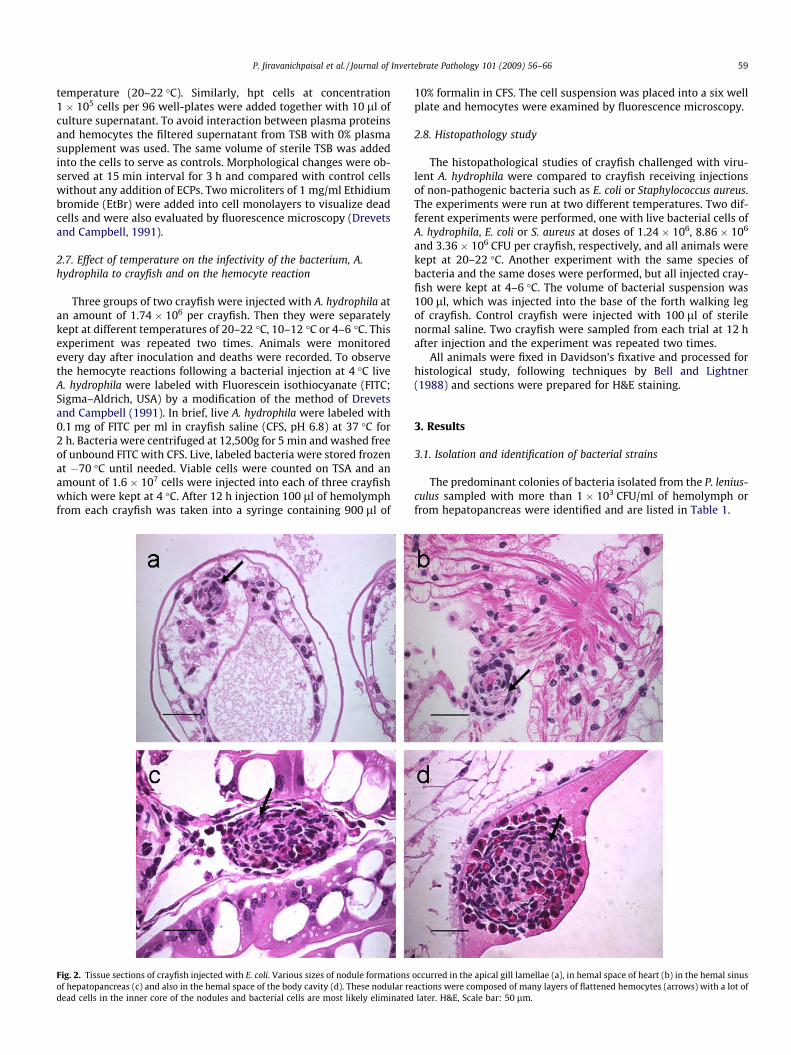
Fig. 3. Tissue sections of crayfish injected with S. aureus. Similar pathological changes asthe nodule formations were present mainly in gill lamellae (a, b and d). Hepatopancrea
from the survivors after 7 days post injection with the avirulentstrains, but we found several extensive black melanized spots tobe present in the hepatopancreas indicating that crayfish mounteda defense response to these avirulent bacteria. However, thesegross signs of melanin deposits were absent in the hepatopancreasof A. hydrophila-injected crayfish.
3.3. Histopathological examinations
At temperatures, 4 or 22 �C, all crayfish injected with the con-trol bacteria E. coli and S. aureus were alive after 12 h injection,whereas all crayfish which received an injection with A. hydrophilaB1 and were kept at 22 �C became moribund at 6 h post injection.Then these crayfish were sacrificed for histopathological studies.Crayfish receiving injections with A. hydrophila showed extensivenecrotic lesions with pyknotic nuclei in various tissues includinggill, heart, interstitial tissue of hepatopancreas, the blood circula-tory system (Fig. 1a–d) and massive hemocytes were aggregatedin the hemal sinuses with presence of many pyknotic nuclei(Fig. 1a–d, arrows). However, no complete nodule formation wasobserved in the lesions of all tissues when injected with A. hydro-phila. In comparison with non-pathogenic bacteria injection,E. coli-injected crayfish showed nodular reactions of hemocytesin the observed tissues including gill, heart, hepatopancreas, aswell as in the hemal sinuses. These nodules were composed ofcompressed multiple hemocyte-cell layers (Fig. 2a–d), which sur-rounded apparently dead hemocytes and most likely also bacteria(Fig. 2a–d, arrows). Similar changes were also found in the S. aur-eus-injected crayfish, but the nodules were present mainly in thegills (Fig. 3a–d). In contrast, all survivors which had been kept at4 �C after the injection had poor or no evidence of hemocytic
in E. coli-injected crayfish were also observed in crayfish injected with S. aureus, buts showing only few hemocytic reactions in hemal sinus (c). H&E, Scale bar: 50 lm.
P. Jiravanichpaisal et al. / Journal of Invertebrate Pathology 101 (2009) 56–66 61
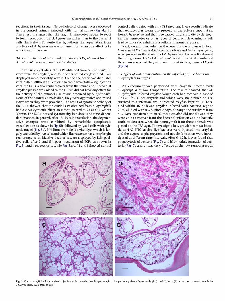
reactions in their tissues. No pathological changes were observedin the control animals injected with normal saline (Fig. 4a–d).These results suggest that the crayfish hemocytes appear to reactto toxins produced from A. hydrophila rather than to the bacterialcells themselves. To verify this hypothesis the supernatant froma culture of A. hydrophila was obtained for testing its effect bothin vitro and in in vivo.
3.4. Toxic activities of extracellular products (ECPs) obtained fromA. hydrophila in in vivo and in vitro studies
In the in vivo studies, the ECPs obtained from A. hydrophila B1were toxic for crayfish, and four of six tested crayfish died. Twodisplayed rapid mortality within 3 h and the other two died laterwithin 46 h. Although all crayfish became weak following injectionwith the ECPs, a few could recover from the toxins and survived. Ifcrayfish plasma was added to the ECPs it did not have any effect forthe activity of the extracellular toxins produced by A. hydrophila.None of the control animals died, they were aggressive and raisedclaws when they were provoked. The result of cytotoxic activity ofthe ECPs showed that the crude ECPs obtained from A. hydrophilahad a clear cytotoxic effect on either isolated SGCs or GCs within30 min. The ECPs induced cytotoxicity in a dose- and time-depen-dent manner. In general, after 15–30 min inoculation, the degener-ative changes were exhibited by remarkable cytoplasmicvacuolization as shown in Fig. 5b, followed by lysed cells with pyk-notic nuclei (Fig. 5c). Ethidium bromide is a vital dye, which is lar-gely excluded by live cells and which fluorescence has a very brightred–orange color. Massive dead cells were displayed by EtBr posi-tive cells after 3 and 6 h post inoculation of ECPs as shown inFig. 5h and l, respectively, while Fig. 5a, e, f, i and j showed normal
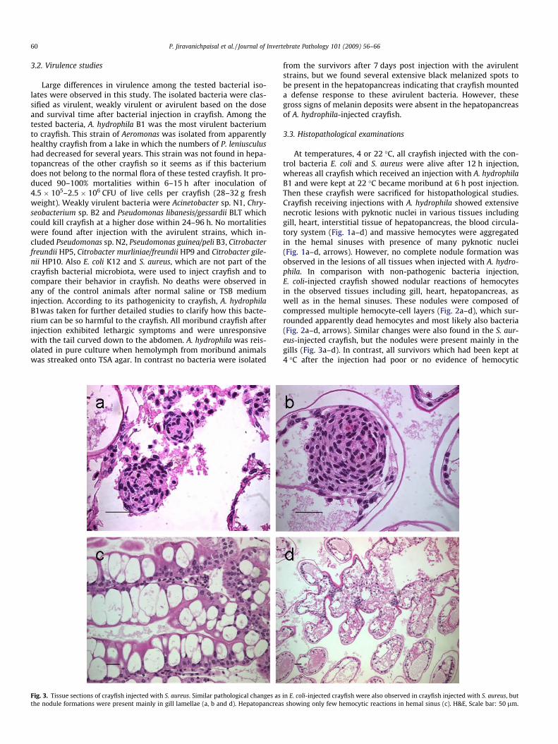
Fig. 4. Control crayfish which received injection with normal saline. No pathological chanobserved H&E, Scale bar: 50 lm.
control cells treated with only TSB medium. These results indicatethat extracellular toxins are present in the culture supernatantfrom A. hydrophila and that they caused crayfish to die by destroy-ing the hemocytes or other types of cells, which eventually willlead to failure of exhibiting a cellular immune response.
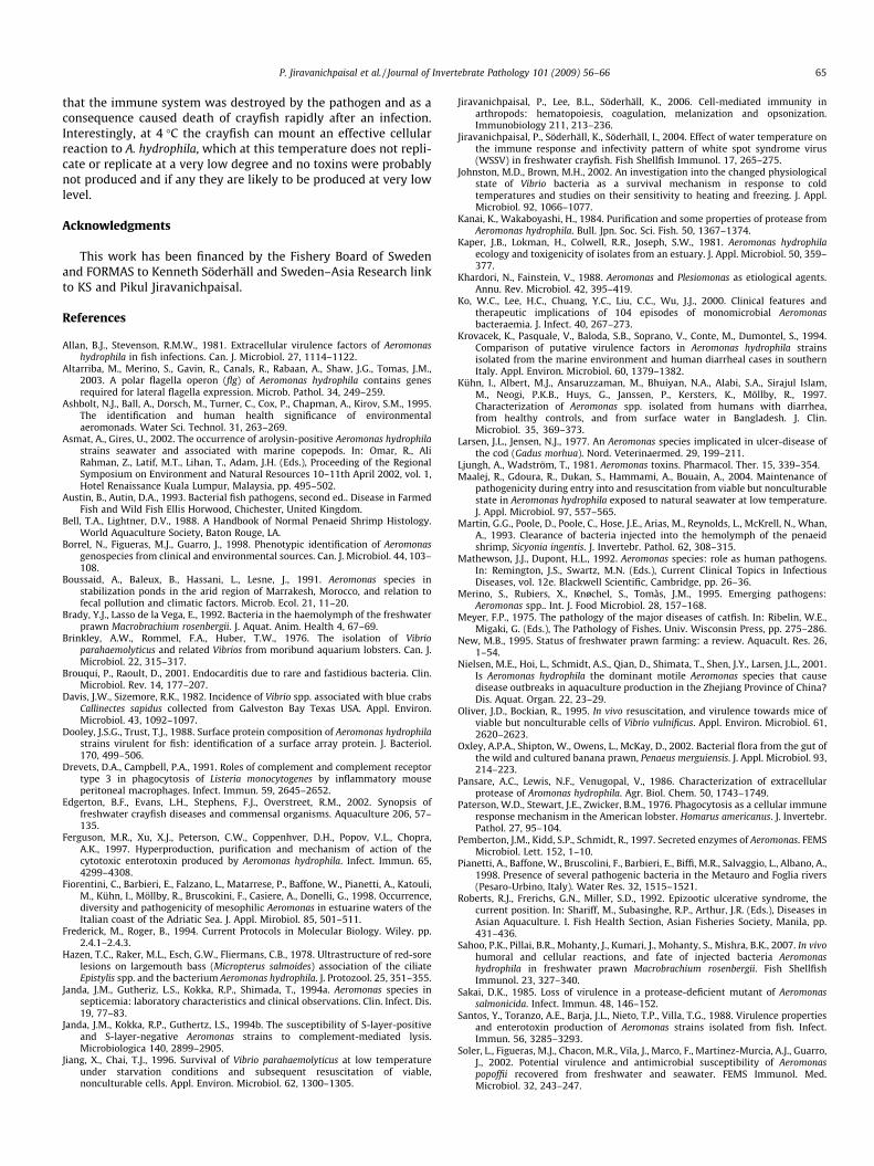
Next, we examined whether the genes for the virulence factors,hlyA gene of V. cholerae-HlyA-like hemolysin and b-hemolysin gene,were present in the genome of A. hydrophila. The results showedthat the genomic DNA of A. hydrophila used in the study containedthese two genes, but they were not present in the genome of E. coli(Fig. 6).
3.5. Effect of water temperature on the infectivity of the bacterium,A. hydrophila to crayfish
An experiment was performed with crayfish infected withA. hydrophila at low temperature. The results showed that allA. hydrophila-infected crayfish which each had received a dose of1.74 � 106 CFU per crayfish and which were maintained at 4 �Csurvived this infection, while infected crayfish kept at 10–12 �Cdied within 36–45 h and crayfish infected with bacteria kept at20 �C all died within 6 h. After 7 days, although the survivors from4 �C were transferred to 20 �C, these crayfish did not die and theywere able to recover from the bacterial infection and no bacteriacould be detected when the hemolymph from these animals wasplated on the TSA agar. To investigate how crayfish combat bacte-ria at 4 �C, FITC-labeled live bacteria were injected into crayfishand the degree of phagocytosis and nodule formation were inves-tigated at different time intervals. After 6–12 h, it was found thatphagocytosis of bacteria (Fig. 7a and b) or nodule formation of bac-teria (Fig. 7c and d) was very effective at the low temperature at
ges in any tissue for example gill (a and d), heart (b) or hepatopancreas (c) could be

Fig. 5. Effect of crude extracellular products (ECPs) obtained from A. hydrophila B1 on hemocytes in vitro. Crayfish hemocytes reacted to the ECPs by cytoplasmic vacuolizationafter 15–30 min. Inoculation with CEPs as shown in Fig. 5b, and this was followed by lysed cells with pyknotic nuclei (Fig. 5c and d). Massive dead cells were increased (g andk) and displayed EtBr positive cells after 3 and 6 h post inoculation as shown in Fig. 5h and l, respectively. While Fig. 5a, e, f, i and j showed control cells treated with only TSBmedium. H&E, Scale bar: 20 lm.
62 P. Jiravanichpaisal et al. / Journal of Invertebrate Pathology 101 (2009) 56–66
4 �C. This is probably the reason why crayfish recover from A.hydrophila infection at this low temperature. The cellular immuneresponse can cope with the injected bacteria, which have a verylow rate of multiplication at this low temperature.
4. Discussion
In a previous report it was demonstrated that in many crayfishspecies including Astacus astacus, Cherax destructoralbidus, Cheraxquadricarinatus and Procambarus clarkii, with apparently healthyconditions, but still they can have a mixed bacterial populationin hemolymph. These bacteria belong to both Gram-negative gen-era and the most frequent species are Pseudomonas, Aeromonas,Acinetobacter, Flavobacterium and Vibrio whereas Micrococcus andStaphylococcus are the most often reported Gram-positive genera(see review in Edgerton et al., 2002; Wong et al., 1995). Bacteria
have also been isolated from the hemolymph of apparently healthyother crustacean species such as spiny lobster, crabs, penaeidshrimp (Edgerton et al., 2002). In addition to bacteremia, mortali-ties associated with enteric bacteria including Citrobacter freundii,Citrobacter sp., Acinetobacterium sp. and Pseudomonas sp., presentin the crayfish gut have been found in several species of Europeanand North American crayfish. Although, infectivity trials with C.freundii cause lesions in normal crayfish if large inoculums areused, the overall infectivity of bacterial isolates was low (Edgertonet al., 2002). Similarly, a variety of bacteria could be isolated fromP. leniusculus hemolymph and in our study, C. freundii and two spe-cies of this bacterium isolated from the hepatopancreas failed tokill crayfish at injection levels of 2.0 � 108 viable cells per crayfish(Table 1). This supports the idea that aquatic crustaceans can toler-ate bacteria in the hemolymph with no apparent detrimental ef-fects, and bacterial presence is not necessarily an indication of
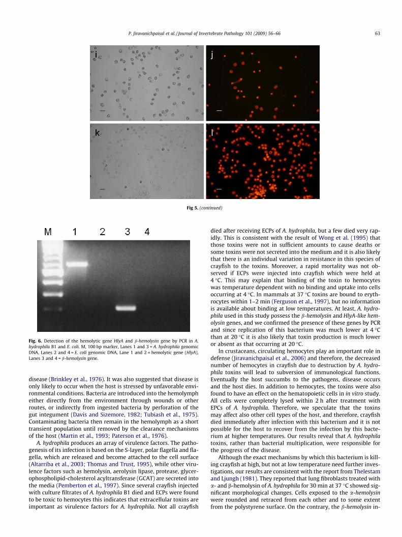
Fig 5. (continued)
Fig. 6. Detection of the hemolytic gene HlyA and b-hemolysin gene by PCR in A.hydrophila B1 and E. coli. M, 100 bp marker, Lanes 1 and 3 = A. hydrophila genomicDNA, Lanes 2 and 4 = E. coli genomic DNA, Lane 1 and 2 = hemolytic gene (HlyA),Lanes 3 and 4 = b-hemolysin gene.
P. Jiravanichpaisal et al. / Journal of Invertebrate Pathology 101 (2009) 56–66 63
disease (Brinkley et al., 1976). It was also suggested that disease isonly likely to occur when the host is stressed by unfavorable envi-ronmental conditions. Bacteria are introduced into the hemolympheither directly from the environment through wounds or otherroutes, or indirectly from ingested bacteria by perforation of thegut integument (Davis and Sizemore, 1982; Tubiash et al., 1975).Contaminating bacteria then remain in the hemolymph as a shorttransient population until removed by the clearance mechanismsof the host (Martin et al., 1993; Paterson et al., 1976).
A. hydrophila produces an array of virulence factors. The patho-genesis of its infection is based on the S-layer, polar flagella and fla-gella, which are released and become attached to the cell surface(Altarriba et al., 2003; Thomas and Trust, 1995), while other viru-lence factors such as hemolysin, aerolysin lipase, protease, glycer-ophospholipid-cholesterol acyltransferase (GCAT) are secreted intothe media (Pemberton et al., 1997). Since several crayfish injectedwith culture filtrates of A. hydrophila B1 died and ECPs were foundto be toxic to hemocytes this indicates that extracellular toxins areimportant as virulence factors for A. hydrophila. Not all crayfish
died after receiving ECPs of A. hydrophila, but a few died very rap-idly. This is consistent with the result of Wong et al. (1995) thatthose toxins were not in sufficient amounts to cause deaths orsome toxins were not secreted into the medium and it is also likelythat there is an individual variation in resistance in this species ofcrayfish to the toxins. Moreover, a rapid mortality was not ob-served if ECPs were injected into crayfish which were held at4 �C. This may explain that binding of the toxin to hemocyteswas temperature dependent with no binding and uptake into cellsoccurring at 4 �C. In mammals at 37 �C toxins are bound to eryth-rocytes within 1–2 min (Ferguson et al., 1997), but no informationis available about binding at low temperatures. At least, A. hydro-phila used in this study possess the b-hemolysin and HlyA-like hem-olysin genes, and we confirmed the presence of these genes by PCRand since replication of this bacterium was much lower at 4 �Cthan at 20 �C it is also likely that toxin production is much loweror absent as that occurring at 20 �C.
In crustaceans, circulating hemocytes play an important role indefense (Jiravanichpaisal et al., 2006) and therefore, the decreasednumber of hemocytes in crayfish due to destruction by A. hydro-phila toxins will lead to subversion of immunological functions.Eventually the host succumbs to the pathogens, disease occursand the host dies. In addition to hemocytes, the toxins were alsofound to have an effect on the hematopoietic cells in in vitro study.All cells were completely lysed within 2 h after treatment withEPCs of A. hydrophila. Therefore, we speculate that the toxinsmay affect also other cell types of the host, and therefore, crayfishdied immediately after infection with this bacterium and it is notpossible for the host to recover from the infection by this bacte-rium at higher temperatures. Our results reveal that A. hydrophilatoxins, rather than bacterial multiplication, were responsible forthe progress of the disease.
Although the exact mechanisms by which this bacterium is kill-ing crayfish at high, but not at low temperature need further inves-tigations, our results are consistent with the report from Thelestamand Ljungh (1981). They reported that lung fibroblasts treated witha- and b-hemolysin of A. hydrophila for 30 min at 37 �C showed sig-nificant morphological changes. Cells exposed to the a-hemolysinwere rounded and retraced from each other and to some extentfrom the polystyrene surface. On the contrary, the b-hemolysin in-
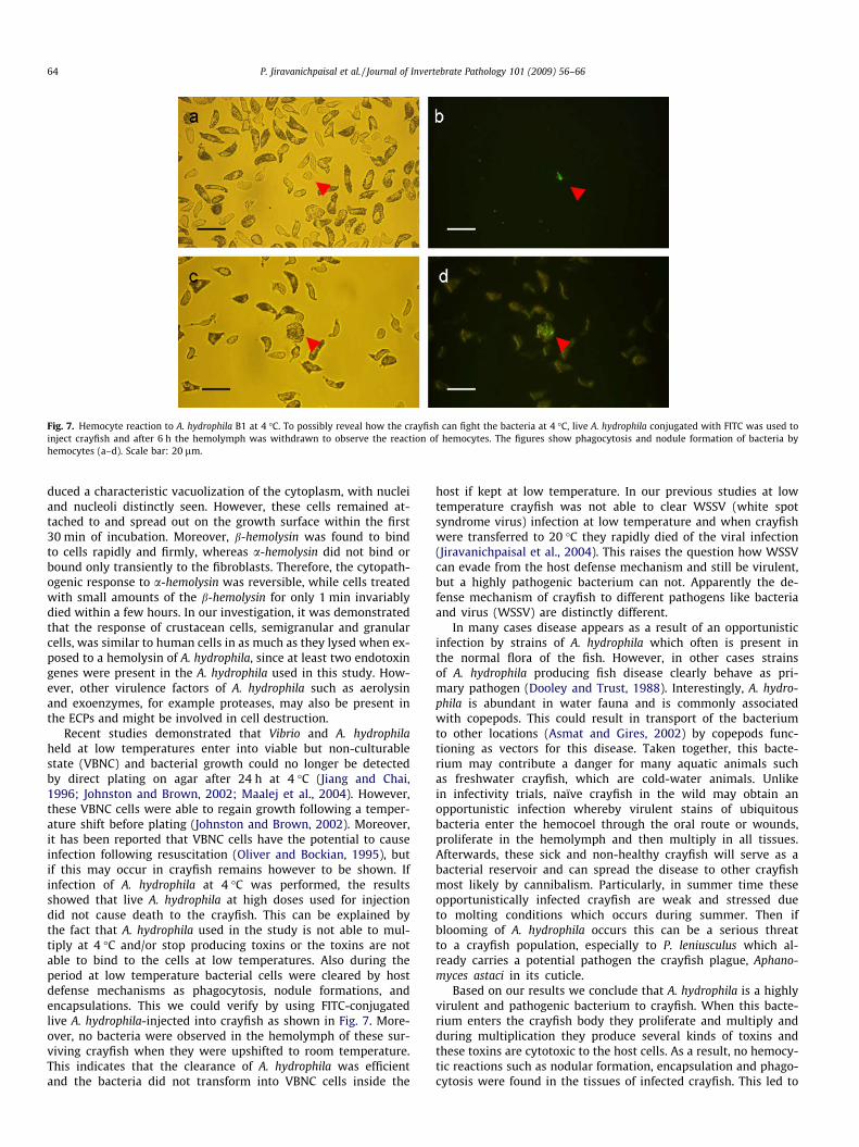
Fig. 7. Hemocyte reaction to A. hydrophila B1 at 4 �C. To possibly reveal how the crayfish can fight the bacteria at 4 �C, live A. hydrophila conjugated with FITC was used toinject crayfish and after 6 h the hemolymph was withdrawn to observe the reaction of hemocytes. The figures show phagocytosis and nodule formation of bacteria byhemocytes (a–d). Scale bar: 20 lm.
64 P. Jiravanichpaisal et al. / Journal of Invertebrate Pathology 101 (2009) 56–66
duced a characteristic vacuolization of the cytoplasm, with nucleiand nucleoli distinctly seen. However, these cells remained at-tached to and spread out on the growth surface within the first30 min of incubation. Moreover, b-hemolysin was found to bindto cells rapidly and firmly, whereas a-hemolysin did not bind orbound only transiently to the fibroblasts. Therefore, the cytopath-ogenic response to a-hemolysin was reversible, while cells treatedwith small amounts of the b-hemolysin for only 1 min invariablydied within a few hours. In our investigation, it was demonstratedthat the response of crustacean cells, semigranular and granularcells, was similar to human cells in as much as they lysed when ex-posed to a hemolysin of A. hydrophila, since at least two endotoxingenes were present in the A. hydrophila used in this study. How-ever, other virulence factors of A. hydrophila such as aerolysinand exoenzymes, for example proteases, may also be present inthe ECPs and might be involved in cell destruction.
Recent studies demonstrated that Vibrio and A. hydrophilaheld at low temperatures enter into viable but non-culturablestate (VBNC) and bacterial growth could no longer be detectedby direct plating on agar after 24 h at 4 �C (Jiang and Chai,1996; Johnston and Brown, 2002; Maalej et al., 2004). However,these VBNC cells were able to regain growth following a temper-ature shift before plating (Johnston and Brown, 2002). Moreover,it has been reported that VBNC cells have the potential to causeinfection following resuscitation (Oliver and Bockian, 1995), butif this may occur in crayfish remains however to be shown. Ifinfection of A. hydrophila at 4 �C was performed, the resultsshowed that live A. hydrophila at high doses used for injectiondid not cause death to the crayfish. This can be explained bythe fact that A. hydrophila used in the study is not able to mul-tiply at 4 �C and/or stop producing toxins or the toxins are notable to bind to the cells at low temperatures. Also during theperiod at low temperature bacterial cells were cleared by hostdefense mechanisms as phagocytosis, nodule formations, andencapsulations. This we could verify by using FITC-conjugatedlive A. hydrophila-injected into crayfish as shown in Fig. 7. More-over, no bacteria were observed in the hemolymph of these sur-viving crayfish when they were upshifted to room temperature.This indicates that the clearance of A. hydrophila was efficientand the bacteria did not transform into VBNC cells inside the
host if kept at low temperature. In our previous studies at lowtemperature crayfish was not able to clear WSSV (white spotsyndrome virus) infection at low temperature and when crayfishwere transferred to 20 �C they rapidly died of the viral infection(Jiravanichpaisal et al., 2004). This raises the question how WSSVcan evade from the host defense mechanism and still be virulent,but a highly pathogenic bacterium can not. Apparently the de-fense mechanism of crayfish to different pathogens like bacteriaand virus (WSSV) are distinctly different.
In many cases disease appears as a result of an opportunisticinfection by strains of A. hydrophila which often is present inthe normal flora of the fish. However, in other cases strainsof A. hydrophila producing fish disease clearly behave as pri-mary pathogen (Dooley and Trust, 1988). Interestingly, A. hydro-phila is abundant in water fauna and is commonly associatedwith copepods. This could result in transport of the bacteriumto other locations (Asmat and Gires, 2002) by copepods func-tioning as vectors for this disease. Taken together, this bacte-rium may contribute a danger for many aquatic animals suchas freshwater crayfish, which are cold-water animals. Unlikein infectivity trials, naïve crayfish in the wild may obtain anopportunistic infection whereby virulent stains of ubiquitousbacteria enter the hemocoel through the oral route or wounds,proliferate in the hemolymph and then multiply in all tissues.Afterwards, these sick and non-healthy crayfish will serve as abacterial reservoir and can spread the disease to other crayfishmost likely by cannibalism. Particularly, in summer time theseopportunistically infected crayfish are weak and stressed dueto molting conditions which occurs during summer. Then ifblooming of A. hydrophila occurs this can be a serious threatto a crayfish population, especially to P. leniusculus which al-ready carries a potential pathogen the crayfish plague, Aphano-myces astaci in its cuticle.
Based on our results we conclude that A. hydrophila is a highlyvirulent and pathogenic bacterium to crayfish. When this bacte-rium enters the crayfish body they proliferate and multiply andduring multiplication they produce several kinds of toxins andthese toxins are cytotoxic to the host cells. As a result, no hemocy-tic reactions such as nodular formation, encapsulation and phago-cytosis were found in the tissues of infected crayfish. This led to
P. Jiravanichpaisal et al. / Journal of Invertebrate Pathology 101 (2009) 56–66 65
that the immune system was destroyed by the pathogen and as aconsequence caused death of crayfish rapidly after an infection.Interestingly, at 4 �C the crayfish can mount an effective cellularreaction to A. hydrophila, which at this temperature does not repli-cate or replicate at a very low degree and no toxins were probablynot produced and if any they are likely to be produced at very lowlevel.
Acknowledgments
This work has been financed by the Fishery Board of Swedenand FORMAS to Kenneth Söderhäll and Sweden–Asia Research linkto KS and Pikul Jiravanichpaisal.
References
Allan, B.J., Stevenson, R.M.W., 1981. Extracellular virulence factors of Aeromonashydrophila in fish infections. Can. J. Microbiol. 27, 1114–1122.
Altarriba, M., Merino, S., Gavin, R., Canals, R., Rabaan, A., Shaw, J.G., Tomas, J.M.,2003. A polar flagella operon (flg) of Aeromonas hydrophila contains genesrequired for lateral flagella expression. Microb. Pathol. 34, 249–259.
Ashbolt, N.J., Ball, A., Dorsch, M., Turner, C., Cox, P., Chapman, A., Kirov, S.M., 1995.The identification and human health significance of environmentalaeromonads. Water Sci. Technol. 31, 263–269.
Asmat, A., Gires, U., 2002. The occurrence of arolysin-positive Aeromonas hydrophilastrains seawater and associated with marine copepods. In: Omar, R., AliRahman, Z., Latif, M.T., Lihan, T., Adam, J.H. (Eds.), Proceeding of the RegionalSymposium on Environment and Natural Resources 10–11th April 2002, vol. 1,Hotel Renaissance Kuala Lumpur, Malaysia, pp. 495–502.
Austin, B., Autin, D.A., 1993. Bacterial fish pathogens, second ed.. Disease in FarmedFish and Wild Fish Ellis Horwood, Chichester, United Kingdom.
Bell, T.A., Lightner, D.V., 1988. A Handbook of Normal Penaeid Shrimp Histology.World Aquaculture Society, Baton Rouge, LA.
Borrel, N., Figueras, M.J., Guarro, J., 1998. Phenotypic identification of Aeromonasgenospecies from clinical and environmental sources. Can. J. Microbiol. 44, 103–108.
Boussaid, A., Baleux, B., Hassani, L., Lesne, J., 1991. Aeromonas species instabilization ponds in the arid region of Marrakesh, Morocco, and relation tofecal pollution and climatic factors. Microb. Ecol. 21, 11–20.
Brady, Y.J., Lasso de la Vega, E., 1992. Bacteria in the haemolymph of the freshwaterprawn Macrobrachium rosenbergii. J. Aquat. Anim. Health 4, 67–69.
Brinkley, A.W., Rommel, F.A., Huber, T.W., 1976. The isolation of Vibrioparahaemolyticus and related Vibrios from moribund aquarium lobsters. Can. J.Microbiol. 22, 315–317.
Brouqui, P., Raoult, D., 2001. Endocarditis due to rare and fastidious bacteria. Clin.Microbiol. Rev. 14, 177–207.
Davis, J.W., Sizemore, R.K., 1982. Incidence of Vibrio spp. associated with blue crabsCallinectes sapidus collected from Galveston Bay Texas USA. Appl. Environ.Microbiol. 43, 1092–1097.
Dooley, J.S.G., Trust, T.J., 1988. Surface protein composition of Aeromonas hydrophilastrains virulent for fish: identification of a surface array protein. J. Bacteriol.170, 499–506.
Drevets, D.A., Campbell, P.A., 1991. Roles of complement and complement receptortype 3 in phagocytosis of Listeria monocytogenes by inflammatory mouseperitoneal macrophages. Infect. Immun. 59, 2645–2652.
Edgerton, B.F., Evans, L.H., Stephens, F.J., Overstreet, R.M., 2002. Synopsis offreshwater crayfish diseases and commensal organisms. Aquaculture 206, 57–135.
Ferguson, M.R., Xu, X.J., Peterson, C.W., Coppenhver, D.H., Popov, V.L., Chopra,A.K., 1997. Hyperproduction, purification and mechanism of action of thecytotoxic enterotoxin produced by Aeromonas hydrophila. Infect. Immun. 65,4299–4308.
Fiorentini, C., Barbieri, E., Falzano, L., Matarrese, P., Baffone, W., Pianetti, A., Katouli,M., Kühn, I., Möllby, R., Bruscokini, F., Casiere, A., Donelli, G., 1998. Occurrence,diversity and pathogenicity of mesophilic Aeromonas in estuarine waters of theItalian coast of the Adriatic Sea. J. Appl. Mirobiol. 85, 501–511.
Frederick, M., Roger, B., 1994. Current Protocols in Molecular Biology. Wiley. pp.2.4.1–2.4.3.
Hazen, T.C., Raker, M.L., Esch, G.W., Fliermans, C.B., 1978. Ultrastructure of red-sorelesions on largemouth bass (Micropterus salmoides) association of the ciliateEpistylis spp. and the bacterium Aeromonas hydrophila. J. Protozool. 25, 351–355.
Janda, J.M., Gutheriz, L.S., Kokka, R.P., Shimada, T., 1994a. Aeromonas species insepticemia: laboratory characteristics and clinical observations. Clin. Infect. Dis.19, 77–83.
Janda, J.M., Kokka, R.P., Guthertz, l.S., 1994b. The susceptibility of S-layer-positiveand S-layer-negative Aeromonas strains to complement-mediated lysis.Microbiologica 140, 2899–2905.
Jiang, X., Chai, T.J., 1996. Survival of Vibrio parahaemolyticus at low temperatureunder starvation conditions and subsequent resuscitation of viable,nonculturable cells. Appl. Environ. Microbiol. 62, 1300–1305.
Jiravanichpaisal, P., Lee, B.L., Söderhäll, K., 2006. Cell-mediated immunity inarthropods: hematopoiesis, coagulation, melanization and opsonization.Immunobiology 211, 213–236.
Jiravanichpaisal, P., Söderhäll, K., Söderhäll, I., 2004. Effect of water temperature onthe immune response and infectivity pattern of white spot syndrome virus(WSSV) in freshwater crayfish. Fish Shellfish Immunol. 17, 265–275.
Johnston, M.D., Brown, M.H., 2002. An investigation into the changed physiologicalstate of Vibrio bacteria as a survival mechanism in response to coldtemperatures and studies on their sensitivity to heating and freezing. J. Appl.Microbiol. 92, 1066–1077.
Kanai, K., Wakaboyashi, H., 1984. Purification and some properties of protease fromAeromonas hydrophila. Bull. Jpn. Soc. Sci. Fish. 50, 1367–1374.
Kaper, J.B., Lokman, H., Colwell, R.R., Joseph, S.W., 1981. Aeromonas hydrophilaecology and toxigenicity of isolates from an estuary. J. Appl. Microbiol. 50, 359–377.
Khardori, N., Fainstein, V., 1988. Aeromonas and Plesiomonas as etiological agents.Annu. Rev. Microbiol. 42, 395–419.
Ko, W.C., Lee, H.C., Chuang, Y.C., Liu, C.C., Wu, J.J., 2000. Clinical features andtherapeutic implications of 104 episodes of monomicrobial Aeromonasbacteraemia. J. Infect. 40, 267–273.
Krovacek, K., Pasquale, V., Baloda, S.B., Soprano, V., Conte, M., Dumontel, S., 1994.Comparison of putative virulence factors in Aeromonas hydrophila strainsisolated from the marine environment and human diarrheal cases in southernItaly. Appl. Environ. Microbiol. 60, 1379–1382.
Kühn, I., Albert, M.J., Ansaruzzaman, M., Bhuiyan, N.A., Alabi, S.A., Sirajul Islam,M., Neogi, P.K.B., Huys, G., Janssen, P., Kersters, K., Möllby, R., 1997.Characterization of Aeromonas spp. isolated from humans with diarrhea,from healthy controls, and from surface water in Bangladesh. J. Clin.Microbiol. 35, 369–373.
Larsen, J.L., Jensen, N.J., 1977. An Aeromonas species implicated in ulcer-disease ofthe cod (Gadus morhua). Nord. Veterinaermed. 29, 199–211.
Ljungh, A., Wadström, T., 1981. Aeromonas toxins. Pharmacol. Ther. 15, 339–354.Maalej, R., Gdoura, R., Dukan, S., Hammami, A., Bouain, A., 2004. Maintenance of
pathogenicity during entry into and resuscitation from viable but nonculturablestate in Aeromonas hydrophila exposed to natural seawater at low temperature.J. Appl. Microbiol. 97, 557–565.
Martin, G.G., Poole, D., Poole, C., Hose, J.E., Arias, M., Reynolds, L., McKrell, N., Whan,A., 1993. Clearance of bacteria injected into the hemolymph of the penaeidshrimp, Sicyonia ingentis. J. Invertebr. Pathol. 62, 308–315.
Mathewson, J.J., Dupont, H.L., 1992. Aeromonas species: role as human pathogens.In: Remington, J.S., Swartz, M.N. (Eds.), Current Clinical Topics in InfectiousDiseases, vol. 12e. Blackwell Scientific, Cambridge, pp. 26–36.
Merino, S., Rubiers, X., Knøchel, S., Tomàs, J.M., 1995. Emerging pathogens:Aeromonas spp.. Int. J. Food Microbiol. 28, 157–168.
Meyer, F.P., 1975. The pathology of the major diseases of catfish. In: Ribelin, W.E.,Migaki, G. (Eds.), The Pathology of Fishes. Univ. Wisconsin Press, pp. 275–286.
New, M.B., 1995. Status of freshwater prawn farming: a review. Aquacult. Res. 26,1–54.
Nielsen, M.E., Hoi, L., Schmidt, A.S., Qian, D., Shimata, T., Shen, J.Y., Larsen, J.L., 2001.Is Aeromonas hydrophila the dominant motile Aeromonas species that causedisease outbreaks in aquaculture production in the Zhejiang Province of China?Dis. Aquat. Organ. 22, 23–29.
Oliver, J.D., Bockian, R., 1995. In vivo resuscitation, and virulence towards mice ofviable but nonculturable cells of Vibrio vulnificus. Appl. Environ. Microbiol. 61,2620–2623.
Oxley, A.P.A., Shipton, W., Owens, L., McKay, D., 2002. Bacterial flora from the gut ofthe wild and cultured banana prawn, Penaeus merguiensis. J. Appl. Microbiol. 93,214–223.
Pansare, A.C., Lewis, N.F., Venugopal, V., 1986. Characterization of extracellularprotease of Aromonas hydrophila. Agr. Biol. Chem. 50, 1743–1749.
Paterson, W.D., Stewart, J.E., Zwicker, B.M., 1976. Phagocytosis as a cellular immuneresponse mechanism in the American lobster. Homarus americanus. J. Invertebr.Pathol. 27, 95–104.
Pemberton, J.M., Kidd, S.P., Schmidt, R., 1997. Secreted enzymes of Aeromonas. FEMSMicrobiol. Lett. 152, 1–10.
Pianetti, A., Baffone, W., Bruscolini, F., Barbieri, E., Biffi, M.R., Salvaggio, L., Albano, A.,1998. Presence of several pathogenic bacteria in the Metauro and Foglia rivers(Pesaro-Urbino, Italy). Water Res. 32, 1515–1521.
Roberts, R.J., Frerichs, G.N., Miller, S.D., 1992. Epizootic ulcerative syndrome, thecurrent position. In: Shariff, M., Subasinghe, R.P., Arthur, J.R. (Eds.), Diseases inAsian Aquaculture. I. Fish Health Section, Asian Fisheries Society, Manila, pp.431–436.
Sahoo, P.K., Pillai, B.R., Mohanty, J., Kumari, J., Mohanty, S., Mishra, B.K., 2007. In vivohumoral and cellular reactions, and fate of injected bacteria Aeromonashydrophila in freshwater prawn Macrobrachium rosenbergii. Fish ShellfishImmunol. 23, 327–340.
Sakai, D.K., 1985. Loss of virulence in a protease-deficient mutant of Aeromonassalmonicida. Infect. Immun. 48, 146–152.
Santos, Y., Toranzo, A.E., Barja, J.L., Nieto, T.P., Villa, T.G., 1988. Virulence propertiesand enterotoxin production of Aeromonas strains isolated from fish. Infect.Immun. 56, 3285–3293.
Soler, L., Figueras, M.J., Chacon, M.R., Vila, J., Marco, F., Martinez-Murcia, A.J., Guarro,J., 2002. Potential virulence and antimicrobial susceptibility of Aeromonaspopoffii recovered from freshwater and seawater. FEMS Immunol. Med.Microbiol. 32, 243–247.
66 P. Jiravanichpaisal et al. / Journal of Invertebrate Pathology 101 (2009) 56–66
Sung, H.H., Hwang, S.F., Tasi, F.M., 2000. Responses of giant freshwater prawn(Macrobrachium rosenbergii) to challenge by strains of Aeromonas spp. J.Invertebr. Pathol. 76, 278–284.
Söderhäll, I., Kim, Y.A., Jiravanichpaisal, P., Lee, S.Y., Söderhäll, K., 2005. An ancientrole for a prokinecticin domain in invertebrate hematopoiesis. J. Immunol. 174,6153–6160.
Söderhäll, K., Smith, V.J., 1983. Separation of hemocyte population of Carcinusmaenas and other marine decapods and prophenoloxidase distribution. Dev.Comp. Immunol. 7, 229–239.
Thelestam, M., Ljungh, Å., 1981. Membrane-damaging and cytotoxic effects onhuman fibroblasts of alpha- and beta-hemolysins from Aeromonas hydrophila.Infect. Immun. 34, 949–956.
Thomas, S.R., Trust, T.J., 1995. A specific PulD homolog is required for the secretionof paracrystalline surface array subunits in Aeromonas hydrophila. J. Bacteriol.177, 3932–3939.
Thune, R.L., Johnson, M.C., Graham, T.E., Amborski, R.L., 1986. Aeromonas hydrophilab-haemolysin: purification and examination of its role in virulence in O-groupchannel catfish, Ictalurus punctatus (Rafinesque). J. Fish Dis. 9, 55–61.
Thune, R.L., Stanley, L.A., Cooper, R.K., 1993. Pathogenesis of Gram-negativebacterial infection in warm-water fish. Annu. Rev. Fish Dis. 3, 37–68.
Tubiash, H.S., Sizemore, R.K., Colwell, R.K., 1975. Bacterial flora of the hemolymph ofthe blue crab, Calinectes sapidus: most probable numbers. Appl. Microbiol. 29,388–392.
Van der Kooj, D., 1988. Properties of Aeromonas and their occurrence and hygienicsignificance in drinking water. Zentralb. Bakt. Hyg. B. 187, 1–17.
Vivas, J., Carracedo, B., Riano, J., Razquin, B.E., Lopez-Fierro, P., Acosta, F., Naharro, G.,Villena, A.J., 2004. Behavior of an Aeromonas hydrophila aroA live vaccine inwater microcosms. Appl. Environ. Microbiol. 70, 2702–2708.
Wadström, T., Ljungh, A., 1991. Aeromonas and Plesiomonas as food and water bornepathogens. Int. J. Food Microbiol. 12, 303–312.
Wong, C.Y.F., Heuzenroeder, M.W., Flower, R.L.P., 1998. Inactivation of twohaemolytic toxin genes in Aeromonas hydrophila attenuates virulence in asuckling mouse model. Microbiologica 144, 291–298.
Wong, F.Y.K., Fowler, K., Desmarchelier, P.M., 1995. Vibriosis due to Vibrio mimicusin Australian freshwater crayfish. J. Aquat. Anim. Health 7, 284–291.
Xia, C., Ma, Z.-H., Rahman, M.H., Wu, Z.-G., 2004. PCR cloning and identification ofthe b-haemolysin gene of Aeromonas hydrophila from freshwater fishes in China.Aquaculture 229, 45–53.
Yasuda, K., Kitao, T., 1980. Bacterial flora in the digestive tract of prawns, Penaeusjaponicus Bate. Aquaculture 19, 229–234.