Sulfate restriction induces hyposecretion of the adhesion proteoglycan and cell hypomotility...
19
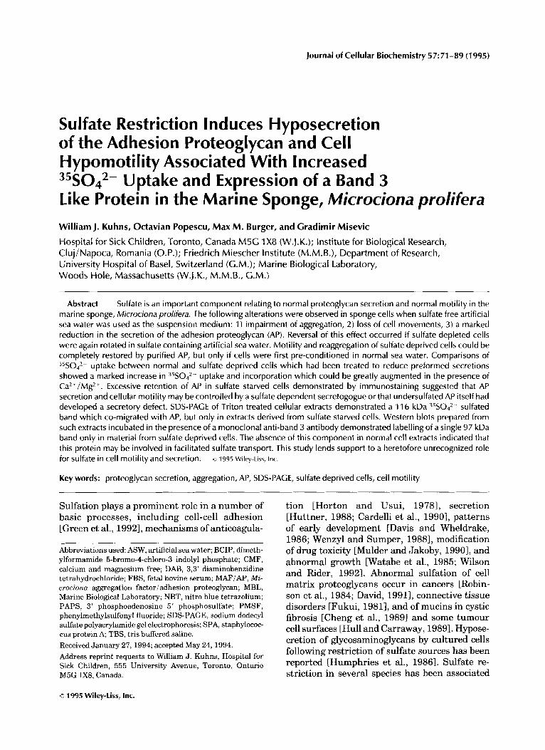
Journal of Cellular Biochemistry 57:71-89 (1995) Sulfate Restriction Induces Hyposecretion of the Adhesion Proteoglycan and Cell Hypomotility Associated With Increased 35S042- Uptake and Expression of a Band 3 Like Protein in the Marine Sponge, Microciona prolifera William J. Kuhns, Octavian Popescu, Max M. Burger, and Cradimir Misevic Hospital for Sick Children, Toronto, Canada M 5 G 1 X8 (W.J.K.); Institute for Biological Research, Cluj/Napoca, Romania (O.P.); Friedrich Miescher Institute (M.M.B.), Department of Research, University Hospital of Basel, Switzerland (C.M.); Marine Biological Laboratory, Woods Hole, Massachusetts (W.J.K., M.M.B., G.M.) Abstract Sulfate is an important component relating to normal proteoglycan secretion and normal motility in the marine sponge, Microciona prolifera. The following alterations were observed in sponge cells when sulfate free artificial sea water was used as the suspension medium: 1) impairment of aggregation, 2) loss of cell movements, 3) a marked reduction in the secretion of the adhesion proteoglycan (AP). Reversal of this effect occurred if sulfate depleted cells were again rotated in sulfate containing artificial sea water. Motility and reaggregationof sulfate deprived cells could be completely restored by purified AP, but only if cells were first pre-conditioned in normal sea water. Comparisons of 35S042- uptake between normal and sulfate deprived cells which had been treated to reduce preformed secretions showed a marked increase in 35S042- uptake and incorporation which could be greatly augmented in the presence of Ca2+/MgZ+. Excessive retention of AP in sulfate starved cells demonstrated by immunostaining suggested that AP secretion and cellular motility may be controlled by a sulfate dependent secretogogue or that undersulfatedAP itself had developed a secretory defect. SDS-PAGE of Triton treated cellular extracts demonstrated a 11 6 kDa 35S042- sulfated band which co-migrated with AP, but only in extracts derived from sulfate starved cells. Western blots prepared from such extracts incubated in the presence of a monoclonal anti-band 3 antibody demonstrated labelling of a single 97 kDa band only in material from sulfate deprived cells. The absence of this component in normal cell extracts indicated that this protein may be involved in facilitated sulfate transport. This study lends support to a heretofore unrecognized role for sulfate in cell motility and secretion. Key words: proteoglycan secretion, aggregation, AP, SDS-PAGE, sulfate deprived cells, cell motility o 1995 WiIey-Liss, Inc. Sulfation plays a prominent role in a number of basic processes, including cell-cell adhesion [Green et al., 19921,mechanisms of anticoagula- ~ Abbreviations used: ASW, artificial sea water; BCIP, dimeth- ylformamide 5-bromo-4-chloro-3 indolyl phosphate; CMF, calcium and magnesium free; DAB, 3,3' diaminobenzidine tetrahydrochloride; FBS, fetal bovine serum; MAFIAP, Mi- crociona aggregation factoriadhesion proteoglycan; MBL, Marine Biological Laboratory; NBT, nitro blue tetrazolium; PAPS, 3' phosphoadenosine 5' phosphosulfate; PMSF, phenylmethylsulfonyl fluoride; SDS-PAGE, sodium dodecyl sulfate polyacrylamide gel electrophoresis; SPA, staphylococ- cus protein A, TBS, tris buffered saline. Received January 27, 1994; accepted May 24, 1994. Address reprint requests to William J. Kuhns, Hospital for Sick Children, 555 University Avenue, Toronto, Ontario M5G 1x8. Canada. tion [Horton and Usui, 19781, secretion [Huttner, 1988; Cardelli et al., 19901, patterns of early development [Davis and Wheldrake, 1986; Wenzyl and Sumper, 19881, modification of drug toxicity [Mulder and Jakoby, 19901, and abnormal growth CWatabe et al., 1985; Wilson and Rider, 19921. Abnormal sulfation of cell matrix proteoglycans occur in cancers [Robin- son et al., 1984; David, 19911, connective tissue disorders [Fukui, 19811, and of mucins in cystic fibrosis [Cheng et al., 19891 and some tumour cell surfaces [Hull and Carraway, 19891.Hypose- cretion of glycosaminoglycans by cultured cells following restriction of sulfate sources has been reported [Humphries et al., 19861. Sulfate re- striction in several species has been associated o 1995 Wiley-Liss, Inc.
-
Upload
independent -
Category
Documents
-
view
1 -
download
0
Transcript of Sulfate restriction induces hyposecretion of the adhesion proteoglycan and cell hypomotility...
Journal of Cellular Biochemistry 57:71-89 (1995)
Sulfate Restriction Induces Hyposecretion of the Adhesion Proteoglycan and Cell Hypomotility Associated With Increased 35S042- Uptake and Expression of a Band 3 Like Protein in the Marine Sponge, Microciona prolifera William J. Kuhns, Octavian Popescu, Max M. Burger, and Cradimir Misevic
Hospital for Sick Children, Toronto, Canada M 5 G 1 X8 (W.J.K.); Institute for Biological Research, Cluj/Napoca, Romania (O.P.); Friedrich Miescher Institute (M.M.B.), Department of Research, University Hospital of Basel, Switzerland (C.M.); Marine Biological Laboratory, Woods Hole, Massachusetts (W.J.K., M.M.B., G.M.)
Abstract Sulfate is an important component relating to normal proteoglycan secretion and normal motility in the marine sponge, Microciona prolifera. The following alterations were observed in sponge cells when sulfate free artificial sea water was used as the suspension medium: 1) impairment of aggregation, 2) loss of cell movements, 3) a marked reduction in the secretion of the adhesion proteoglycan (AP). Reversal of this effect occurred if sulfate depleted cells were again rotated in sulfate containing artificial sea water. Motility and reaggregation of sulfate deprived cells could be completely restored by purified AP, but only if cells were first pre-conditioned in normal sea water. Comparisons of 35S042- uptake between normal and sulfate deprived cells which had been treated to reduce preformed secretions showed a marked increase in 35S042- uptake and incorporation which could be greatly augmented in the presence of Ca2+/MgZ+. Excessive retention of AP in sulfate starved cells demonstrated by immunostaining suggested that AP secretion and cellular motility may be controlled by a sulfate dependent secretogogue or that undersulfated AP itself had developed a secretory defect. SDS-PAGE of Triton treated cellular extracts demonstrated a 11 6 kDa 35S042- sulfated band which co-migrated with AP, but only in extracts derived from sulfate starved cells. Western blots prepared from such extracts incubated in the presence of a monoclonal anti-band 3 antibody demonstrated labelling of a single 97 kDa band only in material from sulfate deprived cells. The absence of this component in normal cell extracts indicated that this protein may be involved in facilitated sulfate transport. This study lends support to a heretofore unrecognized role for sulfate in cell motility and secretion.
Key words: proteoglycan secretion, aggregation, AP, SDS-PAGE, sulfate deprived cells, cell motility
o 1995 WiIey-Liss, Inc.
Sulfation plays a prominent role in a number of basic processes, including cell-cell adhesion [Green et al., 19921, mechanisms of anticoagula-
~
Abbreviations used: ASW, artificial sea water; BCIP, dimeth- ylformamide 5-bromo-4-chloro-3 indolyl phosphate; CMF, calcium and magnesium free; DAB, 3,3' diaminobenzidine tetrahydrochloride; FBS, fetal bovine serum; M A F I A P , Mi- crociona aggregation factoriadhesion proteoglycan; MBL, Marine Biological Laboratory; NBT, nitro blue tetrazolium; PAPS, 3' phosphoadenosine 5' phosphosulfate; PMSF, phenylmethylsulfonyl fluoride; SDS-PAGE, sodium dodecyl sulfate polyacrylamide gel electrophoresis; SPA, staphylococ- cus protein A, TBS, tris buffered saline. Received January 27, 1994; accepted May 24, 1994. Address reprint requests to William J. Kuhns, Hospital for Sick Children, 555 University Avenue, Toronto, Ontario M5G 1x8. Canada.
tion [Horton and Usui, 19781, secretion [Huttner, 1988; Cardelli et al., 19901, patterns of early development [Davis and Wheldrake, 1986; Wenzyl and Sumper, 19881, modification of drug toxicity [Mulder and Jakoby, 19901, and abnormal growth CWatabe et al., 1985; Wilson and Rider, 19921. Abnormal sulfation of cell matrix proteoglycans occur in cancers [Robin- son et al., 1984; David, 19911, connective tissue disorders [Fukui, 19811, and of mucins in cystic fibrosis [Cheng et al., 19891 and some tumour cell surfaces [Hull and Carraway, 19891. Hypose- cretion of glycosaminoglycans by cultured cells following restriction of sulfate sources has been reported [Humphries et al., 19861. Sulfate re- striction in several species has been associated
o 1995 Wiley-Liss, Inc.
72 Kuhns et al.
with a reduction in cell motility and cell migra- tion [Venkatasubramanian and Solursh, 1984; Davis and Wheldrake, 19861. In at least one experimental system, the sea urchin embryo, cell motility could be restored when sulfate de- prived cells were again placed in normal sea water [Venkatasubramanian and Solursh, 19841.
Marine sponges, especially Microciona prolif- era, provide an excellent model to study struc- ture-function aspects of sulfation. The cells are embedded in an abundant extracellular matrix originally termed aggregation factor [Hum- phreys, 19631, and motility studies can be readily carried out on chemically disaggregated cells. Experimental procedures are simplified because controlled experiments can be carried out in artificial seawater (ASW) with or without added sulfate. In addition, early larval forms are preva- lent in the late spring and summer, particu- larly in late June and early July [Simpson, 1968, 19841, thus permitting comparative studies of sulfation in early development.
In the experiments to be reported, our origi- nal findings have been confirmed and extended. Time lapse photography of disaggregated nor- mal and sulfate deprived cells has permitted detailed visual comparison of the effects of sul- fate deprivation upon aggregation and cell motil- ity. Incorporation of 35S042- has been exten- sively studied mostly in summer sponges, and conditions have been modified to optimize 35s042- uptake in sulfate poor cells. The avail- ability of antibodies prepared against secretory AP has enabled us to identify this component by immunostaining methods both in intact cells and in extracts derived from cells in normal or sulfate free medium. This was coupled with the appearance in (-)S042- extracts of a 116 kDa 35S042- sulfated band identified by autoradiogra- phy following gel electrophoresis. Western blot of electrophoretically separated ( - )S042- ex- tracts using anti-band 3 monoclonal antibody identified a single band which migrated at ca. 97 kDa.
MATERIALS AND METHODS Sponges
Specimens of live Microciona proljfera and of Haliclona occulata were collected in the Woods Hole area by the staff of the Marine Resources Center of the Marine Biological Laboratory dur- ing the months of July and August. Sponge samples were utilized for experiments on the day of collection or the following day, but could
be maintained in healthy condition for several days at ambient temperature in tanks of run- ning seawater. Buffer and artificial sea water (ASW) preparations were made up as described previously [Cavanaugh, 19641. Dissociation of sponge cells and aggregation factor assays are described in earlier reports [Humphreys, 19631.
Preparation of Artificial Sea Water With Designated Sulfate Content and Quantitative Sulfate Assays
Artificial sea water (ASW) with or without Ca2+/Mg2+ (MBLSW/CMFSW) was prepared from the highest quality reagent salts contain- ing trace amounts of sulfate (NaC11.5 ppm, KC1 2 ppm). The water used for ASW preparations was 2 x distilled, then passed through mixed bed deionizers (Hydro, Research Triangle Park, NC). Assays from effluent demonstrated anions at less than 2 ppb. Aliquots of ASW preparations with or without added sulfate were assayed for total sulfate content. Samples were analyzed against sulfate standards ranging from 0.5 FM to 50 mM as determined by ion exchange chroma- tography [Jenke, 1981; Weiss, 19861. Sulfate concentrations in the sample designated as CMF (-)S042- and MBL (-)S042- were found to be substantially less than the lowest dilution stan- dard (0.5 pM) and are referred to as sulfate free in the sections to follow.
Pretreatment of Disaggregated Cells
To reduce preformed secretions for incorpora- tion studies, we rotated chemically disaggre- gated cells as previously described for the prepa- ration of AP [Kuhns et al., 19901. Rotations were carried out at 16°C or at 5°C for from 1 to 3 h in CMF ASW, and the cells retrieved by gentle centrifugation and resuspended in fresh me- dium. This was followed by rotations of 24 h or longer in ASW or ASW (-)S042- dur- ing which times cells were pelleted at 8 h inter- vals, the supernatants harvested, and the pellets resuspended in the appropriate ASW at a concen- tration of 2 x l o 7 cells/ml. Cell viability was determined by trypan blue exclusion [Sharpe, 19883. Comparative experiments were carried out on cells derived from summer sponges and from winter sponges.
Fixation and Staining of Microciona Cells
Cell pellets derived from and (-)S042- ASW suspensions were fixed in 10%
Sulfate Restriction Effects in Marine Sponge 73
formalin and embedded in paraffin. Sections of paraffin embedded tissue were cut at 5 pm. Staining of deparaffinized cells was carried out using high iron diamine-Alcian blue for the dem- onstration of sulfated components. This reagent stains black in the presence of sulfated com- pounds [Spicer, 19651. Since AP is a sulfated proteoglycan [Misevic and Burger, 19901 an ap- propriate staining reaction would suggest its presence along with other sulfated components. Immunostaining was performed using as pri- mary antibody monoclonal anti-AP [Misevic, 19891 and alkaline phosphatase or horseradish peroxidase conjugated anti-mouse secondary an- tibody, followed by color development. In con- trol preparations, normal mouse or rabbit I g G was substituted for primary antibody. Blocking tests using purified secreted AP have earlier shown that the primary anti-AP antibodies are specific for this component [Misevic, 19891.
Purification of Adhesion Proteoglycan
AP was purified using Humphrey’s procedure with slight modifications [Jumblatt et al., 1980; Humphreys, 1967; Misevic and Burger, 19901. Finely cut sponge tips weighing 500 g were soaked in 750 ml cold CMF ASW for 1 h. This was followed by two changes of CMF ASW. Cell suspensions of 1-2 x 10⁷/ml were prepared by squeezing tips contained in fine mesh silk bolt- ingcloth into either or (-)S042- ASW and rotating cells at 16°C. Supernatants were harvested at 3 h, and cells rerotated for 24 h in fresh ASW with appropriate sulfate content. The 3 h and 24 h supernatants were spun at 10,000 RPM and then combined for processing. This supernatant was treated with 1 M CaC12 to achieve a 30 mM concentration which yielded a crude AP gel after stirring gently overnight at 4°C. The amounts of crude gel produced by (-)S042- and rotated cells were mea- sured. The gel was spun at 9,000 RPM and redissolved in 30 x volume of 2 mM CaCIB and Tris buffered CMF. Following ultracentrifuga- tion at 105,00Og, the pellet was dissolved in Tris buffered CMF with Ca2+, the solution was brought to 50% cesium chloride w/v, and sub- jected to gradient separation at 140,OOOg. The pellet was then delipidated [Svennerholm and Fredman, 19801 and dialyzed extensively to re- move salts. Protein determinations were carried out on the purified product [Bradford, 19761.
Sponge Cell Aggregation
Aggregation assays were carried out on AP or dilutions of AP using the standard assay system [Humphreys, 1963; Jumblatt et al., 1980; Misevic et al., 19821 using either fresh sponge cells or cells fixed in glutaraldehyde [Jumblatt et al., 19801. Preparations were calibrated as described utilizing a limiting dilution assay and assigning a unit figure equivalent to dilutions required to reach threshold minimal activity.
Microscopic Studies of Cell Motility
Suspensions of Microciona cells adjusted to lo7 cells/ml were divided into two portions, spun, and pelleted. Pellet 1 was resuspended in (-)S042- ASW and pellet 2 was suspended in
ASW. Both suspensions were rotated for 24 h at 16°C during which time cells were spun and pelleted and then resuspended in fresh ASW every 8 h. Aliquots of cells were pipetted onto glass slides in chambers 1.6 cm in diameter and then sealed using glass coverslips. Cell move- ments were studied by time lapse video micros- copy using a Zeiss Axiophot microscope equipped with a 40 x objective. Pictures were taken at 15 s intervals at room temperature over 1-2 h using a Pape Newcon video camera and recorded on a Panasonic optical disc recorder. They were ana- lyzed with Image I software from Universal Imaging.
A grid superimposed on the viewing screen enabled us to measure the tracking record of each of 10 cells at 15 s intervals over a time frame ranging from 2.25 min to 9.75 min. From this data the rates of speed, total movements, and distances traversed by individual cells could be measured.
To determine whether exogenous AP could restore motility in sponge cells which had been maintained in (-)S042- ASW, the following study was carried out: 2-3 p1 of purified AP gel were spotted on to glass slides in chambers as described above. This area was designated on the underside of the slide using a permanent black marking pen. The preparation was main- tained in a moist chamber for 10 min, then aspirated; the chamber was washed with CMF ASW. One hundred microliters of sponge cell suspension at 106/ml were introduced into the chamber, which was sealed with a coverslip. The cells were permitted to settle for 5-10 min. Microscopic observations were then carried out at magnifications of x 50-100 over a period of 2
74 Kuhns et al.
h. Comparisons were made between cells main- tained in (-)S042- ASW or cells treated simi- larly and then conditioning them for 4 to 6 h in
ASW. Cells from a second species of sponge, Haliclona occulata, were utilized in ad- dition to Microciona prolifera.
Effect of Prolonged Sulfate Restriction Upon AP Secretion
Healthy Microciona cells in (-)S042- or ASW at a concentration of 2 x 10⁷/ml
were rotated at 16°C for 8 to 12 h. The cells were then spun at low speed in the centrifuge and the pellets resuspended in (- )S042- or ( ASW. Rotation of cells was continued for 8 h and the process was repeated. This was continued for one or more cycles. In some instances, cells which had been rotated for two or more cycles in (-)S042- ASW, were resuspended in and rotated for 8 h to determine the effect of reintroducing sulfate upon AP secretion. The supernatant fluids obtained after each cycle were measured, spun at 9,000 RPM for 20 min and aliquots procured for protein determinations and assays for AF' using block 1 and block 2 mono- clonal anti-AP antibodies [Misevic, 19891.
Dot Blots
Supernatants derived from rotated Micro- ciona cells were harvested at intervals following periodic replacement with fresh ASW as de- scribed. They were assayed for AP content rela- tive to a positive reference sample. Ten microli- ter aliquots of each supernatant were placed on nitrocellulose paper. At 30 min, reactive sites were saturated using 10 mM Tris buffered sa- line, pH 7.5, with 10% fetal bovine serum (TBS- FBS). This was followed by 3 washes with TBS. Block 1 and block 2 mouse monoclonal anti-AP diluted 1/1,000 in TBSFBS were then added to each antigen spot and followed by incubation for 1 h at room temperature. Excess antibody was removed by washing three times with TBS. Anti- mouse IgG alkaline phosphatase conjugate (Pro- mega) diluted 1/4,000 was added for 1 h at room temperature and the excess then removed by three washes with TBS. Color was developed using NBT-BCIP (Promega) reagents and was quantified by densitometric analyses which re- lated test results to serial dilutions of AP stan- dards.
Sulfate Incorporation Into Sponge Cells Using Carrier Free H:5S0,
HZ5SO4 (2 m Ci/ml:l Ci/mrnol) was purchased from New England Nuclear-Dupont. Aliquots of pretreated Microciona cells in normal or sulfate free ASW were washed with 2,000 x cell volume CMF ( -)S042-, and calibrated to lo7 cells/ml in sulfate free ASW with or without Ca2+/Mg2+. In such an environment of extremely low sulfate, efflux/uptake equilibrium by Microciona cells would probably occur in a matter of minutes [Fan and Templeton, 19921 and under limiting conditions of sulfate, metabolic sources of sul- fate would be expected to contribute only a few percent to the sulfate pool [Hascall et al., 19941. Rotation of cells was carried out at 16°C for periods of time up to 48 h following addition of 2 pCi/ml Hi5S04. In a typical experiment, 100 nmol of isotope was contained in 50 ml of cell suspension and in the case of (-)S042- cell suspensions the contribution of ASW sulfate was < 25 nmol. The effects upon 35s042- uptake of diluting radioactive sulfate with non-radioac- tive sulfate were compared in normal cells washed and transferred to (-)S042- ASW ( < 0.5 KM) as described above vs. cells maintained in (+)S042- ASW (26 mM). To measure cell radio- activity, replicate 1 ml aliquots of 3x washed cells were placed on 25 mm diameter cellulose acetate filter discs (0.45 p.m) at intervals follow- ing the addition of radioactive sulfate. Cells de- posited on filter discs were treated with 100% ethanol to precipitate proteins, washed twice in ethanol, dried, then placed in 20 ml scintillation vials to which was added 15 ml Aquasol-2 liquid scintillation fluid. Samples were counted in a Beckman L56000IC scintillation counter. The results are expressed as dpm/107 cells.
Amino Acid Incorporation Using 3H-leucine
Microciona cell preparations pre-treated in the presence or absence of sulfate were placed in suspension at lo7 cells/ml and rotated in sulfate free ASW with 100 ~1 of a 50 pCi/ml solution of 3H-leucine ( > 300 mCi/mmol New England Nuclear). Incorporation into aliquots was mea- sured following treatment of cells with 100% ethanol.
Preparation of Extracts From Microciona Cells
Five times washed cells in (-)S042- and ASW were concentrated into pellets
containing 2 x lo8 cells. The cells were ex-
Sulfate Restriction Effects in Marine Sponge 75
tracted in 1% Triton X-100 in sulfate free ASW, pH 7.5, and homogenized using 20 strokes in a Dounce homogenizer. The protein concentra- tions of extracts were measured [Lowry et al., 19511. For these studies, protease inhibitors PMSF, leupeptin, pepstatin, and aprotinin were added at concentrations of 25 pg per ml.
S DS- PAC E
SDS-PAGE was performed according to the Laemmli buffer systems [Laemmli, 19701 on gel slabs of 75 x 100 x 0.75 mm at 125 V using a Bio-Rad Protean I1 apparatus at gel concentra- tions of 7.5% and 12.5%. The gel slabs were fixed in 45% methanol/5% acetic acid/50% water by volume, stained with 0.1% Alcian blue as previ- ously described [Misevic and Burger, 1990; Misevic et al., 19821 or with a .05% solution of Coomassie blue, and excess stain removed by rotation of the gel in methanol/acetic acid. The gels were then dried. When gel separations were carried out on material derived from 35S04 incor- poration experiments, dried gels were autoradio- graphed at room temperature using Amersham Hyperfilm HP.
Electrophoresis of Microciona Cell Extracts
Immunoprecipitation using specific mono- clonal antibody permitted separation of total 35S042- macromolecules into specific AP reac- tive fractions.
For immunoprecipitation, 40 p1 of goat anti- mouse IgG was added to 10 mg of sepharose staphylococcus aureus protein A (SPA) beads in 0.5 ml Eppendorf tubes. The mixture was incu- bated at 4°C overnight and then washed to re- move excess IgG. Cell extract (or AP) and mouse anti-AP antibody mixtures were prepared in separate Eppendorf tubes as follows: 1) 40 p1 (120 pg) extract and 25 pl anti-AP; 2) 40 pl(120 pg) (-)S042- extract and 25 p.1 anti- body; 3) 40 p1 TBS and 25 pl antibody; 4) 10 pl purified MAFIAP and 25 p1 antibody. Following incubation, the contents of each tube were added to the modified Sepharose-SPA beads and the complete mixtures incubated overnight at 4°C. Tubes were then spun at low speed, the superna- tant fluids removed, and the beads washed three times with TBS, pH 7.5. SDS-PAGE was per- formed at a concentration of 12.5%. Samples were prepared as follows: to each aliquot of SPA beads was added 20 p1 of SDS sample buffer. The mixture was placed in a boiling water bath for 5 min and then centrifuged. Ten microliters
of each supernatant fluid was applied to the gel. The bead supernatants were handled similarly. The separated proteins on gels were stained and autoradioagraphed as described above.
lmmunoblot of Extract Separated by Gel Electrophoresis
Microciona cell extracts separated on SDS- PAGE were electrophoretically transferred to DEAE-cellulose sheets. Detection of antigen was achieved by incubation with the first antibody following saturation of reactive sites as de- scribed. In these experiments a mouse mono- clonal anti-band 3 antibody specific for the cyto- plasmic amino-terminal protein of band 3 was supplied by Sigma Laboratories. This was fol- lowed by incubation with an anti-mouse IgG alkaline phosphatase conjugate followed by color development using NBT/BCIP.
Chemical Analyses
For amino acid analysis, samples of known protein content in minimal volume were ana- lyzed after hydrolysis in 6 N HC1 at 110°C for 24 h. Analyses of aliquots of 10 p1 were carried out using a Waters Picotag column 3.8 mm x 15 cm at a temperature of 38°C with a Waters gradient [Bidlingmeyer et al., 19841. Column effluents were monitered by UV at 254 nm and calibrated to a Sigma Standard Researcher with a scale factor at 2.0,2.5,5.0,6.0, and 10.0. For cysteine determinations, samples were first converted to cysteic acid by performic acid, evaporated to dryness, and then subjected to acid hydrolysis [Hirs, 19671.
Carbohydrate analysis was carried out accord- ing to the method of Chaplin [ 19821 after metha- nolysis and trimethylsilylation in a Shimadzu GC.14A HPLC using a fused silica SPB.l Su- perco capillary column (0.32 mm X 30 cm).
For determinations of tyrosine-O-sulfate, samples were hydrolyzed under alkaline condi- tions using 6 N KOH and then analyzed as above using a tyrosine-O-sulfate reference stan- dard [Horton, 19901.
For determination of total sulfate, solutions were made up in deionized water, dried in an oven at 105°C for 2 h, then ashed for 2 h at 550°C. The ash was dissolved in deionized water and analyses were carried out by ionic exchange chromatography using a Dionex AS4A anion separation column and an Ag4A guard column [Jenke, 1981; Weiss, 19861.
76 Kuhns et al.
RESULTS Sulfate Deprivation Inhibits AP Secretion
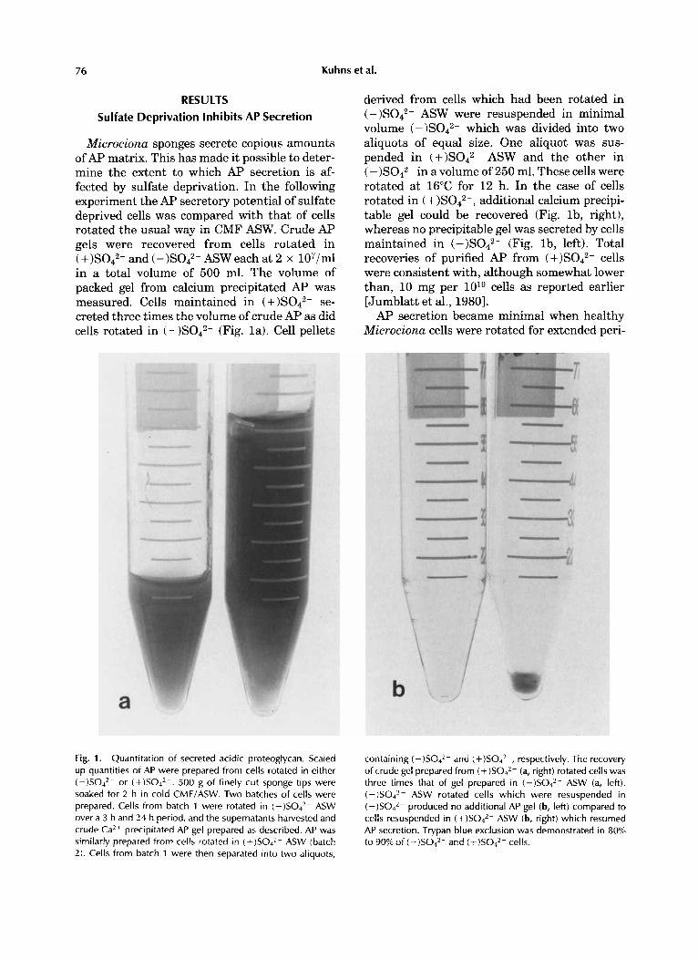
Microciona sponges secrete copious amounts of AP matrix. This has made it possible to deter- mine the extent to which AP secretion is af- fected by sulfate deprivation. In the following experiment the AP secretory potential of sulfate deprived cells was compared with that of cells rotated the usual way in CMF ASW. Crude Ap gels were recovered from cells rotated in
and (-)S042- ASW each at 2 x 10⁷/ml in a total volume of 500 ml. The volume of packed gel from calcium precipitated AP was measured. Cells maintained in (+ )S042- se- creted three times the volume of crude AP as did cells rotated in (-)S042- (Fig. la). Cell pellets
Fig. 1. Quantitation of secreted acidic proteoglycan. Scaled up quantities of AP were prepared from cells rotated in either (-)SO4*- or (+)SO,*-. 500 g of finely cut sponge tips were soaked for 2 h in cold CMF/ASW. Two batches of cells were prepared. Cells from batch 1 were rotated in (-)SO4*- ASW over a 3 h and 24 h period, and the supernatants harvested and crude Ca2+ precipitated AP gel prepared as described. AP was similarly prepared from cells rotated in (+)SO,*- ASW (batch 2). Cells from batch 1 were then separated into two aliquots,
derived from cells which had been rotated in (-)S042- ASW were resuspended in minimal volume (-)S042- which was divided into two aliquots of equal size. One aliquot was sus- pended in ASW and the other in ( -)S042- in a volume of 250 ml. These cells were rotated at 16°C for 12 h. In the case of cells rotated in ( additional calcium precipi- table gel could be recovered (Fig. lb , right), whereas no precipitable gel was secreted by cells maintained in (-)S042- (Fig. lb, left). Total recoveries of purified AF' from cells were consistent with, although somewhat lower than, 10 mg per 1O1O cells as reported earlier [Jumblatt et al., 19801. AP secretion became minimal when healthy
Microciona cells were rotated for extended peri-
containing (-)SO4*- and (+)S04*-, respectively. The recovery of crude gel prepared from (+)SO,*- (a, right) rotated cells was three times that of gel prepared in (-)SO4*- ASW (a, left). (-)SO4*- ASW rotated cells which were resuspended in ( - )S042- produced no additional AP gel (b, left) compared to cells resuspended in ASW (b, right) which resumed AP secretion. Trypan blue exclusion was demonstrated in 80% to 90% of (-)SO4*- and (+)SO,*- cells.
Sulfate Restriction Effects in Marine Sponge 77
ods of time in sulfate deficient ASW as demon- strated by immunoblot using monoclonal anti- AP. Increased secretion could again be demonstrated by healthy cells when (+ )S042- ASW was substituted for (-)S042- ASW in cell suspensions. These alterations are summarized in Table I. In this experiment, Microciona cells were rotated for 32 h in (-)S042- ASW, at which time they were pelleted and resuspended for 8 h in (+ )S042- ASW.
These studies both confirmed that AP secre- tion was retarded in cells maintained in sulfate free ASW and that secretion by these cells could resume in the presence of normal ASW.
Aggregation of Sponge Cells
The following experiment was carried out to determine whether APs derived from cells in (-)S042- or ASW were equally func- tional in aggregation assays. Equal concentra- tions of AP derived from cells at 2 x 10⁷/ml in -S042- or ASW were able to aggregate fresh washed sponge cells or fixed cells in the presence of 10 mM Ca2- in the standard aggrega- tion assay. Cells derived from (-)S042- rotated suspensions aggregated poorly in the presence of purified AP, but aggregation could be restored if such cells were first rotated in ASW and then used in assays. The results suggest that secretory APs from (-)S042- and from (+ )S042- cells are functionally equivalent.
Motility of Sponge Cells
In the following experiments, the significance of motility in the aggregation process was deter- mined by studying well separated stationary cells in chamber preparations. In such a study, random collisions of cells do not occur as they do in standard aggregation assays where gyrotary rotation of cells is carried out. The contrast in motility patterns between cells suspended in (+ )S042- and (- )S042- in stationary cell prepa- rations was quite striking. Cells pretreated and assayed in (+ )Sod2- began to aggregate within minutes, and small aggregates became progres- sively larger until very few non-aggregated sponge cells remained at 2 h (Fig. 3a). A motility study of ten individual cells indicated that they moved at speeds which ranged from 0.1 to 5.2 pm per minute, and in straight or angular direc- tions punctuated at times by turns or loops (Table 11). Net distances (i.e., vector products of horizontal and vertical displacement by single
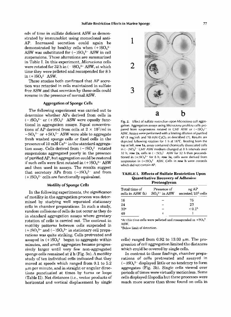
Fig. 2. Effect of sulfate restriction upon Microciona cell aggre- gation. Aggregation assays using Microciona prolifera cells pre- pared from suspensions rotated in CMF ASW or ( - ) S 0 4 * -
ASW. Assays were performed with a limiting dilution of purified AP (1 mg/ml) and 10 mM CaCI2 as described (7). Results are depicted following rotation for 1 h at 16°C. Reading from the top at left, row la, assay contained chemically dissociated cells in (-)SO4*- CMF ASW medium changed at 8 h intervals over 32 h; row 2a, cells in ( - )S042- ASW for 32 h then precondi- tioned in (+)SO4*- for 8 h; row 3a, cells were derived from suspension in (+)S042- ASW. Cells in row b were controls which did not contain AP.
TABLE I. Effects of Sulfate Restriction Upon Quantitative Recovery of Adhesive
Proteoglycan
ng Ap Total time of Presence of cells in ASW (h) Sod2- in ASW secreted/105 cells
16 24 32a 411
75 - 23 - < O . l b
+ 1
-
aAt t h i s time cells were pelleted and resuspended in +S042- ASW. bBelow limit of detection.
cells) ranged from 0.92 to 13.03 Fm. The pro- gression of cell aggregation limited the distances which could be covered by single cells.
In contrast to these findings, chamber prepa- rations of cells pretreated and assayed in ( -)S042- displayed little or no tendency to form aggregates (Fig. 3b). Single cells viewed over periods of times were virtually motionless. Some cells displayed filopodia but these processes were much more scarce than those found on cells in
78 Kuhns et al.
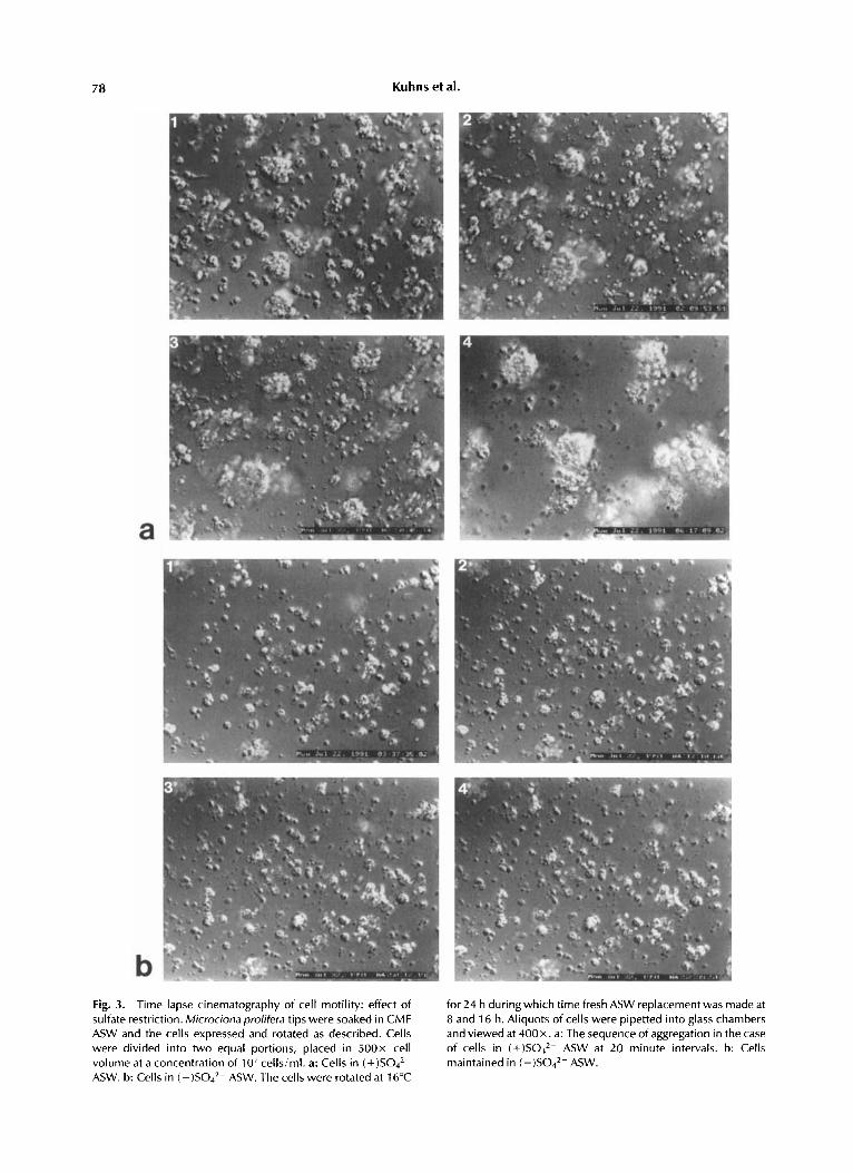
Fig. 3. Time lapse cinematography of cell motility: effect of sulfate restriction. Microcionaprolifera tips were soaked in CMF ASW and the cells expressed and rotated as described. Cells were divided into two equal portions, placed in 5 0 0 ~ cell volume at a concentration of 107cells/ml. a: Cells in ( + ) S 0 4 2 -
ASW. b: Cells in (-)SO4*- ASW. The cells were rotated at 16°C
for 24 h duringwhich time fresh ASW replacementwas made at 8 and 16 h. Aliquots of cells were pipetted into glass chambers and viewed at 400x. a: The sequence of aggregation in the case of cells in (+)SO,*- ASW at 20 minute intervals. b: Cells maintained in ( - )Sod2- ASW.
Sulfate Restriction Effects in Marine Sponge 79
TABLE 11. Summary of Movements of Microciona Cells in + S O P ASW
Total Total Cell observed move- Displace- Net num- time ment Speed menta distance ber ( m i d (um) (umlmin) (urn) (urn)
1 3.75 19.1 5.1 2 2.25 11.7 5.2 3 9.75 30.8 3.2 4 5.5 22.2 4.0 5 5.25 17.3 3.3 6 4.25 17.7 4.2 7 2.5 11.3 4.5 8 4.75 24.7 5.2 9 4.0 0.2 0.1
10 4.75 8.7 1.8
2.110.24 1.811.9 0.616.7 5.11 12.0 8.413.1 2.711.7 0.311.0 2,110.7 0.610.7 5.413.1
2.11 2.62 6.73
13.03 8.95 3.19 1.04 2.21 0.92 6.23
“Horizontali vertical.
(+)S042- ASW. This finding coupled with the demonstration that AP secretion by (-)S042- rotated cells was impaired specifies a critical role for normal secretion in cell motility.
To determine whether highly purified AP ex- posed to healthy (-)S042- ASW cells could re- store motility, cell preparations were mounted in chambers as described and viewed over a period of 2 h. It was found that motility in cells maintained in rotation in (-)S042- ASW could not be restored. However, motility patterns were again observed in such preparations if cells were first preconditioned in ASW (MBLSW) for 6 h (Fig. 4).
This phenomenon appeared to be species spe- cific as judged by a parallel experiment utilizing cells from a second species, Haliclona occulata. Self aggregation of these mechanically dissoci- ated or chemically associated cells occurred readily when cells were rotated at 16°C. When (-)S042- ASW treated cells were exposed to purified Microciona AP, motility was not re- stored even in cells which had first been main- tained 6 h in ASW.
Thus, motility studies of chamber cell prepara- tions indicated that under these conditions ag- gregation was an active physiological process dependent upon inorganic sulfate. In the ab- sence of this nutrient motility and secretion were impaired, and aggregation did not occur. The process was species specific judged from parallel studies using Haliclona, a sponge spe- cies unrelated to Microciona. We conclude that access to inorganic sulfate by Microciona cells is obligatory for cell motility.
lmmunostaining of Microciona Cells With Monoclonal Anti-AP
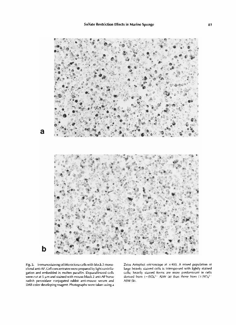
The availability of anti-AP antibodies made it feasible to assess the possibility that cellular retention of AP was a direct result of sulfate starvation. Deparanized cell concentrates were stained with anti AP monoclonal antibodies block 1 and block 2 followed by horse radish peroxi- dase conjugated rabbit antimouse and DAB color developing reagent. Cells which had been main- tained in (-)S042- ASW showed a mixture of many deeply stained cell forms which gave a homogeneous dark color or in some instances stippling, ring, or crescent patterns along with non-stained cells. Of 1,000 cells counted, 45% were scored as AP positive (Fig. 5a). This pat- tern was in contrast to that shown by cells in (+)S042- ASW, in that lightly stained cells were the predominant forms and heavily stained cells comprised a definite minority population; of 1,000 cells counted, 32% were scored as AP positive. This finding suggested that cellular retention of AF’ in (-)S042- rotated cells was a direct result of sulfate restriction.
Chemical Analyses of AP
To determine whether sulfate starvation could induce chemical alterations in secretory AP, com- parative analyses of gel electrophoresis pat- terns; of amino acids, sugars, and amino sugars; and of total sulfate were carried out on purified preparations of secreted AP derived from
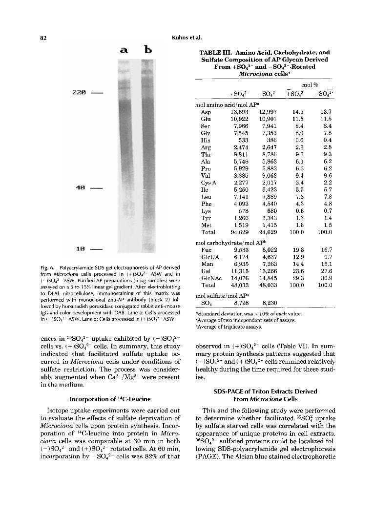
and (-)S042- rotated cells. No signifi- cant differences were observed either in protein distribution (Fig. 6) or in the content of amino acids, amino sugars, or sugars obtained from hydrolysates (Table 111). The quantity of total sulfate in both preparations was similar. This study, if confirmed, suggests that sulfate concen- tration was not limiting for sulfation of secre- tory AP and that the defect causing loss of motion and hyposecretion must reside else- where. However, it does not rule out the possibil- ity that retained or nonsecretory AP might be sulfate deficient if inorganic sulfate became lim- iting.
Incorporation of 35S042
Experiments using radioactive sulfate were designed in order to evaluate comparative up- take and incorporation of 35s04 in normal and sulfate deprived cells. Microciona cells desig- nated for incorporation studies were treated as
80 Kuhns et al.
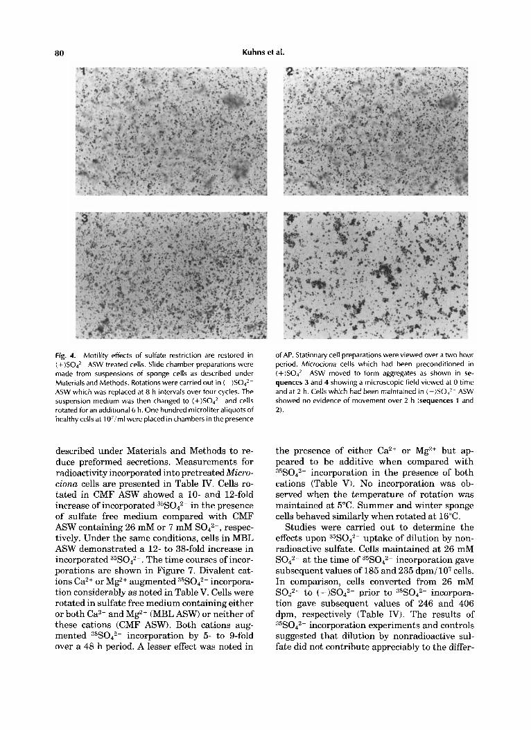
Fig. 4. Motility effects of sulfate restriction are restored in (+ )S042- ASW treated cells. Slide chamber preparations were made from suspensions of sponge cells as described under Materials and Methods. Rotations were carried out in ( - )S042- ASW which was replaced at 8 h intervals over four cycles. The suspension medium was then changed to (+)SO,z- and cells rotated for an additional 6 h. One hundred microliter aliquots of healthy cells at 1 O'iml were placed in chambers in the presence
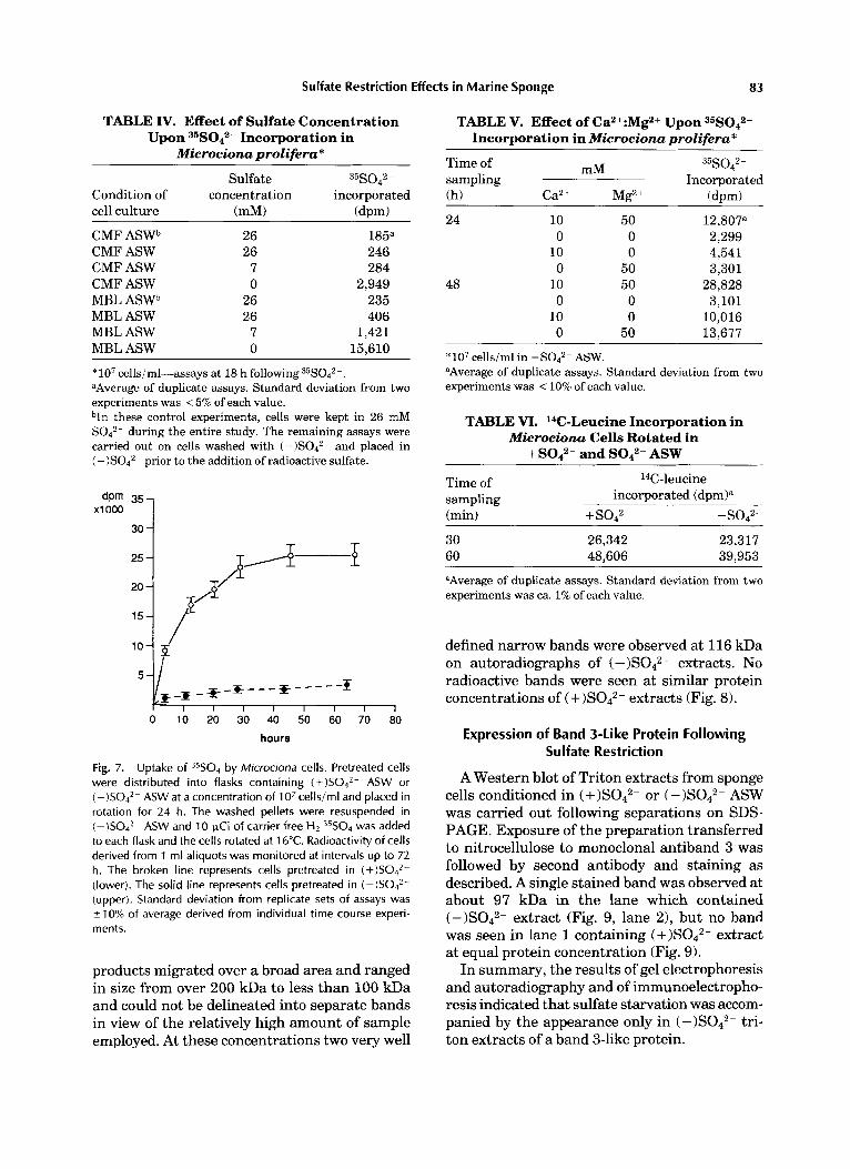
described under Materials and Methods to re- duce preformed secretions. Measurements for radioactivity incorporated into pretreated Micro- ciona cells are presented in Table IV. Cells ro- tated in CMF ASW showed a 10- and 12-fold increase of incorporated 35S042- in the presence of sulfate free medium compared with CMF ASW containing 26 mM or 7 mM S042-, respec- tively. Under the same conditions, cells in MBL ASW demonstrated a 12- to 38-fold increase in incorporated 35S042-. The time courses of incor- porations are shown in Figure 7. Divalent cat- ions Ca2+ or Mg2+ augmented 35S042- incorpora- tion considerably as noted in Table V. Cells were rotated in sulfate free medium containing either or both Ca2+ and Mg2+ (MBL ASW) or neither of these cations (CMF ASW). Both cations aug- mented 35s042- incorporation by 5- to 9-fold over a 48 h period. A lesser effect was noted in
of AP. Stationary cell preparations were viewed over a two hour period. Microciona cells which had been preconditioned in (+ )S042- ASW moved to form aggregates as shown in se- quences 3 and 4 showing a microscopic field viewed at 0 time and at 2 h. Cells which had been maintained in (-)S04*- ASW showed no evidence of movement over 2 h (sequences 1 and 2).
the presence of either Ca2+ or Mg2+ but ap- peared to be additive when compared with 35S042- incorporation in the presence of both cations (Table V). No incorporation was ob- served when the temperature of rotation was maintained at 5°C. Summer and winter sponge cells behaved similarly when rotated at 16°C.
Studies were carried out to determine the effects upon 35s042- uptake of dilution by non- radioactive sulfate. Cells maintained at 26 mM S042- at the time of 35S042- incorporation gave subsequent values of 185 and 235 dpm/107 cells. In comparison, cells converted from 26 mM S042- to (-)S042- prior to 35s042- incorpora- tion gave subsequent values of 246 and 406 dpm, respectively (Table IV. The results of 35S042- incorporation experiments and controls suggested that dilution by nonradioactive sul- fate did not contribute appreciably to the differ-
Sulfate Restriction Effects in Marine Sponge 81
a
b Fig. 5. lmmunostainingof Microciona cells with block2 mono- clonal anti-AP. Cell concentrates were prepared by light centrifu- gation and embedded in molten paraffin. Deparaffinized cells were cut at 5 k m and stained with mouse block 2 anti-AP horse radish peroxidase conjugated rabbit anti-mouse serum and DAB color developing reagent. Photographs were taken using a
Zeiss Axiophot microscope at x400. A mixed population of large heavily stained cells is interspersed with lightly stained cells; heavily stained forms are more predominant in cells derived from ( - )S042- ASW (a) than those from ( + ) S 0 4 2 - ASW (b).
82 Kuhns et al.
Fig. 6. Polyacrylamide SDS gel electrophoresis of AP derived from Microciona cells processed in (+)SOJ2- ASW and in (-)SO4*- ASW. Purified AP preparations (5 pg samples) were assayed on a 5 to 15% linear gel gradient. After electroblotting to DEAE nitrocellulose, immunostaining of this matrix was performed with monoclonal anti-AP antibody (block 2) fol- lowed by horseradish peroxidase conjugated rabbit anti-mouse igG and color development with DAB. Lane a: Cells processed in ( - ) S 0 4 2 - ASW. Lane b: Cells processed in (+)SO4*- ASW.
ences in 35s042- uptake exhibited by (-)S042- cells vs. cells. In summary, this study indicated that facilitated sulfate uptake oc- curred in Microciona cells under conditions of sulfate restriction. The process was consider- ably augmented when Ca2+ /Mg2+ were present in the medium.
Incorporation of 14C-Leucine
Isotope uptake experiments were carried out to evaluate the effects of sulfate deprivation of Microciona cells upon protein synthesis. Incor- poration of 14C-leucine into protein in Micro- ciona cells was comparable at 30 min in both ( - )S042- and ( +)SO,2- rotated cells. At 60 min, incorporation by -S042- cells was 82% of that
TABLE 111. Amino Acid, Carbohydrate, and Sulfate Composition of Ap Glycan Derived
From and -S042--Rotated Microciona cells"
+ S O P - S O P
mol amino acid/mol Ma ASP 13,693 12,997 Glu 10,922 10,901 Ser 7,966 7,941
His 533 386 k g 2,474 2,647 Thr 8,811 8,786 Ala 5,746 5,863 Pro 5,929 5,883 Val 8,885 9,063 Cys A 2,277 2,017 Ile 5,250 5,423 Leu 7,141 7,389 Phe 4,093 4,540 LYS 578 680 TYr 1,266 1,343 Met 1,519 1,415 Total 94,629 94,629
Fuc 9,533 8,022 GlcUA 6,174 4,637 Man 6,935 7,263 Gal 11,315 13,266 GlcNAc 14,076 14,845 Total 48,033 48,033
so* 8.798 8,230
GlY 7,545 7,353
mol carbohydrateimol APb
mol sulfate/mol APa
mol %
14.5 11.5 8.4 8.0 0.6 2.6 9.3 6.1 6.3 9.4 2.4 5.5 7.6 4.3 0.6 1.3 1.6
100.0
19.8 12.9 14.4 23.6 29.3
100.0
13.7 11.5 8.4 7.8 0.4 2.8 9.3 6.2 6.2 9.6 2.2 5.7 7.8 4.8 0.7 1.4 1.5
100.0
16.7 9.7
15.1 27.6 30.9
100.0
~
*Standard deviation was < 10% of each value. "Average of two independent sets of assays. bAverage of triplicate assays.
observed in (+)S042- cells (Table VI). In sum- mary protein synthesis patterns suggested that ( -)S042- and (+ )S042- cells remained relatively healthy during the time required for these stud- ies.
SDS-PACE of Triton Extracts Derived From Microciona Cells
This and the following study were performed to determine whether facilitated 35SO: uptake by sulfate starved cells was correlated with the appearance of unique proteins in cell extracts. 35S042- sulfated proteins could be localized fol- lowing SDS-polyacrylamide gel electrophoresis (PAGE). The Alcian blue stained electrophoretic
Sulfate Restriction Effects in Marine Sponge 83
TABLE IV. Effect of Sulfate Concentration Upon 35S042- Incorporation in
Microciona prolifera*
Sulfate 35S042-
Condition of concentration incorporated cell culture (mM) (dpm)
CMF ASWb CMF ASW CMF ASW CMF ASW MBL ASWb MBL ASW MBL ASW MBL ASW
26 26
7 0
26 26
7 0
185a 246 284
2,949 235 406
1,421 15,610
*lo7 cellsiml-assays at 18 h f ~ l l o w i n g ~ ~ S O ~ ~ - . “Average of duplicate assays. Standard deviation from two experiments was < 5% of each value.
these control experiments, cells were kept in 26 mM S042- during the entire study. The remaining assays were carried out on cells washed with (-)S042- and placed in (-)S042- prior to the addition of radioactive sulfate.
*-*---*--- - -I I I I I I I I
0 10 20 30 40 50 60 70 80
hours
Fig. 7. Uptake of 35S04 by Microciona cells. Pretreated cells were distributed into flasks containing (+)SO4’- ASW or ( - )S042- ASW at a concentration of 1 O7 cells/ml and placed in rotation for 24 h. The washed pellets were resuspended in (-)SO4*- ASW and 10 pCi of carrier free H2 35S04 was added to each flask and the cells rotated at 1 b“C. Radioactivity of cells derived from 1 ml aliquots was monitored at intervals up to 72 h. The broken line represents cells pretreated in (+)S042- (lower). The solid line represents cells pretreated in ( - ) S 0 4 2 - (upper). Standard deviation from replicate sets of assays was 210% of average derived from individual time course experi- ments.
products migrated over a broad area and ranged in size from over 200 kDa to less than 100 kDa and could not be delineated into separate bands in view of the relatively high amount of sample employed. At these concentrations two very well
TABLE V. Effect of Ca2+:Mgz+ Upon 35S042- Incorporation in Microciona prolifera*
~
35SO 2 - Time of mM 4 sampling Incorporated (h) Ca2+ Mg2+ (dDm)
24 10 0
10 0
48 10 0
10 0
50 0 0
50 50
0 0
50
12,807“ 2,299 4,541 3,301
28,828 3,101
10,016 13,677
*lo7 cellsiml in -S042- ASW. “Average of duplicate assays. Standard deviation from two experiments was < 10% of each value.
TABLE VI. 14C-Leucine Incorporation in Microciona Cells Rotated in
+ Sod2- and Sod2- ASW
Time of 14C-leucine sampling incorporated (dpmIa (min) + so42- -sop 30 26,342 23,317 60 48,606 39,953
“Average of duplicate assays. Standard deviation from two experiments was ca. 1% of each value.
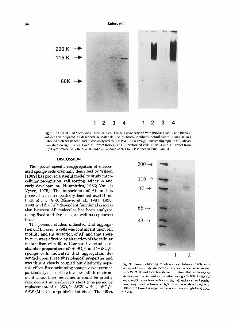
defined narrow bands were observed at 116 kDa on autoradiographs of (-)S042- extracts. No radioactive bands were seen at similar protein concentrations of extracts (Fig. 8).
Expression of Band 3-Like Protein Following Sulfate Restriction
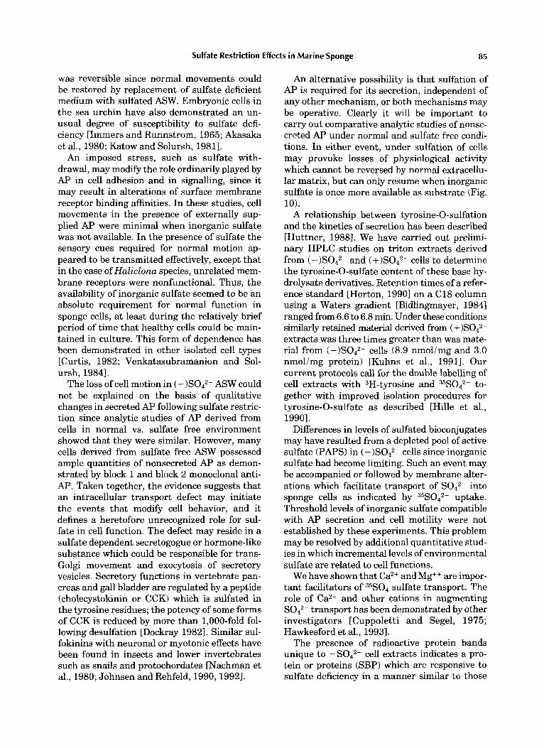
A Western blot of Triton extracts from sponge cells conditioned in (+)S042- or (-)S042- ASW was carried out following separations on SDS- PAGE. Exposure of the preparation transferred to nitrocellulose to monoclonal antiband 3 was followed by second antibody and staining as described. A single stained band was observed at about 97 kDa in the lane which contained (-)S042- extract (Fig. 9, lane 21, but no band was seen in lane 1 containing extract at equal protein concentration (Fig. 9).
In summary, the results of gel electrophoresis and autoradiography and of immunoelectropho- resis indicated that sulfate starvation was accom- panied by the appearance only in (-)S042- tri- ton extracts of a band 3-like protein.
84 Kuhns et al.
Fig. 8. SDS-PAGE of Microciona Triton extracts. Extracts were reacted with mouse block 1 and block 2 anti-AP and prepared as described in Materials and Methods. Antibody bound (lanes 2 and 4) and unbound material (lanes 1 and 3) was analyzed by SDS PAGE on a 12% gel. Autoradiograph on left, Alcian blue stain on right. Lanes 1 and 2: Extract from (+)SO4*- pretreated cells. Lanes 3 and 4: Extract from ( -)SO4*- pretreated cells. A single radioactive band at ca.116 kDa is seen in lanes 3 and 4.
DISCUSSION
The species specific reaggregation of dissoci- ated sponge cells originally described by Wilson 119071 has proved a useful model to study inter- cellular recognition, cell sorting, adhesion and early development [Humphries, 1963; Van de Vyver, 19751. The importance of AF' in this process has been repeatedly demonstrated [Jum- blatt et al., 1980; Misevic et al., 1987, 1990, 19931 and the Ca2+ dependent functional associa- tion between AF' molecules has been analyzed using fixed and live cells, as well as sepharose beads.
The present studies indicated that aggrega- tion of Microciona cells was contingent upon cell motility and the secretion of Ap and that these in turn were affected by alteration of the cellular metabolism of sulfate. Comparative studies of chamber preparations of and (-)S042- sponge cells indicated that aggregation de- pended upon these physiological properties and was thus a closely coupled but distinctly sepa- rate effect. Free swimming sponge larvae seemed particularly susceptible to a low sulfate environ- ment since their movements could be greatly retarded within a relatively short time period by replacement of ASW with (-)S042- ASW (Misevic, unpublished studies). The effect
Fig. 9. lmrnunoblotting of Microciona Triton extracts with anti-band 3 antibody Microciona triton extracts were separated by SDS PAGE and then transferred to nitrocellulose. Immuno- staining was carried out as described using a 1 / I 00 dilution of anti-band 3 monoclonal antibody (Sigma), and alkaline phospha- tase conjugated anti-mouse IgG. Color was developed with NBT-BCIP. Lane 1 i s negative. Lane 2 shows a single band at ca. 97 kDa.
Sulfate Restriction Effects in Marine Sponge 85
was reversible since normal movements could be restored by replacement of sulfate deficient medium with sulfated ASW. Embryonic cells in the sea urchin have also demonstrated an un- usual degree of susceptibility to sulfate defi- ciency [Immers and Runnstrom, 1965; Akasaka et al., 1980; Katow and Solursh, 19811.
An imposed stress, such as sulfate with- drawal, may modify the role ordinarily played by AP in cell adhesion and in signalling, since it may result in alterations of surface membrane receptor binding affinities. In these studies, cell movements in the presence of externally sup- plied AP were minimal when inorganic sulfate was not available. In the presence of sulfate the sensory cues required for normal motion ap- peared to be transmitted effectively, except that in the case of Haliclona species, unrelated mem- brane receptors were nonfunctional. Thus, the availability of inorganic sulfate seemed to be an absolute requirement for normal function in sponge cells, at least during the relatively brief period of time that healthy cells could be main- tained in culture. This form of dependence has been demonstrated in other isolated cell types [Curtis, 1982; Venkatasubramanion and Sol- ursh, 19841.
The loss of cell motion in ( - )S042- ASW could not be explained on the basis of qualitative changes in secreted AP following sulfate restric- tion since analytic studies of AP derived from cells in normal vs. sulfate free environment showed that they were similar. However, many cells derived from sulfate free ASW possessed ample quantities of nonsecreted AP as demon- strated by block 1 and block 2 monoclonal anti- AP. Taken together, the evidence suggests that an intracellular transport defect may initiate the events that modify cell behavior, and it defines a heretofore unrecognized role for sul- fate in cell function. The defect may reside in a sulfate dependent secretogogue or hormone-like substance which could be responsible for trans- Golgi movement and exocykosis of secretory vesicles. Secretory functions in vertebrate pan- creas and gall bladder are regulated by a peptide (cholecystokinin or CCK) which is sulfated in the tyrosine residues; the potency of some forms of CCK is reduced by more than 1,000-fold fol- lowing desulfation [Dockray 19821. Similar sul- fokinins with neuronal or myotonic effects have been found in insects and lower invertebrates such as snails and protochordates [Nachman et al., 1980; Johnsen and Rehfeld, 1990,19921.
An alternative possibility is that sulfation of AP is required for its secretion, independent of any other mechanism, or both mechanisms may be operative. Clearly it will be important to carry out comparative analytic studies of nonse- creted AP under normal and sulfate free condi- tions. In either event, under sulfation of cells may provoke losses of physiological activity which cannot be reversed by normal extracellu- lar matrix, but can only resume when inorganic sulfate is once more available as substrate (Fig. 10).
A relationship between tyrosine-0-sulfation and the kinetics of secretion has been described [Huttner, 19881. We have carried out prelimi- nary HPLC studies on triton extracts derived from and (+)Sod2- cells to determine the tyrosine-0-sulfate content of these base hy- drolysate derivatives. Retention times of a refer- ence standard [Horton, 19901 on a C18 column using a Waters gradient [Bidlingmayer, 19841 ranged from 6.6 to 6.8 min. Under these conditions similarly retained material derived from ( extracts was three times greater than was mate- rial from (-)S042- cells (8.9 nmollmg and 3.0 nmol/mg protein) [Kuhns et al., 19911. Our current protocols call for the double labelling of cell extracts with 3H-tyrosine and 35S042- to- gether with improved isolation procedures for tyrosine-0-sulfate as described [Hille et al., 19901.
Differences in levels of sulfated bioconjugates may have resulted from a depleted pool of active sulfate (PAPS) in (- )S042- cells since inorganic sulfate had become limiting. Such an event may be accompanied or followed by membrane alter- ations which facilitate transport of S042- into sponge cells as indicated by 35S042- uptake. Threshold levels of inorganic sulfate compatible with AP secretion and cell motility were not established by these experiments. This problem may be resolved by additional quantitative stud- ies in which incremental levels of environmental sulfate are related to cell functions.
We have shown that Ca2+ and Mg+ + are impor- tant facilitators of 35s04 sulfate transport. The role of Ca2+ and other cations in augmenting S042- transport has been demonstrated by other investigators [Cuppoletti and Segel, 1975; Hawkesford et al., 19931.
The presence of radioactive protein bands unique to -S042- cell extracts indicates a pro- tein or proteins (SBP) which are responsive to sulfate deficiency in a manner similar to those
86 Kuhns et al.
Exterior
(-)S042-
Cell Surface Membrane
Interior
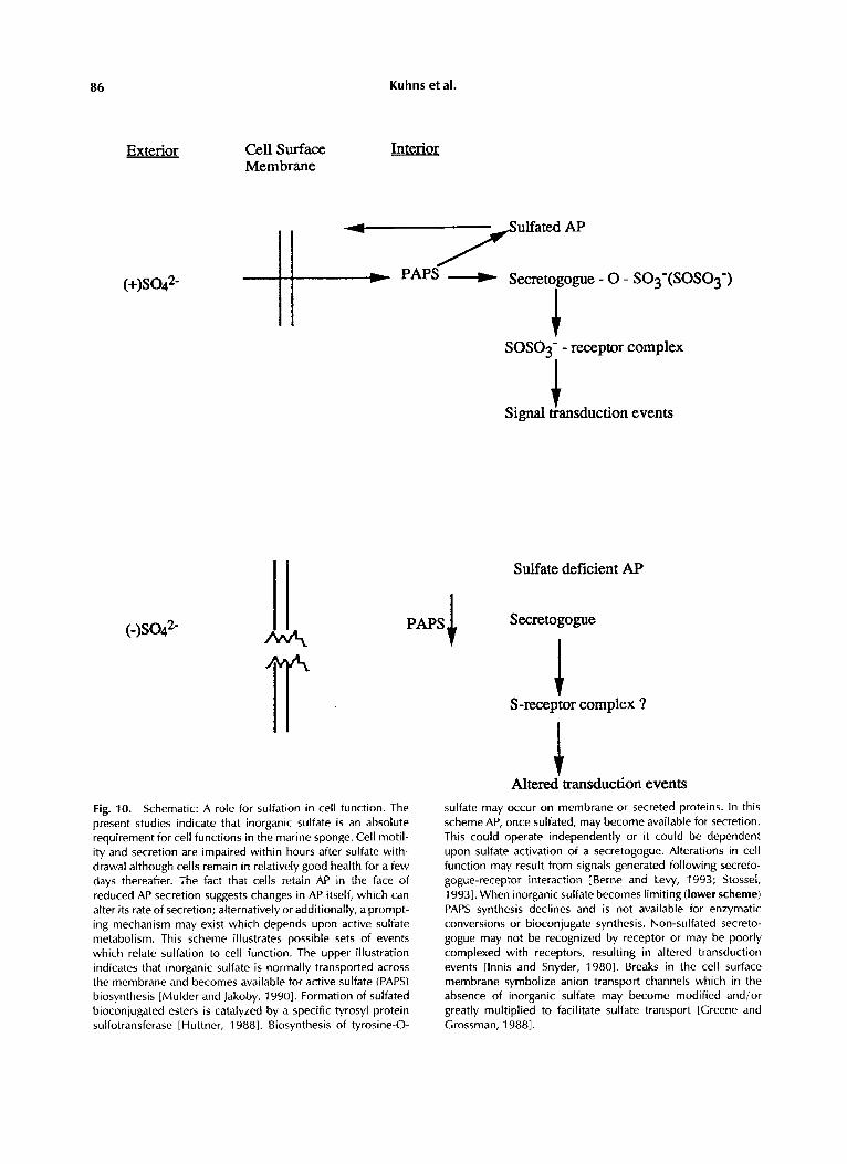
Fig. 10. Schematic: A role for sulfation in cell function. The present studies indicate that inorganic sulfate is an absolute requirement for cell functions in the marine sponge. Cell motil- ity and secretion are impaired within hours after sulfate with- drawal although cells remain in relatively good health for a few days thereafter. The fact that cells retain AP in the face of reduced AP secretion suggests changes in AP itself, which can alter its rate of secretion; alternatively or additionally, a prompt- ing mechanism may exist which depends upon active sulfate metabolism. This scheme illustrates possible sets of events which relate sulfation to cell function. The upper illustration indicates that inorganic sulfate is normally transported across the membrane and becomes available for active sulfate (PAPS) biosynthesis [Mulder and Jakoby, 19901. Formation of sulfated bioconjugated esters is catalyzed by a specific tyrosyl protein sulfotransferase [Huttner, 19881. Biosynthesis of tyrosine-0-
PAPSI
SO SO^' - receptor complex
Signal &amduction events
Sulfate deficient AP
Secretogogue
S-receptor complex ?
Altered' transduction events sulfate may occur on membrane or secreted proteins. In this scheme AP, once sulfated, may become available for secretion. This could operate independently or it could be dependent upon sulfate activation of a secretogogue. Alterations in cell function may result from signals generated following secreto- gogue-receptor interaction [Berne and Levy, 1993; Stossel, 19931. When inorganic sulfate becomes limiting (lower scheme) PAPS synthesis declines and is not available for enzymatic conversions or bioconjugate synthesis. Non-sulfated secreto- gogue may not be recognized by receptor or may be poorly complexed with receptors, resulting in altered transduction events [Innis and Snyder, 19801. Breaks in the cell surface membrane symbolize anion transport channels which in the absence of inorganic sulfate may become modified and/or greatly multiplied to facilitate sulfate transport [Creene and Crossman, 19881.
Sulfate Restriction Effects in Marine Sponge 87
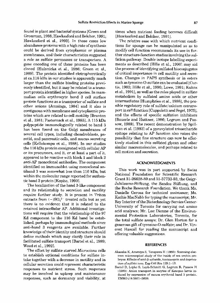
found in plant and bacterial systems [Green and Grossman, 1988; Hawkesford and Belcher, 1991; Hawkesford et al., 19931. In those cases low abundance proteins with a high rate of synthesis could be derived from cytoplasmic or plasma membranes, and their characteristics suggested a role as sulfate permeases or transporters. A gene encoding one of these proteins has been cloned [Hulanicka et al., 1986; Green et al., 19891. The protein identified eletrophoretically at ca.116 kDa in our studies is apparently much larger than the sulfate binding proteins previ- ously identified, but it may be related to a trans- port protein identified in higher species. In mam- malian cells erythrocyte band 3 cell surface protein functions as a transporter of sulfate and other anions [Jennings, 19841 and it also is contiguous with membrane and cytoplasmic pro- teins which are related to cell motility [Branton et al., 1981; Pasternack et al., 19851. A 115 kDa polypeptide immunologically related to band 3 has been found on the Golgi membranes of several cell types, including chondroblasts, pa- rotid, and pancreatic cells, and mucus secreting cells [Kellokompu et al., 19881. In our studies the 116 kDa protein comigrated with cellular AP or its precursors, and it, or at least a part of it, appeared to be reactive with block 1 and block 2 anti-AP monoclonal antibodies. The component identified on immunoblot using monoclonal an- tiband 3 was somewhat less than 116 kDa, but within the molecular range reported for authen- tic band 3 protein [Kopito, 19901.
The localization of the band 3-like component and its relationship to secretion and motility require further study. It is present in Triton extracts from (-)S042- treated cells but as yet there is no evidence that it is related to the retained intracellular AP. Additional investiga- tions will require that the relationship of the 97 Kd component to the 116 Kd band be estab- lished, perhaps by immunological methods since anti-band 3 reagents are available. Further knowledge of their identity and structure should define methods which may clarify their role in facilitated sulfate transport [Bartel et al., 1989; Wood et al., 19921.
The effort by sulfate starved Microciona cells to establish optional conditions for sulfate in- take together with a decrease in motility and in cellular secretion could represent characteristic responses to nutrient stress. Such responses may be involved in upkeep and maintenance responses, such as dormancy and viability, at
times when nutrient feeding becomes difficult [Hawkesford and Belcher, 19911.
The relative ease with which nutrient condi- tions for sponge can be manipulated so as to modify cell function recommends its use in fur- ther structure-function studies involving the sul- fation pathway. Double isotope labelling experi- ments as described [Hille et al., 19901 may aid the process of identifying sulfated bioconjugates of critical importance in cell motility and secre- tion. Changes in PAPS synthesis or in esters such as tyrosine-0-sulfate can be evaluated [Cur- tis, 1982; Hille et al., 1990; Lowe, 1991; Kuhns et al., 19911, as well as the roles played in sulfate metabolism by sulfated amino acids or other intermediates [Humphries et al., 19861, the pos- sible regulatory role of sulfate/calcium cotrans- port in cell function [Cuppoletti and Segel, 19751, and the effects of specific sulfation inhibitors [Bauerle and Huttner, 1986; Legrum and Pas- sow, 19891. The recent demonstration by Spill- man et al. [19931 of a pyruvylated trisaccharide epitope relating to AP function also raises the possibility that fine structure can now be effec- tively studied in this sulfated glycan and other similar macromolecules, and perhaps related to cell motion and secretion.
ACKNOWLEDGMENTS
This work was in part supported by Swiss National Foundation for Scientific Research Grant 31-26628-89 and 31-37733-93, the Geigy- Jubilaums-Stiftung, the Sandoz Stiftung, and the Roche Research Foundation. We thank Ms. Danielle Gerosa for technical assistance; Ms. Kathie MacNabb for typing the manuscript; Mr. Rey Interior of the Biotechnology Service Center- University of Toronto for carrying out amino acid analyses; Mr. Luc Dionne of the Environ- mental Protection Laboratories, Toronto, for the total sulfate assays; Dr. Glen Horton for a generous gift of tyrosine-0-sulfate; and Dr. Vin- cent Hascall for reading the manuscript and offering valuable suggestions.
REFERENCES
Akasaka K, Ameniya S, Terayawa H (1980): Scanning elec- tron microscopical study of the inside of sea urchin em- bryos: Effects of aryl-@-xyloside, tunicamycin and depriva- tion of sulfate ions. Exp Cell Res 129:l-13.
Bartel D, Lepke S, Layh-Schmitt G, Legrum B, Passow H (1989): Anion transport in oocytes of Xenopus larvis in- duced by expression of mouse erythroid band 3 protein. EMBO J 8:3601-3609.
88 Kuhns et at.
Bauerle P, Huttner W (1986): Chlorate-a potent inhibitor of protein sulfation in intact cells. Biochem Biophys Res Commun 1412370-877.
Berne R, Levy M, eds (1993): “Physiology,” 3rd ed. St. Louis, MO: Mosby Yearbook, p. 818.
Bidlingmeyer B, Cohen S, Parvin T (1984): Rapid analysis of amino acids using pre-column derivatization. J Chro- matogr 336:93-104.
Bradford M (1976): A rapid and sensitive method for the quantitation of microgram quantities of protein utilizing the principle of protein-dye binding. Anal Biochem 72:248- 258.
Branton D, CohenM, Tyler J (1981): Interaction of cytoskel- etal proteins on the human erythrocyte membrane. Cell 24:24-32.
Cardelli J, Bush J , Ebert D, Freeze H (1990): Sulfated N-linked oligosaccharides affect secretion but are not es- sential for the transport, proteolytic processing and sort- ing of enzymes in Dictyostelium discoidum. J Biol Chem
Cavanaugh G (1964): “Formulas and Methods of the Marine Biological Laboratory,” 6th ed. Woods Hole, MA Marine Biological Laboratory, p. 67.
Chaplin M (1982): A rapid and sensitive method for the analysis of carbohydrate components in glycoproteins us- ing gas-liquid chromatography. Anal Biochem 123:336- 341.
Cheng PW, Boat T, Cranfill K, Yankaskas J, Boucher R (1989): Increased sulfation of glycoconjugates by cultured nasal epithelial cells from patients with cystic fibrosis. J Clin Invest 84:68-72.
Cuppoletti J , Segel I (1975): Kinetics of sulfate transport by Pericillium notations. Interactions of sulfate, protons and calcium. Biochemistry 14:4711-4718.
Curtis CG (1982): The origins of intracellular sulfate for conjugation reactions. In Mulder G, Caldwell J, van Kem- pen G, Vonk R (eds): “Sulfate Metabolism and Sulfate Conjugation.” London: Taylor and Francis, pp 67-71.
David G (1991): Biology and pathology of the pericellular heparan sulfate proteoglycan. Biochem SOC Trans 19:816- 820.
Davis S, Wheldrake J (1986): Sulfation and the vegetative growth of Dictyostelium discoideum. Eur J Biochem 158:
Dockray GJ (1982): The physiology of cholecystokinin in brain and gut. Br Med Bull 38253-258.
Fan MY, Templeton D (1992): Sulfate metabolism in experi- mental diabetes. Diabete Metab 18:98-103.
Fukui S, Yoshida H, Tan& T, Sakano T, Usui T, Ya- mashina I (1981): Glycosaminoglycan eynthesis by cul- tured skin fibroblasts from a patient with Lowe’s syn- drome. J Biol Chem 256:10313-10318.
Green L, Grossman A (1988): Changes in sulfate transport characteristics and protein composition of anacystic nidu- lans R2 during sulfur deprivation. J Bacteriol 170:583- 587.
Green L, Laudenbach D, Grossman A (1989): A region of a cyanobacterial genome required for sulfate transport. Proc Nat Acad Sci USA 86:1949-1953.
Green P, Tamatoni T, Watanabe T, Miyasaka M, Hasegawa A, Kiso M, Yuen C-T, Stoll M, Feizi T (1992): High affinity binding of the leukocyte adhesion molecule L-selectin to 3’ sulfated Lea and Le” oligosaccharides and the predomi- nance of sulfate in this interaction demonstrated by bind-
265:8847-8853.
174-185.
ing studies with a series of lipid linked oligosaccharides. Biochem Biophys Res Commun 188:244-251.
Hascall V, Calabro A, Midura R, Yanagishita M (1994): Isolation and characterization of proteoglycans. in Meth- ods Enzymol230:409.
Hawkesford M, Davidian JC, Grignon C (1993): Sulfate/ proton cotransport in plasma-membrane vesicles isolated from roots of Brassica napus L.: Increased transport in membranes isolated from sulfur starved plants. Planta
Hawkesford M, Belcher A (1991): Differential protein synthe- sis in response to sulfate and phosphate deprivation. Planta
Hille A, Braulke T, Figura K, Huttner W (1990): Occurrence of tyrosine sulfate in proteins-a balance sheet 1. Secre- tory and lysosomal proteins. Eur J Biochem 188577-586.
Hirs CHW (1967): Determination of cysteine as cystic acid. Methods Enzymol11:59-60.
Horton G (1990): Sulfation of tyrosine residues in coagula- tion factor V. Blood 76:946-952.
Horton D, Usui H (1978): In Schweiger R (ed): “Sulfated Polysaccharides.” Washington, DC: American Chemical Society, p 96.
HulanickaM, Farrett C, Jagura-Burdzy G, Kredich N (1986): Cloning and characterization of the cysAMK region of Salomonella typhimurine. J Bacteriol 168:322-327.
Hull S, Carraway K (1989): Sulfation of the tumor cell surface sialomucin of the 13762 rat mammary adenocarci- noma. J Cell Biochem 40:67-81.
Humphreys T (1963): Chemical dissolution and in uitro reconstruction of sponge cell adhesions. I. Isolation and functional demonstration of the components involved. Dev Biol8:27-47.
Humpreys T (1967): The cell surface and specific cell aggre- gation. In Warren L, Davis B (eds): “The Specificity of Cell Surfaces.” Englewood Cliffs, NJ: Prentice-Hall, pp 195- 210.
Humphries D, Silbert C, Silbert J (1986): Glycosaminogly- can production by bovine aortic endothelial cells cultured in sulfate depleted medium. J Biol Chem 261:9122-9127.
Humphries D, Sugermaran G, Silbert J (1989): Decreasing sulfation of proteoglycan produced by cultured cells. Meth- ods Enzymol179:428-434.
Huttner WB (1988): Tyrosine sulfate and the secretory pathway. Ann Rev Physiol50:363-376.
Immers J , Runnstrom J (1965): Further studies of the effects of deprivation of sulfate on the early development of the sea urchin Paracentrutus lividus J Embryo1 Exp Morphol14:289-305.
Innis R, Snyder S (1980): Distinct cholecystokin in receptors in brain and pancreas. Proc Natl Acad Sci U S A 77:6917- 6921.
Jenke D (1981): Anion peak migration in ion chromatogra- phy. Anal Chem 53:1536.
Jennings M (1984): Oligomeric structure and the anion transport function ofhuman erythrocyte band 3 protein. J Membrane Biol80:105-117.
Johnsen AH, Rklehfeld J F (1993): Lymna D famides, a new family of neuropeptides from the pond snail Lymnaea stagnalis. Eur J Biochem 213:875-879.
Jumblatt J, Schlup V, Burger M (1980): Cell-cell recogni- tion: Specific binding of Microciona sponge aggregation factor to homotypic cells and the role of calcium ions. Biochemistry 19: 1038-1042.
190:297-304.
185~323-329.

Sulfate Restriction Effects in Marine Sponge 89
Katow H, Solursh M (1981): Ultrastructural and time lapse studies of primary mesenchyme cell behavior in normal and sulfate deprived sea urchin embryos. Exp Cell Res
Kellokumpu S, Neff L, Janka-Kellokumpu S, Kopito R, Baron R (1988): A 115 kD polypeptide immunologically related to erythrocyte band 3 is present in Golgi mem- branes. Science 242:1308-1311.
Kopito R (1990): Molecular biology of the anion exchanger gene family. Int Rev Cytol123:177-199.
Kuhns W, Misevic G, Burger M (1990): Biochemical and functional effects of sulfate restriction in the marine sponge, Microcionaprolifera. Biol Bull 179:358-365.
Kuhns W, Burger M, Misevic G (1991): Biochemical changes associated with reductions of cell motility and aggregation following sulfate restriction in the marine sponge, Micro- cionaprolifera. Biol Bull 181:337-339.
Laemmli UK (1970): Cleavage of structural proteins during the assembly of the head of bacteriophage T4. Nature
Legrum B, Passow H (1989): Inhibition of inorganic anion transport across the human red blood cell membrane by chloride dependent association of dipyridamole with a stilbene disulfonate binding site on the band 3 protein. Biochim Biophys Acta 979:193-207.
Lowe G (1991): Mechanisms of sulfate activation and trans- fer. Philos Trans R SOC Lond LBiol135-42.
Lowry 0, Rosebrough N, Farr A, Randall R (1951): Protein measurement with the Folin-phenol reagent. J Biol Chem
Misevic G, Jumblatt J, Burger M (1982): Cell binding frag- ments from a sponge proteoglycan-like aggregation factor. Biochemistry 257:6931-6936.
Misevic G (1989): Immunoblotting and immunobinding of acidic polysaccharides separated by gel electrophoresis. Methods Enzymol179:97-104.
Misevic G, Burger M (19901: The species specific cell binding site of the aggregation factor from the marine Microciona prolifera is a highly repetitive novel glycan containing glucuvonic acid, fucose and mannose. J Biol Chem 265: 20577-20584.
Mulder G, Jakoby W (1990): Sulfation. In Mulder G (ed): “Conjugation Reactions in Drug Metabolism.” Taylor and Francis, Ltd., pp 107-161.
Pasternack G, Anderson R, Let0 T, Marchesi V (1985): Interactions between protein 4.1 and band 3. J Biol Chem
Robinson J, Viti M, Hook M (1984): Structure and proper- ties of an under sulfated heparan sulfate proteoglycan
136:233-245.
227~680-685.
193: 265-275.
260~3676-3683.
synthesized by a rat hepatoma cell line. J Cell Biol98:946- 953.
Sharpe PT (1988): “Methods of Cell Separation.” Amster- dam: Elsevier, p 15.
Simpson TL (1968): The biology of the marine spone Micro- ciona prolifera. I1 Temperature related, annual changes in functional and reproductive elements with a descrip- tion of larval metamorphosis. J Exp Mar Biol Ecol2:252- 277.
Simpson TL (1984): “The Cell Biology of Sponges.” New York: Springer-Verlag.
Spicer S (1965): Diamine methods for differentiating mu- cosubstances histochemically. J Histochem Cytochem 13:
Spillmann D, Hard K, Thomas-Oates J, Vliegenthart J, Misevic G, Burger M, Finne J (1993): Characterization of a novel pyruvylated carbohydrate unit implicated in the cell aggregation of the marine sponge, Microciona prvlif- era. J Biol Chem 268:13378-13387.
Stossel T (1993): On the crawling of animal cells. Science 260:108&1094.
Svennerholm L, Fredman P (1980): A procedure for the quantitative isolation of brain gangliosides. Biochim Bio- phys Acta 617:97-109.
Van de Vyver G (1975): Phenomena of cellular in sponges. In: Moscona A, Monroy A (eds.): “Current Topics in Developmental Biology.” New York: Academic Press, pp
Venkatasubramanian K, Solursh M (1984): Adhesive and migratory behavior of normal and sulfate deficient sea urchin cells in uitro. Exp Cell Res 154:421431.
Watabe T, Hiratsuka A, Ogura K, Endoh K (1985): A reac- tive hydroxymethyl sulfate ester formed regioselectively from the carcinogen 7-12 dihydroxymethylbenz (a) anthra- cene, by rat liver sulfotransferase. Biochem Biophys Res Commun 131:694-699.
Weiss J (1986): “Handbook of Ion Chromatography.” Johnson EL, ed. Sunnyvale, CA: Dionex Gorp.
Wend S, Sumper M (1981): Sulfation of a cell surface glycoprotein correlates with the developmental program during embryogenesis of Voluvx carteri. Proc Natl Acad Sci U S A 78:3716-3720.
Wilson A, Rider C (1992): Evidence that leukosialin CD 43 is intensely sulfated in the murine T-lymphomaline RDM-4. J Immunol148:1777-1783.
Wood P, Muller H, Sovak M, Passow H (1992): Role of lys 558 and lys 869 in substrate and inhibitor binding to the murine b and 3 protein: A study of the effects of site directed mutagenesis of the band 3 protein expressed in the oocytes of Xenopus larvis. J Membr Biol127:139-148.
211-234.
123-140.