Collective judgment predicts disease-associated single nucleotide variants

E X P R E S S I O N O F T C E L L R E C E P T O R G E N E S IN A N
A N T I G E N - S P E C I F I C H Y B R I D O M A A N D
R A D I A T I O N - I N D U C E D V A R I A N T S
BY NICHOLAS R. J. GASCOIGNE,* STANLEY WATERS,* JOHN F. ELLIOTT,* CAROL VICTOR-KOBRIN,*
CHRISTOPHER GOODNOW,* MARK M. DAVIS,* AND CONSTANTIN A. BONA*
From the *Department of Medical Microbiology, Stanford University School of Medicine, Stanford, California 94305; and the *Department of Microbiology, Mount Sinai
School of Medicine, New York, New York 10029
T h e T cell receptor for antigen is similar to that o f B cells (Ig) in that there are a very large number o f specificities and this reper to i re of specificities is clonally distributed. What appears unique about T cell recognit ion is the ability o f T cells to recognize foreign ant igen only in association with a p roduc t of the MHC (1, 2), whereas antibodies, in most cases, require only antigen. Recent serological and biochemical analyses have shown that the T cell receptor exists on the cell surface as a disulfide-linked mul t imer o f two subunits, a and/3 (3-7). These peptide chains are genera ted in a manner similar to Ig in that they are encoded by several sets o f gene segments: variable (V); diversity (D), at least in the case of beta chains; and jo ining (J) regions. These segments rear range dur ing the maturat ion o f an individual cell to p roduce a recep tor combination unique to that cell lineage (8-13). T h e rest o f the T cell receptor protein consists o f a constant (C) region that is itself encoded by three or four separate exons (14- 16).
T h e V regions o f the T cell receptor conserve certain residues that are present in both VH and VL (17-19). This suggests that their overall s t ructure is similar to that of Ig. Some differences have also been no ted (17, 20), whose exact significance remains controversial.
T h e T cell receptor , like Ig, expresses antigenic determinants of V and C regions. T h e antigenic determinants o f the T cell recep tor V regions can be considered as equivalent to the idiotypes of Ig molecules, phenotypic markers of V region genes. T cell idiotypes were identified with antisera or mAbs f rom syngeneic, allogeneic, or xenogeneic animals immunized with T cell clones (3- 7). One such antiidiotypic ant ibody has been used to select o ther T cells, which
This work was supported by grant PCM8408660 (to C.A.B.) from National Science Foundation and by grant A119512 (to M.M.D.) from the National Institutes of Health. NRJG is a Fellow of the Cancer Research Institute. J. F. EIliott's present address is Dept. of Biochemistry, University of Alberta, Edmonton, Alberta, Canada. C. Goodnow's present address is Walter & Eliza Institute, Melbourne, Australia.
J. ExP. MED. © The Rockefeller University Press • 0022-1007/86/01/0113/18 $1.00 1 13 Volume 164 July 1986 113-130
on February 29, 2016
jem.rupress.org
Dow
nloaded from
Published July 1, 1986

114 o~ A N D /3 G E N E S IN T C L O N E V A R I A N T S
were found to have the same H-2 restriction and antigenic specificity and to use the same a and fl chains as the original cell to which the ant ibody had been made (21, 22). This finding indicates that an idiotype can represent a combinatorial de te rminant formed by the interaction of c~ and /3 chains and that the same idiotype can be shared by T cell clones. Also this idiotype can correlate to a part icular MHC and antigen specificity of T cell clones.
In this study we investigate the relationship between the idiotype o f a KLH- specific, I-E-restricted hybr idoma (FN 1-18) (23) and the expression o f particular V,~ and V~ genes. In addition, we have genera ted a series of xoray-induced variants o f this cell line that no longer respond to antigenic stimulus. We show that in each case this is due to failure to produce a stable transcript o f the specific V~ gene, since each o f seven independent ly produced variants have retained their functional T cell receptor genes, yet we could not detect mRNA encoding the FNI-18 /3 chain V region. This contrasts with the o ther reports of T cell variants and may indicate a peculiar sensitivity of the ~ chain locus to x- irradiation.
Mate r i a l s a n d M e t h o d s Mice. C57BL/6, BALB/c, (BALB/c x C57BL/6)F1, AKR/J, P/J, Sm/J, and DBA/1
2-3-rap-old-mice were purchased from The Jackson Laboratory, Bar Harbor, ME. D2.GD, B.10A(5R), B10RSB4, B10RFB2, B10TBR3, and B10RSB2 I-E recombinant inbred strains were kindly donated by Dr. Chella David (Mayo Clinic, MN).
T Cell Hybridomas. These have been described in detail previously (23, 24). In summary, lymph node T cells from KLH-primed CB6F1 mice were fused with the thymoma BW5147, selected in HAT medium and cloned by limiting dilution. They were tested for antigen specificity and genetic restriction elements by their production of IL-2 which was assayed using the HT-2 cell line according to a previously described tchnique (23). The specificities of the hybridomas are summarized in Table I.
Antiidiotype Antibodies. Syngeneic antiidiotype mAbs were produced by immunization of CB6F1 mice with either a KLH-specific, long-term cell line or the FNI-18 hybridoma. The methods used to prepare and to analyze the specificity of these antibodies have been described elsewhere (25). F23-1 mAb specific for Ve chain determinants expressed on ~25% of T cells was kindly provided by Dr. M. Bevan (Scripps Clinic, La Jolla, CA).
Blot Analysis. Northern and Southern blots were performed by standard techniques (26). Probes were labeled either by nick translation (26) or by hexamer priming (27) and hybridized as described (26).
FN1-18 cDNA Library. This was made as previously described (12) in ~ gt 10 (28) from mRNA prepared by standard techniques. The library was screened with Ca and C~probes (detailed below). Selected clones were subcloned into the plasmid vector (pUC9) (29) and the V regions sequenced by the method of Maxam and Gilbert (30).
Probes. Probes used in the Northern and Southern blotting experiments were as follows: (a) 86T5 (8), this contains D, Je3 (J¢~ 1.3) and C~ (C~I) and crosshybridizes with all C~-containing clones; (b) A C~ subclone derived front the TT11 Rsa I-Ava It fragment (10); (c) V~ FNI-18, a pUC9 subclone of the Eco RI-Xmn-1 fragment of this V region (see Fig. 2); (d) V~ 86T1, derived from a thymocyte cDNA clone (8), this represents the V~ of BW5147 and is an Eco RT-Bam HI fragment in pUC9; (e) V~ FNI-18, an Eco RI- Hinf I fragment in pUC9 (see Fig. 2); and (f) V~ T T 11, which represents Eco RI-AIu 1 fragment (10) of the V,~ expressed by BW5147.
Resul ts
MHC Restriction of Various KLH-specific T Hybridomas. T h e MHC elements by which a series o f KLH-specific T hybridomas are restricted have been repor ted
on February 29, 2016
jem.rupress.org
Dow
nloaded from
Published July 1, 1986

GASCOIGNE ET AL.
TABLE I
Genetic Restriction Elements Used To Recognize the KLH by Various T Cell Clones and Hybridomas
115
Genetic restriction used for Other Designation associative recognition reactivities References
FNI-18 I-E k DR4 DR7 MTs none 23 FNI 3-21 I-A d I-A b 24 SW2-3 I-A b nil 25
previously (23, 24). The results of these studies are summarized in Table I and show that one clone, FN 13-21, has an autoreactivity for I-A b in addition to its H-2-restricted specificity for KLH plus I-A d. SW2.3 has only one known specific- ity, for KLH plus I-A b (24, 25). FNI-18 has no known alloreactivity and recognizes KLH in association with an I-E complementation product as well as with human DR4, DR7, or MT3 (23).
Antigenic Determinants Expressed by the Receptor of T Hybridomas. Several syn- geneic mAbs were produced by the immunization of CB6 F1 mice either with an uncloned KLH-specific T cell line or with the FNI-18 hybridoma. B cell hybridomas producing antibodies specific for T cells were selected by RIA using 12"~I-rat anti-mouse K chain antibodies as a second reagent. SP2/0, BW5147, CB6F~ thymocytes and a CB6F1 OVA-specific line were used as specificity controls to rule out the possible binding of mAbs to other T cell antigens such as L3T4, T-200, LAF~ (2 or 3), or the bp54 protein inherited from the BW5147 line as the fusion partner for preparation of the T hybridomas. The results presented in Table II are in agreement with those previously reported (25) and show that all of the T bybridomas bear Thy-l .2 and Ly-l.2, but lack Ly-2.2 or I-A antigens. The $3a.6-18 mAb recognizes a determinant shared by the FN1- 18 hybridoma and by the KLH-specific T cell clone A12-11 (Table II.)
To further substantiate the specificity of $3a.6-18 antibody, we carried out competitive inhibition experiments with a series of mAbs specific for L3T4 antigen (GK1.5), Thy-l .2 (3DH.12), and Lyt-l.2 (NEI107). As negative controls, we used 10-2-16 (anti-I-A k) and F23.1, an antibody specific for a V~ determinant that binds to 25% of T lymphocytes of various strains (7), but that does not bind to FN 1-18 clones. The data presented in Table III show that these antibodies did not inhibit the binding of S3a.6-18 to FNI-18, suggesting that $3a.6-18 does not interact with L~T4, Thy-l .2, or Ly-l.2 antigens associated with the surface of FNt-18 cells.
However, it should be mentioned that we did not succeed in precipitating the receptor of FNI-18 clone with $3a.6-18 from nonionic detergent-solubilized membrane preparation. This can be related to isotype of S3a (~ isotype) since it is known that IgM antibodies have low precipitating abilities.
Effect of S3a. 6-18 Antibody on KLH Recognition. Anticlonotypic antibodies can alter the function of T cell clones and hybridomas, presumably by modulating the T cell receptor and rendering the clones unresponsive to antigen (4, 5). This is the usual effect when the antibodies are in solution, but by attaching the antibodies to beads the effect can be one of activation (5). Previously, Waters et al. (31) have shown that $3a.6-18, an mAb raised against a KLH-specific T cell
on February 29, 2016
jem.rupress.org
Dow
nloaded from
Published July 1, 1986

TA
BL
E
II
Bin
ding
of S
ynge
neic
Ant
iclo
noty
pic
mA
bs t
o V
ario
us T
Cel
l C
lone
s or
Hyb
rido
mas
>
Z
mA
b ls
otyp
e Sp
ecif
icity
F
NI-
18
F
N1
3-2
l S
W2-
3 B
W5
14
7
A12
.11
CI.
3
DI8
C
B6/
F1
Thy
moc
ytes
S
P2
]0
none
2
66
-+9
2 *
3
12
-+4
2
41
7+
52
3
87
±6
8
68
9+
16
8
41
4-+
28
5
88
-+0
1
1,2
15
±3
52
3
06
_+
18
3
DH
.12
t~
T
hy
-1.2
3
3,9
54
_+
81
5
27
,08
7 ±
731
2
1,8
22
± 2
69
8
88
•
186
13,4
89 +
95
7
10
,04
6 ±
21
8
16
,80
2 -+
133
7
,78
0 ±
64
9
78
_+
154
NE
I.1O
7 ~
2b
Ly
t-l.
2
17,9
48 ±
43
14,4
02 +
45
3
10,6
55 ±
23
5
67
8 ±
25
21
,31
2 ±
391
17
,948
_+
20
0
19,5
78 -+
991
3
,26
3 ±
103
7
80
± 3
02
N
EI.
00
6
~ l~
yt-2
.3
1,2
93
±4
71
1
,53
9±
33
8
93
3±
32
5
80
±1
02
8
74
±3
6
64
7±
38
4
70
+2
12
3
,02
7±
99
5
68
±1
00
10
-2-1
6 y
2a
1-A
~ 3
53
-+7
6
77
6+
19
4
59
4±
13
9
63
9±
25
6
93
±1
9t
84
3+
77
8
27
±1
29
1
,27
8±
57
5
08
±8
2
$3a.
6-18
~x
K
LH
}in
e (I
) 1
0,3
03
-+8
68
1
,01
1±
34
4
94
7±
68
8
97
±1
11
9
,54
4±
24
1
54
4-+
14
0
83
1±
12
3
1,4
82
_+
12
6
97
0±
96
5 x
104
cells
wer
e in
cuba
ted
for
45 r
ain
at 4
°C i
n pr
esen
ce o
f 0
.01
% a
zide
and
3%
BSA
wit
h I0
~g
anti
bodi
es.
Aft
er t
hre
e w
ashi
ngs,
th
e ce
ils w
ere
incu
bate
d fo
r 2
h at
4°C
wit
h ll
SI-
rat a
nti
-mo
use
kap
pa
mA
b (5
0,0
00
cpm
). P
rope
rtie
s o
f A
12.
I 1
, CI.
3 a
nd D
I8 h
ave
been
des
crib
ed e
lsew
here
(31
).
* cp
m _
+ S
D o
f tr
ipli
cate
s
Z
,q
f~
©
Z
<
> F. Z
on February 29, 2016jem.rupress.orgDownloaded from
Pub
lishe
d Ju
ly 1
, 198
6

GASCOIGNE ET AL. 117
TABLE III
Inhibition of Binding of ;25I-labeled Anticlonotype Antibodies to FN1- 18 Hybridoma by Unlabeled mAbs with Various Specificities for
Antigens Associated with T Cell Membrane
Unlabeled Specificity 125i.$3a.6.18 antibody
none - - 31,380 + 1,704 $3a.6-18 FNI-18 and A12.11 3,888 + 792
KLH T cell clones GK1.5 L~ T4 34,348 --/- 1,293" 3DH. 12 Thy- 1.2 36,716 +_ 1,886 NEI-107 Lyt-l.2 34,296 +_ 1,352 10-2-16 I-A k 30,556 _ 558 F23.1 Vt~ chain 33,258 + 1,654
5 X l0 s cells were incubated for 45 min. at 4°C in presence of 0.01% azide and 3% BSA with 10/~g cold antibodies. After three washings, the cells were incubated for 2 h at 4°C with $3a.6-18 (105 cpm/well). In independent experiments, we found that GK1.5, 3DH.12, and NEI-107 bind to FN 1-18 cells whereas F23.1 does not.
* cpm - average of triplicates _+ SD.
36 34[ 0 5Z
o Q~o so °~° 28 26 24 A
2a ~~ 2O P8
r6 r4
A 12 I0
F/ / ]J I I I
C 1.0 I0 I00
i X / o
F///A l I I
C I+0 I0
C
o
++ • ,~/~ I I I I
IO0 C 1.0 I0 I00 MONOCLONAL ANTIBODY (#g/ml)
FIGURE 1. Effect of antiidiotype mAbs on IL-2 production by T cell hybridomas. (a) FNI- 8, (b) SW2.3, (c) FN 13-21. (F-I) UPC 10 (IgG~), (O) SW2050, (&) $3a.6-18. cpm of 3H thymidine incorporation by 3 X 10³ HT-2 cells incubated with 1:4-1:32 dilution of supernatant from cell cultures (3 x 10⁴ T cell hybridomas, 5 x 10⁵ 5,000 rad irradiated spleen cells, and 40 #g KLH). Only the results with 1:16 dilution where shown. C, control without antibody added.
l ine, b o u n d to a n d i nh ib i t ed b o t h the p ro l i f e r a t i on a n d he lpe r f u n c t i o n of the KLH-specif ic c lone A12-1 1 w i t hou t a f fec t ing the func t ions of two o t h e r clones, C1 .3 a n d D18, to which it d id no t b ind . Since we have shown that $3a .6-18 b inds to c lone F N I - 1 8 , we s tud ied its effect on the KLH-speci f ic IL-2 p r o d u c t i o n by F N I - 1 8 a n d two o t h e r c lones r e c ogn i z i ng K L H with o t h e r gene t ic res t r ic t ion e lements . T h e da ta p r e s e n t e d in Fig. 1 show that $3a .6-18 inhibi ts the IL-2 p r o d u c t i o n by FN 1-18 b u t no t o f the o t he r two K L H specific-clones, SW2.3 a n d
FN13-21 . T h e s e resul ts suggest tha t $3a .6 -18 b inds to an an t igen i c d e t e r m i n a n t o f the
T cell r e cep to r o f F N I - 1 8 invo lved in the r e c o g n i t i o n o f the K LH - I - E complex .
on February 29, 2016
jem.rupress.org
Dow
nloaded from
Published July 1, 1986

118 a AND /3 GENES IN T CLONE VARIANTS
TABLE IV
Con A-induced IL-2 Production
Clones 2[H]thymidine incorporation
none Con A (5 ug/ml)
FNI-18 605 4- 170" 21,825 4- 688 Variant 11 1,574 4- 258 16,296 4- 148
26 499 4- 53 32,720 4- 892 23 1,186 4- 76 20,564 4- 844
104 1,014 4- 49 12,514 4- 264 106 1,160 4- 192 19,096 4- 528 101 1,122 4- 24 16,416 4- 218 113 1,922 4- 28 18,217 4- 872 116 1,334 4- 154 22,448 4- 404 117 1,198 4- 47 10,595 4- 322
3 × 10⁴ cells were incubated during 48 h in 10% FCS supplemented RPM1 medium and the supernatant (25%) was incubated for 18 h with HT-2 cells. 1 uCi ~[H]thymidine (1 mCi/mmol) was added 12 h before harvesting the cells. Radioactivity was measured in a scintillation coun- ter.
* cpm = mean 4- SD of triplicated cultures.
TABLE V
Binding of Anti-Thy-l.2 and Anticlonotype Antibodies to FN1- 18 Variants
Clones FN 1-18 variants
3DH.12 $3a.6-18
BW5147 886 4- 186 778 + 224 FNI-18 10,751 + 1,108 9,302 - 1,232 Var. 11 11,1564-224 5,6204-458
26 10,446 4- 354 7,554 + 294 104 10,442 + 212 8,882 4- 247 106 10,904 -4- 202 11,625 4- 1,197 113 11,982 4- 378 10,763 4- 399 116 10,931 4- 1,694 12,818 4- 1,328 117 10,174 4- 1,879 8,935 + 1,192
See legend for Table II.
Radiation-induced Mutants of FNl-18. F N I - 1 8 was sub jec ted to 300 rad x-
i r r ad ia t ion a n d su rv iv ing cells were c l o n e d by l imi t ing d i lu t ion . C lones o f cells
tha t g r e w up w e r e tes ted fo r an t i gen reac t iv i ty , and a series o f m u t a n t s were
iden t i f i ed tha t no l o n g e r p r o d u c e d IL-2 in r e sponse to an t i gen bu t w o u l d p r o d u c e
it w h e n t r e a t ed with Con A (Tab le IV). Despi te this loss o f an t i gen react iv i ty ,
s tudies with the $3a .6 -18 s h o w e d tha t in no case was t h e r e a s ignif icant r e d u c t i o n
in b i n d i n g ( T a b l e V). O t h e r w o r k e r s have f o u n d var ian ts o f cell l ines tha t have
m u t a t i o n s in the i r T cell r e c e p t o r genes tha t a re c o r r e l a t e d to changes in
specif ic i ty (32). W e found , h o w e v e r , tha t no IL-2 was p r o d u c e d w h e n F N I - 1 8
var ian ts w e r e i n c u b a t e d with K L H and i r r a d i a t e d sp leen cells f r o m var ious
hap lo types o f I-E r e c o m b i n a n t strains. T h u s , the var ian ts d id no t a c q u i r e new
a l lo reac t iv i t i es o r use o t h e r I-E c o m p l e m e n t a t i o n g e n e p r o d u c t s as gene t i c
on February 29, 2016
jem.rupress.org
Dow
nloaded from
Published July 1, 1986

GASCOIGNE ET AL. 119
restriction elements (Table VI). We therefore tried to analyze this nonrespon- siveness at the level of the gene.
Characterization of the Active V regions of VN l-18. A cDNA library was made from FNI-18 mRNA, and e~ and /3 chain-containing clones were isolated and sequenced. The sequences of both the ~ and B chain V-(D)-J regions expressed (see below) by FNI-18 are shown in Fig. 2b. Both are V regions not previously identified in other T cell clones. We have previously presented the V~ sequence in reference 20; the/3 chain uses the same J region as 86T1 (8), namely Je3 (Je 1.3) (14). va FNI-18 is very divergent from other V~s, ranging from 15 to 26% similar at the protein level (data not shown) (20), where mean similarity is 30 _ 11%. The V~ FNI-18 is 24-38% similar (mean, 36 + 13%) to the other V~ sequences published and uses the same J~ as the T cell clone LB2, which recognizes chicken red blood cells in association with I-A b (20). Fig. 2a also shows the extent of the subcloned probes used in the hybridization studies.
Northern Blot Analysis. Total cytoplasmic RNA from FNI-18, its radiation- induced variants, and FN 13-21 were electrophoresed in formaldehyde/agarose gels, blotted onto nitrocellulose, and hybridized to V~ FNI-18 and C~ probes (Fig. 3, a and b). While all the cell lines express C~ messages (~1.5 kb), only FNI-18 and its variants, but not FN13-21, express V~ FNI-18. In contrast, when blots were hybridized to/3 chain probes (Fig. 3, c and d) only FN 1-18, but neither the seven variants nor FN 13-21, expressed detectable levels of V~ FN 1-18 at the mature message size of 1.3 kb. The DJC message (1.0 kb), of unknown function (33, 34), was found in FNI-18 and all its variants except variant 26. It was not present in FN13-21. FNI-18, variants 11 and 26, and FNI3-21 were also analyzed for expression of the V~ and Ve BW5147 mRNAs. All were positive, as expected. Fig. 3 f shows that they hybridized with 86T1 V~ probe, known to detect Vt~ BW5147 mRNA (18).
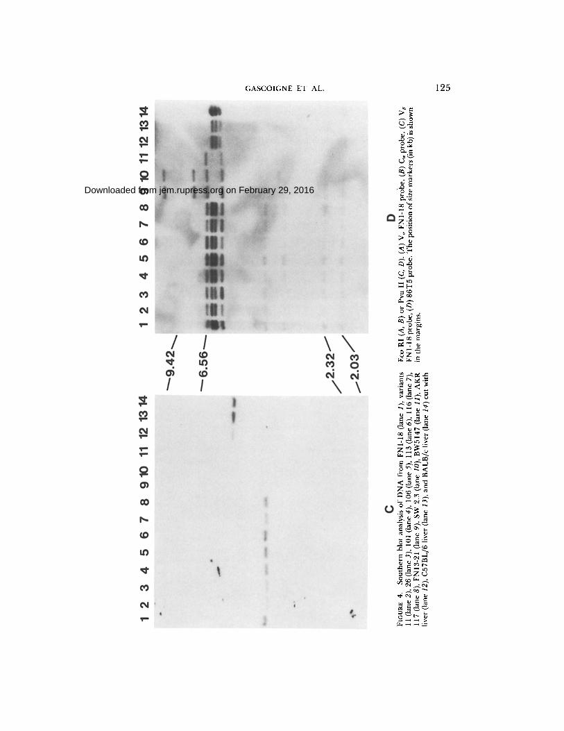
Southern Blot Analysis. Genomic DNA from the same cell lines as above and the hybridoma SW2.3 (25) were prepared, as was DNA from the fusion partner BW5147 and liver cells from mouse strains AKR, BALB/c, and C57BL/6. AKR is the control for BW5147, which is an AKR thymoma, and the other strains are controls for the CB6F~ mice, from which the hybridomas were derived. 10 #g of DNA was digested with the restriction enzymes Eco RI (Fig. 4, a and b) or Pvu II (Fig. 4, c and d). After electrophoresis and blotting, the filters were hybridized to a variety of probes. Fig. 4a shows that V~ FNI-18 exists as a family (n > 5) of related sequences in the genome of all three mouse strains, as are the majority of V~ sequences (19, 20).
A 6.6-kb band is present in all the FNI- t8 series, but not in the other lanes, and probably represents the functional V~. Several of the bands present in the C57BL/6 or BALB/c liver cells are deleted in the hybridomas. This is probably due to deletions that occurred during rearrangement of the functional V~. The pattern of these bands then indicates that the FNI-18 series of variants has not undergone any additional rearrangements and that FN13-21 has undergone a different rearrangement and therefore cannot use the same V~ as FNI-18. SW2.3 has a very similar rearrangement to FN 13-21 and may indeed be identical, although this is not certain from this blot. It should be mentioned that SW2.3 was obtained from an independent fusion and uses I-A b as its restriction element.
on February 29, 2016
jem.rupress.org
Dow
nloaded from
Published July 1, 1986

TA
BL
E
VI
Lac
k of
IL-2
Pro
duct
ion
by F
N l-
18 V
aria
nts
Incu
bate
d w
ith
KL
H a
nd I
rrad
iate
d Sp
leen
Cel
ls fr
om I
-E R
ecom
bina
nt o
r M
ouse
Str
ains
H-2
loc
us
Ori
gin
of
AP
C
wit
h V
ario
us H
aplo
type
s
Clo
nes
K
A~
Aa
EO
Eo
D
FN
I-1
8
Va
rll
Var
26
V
arl0
4
Var
l06
V
ar1
17
V
arl0
1
Var
11
3
Va
rll6
CB
6F~
b/d
b
/d
b/d
b
/d
b/d
b
/d
9,7
78
± 3
66*
1,38
0 4-
82
1,57
0 ±
56
1,58
2 ±
72
1.17
2 ±
361
1,
214
± 2
19
1,
464
± 9
2 1,
082
± 2
52
1,
264
± 3
80
C
57
BL
/6
b b
b b
b b
1,98
2 ±
34
0
1,23
4 ±
51
0
1,81
4 ±
14
0 l,
15
6 ±
21
2
1,50
8 ±
27
4
1,05
4 ±
25
8
1,47
2 ±
14
8 1,
370
± 2
18
1,
056
±
164
BA
LB
/c
d d
d d
d d
1,4
18
±1
84
1
,47
0_
+2
46
1
,26
8±
29
1
1,9
88
4-3
17
1
,49
2±
92
1
,64
4±
16
2
1,2
36
±1
42
1
,52
6±
21
1
1,9
26
4-3
25
A
KR
k
k k
k k
k 1
1,9
42
± 2
42
1,
478
± 9
0 1,
822
± 3
84
1,
830
± 2
54
1
,33
2 ±
28
9
1,77
4 ±
46
6
1,8
02
± 6
14
1,
202
± 3
04
1,
032
±
193
P/J
p
p p
p p
p 1,
030
± 3
76
1,
998
± 2
84
1,
648
±
122
1,64
8 ±
21
2
92
6 ±
64
1,
326
± 2
02
1,
162
±
158
1,36
6 ±
86
1,
674
±
150
Sm
/J
v v
v v
v v
97
6 ±
27
4
1,36
0 4-
57
2
1,66
6 ±
24
6
1,81
4 ±
25
8
1,63
4 ±
120
1,
684
±
138
1,03
4 ±
12
2 1,
746
± 7
6
1,92
2 ±
22
4
DB
A/I
q
q q
q q
q 1,
724
_+ 1
70
1,94
6 ±
25
6
1,30
0 ±
11
0 1,
755
± 4
9
1,15
6 ±
81
1,54
2 4-
45
6
1.25
4 ±
36
0
1,75
4 ±
21
0
1,08
0 ±
94
D
20G
D
d d
d d
b b
1,91
8 ±
25
8
1,50
2 ±
26
6
1,14
4 ±
80
1,
546
± 2
63
8
52
±
120
1,46
0 +
33
2
1,28
2 ±
16
2 1,
542
± 1
66
1,36
0 _+
37
6
BIO
.A(5
R)
b b
b b
k d
10,5
10 ±
24
6
1,54
2 ±
47
8
1,07
2 ±
66
1,86
8 ±
24
7
t,3
66
± 3
26
1,
292
± 4
02
1,
142
± 3
28
1,
066
± 5
6 1,
436
± 2
62
B
I0.R
SB
4
s s
s s
k b
1,60
2 ±
40
2
1,91
2 ±
23
8
1,85
2 ±
15
2 1,
946
± 6
5
1,1
70
± 2
50
1,
600
± 2
40
1,
682
± 2
12
1,
300
± 7
4 1,
512
± 9
6
BI 0
.RFB
2 f
f f
f k
b 1,
528
4- 9
6 1,
820
± 3
38
1,
038
± 6
2
1,34
8 ±
20
7
1,64
2 _+
50
4
1,8
00
± 1
54
1,23
8 _+
49
4
1,62
8 ±
40
1,
554
± 5
2 B
I0.T
BR
3
s k
k k
b b
1,1
84
±2
14
1
,85
8±
20
8
1,1
20
±1
42
1
,69
8_
+2
73
1
,27
6±
48
1
,33
4±
23
4
1,1
12
±1
50
1
,32
2±
26
1
,09
84
-12
0
B10
.RS
B2
s s
s s
k d
83
4--
_7
6
1,6
30
±6
2
1,42
6 ±
72
98
7±
90
1,
372
±
102
1,0
33
±2
91
8
88
±1
74
1
,08
4±
26
4
1,28
6 ±
132
Th
e re
sults
of
S[H
]thy
mid
ine
inco
rpor
atio
n fo
r cu
ltur
es w
itho
ut K
LH
are
not
sho
wn;
the
y w
ere
very
sim
ilar
to t
hose
of
cult
ures
con
tain
ing
KL
H.
In t
he u
se o
f F
N 1
-18
clon
e in
cuba
ted
wit
h C
B6F
I ce
lls,
clon
e w
as 1
,386
± 2
88;
with
AK
R c
ells
1,4
18 ±
15
2; a
nd
wit
h B
I0.A
(5R
) ce
lls 1
,391
+ 2
58
, re
spec
tivel
y.
* 3[
H]t
hym
idin
e in
corp
orat
ion
by 3
x
10 ~
HT
-2 c
eils
inc
ubat
ed w
ith
1:4
-1:3
2 d
ilut
ions
of
supe
rnat
ant
from
cel
l cul
ture
s (3
×
104
T c
ell
hybr
idom
as 5
×
105
5,0
00
rad
irr
adia
ted
spl
een
cells
an
d 4
0 #
g K
LH
).
Onl
y th
e re
sult
s w
ith l
:l 6
dil
utio
n o
f su
pern
atan
t ar
e sh
own.
U
nd
erli
nin
g d
enot
es p
osit
ive
valu
es;
all o
ther
val
ues
are
nega
tive
.
z z Q
z z
on February 29, 2016jem.rupress.orgDownloaded from
Pub
lishe
d Ju
ly 1
, 198
6

GASCOIGNE ET AL. 121
A A L P H A
! 1 I
BamHI o R l Hinfl NcoI EcoRl
1 = F
B E T A
!
I Xmnl Avail
D
, - - - - - - 4
B A L P H A
V Region 30 40 50 60
Q C N F S S T A T Q L Q W F Y q S P G G S L V $ L L S N P 5 G T K H T G R L T 5 CAGTGTAACIFTTTCCTCCAC AGCAACCCAGCTGCAGTGtiTI T TACCAAAGTCC I G GGGGAAGCCTCCaTC A GCC T GTTGT CCAATCC T TCTGGGAC AAAGCACAC T GGAAGAC TGACAT C C
,~ - D ? - , J R e g i o n -
70 80 90 100 T T V T K E R R S S L H I $ S S Q T f D S G T Y L C A M G G S A L G R L H F G A
A~;CACAGTCACTAA~.'G/~CGTCGCAGCTCTTT G CAt;AT T TCC T CCTCCCAGACAAC A£=AC T C A GGCAC T TATC I CTGI GC T AT ~GAGGT I CAG[;C T TAGGGAGW;CTGCAT T TTGGAGC T
, I ~ C Region- 110
G T Q L I V I P D i Q N P GGGACTCAGCTGATTGTCATACCTGACATCCAGAACCCA...
B E T A
- - L e a d e r ~ - - • V Region 1 10 20 30
R P C C G L G A L V Y Q Y P R R T I C K S G T S M R M E C Q A V G F Q A T S V A C GGCCT~Gc~GTGGGcTTGGAGCACTCG~CTATCAATATCC~AGAAGAACCA~CTGTAAGAGIG~AA~l'TC~ATGAGGATGGAG~GTCAAGC~GTG~GTT~ICAG~CAAC~TCTG~AGC~
40 50 60 70 W Y R Q S P Q K T F E L [ A L S T V N S A I K Y E Q N F T Q E K F P I S H P N L
TGGT ATCGTCAATCGCC TCAAAAGACAT T T GAACTGATAG~.ACT T T CTACTGTGAACTCAGCAATCAAk, TATGAACAAAAITTTACC C AGG~AATT T CCCA TC A G T C ATCC CAAC T T A
= - O - I ', J / ~ 1 . 3 ~ 80 90 100
$ F $ S M T V L N A Y L E D R G L Y L C G A R E G T G Y S G N T L Y F i~ E G S R TCCTTTTCATCTATGAC A GTTT T ~TGCATATI; TTGAAGACAGAEGC I T A TATCTCTGTGGTG£ TAGGBAAGGGACAGGGTA IT Cl G GAAJ~I A Ct;C I C T AI TT TGGAGAAGG A AGC CC~L~
J' I ' " - C R e g i o n - -
L I V V E D L R N CTCAT TGT TGTAGAGGATCTGAGAf,k,T.,.
FIGURE 2. (A) The variable regions of the a and/3 chain cDNAs derived from an FNI-18- specific eDNA library were sequenced by the strategy shown in Fig. 2 a, using the technique
32 of Maxam and Gilbert (30). DNA fragments were labeled with P a-dCTP or dATP using the Klenow (large) fragment of DNA pol I. The Barn HI site shown is from the polylinker of pUC9. The Eco RI sites derive from the oligonucleotide linkers added during eDNA cloning. Also shown is the V region probes (thin open boxes) used in Fig. 3. (B) The sequences of V~ F N 1-18 and Va FN 1-18 are shown with their predicted protein sequences. These sequences are analyzed in detail in reference 20.
Hybridization with the C~ probe (Fig. 4b) shows that all hybridomas contain a hybridizing band at 4.7 kb that is not present in BW5147 or the liver cells, and thus must represent a difference due to rearrangement of one of the c~ chain chromosomes.
The Pvu II-digested DNAs were hybridized with beta chain probes. Fig. 4d shows the hybridization of the 86T1 probe. There are three strong bands in the liver cells at 6.2, 6.0, and 5.8 kb, the usual pattern in unrearranged cells (33, 34), and in BW5147 there are two bands at 6.6 and 5.7 kb. All the hybridomas contain the BW5147 bands. The FNI-18 bybridoma also contains two of the germline bands, which implies that there has been no rearrangement to C a (C~2) in at least one chromosome. This is confirmed by the fact that a J region from
on February 29, 2016
jem.rupress.org
Dow
nloaded from
Published July 1, 1986

122 c~ AND /3 GENES IN T CLONE VARIANTS
FIGURE 3. Northern blot analysis of mRNA from FNI-18 (lane 1), variants 11 (lane 2), 26 (lane 3), t 01 (lane 4), 106 (lane 5), 113 (lane 6), 116 (lane 7), 117 (lane 8), and FN 13-21 (lane 9). (A) V~ FNI-18 probe, (B) C~ probe, (C) Va FNI-18 probe, and (D) 86T5 (J~-C~) probe. (E) shows a longer exposure of lanes 2-5 of D. The two arrows show the relative positions of 28 S (~5.0 kb) (upper) and 18 S (~1.8 kb) (lower) ribosomal RNA. (F) Va 86T1 probe. FN1- 18 (lane 1), variant 11 (lane 2), 26 (lane 3), BW5147 (lane 6).
t he 5 ' c lu s t e r (J~3 (J~l .3)) is used by this cell l ine (Fig. 3b) . In a d d i t i o n , F N I - 1 8 has m o r e weak ly h y b r i d i z i n g b a n d s at 3.8, 3.2, 2.3, a n d 2.0 kb, which r e p r e s e n t h y b r i d i z a t i o n to t he D o r J r e g i o n s equences o f 8 6 T 5 . T h e two sma l l e r b a n d s
on February 29, 2016
jem.rupress.org
Dow
nloaded from
Published July 1, 1986

GASCOIGNE ET AL. 123
FIGURE 3.
are of germline pattern and thus represent the unrearranged copies of D and J. The 3.8-kb band represents the rearranged VJD band (see below). FNI-18 expresses a DJC 1.0-kb mRNA (Fig. 3d). Of all the FNI-18 variants, only variable 26 does not express this mRNA and it is also this cell that has no band at 3.2 kb, the size known to correspond to a rearrangement of D to Je3 (Jel.3) from the restriction map (33). Other than this, the patterns of C~ rearrangement in the variants are identical to FN 1-18. The 3.8-kb band corresponds to the copy ofJ~3 (J~1.3) that is rearranged to V~ FNI-18 (see Fig. 4c). FN13.21 and SW2.3 have, as in the ~ chain, very similar rearrangements, although they are not identical, since SW2.3 has a band at 3.4 kb that is not present in FN13.21.
The V region-specific probe (Fig. 4c) indicates that whereas all the liver cells have a single band at 4.3 kb, indicating the unrearranged version of this V~, only FNI-18 and its variants contain a rearranged version at 3.8 kb. The apparent size difference between FN 1-18 and the variants is an artifact of this particular gel. On other blots, the sizes are identical (as in Fig. 4 d). The lack of a Va FN 1- 18 copy in FN13.21 and SW2.3, therefore, indicates that they must use/3 chain V regions that are downstream of V~ FNI-18 and have lost their copy of it during rearrangement.
T
Discussion
We have explored the relationships between T cell idiotypes and the expressed cell receptor V region genes by producing mutated variants of a well-
on February 29, 2016
jem.rupress.org
Dow
nloaded from
Published July 1, 1986

124 a A N D /3 G E N E S I N T C L O N E V A R I A N T S
on February 29, 2016
jem.rupress.org
Dow
nloaded from
Published July 1, 1986
©
O
Z
rr~
,q
FIG
UR
E 4.
S
outh
ern
blot
ana
lysi
s of
DN
A f
rom
FN
I-18
(la
ne 1
), va
rian
ts
11 (
lane
2),
26
(lan
e 3)
, 10
1 (l
ane
4),
106
(lan
e 5)
, 11
3 (l
ane
6),
116
(lan
e 7)
, 11
7 (l
ane
8),
FN13
-21
(lan
e 9)
, SW
2.3
(la
ne 1
0),
BW
5147
(la
ne 1
1),
AK
R
live
r (l
ane
12),
C57
BL
/6 l
iver
(la
ne 1
3), a
nd B
AL
B/c
liv
er (l
ane
14) c
ut w
ith
Eco
RI
(A, B
) or
Pvu
II
(C,
D).
(A
) V
~ F
NI-
18 p
robe
, (B
) C
~ pr
obe,
(C
) V
o F
N l-
18 p
robe
, (D
) 86
T5
prob
e. T
he p
osit
ion
of si
ze m
arke
rs (
in k
b) is
sho
wn
in t
he m
argi
ns.
on February 29, 2016jem.rupress.orgDownloaded from
Pub
lishe
d Ju
ly 1
, 198
6

126 A N D /3 GENES IN T C L O N E V A R I A N T S
characterized T hybridoma which can no longer respond to antigenic stimuli and have not acquired any new allospecificities, yet can produce IL-2 when activated by Con A. The capacity of variants to produce IL-2 after the binding of lectin to another T cell membrane structure and their ability to express Thy- 1.2 and Lyt-l.2 antigens show that the exposure to this dose of x-irradiation did not cause extensive lesions in the genome.
Both the V~ and Ve genes of the parental clone FNI-18 from which the variants had been derived were cloned and sequenced. The 13 chain V region is, like the majority of V~s (17), a single copy gene and, like most V~s (19, 20), V~ FNI-18 exists as a gene family. The FNI-18 variants have lost expression at the mRNA level of their specific Va gene, which would seem to explain their loss of antigen reactivity. However, they still express antigenic molecule(s) associated with T cell membrane recognized by the $3a.6-18 mAb. This determinant was previously identified on another KLH-specific clone (i.e., A12.11) (31). This antibody certainly recognizes a T cell receptor determinant, because it binds to only FNI-18 and A12.11 among our panel of KLH-specific T cell clones, it inhibits both IL-2 production of FNI-18 cells stimulated with KLH and the helper function of A12.11 clones (31), and its binding to FNI-18 was not inhibited by other mAbs directed against antigenic determinants associated with T cell membrane. The binding of this antibody to variants can be explained by the following.
First, that S3a.6-18 recognizes an c~ chain determinant and, therefore, a hybrid heterodimer resulting from the assortment of V,~ FNI-18 and V~ of BW5147 gene products. This explanation is supported by the presence of a Ve BW5147 message identified with a 86T1 probe in Northern blots in variants.
There is evidence for the occurrence of such a hybrid molecule where a complementation between alpha-BW5147 and another/3 chain seems to produce a receptor that gives weak anti-IA b alioreactivity (22). A precedent also exists for a chain-specific T cell receptor determinant. The KJ16 and F23.1 antibodies (35) recognizes a particular V~ found on ~20% of peripheral T cells in most mouse strains (32).
A further possibility is that the mAb may recognize another polymorphic, clonally restricted structure on the cell surface that is important in the T cell recognition, such as a Th equivalent to the "7 chain (36). No candidate molecule has been put forward and its existence seems unlikely from other studies (21, 22).
A third possibility is that $3a.6-18 recognizes a clonotypic marker that is not coded for by V~ and V~ genes. Such markers were recently identified with serological reagents by Allison et al. (personal communication).
The findings presented here are important in respect to other workers' studies of T cell receptor loss mutants. Weiss et al. (37) have made variants of the Jurkat T lymphoma by mutation with y radiation or ethyl methane sulfonate. They were negatively selected for cell surface expression of either T cell receptor or T3, but invariably lost both. These workers have now extended these studies to show that although in all seven cases examined, the T3 6 chain (38) mRNA was present, full-length 13 chain message was detectable in each case. Interestingly, as we have found, the mutants preferentially lost the ~3 chain rather than the c~
on February 29, 2016
jem.rupress.org
Dow
nloaded from
Published July 1, 1986

GASCOIGNE ET AL. 127
chain. Yague et al. (22) describe spontaneous chromosome loss variants where V~ is sometimes lost, but it is likely that this is due to a different mechanism from that occurring in deliberately mutagenized cells.
The mechanism by which x rays produce mutation is still relatively obscure. Malling and de Serres (39), working with Neurospora, found that >90% of the 101 ad-3B x-ray mutants that they studied appeared to be single base substitu- tions, insertions, or deletions. In contrast, a study of x-ray-induced mutants of the Drosophila alcohol dehydrogenase gene (40) revealed 20/28 (71%) to be deletions that included neighboring loci. The screen in this case strongly selected for "non-leaky" mutants and thus may have biased the results against point mutations (40).
The FNI-18 variants do not seem to have suffered any gross lesions in the DNA. This seems also to be true for some of the other reported/3 chain loss variants (41; A. Weiss, personal communication), 4/7 of which were radiation- induced. This implies point mutations or very small deletions rather than large deletions. The mutations would have to be in a region directly or indirectly controlling transcription of Ve FNI-18 mRNA, which must operate in cis since the BW5147 beta chain is still transcribed. An alternative explanation is that the x-ray mutations may have occurred upstream of the Pvu II fragment on which the rearranged Ve FNI-18 is found. This would limit the positions of the mutations to >2 kb 5' to the V region. The mutations induced in the FNI-18 variants must therefore be either small changes in nearby controlling regions of DNA that very strongly decrease the level of transcription or they may be mutations of any size >2 kb upstream of the V~ FNI-18 structural gene. Controlling elements have not been found this far upstream in Ig genes.
Whatever the mechanism, it seems from our studies and others (37, 41) that the/3 chain is more susceptible to mutation than the a chain. In the Drosophila studies mentioned above (40), a clustering of breakpoints of deletions was noted and it may be that the T cell receptor beta chain is particularly susceptible to inactivation by mutagenic agents.
In the/3 chain loss mutants of Weiss and Stobo (37, 41), although the a chain was not lost, the mRNA was expressed at lower levels than normal. When a/3 chain loss mutant was transfected with the/3 chain gene, the level of a chain expression increased back to normal levels and the idiotypic protein and T3 were expressed on the cell surface (41). Thus, there seems to be a regulation of
chain expression by the/3 chain. Since in development the/3 chain is expressed before alpha (42-44), it is reasonable to postulate that its presence in the cell signals the c~ chain to be expressed. In our experiments, we did not observe this lowering of the o~ message in the FN 1-18 beta loss variants since the full-length
chain message from the fusion partner was always expressed. In conclusion, our results show that the variants obtained from the FN 1-18 T
cell hybridomas lost their antigenic specificity without acquiring new alloantigen reactivities. The ability to produce IL-2 after lectin stimulation and to bind antiidiotype mAbs specific for the parental clone was preserved. Loss of the ability to recognize the antigen is, in every case, correlated with failure to produce the FN 1-18-specific Ve mRNA.
on February 29, 2016
jem.rupress.org
Dow
nloaded from
Published July 1, 1986

128 ~ AND /3 GENES IN T CLONE VARIANTS
S u m m a r y
We have analyzed a series o f mutants der ived f rom a KLH-specific, I-E- restr icted T hybr idoma (FN 1-18) which have lost antigen-reactivity while retain- ing both T cell r ecep tor idiotypic de te rminan ts and the ability to respond to Con A. T h e variants have not gained any detectable alloreactivity, nor is there an obvious lesion in the mutants ' beta chain DNA containing the utilized/3 chain genes. This loss o f ant igen reactivity is due to a failure o f stable product ion o f the specific V~-containing m R N A . O u r results indicate that in FNI -18 , the T cell r ecep tor antigenic de te rminan t s are most likely carr ied by the alpha chain alone or by a complementa t ion p roduc t o f the V~ FN 1-18 with the V~ of BW5147. V~ FN 1-18 represents a previously undescr ibed T cell r ecep tor V region.
Received for publication 4 October 1985 and in revised form I1 March 1986.
R e f e r e n c e s 1. Zinkernagel, R. M., and P. C. Doberty. 1979. MHC-restricted cytotoxic T cells:
studies on the biological role of polymorphic major transplantation antigens deter- mining T cell restriction specificity, function and responsiveness. Adv. Immunol. 27:52.
2. Sprent, J., R. Korngold, and K. Molnar-Kimber. 1980. T cell recognition of antigen in vivo: role of the H-2 complex. Springer Semin. Immunopathol. 3:213.
3. Allison, J. P., B. W. Mclntyre, and D. Bloch. 1982. Tumor specific antigen and murine T-lymphoma defined with monoclonal antibody. J. lmmunol. 129:2293.
4. Haskins, K., R. Kubo, J. White, M. Pigeon, J. Kappler, and P. Marrack. 1983. The major histcompatibility complex-restricted antigen receptor on T cells. I. Isolation with a monoctonal antibody. J. Exp. Med. t 57:1149.
5. Meuer, S. C., K. A. Fitzgerald, R. E. Hussey, J. C. Hogdon, S. F. Schlossman, and E. L. Reinherz. 1983. Clonotypic structures involved in antigen-specific human T cell function: relationship to the T3 molecular complex. J. Exp. Med. 157:705.
6. Samelson, L. E., R. N. Germain, and R. H. Schwartz. 1983. MonoclonaI antibodies against the T cell receptor on a cloned T cell hybrid. Proc. Natl. Acad. Sci. USA. 80:6972.
7. Staerz, U. D., M. S. Pasternack, J. R. Klein, J. D. Benedetto, and M.J. Bevan. 1984. Monoclonal antibodies specific for a murine cytotoxic T lymphocyte clone. Proc. Natl. Acad. Sci. USA. 81 : 1799.
8. Hedrick, S. M., E. A. Nielsen, J. Kavaler, D. I. Cohen, and M. M. Davis. 1984. Sequence relationships between putative T cell receptor polypeptides and immuno- globulins. Nature (Lond.). 308:153.
9. Yanagi, Y., Y. Yoshikai, K. Leggett, S. P. Clark, h Aleksander, and T. W. Mak. 1984. A human T cell-specific cDNA clone encodes a protein having extensive homology to immunoglobulin chains. Nature (Lond.). 308:145.
10. Chien, Y., D. M. Becker, T. Eindsten, M. Okamura, D. I. Cohen, and M. M. Davis. 1984. A third type of murine T cell receptor gene. Nature (Lond.). 312:31.
I 1. Saito, H., D. M. Kranz, Y. Takagaki, A. C. Hayday, H. N. Eisen. and S. Tonegawa. 1984. A third rearranged and expressed gene in a clone of cytotoxic T lymphocytes. Nature (Lond.). 312:36.
12. Chien, Y., N. R.J. Gascoigne, J. Kavaler, N. E. Lee, and M. M. Davis. 1984. Somatic recombination in a murine T cell receptor gene. Nature (Lond.). 309:322.
13. Siu, G., S. P. Clark, Y. Yoshikai, M. Malissen, Y. Yanagi, E. Strauss, T. W. Mak, and 1~. Hood. 1984. The human T cell antigen receptor is encoded by variable, diversity,
on February 29, 2016
jem.rupress.org
Dow
nloaded from
Published July 1, 1986

GASCOIGNE ET AL. 129
and joining gene segments that rearrange to generate a complete V gene. Cell. 37:393.
34. Gascoigne, N. R.J. , Y. Chien, D. M. Becker, J. Kavaler, and M. M. Davis. 1984. Genomic organization and sequence of T cell receptor beta chain constant- and joining-region genes. Nature (Lond.). 310:387.
15. Mallissen, M., K. Minard, S. Mjolsness, M. Kronenberg,J. Goverman, T. Hunkapiller, M. B. Prystowsky, Y. Yoshikai, F. Fitch, T. W. Mak, and L. Hood. 1984. Mouse T cell antigen receptor: structure and organization of constant and joining gene segments encoding the bete polypeptide. Cell. 37:1101.
16. Hayday, A. C., D.J. Diamond, G. Tanigawa, J. S. Heilig, V. Folson, H. Saito, and S. Tonegawa. 3985. Unusual organization and diversity of T cell receptor alpha chain genes. Nature (Lond.). 316:828.
17. Patten, P., T. Yokota, J. Rothbard, Y. Chien, K. Arai, and M. M. Davis. 1984. Structure, expression and divergence of T cell receptor beta chain variable regions. Nature (Lond.). 312:40.
38. Barth, R. K., B. S. Kim, N. C. Lan, T. Hunkapiller, N. Sobeieck, A. Winoto, H. Gershenfeld, C. Okada, D. Hansburg, I. L. Weissman, and L. Hood. 1985. The murine T cell receptor employs a limited repertoire of expressed V gene segments. Nature (Lond.). 316:517.
19. Arden, B.,J. L. Klotz, G. Siu, and L. E. Hood. 1985. Diversity and structure of genes of the alpha family of mouse T cell antigen receptor. Nature (Lond.). 316:783.
20. Becker, D. M., P. Patten, Y. Chien, T. Yokota, Z. Eshher, M. Giedlin, N. R. J. Gascoigne, C. Goodnow, R. Wolf, K. Arai, and M. M. Davis. 1985. Variability and repertoire size in T cell receptor V~ gene segments. Nature (Lond.). 317:430.
21. Marrack, P., R. Shimonkevitz, C. Hannum, K. Haskins, andJ. Kappler. 1983. The major histocompatibility complex-restricted antigen receptor on T cells. IV. An anti- idiotypic antibody predicts both antigen and I-specificity.J. Exp. Med. 358:1635.
22. Yague, J., J. White, C. Coleclough, J. Kappler, E. Palmer, and P. Marrack. 1985. The T cell receptor: the alpha and beta chains define idiotype, and antigen and MHC specificity. Cell. 42:81.
23. Waters, S. J., R.J. Winchester, F. Nagase, G. J. Thorbecke, and C. A. Bona. 1984. Antigen presentation by murine and human cells to a murine T cell hybridoma: demonstration of a restriction element associated with a major histocompatibility complex class II determinant(s) shared by both species. Proc. Natl. Acad. Sci. USA. 81:7559.
24. Nagase, F., S.J. Waters, G.J. Thorbecke, and C. A. Bona. 3984. Characterization of a (BALB/c x C57BL/6)FI T cell bybridoma with double specificity: recognition of antigen in context of I-A a and autoreactivity to I-A b. Eur. J. lmmunol. 14:652.
25. Bona, C. A., and S. J. Waters. 1985. Clonotypic markers expressed on a murine T cell hybridoma recognizing foreign antigen in association with murine and human Ia antigen. In Cell Biology of the Major Histocompatibility Complex. B. Pernis and H. Vogel, editors. Academic Press, New York. 289-298.
26. Maniatis, T., E. F. Fritsch, andJ. Sambrook. 1982. Molecular Cloning. A Laboratory Manual. Cold Spring Harbor Laboratory, Cold Spring Harbor, NY.
27. Feinberg, A. P., and B. Vogelstein. 3983. A technique for radio labeling DNA restriction endonuclease fragments to high specific activity. Anal. Biochem. 132:6.
28. Huynh, T. V., R. A. Young, and R. W. Davis. 1984. In DNA Cloning Techniques: A Practical Approach. C. M. Glover, editor. IRI Press, Oxford.
29. Vieira, J., andJ. Messing. 1982. The pUC plasmics, an M13mp7-derived system for insertion mutagenesis and sequencing with synthetic universal primers. Gene. 19:259.
on February 29, 2016
jem.rupress.org
Dow
nloaded from
Published July 1, 1986

130 a AND 13 GENES IN T CLONE VARIANTS
30. Maxam, A. M., and W. Gilbert. 1980. Sequencing end labelled DNA with base- specific chemical cleavages. Methods Enzymol. 65:499.
31. Waters, S. J., P. Luzatti, and C. Bona. 1984. Functional properties of T cell clones with a double specificity for alloantigens and foreign antigens.J. Exp. Med. 160:1300.
32. Sire, G. K., and A. A. Augustin. 1985. V gene polymorphism and a major polyclonal T cell receptor idiotype. Cell. 42:89.
33. Hedrick, S. M., R. N. Germain, M.J. Bevan, M. Doff, I. Engel, P. Fink, N. Gascoigne, E. Heber-Katz, J. Kapp, Y. Kaufman,J. Kaye, F. Melchers, C. Pierce, R. H. Schwartz, C. Sorensen, M. Taniguchi, and M. Davis. 1985. Rearrangement and transcription of a T cell receptor beta chain gene in different T cell subsets. Proc. Natl. Acad. Sci. USA. 82:531.
34. Hedrick, S.J., D. I. Cohen, E. A. Nielsen, and M. M. Davis. 1984. Isolation o f c D N A clones encoding T cell-specific membrane-associated proteins. Nature (Lond.). 309:149.
35. Haskins, K., C. Hannum,J . White, N. Roehm, R. Kubo, J. Kappler, and P. Marrack. 1984. The antigen-specific, major histocompatibility complex-restricted receptor on "F cells. VI. An antibody to a receptor allotype. J. Exp. Med. 1160:452.
36. Saito, H., D. M. Kranz, Y. Takagaki, A. C. Hayday, H. N. Eisen, and S. Tonegawa. 1984. Complete primary structure of a heterodimeric T cell receptor deduced from cDNA sequences. Nature (Lond.). 309:757.
37. Weiss, A., andJ. D. Stobo. 1984. Requirement for the coexpression o fT3 and T cell antigen receptor on a malignant human T cell line.J. Exp. Med. 160:1284.
38. van den Elsen, P., B. A. Sheply, J. Borst, J. E. Caligan, A. F. Markhanm, S. Orkin, and C. Terhorst. 1984. Isolation of cDNA clones encoding the 20K T3 glyco-protein of human T cell receptor complex. Nature (Lond.). 312:413.
39. Mailing, H. V., and F. J. de Serres. 1973. Genetic alterations at the molecular level in x-ray induced ad-3B mutants of Neurospora crassa. Radiation Res. 53:77.
40. Ashburner, M., C. S. Aaron, and S. Bsubota. 1982. The genetics of a small autosomal region of Drosophila melanogaster, including the structural gene for alcohol dehydro- genase. V. Characterization of x-ray induced Adh null mutations. Genetics. 102:421.
41. Ohashi, P. S., T. W. Mak, P. van den Elsen, Y. Yanagi, Y. Yoshikai, A. F. Caiman, C. Terhorst, J. D. Stobo, and A. Weiss. 1985. Reconstitution of an active surface T 3 / T cell antigen receptor by DNA transfer. Nature (Lond.). 316:606.
42. Raulet, D. H., R. D. Garman, H. Saito, and S. Tonegawa. 1985. Developmental regulation of T cell receptor gene expression. Nature (Lond.). 314:103.
43. Snodgrass, H. R., Z. Dembic, M. Steimnetz, H. vonBoehmer. 1985. Expression of T cell antigen receptor genes during fetal development in the thymus. Nature (Lond.). 315:232.
44. Samelson, L. E., T. Lindsten, B. J. Fowlkes, P. van den Elsen, C. Terhorst, M. M. Davis, R. N. Germain, and R. H. Schwartz, 1985. The expression of genes or the T cell antigen receptor complex in precursor thymocytes. Nature (Lond.). 315:765.
on February 29, 2016
jem.rupress.org
Dow
nloaded from
Published July 1, 1986
Copyright © 2022 FDOKUMEN


![Variants of Penney's Game arXiv:2006.13002v1 [math.HO] 19 ...](https://static.fdokumen.com/doc/165x107/6319649f1e5d335f8d0b2299/variants-of-penneys-game-arxiv200613002v1-mathho-19-.jpg)