Noninfectious mixed cryoglobulinaemic glomerulonephritis ...
12
Page 1/12 Noninfectious mixed cryoglobulinaemic glomerulonephritis associated with monoclonal gammopathy of undetermined signicance Adam Flavell ( [email protected] ) Royal Melbourne Hospital https://orcid.org/0000-0002-1278-4303 Robert Fullinfaw Royal Melbourne Hospital Edward R Smith Royal Melbourne Hospital Steve Holt Royal Melbourne Hospital Moira Finlay Royal Melbourne Hospital Thomas Barbour ( [email protected] ) Royal Melbourne Hospital Research article Keywords: cryoglobulinaemia, monoclonal gammopathy of unknown signicance, monoclonal gammopathy of renal signicance, glomerulonephritis Posted Date: March 24th, 2020 DOI: https://doi.org/10.21203/rs.3.rs-18752/v1 License: This work is licensed under a Creative Commons Attribution 4.0 International License. Read Full License Version of Record: A version of this preprint was published on July 23rd, 2020. See the published version at https://doi.org/10.1186/s12882-020-01941-3.
-
Upload
khangminh22 -
Category
Documents
-
view
0 -
download
0
Transcript of Noninfectious mixed cryoglobulinaemic glomerulonephritis ...
Page 1/12
Noninfectious mixed cryoglobulinaemicglomerulonephritis associated with monoclonalgammopathy of undetermined signi�canceAdam Flavell ( adam�[email protected] )
Royal Melbourne Hospital https://orcid.org/0000-0002-1278-4303Robert Fullinfaw
Royal Melbourne HospitalEdward R Smith
Royal Melbourne HospitalSteve Holt
Royal Melbourne HospitalMoira Finlay
Royal Melbourne HospitalThomas Barbour ( [email protected] )
Royal Melbourne Hospital
Research article
Keywords: cryoglobulinaemia, monoclonal gammopathy of unknown signi�cance, monoclonalgammopathy of renal signi�cance, glomerulonephritis
Posted Date: March 24th, 2020
DOI: https://doi.org/10.21203/rs.3.rs-18752/v1
License: This work is licensed under a Creative Commons Attribution 4.0 International License. Read Full License
Version of Record: A version of this preprint was published on July 23rd, 2020. See the published versionat https://doi.org/10.1186/s12882-020-01941-3.
Page 2/12
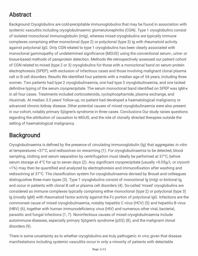
AbstractBackground Cryoglobulins are cold-precipitable immunoglobulins that may be found in association withsystemic vasculitis including cryoglobulinaemic glomerulonephritis (CGN). Type 1 cryoglobulins consistof isolated monoclonal immunoglobulin (mIg), whereas mixed cryoglobulins are typically immunecomplexes comprising either monoclonal (type 2) or polyclonal (type 3) Ig with rheumatoid activityagainst polyclonal IgG. Only CGN related to type 1 cryoglobulins has been clearly associated withmonoclonal gammopathy of undetermined signi�cance (MGUS) using the conventional serum-, urine- ortissue-based methods of paraprotein detection. Methods We retrospectively assessed our patient cohortof CGN related to mixed (type 2 or 3) cryoglobulins for those with a monoclonal band on serum proteinelectrophoresis (SPEP), with exclusion of infectious cases and those involving malignant clonal plasmacell or B cell disorders. Results We identi�ed four patients with a median age of 54 years, including threewomen. Two patients had type 2 cryoglobulinaemia, one had type 3 cryoglobulinaemia, and one lackedde�nitive typing of the serum cryoprecipitate. The serum monoclonal band identi�ed on SPEP was IgM-κin all four cases. Treatments included corticosteroids, cyclophosphamide, plasma exchange, andrituximab. At median 3.5 years’ follow-up, no patient had developed a haematological malignancy oradvanced chronic kidney disease. Other potential causes of mixed cryoglobulinaemia were also presentin our cohort, notably primary Sjögren’s syndrome in three cases. Conclusions Our study raises questionsregarding the attribution of causation to MGUS, and the role of clonally directed therapies outside thesetting of haematological malignancy.
BackgroundCryoglobulinaemia is de�ned by the presence of circulating immunoglobulin (Ig) that aggregates in vitroat temperatures <37°C, and redissolves on rewarming (1). For cryoglobulinaemia to be detected, bloodsampling, clotting and serum separation by centrifugation must ideally be performed at 37°C, beforeserum storage at 4°C for up to seven days (2). Any signi�cant cryoprecipitate (usually >0.05g/L or cryocrit>1%) may then be quanti�ed and analyzed by electrophoresis and immuno�xation after washing andredissolving at 37°C. The classi�cation system for cryoglobulinaemia devised by Brouet and colleaguesdistinguishes three main types (3). Type 1 cryoglobulins consist of monoclonal Ig (mIg) or biclonal Ig,and occur in patients with clonal B cell or plasma cell disorders (4). So-called ‘mixed’ cryoglobulins areconsidered as immune complexes typically comprising either monoclonal (type 2) or polyclonal (type 3)Ig (mostly IgM) with rheumatoid factor activity against the Fc portion of polyclonal IgG. Infections are thecommonest cause of mixed cryoglobulinaemia, notably hepatitis C virus (HCV) (5) and hepatitis B virus(HBV) (6), together with human immunode�ciency virus (HIV) and numerous other viral, bacterial,parasitic and fungal infections (1, 7). Noninfectious causes of mixed cryoglobulinaemia includeautoimmune diseases, especially primary Sjögren’s syndrome (pSS) (8), and the malignant clonaldisorders (9).
There is some uncertainty as to whether cryoglobulins are truly pathogenic in vivo, given that diseasemanifestations including systemic vasculitis occur in only a minority of patients with detectable

Page 3/12
cryoglobulinaemia (10). The systemic vasculitis is exempli�ed by cryoglobulinaemic glomerulonephritis(CGN), which can be classi�ed as type 1 or mixed according to whichever type of cryoglobulin is found inassociation. Classic renal histological features of CGN, including membranoproliferativeglomerulonephritis (MPGN), intracapillary ‘pseudothrombi’, crescents and small vessel vasculitis arenonspeci�c for cryoglobulinaemia as the underlying cause of glomerulonephritis (11). On the other hand,causation due to glomerular deposition of cryoglobulins may be strengthened by the appearance onelectron microscopy (EM) of curvilinear microtubules, suggestive of aggregated cryoglobulins (12, 13), orimmunohistochemistry showing light chain restriction of pseudothrombi in the case of (monotypic) type1 CGN (14, 15).
Monoclonal gammopathy is diagnosed when mIg secreted into the circulation by a proliferating clone ofplasma cells or B cells is detected by means of serum protein electrophoresis (SPEP), immuno�xation(SIFE) or free light chain assays (SFLC), or urine protein electrophoresis (UPEP) or immuno�xation (UIFE)(16). Further evaluation is often required for the malignant clonal disorders, which include multiplemyeloma, Waldenström’s macroglobulinaemia, B cell lymphoma and chronic lymphocytic leukemia.However, in most patients, monoclonal gammopathy of undetermined signi�cance (MGUS) or anotherpre-malignant haematological disorder is diagnosed. Previous studies have established a clearassociation of type 1 CGN not only with malignant clonal disorders, but also with MGUS (14, 17-20). Thishas led to the inclusion of type 1 CGN within the disease classi�cation of monoclonal gammopathy ofrenal signi�cance (MGRS) (21-23). The term MGRS recognizes that certain renal lesions may be causedby nephrotoxic mIg produced by small (i.e. pre-malignant) plasma cell or B cell clones. This diagnosticconcept is underscored in many cases by light chain restricted renal immunostaining (as noted above fortype 1 CGN) (24). Yet surprisingly, type 2 CGN has also been included within MGRS (21-23), despite aweak association with MGUS in the published literature, and the inability to con�rm light chain restrictionowing to the polyclonal Ig component of type 2 cryoglobulins (11). We undertook this study to assesswhether mixed (type 2 or 3) CGN can equally be associated with MGUS.
MethodsCases were identi�ed on retrospective review of clinical and pathological databases at our hospital forthe period 2002-19, and were included if they met all the following criteria: (1) renal histology compatiblewith CGN; and both (2) cryoglobulinaemia >0.05 g/L and (3) a monoclonal band on SPEP at the time ofdiagnostic renal biopsy. Patients were excluded if they met any of the following criteria: (i) type 1cryoglobulinaemia based on immuno�xation of the cryoprecipitate and/or light chain restriction of renalbiopsy tissue, as assessed by immuno�uorescent staining of para�n-embedded tissue after proteasedigestion (para�n-IF) (25); (ii) a malignant clonal disorder based on tests comprising at a minimum bonemarrow aspirate and trephine (BMAT) with �ow cytometry of the aspirate and/or peripheral blood, andcomputed tomography with positron emission tomography (CT-PET); (iii) a potential infectious aetiologyfor mixed cryoglobulinaemia based on tests comprising at a minimum blood cultures, HBV surfaceantigen, and HBV core, HCV, and HIV antibodies; or (iv) vasculitis potentially due to causes other thancryoglobulinaemia based on serologies comprising at a minimum anti-neutrophil cytoplasmic, anti-

Page 4/12
glomerular basement membrane, anti-double stranded DNA, anti-U1-ribonucleoprotein, and anti-cardiolipin antibodies, and lupus anticoagulant.
ResultsOf twenty patients with noninfectious mixed CGN in whom SPEP was performed, four had MGUS,comprising three females and one male (Table 1). Median age at presentation was 54 years (range 47 –66 years). All patients had microscopic haematuria and proteinuria, with a median urinary proteincreatinine ratio (uPCR) of 106mg/mmol (range 70-134mg/mmol). Renal function was signi�cantlyimpaired in three of the four patients, with a median serum creatinine overall of 221μmol/L (range 80-292μmol/L) eGFR of 27mL/min per 1.73m2 (range 15-70mL/min per 1.73m2, MDRD). All patients hadmildly reduced serum albumin levels, and serum complement C4 ± C3 levels were low in three patients.All three female patients had previously been diagnosed with pSS and showed seropositivity forantinuclear, SSA/Ro and SSB/La antibodies. In the weeks following diagnosis of CGN, Patient 2underwent laparotomy for an incidental �nding of cholangiocarcinoma (not previously reported in thesetting of CGN (26)).
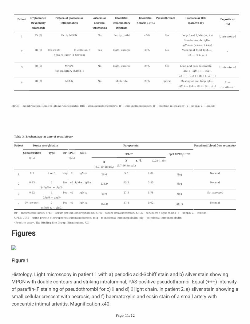
Histological features of CGN included MPGN in three patients, cellular crescents with arteriolar necrosisand thrombosis in one patient, and intracapillary pseudothrombi in three patients (Figure 1 and Table 2).Interstitial �brosis ³25% with mild glomerulosclerosis was also present in three cases.Immunohistochemistry showed variable IgG, IgM and C3 staining in capillary loops and the mesangium,with IgM and/or IgG staining of pseudothrombi in two cases. No case showed light chain restriction onpara�n-IF. EM was performed in three cases, revealing intracapillary curvilinear deposits in one case andunstructured deposits in the other two cases.
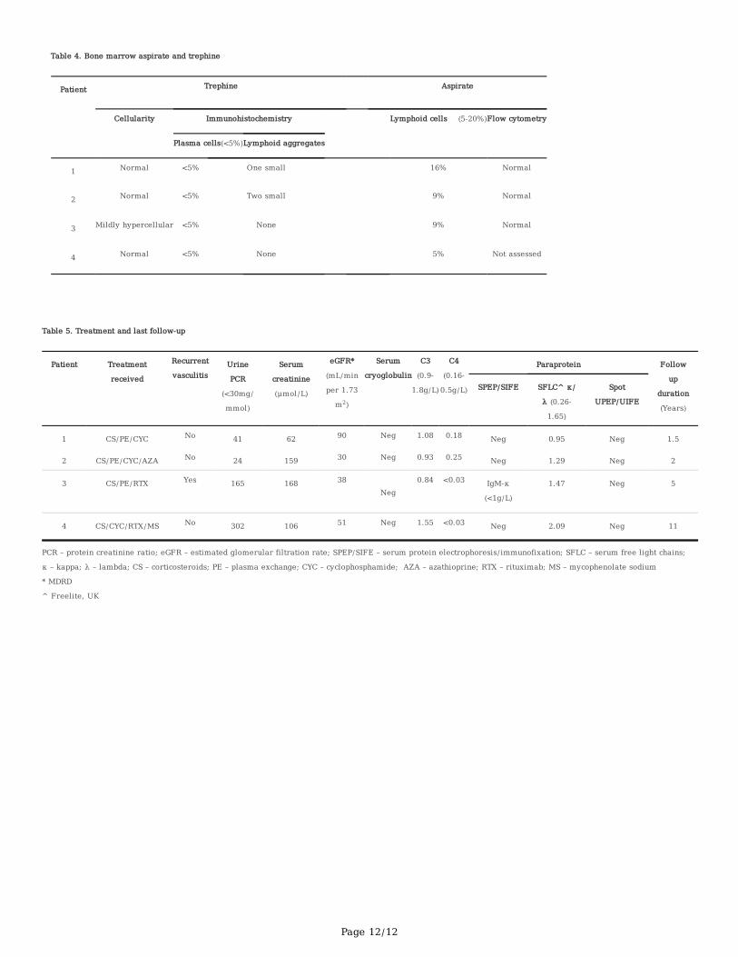
Serum biochemistry at presentation (Table 3) included a median cryoglobulin concentration of 0.43g/L(range 0.1-0.62g/L) in three cases, with a cryocrit of 9% in the fourth case. Immuno�xation of thecryoprecipitate con�rmed type 2 cryoglobulinaemia with a monoclonal IgM-k component in two patientsand type 3 cryoglobulinaemia in one patient, and was not performed in the remaining patient. SPEPrevealed generally small monoclonal bands of median concentration <1g/L (range <1 - 2g/L). In all fourcases, the paraprotein was IgM-k, with an IgG-k paraprotein also present in one case (Patient 2). Nopatient showed bone marrow evidence of a malignant plasma cell or B cell disorder (Table 4).
Treatment consisted of corticosteroids in all patients, cyclophosphamide in three patients, plasmaexchange in three patients, and rituximab in two patients, one of whom (Patient 4) later receivedmaintenance therapy for a diagnosis of seronegative lupus nephritis with mycophenolate mofetil (Table5). At a median follow-up of 3.5 years (range 1.5 – 11 years), no patient had developed a malignantclonal disorder or end-stage renal failure, or was deceased. Renal parameters were improved in all casesbut one (Patient 4), with a median uPCR at last follow-up of 103mg/mmol (range 24 – 302mg/mmol),serum creatinine of 133μmol/L (range 62 - 168μmol/L), and eGFR of 45mL/min per 1.73m2 (range 30-90mL/min per 1.73m2). Serum C4 ± C3 levels remained low in the three patients with this �nding on
Page 5/12
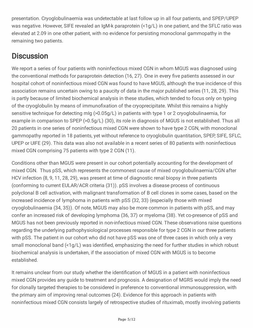
presentation. Cryoglobulinaemia was undetectable at last follow up in all four patients, and SPEP/UPEPwas negative. However, SIFE revealed an IgM-k paraprotein (<1g/L) in one patient, and the SFLC ratio waselevated at 2.09 in one other patient, with no evidence for persisting monoclonal gammopathy in theremaining two patients.
DiscussionWe report a series of four patients with noninfectious mixed CGN in whom MGUS was diagnosed usingthe conventional methods for paraprotein detection (16, 27). One in every �ve patients assessed in ourhospital cohort of noninfectious mixed CGN was found to have MGUS, although the true incidence of thisassociation remains uncertain owing to a paucity of data in the major published series (11, 28, 29). Thisis partly because of limited biochemical analysis in these studies, which tended to focus only on typingof the cryoglobulin by means of immuno�xation of the cryoprecipitate. Whilst this remains a highlysensitive technique for detecting mIg (>0.05g/L) in patients with type 1 or 2 cryoglobulinaemia, forexample in comparison to SPEP (>0.5g/L) (30), its role in diagnosis of MGUS is not established. Thus all20 patients in one series of noninfectious mixed CGN were shown to have type 2 CGN, with monoclonalgammopathy reported in 18 patients, yet without reference to cryoglobulin quantitation, SPEP, SIFE, SFLC,UPEP or UIFE (29). This data was also not available in a recent series of 80 patients with noninfectiousmixed CGN comprising 75 patients with type 2 CGN (11).
Conditions other than MGUS were present in our cohort potentially accounting for the development ofmixed CGN. Thus pSS, which represents the commonest cause of mixed cryoglobulinaemia/CGN afterHCV infection (8, 9, 11, 28, 29), was present at time of diagnostic renal biopsy in three patients(conforming to current EULAR/ACR criteria (31)). pSS involves a disease process of continuouspolyclonal B cell activation, with malignant transformation of B cell clones in some cases, based on theincreased incidence of lymphoma in patients with pSS (32, 33) (especially those with mixedcryoglobulinaemia (34, 35)). Of note, MGUS may also be more common in patients with pSS, and mayconfer an increased risk of developing lymphoma (36, 37) or myeloma (38). Yet co-presence of pSS andMGUS has not been previously reported in non-infectious mixed CGN. These observations raise questionsregarding the underlying pathophysiological processes responsible for type 2 CGN in our three patientswith pSS. The patient in our cohort who did not have pSS was one of three cases in which only a verysmall monoclonal band (<1g/L) was identi�ed, emphasizing the need for further studies in which robustbiochemical analysis is undertaken, if the association of mixed CGN with MGUS is to becomeestablished.
It remains unclear from our study whether the identi�cation of MGUS in a patient with noninfectiousmixed CGN provides any guide to treatment and prognosis. A designation of MGRS would imply the needfor clonally targeted therapies to be considered in preference to conventional immunosuppression, withthe primary aim of improving renal outcomes (24). Evidence for this approach in patients withnoninfectious mixed CGN consists largely of retrospective studies of rituximab, mostly involving patients

Page 6/12
with type 2 CGN and a monoclonal IgM-k cryoglobulin (11, 28, 29, 39). As in previous series (11, 28), ourcohort
received multiple therapies including rituximab but also conventional immunosuppressive agents (11,28). Outcomes were generally favourable, and of the two patients with chronic kidney disease stage 3B atlast follow-up, both had shown signi�cant (25-40%) interstitial �brosis on pre-treatment biopsies(potentially due to pSS-associated interstitial nephritis in one of these cases). Given previous, largercohorts of noninfectious mixed CGN showing ESKD rates of 9-10% at 4 years (11, 29), our study possiblyindicates that MGUS does not always confer a treatment-resistant course. No patient in our seriesreceived bortezomib, for which a single instance of use in refractory noninfectious mixed CGN is reported,in a patient with type 2 CGN and a monoclonal IgM-k component, but no detectable monoclonal band onSPEP (40).
ConclusionsOur study is the �rst to show conclusively that MGUS may be present in a subset of patients withnoninfectious mixed CGN. Even where this is the case, a designation as MGRS may be open to question,given the presence in our cohort of other potential aetiologies for mixed CGN, including pSS in three outof four patients.
DeclarationsEthics approval & consent to participate: N/A
Consent for publication: N/A
Competing interests: none declared
Funding: none
Authors contributions: A Flavell and T Barbour collected & analysed the data and prepared themanuscript. R Fullinfaw assisted with data collection & interpretation. M Finlay collected pathology dataand images. E Smith and S Holt reviewed the manuscript.
Acknowledgements: Nephworks database
Availability of data: all data generated or analysed during this study are included in this published article.
References1. Roccatello D, Saadoun D, Ramos-Casals M, Tzioufas AG, Fervenza FC, Cacoub P, Zignego AL, Ferri C.
Cryoglobulinaemia. Nat Rev Dis Primers. 2018;4(1):11.

Page 7/12
2. Sargur R, White P, Egner W. Cryoglobulin evaluation: best practice? Ann Clin Biochem. 2010;47(Pt1):8-16.
3. Brouet JC, Clauvel JP, Danon F, Klein M, Seligmann M. Biologic and clinical signi�cance ofcryoglobulins. A report of 86 cases. Am J Med. 1974;57(5):775-88.
4. Terrier B, Karras A, Kahn JE, Le Guenno G, Marie I, Benarous L, Lacraz A, Diot E, Hermine O, de Saint-Martin L, Cathebras P, Leblond V, Modiano P, Leger JM, Mariette X, Senet P, Plaisier E, Saadoun D,Cacoub P. The spectrum of type I cryoglobulinemia vasculitis: new insights based on 64 cases.Medicine (Baltimore). 2013;92(2):61-8.
5. Agnello V, Chung RT, Kaplan LM. A role for hepatitis C virus infection in type II cryoglobulinemia. NEngl J Med. 1992;327(21):1490-5.
�. Annear NM, Cook HT, Atkins M, Pusey CD, Salama AD. Non-hepatitis virus associated mixed essentialcryoglobulinemia. Kidney Int. 2010;77(2):161-4.
7. Terrier B, Marie I, Lacraz A, Belenotti P, Bonnet F, Chiche L, Gra�n B, Hot A, Kahn JE, Michel C,Quemeneur T, de Saint-Martin L, Hermine O, Leger JM, Mariette X, Senet P, Plaisier E, Cacoub P. NonHCV-related infectious cryoglobulinemia vasculitis: Results from the French nationwide CryoVassurvey and systematic review of the literature. J Autoimmun. 2015;65:74-81.
�. Galli M, Oreni L, Saccardo F, Castelnovo L, Filippini D, Marson P, Mascia MT, Mazzaro C, Origgi L, OssiE, Pietrogrande M, Pioltelli P, Quartuccio L, Scarpato S, Sollima S, Riva A, Fraticelli P, Zani R, GiuggioliD, Sebastiani M, Sarzi Puttini P, Gabrielli A, Zignego AL, Scaini P, Ferri C, De Vita S, Monti G. HCV-unrelated cryoglobulinaemic vasculitis: the results of a prospective observational study by the ItalianGroup for the Study of Cryoglobulinaemias (GISC). Clin Exp Rheumatol. 2017;35 Suppl 103(1):67-76.
9. Terrier B, Krastinova E, Marie I, Launay D, Lacraz A, Belenotti P, de Saint-Martin L, Quemeneur T, HuartA, Bonnet F, Le Guenno G, Kahn JE, Hinschberger O, Rullier P, Diot E, Lazaro E, Bridoux F, Zenone T,Carrat F, Hermine O, Leger JM, Mariette X, Senet P, Plaisier E, Cacoub P. Management of noninfectiousmixed cryoglobulinemia vasculitis: data from 242 cases included in the CryoVas survey. Blood.2012;119(25):5996-6004.
10. Ramos-Casals M, Stone JH, Cid MC, Bosch X. The cryoglobulinaemias. Lancet. 2012;379(9813):348-60.
11. Zaidan M, Terrier B, Pozdzik A, Frouget T, Rioux-Leclercq N, Combe C, Lepreux S, Hummel A, Noel LH,Marie I, Legallicier B, Francois A, Huart A, Launay D, Kaplanski G, Bridoux F, Vanhille P, Makdassi R,Augusto JF, Rouvier P, Karras A, Jouanneau C, Verpont MC, Callard P, Carrat F, Hermine O, Leger JM,Mariette X, Senet P, Saadoun D, Ronco P, Brocheriou I, Cacoub P, Plaisier E, CryoVas study g.Spectrum and Prognosis of Noninfectious Renal Mixed Cryoglobulinemic GN. J Am Soc Nephrol.2016;27(4):1213-24.
12. Ojemakinde K, Turbat-Herrera EA, Zeng X, Gu X, Herrera GA. The many faces of cryoglobulinemicnephropathy: a clinico-pathologic study of 47 cases with emphasis on the value of electronmicroscopy. Ultrastruct Pathol. 2014;38(6):367-76.

Page 8/12
13. Paueksakon P, Revelo MP, Horn RG, Shappell S, Fogo AB. Monoclonal gammopathy: signi�cance andpossible causality in renal disease. Am J Kidney Dis. 2003;42(1):87-95.
14. Harel S, Mohr M, Jahn I, Aucouturier F, Galicier L, Asli B, Malphettes M, Szalat R, Brouet JC, Lipsker D,Fermand JP. Clinico-biological characteristics and treatment of type I monoclonal cryoglobulinaemia:a study of 64 cases. Br J Haematol. 2015;168(5):671-8.
15. Sidana S, Rajkumar SV, Dispenzieri A, Lacy MQ, Gertz MA, Buadi FK, Hayman SR, Dingli D, Kapoor P,Gonsalves WI, Go RS, Hwa YL, Leung N, Fonder AL, Hobbs MA, Zeldenrust SR, Russell SJ, Lust JA,Kyle RA, Kumar SK. Clinical presentation and outcomes of patients with type 1 monoclonalcryoglobulinemia. Am J Hematol. 2017;92(7):668-73.
1�. Bird J, Behrens J, Westin J, Turesson I, Drayson M, Beetham R, D'Sa S, Soutar R, Waage A,Gulbrandsen N, Gregersen H, Low E, Haemato-oncology Task Force of the British Committee forStandards in Haematology UKMF, Nordic Myeloma Study G. UK Myeloma Forum (UKMF) and NordicMyeloma Study Group (NMSG): guidelines for the investigation of newly detected M-proteins and themanagement of monoclonal gammopathy of undetermined signi�cance (MGUS). Br J Haematol.2009;147(1):22-42.
17. Neel A, Perrin F, Decaux O, Dejoie T, Tessoulin B, Halliez M, Mahe B, Lamy T, Fakhouri F, Jego P, AgardC, Vigneau C, Guenet L, Grosbois B, Moreau P, Hamidou M. Long-term outcome of monoclonal (type1) cryoglobulinemia. Am J Hematol. 2014;89(2):156-61.
1�. Karras A, Noel LH, Droz D, Delansorne D, Saint-Andre JP, Aucouturier P, Alyanakian MA, Grunfeld JP,Lesavre P. Renal involvement in monoclonal (type I) cryoglobulinemia: two cases associated withIgG3 kappa cryoglobulin. Am J Kidney Dis. 2002;40(5):1091-6.
19. Nasr SH, Markowitz GS, Reddy BS, Maesaka J, Swidler MA, D'Agati VD. Dysproteinemia, proteinuria,and glomerulonephritis. Kidney Int. 2006;69(4):772-5.
20. Sethi S, Zand L, Leung N, Smith RJ, Jevremonic D, Herrmann SS, Fervenza FC.Membranoproliferative glomerulonephritis secondary to monoclonal gammopathy. Clin J Am SocNephrol. 2010;5(5):770-82.
21. Leung N, Bridoux F, Hutchison CA, Nasr SH, Cockwell P, Fermand JP, Dispenzieri A, Song KW, Kyle RA,International K, Monoclonal Gammopathy Research G. Monoclonal gammopathy of renalsigni�cance: when MGUS is no longer undetermined or insigni�cant. Blood. 2012;120(22):4292-5.
22. Fermand JP, Bridoux F, Kyle RA, Kastritis E, Weiss BM, Cook MA, Drayson MT, Dispenzieri A, Leung N,International K, Monoclonal Gammopathy Research G. How I treat monoclonal gammopathy of renalsigni�cance (MGRS). Blood. 2013;122(22):3583-90.
23. Leung N, Bridoux F, Batuman V, Chaidos A, Cockwell P, D'Agati VD, Dispenzieri A, Fervenza FC,Fermand JP, Gibbs S, Gillmore JD, Herrera GA, Jaccard A, Jevremovic D, Kastritis E, Kukreti V, Kyle RA,Lachmann HJ, Larsen CP, Ludwig H, Markowitz GS, Merlini G, Mollee P, Picken MM, Rajkumar VS,Royal V, Sanders PW, Sethi S, Venner CP, Voorhees PM, Wechalekar AD, Weiss BM, Nasr SH. Theevaluation of monoclonal gammopathy of renal signi�cance: a consensus report of the InternationalKidney and Monoclonal Gammopathy Research Group. Nat Rev Nephrol. 2018;15:45-59.

Page 9/12
24. Sethi S, Rajkumar SV, D’Agati VD. The Complexity and Heterogeneity of MonoclonalImmunoglobulin–Associated Renal Diseases. Journal of the American Society of Nephrology.2018;29(7):1810-23.
25. Larsen CP, Messias NC, Walker PD, Fidler ME, Cornell LD, Hernandez LH, Alexander MP, Sethi S, NasrSH. Membranoproliferative glomerulonephritis with masked monotypic immunoglobulin deposits.Kidney Int. 2015;88(4):867-73.
2�. Spatola L, Generali E, Angelini C, Badalamenti S, Selmi C. HCV-negative mixed cryoglobulinemia andkidney involvement: in-depth review on physiopathological and histological bases. Clin Exp Med.2018;18(4):465-71.
27. Berenson JR, Anderson KC, Audell RA, Boccia RV, Coleman M, Dimopoulos MA, Drake MT, Fonseca R,Harousseau JL, Joshua D, Lonial S, Niesvizky R, Palumbo A, Roodman GD, San-Miguel JF, Singhal S,Weber DM, Zangari M, Wirtschafter E, Yellin O, Kyle RA. Monoclonal gammopathy of undeterminedsigni�cance: a consensus statement. Br J Haematol. 2010;150(1):28-38.
2�. Foessel L, Besancenot JF, Blaison G, Magy-Bertrand N, Jaussaud R, Etienne Y, Maurier F, Audia S,Martin T. Clinical spectrum, treatment, and outcome of patients with type II mixed cryoglobulinemiawithout evidence of hepatitis C infection. J Rheumatol. 2011;38(4):716-22.
29. Matignon M, Cacoub P, Colombat M, Saadoun D, Brocheriou I, Mougenot B, Roudot-Thoraval F,Vanhille P, Moranne O, Hachulla E, Hatron PY, Fermand JP, Fakhouri F, Ronco P, Plaisier E, Grimbert P.Clinical and morphologic spectrum of renal involvement in patients with mixed cryoglobulinemiawithout evidence of hepatitis C virus infection. Medicine (Baltimore). 2009;88(6):341-8.
30. Batko K, Malyszko J, Jurczyszyn A, Vesole DH, Gertz MA, Leleu X, Suska A, Krzanowski M, SulowiczW, Malyszko JS, Krzanowska K. The clinical implication of monoclonal gammopathies: monoclonalgammopathy of undetermined signi�cance and monoclonal gammopathy of renal signi�cance.Nephrol Dial Transplant. 2018.
31. Shiboski CH, Shiboski SC, Seror R, Criswell LA, Labetoulle M, Lietman TM, Rasmussen A, Sco�eld H,Vitali C, Bowman SJ, Mariette X, International Sjogren's Syndrome Criteria Working G. 2016 AmericanCollege of Rheumatology/European League Against Rheumatism classi�cation criteria for primarySjogren's syndrome: A consensus and data-driven methodology involving three international patientcohorts. Ann Rheum Dis. 2017;76(1):9-16.
32. Zintzaras E, Voulgarelis M, Moutsopoulos HM. The risk of lymphoma development in autoimmunediseases: a meta-analysis. Arch Intern Med. 2005;165(20):2337-44.
33. Nocturne G, Mariette X. Sjogren Syndrome-associated lymphomas: an update on pathogenesis andmanagement. Br J Haematol. 2015;168(3):317-27.
34. Tzioufas AG, Boumba DS, Skopouli FN, Moutsopoulos HM. Mixed monoclonal cryoglobulinemia andmonoclonal rheumatoid factor cross-reactive idiotypes as predictive factors for the development oflymphoma in primary Sjogren's syndrome. Arthritis Rheum. 1996;39(5):767-72.
35. Nishishinya MB, Pereda CA, Munoz-Fernandez S, Pego-Reigosa JM, Rua-Figueroa I, Andreu JL,Fernandez-Castro M, Rosas J, Loza Santamaria E. Identi�cation of lymphoma predictors in patients

Page 10/12
Table 1. Demographic and renal clinical features at time of renal biopsy
Patient Age (Years) Sex Extrarenal clinical
features
Comorbid
conditions
Urine studies Serum studies
Red
cellsPCR
(<30mg/
mmol)
ACR
(<3.5mg/mmol)
Creatinine
(μmol/L)
eGFR
(mL/min per
1.73 m2)*
Albumin
(3.5-
5g/dL)
C3
(0.9-
1.8g/L)
C4
(0.16-
0.5g/L)
ANA/SSA/SSB
1 47 F Purpura, arthritis pSS Pos 70 33 256 18 2.6 0.46 <0.03 Pos
2 60 F Purpura, benign
lymphadenopathy
pSS, CC Pos 126 75 292 15 3.2 1.1 0.27 Pos
3 66 M - - Pos 86 55 186 35 2.7 0.76 <0.03 Neg
4 47 F Purpura, arthritis pSS, hypoGG Pos 134 - 80 70 3.1 1.27 <0.03 Pos
PCR – protein creatinine ratio; ACR – albumin creatinine ratio; eGFR – estimated glomerular filtration rate; ANA – antinuclear antibody; SSA – anti-Ro; SSB – anti-La;
pSS – primary Sjögren’s syndrome; CC – cholangiocarcinoma; hypoGG – hypogammaglobulinaemia
* Modified diet in renal disease (MDRD)
^Nephrotic syndrome
with primary Sjogren's syndrome: a systematic literature review and meta-analysis. Rheumatol Int.2015;35(1):17-26.
3�. Brito-Zeron P, Retamozo S, Gandia M, Akasbi M, Perez-De-Lis M, Diaz-Lagares C, Bosch X, Bove A,Perez-Alvarez R, Soto-Cardenas MJ, Siso A, Ramos-Casals M. Monoclonal gammopathy related toSjogren syndrome: a key marker of disease prognosis and outcomes. J Autoimmun. 2012;39(1-2):43-8.
37. Yang Y, Chen L, Jia Y, Liu Y, Wen L, Liang Y, An Y, Chen S, Su Y, Li Z. Monoclonal gammopathy inrheumatic diseases. Clin Rheumatol. 2018;37(7):1751-62.
3�. Tomi AL, Belkhir R, Nocturne G, Desmoulins F, Berge E, Pavy S, Miceli-Richard C, Mariette X, Seror R.Brief Report: Monoclonal Gammopathy and Risk of Lymphoma and Multiple Myeloma in PatientsWith Primary Sjogren's Syndrome. Arthritis Rheumatol. 2016;68(5):1245-50.
39. Terrier B, Launay D, Kaplanski G, Hot A, Larroche C, Cathebras P, Combe B, de Jaureguiberry JP, MeyerO, Schaeverbeke T, Somogyi A, Tricot L, Zenone T, Ravaud P, Gottenberg JE, Mariette X, Cacoub P.Safety and e�cacy of rituximab in nonviral cryoglobulinemia vasculitis: data from the FrenchAutoimmunity and Rituximab registry. Arthritis Care Res (Hoboken). 2010;62(12):1787-95.
40. Bazari H, Mahindra AK, Farkash EA. Case records of the Massachusetts General Hospital. Case 3-2014. A 61-year-old woman with gastrointestinal symptoms, anemia, and acute kidney injury. N EnglJ Med. 2014;370(4):362-73.
Tables
Table 2. Renal histology
Page 11/12
Table 3. Biochemistry at time of renal biopsy
Patient Serum cryoglobulin Paraprotein Peripheral blood flow cytometry
Concentration
(g/L)
Type RF SPEP
(g/L)
SIFE SFLC* Spot UPEP/UIFE
κ
(3.3-19.4mg/L)
λ
(5.7-26.3mg/L)
κ /λ (0.26-1.65)
1 0.1 2 or 3 Neg 2 IgM-κ 26.6 5.5 4.84 Neg Normal
2 0.43 2
(mIgM-κ + pIgG)
Pos <1 IgM-κ, IgG-κ 231.9 65.3 3.55 Neg Normal
3 0.62 3
(pIgM + pIgG)
Pos <1 IgM-κ 49.0 27.5 1.78 Neg Not assessed
4 9% cryocrit 2
(mIgM-κ + pIgG)
Pos <1 IgM-κ 157.0 17.4 9.02 IgM-κ Normal
RF – rheumatoid factor; SPEP – serum protein electrophoresis; SIFE – serum immunofixation; SFLC – serum free light chains; κ – kappa; λ – lambda;
UPEP/UIFE – urine protein electrophoresis/immunofixation; mIg – monoclonal immunoglobulin; pIg – polyclonal immunoglobulin
*Freelite assay, The Binding Site Group, Birmingham, UK
Patient Noglomeruli
(Noglobally
sclerosed)
Pattern of glomerular
inflammation
Arteriolar
necrosis,
thrombosis
Interstitial
inflammatory
infiltrate
Interstitial
fibrosis (<5%)
Pseudothrombi Glomerular IHC
(paraffin-IF)Deposits on
EM
1 25 (0) Early MPGN No Patchy, mild <5% Yes Loop focal IgM+ (κ-, λ-)
Pseudothrombi IgG+,
IgM+++ (κ+++, λ+++)
Unstructured
2 18 (6) Crescents (5 cellular, 1
fibro-cellular, 2 fibrous)
Yes Light, chronic 40% No Mesangial focal IgM++,
C3++ (κ+, λ+)-
3 20 (5) MPGN,
endocapillary (CD68+)
No Light, chronic 25% Yes Loop and pseudothrombi
IgG++, IgM+++, IgA+,
C3+++, C1q++ (κ ++, λ ++)
Unstructured
4 18 (2) MPGN No Moderate 25% Sparse Mesangial and loop IgG+,
IgM++, IgA+, C3++ (κ -, λ -)Fine
curvilinear
MPGN – membranoproliferative glomerulonephritis; IHC – immunohistochemistry; IF – immunofluorescence; IF – electron microscopy; κ – kappa; λ – lambda
Figures
Figure 1
Histology. Light microscopy in patient 1 with a) periodic acid-Schiff stain and b) silver stain showingMPGN with double contours and striking intraluminal, PAS-positive pseudothrombi. Equal (+++) intensityof para�n-IF staining of pseudothrombi for c) and d) light chain. In patient 2, e) silver stain showing asmall cellular crescent with necrosis, and f) haematoxylin and eosin stain of a small artery withconcentric intimal arteritis. Magni�cation x40.
Page 12/12
Table 4. Bone marrow aspirate and trephine
Patient Trephine Aspirate
Cellularity Immunohistochemistry
Lymphoid cells (5-20%)Flow cytometry
Plasma cells(<5%)Lymphoid aggregates
1 Normal <5% One small 16% Normal
2 Normal <5% Two small 9% Normal
3 Mildly hypercellular <5% None 9% Normal
4 Normal <5% None 5% Not assessed
Table 5. Treatment and last follow-up
Patient Treatment
received
Recurrent
vasculitisUrine
PCR
(<30mg/
mmol)
Serum
creatinine
(μmol/L)
eGFR*
(mL/min
per 1.73
m2)
Serum
cryoglobulin
C3
(0.9-
1.8g/L)
C4
(0.16-
0.5g/L)
Paraprotein Follow
up
duration
(Years)
SPEP/SIFE SFLC^ κ/
λ (0.26-
1.65)
Spot
UPEP/UIFE
1 CS/PE/CYC No 41 62 90 Neg 1.08 0.18 Neg 0.95 Neg 1.5
2 CS/PE/CYC/AZA No 24 159 30 Neg 0.93 0.25 Neg 1.29 Neg 2
3 CS/PE/RTX Yes 165 168 38Neg
0.84 <0.03 IgM-κ
(<1g/L)
1.47 Neg 5
4 CS/CYC/RTX/MS No 302 106 51 Neg 1.55 <0.03 Neg 2.09 Neg 11
PCR – protein creatinine ratio; eGFR – estimated glomerular filtration rate; SPEP/SIFE – serum protein electrophoresis/immunofixation; SFLC – serum free light chains;
κ – kappa; λ – lambda; CS – corticosteroids; PE – plasma exchange; CYC – cyclophosphamide; AZA – azathioprine; RTX – rituximab; MS – mycophenolate sodium
* MDRD
^ Freelite, UK