Untold Stories and Silenced Voices: Lives Inside a North ...
Upload
independentCategory
view
1download
0
Prognostic significance of aberrantly silencedANPEP expression in prostate cancerK D Sørensen*,1, M O Abildgaard1,2, C Haldrup1, B P Ulhøi3, H Kristensen1, S Strand1, C Parker4, S Høyer3,M Borre2 and T F Ørntoft*,1
1Department of Molecular Medicine, Aarhus University Hospital, Brendstrupgaardsvej 100, DK-8200 Aarhus N, Aarhus, Denmark;2Department of Urology, Aarhus University Hospital, Brendstrupgaardsvej 100, DK-8200 Aarhus N, Aarhus, Denmark; 3Institute ofPathology, Aarhus University Hospital, Nørrebrogade, DK-8000, Aarhus C, Aarhus, Denmark and 4Academic Unit of Radiotherapyand Oncology, Institute of Cancer Research and Royal Marsden NHS Foundation Trust, Sutton, SM2 5PT, UK
Background: Novel biomarkers for prostate cancer (PC) are urgently needed. This study investigates the expression, epigeneticregulation, and prognostic potential of ANPEP in PC.
Methods: Aminopeptidase N (APN; encoded by ANPEP) expression was analysed by immunohistochemistry using tissuemicroarrays representing 267 radical prostatectomy (RP) and 111 conservatively treated (CT) PC patients. Clinical end points wererecurrence-free survival (RFS) and cancer-specific survival (CSS), respectively. The ANPEP promoter methylation levels weredetermined by bisulphite sequencing or MethyLight analysis in 278 nonmalignant and PC tissue samples, and in cell lines.
Results: The APN expression was significantly downregulated in PC compared with nonmalignant prostate tissue samples.Aberrant promoter hypermethylation was frequently observed in PC tissue samples, and 5-aza-20-deoxycytidine induced ANPEPexpression in three hypermethylated prostate cell lines, suggesting epigenetic silencing. Negative APN immunoreactivity wassignificantly associated with short RFS and short CSS in the RP and CT cohort, respectively, independently of routineclinicopathological predictors. Combining APN with a known angiogenesis marker (vascular endothelial growth factor ormicrovessel density) improved risk prediction significantly in both cohorts.
Conclusion: Our results suggest negative APN immunoreactivity as a new independent adverse prognostic factor for patients withclinically localised PC and, furthermore, that epigenetic mechanisms are involved in silencing of ANPEP in PC.
Improved diagnostic and prognostic biomarkers for prostatecancer (PC) are needed to prevent overtreatment of indolenttumours, and to ensure early detection of aggressive PC thatrequires treatment. Tissue microarrays (TMAs) have been widelyused for discovery of new candidate biomarkers in cancer. Also,there has been increasing focus on the importance in cancerdevelopment/progression of epigenetic changes, potentially reg-ulating protein expression. Thus, hypermethylation of promoter-associated CpG islands is closely linked to gene silencing in cancer.
Alanyl membrane aminopeptidase (ANPEP) encodes a mem-brane-bound zinc-dependent protease termed aminopeptidase N(APN; aka neutral aminopeptidase, CD13) that belongs to a groupof widely expressed ectopeptidases (Carl-McGrath et al, 2006). The
multifunctional APN is responsible for postsecretory processing of,for example, neuropeptides, regulating their access to cellularreceptors. The APN is also involved in intracellular cell signallingand seems to play important roles in invasion/metastasis of variousmalignancies, including PC (Menrad et al, 1993; Ishii et al, 2001;Hashida et al, 2002). Moreover, APN is important for neoangio-genesis (Petrovic et al, 2007).
Although previous reports have shown that APN is down-regulated in PC cells compared with nonmalignant prostateepithelial cells (Bogenrieder et al, 1997; Liu et al, 2004; Dall’Eraet al, 2007), the mechanism of ANPEP silencing in PC cells has notbeen previously studied. Furthermore, significant prognostic valuehas been demonstrated for ANPEP in several malignancies, but
*Correspondence: Dr KD Sørensen or TF Ørntoft;E-mail: [email protected] or [email protected]
Revised 26 October 2012; accepted 16 November 2012; published online 15 January 2013
& 2013 Cancer Research UK. All rights reserved 0007 – 0920/13
FULL PAPER
Keywords: prostate cancer; prognosis; biomarker; ANPEP; tissue microarray; DNA methylation
British Journal of Cancer (2013) 108, 420–428 | doi: 10.1038/bjc.2012.549
420 www.bjcancer.com | DOI:10.1038/bjc.2012.549

remains to be investigated for PC. High expression of APN incancer cells is associated with poor survival in pancreas (Ikeda et al,2003) and colon cancer (Hashida et al, 2002). Likewise, high APNexpression in tumour-associated endothelial cells predicts poorprognosis in non-small cell lung cancer (Tokuhara et al, 2006). Inthese cancers, APN expression correlates positively with angiogen-esis, as determined by microvessel density (MVD) and/or vascularendothelial growth factor (VEGF) expression, an aspect that hasalso not been investigated for PC before this study.
In the present study, we investigated the prognostic biomarkerpotential of ANPEP for PC by immunohistochemical (IHC)analysis of two tissue microarrays representing (1) a PC patientcohort treated by radical prostatectomy (n¼ 267) and (2) aconservatively managed PC patient cohort (n¼ 111). All patientswere diagnosed with clinically localised PC. We also examined ifANPEP was hypermethylated in PC, and hence could be a novelepigenetic biomarker candidate.
Our results showed that ANPEP downregulation in PC wasfrequently associated with aberrant promoter hypermethylation,suggesting that epigenetic mechanisms are involved in silencing ofANPEP in PC cells. In addition, we identified high APN expressionon tumour cells as a favourable prognostic factor for clinicallylocalised PC in two treatment regimens: curatively intended radicalprostatectomy (RP) and conservative treatment (CT) with nointend to cure.
MATERIALS AND METHODS
Tissue microarrays. Two TMAs were used, representing RP andCT PC patient cohort, respectively. Tissue specimens wereobtained from the Institute of Pathology, Aarhus UniversityHospital (Aarhus, Denmark). The study was approved by the localScientific Ethics Committee. For TMA construction, all tissuespecimens were evaluated by a pathologist to identify representa-tive areas; Gleason score and tumour stage (RP cohort only) werereassigned according to Epstein et al (2005).
The RP cohort TMA (Heebøll et al, 2008) contained 386formalin-fixed, paraffin-embedded (FFPE) tissue samples, includ-ing 267 RP specimens (Table 1) and several primary tumourspecimens from patients with metastatic prostate cancer (MPC),castrate-refractory prostate cancer (CRPC) samples, lymph nodemetastases (LNMs), cancer-adjacent nonmalignant (AN) prostatetissues, and benign prostatic hyperplasia (BPH) samples(Supplementary Table S1). All RP patients were treated at theDepartment of Urology, Aarhus University Hospital, from 1998 to2005.
The CT cohort TMA (Abildgaard et al, 2012) contained tumourspecimens from 111 PC patients diagnosed with clinically localisedPC from 1979 to 1983 in Aarhus County, Denmark (Table 1). Thiscohort has been described previously in detail (Borre et al, 1997).All cases were detected incidentally at transurethral resection of theprostate for BPH or from patients suspected of having PC based onobjective findings or symptoms. Prostate-specific antigen (PSA)testing was not used at the time. The CT patients were managedconservatively, that is, followed expectantly and palliated (endo-crine treatment; estrogens or orchiectomy) at symptoms only. Allpatients were followed retrospectively from the time of diagnosisuntil death (41% died from PC and 59% from other causes).
Immunohistochemistry. Citrate buffer was used for epitopedemasking. For primary staining, monoclonal mouse anti-humanAPN/CD13 Ab-3 antibody (clone 38C12; Thermo Fisher Scientific,Slangerup, Denmark) was diluted 1 : 40 in TBS buffer with 1% BSA.This antibody has been characterised in several previous studies(Rocken et al, 2005; Terauchi et al, 2007; Mawrin et al, 2010). Forsecondary staining, we used the anti-mouse EnVisionþ System
(Dakocytomation, Glostrup, Denmark) with HRP-labelled polymerand DAB solution (Kem-En-Tec, Taastrup, Denmark). Kidney wasused as positive control and ovary as negative control for APN(data not shown). As an additional negative control, primaryantibody was omitted. Blinded scoring for APN immunoreactivity(score¼ 0 (negative); score¼ 1þ (weak); score¼ 2þ (strongintensity)) was performed by a pathologist (BPU) and a medicaldoctor (MOA) with expertise in prostate histopathology. Incases of disagreement, cores were reassessed to reach a consensusscore. The k-statistics showed good interobserver agreement(k index¼ 0.92; Po0.0001).
Cell culture. The LNCaP and DU145 prostate adenocarcinomacells, and BPH1 benign prostatic hyperplasia cells, were cultured aspreviously described (Abildgaard et al, 2012). Cell lines weretreated with 1 to 4mM 5-aza-20-deoxycytidine (5-aza-dC; Sigma)for 48 h. The media was changed daily and cells were harvested atday 5 for extraction of RNA and DNA.
RNA extraction, cDNA synthesis, and quantitativeRT–PCR. See Supplementary Materials and Methods.
Bisulphite sequencing and quantitative methylation-specificPCR analysis. See Supplementary Materials and Methods
Statistical analyses. STATA version 10.1 (StataCorp, CollegeStation TX, USA) was used for all statistical analyses. TheP-values of o0.05 were considered significant. Differences inmedian expression and methylation levels were analysed by theMann–Whitney U-test. Statistical associations between clinico-pathological and IHC data were evaluated by two-sided Fisher’sexact and (w2) tests. Statistical correlations between ANPEP/MYOD1 methylation and APN immunoreactivity were assessed bySpearman’s rank test and logistic regression analysis. For the RPcohort, time to PSA recurrence (cutoff X0.2 ng ml� 1) was selectedas the end point. For the CT cohort, cancer-specific (CSS) survivalwas used as the end point. Survival curves were calculated by theKaplan–Meier method and evaluated by two-sided log-rankstatistics. For recurrence-free survival (RFS) analysis, RP patientswere censored at their last tumour-free clinical follow-up visit.Uni- and multivariate Cox regression analyses were used to test theprognostic value of APN immunoreactivity. The proportionalhazards assumption was verified by the log-negative-log survivaldistribution function for all variables.
RESULTS
APN is downregulated in PC. Using microarray-based transcrip-tional profiling, we have previously (Sørensen et al, 2009) listedANPEP as one of the most significantly downregulated genes in PC(Figure 1A). Here, to investigate the expression pattern of APN indetail, we performed IHC analysis of a TMA containing 386nonmalignant and PC tissue samples (RP cohort TMA;Supplementary Table S1). After exclusion of lost specimens andcores without epithelial cell content, 313 cores (81%) were scoredfor APN staining (Figure 1B and C).
The staining intensities of APN were significantly lower inclinically localised PC (RP specimens) compared with BPH andadjacent nonmalignant (AN) prostate tissue samples (Figure 1B),confirming that APN is downregulated in PC. The vast majority(485%) of benign prostate glands had positive membranousstaining (IHC score¼ 1þ or 2þ ) oriented towards the glandularlumen (Figure 1Ca), whereas most (55%) of the clinically localisedPC samples showed no staining (IHC score¼ 0; Figure 1Cb). Theremaining 45% of clinically localised PC samples were positive(weak or strong intensity) for APN (Figure 1Cc and d), whereasvirtually all hormone-naive metastatic PC, castrate-refractory PC,and lymph node metastasis samples were APN negative
Prognostic value of APN in prostate cancer BRITISH JOURNAL OF CANCER
www.bjcancer.com | DOI:10.1038/bjc.2012.549 421

(Figure 1B), indicating that APN is significantly downregulated inmetastatic compared with localised disease.
Hypermethylation of ANPEP in PC. We used genomic bisulphitesequencing to investigate if downregulation of ANPEP in PC wasassociated with hypermethylation of its promoter-associated CpGisland (CGI; Figure 2A). ANPEP was significantly hypermethylatedin cancer compared with nonmalignant prostate tissue samples(P¼ 0.0023; Mann–Whitney U-test) in a clinical sample setconsisting of 10 localised PC (LPC), 10 metastatic PC (MPC),and 10 nonmalignant prostate tissue samples (5 AN and 5 BPH;Figure 2B and Supplementary Figure S1). There was no significantdifference in ANPEP methylation levels between BPH and AN
prostate tissue samples (P¼ 0.29), nor between LPC and MPC(P¼ 0.53). A single BPH sample had high-density methylation(70%) and originated from a patient diagnosed with PC after 2years. These results suggest that hypermethylation may be afrequent mechanism for silencing of APN in PC.
For validation, we designed a quantitative methylation-specificPCR assay (MethyLight; Figure 2A) and measured ANPEPpromoter methylation in 248 nonmalignant and PC tissue samplesalso analysed on the RP cohort TMA. A region of MYOD1 notcontaining CpG sites was used for normalisation (represents totalinput of bisulphite converted DNA). Methylation of ANPEP/MYOD1 was significantly higher in cancer (LPC, MPC, and CRPC)compared with nonmalignant (AN and BPH) prostate tissue
Table 1. Clinicopathological characteristics of PC patient cohorts
Radical prostatectomy (n¼267)a Conservative treatment (n¼111)a
Median age, years (range) 62 (46–72) 75 (55–95)
Gleason score, n (%)
4–6 111 (41.6) 37 (33.3)7 128 (47.9) 20 (18.0)8–10 28 (10.5) 53 (47.7)Unknown — 1 (0.5)
T stage b, n (%)
T1 — 91 (82.0)T2 159 (59.6) 20 (18.0)T3–4 108 (40.5) 0 (0)
PSA at diagnosis, n (%)
o10 ng ml�1 73 (27.3) —X10 ng ml�1 193 (72.3) —Unknown 1 (0.4) 111 (100)
Nodal status, n (%)
pN0 249 (93.3) —pN1 7 (2.6) —Unknown 11 (4.1) 111 (100)
Metastasis status c, n (%)
M0 267 (100) 111 (100)M1 0 (0) 0 (0)
Surgical margin status, n (%)
Negative 185 (69.3) NAPositive 78 (29.2)Unknown 4 (1.5)
Early endocrine treatment, n (%)
No 267 (0) 68 (61.3)Yes 0 (0) 43 (38.7)
APN IHC, n (%)
Score 0 130 (48.7) 69 (62.2)Score 1þ 79 (29.6) 22 (19.8)Score 2þ 27 (10.1) 4 (3.6)Not determined 31 (11.6) 16 (14.4)Median follow-up time, months (range) 53 (1–131) 61 (1–180)
Abbreviations: APN¼ aminopeptidase N; IHC¼ immunohistochemistry; NA¼not applicable; PC¼prostate cancer; PSA¼prostate-specific antigen.aBoth cohorts were based on all consecutive patients visiting the clinic within a specified time period and diagnosed with clinically localised PC, where tissue blocks were available and informedconsent obtained (see Materials and Methods for additional information).bPathological for radical prostatectomy (RP) cohort and clinical for conservative treatment (CT) cohort.cM0 includes patients without suspicion of metastases at bone scan or X-ray examination as well as patients clinically regarded as having organ-confined disease without objective verification.M1 includes patients with metastases verified by bone scan or X-ray examination as well as patients with manifest clinical symptoms of metastases but without objective verification.
BRITISH JOURNAL OF CANCER Prognostic value of APN in prostate cancer
422 www.bjcancer.com | DOI:10.1038/bjc.2012.549

samples (P¼ 0.0025; Mann–Whitney U-test), but not significantlydifferent between AN and BPH (P¼ 0.14), LPC and MPC(P¼ 0.19), or MPC and CRPC (P¼ 0.95; Figure 2C). There wasa significant inverse correlation between APN immunoreactivityand ANPEP/MYOD1 methylation levels in this sample set (logisticregression OR¼ 0.016, Po0.001; Spearman rho¼ � 0.36,Po0.001), indicating that APN downregulation in PC is associatedwith hypermethylation (Supplementary Figure S2A). Together, ourresults from two independent patient sample sets (Figure 2Band C) indicate that ANPEP is a frequent target of aberrantpromoter hypermethylated in PC.
In further support of epigenetic mechanisms controlling ANPEPgene activity in at least some PC cells, treatment with DNAmethylation inhibitor 5-aza-dC induced ANPEP transcriptionsignificantly in the two hypermethylated PC cell lines LNCaP (10to 39 times) and DU145 (8 to 17 times) as well as in thehypermethylated BPH1 cell line with undetectable endogenousexpression (Figure 2D). Drug-induced gene activation wasassociated with promoter demethylation in all cell lines(Figure 2D).
In clinical samples, ANPEP/MYOD1 methylation was signifi-cantly higher in pT3 vs pT2 tumours and in pN1 vs pN0 tumours(P¼ 0.0051 and P¼ 0.039, respectively; Mann–Whitney U-test;Supplementary Figure S2B). There was no significant correlationwith age at diagnosis, PSA, or Gleason score (data not shown).Although low ANPEP/MYOD1 methylation seemed moderatelyassociated with RFS in the RP cohort, the trend was not significant(Supplementary Figure S3). This may suggest that in addition to
promoter hypermethylation other mechanisms contribute to theregulation of ANPEP in PC.
Association between APN immunoreactivity, clinicopathologicalparameters, and angiogenesis markers. Possible correlationsbetween APN expression in PC and routine clinical parameterswere analysed for the RP cohort (n¼ 267) and for the CT cohort(n¼ 111) by IHC using tissue microarrays. A total of 236 out of 267(88%) and 95 out of 111 (86%) cores, respectively, were assessed forAPN expression after exclusion of lost specimens and cores withouttumour cell content (Supplementary Table S2). Because of fewpatients with IHC score 2þ , all cases positive for APN (IHC scores1þ and 2þ ) were merged into a single group. Loss of APNseemed weakly associated with characteristics of aggressivetumours, that is, high Gleason score in both cohorts and advancedpT stage in the RP cohort (Supplementary Table S3), although thesetrends were only borderline significant. To investigate possiblecorrelations between APN expression and tumour angiogenesis, weused existing VEGF IHC and MVD (CT cohort only) data fromthese cohorts (Borre et al, 1998; Vergis et al, 2008). There was asignificant inverse correlation between APN and VEGF immuno-reactivity in the CT cohort (P¼ 0.018; Fisher’s exact test), but noother significant correlations were observed (SupplementaryTable S3). These results for PC are in contrast to reports fromother solid tumours, where high APN expression correlatespositively with angiogenesis (Hashida et al, 2002; Ikeda et al,2003; Tokuhara et al, 2006).
4pN
010
pN02
5pN
035
pN06
5pN
070
pN08
3pN
091
pN11
2pN
118
pN12
4pT
005
pT01
5pT
030
pT05
0pT
055
pT07
5pT
096
pT10
0pT
107
pT11
7pM
020
pM04
0pM
060
pM08
0pM
084
6ANPEP
(lo
g2) 10
8
12
P =0.001100%
Score 2+
Score 1+
Score 0
P =0.034
P < 0.001 P < 0.001
75%
50%
25%
0%
BP
H (n
=19
)
AN
(n
=7)
RP
(n
=23
6)
MP
C (n
=26
)
CR
PC
(n
=12
)
LNM
(n
=13
)
100 �m 100 �m 100 �m 100 �m
Figure 1. (A) The expression of ANPEP in 10 adjacent nonmalignant prostate (white bars) and 15 PC tissue samples (grey bars) determined byAffymetrix (Santa Clara, CA, USA) Exon Array analysis (data from Thorsen et al, 2008). Horizontal dotted lines indicate median expression ineach group. The P-value for Mann–Whitney U-test is given. (B) Distribution of APN immunoreactivity scores relative to tissue specimen types.Number of samples in each group is given in brackets. BPH¼benign prostatic hyperplasia; AN¼ adjacent nonmalignant prostate; RP¼ radicalprostatectomy specimen from patient with clinically localised PC; MPC¼primary tumour from patient with hormone-naive metastatic PC;CRPC¼primary tumour from patient with castrate-refractory prostate cancer; LNM¼ lymph node metastasis. The P-values for Fisher’s exact testsare given. (C) Representative images of nonmalignant prostate gland (a) and PC (b–d) tissue specimens stained with monoclonal anti-APNantibody. Size bars, 100mm. (Ca) Nonmalignant prostate tissue sample with strong intensity APN staining (score¼2þ ). (Cb) Prostate cancer (PC)tissue sample negative for APN (score¼ 0). (Cc) Prostate cancer tissue sample showing weak APN staining (score¼1þ ). (Cd) Prostate cancer tissuesample with strong intensity APN staining (score¼2þ ).
Prognostic value of APN in prostate cancer BRITISH JOURNAL OF CANCER
www.bjcancer.com | DOI:10.1038/bjc.2012.549 423

Univariate and multivariate survival analyses. To assess thepossible prognostic value of APN, we performed PSA-based RFSanalysis for the RP cohort. By univariate analysis, negative APNimmunoreactivity was significantly associated with short RFS(Figure 3A; P¼ 0.0019). Other variables associated with short RFSin the RP cohort were high preoperative PSA, high Gleason score,advanced T stage, positive lymph nodes, positive surgical margins,and high VEGF staining (Table 2). Except for nodal status and Tstage, all variables remained significant in a multivariate model(Table 2), indicating that APN immunoreactivity is a significantindependent prognostic factor for PC patients with clinicallylocalised disease. Risk stratification (low/intermediate/high) in theRP cohort was significantly improved by combining APN andVEGF in a simple two-marker model (Figure 3B).
Using the same cut-off (negative vs positive immunoreactivity),the prognostic value of APN for clinically localised PC wassuccessfully validated in the independent CT cohort (Figure 3C
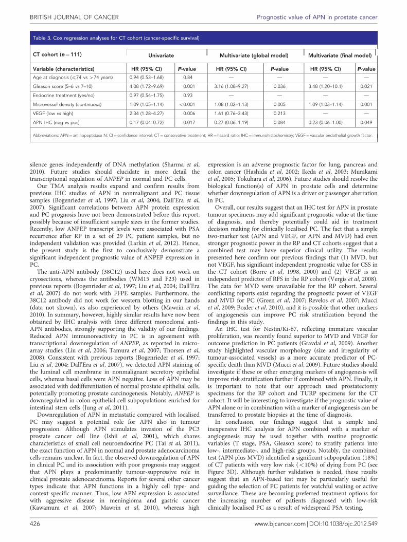
and D and Table 3). In this cohort, negative APN was significantlyassociated with short CSS by univariate (Figure 3C; P¼ 0.0065) aswell as multivariate analyses (Table 3). High Gleason score andhigh MVD were also significantly associated with short CSS in theCT cohort by uni- and multivariate analyses (Table 3). VEGFimmunoreactivity was significant only by univariate analysis in theCT cohort. Prediction of CSS was significantly improved bycombining APN immunoreactivity and MVD (Figure 3D). Therewas no significant association between APN staining and overallsurvival in the CT cohort (data not shown).
DISCUSSION
In summary, our results based on two distinct PC patient cohortsindicate that negative APN immunoreactivity is a significant
TSS
MethyLight
Bis-Seq
CpG islandExon 1
100
10
LNC
aPLN
CaP
(1A
)LN
CaP
(2A
)LN
CaP
(4A
)
LNC
aPLN
CaP
(1A
)
DU
145
DU
145
(1A
)
BP
H1 M
UM
Und
etec
tabl
e
DU
145
DU
145(
1A)
DU
145(
2A)
DU
145(
4A)
BP
H1
BP
H1(
1A)
BP
H1(
2A)
BP
H1(
4A)A
NP
EP
/UB
C (
qRT
–PC
R)
1
LNCaP
BPH1
DU145
100%
80%
60%
Max
imum
met
hyla
tion
(Bis
-Seq
)
40%
20%
0%
1.2
0.8
0.4
AN
PE
P/M
YO
D1
(Met
hylL
ight
)
AN
PE
P/M
YO
D1
(Met
hylL
ight
)
0
AN(n =5)
BPH(n =5)
LPC(n =10)
MPC(n =10)
0.8
0.6
0.4
0.2
0
AN(n =16)
BPH(n =15)
MPC(n =183)
MPC(n =24)
CRPC(n =10)
BP
H1
(1A
)Figure 2. (A) Structure of ANPEP genomic locus. Location of the promoter-associated CpG island is shown by a grey box. DNA regions analysedby bisulphite sequencing (Bis-Seq) and quantitative methylation-specific PCR (MethyLight) are indicated above (dumbbell and white box,respectively). TSS, transcription start site within CpG island. The figure is not to scale. (B) Dot plot of ANPEP promoter methylation in a smallclinical sample set analysed by bisulphite sequencing. Percent methylated CpG sites/total number of CpG sites in the most heavily methylatedclone is given for each sample (see also Supplementary Figure S1). (C) Dot plot of relative ANPEP promoter methylation (normalised to MYOD1) ina second and larger clinical sample set, determined by MethyLight analysis. (B and C) Horizontal dashed lines indicate median methylation foreach group. Sample numbers in each group are given in brackets. (D) Normalised ANPEP expression in three prostate cell lines treated with 1 mM(1A), 2 mM (2A), or 4 mM (4A) 5-aza-20-deoxycytidine or untreated is shown at the top. The expression of ANPEP was determined by qRT–PCR usingUBC for normalisation, and further normalised to ANPEP/UBC expression in a pool of four adjacent nonmalignant prostate tissue samples(arbitrarily set to 100; not shown). The expression of ANPEP was induced by 5-aza-20-deoxycytidine in all cell lines. Bisulphite sequencing results forANPEP in prostate cell lines is shown below. Open and closed circles indicate unmethylated and methylated CpGs, respectively. Each rowrepresents one clone. The CpG sites interrogated by MethyLight analysis are highlighted by black boxes. The promoter methylation levels ofANPEP in treated and untreated cell lines, methylated (M) and unmethylated (UM) control DNA samples, determined by MethyLight analysis, isshown at the bottom.
BRITISH JOURNAL OF CANCER Prognostic value of APN in prostate cancer
424 www.bjcancer.com | DOI:10.1038/bjc.2012.549

independent adverse prognostic factor for patients diagnosed withclinically localised PC. Although further validation is needed, thisindicates that a simple IHC test for APN may add significantprognostic value to currently used routine predictors for PC patientoutcome.
Furthermore, we identified ANPEP as a novel common targetof aberrant promoter hypermethylated in PC. Hypermethylationcorrelated inversely with APN expression, indicating that ANPEPis epigenetically silenced in PC cells. To our knowledge, this is thefirst report of aberrant ANPEP hypermethylation in PC. As furtherexperimental evidence for epigenetic regulation of ANPEPexpression, 5-aza-dC treatment resulted in promoter
demethylation and transcriptional reactivation in three hyper-methylated prostate cell lines. Recently, hypermethylation anddownregulation of ANPEP were reported for some malignantmelanoma cells (Wulfanger et al, 2012).
The methylation levels of ANPEP did not have significantprognostic value in the RP cohort (it was borderline significant,but failed to validate in an independent patient cohort (datanot shown)). A likely explanation is that other mechanisms, inaddition to promoter hypermethylation, contribute to down-regulation of ANPEP in PC cells. Absence of essential transcriptionfactor activities may prevent transcription from unmethylatedpromoters, and the repressive histone mark H3K27me3 can
1.00
0.75
Cum
ulat
ive
surv
ival
Cum
ulat
ive
surv
ival
0.50
APN neg
APN neg
APN pos
APN pos
APN pos & VEGF lo
APN pos & VEGF hi /APN neg & VEGF lo
APN neg & VEGF hi
RP cohort
CT cohort CT cohort
APN pos & MVD lo
APN pos & MVD hi /APN neg & MVD lo
RP cohort
P =0.0019*
P =0.0065*
P <0.0001*
P <0.0001*
0 50 100Recurrence-free survival time (months) Recurrence-free survival time (months)
Number at riskAPN negAPN pos
Number at riskLow risk 69
10953
30287
421
000
Med riskHigh risk
Number at riskLow riskMed riskHigh risk
130106
2838 5
200
Number at riskAPN negAPN pos
4017
6926 5
1615
01520 4 1 02748 12 5 01527 5 0 0
0
150
0 50 100Cancer-specific survival time (months)
150 200 0 50 100Cancer-specific survival time (months)
150 200
0 50 100 150
0.25
0.00
1.00
0.75
Cum
ulat
ive
surv
ival
0.50
0.25
0.00
1.00
0.75
0.50
0.25
0.00
Cum
ulat
ive
surv
ival
1.00
0.75
0.50
0.25
0.00
Figure 3. Kaplan–Meier plots of RFS for the RP cohort (A and B) and CSS for the CT cohort (C and D). The P-values for two-sided log-rank statisticsare given, comparing patients with negative (IHC score¼0) or positive (IHC score¼ 1þ or 2þ ) APN immunoreactivity (A, C), as well as patientswith positive/negative APN staining in combination with low/high VEGF staining (score 0–2 vs score 3–5) in the RP cohort (B), or low/highmicrovessel density (MVD; dichotomised at median) in the CT cohort (D). *Significant P-values.
Table 2. Cox regression analyses for RP cohort (recurrence-free survival)
RP cohort (n¼267) Univariate Multivariate (global model) Multivariate (final model)
Variable (characteristics) HR (95% CI) P-value HR (95% CI) P-value HR (95% CI) P-value
Age at diagnosis (p62 vs 462 years) 1.05 (0.73–1.50) 0.80 — — — —
Preoperative PSA (continuous) 1.05 (1.04–1.07) o0.001 1.04 (1.03–1.06) o0.001 1.04 (1.03–1.06) o0.001
Gleason score (5–6 vs 7-10) 2.81 (1.83–4.31) o0.001 1.60 (0.99–2.60) 0.056 1.72 (1.08–2.75) 0.023
Tumour stage (pT2 vs pT3) 3.35 (2.30–4.88) o0.001 1.54 (0.88–2.69) 0.13 — —
Nodal status (pN0 vs pN1) 3.36 (1.56–7.24) 0.002 1.68 (0.71–3.98) 0.24 — —
Surgical margins (neg vs pos) 3.05 (2.11–4.41) o0.001 1.77 (1.03–3.04) 0.038 2.38 (1.61–3.52) o0.001
VEGF (low vs high) 2.84 (1.97–4.11) o0.001 2.12 (1.41–3.20) o0.001 2.05 (1.38–3.06) o0.001
APN IHC (neg vs pos) 0.55 (0.37–0.80) 0.002 0.64 (0.42–0.98) 0.039 0.54 (0.36–0.80) 0.017
Abbreviations: APN¼ aminopeptidase N; CI¼ confidence interval; HR¼ hazard ratio; IHC¼ immunohistochemistry; PSA¼prostate-specific antigen; RP¼ radical prostatectomy;VEGF¼ vascular endothelial growth factor.
Prognostic value of APN in prostate cancer BRITISH JOURNAL OF CANCER
www.bjcancer.com | DOI:10.1038/bjc.2012.549 425
silence genes independently of DNA methylation (Sharma et al,2010). Future studies should elucidate in more detail thetranscriptional regulation of ANPEP in normal and PC cells.
Our TMA analysis results expand and confirm results fromprevious IHC studies of APN in nonmalignant and PC tissuesamples (Bogenrieder et al, 1997; Liu et al, 2004; Dall’Era et al,2007). Significant correlations between APN protein expressionand PC prognosis have not been demonstrated before this report,possibly because of insufficient sample sizes in the former studies.Recently, low ANPEP transcript levels were associated with PSArecurrence after RP in a set of 29 PC patient samples, but noindependent validation was provided (Larkin et al, 2012). Hence,the present study is the first to conclusively demonstrate asignificant independent prognostic value of ANPEP expression inPC.
The anti-APN antibody (38C12) used here does not work oncryosections, whereas the antibodies (WM15 and F23) used inprevious reports (Bogenrieder et al, 1997; Liu et al, 2004; Dall’Eraet al, 2007) do not work with FFPE samples. Furthermore, the38C12 antibody did not work for western blotting in our hands(data not shown), as also experienced by others (Mawrin et al,2010). In summary, however, highly similar results have now beenobtained by IHC analysis with three different monoclonal anti-APN antibodies, strongly supporting the validity of our findings.Reduced APN immunoreactivity in PC is in agreement withtranscriptional downregulation of ANPEP, as reported in micro-array studies (Liu et al, 2006; Tamura et al, 2007; Thorsen et al,2008). Consistent with previous reports (Bogenrieder et al, 1997;Liu et al, 2004; Dall’Era et al, 2007), we detected APN staining ofthe luminal cell membrane in nonmalignant secretory epithelialcells, whereas basal cells were APN negative. Loss of APN may beassociated with dedifferentiation of normal prostate epithelial cells,potentially promoting prostate carcinogenesis. Notably, ANPEP isdownregulated in colon epithelial cell subpopulations enriched forintestinal stem cells (Jung et al, 2011).
Downregulation of APN in metastatic compared with localisedPC may suggest a potential role for APN also in tumourprogression. Although APN stimulates invasion of the PC3prostate cancer cell line (Ishii et al, 2001), which sharescharacteristics of small cell neuroendocrine PC (Tai et al, 2011),the exact function of APN in normal and prostate adenocarcinomacells remains unclear. In fact, the observed downregulation of APNin clinical PC and its association with poor prognosis may suggestthat APN plays a predominantly tumour-suppressive role inclinical prostate adenocarcinoma. Reports for several other cancertypes indicate that APN functions in a highly cell type- andcontext-specific manner. Thus, low APN expression is associatedwith aggressive disease in meningioma and gastric cancer(Kawamura et al, 2007; Mawrin et al, 2010), whereas high
expression is an adverse prognostic factor for lung, pancreas andcolon cancer (Hashida et al, 2002; Ikeda et al, 2003; Murakamiet al, 2005; Tokuhara et al, 2006). Future studies should resolve thebiological function(s) of APN in prostate cells and determinewhether downregulation of APN is a driver or passenger aberrationin PC.
Overall, our results suggest that an IHC test for APN in prostatetumour specimens may add significant prognostic value at the timeof diagnosis, and thereby potentially could aid in treatmentdecision making for clinically localised PC. The fact that a simpletwo-marker test (APN and VEGF, or APN and MVD) had evenstronger prognostic power in the RP and CT cohorts suggest that acombined test may have superior clinical utility. The resultspresented here confirm our previous findings that (1) MVD, butnot VEGF, has significant independent prognostic value for CSS inthe CT cohort (Borre et al, 1998, 2000) and (2) VEGF is anindependent predictor of RFS in the RP cohort (Vergis et al, 2008).The data for MVD were unavailable for the RP cohort. Severalconflicting reports exist regarding the prognostic power of VEGFand MVD for PC (Green et al, 2007; Revelos et al, 2007; Mucciet al, 2009; Boxler et al, 2010), and it is possible that other markersof angiogenesis can improve PC risk stratification beyond thefindings in this study.
An IHC test for Nestin/Ki-67, reflecting immature vascularproliferation, was recently found superior to MVD and VEGF foroutcome prediction in PC patients (Gravdal et al, 2009). Anotherstudy highlighted vascular morphology (size and irregularity oftumour-associated vessels) as a more accurate predictor of PC-specific death than MVD (Mucci et al, 2009). Future studies shouldinvestigate if these or other emerging markers of angiogenesis willimprove risk stratification further if combined with APN. Finally, itis important to note that our approach used prostatectomyspecimens for the RP cohort and TURP specimens for the CTcohort. It will be interesting to investigate if the prognostic value ofAPN alone or in combination with a marker of angiogenesis can betransferred to prostate biopsies at the time of diagnosis.
In conclusion, our findings suggest that a simple andinexpensive IHC analysis for APN combined with a marker ofangiogenesis may be used together with routine prognosticvariables (T stage, PSA, Gleason score) to stratify patients intolow-, intermediate-, and high-risk groups. Notably, the combinedtest (APN plus MVD) identified a significant subpopulation (18%)of CT patients with very low risk (o10%) of dying from PC (seeFigure 3D). Although further validation is needed, these resultssuggest that an APN-based test may be particularly useful forguiding the selection of PC patients for watchful waiting or activesurveillance. These are becoming preferred treatment options forthe increasing number of patients diagnosed with low-riskclinically localised PC as a result of widespread PSA testing.
Table 3. Cox regression analyses for CT cohort (cancer-specific survival)
CT cohort (n¼111) Univariate Multivariate (global model) Multivariate (final model)
Variable (characteristics) HR (95% CI) P-value HR (95% CI) P-value HR (95% CI) P-value
Age at diagnosis (p74 vs 474 years) 0.94 (0.53–1.68) 0.84 — — — —
Gleason score (5–6 vs 7–10) 4.08 (1.72–9.69) 0.001 3.16 (1.08–9.27) 0.036 3.48 (1.20–10.1) 0.021
Endocrine treatment (yes/no) 0.97 (0.54–1.75) 0.93 — — — —
Microvessel density (continuous) 1.09 (1.05–1.14) o0.001 1.08 (1.02–1.13) 0.005 1.09 (1.03–1.14) 0.001
VEGF (low vs high) 2.34 (1.28–4.27) 0.006 1.61 (0.76–3.43) 0.213 — —
APN IHC (neg vs pos) 0.17 (0.04–0.72) 0.017 0.27 (0.06–1.19) 0.084 0.23 (0.06–1.00) 0.049
Abbreviations: APN¼ aminopeptidase N; CI¼ confidence interval; CT¼ conservative treatment; HR¼hazard ratio; IHC¼ immunohistochemistry; VEGF¼ vascular endothelial growth factor.
BRITISH JOURNAL OF CANCER Prognostic value of APN in prostate cancer
426 www.bjcancer.com | DOI:10.1038/bjc.2012.549
ACKNOWLEDGEMENTS
We thank Dr Niels Tørring for advice on IHC analysis, andAnne Slotsdal, Pamela Celis, Maria Mark, Gitte Høj, MetteRasmussen, Conni Sørensen, and Susanne Skou for excellenttechnical assistance. This work was supported by The LundbeckFoundation, The John and Birthe Meyer Foundation, TheDanish Cancer Society, and The Danish Council for StrategicResearch. The Danish Cancer Biobank (DCB) is acknowledged forbiological material.
CONFLICT OF INTEREST
The authors declare no conflict of interest.
REFERENCES
Abildgaard MO, Borre M, Mortensen MM, Ulhøi BP, Tørring N, Wild P,Kristensen H, Mansilla F, Ottosen PD, Dyrskjøt L, Ørntoft TF, SørensenKD (2012) Downregulation of zinc finger protein 132 in prostate cancer isassociated with aberrant promoter hypermethylation and poor prognosis.Int J Cancer 130(4): 885–895.
Bogenrieder T, Finstad CL, Freeman RH, Papandreou CN, Scher HI,Albino AP, Reuter VE, Nanus DM (1997) Expression and localization ofaminopeptidase A, aminopeptidase N, and dipeptidyl peptidase IV inbenign and malignant human prostate tissue. Prostate 33(4): 225–232.
Borre M, Nerstrøm B, Overgaard J (1997) The natural history of prostatecarcinoma based on a Danish population treated with no intent to cure.Cancer 80(5): 917–928.
Borre M, Nerstrøm B, Overgaard J (2000) Association between immunohisto-chemical expression of vascular endothelial growth factor (VEGF), VEGF-expressing neuroendocrine-differentiated tumor cells, and outcome inprostate cancer patients subjected to watchful waiting. Clin Cancer Res6(5): 1882–1890.
Borre M, Offersen BV, Nerstrøm B, Overgaard J (1998) Microvessel densitypredicts survival in prostate cancer patients subjected to watchful waiting.Br J Cancer 78(7): 940–944.
Boxler S, Djonov V, Kessler TM, Hlushchuk R, Bachmann LM, Held U,Markwalder R, Thalmann GN (2010) Matrix metalloproteinases andangiogenic factors: predictors of survival after radical prostatectomy forclinically organ-confined prostate cancer? Am J Pathol 177(5): 2216–2224.
Carl-McGrath S, Lendeckel U, Ebert M, Rocken C (2006) Ectopeptidases intumour biology: a review. Histol Histopathol 21(12): 1339–1353.
Dall’Era MA, True LD, Siegel AF, Porter MP, Sherertz TM, Liu AY (2007)Differential expression of CD10 in prostate cancer and its clinicalimplication. BMC Urol 7: 3.
Epstein JI, Allsbrook Jr. WC, Amin MB, Egevad LL (2005) The 2005International Society of Urological Pathology (ISUP) ConsensusConference on Gleason Grading of Prostatic Carcinoma. Am J Surg Pathol29(9): 1228–1242.
Gravdal K, Halvorsen OJ, Haukaas SA, Akslen LA (2009) Proliferation ofimmature tumor vessels is a novel marker of clinical progression inprostate cancer. Cancer Res 69(11): 4708–4715.
Green MM, Hiley CT, Shanks JH, Bottomley IC, West CM, Cowan RA,Stratford IJ (2007) Expression of vascular endothelial growth factor(VEGF) in locally invasive prostate cancer is prognostic for radiotherapyoutcome. Int J Radiat Oncol Biol Phys 67(1): 84–90.
Hashida H, Takabayashi A, Kanai M, Adachi M, Kondo K, Kohno N,Yamaoka Y, Miyake M (2002) Aminopeptidase N is involved in cellmotility and angiogenesis: its clinical significance in human colon cancer.Gastroenterology 122(2): 376–386.
Heebøll S, Borre M, Ottosen PD, Andersen CL, Mansilla F, Dyrskjøt L,Ørntoft TF, Tørring N (2008) SMARCC1 expression is upregulated inprostate cancer and positively correlated with tumour recurrence anddedifferentiation. Histol Histopathol 23(9): 1069–1076.
Ikeda N, Nakajima Y, Tokuhara T, Hattori N, Sho M, Kanehiro H, Miyake M(2003) Clinical significance of aminopeptidase N/CD13 expression inhuman pancreatic carcinoma. Clin Cancer Res 9(4): 1503–1508.
Ishii K, Usui S, Sugimura Y, Yoshida S, Hioki T, Tatematsu M,Yamamoto H, Hirano K (2001) Aminopeptidase N regulated by zinc inhuman prostate participates in tumor cell invasion. Int J Cancer 92(1):49–54.
Jung P, Sato T, Merlos-Suarez A, Barriga FM, Iglesias M, Rossell D,Auer H, Gallardo M, Blasco MA, Sancho E, Clevers H, Batlle E (2011)Isolation and in vitro expansion of human colonic stem cells. Nat Med17(10): 1225–1227.
Kawamura J, Shimada Y, Kitaichi H, Komoto I, Hashimoto Y, Kaganoi J,Miyake M, Yamasaki S, Kondo K, Imamura M (2007) Clinicopathologicalsignificance of aminopeptidase N/CD13 expression in human gastriccarcinoma. Hepatogastroenterology 54(73): 36–40.
Larkin SE, Holmes S, Cree IA, Walker T, Basketter V, Bickers B, Harris S,Garbis SD, Townsend PA, Aukim-Hastie C (2012) Identification ofmarkers of prostate cancer progression using candidate gene expression.Br J Cancer 106(1): 157–165.
Liu AY, Roudier MP, True LD (2004) Heterogeneity in primary andmetastatic prostate cancer as defined by cell surface CD profile. Am JPathol 165(5): 1543–1556.
Liu P, Ramachandran S, Ali Seyed M, Scharer CD, Laycock N, Dalton WB,Williams H, Karanam S, Datta MW, Jaye DL, Moreno CS (2006) Sex-determining region Y box 4 is a transforming oncogene in human prostatecancer cells. Cancer Res 66(8): 4011–4019.
Mawrin C, Wolke C, Haase D, Kruger S, Firsching R, Keilhoff G, Paulus W,Gutmann DH, Lal A, Lendeckel U (2010) Reduced activity of CD13/aminopeptidase N (APN) in aggressive meningiomas is associated withincreased levels of SPARC. Brain Pathol 20(1): 200–210.
Menrad A, Speicher D, Wacker J, Herlyn M (1993) Biochemical andfunctional characterization of aminopeptidase N expressed by humanmelanoma cells. Cancer Res 53(6): 1450–1455.
Mucci LA, Powolny A, Giovannucci E, Liao Z, Kenfield SA, Shen R,Stampfer MJ, Clinton SK (2009) Prospective study of prostate tumorangiogenesis and cancer-specific mortality in the health professionalsfollow-up study. J Clin Oncol 27(33): 5627–5633.
Murakami H, Yokoyama A, Kondo K, Nakanishi S, Kohno N, Miyake M(2005) Circulating aminopeptidase N/CD13 is an independent prognosticfactor in patients with non-small cell lung cancer. Clin Cancer Res11(24 Pt 1): 8674–8679.
Petrovic N, Schacke W, Gahagan JR, O’Conor CA, Winnicka B,Conway RE, Mina-Osorio P, Shapiro LH (2007) CD13/APN regulatesendothelial invasion and filopodia formation. Blood 110(1):142–150.
Revelos K, Petraki C, Scorilas A, Stefanakis S, Malovrouvas D,Alevizopoulos N, Kanellis G, Halapas A, Koutsilieris M (2007) Correlationof androgen receptor status, neuroendocrine differentiation andangiogenesis with time-to-biochemical failure after radical prostatectomyin clinically localized prostate cancer. Anticancer Res 27(5B):3651–3660.
Rocken C, Licht J, Roessner A, Carl-McGrath S (2005) Canalicularimmunostaining of aminopeptidase N (CD13) as a diagnostic marker forhepatocellular carcinoma. J Clin Pathol 58(10): 1069–1075.
Sharma S, Kelly TK, Jones PA (2010) Epigenetics in cancer. Carcinogenesis31(1): 27–36.
Sørensen KD, Wild PJ, Mortezavi A, Adolf K, Tørring N, Heebøll S,Ulhøi BP, Ottosen P, Sulser T, Hermanns T, Moch H, Borre M,Ørntoft TF, Dyrskjøt L (2009) Genetic and epigenetic SLC18A2 silencingin prostate cancer is an independent adverse predictor of biochemicalrecurrence after radical prostatectomy. Clin Cancer Res 15(4):1400–1410.
Tai S, Sun Y, Squires JM, Zhang H, Oh WK, Liang CZ, Huang J (2011) PC3 isa cell line characteristic of prostatic small cell carcinoma. Prostate 71(15):1668–1679.
Tamura K, Furihata M, Tsunoda T, Ashida S, Takata R, Obara W,Yoshioka H, Daigo Y, Nasu Y, Kumon H, Konaka H, Namiki M,Tozawa K, Kohri K, Tanji N, Yokoyama M, Shimazui T, Akaza H,Mizutani Y, Miki T, Fujioka T, Shuin T, Nakamura Y, Nakagawa H(2007) Molecular features of hormone-refractory prostate cancer
cells by genome-wide gene expression profiles. Cancer Res 67(11):5117–5125.
Terauchi M, Kajiyama H, Shibata K, Ino K, Nawa A, Mizutani S, Kikkawa F(2007) Inhibition of APN/CD13 leads to suppressed progressive potentialin ovarian carcinoma cells. BMC Cancer 7: 140.
Prognostic value of APN in prostate cancer BRITISH JOURNAL OF CANCER
www.bjcancer.com | DOI:10.1038/bjc.2012.549 427

Thorsen K, Sørensen KD, Brems-Eskildsen AS, Modin C, Gaustadnes M, HeinAM, Kruhøffer M, Laurberg S, Borre M, Wang K, Brunak S, Krainer AR,Tørring N, Dyrskjøt L, Andersen CL, Ørntoft TF (2008) Alternativesplicing in colon, bladder, and prostate cancer identified by exon arrayanalysis. Mol Cell Proteomics 7(7): 1214–1224.
Tokuhara T, Hattori N, Ishida H, Hirai T, Higashiyama M, Kodama K,Miyake M (2006) Clinical significance of aminopeptidase N in non-smallcell lung cancer. Clin Cancer Res 12(13): 3971–3978.
Vergis R, Corbishley CM, Norman AR, Bartlett J, Jhavar S, Borre M,Heeboll S, Horwich A, Huddart R, Khoo V, Eeles R, Cooper C, Sydes M,Dearnaley D, Parker C (2008) Intrinsic markers of tumour hypoxia andangiogenesis in localised prostate cancer and outcome of radical treatment:
a retrospective analysis of two randomised radiotherapy trials and onesurgical cohort study. Lancet Oncol 9(4): 342–351.
Wulfanger J, Schneider H, Wild P, Ikenberg K, Rodolfo M, Rivoltini L,Meyer S, Riemann D, Seliger B (2012) Promoter methylation ofaminopeptidase N/CD13 in malignant melanoma. Carcinogenesis 33(4):781–790.
This work is published under the standard license to publish agree-ment. After 12 months the work will become freely available andthe license terms will switch to a Creative Commons Attribution-NonCommercial-Share Alike 3.0 Unported License.
Supplementary Information accompanies this paper on British Journal of Cancer website (http://www.nature.com/bjc)
BRITISH JOURNAL OF CANCER Prognostic value of APN in prostate cancer
428 www.bjcancer.com | DOI:10.1038/bjc.2012.549
Copyright © 2022 FDOKUMEN