Measurement of Prostate‐Specific Antigen and Human Glandular Kallikrein 2 in Different Body Fluids
Upload
khangminh22Category
view
5download
0
The HumanFemale Prostate
From Vestigial Skene’s Paraurethral Glands and Ductsto Woman’s Functional Prostate
Milan Zaviaèiè
© Milan Zaviaèiè 1999© SAP – Slovak Academic Press, s.r.o., Bratislava 19991st CD-ROM Edition, Bratislava, 1999
All rights reserved. No part of this publication may be reproduced or transmitted in anyform or by any means, electronic or mechanical, including photocopy, recording, orany information storage and retrieval system, without permission in writing fromthe author Milan Zaviaèiè; Comenius University, School of Medicine, Sasinkova 4,811 08 Bratislava, Slovakia.
Layout Academic DTP
ISBN 80-88908-50-7

Contents
Foreword (Dr. Richard J. Ablin, PhD) ....................................................... 5
About the Author ...................................................................................... 7
From Reviewers’ Opinion ......................................................................... 9
Introduction with Acknowledgements ........................................................ 11
I. History of the Female Prostate ................................................................... 17
II. Weight, Size and Macroanatomy of the Female Prostate ............................. 21
III. Histology and Ultrastructure of the Female Prostate ................................... 31
IV. Enzyme Histochemistry of the Female Prostate and its Functional
Implications ............................................................................................... 51
V. Exocrine Function of the Female Prostate: Implications for Gynecology,
Urology, Chronobiology and Forensic Medicine........................................... 61
VI. Neuroendocrine Function of the Female Prostate: Morphological Basis ....... 71
VII. The Female Prostate and Female Ejaculation: Sexologic Implications .......... 77
VIII. Prostate-Specific Antigen: Immunohistochemical Localization in Female
Prostate Tissue, Serological Parameters, and Implications of this Prostate
Marker in the Woman and Man ................................................................... 85
IX. Prostatic and Non-Prostatic Sources of Prostate-Specific Antigen ................ 93
X. Pathology of the Female Prostate with Special Reference to Carcinoma,
Benign Prostate Hyperplasia and Other „Prostatic“ Diseases in Female
Patients ....................................................................................................... 103
XI . Reasons for Rejecting the Term Skene’s Paraurethral Ducts and Glands
in Designating the Prostate in the Human Female (Terminological Note) ..... 119
Summary (in English and Slovak) ................................................................ 123
References ................................................................................................... 135
Index ............................................................................................................ 155


Foreword
�Out of sight, out of mind�, suggested by Nicholas Bruchovsky (l990) to aptly de-scribe the fact that while no other organ causes so much illness in men as the prostate,so little is known about its role in the body, is perhaps even more applicable to thefemale prostate. Morphologically incorporated into the wall of the female urethra, thefemale prostate, first described by Reijnier de Graaf in 1672 (de Graaf 1672), is in factanatomically �... out of sight�.
Recognition of the female prostate has been further hindered by use of the histori-cally acquired terminology �Skene�s paraurethral glands and ducts� , so named afterAlexander Skene, who redescribed the female prostate in 1880 (Skene 1880). Thisterminology implies some other structure of an extraprostatic nature, rather than theprostate itself, is involved and has unfortunately until the recent studies by Zaviaèièand co-workers, promulgated a vestigial notion of this female organ.
While considered in-depth herein, any doubters of the existence of the female pros-tate, will learn that it possesses all the components, i.e., glands, ducts, smooth muscu-lature, characterizing the male prostate, including the cellular, enzymatic and otherfactors necessary for its exocrine and endocrine function.
Encouraged, while discussing the existence of the female prostate and aspects ofthe foregoing, including the presence of prostate-specific antigen (PSA) as an expla-nation for PSA in the serum of females, some 10+ years ago with the eminent urologistWillard E. Goodwin (UCLA School of Medicine), �...to put all that information to-gether and elaborate a little more on it ...�, I am grateful that while other activitiesprecluded my doing so, my esteemed colleague Professor Milan Zaviaèiè has done soand has given me the privilege of writing this �Foreword�. In this regard, the master-fully meticulous studies herein presented by Professor Zaviaèiè in this monographentitled: The Human Female Prostate, have and will contribute immeasurably towardour further understanding and recognition of the female prostate. Commensurate withthis, and from a pragmatic point of view, it is time to alert primary care physicians thatwomen have a prostate and therefore, are affected by the same diseases as their malecounterparts, including prostatitis, benign prostatic hyperplasia and carcinoma (al-though the latter is rare) which need to be treated appropriately, rather than as in the

6
past by invasive urethral dilation and/or antidepressants (the latter implying that theoccurrence of such diseases in the female is a �state of mind� rather than an actualdisease).
In looking to the future, it appears rational, with the demonstration by Zaviaèiè ofthe unequivocal existence of the female prostate and accompanying pathology, to ap-ply knowledge gained from immunobiological studies of the prostate suggesting whathas been termed the �prostatolymphoreticular system� (Ablin and Whyard 1990), tothe female prostate. If the female prostate exhibits the similar immunopermissivenessof its male counterpart, it may also serve as a nidus for various infectious agents,inclusive of HIV. Of particular significance in this regard, as elucidated from the stud-ies by Zaviaèiè, are the substantially increased number of mutually communicatingducts and intraepithelial glands with the urethra and anterior wall of the vagina vs. themale prostate, which contribute, as aptly defined by Zaviaèiè, to the urethro-prostatic-vaginal complex (body). Providing an environment exceptionally favorable for the long-term survival of uropathogens, it is suggested this may not explain relapses of femaleprostatitis, but also the recurrent episodes of urinary tract infections diagnosed as acutecystitis.
In concert with the treatise to follow and the recent editorial position statement bythe Journal of Urology (McGuire 1999 [the official publication of the American Uro-logical Association]) establishing a new section on �Female Urology� and the Ameri-can Board of Urology�s decision to define an area of focus in female urology in pro-gram development, the female prostate should be given equal place with other femalegenitourinary organs and should no longer be a �mysterious female organ�!
Richard J. Ablin, Ph.D.Director, Scientific InvestigationInnapharma, Inc.Suffern, New York 10901PresidentRobert Benjamin Ablin Foundationfor Cancer ResearchMahwah, New Jersey 07430

7
About the Author
Milan ZAVIAÈIÈ, MD, PhD, DSc is professor of pathologyand forensic medicine at the Comenius University Bratislava,Slovak Republic (Slovakia). Professor Milan Zaviaèiè has es-tablished the updated non-vestigial concept of the prostate inthe female. Based on multidisciplinary research, he has presented the female prostateas a functional genitourinary organ in the female with a specific structure, functionand pathology. He has shown that the female prostate parameters are similar or evenidentical with those of the adult male prostate. This recent concept has been based onmorphological, histochemical, forensic-medical, sexological, gynecological, urologi-cal, chronobiological and pathology research.
The results of Prof. Zaviaèiè�s research activities have so far been presented in431 lectures and up to 500 various scientific publications, including full papers, edito-rials, research reports, review articles, book chapters and contributions to proceedingsof scientific congresses, symposia and conferences (40 of them concerning differentaspects of the female prostate).
Professor Zaviaèiè joined the faculty of the Comenius University Medical Schoolas lecturer in pathology in 1963, immediately after his graduation (MUDr.) from theComenius University Medical School, Bratislava. After two years, he was promoted toAssistant Professor, received his CSc degree in 1975 (based on a thesis on the influ-ence of fasting on the structure of the gastric mucosa, Zaviaèiè 1974). Being appointedAssociate Professor in 1981 based on a thesis on the circadian biorhythms in the struc-ture of the gastric mucosa (Zaviaèiè 1979), he published his functional-morphologicalstudy on the female prostate and urethra (Zaviaèiè 1985b) and obtained his DSc in1986. He was appointed head of the Department of Pathology, Comenius UniversityBratislava, two years later, and rose to the position of full professor of pathology andforensic medicine in 1989.
Professor Zaviaèiè is member of the Advisory Council Board of the European So-ciety of Pathology (ESP), member of the National Committee of the Slovak Society ofPathologists, Vice-President of the Slovak Society of Histochemistry and Cytochemis-try, and elected member of International Academy of Sex Research (IASR). He has

8
served as President of the Czechoslovak Society of Pathologists and President of theSlovak Society of Pathologists (two terms), which is member of the World Associationof Societies of Pathology (WASP).
For his work, Prof.Zaviaèiè re ceived several awards from Ministry of Health ofthe Slovak Republic (1991), State Institute for Cancer Research of Japan (1978), JapaneseSociety of Clinical Pathology (1995), Presidium of the Slovak Medical Society (1985,1986, 1988), and Presidium of the Czechoslovak Medical Society (1977, 1980), and asthe appreciation of his research in the female prostate, he received the 1998 CrystalWing for Medicine and Science award.
This Monograph was supported and sponsored :• by a grant-in-aid from Scientific Grant Agency (VEGA) of the Slovak Republic (Project No. 1/5159 / 98);• by Leica (Mikro s. s r.o.Bratislava), Slovak Republic;• by Palma-Tumys, a.s. Bratislava, Slovak Republic.
The CD-ROM version was sponsored :• by Palma-Tumys, a.s. Bratislava, Slovak Republic;• mpro Software - Michal Palkoviè, Bratislava, Slovak Republic.

9
From Reviewers� Opinion
�This book explains excellently a new conception of woman prostate. It was formu-lated by Prof. MUDr. M. Zaviaèiè, DrSc., on the basis of his very successful 17 yearslasting interdisciplinary research and careful selection of pertinent data from interna-tional publications. In contrast to the older view that Skene�s paraurethral ducts andglands are rudimentary structures, the author presents them as woman�s genitourinaryorgan with specific structure, function and pathology. This organ should be properlycalled �woman prostate�. The book is a milestone in the field and contains the newestdetailed information of the anatomy, histology, ultrastructure, cytochemistry, physiol-ogy, pathology and forensic medicine of the woman prostate. Sexological viewpointsare also included�.
Professor Zdenìk Lojda, MD, DSc.,World leading personality in histochemistry,Founder of histochemistry in the Czech andSlovak Republics, Histochemical Laboratory,Charles University, Prague, Czech Republic
�In his monograph �The Human Female Prostate�, Professor Milan Zaviaèiè, MD,DSc of Bratislava introduces novel and inventive knowledge and presents the femaleprostate as a distinct organ which is, as far as its ortology, evolution, anatomy, physiol-ogy, clinical behaviour and pathology are concerned, fully comparable with the pros-tate of the man. This work is unique through its content and exceptionally importantnot only from the standpoint of pathological anatomy but also from the point of view ofclinical practice. The introduced concept of the female prostate implies substantialclinical consequences, which may in the future bring about a change in opinions uponetiopathogenesis of functionally and organically conditioned diseases of the lower uri-

10
nary tract in women. This work brings a strong impulse for intensive clinical researchof the female prostate�.
Professor Jan Breza, MD, DScProfessor of Urology, President of theSlovak Urology Society, Urology DptDerer�s University Hospital, Bratislava
�The monograph �The Human Female Prostate� is an important landmark for ourknowledge of the physiological and pathophysiological functions of woman prostate.It was written by Prof. M. Zaviaèiè, leading researcher in this field during the last 17years. The text represents an excellent and inventive survey of his own experimentsconfronted with recent results of prominent world specialists. It contains a convincingevidence that the female prostate, similarly as the male prostate, has at least dual func-tion: exocrine � production of female prostatic fluid, and neuroendocrine function.Therefore the term �Skene�s glands and ducts as an insignificant and nonfunctionalorgan� is no more correct in the light of contemporary knowledge and should be un-equivocally replaced by the term �woman prostate�. This book helps to �bridge thegap� in our understanding of the importance of the human female prostate not only inthe basic pathology practice but in forensic medicine, gynecologic urology, sexologyand chronobiology as well. It will certainly become a welcome source of up-to-dateinformation not only for specialists but for each doctor who wants to know more aboutthe gland on which such contradictory outlook existed, especially in the past.
Prof. Ing. Miroslav Ferenèík, DScProfessor of Immunology and Immunochemistry,President of the Slovak Immunology Society andChairman, Scientific Board of Institute ofNeuroimmunology, Slovak Academy of Science

11
Introduction with Acknowledgements
The two genders possess a great number of functional and structural parameters whichare more or less identical, yet there are also features characteristic exclusively of onegender and missing in the other. These concern morphological and functional param-eters corresponding to the basic somatic differences between the male and female ofthe genus Homo sapiens, associated with characteristic psycho-behavioral specifici-ties typical of each gender.
In the structures of the male and female sex organs, with evidently different con-figurations in adulthood, homologous organs can be identified in the male and femalesexual anatomy. They are sometimes given terms derived from the opposite gender toemphasize their equal importance, as used for instance by the feminist movement con-cerning some female genital tissues. Thus e.g. the clitoris was referred to as �the fe-male penis� (penis muliebris), presumably also in support of arguments opposing theconcept of oedipism (Oedipus complex � Oedipus, Greek Mythology, Hensyl et al.1990). As early as in 1981, the Federation of Feminist Women�s Health Center wasconcerned with the redefinition of the clitoris and its multiple functions, as publishedin New View of Woman�s Body (Chalker and Gage 1987). This is however absolutelynot in keeping with the understanding of our emphatic recommendation to use theterm female prostate as an equivalent of the designation of this gland in the male, asshown further in this monograph. Our reasoning is certainly no artificial emphasis ofthe equivalency of this organ in the two genders, rather the introduction of the termfemale prostate is based on morphological, functional and clinical evidence analyzedin this monograph, and particularly in Chapter XI concerning terminology (Skene�sparaurethral ducts and glands versus female prostate). Homologous structures developfrom the same embryologic primordium. From conception to about eight weeks ofgestation, the genitals of the male and female fetus are undifferentiated. At that time,due to hormonal stimulation, the genital tissue rearranges itself in characteristic maleor female pattern (Francoeur 1991). The common basis of the sex-specific structuresmay account for the potential broad range of somatic derangements in the later defini-tive differentiation and configuration of the human internal and external genitals withsimultaneous occurrence of elements characteristic of both sexes. Further it may also

12
be involved in derangements of sexo-psychological nature concerning gender identityand gender orientation.
In the fertile woman, parameters characterizing the female sex involve rhythmi-cally recurring changes in the hormonally dependent tissues of the uterus, ovaries,tubes, vagina and breast glands. The parameters characteristic only of the female gen-der and absent in the male include e.g. menstruation, pregnancy and lactation.
Less unequivocal is the situation concerning certain parameters long consideredtypical of the male gender only and in some cultures even identified with the male sexprinciple. One of these controversial and long discussed parameters is the problem ofthe existence and role of the prostate (Skene�s paraurethral glands and ducts) in thewoman. Intensive morphological research and ever more broadening clinical studiesof the female prostate concerning its orthology and pathology, extending over the last50 years and particularly since the early 80s, provided evidence presenting the femaleprostate as an organ fully comparable and even identical with the male prostate, andthat not only as to its structure and function but also as to its pathology.
Our morphological results concerning the female prostate (Skene�s glands) pre-sented in this monograph are based on autopsy findings (as well as rare biopsies) anddetailed histological examinations of the urethra of 150 women, followed by histochemi-cal, immunochemical and further examinations of normal and pathologically changedfemale prostatic tissue, including electron microscopic studies. Many enzymes,glycosamine glycans and glycoproteins were examined, including prostate specificantigen (PSA) and prostate specific acid phosphatase (PSAP) (Zaviaèiè et al. 1983;1985b,c; 1993a, b; 1998d; Zaviaèiè 1987a; Zaviaèiè and Whipple 1993; Zaviaèiè andAblin 1998 c, and references therein).
In our series including as many as 200 female patients of the 2nd University Hospi-tal of Gynecology and Obstestrics in Bratislava, we investigated the biological phe-nomenon of female ejaculation, focusing on questions concerning the openings of theducts of the female prostate and on sexological typology of women with urethral ex-pulsions. Several results obtained in cooperation with the Hospital as well as withfurther medical specialities, including gynecological urology, urology, chronobiology,forensic medicine, etc., were used in developing implications concerning the functionof the female prostate.
The monograph on the female prostate, with its underlying modern concept andrich, colored documentation, consists of several chapters, each focusing on a differentaspect of the topics. There may be some overlap concerning some important pieces ofinformation between the chapters for purposes of emphasis and particularly for treat-ing the given item in the light of different relationships and points of interpretation.The reader is thus provided not only with the latest functional, morphological, patho-physiological and clinical findings but also with a broad range of implications thathave appeared concerning this genitourinary organ of the woman. The basis of theindividual chapters of the monograph are observations and findings derived from theseries investigated by the author and his coworkers and interpreted in the light of hisand their clinical expertise. The relevant morphological and clinical research was con-ducted in cooperation with several institutes of Comenius University and with the

13
support of some departments abroad. The author formed a team consisting of researchworkers, clinicians and technicians based predominantly at his own Institute and at the2nd Hospital of Gynecology and Obstetrics of Comenius University Medical School.Members of the team published over 40 papers dealing with different aspects of thefemale prostate, with more than half of them appearing abroad in CC-covered journals.
The presented monograph is the outcome of almost 20 years of work that the authorand his coworkers devoted to the study of this female genitourinary organ. Their effortcan be summarized in the statement expressed in the subtitle of the book, reflecting thelong way from the vestigial interpretation of Skene�s paraurethral ducts and glands tothe present-day non-vestigial concept of this female organ. It is the subtitle of themonograph �From Vestigial Skene�s Paraurethral Glands and Ducts to Woman�s Func-tional Prostate� that best indicates the author�s aim to communicate the substantiatednotion of the female prostate as a functional genitourinary organ with its own orthology,pathology and a gradually developing clinical understanding, namely a female organwhose parameters are comparable or identical with those of the male prostate. Theauthor emphasizes all important stages of research which supported and corroboratedthe up-to-date notion of the female prostate as presented in the monograph, yet he doesnot avoid mentioning also those personified controversial phases of research which fora long time influenced the issue of the female prostate to a contrary effect.
I would like to start here to express my gratitude to all coworkers from the Depart-ments and Institutes of Comenius University Medical School and of the UniversityHospital in Bratislava for their contribution and assistance in the study of the femaleprostate and in preparing the numerous publications on this topic. The names of mycoworkers appear in these papers as coauthors or they are shown in theacknowledgements. My thanks are due to the late Professor Emeritus, former Head ofour Department, Prof. MUDr. M. Brozman, DrSc (1920-1993) for his support pro-vided at the beginning of my research involvement into the problem of the femaleprostate, a topic considered at that time by some of my colleagues as scientificallyinsignificant.
My special thanks go to Ms. Mária Zajíèková and Ing. Jana Bla�eková, joined lateron also by other workers of the Histochemical Laboratory of the Institute, who over analmost 20-year period have been my closest coworkers. This monograph could hardlyhave been brought into existence without their devoted and technically demandingwork in collecting and processing the material, including sophisticated histochemicalexaminations. Also, I would like to express my thanks to members of the Immunohis-tochemical and Immunochemical Laboratories, to Ms. Ivica Uhnavá and Ms. EmíliaKollárová, and particularly to RNDr. Mária Ru�ièková, whose cooperation in the im-munohistochemical analysis of prostate-specific antigen in tissues of the male andfemale prostate and in searching for other sources of this prostatic marker in the fe-male is being greatly appreciated. Of the doctors of our Insitute who helped in collect-ing autopsy material, I would like to mention especially MUDr. Svetoslav �tvrtina,who assisted in determining the weight and size of the female prostate. I also wish toacknowledge the cooperation of Prof. MUDr. Ján Jakubovský, DrSc, MUDr. �tefanPolák,CSc and of workers of the Electron Microscopic Laboratory of the Institute, Ms.

14
Vlasta Jakubovská (Blahutová) and the late Ing. Milan Belo�oviè, who performed trans-mission and scanning electron microscopic examinations of normal and cancerousfemale prostate tissue and of the fluid of urethral expulsions. Ms. Vlasta Jakubovskáexcelled in bringing up the necessary patience in processing material for electron mi-croscopic examination and in drawing schematic representations of the ultrastructuralappearance of cells of the female prostate gland. The professionally perfect coopera-tion of Mr. Peter Vanerka, phototechnician of the Institute, is highly acknowledged.Further my thanks go to RNDr. Mária Ru�ièková, Ing. Jana Bla�eková and to Ms.Gabriela Svièeková, secretary of the Institute, for retyping the text including the listingof the References. Mr. M. �kultéty, Director of the Slovak Academic Press (SAP), andMUDr M. Palkoviè, Director of mpro Software, have indebted me for their assistancein publishing the manuscript in book form and as CD-ROM. The work of the Aca-demic Painter Lucia Li�ková in drawing some figures (including the cover illustra-tion), mostly according to Huffman�s wax models of the female prostate, is gratefullyacknowledged.
I wish to thank Prof. MUDr. M. Kokavec, DrSc and Assoc. Prof. MUDr. M. Mego,CSc, Head of the Institute of Forensic Medicine, Comenius University Medical School,as well as the workers of this Institute, particularly MUDr. P. Fiala, CSc and MUDr. J.Oberuèová, for their help in organizing the collection of female prostate gland tissuefrom necropsy material of the Institute of Forensic Medicine and for their cooperationin investigating secretory mechanisms of the female prostate by macroenzyme-histochemical methods on underwear in in vivo and in vitro analyses of the femaleejaculate and/or female prostate secretion.
I appreciate the cooperation of Prof. MUDr. Karol Holomáò,CSc, Head of the 2nd
University Hospital of Gynecology and Obstetrics, and of his coworkers MUDr. V.Brázdil,CSc, MUDr. P. �tencl, CSc, MUDr. T. Zaviaèiè, and particularly that of Ing. S.Dole�alová, at that time Head of the Biochemical Laboratory of the Hospital. Theyhelped me in performing some functional-clinical examinations of the complex of thefemale prostate and urethra and biochemical analyses of the female ejaculate. Ourcooperation devoted to the study of the female prostate started in the early 80s and isstill continuing. Concerning the problem of carcinoma of the female prostate, I lookgratefully back to our cooperation with Assoc. Prof. MUDr. M. Borovský, CSc, Headof the 1st Hospital of Gynecology and Obstetrics, Comenius University School of Medi-cine.
Our cooperation with Prof. MUDr. J. Breza, DrSc, Head of the University Depart-ment of Urology, has proved equally successful. With the consent of the Transplanta-tion Committee, he obtained for our studies otherwise hardly available material of thefemale urethra with normal female prostate tissue during organ harvesting for trans-plantation. The material provided met the requirements for successful electron micro-scopic examination.
My thanks go also to the former Head of the 1st Department of Medicine, ComeniusUniversity Medical School, Prof. MUDr. M. Mikulecký, DrSc, for biometric analysisof the female ejaculate in dependence on days of the menstrual cycle and of somefurther data obtained in investigating the female prostate.

15
My sincere thanks are due to my wife, MUDr. Alexandra Zaviaèièová, physician atthe Economic University in Bratislava, my first coworker and coauthor of several pa-pers and lectures on the female prostate. Thanks for her help and support all over mywork on the project and on this monograph and for her invaluable assistance that helpedin solving some specific problems concerning the function of the female prostate.
Last but not least, I wish to acknowledge the cooperation of my colleagues fromabroad, their contribution to the research on the female prostate and their support inestablishing and defending the non-vestigial concept of the female prostate in medico-biological, andrological, sexological, psychological, and sociological sciences in theirenvironment.
Dr. Richard J. Ablin, PhD, occupies a special position among my collaboratorsabroad. To name at least his most important distinctions, he is Scientific Investigator,Director of Innapharma, Suffern, NY, USA, President of the Robert Benjamin AblinFoundation for Cancer Research, Mahwah, USA, and Honorary Lifetime President ofthe International Society of Cryosurgery. Our scientific contacts started in 1985 andhave continued ever since. They resulted in co-authored papers on the female prostate,published or in press in important CC-covered journals. Based on the results of en-zyme histochemical and immunohistochemical studies on the female prostate pub-lished in the early 80s, it was already in 1989 that Dr. Ablin suggested that the femaleprostate may be a potential source of PSA (Ablin 1989) and in further studies he hasbeen proved right. The scientific personality of Dr. Ablin (see Cover Legend, Int JOncol 14:611-613, 1999), renown as the discoverer of PSA and a distinguished authorof several original andrological papers, has promoted our findings on the female pros-tate in penetrating a broader circle of medically, clinically, yet also more generallybiologically oriented scientists. I am most grateful and feel greatly honored that Dr.Ablin wrote the Foreword to my present monograph on the female prostate. I thankhim also for his kind help in completing a certain part of urological and andrologicalliterature cited in the monograph which was not available in Slovakia.
Of the sexologists involved, I would like to mention particularly the renown Ameri-can sexologist Professor Beverly Whipple, PhD, acting President of the American As-sociation of Sex Educators, Counselors and Therapists and member of the ExecutiveCommittee of the World Association for Sexology (WAS), distinguished co-author of abook on the G spot (Ladas et al.1982). Professor Beverly Whipple was one of my firstcoworkers abroad and our intensive scientific contacts resulted in co-authored publica-tions in J Sex Res (1993) and in Slovenský Lekár (1993b). I wish to thank her also forproviding sexologically oriented publications, some of which have been cited in themonograph.
Further, let me mention the Austrian sexologist Dr. Karl F. Stifter, Head of theInstitut für die Therapie psychogener Sexualstörungen in Vienna. He is the author ofthe book Die dritte Dimension der Lust. Das Geheimnis der weiblichen Ejakulation(Stifter 1988). The chapter on the female prostate is predominantly based on our stud-ies published before 1988, the year of appearance of his book.
The non-vestigial concept of the female prostate has found its promoters also amongother sexologists and psychotherapeutists, as Desmond Heath, MD, Mt. Sinai Hospital

16
Medical Center, Child Psychiatry, New York; Dr. John D. Perry, Perry Research Institute,Portland, Maine; Dr. Joseph Bohlen, Southern Illinois University, Springfield, Ill.; Ed-ward W. Eichel, MA, Director of Heterosexual Education Research Council of the US,New York; Nilton César da Silva, MSc, Adjunct Professor of Human Sexuality, UniversidadFederal de Santa Catarina, Santa Catarina, Brazil; Gary Schubach, Ed D, Institute forAdvanced Study of Human Sexuality, Novato, USA; Dr. Janneke van der Velde, Dpt. ofClinical Psychology, Amsterdam, The Netherlands; Dr. F. Cabello, Instituto Andaluz deSexologia, Malaga, Spain; Prof. Dr. Stanislav Kratochvíl, CSc, Psychiatry Hospital,Kromìøí�, Czech Republic. Our forensic-sexological studies concerned with the phe-nomenon of female ejaculation and of the female prostate have met with the interest ofsuch renown scientific personalities as Dr. John Money, Baltimore, USA; Stephen J.Hucker, MB, BS,FRCPC, FRC Psych., Division of Forensic and Correctional Psychia-try, Kingstone, Canada; Dr. Sune M. Innala and Dr. Kurt E. Ernulf, PsychologiskaInstitutionen, Göteborg Universitet, Sweden, and several other scientists.
Our research into the problem of the female prostate met with a positive responsealso in the great authority in pathology, Professor F.K. Mostofi, MD, FRCPA, Dpt.Genitourin. Path., AFIP, Washington, DC, USA, who wrote me �... I congratulate youon your research on prostates in female patients.� I wish to thank him for his apprecia-tion of our papers on the female prostate.
To all those mentioned above, yet also to those whose names do not appear explic-itly, I am most grateful for their cooperation of a rather varied nature and for the sup-port I received during my reserch on the female prostate and in preparing the relevantpublications.

17
I.History of the Female Prostate
As early as in 1672, De Graaf presented the first description of the female prostateand he was also the first to use this term. In his work �De mulierum organisgenerationi inservientibus...�, Reijnier De Graaf (1641-1673), a Dutch physiolo-gist and histologist, described one year before his death the structure of the femaleprostate amazingly exactly for his period of time as being formed by glands andducts around the female urethra. De Graaf was also the first to attempt to formu-late the role of the female prostate on writing �The function of the prostate (corpusglandulosum) is to generate a pituitoserous juice which makes women more libidi-nous with its pungency and saltiness and lubricates their sexual parts in agreeablefashion during coitus (Jocelyn and Setchell 1972). Although De Graaf�s idea onthe homology of the female para(urethral) glands and ducts with the male prostatewas basically but of intuitive nature, nevertheless he is undoubtedly the discovererof the female prostate.
The American gynecologist Alexander J. C. Skene (1838-1900) played a rathercontroversial role in the problem of the female prostate. His idea, voiced 200 yearsafter de Graaf and identifying the female prostate with two paraurethral ducts (twoimportant glands of the female urethra) opening on the sides of the urethral orifice(Skene 1880), has exerted an inhibitory effect on further advances in the problem ofthe female prostate, and that despite the fact that dating back as many as 50 years,Huffman expressed his disagreement with several conclusions drawn by Skene (Huffman1948; 1951). Even to date, the female prostate is known to many urologists (gyneco-logical urologists) and gynecologists under Skene�s name, and this term (Skene�s ep-onym) is still commonly used to designate the female prostate, though the substantia-tion of this use has long been defuted.
In a separate chapter of this monograph, treating problems of terminology in detail,we present the reasons justifying only the use of the term female prostate in designat-ing prostatic tissue, similarly as in the male. The reasoning is based on repeated sub-stantiated recommendations which appeared in previous publications (Zaviaèiè et al.1985b; 1997a, b; Zaviaèiè 1987a; Zaviaèiè and Whipple 1993; Sesterhenn et al. 1998;Zaviaèiè and Ablin 1998a, b).

18
In the past, the problem of the female prostate interested such authorities in medi-cine and biology as Astruc (1737) , Virchow (1853) and several others, as presented byHuffman (1948) and later by Stifter (1988). It has to be emphasized that the epoch ofthe great pathologist Rudolf Virchow (1821-1902) was rather supportive of the studyof the female prostate and Prof. Virchow himself considered it to be a genitourinaryorgan of the female in its own right, to which he gave his considerable attention. Hewas first to describe in the glands of the female prostate concrements �corpora amylacea�(Virchow 1853), which had before been known to occur only in the male prostate.Despite the great interest and the favorable attitude of Rudolf Virchow and of his timetowards this field of study, there was no breakthrough in the research into the problemof the female prostate since at that time autopsy and macrodiagnostics were predomi-nant in pathology, while biopsy and histological methods were in research activitiesrather at their beginning. Yet the outstanding scientific authority of Prof. Virchow pro-moted the study of the female prostate for many years even after his death. The clinicalinterest in the female prostate was at that time and also later on less intensive than thatof morphologists.
At present we witness an increasing interest of urologists and gynecologists in thefemale prostate, associated to a great deal with the new information on the femaleprostate-specific antigen and its potential implications in the female. Similarly as theprostate in the male, the female prostate is considered to be the main producer of thisprostatic marker in the woman (Zaviaèiè and Ablin 1998a, b, c; Zaviaèiè et al. 1998c),and that even in light of considerable research attention that has been given to thesearch for extraprostatic sources of PSA. The lack of clinical interest may be accountedfor by the fact that compared to the male prostate, the female is far less affected bydiseases, and those which do occur are usually of minor severity. As yet, however,exact clinico-pathological data on diseases of the female prostate and on their actualincidence are not available.
The term female prostate was commonly used till the beginning of the 20th century.At that time the term was based mainly on embryological data showing that Skene�sglands and the male prostate arise from the same embryonic primordium, the urogeni-tal sinus. Even to date, many textbooks present these embryological findings as thesole argument in favor of the homology between Skene�s glands (female prostate) andthe male prostate (Campbell 1954; Egloff 1972; Kurman 1994). Unfortunately, andapparently justifiably, for the majority of scientists embryological findings supportingthe notion of homology of the two genitourinary structures have been insufficient foraccepting unequivocally the existence of the prostate in the woman.
On the other hand, from the early years of our century, we can trace an oppositetrend considering the female prostate referred to as Skene�s paraurethral ducts andglands as an insignificant, rudimentary, vestigial female organ, which does not playany role in the life of the woman. The vestigial concept of the female prostate has beenbased mainly on gross macroscopic differences between the size of the prostatic glandsin the two genders. The difference in size, in disfavor of the female prostate, providedfor many a welcome implication that it would be unable to function, or at least tofunction comparably to the male prostate. The human body provides several examples

19
of small organs defuting this hypothesis. Thus e.g., the pituitary is despite its small sizea central endocrine organ controlling the function of other endocrine glands and throughthem the body as a whole. On the other hand, the female prostate was considered to beclinically rather unproblematic and this notion has apparently supported the vestigialconcept, though even this favored and often repeated statement fails to be based onobjectively established evidence.
The male prostate presents a classical example of an androgen-dependent organ,while the question whether, and if then to what extent is the function of the femaleprostate hormonally dependent has not been fully answered as yet. Nevertheless, ourfirst electron microscopic study of the ultrastructure of the normal female prostateindicated that in the function of the female prostate and particularly in its secretorycells, estrogens may play an equally significant role (Zaviaèiè et al.1998d) as doandrogens in the maturation and function of the male prostate. The relationship be-tween the prostate and androgens, which andrologists have so far even uncriticallyconsidered to be an absolute one, does not appear to apply biologically in general. Asfar as the woman is concerned, female sex hormones will probably turn out to play afar greater role in prostate function than do androgens. In urologists, endocrinolo-gists and gynecologists, this assumption should stimulate a greater interest in study-ing the relationship between the female prostate and sex hormones, which has so farreceived but little attention, and even that goes as far back as the late 30s(Korenchevsky 1937).
The history of the study of the female prostate extending from the vestigial posi-tion to the present-day non-vestigial concept developing since the early 80s was treatedof in our previous publications which presented evidence on the unequivocally non-vestigial position of the female prostate. Attention of those interested in this topicshould be directed especially to the DSc Thesis of the author of this monograph and anumber of earlier publications (Zaviaèiè 1985b; 1987a; Zaviaèiè and Whipple 1993;Zaviaèiè et al. 1983; 1985b) as well as some recent ones (Zaviaèiè and Ablin 1998a, b,c; Ablin and Zaviaèiè 1999). A historically broadly conceived overview on the femaleprostate and its function in the female ejaculation phenomenon, along with differentattitudes on female ejaculation as observed in individual sex cultures, including thoseof ancient India and Japan, can be found in Stifter�s work (Stifter 1988).
Huffman�s publications (Huffman 1948; 1951) provide important data concerningspecifically the orthology and pathology of the female prostate but dealing also withhistorical aspects. Earlier studies, dating back to the first half of the 20th century, de-serve also to be mentioned (Evatt 1911; Johnson 1922; Korenchevsky 1937; Petrowaet al. 1939; Caldwell 1941; Folsom and O�Brien 1943; 1945; Deter et al. 1946). Theyhave contributed to the gradual shaping of views on this female organ and influencedHuffman�s conclusions with their lasting impact on the problem of the female prostate.
The present intensive research on the female prostate, which started in the early80s, has succeeded in presenting this small organ of the genitourinary system of thewoman as an organ with defined structure and function. Ever more morphological andclinical parameters keep accumulating which are shown to be identical or at least wellcomparable with those of the male prostate.

20
Our monograph is concerned with morphological, functional-morphological andclinical aspects of the normal and pathologically changed female prostate. In pathol-ogy, the main focus is on carcinoma, benign hyperplasia and inflammation of the fe-male prostate. The value of several prostatic markers and especially of prostate-spe-cific antigen (PSA) in the study of the normal and pathologically changed prostate isbeing emphasized. Further updated findings concern clinical, sexological, forensic-sexological, gynecologic-urological, chronobiologic and forensic-medical aspects.
The major part of the monograph is focused on research carried out over the last 20years, i.e. over a period of intensive advances and increasing clinical interest in thisfemale genitourinary organ. The intensive research and publication activities of theselast two decades have affected our opinion on this organ, as documented by differentviews of the same authors expressed in publications that appeared within a relativelyshort period of time. Thus e.g. Sesterhenn co-authored the study of Wernert on thefemale prostate (Wernert et al. 1992) with the following conclusion: �They� (glands ofthe female prostate) �remain immature throughout life from the fetal period up to theadvanced age ..... No indications can be found for a proper biological function�. Sixyears later, the same author wrote in Congress Abstracts (Sesterhenn et al. 1998): �Thefemale prostate is not a myth and is not equivalent to Skene�s glands. ... It does explaindetectable serum PSA levels in females�. We are confident that such shifts in opinionoccurred also due to the considerable influence of our numerous studies on the femaleprostate published over the last 15 years, as well as our recent contributions publishedwith Dr. R. J. Ablin, PhD in �Correspondence� of the Journal of the National CancerInstitute (Zaviaèiè and Ablin 1998a), in �Letter to the Editor� of the Journal of Urology(Zaviaèiè and Ablin 1998b), in the Invited Review in the Journal of Histology andHistopathology (Zaviaèiè and Ablin 1998c), and in �Commentary� of the journal Lan-cet (Ablin and Zaviaèiè 1999).

21
II.Weight, Size and Macroanatomyof the Female Prostate
Weight and size of the female prostate
Over long years, the dimensions and the weight of the female prostate would remainunknown variables, although they represent the basic data shown for every humanorgan. Certainly, this also supported the perception of this female organ as a vestigialone compared to the same organ in the male the dimensions and the weight of whichhave been well known since long. Size and weight data of the female prostate havebeen published but recently (Zaviaèiè et al. 1998a).
In determining the weight of the male prostate, the individual structural compo-nents (glands, ducts and smooth musculature (the musculofibrous part) are not sepa-rated and the prostatic portion of the urethra is not removed; consequently, the totalorgan is weighed. Thus, the weight of the male prostate represents the sum of theweights of the individual parts of the prostatic components, including the prostaticportion of the urethra. Its size (in cm) is given by the length (measured along the axisof the urethra), width (transversal diameter), and height (vertical diameter from thebase of the prostate to its cranial top) of this chestnut-shaped organ (Fig.II/2).
A similar principle has been applied to the determination of the weight of the fe-male prostate, being the sum of the entire organ including the female urethra. The sizeof the female prostate (expressed in cm) is given by the length, width and cranio-caudal height of the female urethra (see Fig.II/4). The whole female urethra corre-sponds to the prostatic part of the male urethra (Egloff 1972; Zaviaèiè 1987a).
Based on data from our own series involving histologically verified normal maleprostates, as well as based on literary data, the weight and size parameters of the nor-mal female prostate were compared to the corresponding measures of the adult maleprostate (Zaviaèiè et al. 1998a).

22
Fig.II/1 Normal male prostate (P) and its relationships to the urethra (U) and the urinary blad-der (UB). The male prostate surrounds the prostatic portion of the urethra which issited approximately in the center of this male organ.
We present data obtained from the determination of weights and sizes of 20 normalmale prostates (obtained at necropsy) and data concerning the female prostate ob-tained from 15 necropsy cases (Zaviaèiè et al. 1998a).
Measurements of male and female prostates showed that the normal prostate of theadult woman weighs 5.2 g in average, whereas the corresponding figure for the normaladult man is 23.7 g (Zaviaèiè et al. 1998a). The mean size of the female and maleprostate is 3.3 cm x 1.9 cm x 1 cm (length x width x height)(Fig.II/4) and 3.4 cm x 4.5cm x 2.9 cm (Fig.II/2) respectively. For results of a more detailed non-parametric bio-metrical analysis, see Table 3 of the above mentioned paper (Zaviaèiè et al. 1998a).
Compared to the mean weight and size of the normal male prostate as usually ascer-tained in our necropsy material, which is in keeping with the majority of the relevantliterary data (Thackray 1978; Sinelnikov 1981; Williams et al. 1989; Petersen 1994), thefemale prostate achieves about one fifth to one fourth of the male prostate weight. If weagree with the generally accepted finding that, in the majority of women, the richest

23
prostatic tissue is in the anterior urethra behind the urinary meatus, as already reportedby Huffman (1948) and later by Zaviaèiè et al. (1985b) and Wernert et al. (1992), namelyin the anterior distal half of the urethra (the meatal type of the female prostate; Zaviaèièet al. 1985b; Zaviaèiè et al. 1987a), a potential correction concerning virtually 70% ofwomen seems to be plausible. The weight of the female prostate would then vary withinthe 2.6 � 5.2 g range, and its size would be 1.7 cm x 1.9 cm x 1 cm (L x W x H). Thus, theprostate of the adult female would roughly reach one tenth to one fourth of the meanweight of the normal adult male�s prostate. These specifications replace the rather vagueformulation used so far, stating that the female prostate (Skene�s glands and ducts) is�much smaller than the male prostate� (Zaviaèiè et al. 1998a).
Macroanatomy of the female prostate
The principal macroscopic difference between the male and the female prostate lies inthe localisation of the prostatic tissue. In the male, the prostate surrounds the prostaticportion of the urethra, with the urethra being approximately in the center of this organ(Fig.II/1), whereas the female prostate is situated in the wall of the urethra (the wholefemale urethra corresponds to the prostatic part of the male urethra (Egloff 1972;Zaviaèiè 1987a), and there is no prostatic tissue beyond the urethra in the female)(Figs.II/3 and 4). The thickness of the urethral wall and the length of the female urethra thuslimit the size of the prostate, which has to be � and actually is � smaller than the maleprostate. This even macroscopically evident difference in appearance between the maleand the female prostate (Figs. II/1,3,4) has proven to be the handicap of the femaleprostate, running through the whole history of the attempts to understand this organ(Zaviaèiè et al. 1998a).
It nevertheless has to be emphasized that, despite the smaller space available forthe female prostate, this organ in the female possesses all the components (glands,
Fig.II/2 The size of the male prostate (in cm) is given by the length, width and height of thismale organ.

24
ducts, smooth musculature) which characterize the male prostate, including the cellu-lar, enzymatic and other equipment necessary for its exocrine (production of femaleprostatic fluid) and neuro-endocrine function (Zaviaèiè et al. 1985b; Zaviaèiè et al.1987a; Zaviaèiè and Whipple 1993 and references therein; Zaviaèiè et al. 1997b andreferences therein). These findings have implications with respect to several medicaldisciplines (Zaviaèiè et al. 1985b; Zaviaèiè 1987a; Zaviaèiè and Whipple 1993; Zaviaèièand Ablin 1998a, b, c).
The individual parts of the female prostate can be visualized on cross sectionsusing both conventional histological methods and selected enzyme-histochemical meth-ods, as well as by immunohistochemical identification of prostate-specific antigen(PSA) and epithelial markers. Some assays only visualize a single prostatic compo-nent, e.g. PSAP only identifies prostatic glands (Figs. IV/1, 2, 3), yet no prostatic ducts(Zaviaèiè 1984b), other enzymes, such as glycerol-3-phosphate dehydrogenase, areable to visualize both the ductal and the glandular parts of the female prostate (Zaviaèiè1984a). Similar results were obtained concerning the immunohistochemical expres-sion of PSA (Figs.VIII/1, 3)(Zaviaèiè and Ablin 1998c; Zaviaèiè et al. 1998c). Expres-sion of cytokeratins, particularly the mixture of AE
1/AE
3 provides optimal possibilities
to study the epithelial components of the female prostate (Figs.III/8, 9). This can be anadvantage in demonstrating the difference in the abundance of prostatic tissue between
Fig.II/3 The female prostate in the wall of the female urethra (U). The relationship is shownbetween the female urethra with the prostatic tissue, the urethro-vaginal septum (UVS)and the vaginal canal (V).

25
the anterior and the posterior urethra in the meatal type of the female prostate. Wernertand coworkers (1986; 1987) reported on their good experience with using cytokeratins,epithelial markers (AE
1/AE
3) to visualize structures of the male prostate, only to find
later that the same approach yields excellent results also in studying the female pros-tate (Wernert et al. 1992).
The spatial representation of the female prostate on wax models (Huffman 1948)can be considered a milestone in the study of the detailed spatial anatomy of the femaleprostate and the onset of its modern history. The three-dimensional representation ofthis small organ provides a plastic picture which has not been overcome even after fiftyyears, although virtual computer programs could presumably offer further insight intothe structure of the female prostate. A considerable part of Huffman�s conclusions(1948; 1951) has gained general acceptance, and we are able to confirm several ofthem on a far larger autopsy material than available to Huffman (1948).
In our material, the whole urethra removed at autopsy was transversally dividedinto 6 � 8 segments, depending on the overall length of the organ, and the segmentswere embedded in paraffin. More than 20 paraffin sections were prepared from oneblock.
On assessing the presence or absence of ductal and glandular components of thefemale prostate in the individual segments of the entire urethra examined at autopsy,we could distinguish, with certain simplification, several �types� of the female pros-tate which do not have counterparts in adult individuals of the opposite sex (Zaviaèiè1987a; Zaviaèiè et al. 1998a).
Among all the types, the �anterior (meatal) type� of the female prostate is the richestin prostatic tissue which is located in the distal half of the female urethra in segments ofthe anterior urethra behind the urinary meatus (Fig.II/5). This type occurred most fre-quently in our material (66%) and, in our opinion, it represents the most characteristicappearance of the female prostate (Zaviaèiè et al. 1998a), see also cover illustration in
Fig.II/4 The size of the female prostate (in cm) is given by the length, width and height of thefemale urethra. Schematic representation of the female prostate structure in the wall ofthe urethra.

26
this Monograph and on the CD-ROM cover.Huffman (1948) and Wernert et al. (1992) arrived at the same conclusionemphasizing that the distal part of the female urethra is most abundant in prostatictissue. It has to be noted, however, that the musculofibrous tissue forms a much largerpart of the female prostate than is the case in the male prostate. The ratio between theglandulo-ductal and the muscular component exhibits considerable inter-individual varia-tions, yet the musculofibrous component invariably exceeds the glandulo-ductal one. Inexceptional cases, conglomerates of prostatic glands could be seen along with corporaamylacea(?) (Fig.III/4), so that the tissue might have been mistakenly considered onhistological examination to be male prostatic tissue (Zaviaèiè et al. 1985b). Prostaticcalculi in the female prostate were first described by Virchow (1853).
The „posterior type“ of the female prostate can be characterized by prostatic tissuebeing most abundant in the wall of the posterior urethra extending to the neck of theurinary bladder (Fig.II/6). This type could be identified in 10% of autopsies (Zaviaèièet al. 1998a). In concordance with our experience with gynecological examination offemale patients at Department of Gynecology (Zaviaèiè et al. 1988b) and with thedetection of the G spot in the vagina during autopsy at Department of Pathology, onlyin this relatively low number of cases was there a relation between the main portion ofthe female prostate tissue and the localisation of the Graefenberg (G) spot (Perry andWhipple 1981) on the anterior wall of the vagina. Eichel et al. (1988) and Eichel (1997)pointed out the importance of the meatal type of the female prostate with respect to the
Fig.II/5 „Anterior (meatal)“ type of the female prostate (according to Huffman’s wax model(1948)).

27
achieving of coital orgasm in the female, when the anterior portion of the female ure-thra containing the greatest amount of prostatic tissue is directly stimulated by pres-sure and counterpressure of the male and female genital regions. Eichel (1997) thusshifted the attention from the classic G-spot, an erotic zone on the anterior wall of thevagina (Graefenberg 1950; Perry and Whipple 1981; Ladas et al. 1982), correspondingto the posterior urethra and the neck of the urinary bladder (Zaviaèiè et al. 1985b;Zaviaèiè 1987a), to the vaginal introitus where the urethral meatus and the onset of theanterior urethra are projected. In the majority of women, it is the wall of the anteriorurethra, and especially its dorsolateral parts facing the anterior wall of the vagina thatcontains the greatest amount of prostatic tissue (Huffman 1948; Zaviaèiè et al. 1985b;Zaviaèiè 1987a; Wernert et al. 1992). In this respect, Eichel (1997) speaks of �reloca-tion� of the G-spot.
The type of the prostate distributed �over the whole length of the female urethra�(Fig.II/7) was present in 6% of the autopsy cases (Zaviaèiè et al. 1998a). Although thistype of the female prostate is rather rare, it is this configuration shown in the paper byHuffman (1948, Fig.1) that has often been considered the classical model of the femaleprostate and presented as such in several papers. In the same study (Figs.2 and 4),Huffman showed the configuration of the female prostate now referred to as the �meataltype�, actually the most frequently occurring configuration of the female prostate(Zaviaèiè et al. 1985b; Zaviaèiè 1987a; Zaviaèiè and Whipple 1993).
Fig.II/6 �Posterior� type of the female prostate with prostatic tissue mostly located in the wallof the posterior urethra.

28
In the female prostate, paraurethral glands and ducts were reported to be locatedunder the luminal surface and in deeper parts of the urethral wall dorsally anddorsolaterally rather than ventrolaterally. A corresponding localisation, predominantlyin the dorsal and lateral part of the female urethra was also reported by Wernert et al.(1992). In this context, Sesterhenn et al. (1998) speak of �the glands (of the femaleprostate) deep in the posterior wall of the urethra�. This location of prostatic tissue inclose vicinity of the ventral wall of the vagina enables mechanical expulsion of thecontents of the glands and ducts of the female prostate by pressure of the penis duringpenocoital friction or by contractions of the muscles around the urethra during orgasm.Similarly, the discharge of the contents of the female prostate can be enhanced bycongestion on excitation at the onset of the female�s sexual response, and even by non-sexual stimuli eliciting a local effect, such as micturition, defecation and physical andmotor activity (Zaviaèiè et al. 1983; 1988a).
The �rudimentary� female prostate in our material was characterized by scarcity ofglands and ducts in the majority of sections evaluated. This picture was seen in 8% ofthe cases (Zaviaèiè et al. 1998a). Yet, when examining all segments of the urethraalong its entire length in detail, thorough examination of the female urethra wall in-variably revealed at least one or a few small ducts and paraurethral glands. If thesefindings are also taken as representing the presence of the female prostate, then anywoman evidently has prostate, the richess of the structure of which may cover a broadrange, including the �rudimentary� form. Leaving the 8% cases of �rudimentary� fe-male prostate seen in our material out of consideration, we can say that the rate ofsuccessfully identified female prostate is 90%. Wernert et al. (1992) demonstratedPSA and/or PSAP immunohistochemical positivity of the female prostate in 22 out of33 cases of their small series, i.e. in 66.7% (actually, this means 48-82% with 95% CI;Zaviaèiè et al. 1998a). The figures reported by these authors are thus statistically com-parable with the data published by Tepper et al. (1984) and Pollen and Dreilinger (1984)who identified female prostatic tissue in 70% and 80% of the examined cases, respec-
Fig.II/7 Type of the female prostate extending �over the whole length of the female urethra�(according to Huffman�s wax model (1948)).

29
tively, although their series were even smaller than that of Wernert et al. (1992).Sesterhenn et al. (1998) claimed that female prostate can be identified in as many as80% of women. Interestingly, the above authors observed discrepancies in PSA and/orPSAP immunohistochemical positivity between the investigated samples.
The rarely occurring �middle� and �dumbbell� configurations of the female prostatedescribed with a certain simplifying abstraction (Zaviaèiè et al. 1985b; Zaviaèiè 1987a)are presumably of no practical relevance. Their occurrence rates are even lower than thatof the �rudimentary� type. It has to be emphasized that the meatal type is the most fre-quent one (66%; 95% CI 58-74%), while the posterior type occurs in about 10% ofwomen. The two latter types are thus to be found in more than three quarters of adultwomen (Zaviaèiè et al. 1998a). For pathologists, anatomists and other researchers inter-ested in the understanding of the female prostate�s structure, the best approach is to studythe distal half of the female urethra (the first two to three segments of the anterior ure-thra) where most of the prostatic tissue is to be found in the majority of women.
Huffman�s (1948) wax models of the female prostate (cf. Fig.3 in his paper, inparticular) clearly show that the female prostate does not have two (paraurethral) ducts
Fig.II/8 Numerous (paraurethral) ducts of the female prostate. The ducts have no separate open-ings on the sides of the urethral orifice (urethral meatus), and effectively enter theurethra along its whole length (according to Huffman�s wax model (1948)).

30
as claimed by Skene (1880), and that they do not open on both sides of the urethralorifice (Fig.II/8) as incorrectly presented in anatomical (Rauber and Kopsch 1929;Schaeffer 1944; Moore 1980; Williams and Warwick 1980; Williams et al. 1989) andspecialized gynecologico-urological literature (Novak and Woodruff 1979; Kurman1994). On the contrary, the ducts of the female prostate (paraurethral ducts) have beenshown to have no separate openings into the vulva on the sides of the female urethra;rather, they effectively enter the urethra behind the meatus along the whole length ofthe former (Huffman 1948; Zaviaèiè et al. 1983; Wernert et al. 1992). Similarly as inthe man, the female prostate discharges its contents through the urethra by mechanismof continual secretion or upon urethral expulsions (Zaviaèiè et al. 1988b, c). Both inthe male and female, the urethra represents the common passageway for urine andprostatic secretions. In our series which included as many as 200 female patients of the2nd University Hospital of Gynecology and Obstetrics, Bratislava, we have not seen asingle instance of female prostate (paraurethral) ducts opening on the sides of the ure-thral orifice. Only in multiparous women, where the urethral meatus can be consider-ably distended, spot-like openings of the female prostatic ducts could occasionally beseen, yet always behind the orifice, in the depth of the urethral lumen. These observa-tions only concerned 5 multiparas, and the findings were not unequivocal even in thesecases (Zaviaèiè et al. 1983). Gynecologists and urologists must nevertheless continueto look for openings of prostatic ducts in female patients to broaden our clinical under-standing of this female organ. Autopsy material does not lend itself for the identifica-tion of female prostatic duct openings. The issue of female prostate ducts opening thusremains a hot clinical topics. Our observations of the female prostate ducts not open-ing on the sides of the female urethra meatus as has been erroneously claimed so far inthe literature but rather penetrating the lumen behind the urethral meatus along itsentire course, as postulated by Huffman (1948) followed by other authors, includingourselves (Zaviaèiè 1987a; Zaviaèiè et al. 1983; 1985b; Wernert et al, 1992) have beensupported by clinical papers dealing with urethroplasty and urethrolysis for the treat-ment of the urethral syndrome and for the correction of distal urethral resistance tourination in the female (Richardson 1969; 1972; Richardson and Stonington 1969).These surgical procedures are designed to interrupt the continuity of fibroelastic tissuewhich surrounds the distal third of the urethra resulting in increased resistance to thefree flow of urine. In Figure 1 of the Richardson and Stonington�s (1969) paper, show-ing the anatomical relationships of the urethra to the surrounding tissues, there are noducts or any other structures of the female prostate seen; at the same time, no ducts ofthe female prostate are shown to open to the vulva on the sides of the female urethrameatus as assumed by Skene (1880). Neither do Richardson and Stonington (1969)mention that the surgeon should pay any special attention to female prostate ducts(paraurethral ducts) during the intervention which would be necessary did the ductsopen to the vulva on the sides of the female urethra meatus. With respect to the exter-nal urethroplasty technique, Richardson (1969) literally states: �No attempt is madeduring resection either to include or avoid Skene�s glands�.

31
III.Histology and Ultrastructureof the Female Prostate
Histologically, the female prostate is composed of the same elements as the prostate ofthe male: prostatic glands, ducts and smooth musculature. The ducts of the femaleprostate are more numerous � not only two ducts as Skene (1880) maintained -, and theglands are less numerous, i.e. the glandular and the ductal components are in an in-verse ratio compared to the male prostate. These historical statements concerning thefemale prostate were analyzed in detail in our material by investigating prostatic tissueover the entire length of the female urethra, from the meatus of the anterior urethra tothe orifice of the posterior urethra in the urinary bladder.
The ducts of the female prostate (paraurethral ducts) are a more substantial compo-nent than the prostatic (paraurethral ) glands, while this relation is inverse in the maleprostate. The male prostate has some 12 � 20 excretory ducts (Williams and Warwick1980; Williams et al. 1989). The number of ducts in the female prostate is not kown, itundoubtedly exeeds several times that of the male prostate. When counting the dorso-lateral and the ventrolateral ducts on Huffman�s (1948) wax model I, representing thetotal length (2.8 cm) of the urethra of a 20-year-old virgin, we arrive at a number ofducts exceeding 40, and this only with respect to large and medium-sized ducts, as thesmall-caliber ducts are not represented on the model (Fig.II/8)(also see our mini-re-view Zaviaèiè et al. 1998a). The ducts of the female prostate are long tubular forma-tions with the walls consisting of pseudostratified columnar epithelium (Fig.III/1).Similarly as the glands, they also sometimes contain female prostatic secretion (Fig.III/2). Stratified squamous epithelium was observed in the large paraurethral ducts, oftenat the site of their opening into the urethra (Fig.III/3)(Zaviaèiè et al. 1983); this is oneof the epithelium types lining the female urethra (Zaviaèiè et al. 1985a and referencestherein). The paraurethral ducts, the ducts of the female prostate canot be considered apassive component the role of which is just to transport the female prostatic secretion,the product of the female prostatic glands, into the urethra. The wall of the ducts wasfound to be richly equipped with neuroendocrine cells (Zaviaèiè 1986a, b; Zaviaèiè etal. 1997b and references therein) indicating that the ducts may be responsible for the

32
Fig.III/1 A large empty duct of the female prostate with pseudostratified columnar epitheliumin the duct�s lining. Female, 58-year-old, HE x 360
Fig.III/2 A large duct of the female prostate with pseudostratified columnar epithelium in thelining of the duct. Dense prostatic secretion can be seen in the lumen. Female, 31-year-old, HE x 175

33
major portion of the neuroendocrine function of the female prostate. The presence ofthese cells has been known since the early 40s (Pretl 1944), and has been repeatedlyconfirmed (Zaviaèiè 1986a, b; Wernert et al. 1992; Zaviaèiè et al. 1997b and refer-ences therein). Compared to the knowledge on the male prostate that concerning thefemale prostate as a neuroendocrine gland, a part of the female�s diffuse neuroendo-crine system, and the knowledge on the hormonal polypeptides produced by the fe-male prostate remains rather insufficient.
In our series, prostatic (paraurethral) glands were seen as solitary alveolar or tubulo-alveolar glands, sometimes occurring in variously dense glandular conglomerates(Figs.III/4, 5). They either formed the terminal endings of the female prostate ducts orwere localized directly in the epithelial lining of the prostatic ducts as so-calledintraepithelial prostatic glands, already demonstrated by Huffman (1948). In Fig.III/6,a detail can be seen of a female prostatic gland containing secretory and basal cells.Sometimes, problems may arise in distinguishing female prostatic glands from muci-nous Littré�s glands. Littré�s glands are strikingly bright, weakly PAS positive, dia-stase-resistant mucinous cells which can be distinguished from the finely granulatedsomewhat darker cells of the female prostatic glands even by conventional stainingmethods (Fig.III/7). Contrary to the data reported by Elgamal et al. (1994) on PSA
Fig.III/3 A large duct of the female prostate close to the opening to the urethra, with stratifiedsquamous epithelium in the lining (left margin). Intraductal prostatic glands with acidphosphatase activity. No enzyme in the ductal epithelium. Female, 33-year-old. Naph-thol ASBI phosphate, hexazonium-p-rosaniline, x 90
34
positivity of male Littré�s accessory sex glands, female urethral Littré�s glands do notexpress PSA (Zaviaèiè et al. 1998a), whereas female prostatic glands typically expressthis prostatic marker (Pollen and Dreilinger 1984; Tepper et al. 1984; Zaviaèiè et al.1994; Zaviaèiè and Ablin 1998a, c; Zaviaèiè et al. 1998c; also see Chapter VIII andFigs. VIII/1 � 3 of this Monograph). Female prostatic ducts (Fig.III/8) and glands (Fig.III/9) are easily identified and contrasted on immunohistochemical examination of theAE1/AE3 epithelial marker mixture.
Glands of the female prostate (paraurethral glands) are lined with columnar, cuboi-dal or moderately tall cylindrical cells. Even light microscopy enables to differentiatebetween secretory and basal cells in the female prostate glands by the shape of thecells, the appearance of their nuclei and their localization (Fig.III/6). Some paraure-thral glands and ducts were empty, others contained eosinophilic homogeneous or finelyvacuolized secretion which expressed PSA and showed diastase-resistant PAS positiv-ity (Zaviaèiè et al. 1983). Interestingly enough, the finding of retained secretions inglands and ducts of the female prostate would in the past, be sufficient for the assess-ment of the secretory activity of the female prostate, and even conclusions would bedrawn on this basis of the female prostate as being a functional genitourinary organ inchildhood in contrast to the male prostate which at this age remains unfunctional(MacKenzie and Beck 1936; Moore 1936; Andrews 1951). Naturally, the evidence for
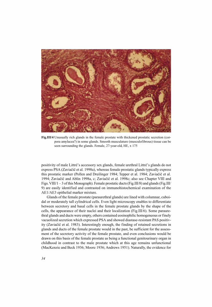
Fig.III/4 Unusually rich glands in the female prostate with thickened prostatic secretion (cor-pora amylacea?) in some glands. Smooth musculature (musculofibrous) tissue can beseen surrounding the glands. Female, 27-year-old, HE, x 175

35
Fig.III/5 Groups of glands of the female prostate embedded in vascularized musculofibroustissue. Female, 52-year-old. HE, x 360
Fig.III/6 Two female prostatic glands lined with columnar secretory cells. The basal (reserve) cellsare located below the secretory cells. The shape and the appearance of the nuclei of thebasal cells differ from those of the secretory cell nuclei. Female, 52-year-old, HE, x 360

36
Fig.III/8 Large ducts of the female prostate with pseudostratified columnar epithelium in thelining, with expression of AE1/AE3 epithelial markers in the cell membranes and onthe surface of epithelial cells bordering the duct lumen. Female, 63-year-old, x 360
Fig.III/7 Littré�s gland with strinkingly bright mucinous cells containing uniformly basally lo-cated dark nuclei. A portion of female prostatic gland in the vicinity. Female, 34-year-old, HE, x 360

37
the secretory activity of the female prostate is currently based on much more objectiveparameters, including, a.o., the observation of active secretory configuration of secre-tory prostatic cells under electron microscopy of female prostatic glands (Zaviaèiè etal. 1998d). Nevertheless, the female prostatic glandular and ductal contents need to bepaid attention to even today. The female prostatic secretion seems to stagnate in thecavitary system of the female prostate unless mechanisms helping the female prostateto empty are involved. Female prostates of bed-ridden patients after prolonged hospitalstays examined at autopsy frequently contained more stagnating prostatic secretionthan seen at forensic-medical analyses in the same structures in �fully healthy� womenwho died suddenly. In these latter cases, the mechanisms helping the prostate to emptycould have been operating up to the death without any restriction (unpublished obser-vations). Also, a layer on the surface of secretory cells of the prostatic glands and onthe luminal surface of the urothel of prostatic ducts and the female urethra showed thesame diastase-resistant PAS positivity as did prostatic secretion. This clearly config-ured layer on the luminal surface and in the apex of secretory cells of prostatic glandsin both sexes as well as on the surface lining of the cavitary system of the femaleprostate and urethra appears to present a complex interplay of glycosamine glycans,glycoproteins (including PSA and (human) urine protein 1) and enzymic proteins. Thus,in addition to the secretory role of urine protein 1 as indicated by the association of thislayer with the female and male secretory cells, it may also have a protective role, espe-
Fig.III/9 Female prostatic glands expressing AE1/AE3 epithelial markers. Female, 39-year-old, x 90

38
cially in the ureoepithelium, against the aggressive effects of urine (Zaviaèiè et al.1997a).
Female prostatic (paraurethral) glands have been known since mid-80s to manifesthigh acid phosphatase (PSAP) activity on various enzyme-histochemical (Zaviaèiè1984b) and immunohistochemical methods (Tepper et al. 1984; Pollen and Dreilinger1984), as well as proteosynthetic enzymes activity (glucose-6-phosphatase, E-600-sensitive esterase)(Zaviaèiè 1984a). The identification of female secretory prostaticcells under light microscope has been promoted by both histochemical andimmnunochemical studies of PSAP and by immunohistochemical findings of PSAexpression in the apical part and the surface of these cells (Pollen and Dreilinger 1984;Tepper et al. 1984; Zaviaèiè et al. 1994; Zaviaèiè 1997; Zaviaèiè and Ablin 1998c).Further insight could be gained by immunohistochemical demonstration of humanprotein 1 in female prostatic glands which, compared to conventional staining meth-ods, provided better possibilities to distinguish secretory from basal cells of femaleprostatic glands under the light microscope (Zaviaèiè et al. 1997a; and Fig. 1).
The ultrastructure of the normal prostatic gland in the adultwoman
The first electron microscopic study of the female prostate concerned a case of femaleprostate carcinoma in inadequately fixed material (Sloboda et al. 1998). This ultra-structural study nevertheless confirmed the immunohistochemical and histologicalconclusions made on the existence of secretory (luminal) cells in the female prostate(Zaviaèiè et al. 1997a; 1998a). Sloboda�s paper (Sloboda et al. 1998) showed the tu-mor cells to possess the basic ultrastructural parameters of secretory cells: numeroussecretory elements (secretory vacuoles and granules) in the apical cytoplasm, and shortstubby microvilli on their surface. Thus, carcinoma of the female prostate appears todevelop from the same cells as does male prostate cancer (Sloboda et al. 1998 andreferences therein).
Over prolonged periods of time, the investigation of the female prostate�s ultra-structure using transmission electron microscopy would be hindered by problems con-cerning the obtaining of optimally fresh material of the female urethra which wouldlend itself for electron microscopic investigations. Suitable material (of the femaleurethra � female prostate) could only be obtained recently during harvesting of otherorgans for transplantation purposes. Ultrastructural parameters of the normal adulthuman female prostate gland were published only recently (Zaviaèiè et al. 1998d).
Also, Gittes and Nakamura (1996) obtained several specimens of the female ure-thra and its surrounding soft tissue en bloc from brain-dead donors from whom mul-tiple organs were harvested for transplantation. This optimally fresh material wasonly used to immunohistochemically determine PSA in the glands of the femaleprostate, no electron microscopic investigation of the female prostatic tissue washowever done.

39
In our electron microscopic study (Zaviaèiè et al. 1998d), the female urethra con-taining prostatic tissue was obtained from three brain-dead female patients aged 19, 34and 47 years. They all had suffered cranio-cerebral trauma during traffic accidents,and were pronounced dead after the cerebral blood circulation had stopped (as con-firmed by cerebral panangiography). The systemic blood circulation was howevermaintained within normal limits until permission could be obtained to harvest severalorgans for transplantation purposes. The urethra was removed first by transvaginal /perineal approach. Immediately after the removal, the distal urethra containing mostprostatic tissue (meatal type of the female prostate; Zaviaèiè et al. 1998a and refer-ences therein) was cut under a drop of 3% glutaraldehyde solution into small blocks
Fig.III/10 A secretory cell with the typical bizarre appearance of secretory vacuoles (SV). Longermicrovilli can be seen on the cell surface, with irregular orientation towards differentdirections. The short arrows point to polymorphous SV in the cytoplasm. Some ofthem are in contact with the cell membrane (long arrows). Some SV contain 2 � 4dense microgranules, others contain wisps of little electrodense material (long doublearrow). In some SV, lysosome-like particles and pinocytic vacuoles (arrowheads) canbe seen in close vicinity to the cell membrane. The cytoplasmic protuberance (P) alsocontains SV. Moderately dilated sacs of the perinuclear cisterna are in direct contactwith RER. Nuclear pores of varying density (thick arrowheads) and a simple nuclearbody (between the arrowheads) can be seen. Uranyl acetate and lead citrate (UALC),x 18,000

40
with an edge length of approx. 0.2 mm, and fixed in 3% glutaraldehyde for 2 hours. Fordetails of further steps of the fixation procedure, see Zaviaèiè et al. (1998d).
Glands of the female prostate are formed by tall cylindrical secretory (luminal)cells limiting the lumen of the female prostate gland. Basal (reserve) cells are locatedbelow the secretory cells or between their base and the basement membrane. Epithelialcells may occasionally be seen interposed between basal and secretory cells or in theirvicinity; in our study they have been referred to as intermediary cells (Zaviaèiè et al.1998d).
The ultrastructure of the normal female prostate gland was compared with the avail-able information on the ultrastructure of the normal adult male prostate reported byseveral researchers (Brandes et al. 1964; Fisher and Jeffrey 1965; Mao et al. 1966;Tannenbaum et al. 1967; Fisher and Sieracki 1970; Sinha and Blackhard 1973; Brandes1974; Dermer 1978; Srigley et al. 1988). In their monograph on the prostate, Fitzpatrickand Krane (1989) devoted a chapter to the ultrastructure of this organ.
In our material (Zaviaèiè et al. 1998d), secretory cells resting on a layer of basalcells were found to be the predominant cell type of the female prostatic gland. The
Fig.III/11 Schematic representation of the ultra-structure of a secretory (luminal) cellwith an abundant population of polymor-phous secretory vacuoles.Symbols: SV, secretory vacuole; MV,microvillus; M, mitochondria; RER,rough endoplasmic reticulum; N, nucleus

41
basic structure of the female prostate acinus and the location of prostatic secretory andbasal cells were in conformity with the structural pattern of the prostatic glands of theadult male (Fisher and Jefrey 1965; Fisher and Sieracki 1970; Brandes 1974; Dermer1978; Srigley et al. 1988).
Similarly as in the adult male prostate (Fisher and Jeffrey 1965; Brandes 1974;Srigley et al. 1988), the columnar secretory cells of the female prostate are character-ized by polymorphous secretory elements in the cytoplasm: secretory vacuoles andsecretory granules, lending these cells a special appearance. The secretory vacuoles
Fig.III/12 Secretory cells (A,B,C,D,E) with secretory vacuoles (SV) and secretory granules(SG). Numerous polymorphous SV (long arrows) of varying structure and contentdensity can be seen in cell A. Cell B contains denser SG and some RER profiles(thick arrow). A nuclear body can be seen in the nucleus (between the arrowheads).Stubby microvilli on the surface of cells A and B (arrows). Cells C and D containmostly uniform SG of medium density with peripherally located flakes of electrodensematerial. Some of them are within the surface membrane (double arrow). AbundantRER in both cels. Cell C has a nucleus with two striking nucleoli (double thick ar-row) and contains supranuclear optically empty vacuoles. Cell E has a large apicalcytoplasmic protuberance (P) and contains much RER and few secretory elements.Zonulae occludentes in the apical part between the cells (between arrows). Some-what below, zonulae adherentes (between the arrowheads). The nuclei of all cellshave numerous nuclear pores. UALC, x 12,000

42
mostly had a similarly bizarre appearance (Fig.III/10) as seen in mature male secretorycells with rather polymorphic structures within the vacuoles. In some instances, theycontained remnants of mitochondria or other organelles. Some female secretory pros-tatic cells also contained homogeneous medium-dense granules with flakes of moredense material (Fig.III/12). A mixture of polymorphous populations of secretory ele-ments, secretory vacuoles as well as secretory granules was seen in other secretorycells (Fig.III/14).
The merocrine content-releasing mechanism of the secretory vacuoles (secretoryvacuoles fuse with the luminal plasma membrane and the contents get dissolved)and the apocrine discharge of apical blebs into the lumen of female prostatic glandsobserved in our studies was consistent with the known secretory mechanism of thesame cells in the adult male prostate (Brandes 1974). Lysosomes and lysosome-likedense bodies were often seen in the supranuclear parts and in blebs of secretory cellsin our samples. Our ultrastructural findings support the results of numerous enzyme-histochemical studies which identified high activities of lysosomal and cytoplasmicPSAP in the apical parts of the female secretory cells (Pollen and Dreilinger 1984;Tepper et al. 1984; Zaviaèiè 1984b; Aumüller and Seitz 1985). The appearance of
Fig.III/13 Schematic representation of the ultrastructureof a secretory (luminal) cell with secretorygranules.Symbols: SG, secretory granule; MV, mi-crovillus; P, protuberance of the apical cyto-plasm; LY, lysosome; RER, rough endoplas-mic reticulum; G, Golgi complex; M, mito-chondria; N, nucleus; NU, nucleolus

43
other cell organelles of female prostate secretory cells also corresponded to that ofmature secretory cells of the male prostate. Besides apical protuberances of the cyto-plasm and blebs, the surface of these cells was also covered by numerous short stubbymicrovilli, similarly as seen on the surface of secretory cells of the male prostate(Brandes et al. 1964; Fisher and Jeffrey 1965; Brandes 1974; Srigley et al. 1988). Acomparison of the normal fine structure of secretory cells of the female and maleprostate showed that the secretory cells of the adult female prostate were consistentin appearance with the prostatic secretory cells of the adult male. The present focuson female PSA (Borchert et al. 1997; Diamandis 1998; Zaviaèiè and Ablin 1998a, c)has increased the interest in the female prostate, which produces the major part ofthis prostatic marker in the healthy female and which has several functional implica-tions (Sesterhenn et al. 1998; Zaviaèiè and Ablin 1998a, b, c; Zaviaèiè et al. 1998c).The ultrastructural organization of female secretory cells in our material suggests anactive secretory function as judged upon by the presence of abundant secretory ele-ments � secretory vacuoles and secretory granules, rough endoplasmic reticulum,developed Golgi complexes, numerous mitochondria and apical blebs. The subcellu-
Fig.III/14 Secretory cells of the female prostate with a mixture of secretory granules (SG) andseretory vacuoles (SV). Individual short microvilli (arrow) on the surface. Occa-sional para- and supranuclear RER profiles (double arrow). A lipid droplet (LD) andempty vacuoles (V) above the nucleus. Protuberances (P) and V and RER profiles inthe apical cytoplasm. ULAC, x 18,000

44
lar equipment of female and male secretory cells with �secretory� organelles is com-parable. In the light of the exocrine function of the male prostatic glands (Ablin 1997and references therein) and of the same function of female prostatic glands (Zaviaèièand Whipple 1993 and references therein), the secretory (luminal) cells play an ex-ceptionally important role in both these organs.
The other basic cell type seen in the female prostate, i.e. the basal (reserve) cells,was found to be identical with the basal cells of the adult male prostate as to the loca-tion and the appearance of the cytoplasm (Fisher and Jeffrey 1965; Dermer 1978; Srigleyet al. 1988). In the cytoplasm of female prostatic basal cells, dilated profiles of therough endoplasmic reticulum and moderate numbers of mitochondria could be visual-ized (see the schematic representation in Fig. III/16). Besides these organelles, Srigleyet al. (1988) also described a poorly developed Golgi complex in male prostatic basalcells. Neither us nor other researchers did find any secretory vacuoles or granuleseither in female or in male prostatic basal cells. Their absence provides the most char-acteristic difference between mature basal cells and secretory prostatic cells (Fisherand Jeffrey 1965; Dermer 1978; Srigley et al. 1988). Both the appearance of prostaticbasal cell nuclei and the distribution of their chromatin in the female and male prostate
Fig.III/15 Schematic representation of the ultrastructure ofa secretory (luminal) cell with a mixture of secre-tory elements (secretory vacuoles together withsecretory granules in one secretory cell).Symbols: SV, secretory vacuole; SG, secretorygranule; B, bleb; LY, lysosome; MV, microvil-lus; RER, rough endoplasmic reticulum; LD,lipid droplet; V, empty vacuole; M, mitochon-dria; N, nucleus

45
were comparable (Fisher and Jeffrey 1965; Dermer 1978; Srigley et al. 1988). Sincefemale basal cells rest on the basement membrane of female prostatic glands, theirpotential myoepithelial nature may be considered as tentatively discussed in the caseof male prostate basal cells (Fisher and Sieracky 1970). Unlike breast and salivaryglands where myoepithelium is present, the female and the male prostate contain anon-glandular contractile system composed of smooth muscle cells and/ormusculofibrous tissue (Huffman 1948; Dermer 1978; Zaviaèiè et al. 1983; Zaviaèiè1987a; Wernert et al. 1992). In keeping with this, we did not find structures suggestingcontractile function in female basal prostatic cells. Besides sexual stimuli, the tonusand the congestion in musculofibrous tissue surrounding the glands and ducts of thefemale prostate, which change during micturition and defecation, may well contributeto the evacuation of the female prostate (Zaviaèiè et al. 1983).
Fig.III/16 Schematic representation of the ultrastructure of a basal (reserve) cell of the femaleprostatic gland. Swollen mitochondria and profiles of the rough endoplasmic reticu-lum can be seen in the dense cytoplasmic ground substance.Symbols: M, mitochondria; RER, rough endoplasmic reticulum; N, nucleus
46
Glandular epithelial cells with little differentiated cytoplasm, seen along with secre-tory and basal cells, were termed by us type 1 and 2 intermediary cells. The mostpronounced difference between them is the virtually complete absence of secretorygranules and vacuoles in type 1 cells (Fig.III/17) and their appearance in the littledifferentiated cytoplasm of type 2 intermediary cells. In the female prostate, interme-diary cells may represent cell forms between basal and secretory cells, demonstratedby some authors under electron microscopy in the male prostate only (Mao and Angrist1966). Dermer (1978) and Srigley et al. (1988) suggested that male prostate basal cellsmay be a source of secretory cells. Xue et al. (1998) developed a prostatic stem cellmodel in which both exocrine and neuroendocrine cells of the male prostate are de-rived from a subpopulation of basal cells that give rise to luminal (secretory) cellsthrough intermediate cells (pluripotent amplifying cells). Our findings support theopinion on the similar role of basal cells in the renewal of exocrine (secretory) ele-ments in the female prostate glands.
The principal structural parameters of secretory (luminal), basal (reserve) and in-termediary cells of the adult human female prostate gland are schematically shown inFigures 11, 13, 15, 16, and 18.
In the male prostate, immature stratified glandular epithelium differentiates duringpuberty into two principal cell types of the adult prostate glands: the secretory colum-nar epithelium and the basal cells. This differentiation process of the male prostateduring puberty is obviously initiated by the commencing testicular androgen secretion(Wernert and Dhom 1989; Wernert et al. 1992). These andrological findings are inaccordance with the earlier anatomical observations by Andrews (1951) that the maleprostate gland remains without any secretory activity between about 4 months andthirteen years of age. Heath (1984; 1987) believes that the female prostate begins func-tioning very early, nevertheless erotic and orgasmic functional sequences are graduallyinhibited and, as hypothesized by Money (1987), also societal prohibition plays a rolein the inhibition. In spite of the fact that under normal conditions, there are no andro-genic stimuli either during puberty or throughout life of a woman, the epithelium offemale prostatic glands gets clearly differentiated into mature secretory and basal cells.It seems to be justified to address the question whether it actually is androgens onlythat are involved in an important manner in the processes of differentiation and matu-ration of prostatic glandular cells and whether estrogens may not be called into play inthe differentiation of glandular cells of the female prostate, which is equipped withreceptors for this hormone. Estrogen-receptor-associated protein (ER-D5) was foundin the cytoplasm of cells lining female prostate glands, yet not in ductal cells (Wernertet al. 1992). Our considerations concerning the potential participation of estrogens inthe differentiation of the glandular epithelium of the human female prostate are stronglysupported by the findings of Merck et al. (1980). These authors observed regressedglandular cells in the male prostate to acquire a new phenotype after the administrationof estradiol-17-beta-17-cyclopentylpropionate to castrated and hypophysectomizeddogs. The characteristic features of these cells included newly formed secretory gran-ules. The authors suggested that androgens and estrogens have an equal capacity tocontrol the expression of prostatic secretory granules.

47
Our results concerning the female prostate (see references in this Monograph) in-cluding our electron microscopic study (Zaviaèiè et al. 1998d) with the discussionpresented in the latter paper and in this Monograph substantiate our disagreement withthe skeptical conclusions voiced in the study by Wernert and coworkers (1992) that�these glands (of the female prostate) resemble strongly the male prostate glands be-fore puberty ... glands (of the female prostate) remain immature throughout life (of thefemale) from the fetal period up to advanced age... No indications can be found for aproper biological function...� On the contrary, our electron microscopic analysis of thenormal female prostate showed that, similarly as in the male after puberty, prostaticglands in the adult female form morphologically mature secretory and basal cells.There is no ground to believe that these cells would not play the same role in thepathophysiology of the female as they do in the male prostate. Supporting evidencehas been provided by several implications of the exocrine function of the female pros-tate in forensic medicine, gynecologic urology, chronobiology and sexology (Zaviaèiè
Fig.III/17 Intermediary cell type 1 (IC1) between two secretory cells (SC). The nucleus with asmooth surface, inconspicuous simple nucleolus (NU) and much disperse chromatin.Only one or two small SG (arrow), profiles of undilated RER (arrowhead), free ribo-somes and centriole (C) in the cytoplasm. The cell rests on the basement membrane(BM) and is attached to it by separate hemidesmosomes (long arrow). Zonulaeadherentes connect the cell with the cytoplasm of the neighboring SC (thick arrow).UALC, x 18,000

48
1987a; Zaviaèiè and Whipple 1993; Zaviaèiè and Ablin 1998a, b, c). On giving equalconsideration to ultrastructural parameters of the glands of the human male and femaleprostate, it might be appropriate to mention some rodent species with comparable struc-tures of prostatic glands in males and females, observed at the ultrastructural level(Ichihara 1976; Gross and Didio 1987).
The recent ultrastructural observations further support the justified view that pros-tate should be seen as woman�s specific functional genitourinary organ. Increasingevidence substantiates the notion that the woman�s prostate is an actually functionalgland in its own right rather than an afunctional vestigial Skene�s gland. Its structurehas been well defined and the adult woman prostate�s ultrastructural equipment alsocorresponds to that of the glandular cells of the adult male prostate. Comparabilitywith the male prostate has not only been established with respect to the exocrine andprobably also neuroendocrine function, but also with regard to the pathology of thefemale prostate. The occurrence of pathologic conditions may be less frequent in thefemale, yet there is increasing evidence indicating that the female prostate can be af-fected by the same diseases as does the male prostate, including cancer, benign pros-tate hyperplasia and prostatitis (Folsom and O�Brien 1943; Zaviaèiè et al. 1985b; 1993a;1994; 1995; Zaviaèiè 1987a; Zaviaèiè and Whipple 1993; Gittes and Nakamura 1996;Sloboda et al. 1998 and references therein; Sesterhenn et al. 1998; Zaviaèiè and Ablin1998a, b, c).
Fig.III/18 Schematic representation of the ultrastructure of type 1 and 2 intermediary cells ofthe female prostatic gland. The cytoplasm of type 2 intermediary cell (IC2) is moredifferentiated than that of IC1, and contains sparse secretory elements (secretorygranules).Symbols: SG, secretory granule; G, Golgi complex; M, mitochondria; C, centriole;RER, rough endoplasmic reticulum; N, nucleus

49
The prostate can thus be considered as another organ exhibiting different size andweight (certainly, depending on additional parameters such as body stature and weight,and age as do most human organs and as evident from tables showing the parameters ofhuman organs; Thomas 1990) and different functional capacity in man and woman, yetthe same qualitative parameters in both genders. Sandritter�s Macropathology (Tho-mas 1990) however does not show any data concerning the weight of the female pros-tate since such data have only been published very recently (Zaviaèiè et al. 1998a).
Differences in somatic organ parameters between the genders may become impor-tant with respect to transplantation medicine, e.g. in looking for adequate replacementof male organs by female organs. The heart of a 40- and 50-year-old woman is 35 glighter and smaller than that of a man of the same age, stature and almost the sameweight (Thomas 1990). Then, such differences may become a serious or even deter-mining indication criterion with respect to the selection of the donor for human hearttransplantation. Naturally, the differences in organ parameters between males and fe-males should not be an adequate argument to support the simplistic view that someorgans in women are inferior to those of men. A similar conclusion also applies to thefemale prostate: it cannot be considered inferior just because it is smaller and has asmaller weight than the male prostate.

50

51
IV.Enzyme Histochemistryof the Female Prostate and itsFunctional Implications
The first enzyme histochemical papers dealing with the female prostate (Skene�s glands)appeared in early 80s, at a time, when homology between vestigial Skene�s glands andthe male prostate was essentially based on the embryology-derived knowledge of bothtissues originating from the same embryonal urogenital sinus. Suddenly, evidence couldbe brought for Skene�s glands having a typical enzyme equipment comparable withthat of the male prostate (Zaviaèiè 1985a), including the presence of prostatic andlysosomal acid phosphatase (Zaviaèiè 1984b), other hydrolases and numerous oxido-reductases (Zaviaèiè 1984a).
Enzyme-histochemical investigations, and among them and due to understandablereasons, the evidence for acid phosphatase in Skene�s glands but not in paraurethralducts, were the strongest reason for the recognition of the existence of the female pros-tate in early 80s, prior to the PSA era. At a time, when identification of female prostaticglands faced some problems, in particular when located intraepithelially in the prostaticduct walls, azo-coupling and precipitation techniques according to Lojda et al. (1976)were an easy approach to resolve the problem. Acid phosphatase activity established bythe azo-coupling (Fig.IV/1) and Gomori�s precipitation method (Fig.IV/2) most prob-ably demonstrated �lysosomal� acid phosphatase (EC 3.1.3.2) rather than its isoenzyme,cytoplasmic �prostatic� phosphatase (Vries et al. 1980). Also, the lysosomal fraction canbe assumed with a high probability to be released to the female ejaculate along withcytosolic prostatic acid phosphatase, as suggested by macro-enzyme-histochemical studiesproviding evidence for a high activity of the former enzyme in association with continualsecretion and ejaculation-induced emptying of the female prostate (Zaviaèiè et al. 1987a,b; 1988a, b, c). As determined biochemically, acid phosphatase activity was several timeshigher in the fluid of female urethral expulsions than in the urine (Addiego et al. 1981;Belzer 1981; Longo 1982; Stifter 1988).
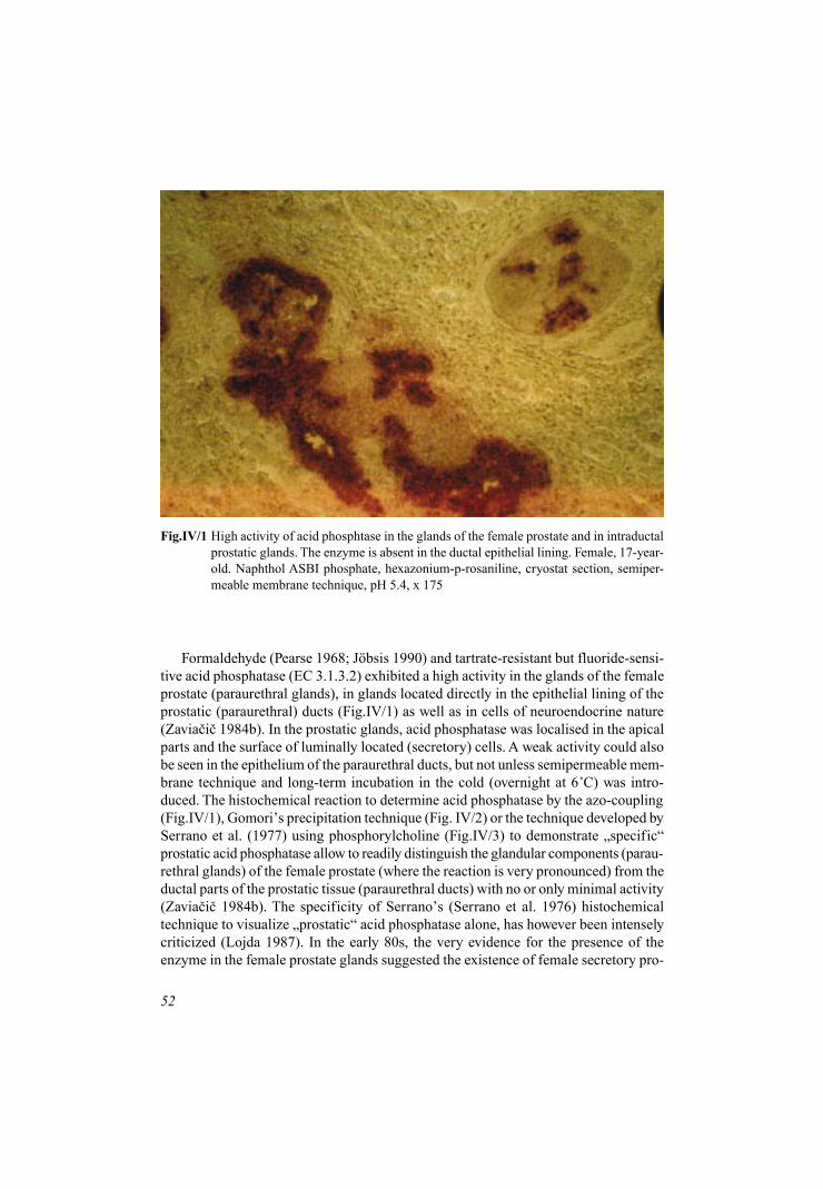
52
Formaldehyde (Pearse 1968; Jöbsis 1990) and tartrate-resistant but fluoride-sensi-tive acid phosphatase (EC 3.1.3.2) exhibited a high activity in the glands of the femaleprostate (paraurethral glands), in glands located directly in the epithelial lining of theprostatic (paraurethral) ducts (Fig.IV/1) as well as in cells of neuroendocrine nature(Zaviaèiè 1984b). In the prostatic glands, acid phosphatase was localised in the apicalparts and the surface of luminally located (secretory) cells. A weak activity could alsobe seen in the epithelium of the paraurethral ducts, but not unless semipermeable mem-brane technique and long-term incubation in the cold (overnight at 6°C) was intro-duced. The histochemical reaction to determine acid phosphatase by the azo-coupling(Fig.IV/1), Gomori�s precipitation technique (Fig. IV/2) or the technique developed bySerrano et al. (1977) using phosphorylcholine (Fig.IV/3) to demonstrate �specific�prostatic acid phosphatase allow to readily distinguish the glandular components (parau-rethral glands) of the female prostate (where the reaction is very pronounced) from theductal parts of the prostatic tissue (paraurethral ducts) with no or only minimal activity(Zaviaèiè 1984b). The specificity of Serrano�s (Serrano et al. 1976) histochemicaltechnique to visualize �prostatic� acid phosphatase alone, has however been intenselycriticized (Lojda 1987). In the early 80s, the very evidence for the presence of theenzyme in the female prostate glands suggested the existence of female secretory pro-
Fig.IV/1 High activity of acid phosphtase in the glands of the female prostate and in intraductalprostatic glands. The enzyme is absent in the ductal epithelial lining. Female, 17-year-old. Naphthol ASBI phosphate, hexazonium-p-rosaniline, cryostat section, semiper-meable membrane technique, pH 5.4, x 175

53
Fig.IV/2 High activity of acid phosphatase in the glands of the prostate of a 15-year-old female.Sodium beta-glycerophosphate, Gomori�s method, pH 5.4, frozen section, x 175
Fig.IV/3 High acivity of prostatic (specific) acid phosphatase in the glands of the prostate of a37-year-old female. Method according to Serrano et al. with phosphorylcholine, x 180

54
static cells and thus an exocrine role for the female prostate. Prostatic ducts do nothave a similar role. However, they are far from being a mere passive ductal part of theprostate since (as will be shown below) they are equipped with cells for neuroendo-crine function. In addition to secretory cells of female prostatic glands, also occasionalcells and nerve fibres of cells showed reaction, which corresponded well with argyro-philic cells as seen when using the Grimelius� method (Zaviaèiè 1986a) or when usingpolyclonal antibodies against human protein 1 (urine protein 1) and other neuroendo-crine markers to study female prostate (Zaviaèiè et al. 1997b). The findings concern-ing acid phosphatase and some other phosphatases repeatedly confirmed the existenceof neuroendocrine cells in the female prostate, although their presence in the femaleand the male prostate urethra has been known since the late 50s (Koch and Engelhard1959) and even early 40s (Pretl 1944) respectively.
The determination of acid phosphatase, naphthyl esterase and glucose-6-phosphatasehelps to distinguish the glandular component of the female prostate (where relativelyhigh activities are observed) from the epithelial lining of the ducts, where these en-zymes are missing. The secretory function of the female prostate glands has been sup-ported by the findings of E-600 sensitive naphthyl esterase (Fig.IV/4) and glucose-6-phosphatase (Fig.IV/5) in luminally located secretory cells of female prostatic glands.
Fig.IV/4 Markedly high activity of E-600 sensitive esterase in the cytoplasm of prostatic glandcells. The activity is considerably weaker or missing in the pseudostratified columnarepithelial lining of the ducts. Female, 17-year-old. Alpha-naphthyl acetate andhexazonium-p-rosaniline, x 175
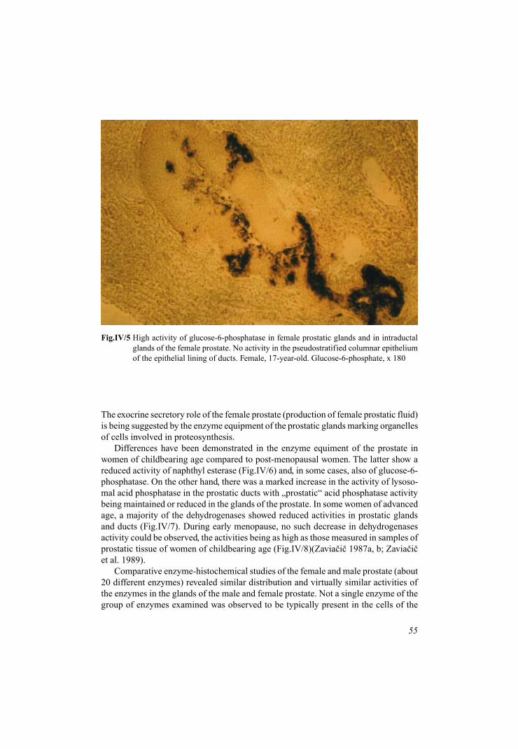
55
Fig.IV/5 High activity of glucose-6-phosphatase in female prostatic glands and in intraductalglands of the female prostate. No activity in the pseudostratified columnar epitheliumof the epithelial lining of ducts. Female, 17-year-old. Glucose-6-phosphate, x 180
The exocrine secretory role of the female prostate (production of female prostatic fluid)is being suggested by the enzyme equipment of the prostatic glands marking organellesof cells involved in proteosynthesis.
Differences have been demonstrated in the enzyme equiment of the prostate inwomen of childbearing age compared to post-menopausal women. The latter show areduced activity of naphthyl esterase (Fig.IV/6) and, in some cases, also of glucose-6-phosphatase. On the other hand, there was a marked increase in the activity of lysoso-mal acid phosphatase in the prostatic ducts with �prostatic� acid phosphatase activitybeing maintained or reduced in the glands of the prostate. In some women of advancedage, a majority of the dehydrogenases showed reduced activities in prostatic glandsand ducts (Fig.IV/7). During early menopause, no such decrease in dehydrogenasesactivity could be observed, the activities being as high as those measured in samples ofprostatic tissue of women of childbearing age (Fig.IV/8)(Zaviaèiè 1987a, b; Zaviaèièet al. 1989).
Comparative enzyme-histochemical studies of the female and male prostate (about20 different enzymes) revealed similar distribution and virtually similar activities ofthe enzymes in the glands of the male and female prostate. Not a single enzyme of thegroup of enzymes examined was observed to be typically present in the cells of the

56
Fig.IV/6 Low activity of E-600 sensitive esterase in the glands and ducts of the prostate of a 73-year-old-female. Alpha-naphthyl acetate and hexazonium-p-rosaniline, x 180
Fig.IV/7 Low activity of mitochondrial glycerol-3-phosphate dehydrogenase in the epitheliumof the female prostate ducts. Female, 71-year-old, x 180

57
Fig.IV/8 High activity of mitochondrial glycerol-3-phosphate dehydrogenase in the epitheliumof the female prostate ducts and urethra. Female, 38-year-old, x 180
male prostatic glands while being absent in the female prostate structures (Zaviaèiè1985a).
At the same time when the first papers concerning the enzyme equipment of thefemale prostate started appearing (Zaviaèiè 1984a, b; 1985a), also the first immuno-histochemical works were published providing evidence for the presence of prostate-specific antigen (PSA) in female prostate structures, in particular in glands whichcould also be easily identified (Mallon 1983; Tepper et al. 1984; Pollen and Dreilinger1984), similarly as in selected enzyme-histochemical studies.
Being an important component of the male prostate secretion, acid phosphataseparticipates in the hydrolysis of phosphorylcholine in seminal plasma, and is consid-ered to be a marker of the development of the secondary sex characteristics in the male(Spring-Mills and Hafez 1980). The role of the high activity of acid phosphatase in thefemale prostate is not clear, it nevertheless cannot be ruled out that female prostaticacid phosphatase plays a similar role as in the male with respect to the female second-ary sex characteristics. This enzyme could be demonstrated in a woman aged over 80as well as in early childhood long before the onset of puberty (Zaviaèiè et al. 1983;1989). Kellokumpu-Lehtinen (1980) used light and electron microscopy to study acidphosphatase distribution in the urogenital system of 7-13-week-old human fetuses ofboth sexes. At this young age already, the lining of the female urethra displayed acidphosphatase activity comparable with the activity of the same enzyme in the urethra of
58
male fetuses. The same author could observe lysosomal acid phosphatase activity inthe prostatic epithelium upon its appearance in week 10, and a few epithelial cellsshowed acid phosphatase-positive apical granules suggestive of the appearance of secre-tory acid phosphatase. We believe that (lysosomal?) acid phosphatase may have a simi-lar significance in the prostate as in some other secretory active cells in removingexhausted cell organelles during the active and resting phase of their metabolic in-volvement (Zaviaèiè et al. 1980).
Contrary to acid phosphatase, the equipment of lysosomes with beta-D-glucu-ronidase and acetyl-beta-D-glucosaminidase in the female prostatic gland is but verypoor. E-600 sensitive naphthyl esterase and glucose-6-phosphatase have been demon-strated in cells of female prostatic glands; similarly as acid phosphatase, this enzyme isof help in differentiating between female prostate glands and ducts. Together with otherenzymes, the finding of the above enzymes represents the expression of secretory ac-tivity of the cells. The equipment of glandular cells with granular endoplasmic reticu-lum (Figs.III/10, 12, 14) is indicative of the secretory role of the female prostate gland.
The high activity of glycerol-3-phosphate dehydrogenase in the ducts and glandsof the female prostate which invariably yields the highest activity of all dehydrogena-ses examined (Fig.IV/8)(the same holding true for the male prostate) may indicatesynthesis of triglycerides and phospholipids in these parts. It has not yet been deter-mined whether these substances are components of female prostatic secretion. Being anon-polar lipid, cholesterol is a component of the male prostatic fluid, and is presumedto support sperm stabilisation, protecting it from environmental shock and heat dam-age (Spring-Mills and Hafez 1980).
Assessing, from a different angle, the value of histochemical works dealing withthe enzyme equipment of the female prostate (Skene�s glands), which has been shownto be comparable with that of the male prostate (Brandes and Bourne 1956; Kirchheimet al. 1964; Brandes 1966; Aso et al. 1968), those works can be said to become, at thetime when published in mid-80s, the basis for the �histochemical concept of male andfemale prostate homology�, and all this at a time when it was only embryology-derivedknowledge on the origin of Skene�s glands and the male prostate that pointed to such ahomology.
Differences in the enzyme equipment of the prostate between women of childbear-ing age and post-menopausal women (concerning a decrease in the activities of naph-thyl esterase, glucose-6-phosphatase and a majority of dehydrogenases in elderly women,and a marked increase in the activity of �lysosomal� acid phosphatase in the prostaticducts in elderly individuals) support the possibility of hormonal dependence in thisfemale genitourinary organ function (Zaviaèiè et al. 1989). This important issue hasnot been dealt with in detail so far, and our assumptions in this matter have not beeneither supported or confirmed by other methodological approaches. There are howeversome sexological reports showing that some of the patients which could be consideredfemale ejaculators lost this ability at higher age (Ladas et al. 1982). This issue is ofimportance since the male prostate is a classical hormone-dependent organ the epithe-lium of which starts differentiating under the influence of the commencing productionof sex hormones, and which is substantially dependent with respect to its function on a
59
sufficient level of male sex hormones throughout life of the man. The same issue in thefemale prostate should be treated with caution because of relatively little informationavailable in this respect. Nevertheless, estrogens seem to play the same role in thefemale prostate as do androgens for the male prostate. This is suggested by the findingof typical secretory vesicles and granules in female prostatic secretory cells. Also,additional ultrastructural parameters of these cells of the female prostate correspond tothose of the secretory cells of the adult male prostate, and this despite the fact thatandrogens said to be the key factor for the formation of secretory elements in maleprostatic secretory cells, are lacking in normal females. However it may be, secretoryelements (secretory vacuoles and secretory granules) are seen in the secretory cells ofthe female prostate, and their numbers and appearance is even comparable to thoseseen in the same prostatic cells of the adult male (Zaviaèiè et al. 1998d).

60

61
V.Exocrine Function of the FemaleProstate: Implications forGynecology, Urology, Chronobiologyand Forensic Medicine
The exocrine function of the female prostate consists of the production of prostaticfluid, and this role is equally a major one as it is the case in the male prostate. Similarlyas prostatic glands in the adult male, female prostatic glands are equipped by maturesecretory (luminal) cells (Zaviaèiè et al. 1998d) responsible for the production of pro-static fluid in both genders. Unlike male�s fluid, pure female prostatic fluid could nothave been isolated as yet. So far, female prostatic fluid has been studied but as a part ofthe female ejaculate, a fluid released during the phenomenon of female ejaculation,and it has been investigated with respect to the demonstration of continual secretion ofthe female prostate. If urologists and gynecologists succeeded in isolating pure femaleprostatic fluid, it would significantly speed up the identification of additional compo-nents considered to be typical components of male�s prostatic fluid, the production ofwhich by the female prostate is uncertain.
According to our experience, the volume of the fluid obtained after a single G-spotstimulation ranged between 1 � 5 mL. A distinct layer of whitish opaque cellular com-ponent appeared on the bottom of the test tube 10 � 15 minutes after collection, cov-ered by urinous layer. A thin layer of transparent mucosubstances was sometimes seenat the borderline between the two above layers (Zaviaèiè et al. 1984a, c; 1988c).
The cellular component of the female ejaculate is formed by squamous cells of thevaginal type originating from the lining of the female urethra, large ducts of the femaleprostate (paraurethral ducts), and the bladder trigone. During the proliferative and ovu-latory phase of the cycle, typical flat squamous cells with pycnotic nuclei were seen inShorr-stained smears of the cellular component of the female ejaculate (Fig.V/1). Scan-ning electron microscopy showed clusters of flat squamous cells with overlapping edges

62
Fig.V/2 During the secretory phase of the cycle (late luteal phase), squamous cells were fre-quently seen on Shorr-stained hormonal urocytology, shrinking and curled up, withtheir edges bent. Day 27 of 28-day cycle
Fig.V/1 During the proliferative and ovulatory phases of the cycle, typical flat squamous cellswith pycnotic nuclei were seen in smears of the Shorr-stained cellular component. Day14 of 28-day cycle

63
and knob-like nuclei in the cell center. During the late luteal (secretory) phase of thecycle, squamous cells were frequently seen under Shorr hormonal urocytology stain-ing, shrinked, curled and with bent edges (Fig.V/2). Scanning electron microscopyshowed regressively altered squamous cells at different stages of folding-up. Thus, aqualitative rhythm can be distinguished with respect to the uroepithelial squamouscells pattern (cell component) in female ejaculate during the menstrual cycle. This cellcomponent pattern can be used for hormonal urocytology in cases when vaginal smearscannot be obtained (Zaviaèiè et al. 1984a, b; 1988c).
The typical cellular changes during the menstrual cycle seen under light micro-scope represent the basis for functional urethral and urinary cytology (Langreder1961; O�Morchoe and O�Morchoe 1967; Lencioni and Staffieri 1969; Egloff 1972;Hermosa 1978; Bernhard et al. 1979) which are more preferred in South America(Hermosa 1978) than in other parts of the world (Koss 1979). The drawback of theurinary sediment cytology from centrifuged urine is the small amount of cells whichcan be evaluated in urine cytograms, as well as artifacts which may develop duringurine centrifugation. The squamous cells of the female ejaculate are perfectly pre-served, remain unaltered by centrifugation, and are present in amounts that cannotbe obtained by any other similar method, including the procedure used to collect
Fig.V/3 Daily volumes of the cell component during one-and-half menstrual cycle, multiparavolunteer, 37-year-old. PP, proliferative phase (white columns); SP, secretory phase(hatched columns and circles). Runs shown by arrows (dashed for missing data); sometheir ranks are given in numbers (in parentheses including missing data).Global lineartrend for all SP data and its 95% confidence corridor (dashed straight lines and hyperbo-las). Right panel: The median (x~) and its 95 % confidence lower (LL) and upper (UL)limit both for PP and SP. Random variable of the Mann-Whitney test (U) and Shapiro-Wilk test (W).

64
Fig.V/4 Markedly high acid phosphatase activity in a spot of 3 mL ejaculate, 46-year-old male.Naphthol ASBI phosphate, hexazo-p-rosaniline, cotton, 2:1
material for urethral cytology. The cellular component of the female ejaculate can bepreferred for hormonal urocytology over urinary sediment cytology from centrifugedurine.
Quantitative data on the amounts of uroepithelial squamous cells released into thefluid of urethral expulsions were obtained from a 37-year-old female ejaculator from agroup with �easily induced expulsions� (Zaviaèiè et al. 1988b). Digital stimulation ofthe G spot was repeated consecutively five times every day over the period of one anda half menstrual cycle. As shown by the quantitative biometrically analyzed data, theproportions of the cellular component were far higher during the secretory phase of thecycle than during the proliferative phase (Fig.V/3). The quantitative circatrigintan rhythm(30±5 days) of the cell component volume of the female ejaculate (female urethralexpulsion fluid) with the acrophase during the secretory phase of the menstrual cyclereduced the height of the urethral epithelium, this weakening the sphincteric closingmechanism of the female urethra. In contrast to the first half of the cycle when farfewer cells were released into the ejaculate, the epithelium remained in situ, and thesphincteric closing mechanism of the urethra could operate effectively (Zaviaèiè et al.1984c; Mikulecký et al. 1986).
Gynecologists and sexologists are familiar with the rhythmic menstrual cycle of womenin terms of phases. However, there are additional rhythmic components during the men-strual cycle. The rhythm of the cellular component of the female ejaculate (Zaviaèiè et al.

65
1984c) is of practical value for gynecologic urology and with respect to problems ofurinary incontinence in women. With respect to the role of the urethral epithelium in theclosing mechanism of the female urethra it may be assumed that conditions for the devel-opment of incontinence in women are more favorable during the secretory phase of themenstrual cycle (Zaviaèiè et al. 1984c; Mikulecký et al. 1986). The treatment of urinaryincontinence by estrogens is thus justified as estrogens positively influence the thicknessof both the urethral epithelium and the urethral wall (Bhatia et al. 1989). The closingmechanism of the female urethra important from the viewpoint of urine incontinence ishowever even more complicated: also, striated muscle fibers around the smooth muscu-lature (musculofibrous tissue) in the urethral wall around the spongy body and the glandsand ducts of the female prostate participate in this mechanism. All these structures areinvolved in the mechanism of continence, and play a role in sexual reactions of the fe-male (Graefenberg 1950; Ricci et al. 1950; Langreder 1956; Hutch and Rambo 1967).
Female ejaculate as a phenomenon of the exocrine function of the female prostate(the fluid of the female urethral expulsions) and female ejaculation have importantpractical implications for the practice of forensic medicine.
The forensic-medical expertise has used determination of spermatozoa as the mostsensitive and most reliable method for the identification of semen. However, when dueto whatever reasons no spermatozoa could be determined in the individual investigated(e.g. in azoospermic male or after vasectomy), the evidence of acid phosphatase (Fig.V/
Fig.V/5 Acid phosphatase in a spot of female ejaculate determined by azo-coupling method.Female ejaculate (1.2 mL) obtained on day 8 of cycle. Naphthol ASBI phosphate,hexazo-p-rosaniline, cotton, 2:1

66
Fig. V/6 Markedly high acidphosphatase positivityin a spot on lingerie inurethral projection(U). Girl, 11-year-old,cotton lingerie wornfor 4 days. NaphtholASBI phosphate, he-xazo-p-rosaniline, 2:1
4) alone would be considered adequate to identify a semen spot in the past (Rusfeld1946; Kaye 1947). The quantitative aspects of the enzyme determined were empha-sized as early as in the 70s (Schumann et al. 1976; Findley 1977). Later on, this test incombination with positivity of other components (spermine, fructose) would only beaccepted as reliable proof in forensic-medical expertise.
With respect to expert evidence of rape in women, the acid phosphatase test wasfound to have no forensic relevance in identifying sperm spots which contain no sper-matozoa, since the same acid phosphatase activity can be established in in vitro formedspots of female ejaculate (Fig.V/5) (Zaviaèiè et al. 1987b) and in in vivo producedspots on worn female lingerie (Figs. V/6, 7) (Zaviaèiè et al. 1988d). Thus, positivelyreacting spots may originate from the woman herself, without any male involvement.Macro-enzyme-histochemical findings of acid phosphatase on underwear (Figs. V/6,7) in parts in constant contact with the female genitals are indicative of the existence

67
Fig.V/7 Markedly high acid phosphatase positivity in a spot on cotton underwear in the area ofurethral projection (U). Female, 42-year-old (28/5). Underwear worn for 4 days (be-tween days 14 and 17 of cycle). Naphthol ASBI phosphate, hexazo-p-rosaniline, 2:1
Fig.V/8 Acid phosphatase positivity in a spot on cotton underwear in the urethral projectionarea. Spot spreads distally because the underwear had been worn for several days. Girl,7-year-old, death of asphyxia (suffocation) due to lung compression by elevator cage.Ejaculation due to asphyxia. Naphthol ASBI phosphate, hexazo-p-rosaniline. 2:1

68
Fig.V/9 High acid phosphatase positivity in a large spot on underwear, extending distally (V)from the urethral projection (U). New, not worn cotton underwear. The transversalsection is artificial. Female, 57-year-old, suicide by hanging. Agonal ejaculation dueto asphyxia. Naphthol ASBI phosphate, hexazo-p-rosaniline, 2:1
of continual secretion by the female prostate (Zaviaèiè et al. 1988d). Initiation of thefemale prostate secretion precedes the onset of puberty (Zaviaèiè et al. 1989), simi-larly as is the case in the male (Aumüller et al. 1983).
Asphyxia can evoke agonal urethral expulsions (ejaculation) in the woman, e.g.upon strangulation, suffocation (Fig.V/8) and hanging (Fig.V/9)(Zaviaèiè et al. 1987a;1988d), similarly as it occurs under the same circumstances in about 30% of men(Resnik 1972). It has been hypothesized (Zaviaèiè 1994) that the fenomenon of thefemale ejaculation may play a role as a motivation in sexual asphyxiophilia(Kozcwarism) in women. According to this hypothesis, women of the ejaculatory type(female ejaculators) desire to induce ejaculatory orgasm (orgasm with ejaculation) byasphyxia. This kind of female orgasm is usually assessed as a sensation of greaterdelight than orgasm without ejaculation, and this may be the reason for repetitive, life-threatening deviant sexual practice.
As suggested above, forensic medical studies of the female prostate and femaleejaculation not only focussed on special forensic medical problems but also on theassociated possibilities to study the secretory mechanism of the female prostate. Wecould show that, besides the ejaculatory mechanism by which the female prostate isemptied (and which has received inaproppriately great attention), there also is con-

69
Fig.V/10 Prostate-specific acid phosphatase expression in the luminal part of cells bordering theprostatic duct, in a prostatic intraductal gland, and mainly in prostatic fluid in the duct.Female, 38-year-old. Two-step indirect immunoperoxidase technique and PSAP anti-bodies, x 180
tinual release of small amounts of female prostatic secretion into the urethra, as evi-denced by spots on underwear showing acid phosphatase positivity. This mode of se-cretion of the female prostate is fully comparable with the continual spontaneous se-cretion by the male prostate (Mann 1974).
The most typical components of the male prostatic fluid include acid phosphatase,citric acid, zinc, and spermine (Spring-Mills and Hafez 1980). Similarly as in the maleprostatic fluid, acid phosphatase can also be identified biochemically and histochemi-cally (Adiego et al. 1981; Sensabaugh and Kahane 1982; Belcer et al. 1984; Zaviaèièet al. 1984a; 1987b; 1988d; Stifter 1988) and fructose biochemically (Zaviaèiè et al.1988a) in the female prostatic secretion, including that in situ in the female prostaticducts and glands (Fig.V/10) and female ejaculate (Fig.V/5), representing a substantialportion of the latter. Additional components which would correspond to the knownmale prostatic components are being looked for. So, crystals were found on scanningelectron microscopy (Zaviaèiè et al. 1984a) which could resemble spermine crystalsby their appearance (three-sided bodies)(unpublished observation). Apparently, the fe-male prostatic tissue should also be analyzed for the presence of zinc; the male pros-tate contains this metal which has for long been considered a typical prostatic param-eter in vertebrates (Bertrand and Vladesco 1921). However, fundamental differences

70
were observed by histochemical and atomic absorption spectrophotometry methods inthe distribution of zinc within the various zones of the male prostate (Györkey et al.1967). Should spermine and zinc be demonstrated to be present in the female prostate,further markers will have to be excluded from the methodological armamentarium offorensic medicine used to provide evidence for rape, since they would lose their foren-sic relevance (after acid phosphatase, maybe also spermine and zinc). It seems prema-ture to speak about the role of zinc in the female prostate. If however the presence ofzinc is convincingly demonstrated, it may not be ruled out to play an important role inreproductive physiology in women similarly as is the case in men (Byar 1974). Aninverse relationship between zinc concentration and pathology of the prostate is knownin males. So, zinc is reduced to about one third of the normal level in malignant condi-tions of the prostate (Bush et al. 1974; Muntzig et al. 1974). So far, zinc levels have notbeen monitored in female prostate cancer or normal female prostatic tissue.
There are substantial changes brought about by DNA analyses with respect to pro-viding evidence for rape where the suspect�s sperm contains no spermatozoa or if thereis a technical failure to demonstrate them (Zaviaèiè and Ablin 1998a). Now, we areable to not only find out whether a biological piece of evidence originates from a manor a woman, but even to individually exactly identify the suspect individual. The valueof the forensic medical expertise which, prior to the period of DNA analyses, wouldmore or less determine the guilt or innocence of the accused now increasingly turnshistorical.

71
VI.Neuroendocrine Function of theFemale Prostate: Morphological Basis
Since its very beginnings, the recent studies of the female prostate have set out on thepath of looking for the parameters of the male prostate which have been known andconsidered as typical of male prostatic tissue. One the one hand, this has to a certaindegree limited the possibility of finding entirely new parameters of the female prostatewhich may be missing in the male prostate; on the other one, investigations of thefemale prostate have been moving on a realistic comparable level which has contrib-uted to the implications of the female prostate being accepted easier since they areknown to be identical and of similar importance as for the male prostate.
One of these areas have been studies of the female prostate focussing on the iden-tification of neuroendocrine cells in the female prostatic tissue and on the evidence ofpolypeptides and hormones produced by the female prostate. Thus, this has concernedthe basic concept of whether the female prostate can be seen as a female neuroendo-crine organ � the neuroendocrine function of the male prostate has been well knownand understood, including a broad range of polypeptides produced by the male pros-tatic neuroendocrine cells. Even if it were so, the neuroendocrine function can not beexpected to be more important than the exocrine function of the female prostate. Thepreferential concept of the male prostate as a predominantly exocrine gland producingprostatic fluid, remains the primary and most important one, and this will likely be thecase with the female prostate, even if the neuroendocrine function of the male prostateis much better understood than the same function in the female prostate.
Argyrophil and argentaffin neuroendocrine cells (endocrine-paracrine cells, APUDcells) have been described in the human male prostate and male prostatic urethra along time ago (Pretl 1944; Azzopardi and Evans 1971; Kazzaz 1974; Police et al. 1979;Capella et al. 1981; Fetissof et al. 1983; Di Sant� Agnese and De Mesy Jensen 1984a,b). The existence of these cells in the female urethra has also been established sincelong (the whole female urethra corresponds to the pars prostatica of the male urethra)as shown by Koch and Engelhard (1959), Lendon et al. (1976), and Zaviaèiè (1986a, b).Numerous neuroendocrine argyrophil Grimelius-positive cells of the closed (Fig.VI/1)

72
and open (Fig.VI/2) type were found in the female prostate ducts, argentaffin cellswere rare (Zaviaèiè 1986a, b). Open and closed types of neuroendocrine cells werealso found in a study concerned with human protein 1 (urine protein 1) using polyclonalantiurinary protein 1 antibody (Figs. VI/3, 4)(Zaviaèiè et al. 1997b). If there is a simi-lar different induction of the production of the hormone polypeptide by open and closedtypes of neuroendocrine cells in the genitourinary area as established in the gastro-entero-pancreatic system (Fujita and Kobayashi 1973), then it may be assumed thatthere actually is a number of stimuli to act on the closed types of neuroendocrine cellsin the female prostate and urethra, where these types predominate. Local, paracrineproduction of hormone polypeptides may be assumed for the closed types, which couldbe mediated by several cells in parallel, as they are frequent communications betweenthem (processes). Neuroendocrine cells located among epithelial cells which line theduct without any contact with the cavital system lumen may react to a variety of me-chanical stimuli, including the pressure of the corpus spongiosum in the urethra uponcongestion and tumescence of the genitals during sexual response in the woman, chang-ing muscular tension of the pelvic bottom during micturition and defecation. The opentypes are of the receptor-secretory cell nature, communicating by their projectionswith the lumen of the prostatic-urethral complex. These types could get stimulationdirectly from the lumen of the prostatic ducts, and could release the hormone polypep-
Fig.VI/1 Large oval argyrophil cell with a short thick process not reaching to the lumen of thefemale prostate duct (closed neuroendocrine cell type). Grimelius� argyrophil method,x 1,150

73
tide into their lumen, as known for somatostatin in the gastrointestinal tract (Uvenas-Wallenstein 1980).
The hormone polypeptides produced by the male prostate have repeatedly beendemonstrated immunohistochemically in real neuroendocrine cells. Many of thesecells have been found to contain serotonin (Fetissof et al. 1983) and somatostatin orsomatostatin-like polypeptides (Di Sant�Agnese and De Mesy-Jensen 1984b). In ad-dition, immunoreactivity for bombesin (gastrin-releasing peptide), thyroid stimulat-ing hormone and calcitonin has been regularly found in a small population of maleprostatic neuroendocrine cells, and the less frequent neuroendocrine products dem-onstrated include the alpha subunit of human chorionic gonadotropin, adrenocorti-cotropic hormone, leu-enkephalin, beta-endorphin, and glucagon (Di Sant�Agneseand De Mesy Jensen 1984a; Fetissof et al. 1987; Abrahamsson et al. 1989; DiSant�Agnese 1990).
As for the female prostate, production of serotonin in the female prostatic glandsand ducts has only been reported by Wernert et al. (1992), and we have the same expe-rience with this polypeptide in the female prostate (unpublished observation). Accord-ing to our experience with the rich equipment of neuroendocrine cells and the identifi-cation of parts of nerve fibres, in particular in the female prostatic ducts using silver-staining methods (Zaviaèiè 1986a) and mainly Grimelius� (1968) and to a lesser extent
Fig,VI/2 Open argyrophil cell type in the female prostate duct. The cell spreads across the wholeductal wall, and its process reaches the surface of the ductal lumen. Argyrophil methodaccording to Grimelius, x 1,150

74
Sevier-Munger�s method (1965), and their almost total absence when using argen-taffin techniques (Masson-Hamperl�s method as modified by Singh 1964), the richpresence of neuroendocrine cells expressing chromogranin A and human protein 1(urine protein 1) and neuron-specific enolase (Zaviaèiè et al. 1997b), it is justified tosee female prostatic ducts as an exclusive neuroendocrine part of the female prostaterather than a passive ductal portion which drains female prostatic secretion to the ure-thra. Because of the predominating equipment with neuroendocrine cells, prostaticducts may also have a different pathology as compared to the remaining parts of thefemale prostate. Of the group of genitourinary neuropeptides, the greatest interest hasbeen focused on vasoactive intestinal polypeptide (VIP) which has been demonstratedimmunohistochemically in nerve fibres at several sites of the human female reproduc-tive organs (Alm et al. 1980). The presence of nerve fibres or their parts, which caneasily be visualized by argyrophilia, may suggest the presence of this polypeptide inthe female prostatic ducts. Based on a certain analogy with the male reproductive or-gans (Larsen et al. 1981), this polypeptide might be assumed to participate in the regu-lation of the blood flow in the corpus spongiosum of the female urethra, in develop-ment of excitation with congestion and tumescence of the genitals during the first
Fig.VI/3 Portion of a female prostate duct close to the meatus of the urethra with urinary protein1 in the cytoplasm of triangle and barrel-shaped neuroendocrine cells located deep inthe epithelial lining of the duct (closed neuroendocrine cell type) and in the mem-branes of the uroepithelium. Female, 19-year-old. Rabbit polyclonal antiurine protein1 antibody, biotin-streptavidin-peroxidase technique, x 320

75
phase of the EPOR model (Masters 1960). However, VIP may also directly regulate thesecretion by female prostatic cells as VIP has been shown to be involved in the localcontrol of the secretory process at other sites (Lindkaer et al. 1978). Also, the existenceof the �urethro-prostatic-vaginal body� which represents one interconnected and mu-tually communicating system, supports similar considerations. In addition to Huffman�s(1948) three-dimensional demonstrations of the urethra-female prostate complex, his-tochemical (Zaviaèiè 1987a) and other works (Heath 1984) have also made a contribu-tion to the acceptance of the common body concept. Probably, we have to account foroverlapping of prostatic and urethral parameters and for the involvement of the ante-rior vaginal wall and/or of the erectile tissues of the female genital. These interconnec-tions may also be influenced by broader physiological, pathophysiological and patho-morphological characteristics of this area.
The spectrum of neuroendocrine polypeptides produced, at least those typical ofthe prostate and other genitourinary structures in the male, is worth of being investi-gated with respect to the female prostate as another female neuroendocrine organ.When it comes to the investigation of the female prostate as a neuroendocrine organ,we are still in the early stages, and new knowledge in this respect is to be expected.
Fig.VI/4 A female prostate duct showing urinary protein 1 immunoreactivity in individual neu-roendocrine cells, mostly located luminally and communicating with the ductal lumen(open neuroendocrine cell types) and in the membranes of the pseudostratified colum-nar epithelium of the duct lining. Female, 19-year-old. Rabbit polyclonal antiurineprotein 1 antibody, biotin-streptavidin-peroxidase technique, x 100

76
Note: Nonspecific binding of proteins was inhibited by incubating the tissue in a blockingreagent (protein block serum-free; DACO, CA, USA) for 5 min, and endogenous per-oxidase or pseudoperoxidase activity was quenched by incubating the tissue in 3%hydrogen peroxide in water for 5 min prior to the application of the primary antibody(as recommended by DACO).Controls were incubated without primary antibodies.

77
VII.The Female Prostate and FemaleEjaculation: Sexologic Implications
The biological phenomenon of female ejaculation has always been an attractive com-ponent of traditional sex cultures, particularly of those of ancient India, Japan, as wellas other territories in Asia and Africa. Stifter (1988) presented a historically well foundedoverview on the position of the female ejaculation phenomenon in individual sex cul-tures, documented by copies of original texts and works of art. This specific manifes-tation of female erotism was mentioned already in the ancient Indian text Ananga-Rang (Ananga means erotism and Rang means colors of different shades of humansexuality). It was published in the 16th century, 200 years later than the much betterknown Kamasutra, which besides erotic passages on human sexuality deals also withsocial and cultural aspects of life in the given period of time. Ananga-Rang presents adetailed female genital anatomy with representations of the erotically especially sensi-tive area of the vagina (saspanda nadi), whose (penocoital) stimulation leads to theproduction of great amount of �love juice� in the female. The erotically highly sensi-tive site of the vagina indicated in Ananga-Rang is topically identical with the vaginalerogenic zone described by Graefenberg (1950). Designated G-spot, it was introducedinto human sexology by Perry and Whipple (1981). The role and significance of the G-spot in the contemporary sexual life of the woman was analyzed by Whipple (1994).Thus the concept of female ejaculation and the role of the G-spot in modern westernhuman sexology was mentioned already in Ananga-Rang in the 16th century (Samak1997). The female ejaculation phenomenon of the ancient sex cultures has always beena component of the sexual image of the woman and has remained an attractive phe-nomenon of female sexuality, although in the western knowledge of sexuality its evalu-ation has involved problems and discussions (Winton 1989).
The long-lasting controversy and doubt whether there is an association between thefemale prostate and the ejaculation phenomenon (Whipple and Komisaruk 1991; 1992)and thus whether the female prostate contributes to the female ejaculate by its prostaticcomponent appears to be definitely settled. The majority of authors no longer questionthe fact that during their sexual response or/and orgasm women emit a certain fluid

78
that differs from vaginal lubrication (Graefenberg 1950; Sevely and Bennett 1978;Belzer 1981; Addiego et al. 1981; Perry and Whipple 1981; Bohlen 1982; Sensabaughand Kahane 1982; Ladas et al. 1982; Fischer 1983; Belzer et al. 1984; Bullough etal.1984; Zaviaèiè et al. 1984a; 1987a; Zaviaèiè 1985b; 1987a; Stifter 1988; Zaviaèièand Whipple 1993) and that the female prostate is a substantial source of the fluid offemale ejaculation (urethral expulsions) (Sevely and Bennett 1978; Zaviaèiè 1985b;Zaviaèiè et al. 1988a, b, c; Zaviaèiè and Whipple 1993; Zaviaèiè and Ablin 1998a, b, c).
Graefenberg (1950) described induction of orgasm in women by penocoital or digi-tal stimulation of a specifically sensitive area on the anterior wall of the vagina accom-panied by release of a milky opalescent fluid from the urethra. The mode of release aswell as the resemblance of the urethral fluid to the male ejaculate has resulted in thephenomenon being designated female ejaculation.
Based on our experience with the results of digital stimulation of the G-spot underlaboratory conditions, we described three types of responses to this kind of stimulation(Zaviaèiè et al. 1988b). They differed as to the duration of stimulation, the response ofsubjects to digital massage of the G-spot and the culmination accompanied by urethralexpulsion of fluid.
The subjects of the group with �relatively-hard-to-induce-expulsion� were charac-terized by great personal involvement in the stimulation and considerable demands onvaginal digital friction concerning both the procedural technique and duration. Wepresent one case to illustrate the type. The subject was a married 29-year-old nullipara.Her pattern of sexual stimulation was limited to the vagina. The clitoris had never beeninvolved in her self-stimulation or in heterosexual petting activity of her husband. Shehad experienced urethral expulsions of fluid during her premarital sexual activity. Shedeveloped urethral expulsions immediately before or simultaneously with orgasm dur-ing long-lasting sexual intercourse. For this purpose she preferred the face-to-faceposition, with her hips (not waist) placed on a support and elevated and her lower limbsraised. She could induce urethral expulsions by masturbation, using a dildoe (artificialmembrum) or with her fingers manipulating the anterior wall of the vagina. In order toinduce urethral expulsions in laboratory conditions, massage of the G-spot had to beapplied for at least 15 minutes. On using a vibrator, we achieved only a slight, 1- to 2-minute reduction of the time needed for evoking urethral expulsions. Within 10 to 30seconds from the onset of stimulation, marked lubrication of the vagina was observed.During the following 10 minutes, the subject developed distinct hyperapnea anderythema of the face appeared. There was pronounced tumescence of the external geni-tals with intensive red coloration of the labia minora pudendi and protrusion of theuterus towards the introitus of the vagina. On intensive and long-lasting regular fric-tion of the G-spot, she developed orgasm with rhythmic 8 to 10 contractions of thedeep circumvaginal muscles, digitally confirmed by the doctor who was performingthe stimulation. Perineal as well as carpopedal spasms were observed frequently. Atthe peak of orgasm, apnea was recorded. Simultaneously with orgasmic contractions,the urinary meatus opened and a whitish opalescent fluid was ejected in one or twoportions into the test vessel. The urethral expulsions were followed by manifestationsof the resolution phase.

79
The subjects of the group with �easily-induced-expulsion� were minimally involvedin the stimulatory technique itself. Release of fluid from the urethra occurred after avery short time (maximally one and a half minute) of G-spot stimulation. Neither dur-ing stimulation nor on ejaculation was any other visible manifestation of sexual arousalobserved in these subjects. The prototype of this response pattern was a 37-year-oldmultipara, in whom over 160 stimulations resulted in ejaculation. We analyzed thefemale ejaculation phenomenon and the urethral fluid obtained from this woman re-peatedly in several papers (Zaviaèiè et al. 1984a, b, c). The subject preferred clitoral(mostly direct) stimulation for achieving orgasm. She achieved orgasm during inter-course, yet invariably only on accompanying digital stimulation of the clitoris. Shedescribed her orgasm as having a vehement course during which she experienced re-peated contractions of the vulva (regularly 8 to 12 rhythmic contractions) with spasmsof the perineum and rectum. Before volunteering for the program, she had not experi-enced ejaculation. Now she is able to induce urethral expulsions on concentrated stimu-lation of the G-spot by a dildoe or digitally by herself or her husband. Within 30-50seconds of digital massage of the G-spot, which was far less intensive than that appliedin subjects of the previous group, distinct tumescence of the G-spot was observed.Frequently, within one minute of onset of the massage, anteroposterior reduction of thevagina was observed. The doctor stimulating the G-spot felt under his/her fingers theuterus protruding forward into the ostium of the vagina. At the moment of descent ofthe uterus, the urinary meatus opened and under slight pressure a whitish opalescentfluid flowed out in one stream. Besides the descent of the uterus, no other perceptiblemanifestation of female sexual response, seen typically in subjects of the previousgroup, was observed. Although subjects of this group reported the sensation experi-enced at ejaculation as agreable, they strictly differentiated it from the sensations ac-companying orgasm achieved by stimulation of the clitoris. These subjects stressedrepeatedly that ejaculation does not represent the climax of their sexual response.
Between these two extreme groups of basically different response patterns thereare subjects of the �intermediate group� whose response during stimulation and expul-sion resembled the �easily-induced-expulsion group� yet was not identical with it.Compared to subjects of the �relatively-hard-to-induce-expulsion group�, the requiredstimulation was far less intensive and lasted for about five minutes.
In cooperation with the 2nd University Hospital of Gynecology and Obstetrics, weexamined more than 200 women and these investigations are continuing. According tohistory data of Slovak women, they experience relatively frequently (40-50 %) ejacu-lation at coital activity (mostly in positio obversa, with the penis stimulating the ante-rior wall of the vagina). These findings are in keeping with data reported by Bulloughet al. (1984). The corresponding data appear to be of interest also in light of the factthat in the USA this phenomenon received great publicity in the media in the early 80s,which might have biased the results concerning its frequency. Thus e.g. the well-knownAmerican sexologist Professor Beverly Whipple, co-author with Ladas and Perry ofthe book �The G Spot and Other Recent Discoveries About Human Sexuality� , is theauthor of over 200 radio and TV programs on human sexuality, including the phenom-enon of female ejaculation (Whipple 1998). Data on the occurrence rate of ejaculation

80
in the female population cover a considerable range from 10 % (Perry and Whipple1981), to 19 % (Kratochvíl 1993;1994) up to respective 40 % and 54 % (Bullough etal. 1984; Darling et al. 1990), which are close to our findings. Later on, Whipple andKomisaruk (1991) reported ejaculation experience in as many as 69 % of women.Cabello (1997) has suggested that most women ejaculate, sometimes with retrogradedirection towards the bladder, as occurs in retrograde ejaculation in some men. Stifter(1988) voiced a similar opinion, convinced that the ejaculation capacity is a generallypresent sexological phenomenon given to every woman. In some cases, the womanmay not be aware of her ejaculation, particularly when the amount of the emitted fluidis small. In many women however, the ejaculation phenomenon (urethral expulsions)is so characteristic, mostly associated with orgasm, that it can not be overlooked. Thesewomen usually prefer and even try to achieve orgasm associated with fluid expulsionssince this type affords a subjectively higher degree of satisfaction than orgasm withoutejaculation (Zaviaèiè 1994). It is however important to inform women on this normalmanifestation of female sexuality to avoid their frustration and that of their partners onincorrectly interpreting this phenomenon and considering it to be orgasmic micturition(urinating sex).
The usual masturbation activity or petting stimulation of the clitoris-labial com-plex results in ejaculation less frequently than does stimulation of the vagina. Vibratorsare nowadays commercially available which are specially shaped to stimulate the G-spot on the anterior wall of the vagina. At masturbation or petting activities with thepartner, they can relatively simply induce orgasm with ejaculation. Even women withcomplete spinal cord injury as high as T-7 do experience vaginal orgasm as a result ofusing vaginal or cervical self-stimulators (Whipple et al. 1996a, b; Komisaruk et al.1997; Whipple and Komisaruk 1998). When the clitoris-labial complex is stimulated,ejaculation occurs at inadequately long extension of the plateau phase, at repeateddifficulties in achieving orgasm, after long sexual abstinence, or at oral-genital contact(at cunnilingus or lambit of the vulva). Some women induce expulsions by suprapubicmassage of the cervix of the urinary bladder through pressure or friction with fingersin the spatium praevesicale Retzii. Such a patient volunteered to cooperate in a clinicalstudy aimed at demonstration of fructose in the female ejaculate (Zaviaèiè et al. 1988a).We found that different ways of stimulation inducing female ejaculation (vaginal andclitoral) can affect the amount of PSA in female ejaculate. On massaging the urethrathrough the anterior vaginal wall (G-spot?), using a maneuver not unlike that oftransrectal friction of the prostate in the male for obtaining male prostatic secretion,PSA can reach exceptionally high values of 7.0 up to 33.0 ng/mL of female ejaculate(Zaviaèiè et al. 1999). As early as the beginning of this century, in Freud�s era ofpsychoanalysis, some female patients with nocturnal orgasm reported ejaculation epi-sodes, called female pollution (Heyn 1924). They were considered outlet manifesta-tions of female sexual abstinence, similarly as seen in the male under comparablecircumstances. They occur during dreams with erotic content (wet dreams). Later on,attention was given to such episodes also in psychiatric patients (Winokur et al. 1959).
Not even to date, however, has the female prostatic fluid been isolated in pure formand it has been investigated only as part of the female ejaculate. While in males it is
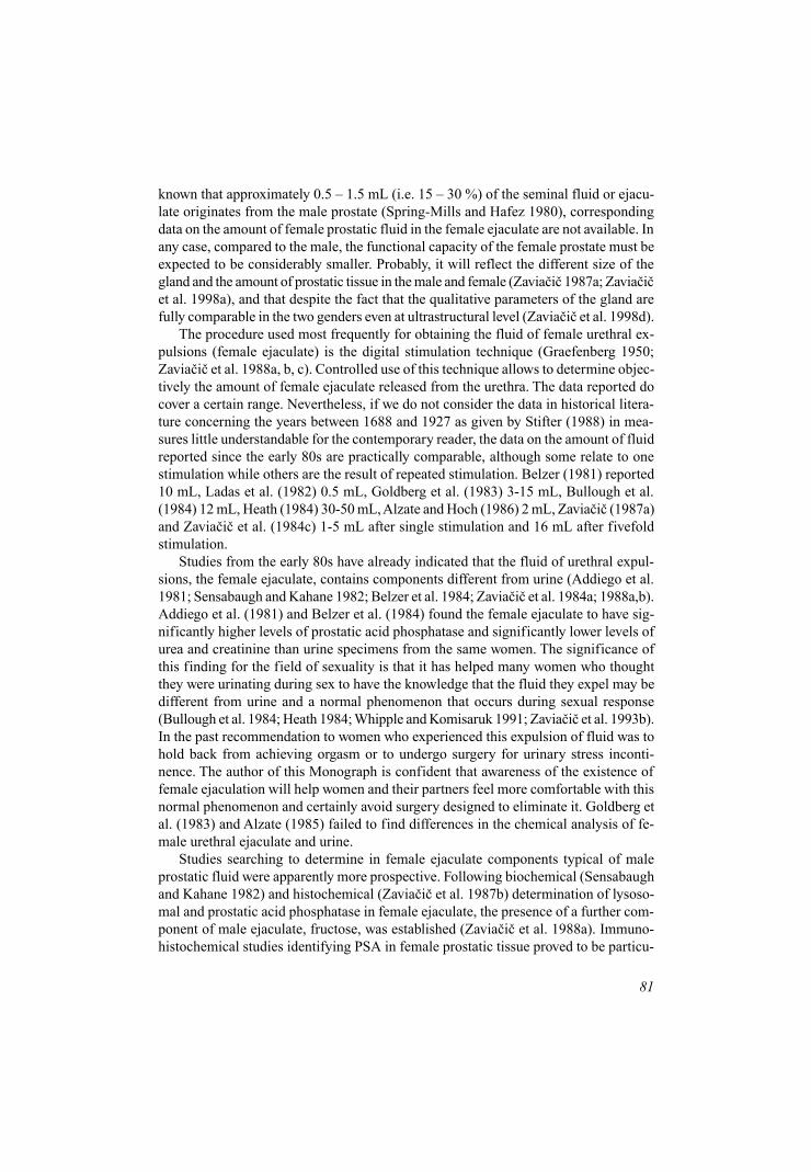
81
known that approximately 0.5 � 1.5 mL (i.e. 15 � 30 %) of the seminal fluid or ejacu-late originates from the male prostate (Spring-Mills and Hafez 1980), correspondingdata on the amount of female prostatic fluid in the female ejaculate are not available. Inany case, compared to the male, the functional capacity of the female prostate must beexpected to be considerably smaller. Probably, it will reflect the different size of thegland and the amount of prostatic tissue in the male and female (Zaviaèiè 1987a; Zaviaèièet al. 1998a), and that despite the fact that the qualitative parameters of the gland arefully comparable in the two genders even at ultrastructural level (Zaviaèiè et al. 1998d).
The procedure used most frequently for obtaining the fluid of female urethral ex-pulsions (female ejaculate) is the digital stimulation technique (Graefenberg 1950;Zaviaèiè et al. 1988a, b, c). Controlled use of this technique allows to determine objec-tively the amount of female ejaculate released from the urethra. The data reported docover a certain range. Nevertheless, if we do not consider the data in historical litera-ture concerning the years between 1688 and 1927 as given by Stifter (1988) in mea-sures little understandable for the contemporary reader, the data on the amount of fluidreported since the early 80s are practically comparable, although some relate to onestimulation while others are the result of repeated stimulation. Belzer (1981) reported10 mL, Ladas et al. (1982) 0.5 mL, Goldberg et al. (1983) 3-15 mL, Bullough et al.(1984) 12 mL, Heath (1984) 30-50 mL, Alzate and Hoch (1986) 2 mL, Zaviaèiè (1987a)and Zaviaèiè et al. (1984c) 1-5 mL after single stimulation and 16 mL after fivefoldstimulation.
Studies from the early 80s have already indicated that the fluid of urethral expul-sions, the female ejaculate, contains components different from urine (Addiego et al.1981; Sensabaugh and Kahane 1982; Belzer et al. 1984; Zaviaèiè et al. 1984a; 1988a,b).Addiego et al. (1981) and Belzer et al. (1984) found the female ejaculate to have sig-nificantly higher levels of prostatic acid phosphatase and significantly lower levels ofurea and creatinine than urine specimens from the same women. The significance ofthis finding for the field of sexuality is that it has helped many women who thoughtthey were urinating during sex to have the knowledge that the fluid they expel may bedifferent from urine and a normal phenomenon that occurs during sexual response(Bullough et al. 1984; Heath 1984; Whipple and Komisaruk 1991; Zaviaèiè et al. 1993b).In the past recommendation to women who experienced this expulsion of fluid was tohold back from achieving orgasm or to undergo surgery for urinary stress inconti-nence. The author of this Monograph is confident that awareness of the existence offemale ejaculation will help women and their partners feel more comfortable with thisnormal phenomenon and certainly avoid surgery designed to eliminate it. Goldberg etal. (1983) and Alzate (1985) failed to find differences in the chemical analysis of fe-male urethral ejaculate and urine.
Studies searching to determine in female ejaculate components typical of maleprostatic fluid were apparently more prospective. Following biochemical (Sensabaughand Kahane 1982) and histochemical (Zaviaèiè et al. 1987b) determination of lysoso-mal and prostatic acid phosphatase in female ejaculate, the presence of a further com-ponent of male ejaculate, fructose, was established (Zaviaèiè et al. 1988a). Immuno-histochemical studies identifying PSA in female prostatic tissue proved to be particu-

82
larly important (Mallon 1983; Tepper et al. 1984; Pollen and Dreilinger 1984; Zaviaèièet al. 1994). PSA in male seminal plasma was long referred to as gamma seminoprotein(Hara et al. 1971) and/or as p30 (Sensabaugh 1978).
The presence of fructose in the female urethral ejaculate (Zaviaèiè et al. 1988a),once confirmed also by other authors, may be considered also with respect to the im-portant role of this sugar in the physiology of human reproduction. Macroenzymatichistochemical findings of acid phosphatase on parts of lingerie which were in constantcontact with the female genitals are indicative of the existence of continual secretionof the female prostate (Zaviaèiè et al. 1988d), as has long been known concerning themale prostate (Mann 1974). Contrary to the male, the ejaculatory mechanism of thefemale prostate (urethral expulsions) is not directly related to reproduction, it is ratheran attractive phenomenon of the woman�s sexuality. On the other hand, continual se-cretion of the female prostate, with the secretion containing fructose and other pros-tatic components flowing under gravitation in small amounts from the urethra to thevagina, may be of importance for reproduction. It is this amount of fructose producedby the female that may provide a permanent basal low level of this sugar in the vaginalenvironment. After coital ejaculation of the male, the level of this sugar is substantiallyincreased in the vagina by the amount of fructose derived from the seminal vesicles ofthe male. Since fructose represents the main energy source for the motility of sperms,the woman herself may with her own fructose affect to some extent this parameterimportant for the reproduction process. It appears to be biologically conceivable thatdue to the particular significance of this parameter for natural selection of the highest-quality sperm for successful fertilization of the egg, the process would be warrantedalso by the female. This apparently calls for greater attention to be paid to the analysisof continual (spontaneous) secretion of the female prostate, which may importantlycome into play in reproduction biology.
In our studies, female ejaculate samples were always obtained, after complete mic-turition of the proband, by the female herself or with the help of her partner. Perry andWhipple (1981) first termed the special erotically sensitive area on the anterior vaginalwall which was to be stimulated to induce urethral expulsions the G-spot, after theobstetrician and gynecologist Ernst Graefenberg (1881-1957). Graefenberg (1950) de-scribed this zone of erogenous feeling and the expulsion of fluid that occurs simulta-neously with orgasm. Perry and Whipple (1981) identified the hypogastric plexus andthe pelvic nerve as the sensory pathways in sexual response of women when there isvaginal stimulation. This is different from clitoral stimulation, when the major sensorypathway is via the pudendal nerve, as identified by Masters and Johnson (1966).
Despite the fact that the G-spot can easily be identified in gynecologico-sexualinvestigation on digital vaginal examination, at autopsy digital identification of the G-spot on the anterior wall of the vagina is rather problematic. Contrary to in vivo condi-tions, when identification is facilitated by the tumescence of the spot on friction, thischaracteristic phenomenon is conceivably absent on post mortem tissue. Clinical ex-perience of the examiner with identification of the spot in female patients has proveduseful in finding the relevant area also post mortem. We generally succeeded in theprocedure and were usually able to mark the supposed area of the G-spot on the ante-

83
rior wall of the vagina in order to perform detailed histological examination after itsfixation. Although the knowledgeable doctor can clinically identify the G-spot withoutproblems, its anatomical substance remains unclear. We detected collagen and elasticfibers in the given area, often in plexiform arrangement with muscle fibers, rich inblood vessels and nerve fibers. On comparing the equipment of different parts of theanterior vaginal wall, the area of the G-spot and the distal areas of the vagina exhibitedgreater richness in these elements than did the proximal parts. In some cases thesedifferences were pronounced, in others they were but minimal. Nevertheless, the ante-rior wall of the vagina appears to be better equipped with the given structures than theposterior wall of the vagina. We failed in our expectation to detect in the G-spot spe-cific morphological structures that would be absent in other parts of the anterior vagi-nal wall. We are rather inclined to agree with the opinion that the G-spot is part of theHalban fascia, considered by some authors to be analogous with the corpus spongiosumpenis (Minh 1981), playing an important role in sexual physiology as a source of deepsensations during intercourse (Minh et al. 1979). The role of the Halban fascia in sup-porting the bladder is well known and must be carefully preserved in all operations bythe vaginal route (Minh et al. 1979). Our finding of the richer presence of nerve fibersin samples from the anterior wall compared to the posterior wall of the vagina is inkeeping with results of the study on the innervation of the human vaginal mucosausing as neuronal marker the protein gene product 9.5 (Hilliges et al. 1995). Lenck andVanneuville (1992) voiced the opinion that the G-spot was identical with the urethralsphincter. They reached this conclusion by correlating clinical (sexological),ultrasonographic, and anatomical findings. Generalizing, they situated vaginal andclitoral sensitivity in the same entity of the urethro-clitorido-vulval complex (Lenckand Vanneuville 1992). O�Connell et al. (1998) presented new aspects on human fe-male urethral and genital anatomy. They showed that the female urethra, distal vaginalwall and erectile tissue are packed into the perineum caudally (superficially) to thepubic arch. The perineal urethra is embedded in the anterior vaginal wall and is sur-rounded by erectile tissue in all directions, except posteriorly where it relates to thevaginal wall.
These studies have broadened our attention beyond the so far studied areas of thefemale urethra, female prostate, and the anterior wall of the vagina, which form onefunctional-morphological complex (Heath 1984; Zaviaèiè 1987a), so as to involve alsotheir connections with further, especially erectile structures of the female genitals (cli-toris), focusing thus on an even greater complex.

84

85
VIII.Prostate-specific Antigen: Immuno-histochemical Localization in FemaleProstate Tissue, Serological Param-eters, and Implications of this Pros-tate Marker in the Woman and Man
At present, prostate-specific antigen (PSA) is generally accepted as the most usefulbiological marker of male prostate carcinoma, and has become the mainstay of theprostate carcinoma screening and staging, in monitoring response to therapy, and inpredicting the outcome. In spite of this optimism, the role of PSA as a sole criterion inscreening and staging of male prostate cancer remains controversial. First, the princi-pal concerns are inherently related to the fact that PSA, although specific of the pros-tatic tissue, is not tumor specific; prostatic cancer is heterogenous, with subpopula-tions of cells that vary as to their synthesis and possible secretion of PSA. A secondconcern is whether screening will detect what is generally thought to be latent or clini-cally indolent cancers, present in approximately 30% of males aged over 50, and whethermorbidity and mortality associated with the treatment of these tumors will exceedthose of the disease itself (Ablin 1996; 1997). Just to present the complete picture, afurther concern is the cost-related clinical utility of screening (Coley et al. 1997).
From the viewpoint of staging, wide variations in PSA levels exist in many patientswith either localized or advanced metastatic disease (Brawer and Lange 1989; Scardino1989). Therefore, except within broad ranges, PSA serum levels but poorly predicttumor stage on an individual basis.
In terms of the use of PSA for the screening for early detection of prostatic carcinomain males, a major limitation has been false positive elevations produced by benign pros-tate hyperplasia. In addition, prostatitis (Robles et al. 1988) and prostatic ischemia and/or infarction (Glenski et al. 1992) can be associated with elevated levels of the antigen.

86
Stamey et al. (1989) demonstrated that the gland weight in the male is the mostimportant non-cancer variable in increases in PSA levels. It has been proposed on thisbasis (Littrup 1991a, b; Lee et al. 1992) that what has been said in the introductionholds true despite some indications in recent reviews on PSA (Ablin 1996; 1997) whichprompt us to be more cautious. In his recent papers, Ablin states that �PSA is far frombeing the perfect �tumour� marker as show elevations of PSA in other irregularities ofthe prostate, notably in benign prostatic hyperplasia, and increasing frequency andnumber of non-prostatic tissues, including those in women expressing PSA have im-plications for future immunoassays for PSA and strategies for immunotherapy usingPSA-based monoclonal antibodies or vaccines, as well as for the molecular basis forits anomalous expressions and physiological functions.� (Ablin 1996; 1997).
Measurement of gland volumes by transrectal ultrasound (Miyashita et al. 1984)may help detect PSA level increases due to cancer or benign prostate hyperplasia.Investigations of the distribution of PSA levels in a large population of healthy mensuggested the importance of age and prostatic volume. Dalkin et al. (1993) and Oesterlinget al. (1993a,b) independently found that serum PSA levels correlate directly withpatient�s age, in association with the increase in prostatic volume with advancing age.
Immunohistochemical evidence of prostate-specific antigen (PSA) as presently themost reliable prostatic marker, plays a crucial role in the identification of normal andpathologically altered tissue in the male (Nadji et al. 1981; Epstein and Eggleston1984; Purnell et al. 1984; Stein et al. 1984; Svanholm 1986; Jöbsis 1990; Keillor andAterman 1993). Immunohistochemical staining for PSA is weaker in poorly differen-tiated primary tumours and in metastases, and may even be absent in the latter. Giventhat tissue PSA has be shown to be age-dependent, to correlate with androgen levels(Goldfarb et al. 1986), and that male prostate cancer is heterogeneous in its reponsivenessto antiandrogen therapy, one must be cautious when interpreting the relevance of weakerstaining in PSA. Recent studies by Pretlow et al. (1991) in which the heterogeneity inPSA expression observed immunohistochemically in prostate cancer and benign pros-tate hyperplasia tissues were confirmed by quantification of PSA in extracts preparedfrom the same tissues as examined immunohistochemically. In accordance with earliercomparative studies of the PSA diversity in normal, benign and malignant prostatictissue (Ablin 1972) PSA was found to be expressed at a significantly lower level inmalignant than in benign prostatic hyperplasia. Hamdy et al. (1992) reported veryinteresting results which supported cell-type specificity of antibodies to PSA. Usingflow cytofluorography, these authors detected circulating PSA-positive cells in theperipheral blood of male patients with metastatic prostate carcinoma. The presence ofPSA-positive cells showed a higher degree of sensitivity and specificity in predicting apositive bone scan indicative of the presence of bone metastases than did serum PSAlevels (Hamdy et al. 1992).
Immunohistochemical demonstration of PSA also appears to be of equal impor-tance with respect to the identification of normal (Pollen and Dreilinger 1984; Tepperet al. 1984; Wernert et al. 1992; Zaviaèiè et al. 1994) and pathologically altered (can-cerous) prostatic tissue in the female (Svanholm et al. 1987; Wernert 1991; Zaviaèiè etal. 1993a; Sloboda et al. 1998 and references therein).

87
Immunohistochemical determination of PSA by peroxidase-antiperoxidase (PAP)technique and by the method of biotin-streptavidin-alkaline phosphatase (BSAP) or bythe biotin-streptavidin-peroxidase (BSP) method yielded concordant expression of theexamined marker in the highly specialized apically superficial layer of male and fe-male secretory (luminal) cells of the prostatic glands and membranes of secretory andbasal cells and membranes of columnar cells of the female prostate ducts lining (Figs.VIII/1,2,3,4,5)(Zaviaèiè et al. 1994; Zaviaèiè 1997), which is in agreement with PSAexpression reported by other authors. We may agree with Pappoti et al. (1989) whoclaim that, in a majority of tissues, immunohistochemical determination of PSA ismore efficient when rabbit polyclonal antibodies rather than mouse monoclonal anti-bodies are used. Similar differences could also be observed in immunohistochemicaldetermination of PSA in female prostatic tissue. Only some glandular cells reactedwith monoclonal antibodies, the reaction being less massive than that with polyclonalantibodies (Zaviaèiè and Ablin 1998c). A comparison of hetero (polyclonal) antiseraand monoclonal antibody raised against malignant prostatic tissue showed that mono-clonal antibody was less sensitive (Schevchuk et al. 1983). In a routine immunohis-tochemical demonstration of PSA and PSAP monoclonal antibodies appear less usefulthan polyclonal antibodies in the diagnosis of prostatic carcinoma (Gallee et al. 1986).
Fig.VIII/1 Distinct expression of PSA in the apical parts of cells of two intraductal prostaticglands and in apical parts and on the surface of cells of the pseudostratified colum-nar epithelium lining the lumen of a female prostate duct, and in membranes ofthese cells. Female, 41-year-old, peroxidase-antiperoxidase (PAP) technique, x 170

88
In addition to secretory cells, PSA is expressed in membranes and on the surface ofcells of the pseudostratified columnar epithelium of the lining of the ducts of the fe-male (Figs. VIII/1,2) and male prostate. This unique layer of cells of the urogenitalsystem is distinctly formed and abundantly equipped with glycosamine glycans, gly-coproteins and enzymic proteins. Besides numerous enzymes (Zaviaèiè 1984a,b;1985a,b) and the presence of urinary protein 1 as shown very recently (Zaviaèiè et al.1997 a), distinct immunological properties could be observed between male and fe-male prostate given by the presence of an antigen specific for the prostate � PSA. Thefindings of immunohistochemical studies focusing on PSA (Pollen and Dreilinger 1984;Tepper et al. 1984; Wernert et al. 1992; Zaviaèiè et al. 1993a; 1994; Zaviaèiè 1997;Sloboda et al. 1998 and references therein) have broadened our understanding andenhanced the biological value of PSA since this prostate marker has been found rel-evant not only in studies of the male but equally so of the female prostate.
For males, the reference range is about 1 � 2 ng/mL and values above 3 � 4 ng/mLare indicative of prostate cancer, benign prostatic hyperplasia or prostatitis (Borchertet al. 1997). A short update on the female prostate (Zaviaèiè and Ablin 1998a, b, c) hasstrongly suggested the possibility that, similarly as in the male, the prostate (Skene�sgland) is a regular source of PSA in the serum and/or urine of the female as well. The
Fig. VIII/2 Marked PSA expression in the apical part and on the surface of cells lining thelumen of a small female prostate duct, and in membranes of columnar cells liningthe duct. A small female prostatic gland expressing PSA. Female, 27-year-old, bi-otin-streptavidin-alkaline phosphatase (BSAP) method, x 170

89
contribution of the female prostate to PSA formation and its quantities in urine hasbeen confirmed by the results reported by Cabello (1997) who could demonstrate sig-nificant differences in urinary PSA levels in the same women between samples drawnprior to and after orgasm. The increased post-orgasmic urinary PSA levels can be ex-plained by the emptying of the female prostate due to orgasm-induced contractions ofmuscles surrounding the female urethra when the prostate contents with prostatic com-ponents including PSA are pressed into the urethra, thus getting to the urine.
A healthy female with a normal prostate typically shows a broad range of serumPSA values from effectively unappreciable amounts to the highest reported ones of 0.9ng/mL (Borchert et al. 1997). The latter value is very close to the normal referencerange for the male. Serological and/or urinary parameters of PSA in females should nolonger be considered surprising since they are very well in line with the nonvestigialconcept of the female prostate (Zaviaèiè et al. 1985b; Zaviaèiè 1987a; Zaviaèiè andWhipple 1993; Zaviaèiè and Ablin 1998a, b, c) whose structure and function have beenwell established and its pathology broadly studied. These parameters are very similarto those of the male prostate (Zaviaèiè 1987a; Zaviaèiè and Ablin 1998a, b). Increasedserum PSA values may be the result of pathological changes of the female prostateitself, e.g. in carcinoma of the prostate in the female when levels as high as 5.9 ng/mLcan be encountered (Dodson et al. 1994); or the increase may present a summation of
Fig.VIII/3 Moderate to strong PSA positivity in groups of normal female prostate (Skene�s)glands. Female, 19-year-old, biotin-streptavidin-peroxidase (BSP) technique, x 180

90
values derived from PSA production of the normal and pathological female prostate(Skene�s gland)(Zaviaèiè and Ablin 1998a, b, c) and PSA of possibly non-prostatictissue origin, e.g. in female breast cancer (Diamandis et al. 1994; Yu et al. 1994; 1996;Borchert et al. 1997). In female patients with breast fibroadenoma, serum PSA valuesare very high compared with female breast cancer, and may amount up to 55.1 ng/mL(Borchert et al. 1997). According to our results with immunohistochemical determina-tion (Zaviaèiè et al. 1998b, c), normal female breast epithelial tissue produces no PSA.
One should be aware of the fact that every serum and/or urine PSA determinationin the female inevitably comprises PSA produced by the female prostate (Skene�s gland).
Novel, more sensitive serological methods introduced into clinical practice, e.g.IMMULITE-r immunochemiluminescent third-generation assay (Diagnostic ProductsCorp., Los Angeles, CA) or further modern tests may enhance our knowledge of fur-ther parameters which characterize female PSA.
Reading the rapidly increasing numbers of papers concerned with the PSA markerin a broad range of relationships, it must however be inferred that only a minor part ofauthors know the names of the actual discoverers of this prostate marker. The majorityof publications supply the reader with incorrect and misleading information (Diamandisand Yu 1995), including the most recent Cover Legend which appeared in Cancer Re-search (Weinhouse 1998).
Fig.VIII/4 PSA expression in the apical part, on the surface and on membranes of columnarcells lining the lumen of a narrow duct of the female prostate. Female, 19-year-old,biotin-streptavidin-peroxidase (BSP) technique, x 180

91
It has to be noted that the tissue specific antigens of the human prostate, now knownas PSA, were first identified by Richard J.Ablin and his coworkers (W.A. Soanes,P.Bronson, E.Witebsky); they have a justified priority claim as they published theirresults in two renown journals (Ablin et al. J.Immunol. 1970a; Ablin et al. J.Reprod.Fertil. 1970b), nine years before the paper by Wang et al. (1979). Nevertheless,the discovery of PSA is often attributed to the latter authors (M.C.Wang, L.A.Valenzuela,G.P.Murphy, T.M.Chu). Such misleading information (�Dr.Chu�s work in collabora-tion with Ming C.Wang, eventually resulted in the discovery and purification of PSAfrom the prostate�, Wang et al. Invest. Urol. 1979) also appeared in the April issue ofJournal of Cancer Research (Weinhouse 1998). Even before our reference, the firstidentification of PSA by the team of Richard J.Ablin has been pointed out by the lateDr.William H.Cooner from Emory University School of Medicine (Cooner 1993) andby the group of Dr.Joseph E.Oesterling [(J.M.Monda, M.J.Bary, J.E.Oesterling) (Mondaet al. 1994)]. Although some of the authors incorrectly mentioned as PSA discoverersdid publish some important partial findings concerning PSA, their work was essen-tially based on the groundbreaking studies of the Ablin team.
It seems safe to state that the situation concerning the priority claim with respect toPSA discovery may be best expressed as follows: �PSA was initially identified byR.J.Ablin, W.A.Soanes, P.Bronson, and E.Witebsky in 1970 (Ablin et al. 1970a, b),
Fig.VIII/5 Marked PSA expression in the columnar epithelium lining a female prostate duct.Female, 19-year-old, biotin-streptavidin-peroxidase (BSP) technique, x 360

92
and was purified and characterized by M.C.Wang, L.A.Valenzuela, G.P.Murphy andT.M.Chu in 1979 (Wang et al. 1979).� Wang et al. (1979) characterized PSA as amonomer with molecular mass of 33-34 kDa. Seven years later, PSA was shown to bea 240-amino-acid single-chain glycoprotein (Watt et al. 1986). The primary gene struc-ture and the amino acid sequence of PSA exhibit a high degree of similarity with thoseof other serine proteases of the kallikrein gene family. PSA participates in the liquefac-tion of the semen following ejaculation (Lilja 1985; Watt et al. 1986; Lilja et al. 1987).PSA has been shown to exist in multiple forms and, being a protease, it forms com-plexes with protease inhibitors in the serum (Stenman et al. 1991), mainly with alpha-1-antichymotrypsin (ACP). Stenman et al. (1991) and Lilja et al. (1991) believe thatthe major proportion of serum PSA is ACT-bound to form PSA-ACT complex.
In his Letter to the Editor which appeared as Commentary in the magazine Oncol-ogy, Ablin (1998) presented a good overview of the individual steps in the investiga-tion of prostatic antigens which mouthed into the discovery of PSA. In his Letter, heresponded to incorrect information presented by some authors of articles appearing inthe same journal of Oncology [(Pannek and Partin 1997) according to Ablin 1998)]concerning the historical chronology of the PSA identification. The statements of theseauthors have been even more surprising in the view of the fact that Dr.Partin previously(1994) published a paper on the initial identification of PSA by Ablin�s group (Partinand Oesterling 1994).

93
IX.Prostatic and Non-prostatic Sourcesof Prostate-specific Antigen
In the male, the main PSA producer are epithelial cells of the prostate (Partin andOesterling 1994; Zaviaèiè et al. 1994; Ablin 1996; 1997 and references therein; Zaviaèiè1997). Nevertheless, this prostate marker was immunohistochemically detected notonly in the normal male prostate and male periurethral glands but also in the normalfemale prostate (Skene�s gland) and in diseased male and female prostate tissues in-cluding cancer in both sexes (Frazer et al. 1992; Zaviaèiè et al. 1993a; Elgamal et al.1994; Zaviaèiè et al. 1994; Ablin 1997; Breul et al. 1997; Zaviaèiè 1997; Sloboda et al.1998 and references therein; Zaviaèiè and Ablin 1998a, b, c and references therein).
Some authors consider the breast to be the principal PSA producer in the female(Diamandis and Yu 1995; Yu et al. 1996; Diamandis 1998). In their opinion the mainPSA producer in the female is not only diseased female breast tissue and fluids, espe-cially benign breast disease and cancer, but also normal female breast tissue (Yu et al.1996; Mannello et al. 1996; Diamandis 1998). So far, preliminary immunohistochemi-cal data concerning PSA examination in the normal female and male breast tissue havebeen published in one short communication (Zaviaèiè et al. 1998b). The quantitativeimmunochemical data reported by the Diamandis� team (Yu et al. 1996; Diamandis1998) as being typical of normal breast tissue do not represent values for normal fe-male breast tissue since their �normal� group of female patients consisted of 38 womenwith hyperplastic breasts, and tissue samples were obtained during breast reductionsurgery. This type of female breast tissue is far from being normal breast tissue � eventhe authors of the original paper admit so (Yu et al. 1996).
In the male, there are no doubts on the primary role of the normal or pathologicallyaltered prostate with respect to PSA production. More recent reports providing evi-dence for a similar nonvestigial role of the prostate in the female (Zaviaèiè et al. 1985b;Zaviaèiè 1987a; Zaviaèiè and Whipple 1993; Zaviaèiè and Ablin 1998a, b, c) are eithernot sufficiently known or have been ignored (Wernert et al. 1992; Borchert et al. 1997;Elgamal et al. 1997; Ignatoff 1997; Diamandis 1998).

94
In our recent works (Zaviaèiè et al. 1998b, c), we could verify, using immunohis-tochemical methods on necroptic and bioptic material, whether and to what extentnormal and pathologically altered male and female tissues participate in the PSA pro-duction. Considering the fact that in healthy women, serum PSA levels range fromnon-detectable to high, reaching up to 0.9 ng/mL, a value approaching that of thenormal reference range for males (1-2 ng/mL, according to Borchert et al. 1997), itappears justified to address the issue of which organ or tissue in the healthy and dis-eased female is actually responsible for the production of this prostate marker.
Using immunohistochemical methods PSA was examined in tissues taken duringmany biopsies and autopsies (a total of 115 cases). The bioptic material of the femaleand male breasts involved 39 cases of benign breast disease (35 women and 4 men).The majority of the cases concerned fibroadenomas and cysts of the female breast; inthe male patients, they concerned gynecomastia. In 18 women, breast cancer was diag-nosed. Our material did not include any case of male breast cancer.
Normal breast tissue was obtained at autopsy of 52 deceased (23 women and 29men). Histological examination of normal female breast tissue revealed only age-dependent involutionary postmenopausal atrophic changes, typical of female sub-jects aged over 50 years, and resting female breast tissue in younger female individu-als. The normal male breast only contains ducts and no lactiferous glands (Zaviaèièet al. 1998c).
The female urethra with prostate tissue was obtained from 37 cases at autopsy, andtreated as published elsewhere (Zaviaèiè 1984a,b). The whole male prostate was ob-tained at autopsy from 33 individuals, and PSA was immunohistochemically deter-mined in the peripheral part of the prostate.
Tissue samples from the axilla and the perineum containing apocrine sweat glandswere taken from 42 necropsies (16 females and 26 males) for PSA determination(Zaviaèiè et al. 1998c).
PSA was determined in six gastrobioptic samples obtained from patients with dif-ferent diseases of the stomach, including two cases of signet-ring cell carcinoma of thestomach (two males aged 69 and 53 years).
PSA expression was demonstrated in samples from 52 normal and 57 pathologi-cally changed female and male breasts (Zaviaèiè et al. 1998c). The 57 breast tissuesexamined included 39 benign (22 cases of dysplasia of the female breast, 4 cases ofgynecomastia in the male, 6 fibroadenomas, and 7 cases of inflammatory breast dis-eases) and 18 malignant diseases (13 cases of ductal and 5 cases of lobular carcinoma).
PSA expression could not be observed in any epithelial structures of the normalmale and female breast (Zaviaèiè et al. 1998b, c).
PSA positivity could be identified in 17 out of 39 cases of benign breast diseases inboth sexes (44%).
Out of the 18 cases of female breast cancer examined, 7 cases of ductal carcinomawere PSA positive (38%), and 6 cases were negative. All lobular carcinomas examinedwere PSA negative.
The data on PSA expression in normal necroptic prostatic tissue in both sexes (37female and 33 male prostates) were grouped according to the age of the individuals;

95
also, results of immunohistochemical examination of PSA in 42 samples of normalaxillary and perineal tissue have been published (Zaviaèiè et al. 1998c).
All the samples of the normal male and female prostate tissue showed moderate tohigh PSA expression in the glands and ducts (Zaviaèiè et al. 1998c).
Extraprostatic tissues of the axilla and the perineum showed weak PSA positivityin the apocrine sweat glands.
Moreover, PSA could be immunohistochemically detected, sometimes at the detec-tion limit, in membranes of adipocytes of the female and males breast fat tissue (Fig.IX/1), in small vessel endothelium and some blood cells, perineal glands, and in basalmembranes of various genito-urinary and gastrointestinal organs (Zaviaèiè et al. 1998c).Examination of gastrobiopsies yielded characteristic findings of PSA expression inparietal cells of the fundal gastric mucosa. Unlike the signet-ring cell variant of ductalfemale breast carcinoma whose cells displayed intense PSA positivity (Fig. IX/2), themucus accumulating cells of the signet-ring cell carcinoma of the stomach exhibitedPAS (periodic acid-Schiff) positivity but no PSA expression.
In keeping with the findings of Papotti et al. (1989), the best results of PSA deter-mination were obtained with rabbit polyclonal antibodies. The polyclonal antibodyreacts with several epitopes, whereas the monocolonal antibody only recognizes a singleone (Ferenèík 1993). PSA expression was intensified by monoclonal antibody antigenrevitalisation in microwave oven; the procedure however had a negative effect on thestructure of the preparation and interfered with the expression assay.
The results of our immunohistochemical studies provided a clear-cut answer as to therole of the normal female and male breast tissue in the production of PSA. In contradic-tion to the conclusion arrived at by the Diamandis� group (Yu et al. 1996; Howarth et al.1997; Diamandis 1998) and in agreement with our preliminary study (Zaviaèiè et al.1998b) we could confirm that normal female breast epithelium does not produce PSA(Zaviaèiè et al. 1998c). The discrepancy between the results of the quantitative immu-nochemical determination obtained by the above authors and our findings may be ex-plained by the fact that despite the higher sensitivity of quantitative immunochemicalmethods compared to immunohistochemical methods, the former are used with hetero-geneous homogenates which, in the given case, contain adipose tissue and small vesselsin addition to female breast glands; adipose tissue and small vessels have been reportedto show positivity on immunohistochemical examination. This may account for the dis-crepancies between our results obtained using the immunohistochemical method andthose of the Diamandis� group, which made them erroneously conclude that normalfemale breast tissue produces PSA. Moreover, as already mentioned, their results wereobtained in material from hyperplastic female breasts rather than from normal tissue.Our immunohistochemical results apply to normal resting and postmenopausal atrophicfemale breast tissue representing age-dependent physiological variations of the normalfemale breast (Ahmed 1992). Our immunohistochemical findings lead to the conclusionthat the normal female breast cannot be a major PSA producer and can, by no means,explain the PSA levels observed in normal healthy women.
No PSA expression could be detected in the samples of the 29 normal male breastsexamined by us (Zaviaèiè et al. 1998c). Unlike female breast tissue, that of males only
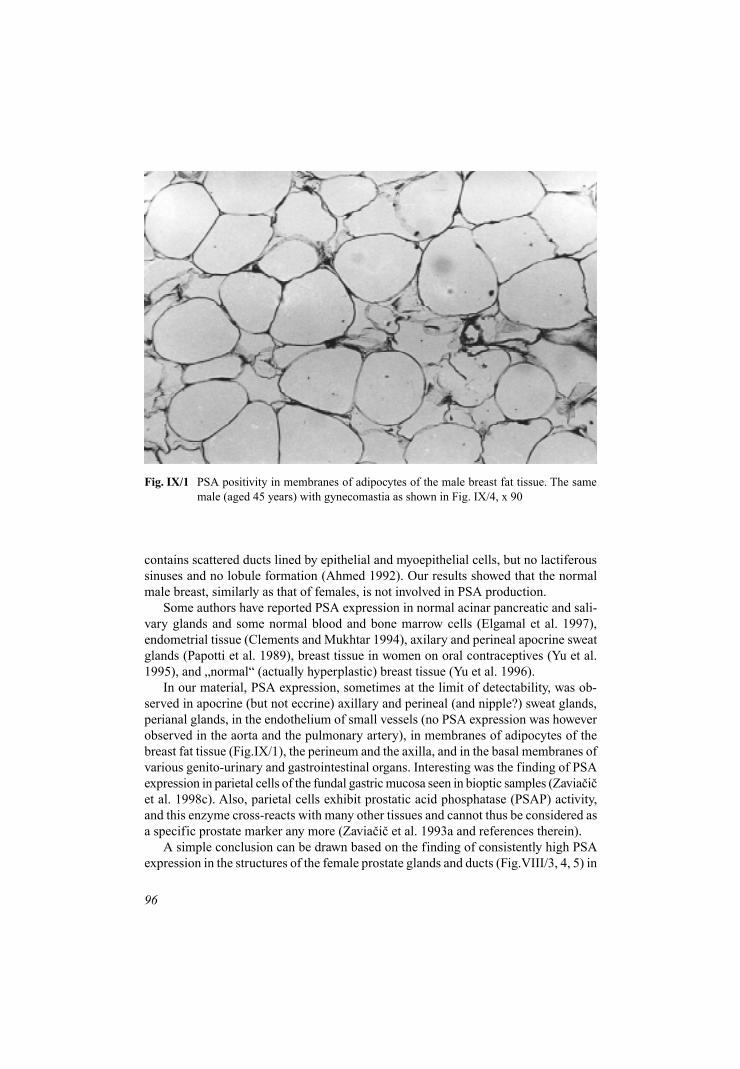
96
contains scattered ducts lined by epithelial and myoepithelial cells, but no lactiferoussinuses and no lobule formation (Ahmed 1992). Our results showed that the normalmale breast, similarly as that of females, is not involved in PSA production.
Some authors have reported PSA expression in normal acinar pancreatic and sali-vary glands and some normal blood and bone marrow cells (Elgamal et al. 1997),endometrial tissue (Clements and Mukhtar 1994), axilary and perineal apocrine sweatglands (Papotti et al. 1989), breast tissue in women on oral contraceptives (Yu et al.1995), and �normal� (actually hyperplastic) breast tissue (Yu et al. 1996).
In our material, PSA expression, sometimes at the limit of detectability, was ob-served in apocrine (but not eccrine) axillary and perineal (and nipple?) sweat glands,perianal glands, in the endothelium of small vessels (no PSA expression was howeverobserved in the aorta and the pulmonary artery), in membranes of adipocytes of thebreast fat tissue (Fig.IX/1), the perineum and the axilla, and in the basal membranes ofvarious genito-urinary and gastrointestinal organs. Interesting was the finding of PSAexpression in parietal cells of the fundal gastric mucosa seen in bioptic samples (Zaviaèièet al. 1998c). Also, parietal cells exhibit prostatic acid phosphatase (PSAP) activity,and this enzyme cross-reacts with many other tissues and cannot thus be considered asa specific prostate marker any more (Zaviaèiè et al. 1993a and references therein).
A simple conclusion can be drawn based on the finding of consistently high PSAexpression in the structures of the female prostate glands and ducts (Fig.VIII/3, 4, 5) in
Fig. IX/1 PSA positivity in membranes of adipocytes of the male breast fat tissue. The samemale (aged 45 years) with gynecomastia as shown in Fig. IX/4, x 90

97
37 autoptic cases that, similarly as in the male, the prostate (Skene�s glands) is a regu-lar and major PSA producer also in the female. The variability of the female prostate(Zaviaèiè et al. 1983; 1985b; Zaviaèiè 1987a; Zaviaèiè and Whipple 1993; Zaviaèièand Ablin 1998c; Zaviaèiè et al. 1998a) as well as its variable size and weight com-pared to the male prostate (the adult female prostate weighs slightly more than 5g,representing about 20 � 25% of the adult male prostate � Zaviaèiè et al. 1998a) mayaccount for the broad variation in the serum and urine PSA values in healthy femalesranging from undetectable levels to as high as 0.9 ng/mL (Borchert et al. 1997). Thisexplanation of the female prostate role as the PSA producer is well in keeping with thecurrent non-vestigial concept of the prostate in women as a genito-urinary organ witha structure, function and pathology comparable to those typical of the male prostate(Zaviaèiè et al. 1983; 1985b; Zaviaèiè 1987a; Zaviaèiè and Whipple 1993; Zaviaèiè etal. 1994; Zaviaèiè and Ablin 1998a,b,c and references therein). This role in the female,with the female prostate PSA production being comparable to that by the male pros-tate, offers the simplest explanation for the origin of PSA in females. PSA immunore-activity has however been identified in a number of extraprostatic tissues and secre-tions in males and females. Immunoreactivity has been reported in a variety of neo-plastic and benign tumorous tissues including apocrine sweat gland carcinomas andapocrine breast carcinomas (Papotti et al. 1989; Allanen et al. 1996), salivary glandand salivary duct carcinoma (Van Krieken 1993), colon cancer (Wilbur et al. 1987),
Fig. IX/2 Moderate PSA positivity in cells of the signet-ring cell variant of breast ductal carci-noma. Female, 78-year-old, x 90

98
pancreatic acinar cell carcinoma (Kuopio et al. 1995), biliary tract carcinomas (Wilburet al. 1987), urinary bladder carcinomas (Grignon et al. 1991), mature cystic teratomasof the ovary (McLachlin and Srigley 1992), male and female breast tumours (Diamandiset al. 1994; Yu et al. 1994; 1996; Monne et al. 1994); no PSA was however found inmale breast cancer (Dawson 1992).
Our materials which consisted of pathologically changed female and male tissueseffectively without exception derived from biopsies and autopsies taken routinely, andthus represented a random collection of samples. Unlike normal tissue of the femaleand male breast whose epithelial structures did not express PSA (Zaviaèiè et al. 1998b,c), apocrine metaplasia in benign female breast disease (Fig.IX/3) and male gyneco-mastia (Fig.IX/4) exhibited distinct PSA positivity (Zaviaèiè et al. 1998c).
Our immunohistochemical findings in tissues from benign female and male breastdiseases are in conformity with the immunochemical data reported by the Diamandis�group who referred the highest PSA production for these tissues compared to carci-noma and �normal� breast tissue (Yu et al. 1996; Diamandis 1998). PSA was expressedin 17 out of 39 cases of benign breast disease (35 women and 4 men) in our series(Zaviaèiè et al. 1998c).
Also, PSA expression was observed in tissue from female breast cancer, especially indifferentiated types of ductal carcinoma. In undifferentiated types, PSA expression wasdeclining (Fig.IX/5) or missing at all, and no PSA expression could be detected in tissue
Fig.IX/3 Moderate PSA positivity in the biopsy of female breast apocrine metaplasia in be-nign breast disease. Female, 51-year-old, x 360

99
from lobular carcinoma of the breast. The signet-ring cell variant of ductal female breastcarcinoma displayed high PSA expression (Fig.IX/2). This may be a specific feature ofthe cancerous breast tissue since, in two cases of signet-ring cell carcinoma with differ-ent localization (gastric mucosa), the cancer cells were characterized by periodic acid-Schiff (PAS) positivity of mucus accumulating cells; yet, in contracst to cells of this typeof cancer in the female breast, they did not express PSA (Zaviaèiè et al. 1998c). Only 7out of the total of 18 cases of cancer of the female breast in our study displayed PSApositivity, and 11 were negative (Table 1 in Zaviaèiè et al. 1998c). Based on our material,we were unable to address conclusively the issue concerning the relationship betweenPSA production and the equipment with steroid hormone receptors of normal and patho-logically altered female breast and other tissues (Yu et al. 1994; Bodey et al. 1997; Borchertet al. 1997; Diamandis 1998). Further studies are needed to confirm or reject the hypoth-esis on PSA being exclusively produced in structures equipped with steroid hormonereceptors, as promoted by Diamandis (1998). In contrast to our immunohistochemicalstudy where a majority of cancerous female breasts produced no PSA (PSA negativitywas found in 11 out of a total of 18 cases of breast cancer), in a paper published in theMarch 1999 issue of British Journal of Cancer, Majumdar and Diamandis (1999) re-ported about 70% of breast cancer tissues to produce PSA. They claim that mutationsassociated with the androgen response elements have been shown previously to result inan 80% decrease in PSA gene expression (Majumdar and Diamandis 1999).
Fig. IX/4 Moderate PSA positivity in an autopsy case of male breast apocrine metaplasia ingynecomastia. Male, 45-year-old, x 90

100
Ablin (1996; 1997) and Zaviaèiè and Ablin (1998a, b, c) consider the expression ofPSA in male and female non-prostatic cancerous tissues, e.g. in the female breast, asapparently anomalous, being a manifestation of the neoplastic transformation process.
Immunohistochemically detectable expression of PSA in the normal (Pollen andDreilinger 1984; Tepper et al. 1984; Zaviaèiè et al. 1994) and cancerous female pros-tate tissue (Zaviaèiè et al. 1993a; Sloboda et al. 1998 and references therein) allows toidentify female prostate tissue and, to the pathologist, it provides the means to differen-tiate between true female prostatic carcinoma and other adenocarcinomas of the fe-male urethra which frequently occur in relation to urethral diverticulum, the latter be-ing PSA negative (Tanabe et al. 1982; Oliva and Young 1996; Amin and Young 1997)and are not assumed to be of prostatic origin in the female, rather, their mesonephricorigin has been suggested (Dodson et al. 1995; Sloboda et al. 1998).
Convincing clinical evidence is accumulating on PSA production by the femaleprostate. Sexological studies have demonstrated differences in pre- and post-orgasmicurine PSA levels (due to the release into the urine of the urethral contents includingPSA by contractions of muscles surrounding the urethra during the orgasm)(Cabello1997); moreover, the study of Dodson et al. (1994) who observed preoperatively anincreased serum PSA level in a female patient with carcinoma of the prostate (femaleurethra) which promptly decreased after excision of the tumour has been frequentlyreferred to. Neither us nor the authors of the report (Dodson et al. 1994) have ever
Fig.IX/5 Very weak PSA positivity in the biopsy tissue from inflitrating ductal carcinoma ofthe breast (G-2). Female, 32-year-old, x 90

101
questioned the prostatic origin of this marker in the female or considered extraprostaticproduction of PSA by female breast tissue.
The rather weak and variable expression of PSA by some normal extraprostatictissues (membranes of adipocytes, apocrine sweat glands, parietal cells, endothelium,etc.) does certainly not qualify these structures as major PSA sources in the healthymale or female.
The principal source of PSA is the prostate in both sexes. There are however dis-tinct quantitative differences in the production of PSA between the male and the fe-male prostate, probably reflecting the lesser efficiency of the female organ.
Pathologically altered male and female breast, especially in benign breast diseases(Figs.IX/3,4) produces higher PSA levels compared to both normal and cancerousbreast tissue. In the latter as well as in other pathologically altered tissues of male andfemale patients, known to produce PSA, the total amounts of this prostate marker (PSA)represent the summation of regular prostatic and anomalous diseased extraprostatictissue production (Zaviaèiè et al. 1998c).

102

103
X.Pathology of the Female Prostatewith Special Reference to Carci-noma, Benign Prostate Hyperplasiaand Other �Prostatic� Diseasesin Female Patients
Permanent awareness and position of a given organ in the hierarchy of organs in thehuman body is established not only by understanding its physiology and pathophysiol-ogy but also and particularly by insight into its pathology. As our knowledge on thepathology of the organ broadens, we attempt to understand the essential mechanismsinvolved in the development of the diseases affecting the organ and search for wayshow to treat them. These general statements do fully apply to the problem area of thefemale prostate. In the majority of women, the prostate is considered to be an organdevoid of problems. This simplistic, traditionally repeated approach is absolutely un-substantiated. Objective data from pathologists, urologists and gynecological urolo-gists documenting the actual incidence of female prostatic diseases on the basis ofadequately large material are not available as yet. The pathology of the female prostatehas so far been dealt with mostly in case reports. The situation would change rapidly ifautopsy of the female urethra along with the prostatic tissue became a routine part ofthe post mortem examination, and that also in forensic cases, and if gynecologists,urologists, and gynecological urologists (urogynecologists) approached the diseasesas actual diseases of the female prostate rather than of the urethra. Many clinicallyvaluable data could be obtained from patients by routine use of the already existingquestionnaires, adequately adapted for establishing relevant findings, which are com-monly used to record and document symptoms and complaints of patients in urologicaland andrological departments. The obtained data might not only promote early diagno-sis of cancer, benign hyperplasia and prostatitis in female patients but they could also

104
become an important basis for studying the actual incidence of diseases of the prostatein the female population. The present chapter, virtually based on case reports and analysisof small series, focuses on carcinoma, benign hyperplasia and inflammation of thefemale prostate, diseases which represent also the most important diagnostic and thera-peutic entities of the diseased prostate in the male.
Carcinoma of the female prostate
Tumors of Skene�s paraurethral glands, the female prostate, are very rare when com-pared with their incidence in the male prostate. Primary urethral carcinomas are of theless frequently occurring carcinomas of the female reproductive system, with a re-ported incidence of 0.16 to 0.70 or 0.95 % (Egloff 1972). They are mostly squamous orurothelial in nature (Levine 1980; Kamat et al.1981; Groben et al. 1985). Approxi-
Fig. X/1 Cases of adenocarcinoma of the female prostate (female urethra) with results of PSAand PSAP immunohistochemical examination. Data from the Table in Sloboda et al.(1998) � modified.
Immunohistochemical examination of PSA andPSAP in cases of adenocarcinoma of the femaleurethra-female prostate (Skene�s gland)
Author N cases PSA PSAP
Svanholm et al. 1987 1 + +Spencer et al. 1990 1 + +Mostofi and Sesterhenn 3 + +cited by Wernert 1991Kamoto et al. 1993 1 � �Zaviaèiè et al. 1993 1 + +Dodson et al. 1994 1 + +Dodson et al. 1995 12 � 0Ebisuno et al. 1995 1 + +Miyai and Ebisuno 1995 1 + +Zaviaèiè et al. 1995* 1 + +Oliva and Young 1996 5 + +
13 � �
+ Positive expression of PSA or PSAP in cancerous cells� Lack of expression of prostate markers in cancerous cells0 No data on PSAP* This case report appeared in the form of abstract (Congress of
ESP, 1995) and as a full publication (Sloboda, Zaviaèiè et al.1998)
** Serum level of PSA was increased preoperatively and promptlydecreased after surgical excision of the tumor

105
mately 10 % of malignant tumors of the female urethra were reported to be adenocar-cinomas derived from paraurethral ducts and glands (Huffman 1951; Schnitzer 1964;De Haan 1965; Adler 1968; Roberts and Melicow 1977).
The morphological similarity between adenocarcinoma of the female urethra andmale prostatic carcinoma has been known for many decades (Huffman 1951). Femaleurethral adenocarcinoma is generally assumed to arise from the female prostate � Skene�sparaurethral ducts and glands (Huffman 1951; Zaviaèiè et al. 1993a). In cases of ure-thral adenocarcinomas, immunohistochemical examinations of prostatic markers wererare till the 90s. In their paper published in Pathology Research and Practice, Slobodaet al. (1998) however, presented a table with 15 cases of adenocarcinoma of the femaleprostate (female urethra), with immunohistochemical staining for PSA and PSAP, sup-porting the diagnosis of female prostate carcinoma (Fig. X/1). This paper has providedthe first published results of electron microscopic examination of formalin-fixed can-cerous tissue showing the ultrastructure of female prostate carcinoma, which was com-parable to that of male prostate carcinoma.
The results of Svanholm et al. (1987), Spencer et al. (1990), Mostofi and Sesterhenn(cited by Wernert 1991), Zaviaèiè et al. (1993a), Dodson et al. (1994), Ebisuno et al.(1995), Miyai and Ebisuno (1995), Oliva and Young (1996), Sloboda et al. (1998), andSesterhenn et al. (1998) concerning PSA and PSAP positivity provided crucial contri-bution to the diagnosis of female prostate carcinoma. Immunohistochemical expres-
Fig. X/2 Solid parts of renal cell carcinoma of the left kidney. On the margin renal tissue withglomerulus. HE, x 175.

106
Fig. X/4 Metastasis of renal cell carcinoma in the liver. Tumorous cells show negative cytoplas-mic staining for PSA. Immunoperoxidase (PAP) technique, x 175.
Fig. X/3 Tumor cells of adenocarcinoma of the female prostate (tumor of the �anterior wall of thevagina�) with distinct expression of PSA. Immunoperoxidase (PAP) technique, x 175.

107
sion of the prostatic marker PSAP in cancerous tissue of the female prostate is notidentical with the diagnostic and interpretative value of PSA. Unlike the high PSAspecificity (although this marker is far from being a perfect �tumor� marker � Ablin1996; 1997), PSAP cross-reacts with many other tissues (Zaviaèiè et al. 1993a), suchas pancreatic islet cells, gastric parietal cells, liver cells, renal tubular epithelium, epi-thelial cells of seminal vesicles and breast carcinomas, as well as some carcinoids andadenocarcinomas of the urinary bladder. This makes PSA immunohistochemical stain-ing the approach of choice both for normal and cancerous prostatic tissue of the maleand for normal and cancerous prostatic tissue of the female (Zaviaèiè et al. 1993a).
For the pathologist, positive immunohistochemical staining of tumor cells will ap-parently become an important indicator for distinguishing between �true prostatic�adenocarcinomas and other adenocarcinomas of the female urethra (Sloboda et al.1998). The latter are PSA negative and occur frequently in relation to the urethraldiverticulum (Oliva and Young 1996; Amin and Young 1997). They are not assumed tobe of prostatic origin (originating from the female prostate), rather their mesonephricorigin has been suggested, and one would agree with Dodson et al. (1995) that femaleurethral adenocarcinomas may have more than one tissue origin. The evident prostaticorigin of adenocarcinomas of the female urethra characterized by PSA expression intumor cells is supported by the finding of preoperatively increased serum PSA level ina female patient with tumor of the urethra which promptly decreased after removal ofthe tumor (Dodson et al. 1994). This highly important observation has also provided
Fig. X/5 Well differentiated glands of adenocarcinoma of the female prostate. HE, x 180.

108
clinical evidence corroborating the non-vestigial concept of the female prostate as agenitourinary organ which evidently exerts its function, and that differently under nor-mal and under pathological conditions. The logical message of the given serologicalfindings is that serum PSA examination should become a regular constituent of preop-erative and postoperative monitoring of female patients with prostatic (urethral) tu-mors.
One of the cases listed in the table given by Sloboda et al. (1998) is our patientwhose case report was published in Virchows Arch A Pathol Anat six years ago (Zaviaèièet al. 1993a). We reported on adenocarcinoma of the female prostate (paraurethralSkene�s glands) in a 70-year-old woman. On gynecological examination a flat tumor(1.2 cm in diameter) was observed on the anterior wall of the vagina in the suburethralregion, approximately 1.5 cm behind the vaginal orifice. A biopsy specimen takenfrom the tumor was sent for histological examination. In the meantime the patient diedof cardiorespiratory failure. Autopsy revealed renal cell carcinoma of the left kidney(Fig.X/2) with metastases in the lung, liver, lymph nodes and ribs. On the anterior wallof the vagina, at the site where the gynecologist had diagnosed the tumor, the mucousmembrane was protruding over an area of about 1 cm. Vertical section showed tumortissue to be below the vaginal mucosa and to spread diffusely into the depth of theurethrovaginal septum, towards the urethra. Both biopsy and autopsy of the tumor in
Fig. X/6 Carcinoma of the female prostate with cribriform multiple gland-like and solid cancer-ous tissue composed of clear vacuolated cells. Thin fibrous tissue strips between neo-plastic cells. HE, x 180.

109
Fig. X/8 Positive staining for PSA in clear neoplastic cells forming cribriform multiple gland-likelumina of adenocarcinoma. Avidin-biotin-alkaline phosphatase complex, x 180.
Fig. X/7 Undifferentiated parts of female prostate carcinoma infiltrating perineural space. HE,x 180.

110
the anterior vaginal wall showed adenocarcinomatous structure with cribriform mul-tiple gland-like lumina. Immunohistochemical examination of PSA and PSAP showedstrong cytoplasmic positivity in cells of the tumor in the vagina (Fig.X/3), while stain-ing of prostatic markers (PSA, PSAP) was negative in the renal tumor and in the me-tastases (Fig.X/4). Classification of the tumor as non-metastasizing adenocarcinomaof the female prostate (Skene�s paraurethral glands and ducts) was made on the basisof the microscopic appearance of the tumor of the anterior vaginal wall and the resultsof immunohistochemical examination of prostatic markers, particularly PSA..
The case reported by Svanholm et al. (1987) was also a non-metastasizing carci-noma of Skene�s glands (female prostate) with coincidence of ovarial carcinoma.Spencer�s case (Spencer et al. 1990), on the other hand, concerned urethral adenocar-cinoma (female prostate carcinoma) with PSA and PSAP positivity not only in cancer-ous cells but also in regional metastases.
Our second case of carcinoma of the female prostate diagnosed in our Departmentwas recently published in Pathology Research and Practice (Sloboda et al. 1998). Wereported on a 46-year-old woman, mother of two children, with stress incontinenceover the last year and complaint of urethritis. She was examined at the Urology Depart-ment of the Hospital in Karlskrona, Sweden. A paraurethral tumor was found and ure-thral diverticulum was suspected. We received a surgical specimen of the tumor fromKarlskrona and examined it histologically and electron microscopically in our Depart-ment in Bratislava, Slovakia. Histologically, a part of the tumor tissue consisted of welland less well differentiated structures of adenocarcinoma (Fig. X/5). The predominantpart of the tumor was composed of clear vacuolated cells with large nuclei, formingglands, glandular cribriform and solid cancerous tissue (Fig.X/6). Anaplastic areas ofthe carcinoma infiltrating perineural spaces were also seen (Fig.X/7). Examination ofprostatic markers demonstrated PSA (as well as PSAP) positivity in all cancerous cellsin well-differentiated and cribriform clear cell glandular structures of the adenocarci-noma (Fig.X/8) and in anaplastic cells of solid tumor areas. Electron microscopic ex-amination allowed for the first time to demonstrate the subtle cellular architecture oftumor cells of female prostate carcinoma. In the female, similarly as in the male, pros-tate carcinoma probably originates from secretory (luminal) cells of the prostate glands.In this case, diagnosis of adenocarcinoma of the female prostate (Skene�s paraurethralducts and glands) was based on the histological and ultrastructural pattern resemblingmale prostate carcinoma, on the results of immunohistochemical positivity of prostaticmarkers � mainly PSA � in cancerous cells, and on the results of aspiration cytology ofan inguinal lymph node.
Benign hyperplasia of the female prostate
According to Folsom and O�Brien (1943), John Caulk (1921) was the first to suggestresection for bladder neck obstruction in the female. Folsom and O�Brien (1943) re-ported that from the early 30s publications appeared repeatedly supporting the exist-

111
ence of benign prostate hyperplasia in the female (Nesbit 1933; Fite 1934; Van Houtum1939 � cited in Folsom and O�Brien 1943). Folsom and O�Brien published a paper inJAMA entitled �The Female Obstructing Prostate�, stating that the disorder referred toas female prostatism is a clinical entity occurring in women much more frequentlythan recognized and treated properly. Physicians often fail to ask the proper questionsto get an accurate and revealing history. Folsom and O�Brien believed that many womenhad some type of prostatism. In the male, the term prostatism has been replaced by theterm lower urinary tract symptoms (LUTS), the disorder being practically the same.Clinically, besides bladder irritation, these female patients present with some degreeof difficulty in voiding up to complete retention. Three out of 15 cases with femaleobstructing prostate develop complete retention. The majority of reported cases showedfibromuscular hypertrophy with fibrous hyperplasia, although true adenomatous en-largement of the female prostate also occurred in these female patients resulting incomplete obstruction and complete retention of urine in the bladder (Folsom and O�Brien1943). In some cases, autopsy showed bilateral hydronephrosis and hydroureter withtrabeculated bladder wall, bladder stones and a prostatic bar. In these cases, the com-plications found in women were typical of benign male prostate hyperplasia. With theexception of the contribution of Nesbit (1943), the discussion to the lecture of Folsomand O�Brien, published in the same issue of JAMA immediately following their paper,was carried on strictly along the line that �... the female does not have a prostate�(Ockerblad 1943; Kretschmer 1943; O�Conor 1943). Interestingly enough, many clini-cians remembered the unequivocally negative interpretation of the existence of thefemale prostate as presented in the discussion on the obstructing prostate rather thanthe very text of the paper by Folsom and O�Brien (1943) with the brilliantly presentednon-vestigial concept. It is to be emphasized that the published discussion to theirpaper provided further generations of physicians sufficient reasons to accept the vesti-gial concept of the female prostate under Skene�s name as an organ insignificant andunnecessary in the woman�s life. It can not be excluded that similarly as carcinoma ofthe female prostate used to be considered adenocarcinoma of the urethra, so also casesof benign hyperplasia of the female prostate may have been reported under a differentname. Sharma et al. (1988) published two case reports of bladder outflow obstructiondue to fibromatous (fibroma) and glandular hyperplasia of female prostatic tissue. Thepaper appeared under the descriptive title: �Benign Periurethral Mass Lesions: TheFemale Prostate�.
Recent findings have provided convincing support to the belief of Folsom andO�Brien and of several clinicians who accepted the existence of the female prostateand considered its pathology to be comparable with that of the male prostate, includingthe existence of benign female prostate hyperplasia and/or prostatism (LUTS). Furthercontinual research has to be carried out on large pathological material to verify thenotion of Folsom and O�Brien (1943) that it is obstruction of the enlarged femaleprostate in the posterior part of the female urethra that causes retention of urine in thebladder. The relatively low occurrence rate of the so-called posterior type of the femaleprostate found only in about 10 % of women (Zaviaèiè 1987a; Zaviaèiè et al. 1998a),as concluded on the basis of detailed examination of the urethra in 150 autopsies, fails

112
to be in agreement with earlier published data reviewed by Deter et al. (1946). I quotefrom his paper: �The closer the bladder sections were taken, the greater the number of(prostatic) glands�. In keeping with the findings of Huffman (1948), our data concern-ing distribution of female prostatic tissue predominantly in the distal (anterior) urethrarather than in the posterior urethra, might conceivably account for the relatively lowerincidence of benign prostatic hyperplasia in women compared to the high incidence ofthis disorder in men. In spite of our, probably largest series of autopsies of the femaleurethra (150 cases), we failed to see in our material (Zaviaèiè 1987a; Zaviaèiè andWhipple 1993; Zaviaèiè and Ablin 1998a; Zaviaèiè et al.1998a) such characteristicpictures of fibromuscular and adenomatous hyperplasia of the female prostate as re-ported and distinctly documented in pictures by Folsom and O�Brien (1943). On theother hand, in their abstract Sesterhenn et al. (1998) reported not only on the normalprostate and on prostatic carcinoma but also on prostatic hyperplasia in female pa-tients. It appears to be necessary to detect and study the combined incidence of pros-tatitis and benign female prostate hyperplasia, a combination frequently seen in themale, and to attempt to define clinically and pathologically which actual diseases andconditions are involved in LUTS in the female.
Female prostatitis (Female Urethral Syndrome?)
The urethral syndrome in women is described as lower urinary tract symptoms with aplethora of subjective complaints of retropubic pressure, dyspareunia, urinary frequencyand dysuria, yet associated with a lack of objective findings (Bashi 1988; Gittes andNakamura 1996). The symptoms resemble lower urinary tract infection but there areno abnormal midstream urinalysis findings, no positive bacterial culture (Obrink 1979),and no obvious findings on pelvic examination (Gallagher et al. 1965; Messing 1986).The urethral syndrome remains a diagnosis per exclusion. More serious causes of uri-nary frequency, dysuria and suprapubic pressure must be considered and excluded.Only a combination of careful history, physical examination, inspection of urine, urineculture, urine cytology and cystourethroscopy with biopsy can establish the diagnosis(Bodner 1988). According to Jackson (1976), the pathological changes of the tissuesclinically manifested as urethral syndrome, produce signs which are radiologicallyrecognizable. Excretory urography findings correlate closely with cystographic find-ings. Transabdominal ultrasonographic measurement of mucose thickness around thebladder neck and internal examination for tenderness at the anterior wall of the vaginaare thought to be useful approaches to diagnosis and follow-up of urethral syndrome(Sagaya et al. 1992).
Many factors have been suggested as causative of this syndrome, including non-specific infection, urethral obstruction and spasm, senile atrophy, psychosomatic andtraumatic factors, muscular abnormality of the pelvic floor, spasm of the external sphinc-ter (Cardon et al. 1979; Kaplan et al. 1980; Stamm et al. 1981; Schmidt and Tanagho1981; Schmidt 1985; Bashi 1988; Bernstein et al. 1992). Splatt and Weedon (1981)

113
found in biopsies of tissue obtained on Richardson urethroplasty and in autopsy casesof females with urethral syndrome a significant increase in fibrous tissue in the ure-throvaginal septum, accompanied by loss of smooth muscle fibers and local clumpingof elastic tissue.
The variety of treatments practiced over the years reflect the many �causative�factors of this syndrome. They cover the full spectrum from aggressive surgical exci-sion of periurethral tissue (Richardson 1969), internal urethral cutting procedure andforceful overdilatation of the urethra (Rieser 1968; Evans 1971; Bergman et al. 1989)to the use of anxiolytic and tricyclic antidepressant drugs. Although all treatmentswere judged ineffective and it has been suggested that treatment be supportive and�harmless�, aggressive management continues to be widely practiced even to date.
This survey clearly shows that despite intensive research, the problem of femaleurethral syndrome remains still an open question, both as to diagnosis and treatment. Itis however surprising that in searching the structure whose damage is the cause of thebroad clinical symptomatology in women with urethral syndrome, the female prostatewas not considered for a long period of time, and that despite the fact that similarsymptoms in man would suggest prostatitis and would lead to microscopic examina-tion of prostatic secretion for the presence of inflammatory cells (Drach et al. 1978). Itwas only in 1996 that Gittes and Nakamura called attention to the fact that the femaleurethral syndrome concerns prostatitis, namely inflammation of the female prostatewith a broad range of clinical manifestations accompanying the syndrome.
Experts in urinary infection attempted to detect the offending organism, Chlamy-dia species, and found antibiotic therapy empirically effective (Stamm et al. 1980;1981). Yet before Gittes and Nakamura (1996) none of the investigators related thediagnosis of the infection anatomically to the tissue of the female prostate (Skene�sparaurethral ducts and glands). On examining the female prostate, Gittes and Nakamura(1996) recommend careful pinpoint palpation of the soft tissue of the anterior wall ofthe vagina on each side of the urethra (similarly to the examination mode described byHuffman already in 1951). Upward fingertip pressure against the pubic bone laterallyto the urethra elicits localized tenderness when the paraurethral glands are infected.The examination has certain parallels with digital rectal examination that localizeslimited prostate gland tenderness in the male (Gittes and Nakamura 1996). Providedthat Chlamydia species have invaded the female prostatic gland, antibiotic treatmentwith tetracycline or erythromycin usually eliminates the symptoms (Stamm et al. 1980).Russi (1996) recommended administration of doxycycline. Objective vaginal re-ex-amination during follow-up usually confirms the decrease or absence of tenderness inthe limited area of the anterior vaginal wall. Nevertheless, relapses of female prostati-tis (recently termed also �recurrent cystitis� � Moore 1975) may occur months or yearslater. Relapses reflect the easy spread of infection through the rich, mutually commu-nicating system of prostatic glands, including intraepithelial glands (Huffman 1948;Zaviaèiè et al.1998a) and ducts of the female prostate, with the urethra and anteriorwall of the vagina. These anatomical structures are parts of the urethro-prostatic-vagi-nal entity (Zaviaèiè et al. 1998a). The existence of this common genitourinary entitymay explain the easy spread of infection and relapses of prostatitis (Skeneitis) in the

114
female. The anatomical continuity between the female urethra and the urinary bladderpredisposes the spread of inflammatory diseases of the female urethra, and the femaleprostate embedded in it, to the urinary bladder with manifestations of cystitis, or pos-sibly also a simultaneous inflammatory response of all these structures (urethro/prostato/cystitis). The rich mutual communication in the cavities of the system of the femaleurethra and prostate provides exceptionally good conditions for long-term survival ofuropathogens in these areas, possibly accounting not only for relapses of female pros-tatitis but also potential recurrent episodes of urinary tract infections (UTI) appearingas acute cystitis. Acute cystitis is the commonest form of UTI, whose incidence inyoung women in the USA was reported to be 0.5-0.7 episodes per person per year(Hooton et al. 1996). About 25 % of women with acute cystitis develop frequentlyrecurring infections (Foxman 1990). Stapleton (1999) was concerned with factors pre-disposing to recurrent UTI. Unfortunately, in her contribution there is no informationon the female prostate and/or female prostatitis and UTI. We believe (Ablin and Zaviaèiè1999 � Commentary, Lancet) that the contributory role of the female prostate to recur-rent UTI is much more important than the majority of factors related to recurrent UTIas given by Stapleton (1999).
The paper by Gittes and Nakamura (1996) has considerable implications for clini-cal practice. The authors state that female prostatitis has actually to be treated as aninflammatory disease of the prostate (here the commonly given designation as Skene�sparaurethral glands and ducts appears to be useless and counterproductive) and not asa disease of the urethra, as done so far. It is certainly time to stop performing forcefuloverdilatations of the urethra (Bergman et al. 1989) and administering anxiolytic andtricyclic antidepressant drugs to patients suffering from female prostatitis, an ailmentaffecting a great number of the female population in the USA as stated by Karram(1990), who reported more than 5 million visits of female patients with this problem inthe office per year. Inflammation of the female prostate can be successfully treated bytaking advantage of all the available possibilities of conservative therapy which haveproved effective in treating inflammation of the prostate in the male.
Once physicians become aware of the fact that prostatitis as well as other diseasesof the female prostate, such as benign hyperplasia and carcinoma, have to be treated asfemale prostatic and not other diseases, they will be equipped with therapeutic strate-gies and patterns verified and successfully used for years in the treatment of the samediseases in the male. New perspectives open up also for large pharmaceutical compa-nies which have so far produced drugs for the treatment of the male prostate and havethus saturated the needs of one half of the population. They should broaden their pro-duction and sale of �prostatic� drugs to satisfy also the needs of the female populationsuffering from diseases of the prostate.
It appears to be precocious to pronounce any conclusions on the incidence andprevalence of these female urological diseases of the prostate, especially on femalebenign prostate hyperplasia, prostatitis, or the combination of both diseases in onefemale patient. As mentioned in the introduction to this chapter, urologists and gyne-cological urologists, on taking the patient�s history, have to ask targeted questionsconcerning the frequency of micturition and the degree of difficulty in voiding the

115
urinary bladder. This could be done also by questionnaires as are used in male patientswith benign prostate hyperplasia, after appropriate modification of the questions pre-sented. A further key requirement should be invariable rather than exceptional exami-nation of the urethra at autopsy. Pathologists could thus help establish the as yet miss-ing objective data on the actual incidence of diseases of the prostate in the femalepopulation.
Nevertheless, there are certainly some diseases of the urogenital tract that are andwill remain characteristic of one gender while absent or rare in the other one.
Thus e.g. a situation which would result in paraphimosis or phimosis in the male ispractically unknown in the female, presumably due to the substantially larger spacebetween the prepuce and the glans clitoridis than between the prepuce and the glanspenis in the male. An exception might present an enormous hypertrophy of the glansclitoridis with a short hood (prepuce), which at autoerotic stimulation of the clitoris orat petting activities may occasionally after an initially pleasant feeling induce one oftraction and even discomfort in the region of the vulva (unpublished observation).Phimosis and particularly paraphimosis are relatively frequently occurring disordersin boys and men causing discomfort. Yet especially paraphimosis may present a seri-ous complication requiring urgent surgical intervention. The difference between thegenders is reflected also by the fact that the prepuce may become one of the causes ofdyspareunia in the male but in the female it has not been mentioned among the causesof dyspareunia (Wabrek and Wabrek 1975). Priapism, on the other hand, does not af-fect males only. Painful priapism of the clitoris was described which resulted fromretrograde neoplastic embolization of the corpora cavernosa of the clitoris by cervicalcarcinoma, and required surgical intervention (Lozano and Casta±eda 1981).
Concomitant infection of the ducts and glands of the female prostate invariablyaccompanies acute gonorrheal urethritis and may persist as an undiscovered source ofreinfection during the later stages of the disease (Huffman 1951). Female prostatesecretion may contribute also to the development of vaginal flow. If the female prostateexhibits the immunopermissiveness observed in the male prostate (Ablin 1991), it mayalso serve as a site for viral latency and origin of infection in women with the humanimmunodeficiency virus (Zaviaèiè and Ablin 1998a). The ducts of the female prostate(paraurethral ducts) may at times harbor Trichomonas vaginalis. In recurrent or intrac-table vaginal trichomoniasis the female prostate ducts play a role in persistentreinfestations of the vagina (Huffman 1951).
In other diseases of the female prostate (Skene�s glands), its involvement in acuteand chronic inflammation of the urethra has been repeatedly emphasized. Huffman(1951) states: �The paraurethral ducts and glands (female prostate gland) are of clini-cal significance not only because they are foci for acute and chronic inflammation ofthe female urethra but also because of the diverse roles which they play in other lesionsof the urethra and the urethrovaginal tissues. Suburethral abscesses develop in ob-structed and infected retention cysts of the paraurethral (prostatic) ducts. These cystsand abscesses are important factors in the etiology of acquired urethral diverticula.Urethrovaginal fistulas may follow rupture of paraurethral (prostatic) duct abscessesinto the vagina and urethra or severance of the channel of a prostatic (paraurethral)

116
duct during surgical procedures on the anterior vaginal wall�. Skene�s duct cysts, andmainly those larger than 2 cm, induce clinical symptoms including obstruction, dys-pareunia and dysuria. Superposed infection may cause significant discomfort as well.Treatment usually consists of antibiotic management (Raz 1996).
After John W.Huffman (1951) apparently nobody did focus in so concentrated aform on the clinical significance of the female prostate. Relevant information is scat-tered in urological and gynecological journals, mostly as case reports. Although ourknowledge has substantially broadened, particularly on carcinoma of the female pros-tate, concerning its other diseases Huffman�s findings are to be accepted even morethan 40 years after their publication. One of the reasons may lie in the circumstancethat interest in the female prostate and its diseases has appeared only in the last 10 � 15years, following a relatively long period when this female organ was more or lessignored. We can only hope that a gynecologist or urologist, a new �Huffman�, will turnup who would study this female organ with the same enthusiasm as did his/her prede-cessor half a century ago. The publication by Gittes and Nakamura (1996) initiated anunexpectedly great response (Russi 1996; Peterson 1996; Zaviaèiè and Ablin 1998b),demonstrating that interest in the female prostate is much more intensive than it usedto be, and that also among clinicians. However, despite the caution voiced by somedoctors who long ago considered urethral dilatation alone unsatisfactory for treatment(Richardson and Stonington 1969), the attitude of some urologists to therapeutic dila-tation of the urethra is still positive. In Comments to Gittes and Nakamura (1996)Peterson (1996) wrote: �A caliber of 38F in adults is necessary, and mild, temporaryurethral bleeding is a favorable prognostic sign. Benefit is reflected in the patients whovoluntarily return after varying intervals for retreatment when symptoms recur. Thesecond dilatation is seldom as uncomfortable as the first� (Peterson 1996).
The present boom of interest in the female prostate especially in clinicians mightbe promoted also by their insight that if we persist in doubting the existence of thefemale prostate as a functional organ we will hardly be able to explain without a greatdeal of speculation the origin of PSA in serum and urine of the healthy female (up to0.9 ng/mL in female serum, Borchert et al. 1997). Contrary to the data of Diamandis etal.(1994), Yu et al. (1996), and Diamandis (1998), our immunohistochemical examina-tions of normal epithelial tissue of the female breast, as well of the male breast, failedto demonstrate PSA expression (Zaviaèiè et al. 1998b,c). Like in the male so in thefemale, the main producer of PSA is the prostate, whose secretory capacity determinesthe concentration of this prostatic marker in serum and urine (Zaviaèiè and Ablin 1998a,b, c). Even in cases of abnormal PSA production by hyperplastic or cancerous tissue ofthe female breast (Borchert et al. 1997) and of some other organs (Zaviaèiè et al. 1998c),the production of the female prostate does participate in the total serum and urinaryPSA concentration, reflecting the sum of prostatic and extraprostatic PSA production(Zaviaèiè and Ablin 1998a; Zaviaèiè et al. 1998c).
Concluding this chapter on the pathology of the female prostate it can be stated thatseveral findings on the diseased female prostate published in the past appear to emergein new associations and relationships unknown before. This may be accounted for es-pecially by advances in laboratory medicine marked by the introduction of highly sen-

117
sitive methods in immunochemical and immunohistochemical demonstration of pros-tatic markers. These approaches have not only broadened the diagnostic possibilitiesand made them more accurate in detecting diseases of the female prostate but have alsoprovided means to monitor their course and assess the efficacy of the therapy applied.We are only in the initial phase, and further advances can be achieved by efficientsupport of clinical research into the problem of the female prostate. In this Monograph,we direct attention to some topical issues of the clinical problem area of the femaleprostate.

118

119
XI.Reasons for Rejecting the TermSkene�s Paraurethral Ducts and Glandsin Designating the Prostate in theHuman Female (Terminological Note)
The term female prostate was commonly used in the past and occurred frequently alsoin the first decades of our century, as seen in titles of papers given in the References ofthis Monograph. Later on, the term appeared mostly only in sexologically orientedliterature (Sevely and Bennett 1978; Addiego et al. 1981; Belzer 1981; Perry andWhipple 1981; Ladas et al. 1982; Bohlen 1982; Mallon 1983; Belzer et al. 1984;Bullough et al. 1984; Heath 1984; Zaviaèiè 1987a), although there were also somesexologists who voiced arguments against its use (Alzate and Hoch 1986; 1988; Alzate1990). The term female prostate has been commonly used in veterinary literature indescribing prostatic tissue of females of different animal species (Shehata 1972; 1975;1980; Ichihara 1976; Gross and Didio 1987). Papers published in sexologic orsexologically orientated journals have considerably contributed to the use of the termfemale prostate at a time when other medical journals stubbornly kept to the officialterminology and refused to use any other designation than the official one of �Skene�sparaurethral ducts and glands�.
In the 80s, and particularly in the later years of the decade, we witnessed the ap-pearance of papers on the female prostate or female prostatic tissue using these termsalso in journals of other than sexologic orientation (Longo 1982; Pollen and Dreilinger1984; Tepper et al. 1984; Zaviaèiè 1984a, b; 1985a, b; 1986a, b; 1987b; Zaviaèiè et al.1983; 1985b, c, 1987a, b; 1988d; 1989). Looking back at the last 15 years, I feel obligedto mention particularly the name of Professor R. Wegmann, Editor-in-Chief, who asearly as in 1984, without any argument, accepted for publication in his journal Mo-lecular and Cellular Biology our pioneer work on the enzymatic equipment of thefemale prostate (Zaviaèiè 1984a, b) with the term �adult human female prostate� ap-
120
pearing in the title of the paper. In the majority of other journals (with the exception ofthe Histochemical Journal, Editor Prof. P. J. Stoward), even later on, we had to explainand defend the term again and again in our correspondence with the editors.
At present, the use of the term Skene�s paraurethral ducts and glands for the fe-male prostate fails to reflect the results published from the beginning of the 80s to datein the field of research into the problem of the female prostate. In his conclusions onthe female prostate, the American gynecologist Alexander Skene was incorretly con-cerned only with two paraurethral ducts, which however defies the actual state, asrepeatedly described by several authors (Huffman 1948; 1951; Zaviaèiè et al. 1983;Zaviaèiè 1987a; Wernert et al. 1992; Zaviaèiè and Whipple 1993). Although Skenestudied the female prostate 200 years after de Graaf, his contribution to the under-standing of the structure and function of the female prostate does hardly justify the useof his name as eponym. Should however an eponym be used, it would only be appropri-ate to designate the female prostate by the name of de Graaf who was the first to referto it by this term. The fact that after more than 300 years we have returned to thedesignation used by de Graaf is causally fully substantiated by convincing evidence.
Particularly important in this respect were the results of Mallon (1983), Pollen andDreilinger (1984), Tepper et al. (1984), Zaviaèiè et al (1994) and Zaviaèiè (1995; 1997),showing expression of (male) prostate specific antigen (male PSA) in structures ofSkene�s paraurethral glands and ducts, namely demonstrating similar antigenicity be-tween the male prostate gland and Skene�s gland. Equally corroborating proved theresults of histochemical studies on the enzyme equipment of the female prostate(Zaviaèiè 1984a, b), unknown before the early 80s. The enzyme equipment of the maleand female prostate was found to be comparable (Zaviaèiè 1985a), including bothimmunohistochemical (Pollen and Dreilinger 1984) and histochemical (Zaviaèiè et al.1984b) demonstration of prostatic and lysosomal acid phosphatase. It was the expres-sion of PSA in Skene�s glands and ducts and the finding of characteristic �prostatic�and lysosomal acid phosphatase in Skene�s glands, i.e. evidence of their exocrine func-tion and equipment for neuroendocrine function, along with the increasing number ofpublications on diseases of the female prostate formerly considered to be characteris-tic diseases of the male prostate, and last but not least awareness of implications of theexocrine function of the female prostate in gynecologic urology, forensic medicine,sexology and chronobiology that provided conclusive evidence that the tissue con-cerned was actually prostatic tissue in the female, thus substantiating and justifyingthe preference of the term female prostate over that of Skene�s glands and ducts (Zaviaèiè1987a).
It appears to be illogical to use the term prostate for the tissue in the male and adifferent term (Skene�s glands and ducts or paraurethral glands and ducts) for thesame tissue in the female. The use of the term Skene�s paraurethral glands and ductswrongly implies that some other structure rather than the prostate is involved, promot-ing the vestigial notion concerning this female organ. In light of the new informationand knowledge on this genitourinary organ, we advocate the renaissance of the termfemale prostate, its strict use in the same meaning as applies to this organ in the male,and inclusion of the term in Nomina Anatomica. The term prostata feminina does not

121
appear in the Fifth Edition of Nomina Anatomica published in 1983, or in any of theprevious editions issued in 1955, 1961, 1963, 1966,1968, 1977 (Allan et al. 1983).Neither did, for that matter, the Sixth Edition of Nomina Anatomica along with theNomina Histologica and Nomina Embryologica (Third Edition) from 1989 give theterm prostata feminina. Since the time of the Paris Anatomical Terminology (ParisiensiaNomina Anatomica 1955), the Nomina Anatomica have avoided the use of the eponymand Skene�s name has not appeared in relation to the female prostate. The organ isgiven under the anatomico-histological term paraurethral ducts and glands. In clinicalpractice, with its relatively liberal approach to terminology and ready acceptance ofsynonyms and eponyms, the designation of the female prostate under Skene�s namehas persisted. Gynecologists and urologists appear to be rather conservative in thisrespect. Skene did not detect the female prostate and there is thus no reason to use hisname considering the meaning of the Greek word eponymos, i.e. giving a name tosomebody or something. Eponyms are usually conferred in honor of a person closelyrelated to the given phenomenon, mostly by having detected or discovered it. The prob-lem would however not be eliminated if we just avoided Skene�s name and used in-stead the term paraurethral glands and ducts, while the objectively justified term fe-male prostate would be missing in Nomina Anatomica. Nor do we see a solution inusing the term prostate for the prostate gland in the male and female prostate for that inthe female, although this might be regarded as a compromise. The optimal, most exactmode reflecting the actual situation appears to be the use of the terms the male prostateand the female prostate, separately for each gender, as we have repeatedly recom-mended (Zaviaèiè 1987a; Zaviaèiè and Whipple 1993; Zaviaèiè and Ablin 1998a, b, c).
The situation is probably not unlike that applying to the term mamma masculinaand glandula mammaria � mamma feminina, both to be found in Nomina Anatomica,in spite of differences in the size of these structures in the male and female, which insome cases are certainly greater than between the male and female prostate. Moreover,the male breast consists only of scattered ducts lined by epithelial and myoepithelialcells, embedded in fibrofatty connective tissue, lactiferous sinuses are absent and thereis no lobule formation (Ahmed 1992). And yet, not even with respect to missing struc-tures in the male breast compared to the female breast, and considering the exception-ally rare pathology of this male gland confined to gynecomastia and carcinoma, affect-ing the male at a much later age than the female (Ahmed 1992), none of the differenceshas been considered so significant as to justify exclusion of the term mamma masculinafrom Nomina Anatomica. Even the greatest sceptic has to admit that the number ofcorresponding parameters between the female and male prostate is incomparably higherthan between the female and male breast. Nevertheless, the term the female prostatedoes not occur in Nomina Anatomica, while the term the male breast (mammamasculina) is listed along with the term the female breast (mamma feminina) in theFifth and Sixth Edition of Nomina Anatomica issued in the respective years of 1983and 1989.
Summarizing the knowledge on the female prostate, including recently publishedinformation, it is to be stated that in the female the notion of the prostate as an insig-nificant, afunctional, vestigial Skene�s gland (Skene�s paraurethral glands and ducts in

122
the female) has completely lost its substantiation. It can however hardly be expectedthat so controversial a problem as that of the female prostate has been for over 300years would be simply and unequivocally solved for each and every person involved inthis field of interest. The recent more than 15 years of intensive research into the func-tion and pathology of the female prostate, building on the high-quality morphologicalstudies of the 40s and 50s, have clearly justified the inclusion of this small organfunctioning as the female prostate into Nomina Anatomica under the term it deserves.In light of the copious discussions mentioned in this monograph and published in ourpapers, the equivalent position of the female prostate with other female genitourinaryorgans and particularly with the male prostate should by now be accepted. This wouldapply not only to urologists, gynecologists and pathologists, but practically to all phy-sicians and doctors, even those not closely involved in the given problem area. Theattitude of physicians can clearly influence also the level of information reaching thelay population who still consider the female prostate as a �mystery female organ�(Zaviaèiè and Ablin 1998c).

123
Summary
Comparably to the majority of organs in the male and in the female, the parameters ofthe female prostate (size, weight, and apparently also functional capacity) are smallerthan those of the male prostate.
While the male prostate surrounds the urethra, the female prostate lies in the wallof the female urethra. This is the basic macroscopic difference between the male andfemale prostate glands. The thickness of the wall and the length of the female urethrathus limit the size of the prostate, which for these reasons is smaller than the prostate inthe male. Nevertheless, despite the smaller space available for the female prostate, itpossesses all the structural components characteristic of the male prostate.
The mean weight of the prostate of the adult female is 5.2 g and its size is 3.3 cm(length) x 1.9 cm (width) x 1.0 cm (height). If we consider the meatal type, the mostfrequent type of the female prostate occurring in approximately 70 % of adult womenand presenting the greatest amount of prostatic tissue in the distal half of the femaleurethra, then the weight of the female prostate would vary within the range of 2.6 to 5.2g and represent roughly one tenth to one fifth or one quarter of the mean weight (23.7g) of the adult male prostate.
The female prostate possesses histologically the same structures as the prostate ofthe male, i.e. glands, ducts, and smooth musculature. The ducts are more numerousthan the glands and they exceed in number also the ducts in the male prostate. Thesmooth musculature (musculofibrous tissue) are also more abundant in the femalecompared to the male prostate. The prostatic (paraurethral) ducts do not open into thevulva on the sides of the meatus of the female urethra, they rather penetrate into thelumen of the urethra along its whole length, and it is through the urethra and not throughseparate openings that the female prostate discharges its contents.
Similarly as the prostate in the male, the female prostate has at least two mainfunctions: exocrine � production of female prostatic fluid � and neuroendocrine func-tion. The exocrine function of the female prostate is reflected by its particular struc-ture, including the presence of secretory and basal cells with their characteristic ultra-structural appearance. Tall cylindrical secretory (luminal) cells are the predominanttype both in the female and male prostatic glands. Apical cytoplasm contains abundant

124
secretory elements (secretory vacuoles and granules), rough endoplasmic reticulum,developed Golgi complexes and numerous mitochondria. These organelles character-ize the active secretory configuration of female prostatic secretory cells with apocrine(apical blebs) and merocrine (secretory vacuoles and granules) type of secretion.
Basal (reserve) cells were seen to be located between the secretory cells and thebasement membrane. Their ground cytoplasm is dense with rough endoplasmic reticu-lum and mitochondria, without secretory elements. Their nuclei, unlike those of secre-tory cells, possess more peripheral condensed chromatin, dense dispersed chromatin,and sporadic nucleoli.
Besides the two basic types of mature prostatic cells there are also intermediary cells,located between the basal and secretory cells or in their close vicinity. Their cytoplasmexhibits numerous profiles of rough endoplasmic reticulum and free ribosomes. Secre-tory granules are practically absent (type 1 intermediary cells), so that they resemblebasal (reserve) cells. In some of them, however, similarly as in secretory cells, secretoryelements do appear (type 2 intermediary cells). The finding of intermediary cells sup-ports the assumed function of basal (reserve) cells in the renewal of cells in the femaleprostate glands, comparably to the role played by intermediary cells in the male prostate.Ultrastructural analysis of the normal female prostate performed by transmission elec-tron microscopy confirmed that, similarly as in the postpubertal male, the prostatic glandsin the adult female display morphologically mature secretory and basal cells.
Like in the male also in the female prostate, especially in its luminally locatedsecretory cells, lysosomal and prostatic (specific) acid phophatase was determinedhistochemically using Gomori�s, azo-coupling and Serano�s methods. Histochemicaldemonstration of E-600 sensitive esterase and glucose-6-phosphatase with further en-zymes indicates secretory activity of female prostatic cells. These enzymes involved inproteosynthesis participate in the production of female prostatic fluid. Concerning thespectrum of the enzymes determined, the enzyme equipment of the male and femaleprostate is comparable. Differences were however observed in the activity of someprostatic enzymes (naphthyl esterase, glucose-6-phosphatase, some dehydrogenases)in women of childbearing age compared to women after menopause, potentially in-dicative of a varying functional capacity of the prostate in the course of woman�s life.
Prostate-specific antigen (PSA) is currently the most frequently used marker foridentification of normal and pathologically altered prostatic tissue in the female.Immunohistochemically, PSA is expressed in the highly specialized apically-superfi-cial layer of female secretory cells of the prostate as well as in uroepithelial cells atother sites of the female urogenital tract. In clinical practice, PSA is a valuable markerin diagnosis and monitoring of diseases of the female prostate, particularly carcinoma.Besides other evidence, the non-vestigial concept of the female prostate is at presentbased on the demonstration of similar antigenicity between the male prostate and Skene�sparaurethral glands, as evidenced by PSA and prostate-specific acid phosphatase (PSAP)positivity. Expression of the highly specific male prostate antigen in Skene�s glandsjustifies by itself the use of the term prostate in the female. It defies logical consider-ation to refer to prostatic tissue in the male as prostate and to the same tissue in thefemale as Skene�s paraurethral glands and ducts.

125
Considering the immunohistochemical absence of PSA expression in normal breastepithelial structures and the minimal and variable PSA expression in some normalextraprostatic tissues (membranes of adipocytes, apocrine sweat glands, small vesselendothelium, gastric parietal cells), it is evident that in the female, similarly as in themale, the prostate is the principal source of PSA. With respect to pathological breasttissue, and/or other pathological tissues known to produce PSA, the total amount ofPSA in serum and urine is the summation of the production by the normal prostatic andthe anomalous extraprostatic pathological tissue in the male and female patient.
For neuroendocrine production, the female prostate is richly equipped with neu-roendocrine cells, especially in the lining of prostatic ducts. These cells display immu-nohistochemical positivity for neuron specific enolase, chromogranin A, and urinaryprotein 1. Using silver staining, these cells were found to be positive by the Grimeliusand less by the Sevier and Munger method, and exceptionally also by the Masson andHamperl argentaffin method. The majority of these cells are in contact with the lumenof the female prostate ducts and represent the open type of neuroendocrine cells. In themale, the range of hormonal polypeptides produced by neuroendocrine cells of theprostate is well known. In the female, however, immunohistochemically only the pro-duction of serotonin by female prostatic neuroendocrine cells has so far been estab-lished. To date we are but at the beginning in the study of the female prostate as afurther neuroendocrine organ, a constituent part of the diffuse neuroendocrine systemof the woman.
Our insight into the exocrine function of the female prostate in producing femaleprostatic fluid is much more developed than our knowledge on its neuroendocrinefunction. It is true that pure female prostatic fluid has not yet been isolated and it hasbeen studied only as a component of the female ejaculate, whose substantial compo-nent it actually is. Nevertheless, important features of the biological phenomenon offemale ejaculation and of the fluid of urethral expulsions (female ejaculate) have beengradually revealed, which appear to have implications for a variety of medical disci-plines.
Morphologically, the female ejaculate was found to be produced by cellular, muci-nous, and urinary components. The cellular component of the female ejaculate is formedby squamous cells of the vaginal type, originating mainly from the lining of the femaleurethra and large prostatic (paraurethral) ducts. The qualitative rhythm of uroepithelialsquamous cells (cellular component of the female ejaculate) during the menstrual cycleallows to use the cellular component for hormonal urocytology in those cases wherevaginal smears can not be obtained. On biochemical analysis, the fluid of urethralexpulsions (female ejaculate) was found to have a significantly higher concentrationof components arising from the female prostate, namely prostatic acid phosphataseand especially prostate-specific antigen, and a significantly lower concentration ofurea and creatinine than urine specimens taken from the same women.
Chronobiological analysis of female ejaculate during the menstrual cycle exhibiteda quantitative circatrigintan rhythm (30 ± 5 days) of cellular component volume of thefemale ejaculate (female urethral expulsion fluid) with acrophase during the secretoryphase of the menstrual cycle. At that time reduction in the height of the urethral epithe-

126
lium appears, weakening the closing sphincter mechanism of the urethra. During thesecretory phase of the menstrual cycle conditions develop which facilitate the occur-rence of urinary incontinence, in contrast to the first half of the cycle when fewer cellsare released into the ejaculate, the epithelium remains in situ, and the closing sphinctermechanism of the urethra can operate effectively.
Forensic aspects of the female ejaculate, containing female prostatic fluid, and thefemale ejaculation phenomenon as such concern two issues: critique of the signifi-cance of the acid phosphatase test in evidence of rape in women and the possibility tostudy modes of secretory mechanisms of the female prostate. In providing expert evi-dence of rape in women, the acid phosphatase test was found to be without forensicsignificance in identifying sperm spots which contain no spermatozoa, since the sameacid phosphatase positivity was established in in vitro formed spots of female ejaculateand in in vivo originated spots on used female lingerie. Thus positively reacting spotsmay originate from the woman herself, without male participation. At present, in thetime of forensic DNA analysis, macroenzyme histochemical findings are of historicalrather than forensic importance in providing evidence of rape in women.
On the other hand, macroenzyme histochemical findings of acid phosphatase onthose parts of used underwear which are in constant contact with the female genitalsare indicative of the existence of continual secretion of the female prostate. Thehypothesis has been put forward that the continual type of female prostate secretion(with fructose in the female prostatic fluid) may come into play in the human repro-ductive process as far as motility of spermatozoa is concerned. Thus both the maleand the female appear to affect the concentration of fructose in the vaginal environ-ment. The basal amount of fructose in the vagina is represented by female fructose,which at continual secretion of the female prostate flows under gravitational forcesfrom the urethra into the vagina. At coital ejaculation of the male into the vagina,this basal level is considerably increased by fructose from the seminal vesicles. Spermmotility could thus be affected also by the female, though certainly to a far lesserdegree than by fructose which is of male origin. Since this parameter is of particularimportance for successful fertilization of the egg by the highest-quality sperm, it isconceivable that female mechanisms may also be involved in securing the desirableresult. Similarly as in the male, initiation of female prostate secretion precedes theonset of puberty.
The ejaculation mechanism, the biological phenomenon of urethral expulsions inthe female, which contrary to the male is not directly involved in reproduction yetremains an attractive phenomenon of female sexuality, is a further mode of evacuationof the prostate in the female. It is most frequently induced by stimulation of the eroti-cally sensitive spot on the anterior wall of the vagina, the so-called G-spot. Stimulationof the clitoris or suprapubic massage of the urinary bladder neck are relatively raremodes of inducing urethral expulsions. Asphyxia accompanying strangulation, suffo-cation and hanging, typical phenomena in the experience of forensic doctors at au-topsy, were also found to trigger urethral expulsions (female ejaculation). Knowledgethat the female ejaculation phenomenon may potentially play a role also in the motiva-tion of life threatening eroticizing paraphilic behavior � sexual asphyxiophilia

127
(Koczwarism) in women could improve diagnosis of these lethal cases often mistakenfor suicide rather than fatal accidents that they actually are.
Study of the pathology of the female prostate has remained underresearched due tothe long-lasting lack of interest in this female organ shown by urologists, gynecolo-gists, gynecological urologists as well as pathologists. When critically evaluating ourpresent knowledge on the pathology of the female prostate, we have to admit thatcompared to the clinical studies of Huffman published fifty years ago, considerableadvancement has been made only in the problem of female prostate carcinoma. Immu-nohistochemical expression of PSA and PSAP in tumor cells and in metastases hasbeen used to advantage in diagnosing female prostate carcinoma and serum PSA de-termination has proved beneficial in monitoring the disease. For the pathologist, posi-tive immunohistochemical PSA staining will apparently become an important indica-tor in distinguishing between �true prostatic� adenocarcinomas and other, yet PSAnegative adenocarcinomas of the female urethra, occurring frequently in relation tourethral diverticulum. The latter are not assumed to originate from the female prostate,but have been suggested to be rather of mesonephric origin. As far as other diseases ofthe female prostate are concerned, such as benign prostate hyperplasia, prostatitis (fe-male urethral syndrome), and prostatism, our knowledge is based mainly on case re-ports while a conceptual description of these diseases is still lacking. Neither patholo-gists nor gynecological urologists have provided reliable data on the incidence of dis-eases of the female prostate. As a consequence, unlike the male prostate, the femaleprostate is largely considered not to be affected by �prostatic� diseases and this falsetraditional view may bias some authors to believe in the vestigial concept of the femaleprostate. Mapping of the actual incidence of prostatic diseases in the woman and in-sight into functions of the non-vestigial female prostate may open completely newclinical possibilities in the therapy of this organ. Diseases of the female prostate havebeen mostly incorrectly diagnosed as diseases of the female urethra and also treated assuch. Thus e.g. dilatation of the urethra and tranquillizers were used in urethral syn-drome (female prostatitis). Dilatation of the urethra has been applied also in otherforms of urethral obstruction in female patients with retention syndrome, which maybe caused by some other female prostatic disease as well (benign hyperplasia of thefemale prostate?). All other potential pathological changes frequently occurring in thisregion have however to be excluded. In treating the pathologically altered female pros-tate, urologists have not even tried to use the broad possibilities of highly effective andverified conservative treatment and the successful therapeutic strategies applied in thesame diseases of the prostate in men. These remarks are intended to stimulate theinterest of clinicians into problems of the female prostate and to further develop theirinsight acquired in relation to female PSA determined by means of sophisticated highlysensitive laboratory methods (IMMULITE � immunochemiluminescent third genera-tion assay).
Compared to its male counterpart, the female prostate does not only have a similarstructure, expression of prostate markers, enzyme equipment, the same exocrine andpossibly neuroendocrine function but it may also be the site of origin of similar serious�prostatic� diseases, such as prostatitis, prostatism, benign prostate hyperplasia and

128
carcinoma. Expression of the antigen specific for the male prostate, PSA, in femaleSkene�s paraurethral glands and ducts, along with structural and functional parametersas well as diseases similar to those of the male prostate have provided convincingevidence on the existence of a functional prostate in women. All this seems to fullyjustify the definitive preference of the term �prostate� over that of �Skene�s paraure-thral glands and ducts�. The use of Skene�s eponym and/or the term paraurethralglands and ducts in refering to the female prostate incorrectly implies that some otherstructure rather than the prostate is involved and promotes the vestigial notion of thisfemale organ.
The fluid of female urethral expulsions (female ejaculate) contains female pros-tatic components, mainly PSA and PSAP, and also fructose, which clearly confirms,particularly with respect to the first two parameters, participation of the prostate in theproduction of female ejaculate. The female ejaculate (fluid of female urethral expul-sion) exhibits properties important for specific issues in urology, gynecological urol-ogy, chronobiology, sexology, forensic sexology, forensic medicine and reproductivemedicine. Insight into the female ejaculation phenomenon may lead to resolution ofseveral problems concerning issues of female urinary incontinence, secretory modesof the female prostate, evidence of rape in women, exfoliative hormonal urocytology,as well as the quality of female sexual life, presenting female ejaculation as a normaland often attractive phenomenon of the woman�s sexuality.

129
Súhrn
Podobne ako pri väè�ine orgánov mu�a a �eny, je aj �enská prostata orgánom, ktoréhoparametre (ve¾kos�, hmotnos� a zrejme aj funkèná výkonnos�) sú men�ie v porovnanís prostatou mu�a.
Zatia¾ èo prostata mu�a obklopuje uretru, prostata �eny je ulo�ená v stene �en-skej moèovej rúry. Toto je základný makroskopický rozdiel medzi prostatovými �¾aza-mi mu�a a �eny. Hrúbka steny a då�ka �enskej uretry tak obmedzujú ve¾kos� �enskejprostaty, ktorá je z uvedených dôvodov men�ia ako mu�ská prostata. Napriek men�ie-mu priestoru, ktorý má �enská prostata k dispozícii, tvoria ju v�etky �truktúrne kom-ponenty charakteristické pre mu�skú prostatu.
Priemerná hmotnos� prostaty dospelej �eny je 5,2 g a jej ve¾kos� 3,3 cm (då�ka) x1,9 cm (�írka) x 1 cm (vý�ka). Ak pova�ujeme �meatálny typ� za najèastej�í typ �en-skej prostaty vyskytujúci sa pribli�ne u 70 % dospelých �ien, s najväè�ím mno�stvomprostatového tkaniva v distálnej polovici �enskej uretry, potom hmotnos� �enskej pros-taty kolí�e od 2,6 g do 5,2 g a predstavuje zhruba 1/5 a� 1/4 respektíve 1/10 priemernejhmotnosti prostaty (23,7 g) dospelého mu�a.
�enská prostata má histologicky tie isté èasti ako prostata mu�a, t.j. �¾azy, vývodya hladkú svalovinu. Vývody sú poèetnej�ie ako �¾azy a prevy�ujú tie� poèet vývodovmu�skej prostaty. Hladkej svaloviny (svalovofibrózneho tkaniva) je viac v �enskej pros-tate v porovnaní s prostatou mu�a. Prostatové (parauretrové) vývody nevyús�ujú dovulvy po stranách vchodu (meatu) �enskej uretry, ale vnikajú do lumenu uretry v ce-lom jej priebehu. Tak cez moèovú rúru a nie cez osobitné vývody �enská prostatauvo¾òuje svoj obsah.
�enská prostata má rovnako ako prostata mu�a najmenej dve funkcie: exokrinnú �produkciu �enskej prostatovej tekutiny a neuroendokrinnú funkciu.
Exokrinná funkcia �enskej prostaty je obrazom jej osobitnej �truktúry s prítom-nos�ou sekreèných a bazálnych buniek charakteristického ultra�truktúrneho vzh¾adu.
Vysoké cylindrové sekreèné (luminálne) bunky sú najèastej�ími bunkami �enskýchprostatových �liaz, rovnako ako je tomu v �¾azách mu�skej prostaty. Apikálna cytop-lazma obsahuje poèetné sekreèné elementy (sekreèné vakuoly a granuly), drsné endo-plazmové retikulum, vyvinutý Golgiho komplex a poèetné mitochondrie. Tieto orga-
130
nely charakterizujú aktívnu sekreènú konfiguráciu �enských prostatových sekreènýchbuniek s apokrinným (apikálne mechúriky) a merokrinným (sekreèné vakuoly a gra-nuly) typom sekrécie.
Medzi sekreènými bunkami a bazálnou membránou sú ulo�ené bazálne (rezervné)bunky. Ich základná cytoplazma je denzná s drsným endoplazmovým retikulom a mi-tochondriami, ale bez sekreèných elementov. Jadrá týchto buniek na rozdiel od jadiersekreèných buniek majú viac periférneho kondenzovaného chromatínu, denzný dis-perzný chromatín a zriedkavé jadierka.
Okrem dvoch základných typov prostatových buniek sa na�li tie� prechodné bunky,ulo�ené medzi bazálnymi a sekreènými bunkami alebo v ich tesnej blízkosti. Ich cytop-lazma obsahovala poèetné profily drsného endoplazmového retikula a vo¾né ribozómy.Sekreèné vakuoly a granuly v nich väè�inou chýbali (1. typ prechodných buniek), tým sapodobali na bazálne bunky. V niektorých v�ak, rovnako ako v sekreèných bunkách, satieto sekreèné elementy postupne objavovali (2. typ prechodných buniek). Nález pre-chodných buniek podporuje úlohu bazálnych (rezervných) buniek pri obnove buniek�¾azy �enskej prostaty. Rovnakú úlohu majú prechodné bunky v mu�skej prostate. Ultra-�truktúrna analýza normálnej �enskej prostaty transmisným elektrónovým mikroskopomukazuje, rovnako ako u mu�a po puberte, �e prostatové �¾azy dospelej �eny tvoria morfo-logicky zrelé sekreèné a bazálne bunky.
Rovnako ako u mu�a, aj v �¾azách �enskej prostaty, osobitne v luminálne lokali-zovaných sekreèných bunkách, sa histochemicky dokázala lyzozómová a prostatová(�pecifická) kyslá fosfatáza a to pomocou metód pod¾a Gomoriho, Seranu a azokopu-laèných techník. Histochemický dôkaz E-600 senzitívnej esterázy, glukóza-6-fosfatá-zy a ïal�ích enzýmov poukazuje na sekreènú aktivitu buniek �enskej prostaty. Tietoenzýmy sa zúèastòujú na proteosyntéze pri produkcii �enskej prostatovej tekutiny. Èosa týka spektra dokazovaných enzýmov, enzýmové vybavenie mu�skej a �enskej pros-taty je porovnate¾né. Pozorovali sa v�ak rozdiely v aktivitách niektorých enzýmov (na-ftyl esterázy, glukóza-6-fosfatázy, niektorých dehydrogenáz) v prostate �ien fertilné-ho veku a �ien po menopauze, èo by mohlo poukazova� na rozdielnu funkènú kapacituprostaty poèas �ivota �eny.
Prostatový �pecifický antigén (PSA) je v súèasnosti najèastej�ie vyu�ívaným mar-kerom na identifikáciu normálneho a patologicky zmeneného prostatového tkaniva�eny. Imunohistochemicky je PSA lokalizovaný vo vysoko �pecializovanej apikálno-povrchovej vrstve �enských prostatových sekreèných buniek ako aj uroepitelovýchbuniek na iných miestach �enského moèovopohlavného systému. V klinickej praxi jePSA významným markerom u¾ahèujúcim diagnostiku a monitorovanie ochorení �en-skej prostaty, najmä karcinómu. Súèasná nevestigiálna koncepcia prostaty u �eny saokrem iných dôkazov zakladá na demon�trácii rovnakých antigénových epitopov me-dzi mu�skou prostatou a Skeneovými parauretrovými �¾azami, èo dokazuje najmä prí-tomnos� PSA a prostatovej �pecifickej kyslej fosfatázy (PSAP). Expresia vysoko �pe-cifického antigénu mu�skej prostaty v Skeneových �¾azách a vývodoch oprávòuje samaosebe pou�itie termínu ��enská prostata�. Javí sa nelogickým hovori� o prostatovomtkanive mu�a ako o prostate a o tom istom tkanive �eny ako o Skeneových parauret-rálnych �¾azách a vývodoch.

131
Ak nebereme do úvahy chýbanie expresie PSA v normálnych epitelových �truktú-rach prsníka �eny a mu�a pri imunohistochemickom vy�etrení a jeho minimálnu a va-rírujúcu expresiu v niektorých normálnych extraprostatových tkanivách (v membrá-nach tukových buniek, apokrinných potných �¾azách, endotele malých ciev, �alúdko-vých parietálnych bunkách), je zrejmé, �e u �eny rovnako ako u mu�a je prostatahlavným zdrojom PSA. V patologicky zmenených tkanivách prsníka a v ïal�ích pato-logických tkanivách, o ktorých je známe, �e tvoria PSA, je celkové mno�stvo PSAv sére a v moèi súètom normálnej prostatovej a anomálnej extraprostatovej produk-cie patologicky zmenenými tkanivami mu�a a �eny.
Pre neuroendokrinnú funkciu má �enská prostata bohaté vybavenie neuroendo-krinnými bunkami, nachádzajúcimi sa osobitne vo výstielke prostatových vývodov.Tieto bunky vykazujú imunohistochemickú pozitivitu neurónovej �pecifickej enolázy,chromograninu A a moèového proteínu 1. Pri striebreniach sú tieto bunky pozitívnepri Grimeliusovej, menej Sevierovej a Mungerovej metóde a výnimoène aj pri Mas-sonovej a Hamperlovej argentafinnej metóde. Väè�ina týchto buniek komunikuje s lu-menom vývodov �enskej prostaty a predstavuje otvorený typ neuroendokrinných bu-niek. Spektrum hormónových polypeptidov produkovaných neuroendokrinnými bun-kami mu�skej prostaty je andrológom u� dobre známe. Na rozdiel od toho bola doterazimunohistochemicky dokázaná v �enských prostatových bunkách len produkcia sero-tonínu. Musíme kon�tatova�, �e pri �túdiu �enskej prostaty, ako ïal�ieho neuroendo-krinného orgánu, ktorý je súèas�ou difúzneho neuroendokrinného systému �eny, smelen na zaèiatku.
Na�e znalosti o exokrinnej funkcii �enskej prostaty pri produkcii �enskej prostato-vej tekutiny sú podstatne konkrétnej�ie v porovnaní so znalos�ami o jej neuroendo-krinnej funkcii. Napriek tomu, �e sa doteraz nepodarilo izolova� èistú �enskú prostato-vú tekutinu, ktorá bola doteraz �tudovaná len ako súèas� �enského ejakulátu, ktoréhopodstatnou èas�ou skutoène aj je, postupne sa objavili dôle�ité implikácie biologické-ho fenoménu �enskej ejakulácie a tekutiny uretrových expulzií (�enského ejakulátu)v najrôznej�ích medicínskych disciplínach.
Ukázalo sa, �e �enský ejakulát morfologicky tvorí bunková, mucinózna a moèovázlo�ka. Bunkový komponent �enského ejakulátu tvoria dla�dicové bunky vaginálnehotypu, ktoré pochádzajú hlavne z výstielky �enskej moèovej rúry a velkých prostato-vých (parauretrových) vývodov. Kvalitatívny rytmus uroepitelových dla�dicových bu-niek (bunkový komponent �enského ejakulátu) poèas men�truaèného cyklu ponúkamo�nos� vyu�i� ho na hormónovú urocytológiu v tých prípadoch, kde nie je mo�nézíska� stery zo sliznice po�vy. Pri biochemickej analýze tekutiny uretrových expulzií(�enského ejakulátu) boli signifikantne vy��ie hladiny zlo�iek pochádzajúcich zo �en-skej prostaty, prostatová kyslá fosfatáza a najmä prostatový �pecifický antigén, so sig-nifikantne ni��ími hodnotami moèoviny a kreatinínu v porovnaní s hodnotami vo vzor-kách moèu od tých istých �ien.
Chronobiologická analýza �enského ejakulátu poèas men�truaèného cyklu ukazujekvantitatívny cirkatrigintánny rytmus ( 30 ± 5 dní) objemu bunkovej zlo�ky �enskéhoejakulátu (tekutiny �enských uretrových expulzií) s akrofázou poèas sekreènej fázymen�truaèného cyklu. V tomto èase dochádza k zní�eniu vý�ky epitelovej výstielky

132
uretry, èo vedie k oslabeniu uzáverového sfinkterového mechanizmu uretry. Poèas sek-reènej fázy men�truaèného cyklu sa vytvárajú u �eny podmienky u¾ahèujúce výskytinkontinencie moèu, na rozdiel od prvej polovice cyklu, kedy sa ove¾a menej buniekuvo¾òuje do ejakulátu, epitélie zostávajú in situ a sfinkterový uzáverový mechanizmusuretry mô�e efektívnej�ie fungova�.
Forenzno medicínske aspekty �enského ejakulátu, (ktorý obsahuje �enskú prosta-tovú tekutinu) a �enského ejakulaèného fenoménu sa týkajú dvoch otázok: kritiky výz-namu kyslej fosfatázy ako testu pri dôkaze znásilnenia �eny a mo�nosti �tudova� spô-soby sekreèných mechanizmov �enskej prostaty. Pre vypracovanie znaleckého posud-ku pri znásilnení �eny sa ukázalo, �e test na kyslú fosfatázu pri identifikácii �kvàn zospermy, ktorá neobsahuje spermatozoá, nemá forenzný význam, preto�e rovnakú pozi-tivitu kyslej fosfatázy vykazujú in vitro vytvorené �kvrny zo �enského ejakulátu a invivo vzniknuté �kvrny na pou�ívanej �enskej bielizni. Pozitívne reagujúce �kvrny mô�utak pochádza� od samotnej �eny, bez úèasti mu�a. V súèasnej dobe forenzných DNAanalýz pou�ívaných pri dokazovaní znásilnenia �eny majú demon�trované makroenzý-movohistochemické nálezy viac historický ako justièný význam.
Pravda, makroenzýmovohistochemické nálezy kyslej fosfatázy na pou�ívanej �en-skej bielizni na tých miestach, ktoré sú v trvalom kontakte s �enskými genitáliami pou-kazujú na existenciu kontinuálnej sekrécie �enskej prostaty. Je postavená hypotéza, �ekontinuálny typ sekrécie �enskej prostaty (s fruktózou v �enskej prostatovej tekutine)mô�e hra� urèitú úlohu v reprodukènom procese èloveka z h¾adiska ovplyvnenia pohyb-livosti spermií. Zdá sa, �e mu� a �ena by mohli spoloène vplýva� na hladinu fruktózyv prostredí vagíny. Základné (bazálne) mno�stvo fruktózy vo vaginálnom prostredí re-prezentujúce �enskú fruktózu pri kontinuálnej sekrécii �enskej prostaty, keï jej sekrétv malých mno�stvách steká gravitáciou z uretry do vagíny, by sa po koitálnej ejakuláciimu�a do vagíny podstatne zvy�ovalo o fruktózu pochádzajúcu z jeho seminálnych vezi-kúl. Tak by aj �ena mohla ovplyvòova�, aj keï ïaleko men�ou mierou ako mu�, svojoufruktózou pohyblivos� spermií. Vzh¾adom na to, �e tento parameter má osobitý význampre úspe�nos� oplodnenia vajíèka biologicky najkvalitnej�ou spermiou, je skutoène mo�-né, aby bol tento proces pre svoju dôle�itos� istený aj druhým (�enským) pohlavím. Za-èiatok sekrécie �enskej prostaty, rovnako ako u mu�a, predbieha zaèiatok puberty.
Ejakulaèný mechanizmus (biologický fenomén �enskej ejakulácie, uretrové expul-zie �eny), ktorý nemá na rozdiel od mu�a bezprostredný význam pre reprodukciu, alebol a zostal atraktívnym fenoménom �enskej sexuality, je ïal�ím spôsobom, ako savyprazdòuje prostata �eny. Je vyvolaný najèastej�ie stimuláciou eroticky citlivého miestana prednej stene vagíny (tzv. G spotu). Menej èasto sa vyvolávajú expulzie stimuláciouklitorisu, alebo suprapubickou masá�ou krèka moèového mechúra. Ukázalo sa, �e dô-le�itú úlohu pri vyprovokovaní uretrových expulzií (ejakulácie �eny) mô�e ma� asfy-xia aká sprevádza �krtenie, dusenie a obesenie, teda typických fenoménov s ktorýmisa be�ne stretáva súdny lekár pri súdnej pitve. Poukázalo sa aj na mo�nú úlohu �enské-ho ejakulaèného fenoménu v motivácii �ivot ohrozujúceho erotizujúceho parafilnéhosprávania sa � sexuálnej asfyxiofílie (Koczwarizmu) u �eny a na mo�nos� zlep�eniadiagnózy takýchto úmrtí, ktoré bývajú èasto chybne pova�ované za samovra�dy a nieza smrtelné nehody, akými v skutoènosti aj sú.

133
�túdium patológie �enskej prostaty zostáva nedostatoène spracovanou problemati-kou v dôsledku dlho pretrvávajúceho nezáujmu urológov, gynekológov, gynekologic-kých urológov, ale aj patológov o tento �enský orgán. Ak kriticky zhodnotíme na�edoteraj�ie vedomosti o patológii �enskej prostaty, musíme prizna�, �e v porovnaní s kli-nickou prácou Huffmana, publikovanou pred pä�desiatimi rokmi, do�lo k významné-mu roz�íreniu na�ich znalostí len v prípade karcinómu �enskej prostaty. Pre diagnózukarcinómu �enskej prostaty sa s úspechom vyu�íva imunohistochemická pozitivita PSAa PSAP v nádorových bunkách a v metastázach tohto karcinómu, ale aj stanoveniePSA v sére pacientiek pre jeho monitorovanie poèas ochorenia. Pre patológov imuno-histochemická pozitivita PSA a PSAP v nádorových bunkách je dôle�itým indikáto-rom na odlí�enie �pravých prostatových� adenokarcinómov od iných adenokarcinó-mov �enskej uretry, ktoré èasto vznikajú z divertikulov uretry a sú PSA negatívne. Otýchto sa predpokladá, �e nepochádzajú zo �enskej prostaty, ale majú mezonefroidnýpôvod. V prípade iných ochorení �enskej prostaty, benígnej prostatovej hyperplázie,prostatitídy (�enského uretrového syndrómu) a prostatizmu nebola problematika tých-to ochorení zatia¾ spracovaná koncepène a opiera sa skôr len o kazuistiky. Doteraznemáme k dispozícii údaje patológov a gynekologických urológov o skutoènej inci-dencii ochorení prostaty u �eny, èo vyvoláva dojem, �e �enská prostata na rozdiel odprostaty mu�a �prostatovými� ochoreniami netrpí, a tak sa to aj traduje, èo opä� pod-poruje vestigiálne chápanie tohto malého, fungujúceho �enského orgánu niektorýmiodborníkmi. So zmapovaním incidencie prostatových ochorení �eny a osvojením sisúèasnej nevestigiálnej koncepcie fungujúcej �enskej prostaty sa otvárajú celkom novéklinické mo�nosti pri lieèbe ochorení �enskej prostaty. Ochorenia �enskej prostaty sadoteraz nesprávne diagnostikujú väè�inou ako ochorenia �enskej uretry a takto sa ajlieèia, napríklad pri uretrovom syndróme (�enskej prostatitíde) dilatáciou �enskej uret-ry a trankvilizérmi. Dilatácia uretry sa pou�íva aj pri iných formách ob�trukcie uretryu �ien s retenèným syndrómom, ktorého príèinou by mohlo by� aj iné �enské prostato-vé ochorenie (benígna hyperplázia �enskej prostaty ?), pravda, s dôsledným vylúèe-ním v�etkých iných mo�ných patologických zmien èasto sa vyskytujúcich v tejto ob-lasti. Urológovia doteraz pri lieèbe patologicky zmenenej �enskej prostaty vôbec ne-vyu�ívajú a ani sa nepokúsili vyu�i� �iroké mo�nosti overenej vysoko efektívnejkonzervatívnej lieèby a osvedèené terapeutické stratégie, pou�ívané pri tých istýchochoreniach prostaty u mu�a. Tieto kon�tatovania mô�u podnieti� ïal�í záujem klinic-kých lekárov o tento �enský orgán a ïalej iniciova� získavanie potrebných klinickýchpoznatkov, ktorých impulzy sa u� objavili v súvislosti so �enským PSA pri pou�itísúèasných vysoko citlivých laboratórnych metód (IMMULITE- imunochemiluminis-cenèná metóda tretej generácie).
�enská prostata, v porovnaní s rovnakou �¾azou mu�a, nemá len rovnakú �truktú-ru, expresiu prostatových markerov, enzýmové vybavenie, tú istú exokrinnú a mo�núaj neuroendokrinnú funkciu, ale mô�e by� aj zdrojom rovnako záva�ných �prostato-vých� ochorení, t.j. prostatitídy, prostatizmu, benígnej prostatovej hyperplázie a karci-nómu. Expresia antigénu �pecifického pre prostatu mu�a, t.j. PSA v �enských Skene-ových parauretrových �¾azách a vývodoch, �truktúrne a funkèné parametre a ochore-nia rovnaké ako pri prostate mu�a, podávajú presvedèivé dôkazy o existencii funkènej

134
prostaty u �eny a vedú k definitívnemu uprednostneniu termínu �prostata� pred ozna-èením �Skeneove parauretrové vývody a �¾azy�. Pou�ívanie Skeneovho eponyma, ale-bo termínu parauretrové �¾azy a vývody na oznaèenie prostaty �eny nesprávne nazna-èujú, �e sú prítomné akési iné �truktúry ako prostata, èo napomáha vestigiálnemu pos-taveniu tohto �enského orgánu.
Tekutina �enských uretrových expulzií (�enského ejakulátu) obsahuje �enské pros-tatové komponenty, hlavne PSA a PSAP, prípadnene aj fruktózu, èo najmä v prípadeprvých dvoch parametrov povrdzuje jasnú úèas� �enskej prostaty pri tvorbe �enskéhoejakulátu. �enský ejakulát (tekutina uretrových expulzií) vykazuje parametre dôle�itépre �peciálne otázky urológie a gynekologickej urológie, chronobiológie, sexuológie,forenznej sexuológie, súdneho lekárstva a reprodukènej medicíny. Týkajú sa proble-matiky inkontinencie moèu u �eny, sekreèných mechanizmov �enskej prostaty, doka-zovania znásilnenia �eny, exfoliatívnej hormónovej urocytológie a kvality sexuálneho�ivota �eny s vysvetlením �enskej ejakulácie ako normálneho (a èasto atraktívneho)fenoménu jej sexuálneho �ivota.

135
References
ABLIN RJ (1972) Immunologic studies of normal, benign and malignant human prostatictissue. Cancer 29: 1570-1574
ABLIN RJ (1989) Prostate-specific antigen in the female prostate. (Letter). Clin Chem 35:507-508
ABLIN RJ (1991) HIV �related protein in the prostate: a possible reservoir of virus. Amer JClin Pathol 95: 759-760
ABLIN RJ (1996) Prostate-specific antigen: A prognostic indicator of prostate pathophysi-ology. http:// www. oncolink.upenn.edu/disease/prostate Ablin.html, pp 1-10 /29 Oct1996/
ABLIN RJ (1997) A retrospective and prospective overview of prostate-specific antigen. JCancer Res Clin Oncol 123: 583-594
ABLIN RJ (1998) Prostate-specific antigen: Chronology of its identification. (Letter to theEditor). Oncology 12: 1016
ABLIN RJ, WHYARD TC (1990) Immunobiological implication of select bioactive moleculesin the prostate with a known and unknown target. pp 149-171. In: The Prostate as anEndocrine Gland. Farnsworth WE, Ablin RJ (Eds.) CRC Press Inc. Boca Raton
ABLIN RJ, ZAVIAÈIÈ M (1999) Contributory role of the female prostate to recurrent femaleurinary tract infection. (Commentary). Lancet (in preparation)
ABLIN RJ, BRONSON P, SOANES WA, WITEBSKY E (1970a) Tissue- and species-specific anti-gens of normal human prostatic tissue. J Immunol 104: 1329-1339
ABLIN RJ, SOANES WA, BRONSON P, WITEBSKY E (1970b) Precipitating antigens of the nor-mal human prostate. J Reprod Fertil 22: 573-574
ABRAHAMSSON PA, FALKMER S, FALT K, GRIMELIUS L (1989) The course of neuroendocrinedifferentiation in prostatic carcinomas. An immunohistochemical study testingchromogranin A as an �endocrine marker�. Path Res Pract 185: 373-380
ADDIEGO F, BELZER EG, COMMOLI J, MOGER W, PERRY JD, WHIPPLE B (1981) Female ejacula-tion: A case study. J Sex Res 17: 13-21
ADLER M (1968) Bericht über ein primäres Carcinoma urethrae als Beitrag zur Problematikder gynäkologisch-urologischen Grenzfälle. Zbl Gynäk 90: 123-124
AHMED A (1992) Diagnostic Breast Pathology. A Text and Colour Atlas. ChurchillLivingstone. Edinburgh, London, Madrid, Melbourne, New York, 151 pp

136
ALLAN FD, AL-WIR MUNIB, AMPRINO RM, ANTUNEZ LL, AREY LB, ARNOLD M, ASLING CW,BAIRATI A, BALINSKY BI, BARONE R, BASMAJIAN JV, BAUMEL JJ, BJORKMAN N, BODIAN
D, BONJEAN AP, BORODIN J, BOSSY JG, BUNGE RP, CHAMBON F, CHENG SZE-CHING, CHATAIN
I, CIHAK R, COPENHAVER WM , COWAN WM, CRAFTS RC, CRAWFORD GN, DAS AC,DELMAS A, DIDIO LJA, DONATH T, DYSON M, ELHAN A, ERHART EA, EVANS HE, FABER
J, FAUTREZ J, FIX J, GILMORE SA, GLENISTER TW, GOMEZ-ALVAREZ S, HAST MH,HULLINGER R L, HUREAU J, IURATO S, JANSEN MT, JONES DS, KEEN EN, KOERING MJ,KONIG B JR, KRETSCHMANN HJ, KRMPOTIÆ- NEMANIÆ J, KUBIK S, KUPRIAYANOV VV,MACCONAILL MA, MCKENZIE J, MAHRAN Z, MARTIN AH, MARTIN GF, MARTINEZ JL,MEHTA LATA N, MEITNER ER, MIZUNA N, MONESI V, MOSCOVICI M, NAKAI J, NIEUWENHUIS
P, NISHIMURA H, OELRICH TM, OKAMOTO M, PRATES JC, O�RAHILLY R, PALFREY AJ,PAULE WJ, PEARSON B W, PINA ESPERANCA JA, PING-YU, RABISCHONG P, RAKHAWY MT,RIDEAU AY, ROSTGAARD J, RUDNICK D, SATIUKOWA G, SINGER R, SOEMAITI AHMAD
MUHAMMAD, TELFORD IR, THEILER K, TROTTER M, WARWICK R, WEBER AF, WENDELL-SMITH C, WERNECK HJL, WOLKOVA O, WOO JU-KANG, WOODBURNE RT, ZHAO-TE LI
(1983) Nomina Anatomica 5th ed. with Nomina Histologica and Nomina Embryologica2nd ed. Williams Wilkins. Baltimore, London
ALLANEN KA, KUOPIO T, KOSKINEN PJ, NEVALAINEN TJ (1996) Immunohistochemical label-ling for prostate specific antigen in non-prostatic tissues. Pathol Res Pract 192: 233-237, 1996
ALM P, ALUMETS J, HAKANSON R, HELM G, OWMAN CH, SJOBERG NO, SUNDLER F (1980)Vasoactive intestinal polypeptide nerves in the human female genital tract. Amer J ObstetGynec l36: 349-351
ALZATE H (1985) Vaginal eroticism: A replication study. Arch Sex Behav 14: 529-537ALZATE H (1990): Vaginal erogeneity, the �G spot� and �female ejaculation�. (Letters to the
Editor). J Sex Educ Ther 16: 137-140ALZATE H, HOCH Z (1986) The �G spot� and �female ejaculation�: A current appraisal. J
Sex Marit Ther 12: 211-220ALZATE H, HOCH Z (1988) A reply to Zaviaèiè. (Letter to the Editor). J Sex Marit Ther 14:
299-301AMIN MB, YOUNG RH (1997) Primary carcinomas of the urethra. Semin Diagn Pathol 14:
147-160ANDREWS GS (1951) The histology of the human foetal and prepubertal prostates. J Anat
85: 44-54ASO Y, HOSHINO Y, OKADA K, MURAHASKI I, TAKAYASA H (1968) Histochemical changes of
phosphatases in the prostatic gland. Invest Urol 6: 304-314ASTRUC J (1737) A Treatise of the Venereal Disease. Translated by W. Barrowly. London,
250 ppAUMÜLLER G, SEITZ J, BISCHOF W (1983) Immunohistochemical study on the initiation of
acid phosphatase secretion in human prostate. J Andrology 4: 183-191AUMÜLLER G, SEITZ J (1985) Cytochemistry and histochemistry of acid phosphatase VI:
Immunoelectron microscopic studies on human prostatic and leukocytic acid phos-phatases. Prostate 7: 161-169
AZZOPARDI JG, EVANS DJ (1971) Argentaffin cells in prostatic carcinoma: Differentiationfrom lipofuscin, melanin in prostatic epithelium. J Pathol 104:247-251

137
BASHI SA (1988) The urethral syndrome. Int Urol Nephrol 20: 367-374BELZER EG (1981) Orgasmic expulsions of women: A review and heuristic inquiry. J Sex
Res 17: 1-12BELZER EG, WHIPPLE B, MOGER W (1984) On female ejaculation. J Sex Res 20: 403-406BERGMAN A, KARRAM M, BHATIA NN (1989) Urethral syndrome. A comparison of different
treatment modalities. J Reprod Med 34: 157-160BERNHARD G, THIERRY B, HAOUR P (1979) Aspects de la cytologie urétrale au cours de la
grossesse et du post-partum. Sem Hôp Paris 55: 769BERNSTEIN AM, PHILIPS HC, LINDEN W, FENSTER H (1992) A psychophysiological evalua-
tion of female urethral syndrome: Evidence for a muscular abnormality. J Behav Med15: 299-312
BERTRAND G, VLADESCO R (1921) Intervention probable du zinc dans les phenomènes defécondation chez les animaux vertèbres. Compt Rend 173: 176-180
BHATIA NN, BERGMAN A, KARRAM MM (1989) Effects of estrogen on urethral function inwomen with urinary incontinence. Amer J Obstet Gynecol 160: 176-181
BODEY B, BODEY BI, KAISER HE (1997) Immunocytochemical detection of prostate specificantigen expression in human breast carcinoma cells. Anticancer Res 17: 2577-2581
BODNER DR (1988) The urethral syndrome. Urol Clin North Amer 15: 699-704BOHLEN JG (1982) Female ejaculation and urinary stress incontinence. J Sex Res 18: 360-368BORCHERT GH, GIAI M, DIAMANDIS EP (1997) Elevated levels of prostate-specific antigen in
serum of women with fibroadenomas and breast cysts. (Correspondence). J Natl Can-cer Inst 89: 587-588
BRANDES D (1966) The fine structure and histochemistry of prostatic glands in relation tosex hormones. Int Rev Cytol 20:207-276
BRANDES D (1974) Fine structure and cytochemistry of male sex accessory organs. pp 18-113. In: Male Accessory Sex Organs. Structure and Function in Mammals. Brandes D(Ed.) Academic Press. New York, San Francisco, London
BRANDES D, BOURNE GH (1956) Histochemistry of the human prostate: normal and neo-plastic. J Path Bact 71: 33-36
BRANDES D, KIRCHHEIM D, SCOTT WW (1964) Ultrastructure of the human prostate: normaland neoplastic. Lab Invest 13: 1541-1560
BRAWER MK, LANGE PH (1989) Prostate-specific antigen: Its role in early detection, stag-ing, and monitoring patients with prostatic carcinoma. J Endourol 3: 227-236
BREUL J, PICKL U, SCHAFF J (1997) Extraprostatic production of prostate specific antigenunder hormonal control. J Urol 157: 212�213
BRUCHOVSKY N (1990). Preface. In: The Prostate as an Endocrine Gland. Farnsworth WE,Ablin RJ (Eds.) CRC Press Inc. Boca Raton
BULLOUGH B, DAVID M, WHIPPLE B, DIXON J, ALLGEIER ER, DRURY KC (1984) Subjectivereports of female orgasmic expulsions of fluid. Nurse Pract 9: 55-59
BUSH IM, BERMAN E, NOURKAYHAN S, BUSH RB, SADOUGHI N, GUINAN P, ZAMM A, ABLIN RJ(1974) Zinc and the prostate. 69th Annual Meeting of American Urological Association,Williams and Williams. St. Louis, Baltimore, p.110 (Abstract)
BYAR DP (1974) Zinc in male sex accessory organs: Distribution and hormonal response.pp.161 � 171. In: Male Accessory Sex Organs. Structure and Function in Mammals.Brandes D (Ed.) Academic Press. New York, San Francisco, London

138
CABELLO F (1997) Female ejaculation: Myths and reality. pp. 1- 8. In: Sexuality and HumanRights. Borrás-Valls JJ, Pérez-Conchillo M (Eds.) Nau Llibres. Valencia
CALDWELL GT (1941) The glands of the posterior female urethra. Texas State J Med 36:627-632
CAMPBELL M (1954) Urology. Saunders. Philadelphia, 1579 ppCAPELLA C, USELINI L, BUFFA R, FRIGERIO B, SOLCIA N (1981) The endocrine component of
prostatic carcinomas, mixed adenocarcinoma�carcinoid tumors and non-tumor pros-tate. Histochemical and ultrastructural identification of the endocrine cells. Histopa-thology 5: 175-192
CARDON CC, OSBORNE D, SEGURA JW (1979) Psychologic characteristics of patients withfemale urethral syndrome. J Clin Psychol 35: 312-313
CAULK JR (1921) Contracture of the vesical neck in the female. J Urol 6: 341-343CHALKER R, GAGE R (1987) The redefinition of the clitoris. Abstracts of 8th World Congress
for Sexology, Heidelberg, FRG, p.229CLEMENTS J, MUKHTAR A (1994) Glandular kallikreins and prostate-specific antigen are
expressed in the human endometrium. J Clin Endocrinol Metab 78: 1536- 1539COLEY CM, BARRY MJ, FLEMING C, FAHS MC, MULLEY AG (1997) Early detection of
prostate cancer. II. Estimating the risks, benefits, and costs. Ann Intern Med 126:468-479
COONER WH (1993) PSA in a clinical setting. pp. 22-31. Proceedings of the Eighth Inter-national Symposium on Transrectal Ultrasound in the Diagnosis and Management ofProstate Cancer. American Institute of Ultrasound in Medicine, Rockville
Cover Legend: Richard J Ablin (1999) Int J Oncol 14: 611-613
DALKIN BL, AHMANN F, SOUTHWICK P, BOTTACCINI MR (1993) Derivation of normal prostate-specific antigen (PSA) level by age. J Urol 149: 413 A (Abstract)
DARLING CA, DAVIDSON JK, CONWAY-WELCH C (1990) Female ejaculation: Perceived ori-gins, the Graefenberg spot / area, and sexual responsiveness. Arch Sex Behav 19: 29-47
DAWSON PJ, PALNE TM, WOLMAN SR (1992) Immunocytochemical characterization of malebreast cancer. Mod Pathol 5: 621-625
DE GRAAF R (1672) De mulierum organis generationi inservientibus. Tractatus novusdemonstrans tam homines et animalia caetera omnia, quae viviparadicuntur, haud mi-nus quam vivipara ab ovo originem ducere. Leyden, 66 pp
DE HAAN QC (1965) Paraurethral gland adenocarcinoma. Amer J Obstet Gynecol 93: 903-904
DERMER GB (1978) Basal cell proliferation in benign prostatic hyperplasia. Cancer 41:1857-1862
DETER RL, CALDWELL GT, FOLSOM AI (1946) A clinical and pathological study of the poste-rior female urethra. J Urol 55: 651-662
DIAMANDIS EP (1998) Response to Zaviaèiè and Ablin�s correspondence The female pros-tate. (Correspondence). J Natl Cancer Inst 90: 713-714
DIAMANDIS EP, YU H (1995): New biological functions of prostate�specific antigen? (Edi-torial). J Clin Endocrinol Metab 80: 1515-1517
DIAMANDIS EP, YU H, SUTHERLAND DJA (1994) Detection of prostate specific antigen im-munoreactivity in breast tumors. Breast Cancer Res Treat 32: 301�310
139
DI SANT�AGNESE PA (1990) Prostatic endocrine-paracrine cells and neuroendocrine differ-entiation in prostatic carcinoma. pp 87-108. In: Progress in Reproductive and UrinaryTract Pathology. Damjanov I, Cohen AH, Mills SE, Young RH (Eds.) Field and Wood.New York
DI SANT�AGNESE PA, DE MESY-JENSEN KL (1984a) Neuroendocrine differentiation in pros-tatic carcinoma. Hum Pathol 18: 849-856
DI SANT�AGNESE PA, DE MESY-JENSEN KL (1984b) Somatostatin and/or somatostatinlikeimmunoreactive endocrine-paracrine cells in the human prostate gland. Arch PatholLab Med 108: 693-695
DODSON MK, CLIBY WA, KEENEY GL, PETERSON MF, PODRATZ KC (1994) Skene�s glandadenocarcinoma with increased serum level of prostatic-specific antigen. Gynecol Oncol55: 304-307
DODSON MK, CLIBY WA, PETTAVEL PP, KEENEY GL, PODRATZ KC (1995) Female urethral adeno-carcinoma :evidence for more than one tissue of origin? Gynecol. Oncol 59: 352-357
DRACH GW, FAIR WR, MEARES EM, STAMEY TA (1978) Classification of benign diseasesassociated with prostatic pain: Prostatitis or prostatodynia? (Letter). J Urol 120:266
EBISUNO S, MIYAI M, NAGAREDA T (1995) Clear cell adenocarcinoma of the female urethrashowing positive staining with antibodies to prostate-specific antigen and prostatic acidphosphatase. Urology 45: 682-685
EGLOFF B (1972) Pathologische Anatomie der weiblichen Urethra. pp 433-488. In: Handbuchder pathologischen Anatomie. VII /4 Weibliche Geschlechstorgane. Lubarsch O, HenkeF, Rössle R, Uehlinger E (Eds.) Springer Verlag. Berlin, Heidelberg, New York
EICHEL EW (1997) Coital orgasm defined by the C.A.T. research. Abstract book of 13thWorld Congress Sexuality and Human Rights, Valencia, Spain, June 25-29, 1997
EICHEL EW, EICHEL JD, KULE S (1988) The technique of coital alignment and its relation tofemale orgasmic response and simultaneous orgasm. J Sex Marit Ther 14: 129-141
ELGAMAL AA, VAN DE VOORDE W, VAN POPPEL H, LAUWERYNS J, BAERT L (1994) Immunohis-tochemical localization of prostate-specific markers within the accessory male sex glandsof Cowper, Littré and Morgagni. Urology 44 : 84-90
ELGAMAL AA, ECTORS NL, SUNARDHI�WIDYAPUTRA S, VAN POPPEL HP, VAN DAMME BJ, BAERT
LV (1997) Letter to the Editor. J Urol 157: 1373-1374EPSTEIN JI, EGGLESTON JC (1984) Immunohistochemical localization of prostate-specific
acid phosphatase and prostate-specific antigen in stage A2 adenocarcinoma of the pros-tate. Prognostic implications. Hum Pathol 15: 853-859
EVANS AT (1971) Etiology of urethral syndrome. Preliminary report. J Urol 105: 245-250EVATT EJ (1911) A contribution to the development of the prostate gland in the human
female and a study of the homologies of the urethra and vagina of the sexes. J AnatPhysiol 45: 122-130
FERENÈÍK M (1993) Handbook of Immunochemistry. 1st ed. Chapman and Hall, London,Glasgow, New York, Tokyo, Melbourne, Madras, 519 pp
FETISSOF F, DUBOIS MP, ARBEILLE-BRASSART B, LANSON Y, BOIVIN F, JOBARD P (1983) Endo-crine cells in the prostate gland, urothelium and Brenner tumours. Virchows Arch CellPath 42: 53-64

140
FETISSOF F, ARBEILLE B, GUILLOTEAU D, LANSON Y (1987) Glycoprotein hormone chain im-munoreactive endocrine cells in prostate and cloaca-derived tissues. Arch Pathol LabMed 111: 836-840
FINDLEY TP (1977) Quantitation of vaginal acid phosphatase and its relationship to time ofcoitus. Amer J Clin Path 68: 238�242
FISCHER J (1983) Female ejaculation. Sexualmedizin 12: 262-264FISHER ER, JEFFREY W (1965) Ultrastructure of human normal and neoplastic prostate with
comments relative to prostatic effects of hormonal stimulation in the rabbit. Amer JClin Path 44: 119-134
FISHER ER, SIERACKI JC (1970) Ultrastructure of human normal and neoplastic prostate.Pathol Annu 5: 1-26
FITZPATRICK JM, KRANE RJ (Eds.) (1989) The Prostate. Churchill Livingstone. Edinburgh,London, Melbourne, New York, 199 pp
FOLSOM AI, O�BRIEN HA (1943) The female obstructing prostate. JAMA 121: 573-580FOLSOM AI, O�BRIEN HA (1945) The female urethra. The connecting link between the urolo-
gist and the gynecologist. JAMA 128: 408-414FOXMAN B (1990) Recurring urinary tract infection: incidence and risk factors. Amer J
Public Health 80: 331-333FRANCOEUR RT (1991) Becoming a Sexual Person. 2nd ed. Macmillan Publishing Co. New
York, Toronto, 677ppFRAZER HA, HUMPHREY PA, BURCHETTE JL, PAULSON DF (1992) Immunoreactive prostatic
specific antigen in male periurethral glands. J Urol 147: 246�248FUJITA T, KOBAYASHI S (1973) The cells and hormones of the GEP endocrine system. The
current of studies. pp. 1-16. In: Gastro-Entero-Pancreatic System. A Cell BiologicalApproach. Fujita T (Ed.) Igaku Shoin. Tokyo
GALLAGHER GJA, MONTGOMERIE JZ, NORTH JDK (1965) Acute infections of the urinary tractand the urethral syndrome in general practice. Br Med J 1: 622-662
GALLEE MPW, VAN VROONHOVEN CCJ, VAN DER KORPUT AGM (1986) Characterization of mono-clonal antibodies raised against the prostatic cancer cell line PC-82. Prostate 9: 33-37
GITTES RF, NAKAMURA RM (1996) Female urethral syndrome. A female prostatitis? West-ern J Med 164: 435-438
GLENSKI WJ, MALEK RS, MYRTLE JF, OESTERLING JE (1992) Sustained substantially increasedconcentration of prostate-specific antigen in the absence of prostatic malignant dis-ease: an unusual clinical scenario. Mayo Clin Proc 67: 249-252
GOLDBERG DC, WHIPPLE B, FISHKIN RE, WAXMAN H, FINK PJ, WEISBERG M (1983) TheGräfenberg spot and female ejaculation: A review of initial hypotheses. J Sex MaritTher 9: 27-37
GOLDFARB DA, STEIN BS, SHAMSZADEH M, PETERSEN RO (1986) Age-related changes intissue levels of prostatic acid phosphatase and prostate specific antigen. J Urol 136:1266-1269
GRAEFENBERG E (1950) The role of the urethra in female orgasm. Int J Sexol 3: 145-148GRIGNON DL, RO JY, AYALA AG, JOHNSON DE, ORDONEZ NG (1991) Primary adenocarci-
noma of the urinary bladder: A clinicopathological analysis of 72 cases. Cancer 67:2165�2172

141
GRIMELIUS L (1968) A silver nitrate stain for alpha2 cells in human pancreatic islets. Acta
Soc Med 73: 243-270GROBEN P, KARIS M, REDDICK RL, SIEGAL GP (1985) Primary transitional cell carcinoma of
the female urethra with features of clear cell adenocarcinoma.Urol Int 40: 294-297GROSS SA, DIDIO LJ (1987) Comparative morphology of the prostate in adult male and
female Praomys (Mastomys) Natalensis studied with electron microscopy. J SubmicroscCytol 19: 77-84
GYÖRKEY F, MIN K-W, HUFF JA , GYÖRKEY Ph (1967) Zinc and magnesium in human pros-tate gland: normal, hyperplastic and neoplastic. Cancer Res 27: 1348-1353
HAMDY FC, LAWRY J, ANDERSON JB, PARSONS MA, REES RC, WILLIAMS JL (1992) Circulatingprostate specific antigen�positive cells correlate with metastatic cancer. Br J Urol 69:392- 396
HARA M, KOYANAGI Y, INOUE T, FUKUYARA T (1971) Some physicochemical characteristicsof gamma-seminoprotein: an antigenic component specific for human seminal plasma.Jpn J Leg Med 25: 322-324 (in Japanese, abstract in English)
HEATH D (1984) An investigation into the origins of a copious vaginal discharge duringintercourse: �Enough to wet the bed�- that �is not urine�. J Sex Res 20: 194-215
HEATH D (1987) Female ejaculation: its relationship to disturbance of erotic function. MedHypoth 24: 103-106
HENSYL WR, BORYSEWICZ ML, DUH S-H, KOCH TR, SCHAFFNER DL, SEQUEIROS J (1990)Stedman�s Medical Dictionary. 25thed. Williams and Wilkins. Baltimore, 1784 pp
HERMOSA AR (1978) Urocytogram. In: Gynecological Cytopathology. Ayala MJ, Ortiz FN(Eds.) Mosby Co. Saint Louis
HEYN A (1924) Über sexuelle Träume (Pollutionen) bei Frauen. Archiv für Frauenkundeund Konstitutionsforschung 10: 60-69
HILLIGES M, FALCONER C, EKMAN-ORDEBERG G, JOHANSSON O (1995) Innervation of thehuman vaginal mucosa as revealed by PGP 9.5 immunohistochemistry. Acta Anat 153:119-126
HOOTON TM, SCHOLES D, HUGHES JP (1996) A prospective study of risk factors for urinarytract infection in young women. N Engl J Med 335:468-474
HOWARTH DJ, ARONSON IB, DIAMANDIS EP (1997) Immunohistochemical localization of pros-tate-specific antigen in benign and malignant breast tissues. Br J Cancer 75: 1646-1651
HUFFMAN JW (1948) The detailed anatomy of the paraurethral ducts in the adult humanfemale. Amer J Obstet Gynecol 55: 86-101
HUFFMAN JW (1951) Clinical significance of the paraurethral ducts and glands. Arch Surg62: 615-626
HUTCH JA, RAMBO ON (1967) A new theory of the anatomy of the internal urinary sphinc-ter and the physiology of micturition. III. Anatomy of the urethra. J Urol 97: 696-704
ICHIHARA I (1976) The fine structure of the epithelium of prostate glands in adult femalemastomys erythroleucus Temm. Anat Anz 140: 477-484
IGNATOFF JM (1997) Letter to the Editor. J Urol 157:1373
JACKSON EA (1976) Urethral syndrome in women. Radiology 119: 287-291

142
JOCELYN HD, SETCHELL BP (1972) Reijnier De Graaf: On the human reproductive organs.An annotated translation of tractatus de virorum organis generationi inservientibus(1668) and the mulierum organis generationi inservientibus tractatus novus (1672).New treatise concerning the generative organs of women. J. Reprod Fertil 17: 77-222(Supplement)
JOHNSON FP (1922) Homologue of the prostate in the female. J Urol 8: 13-34JÖBSIS AC (1990) Prostate. pp. 367-372. In: Histochemistry in Pathology. Filipe MI, Lake
BD (Eds.) Churchill Livingstone. Edinburgh
KAMAT MR KULKARNI JN, DHUMALE RG (1981) Primary carcinoma of female urethra: re-view of 20 cases. J Surg Oncol 16: 105-109
KAMOTO T, NOGUCHI T, OKABE T, TAKAI I, MATSUMO M (1993) A case of clear cell adenocar-cinoma of the female urethra. Hinyokika Kiyo 39: 965-969 (in Japanese, Abstract inEnglish)
KAPLAN WE, FIRLT CF, SCHOENBERG HW (1980) The female urethral syndrome: Externalsphincter spasm as etiology. J Urol 124: 48-49
KARRAM MM (1990) The painful bladder: Urethral syndrome and interstitial cystitis. CurrOpinion Obstet Gynecol 2: 605-611
KAYE S (1947) Identification of seminal stains. J Crim Law Criminol 38: 19-26KAZZAZ BA (1974) Argentaffin and argyrophil cells in the prostate. J Pathol 112: 189-193KEILLOR JS, ATERMAN K (1993) The response of poorly differentiated prostatic tumors to
staining for prostate specific antigen and prostatic acid phosphatase: a comparativestudy. J Urol 137: 894-896
KELLOKUMPU-LEHTINEN P (1980) The histochemical localization of acid phosphatase in hu-man fetal urethral and prostatic epithelium. Invest Urol 17:435-440
KIRCHHEIM D, GYORKEY F, BRANDES D, SCOTT WW (1964) Histochemistry of the normal,hyperplastic and neoplastic human prostate gland. Invest Urol 1: 403-420
KOCH G, ENGELHARD G (1959) Über das argyrophile Helle-Zellen-Organ in der Harnrohren-Schleimhaut des Säugetieres. Naunyn-Schmiederg�s Arch Exp Path Pharmak 236: 463-473
KOMISARUK BR, GERDES CA, WHIPPLE B (1997) �Complete� spinal cord injury does notblock perceptual responses to genital self-stimulation in women. Arch Neurol 54: 1513-1520
KORENCHEVSKY V (1937) The female prostate gland and its reaction to the male sexualcompounds. J Physiol 90: 371-376
KOSS LG (1979) Cytologic evaluation of the endocrine status of the woman. In: DiagnosticCytology and its Histopathologic Bases, Vol II. Koss LG (Ed.) Lippincott Co. Philadel-phia, Toronto
KRATOCHVÍL S (1993) Sexual responsiveness and orgasm in women. AUPO Fac Phil Psychol30: 67-100 (in Czech, Summary in English)
KRATOCHVÍL S (1994) Orgasmic expulsions in females. Ès Psychiatr 90: 71-77 (in Czech,Summary in English)
KRETSCHMER HL (1943) Discussion to the lecture of AI Folsom and HA O�Brien �Thefemale obstructing prostate� on the 93 Annual Session of American Medical Associa-tion, Atlanta City, N J, June 10, 1942. JAMA 128: 580

143
KUOPIO T, EKFORS TO, NIKKANEN V, NEVALAINEN TJ (1995) Acinar cell carcinoma of thepancreas: Report of three cases. APMIS 103: 69�78
KURMAN J (1994) Blaustein�s Pathology of the Female Genital Tract. 4th ed. Springer Verlag.New York, 1280 pp
LADAS AK, WHIPPLE B, PERRY JD (1982) The G Spot and Other Recent Discoveries aboutHuman Sexuality. Holt, Rinehart and Winston. New York, 236 pp
LANGREDER W (1956) Die weibliche Urethra, funktionelle Anatomie, Pathologie und Therapiedes Verschlussmechanismus. Zbl Gynäkol 78: 561-609
LANGREDER W (1961) Gynäkologische Urologie. Georg Thieme Verlag. StuttgartLARSEN JJ, OTTESEN B, FAHRENKRUG J, FAHRENKRUG L (1981) Vasoactive intestinal polypep-
tide (VIP) in the male genitourinary tract. Invest Urol 19: 211-213LEE F, LITTRUP PJ, LOFT-CHRISTENSEN L, KELLY BS JR, MCHUGH TA, SIDERS DB, MITCHELL
AE, NEWBY JE (1992) �Predicted� prostate specific antigen results using transrectalultrasound gland volume: differentiation of benign prostatic hyperplasia and prostatecancer. Cancer 70: 211-220 (Suppl.)
LENCIONI LJ, STAFFIERI JJ (1969) Urocytogram diagnosis of sexual precocity. Acta Cytol(Baltimore) 13: 382-386
LENCK LCH, VANNEUVILLE G (1992) The urethral sphincter (G point). Clinico-anatomicalcorrelations. Rev Fr Gynécol Obstét 87: 65-69 (in French, Summary in English)
LENDON RG, DIXON JS, GOSLING JA (1976) The distribution of endocrine-like cells in thehuman male and female urethral epithelium. Experientia (Basel) 32: 377-378
LEVINE RL (1980) Urethral cancer. Cancer 45: 1965-1972LILJA H (1985) A kallikrein �like serine protease in prostatic fluid cleaves the predominant
seminal vesicle protein. Clin Invest 76: 1899-1903LILJA H, OLDBRING J, RANNEVIK G, LAURELL CB (1987) Seminal vesicle secreted proteins
and their reactions during gelation and liquefaction of human semen. J Clin Invest 80:281�285
LILJA H, CHRISTENSON A, DAHLÉN U, MATIKAINEN MT, NILSSON O, PETTERSSON K, LOVGREN T(1991) Prostate-specific antigen in serum occurs predominantly in complex with alpha1 � antichymotrypsin. Clin Chem 37: 1618-1625
LINDKAER JS, FAHRENKRUG J, HOLST JJ, VAGN NIELSEN O, SCHAFFALITZKY DE MUCKADELL (1978)Secretory effects of vasoactive intestinal polypeptide (VIP) on the isolated perfusedporcine pancreas. Amer J Physiol 235: E 387 (Abstract)
LITTRUP PJ, KANE RA, WILLIAMS CR, EGGLIN TK, LEE F, TORP-PEDERSEN S, CHURCH PA (1991a)Determination of prostate volume with transrectal US and cancer screening. I. Com-parison with prostate�specific antigen assays. Radiology 178: 537�542
LITTRUP PJ, WILLIAMS CR, EGGLIN TK, KANE RA (1991b) Determination of prostate volumewith transrectal US for cancer screening. II. Accuracy of in vitro and in vivo tech-niques. Radiology 179: 49-53
LOJDA Z (1987) Personal communicationLOJDA Z, GOSSRAU R, SCHIEBLER TH (1976) Enzymhistochemische Methoden. Springer
Verlag. Berlin, Heidelberg, New York, 302 ppLONGO VJ (1982) The female prostate. Urology 20: 108-109LOZANO GBL, CASTAÒEDA PF (1981) Priapism of the clitoris. Br J Urol 53:390

144
MACKENZIE DW, BECK S (1936) A histopathological study of the female bladder neck andurethra. J Urol 36: 414-442
MAJUMDAR S, DIAMANDIS EP (1999) The promoter and the enhancer region of the KLK 3(Prostate specific antigen) gene is frequently mutated in breast tumours and in breastcarcinoma cell lines. Br J Cancer 79: 1594-1602
MALLON RB (1983) Prostatic tissue in females. (Letter). J Sex Educ Ther 9: 6MANN T (1974) Secretory function of the prostate, seminal vesicles and other male acces-
sory organs of reproduction. J Reprod Fertil 37: 179-188MANNELLO F, BOCCHIOTTI GD, BIANCHI G, MARCHEGGIANI F, GAZZANELLI G (1996) Quantifi-
cation of prostate-specific antigen immunoreactivity in human breast cyst fluids. BreastCancer Res Treat 38: 247-252
MAO P, ANGRIST A (1966) The fine structure of the basal cell of human prostate. Lab Invest15: 1768-1782
MAO P, NAKAO K, ANGRIST A (1966) Human prostatic carcinoma: an electron microscopestudy. Cancer Res 26: 955-973
MASTERS WH (1960) The sexual response cycle of the human female. I. Gross anatomicconsideration. West J Surg Obstet Gyn 68: 57-72
MASTERS W, JOHNSON V (1966) Human Sexual Response. LittleBrown Co. BostonMCGUIRE EJ (1999) Urological diseases of women, and women in urology. (Editorial). J
Urol 161:4MC LACHLIN SM, SRIGLEY JR (1992) Prostatic tissue in mature cystic teratomas of the ovary.
Amer J Surg Pathol 16:780�784MERK FB, LEAV I, KWAN PW, OFNER P (1980) Effects of estrogen and androgen on the
ultrastructure of secretory granules and intercellular junctions in regressed canine pros-tate. Anat Rec 197: 111-132
MESSING EM (1986) Urethral syndrome. pp 1087-1091. In: Campbell�s Urology. WalshPC, Gittes RF, Perimoller AD, Stamey TA (Eds.) W B Saunders. Philadelphia
MIKULECKÝ M, ZAVIAÈIÈ M, HALBERG F, KUBÁÈEK L, VALACHOVÁ A, (1986) Cosinor demon-stration of circatrigintan (circavigintan) biorhythm in cell component volume of fe-male urethral ejaculate. Chronobiologia 13: 155-159
MINH HN (1981) Similarity of male and female sex organs: a new approach to embryogen-esis. Contraception Fertilité Sexualité 9: 279-283
MINH HN, SMADJA A, HERVÉ DE SIGALONY JP (1979) Halban�s fascia: its role in sexualphysiology. Gynécologie 30: 267-273 (in French, Summary in English)
MIYAI M, EBISUNO S (1995) A case of clear adenocarcinoma of the female urethra. HinyokikaKiyo 41: 479-483 (in Japanese, Abstract in English)
MIYASHITA H, WATANABE H, OHE H, SAITHOH M, OOGAMAY, IIJIMA S (1984) Transrectalultrasonotomography of the canine prostate. Prostate 5: 453- 457
MONDA JM, BARRY MJ, OESTERLING JE (1994) Prostate specific antigen cannot distinguishstage T1
a (A1) prostate cancer from benign prostatic hyperplasia. J Urol 151: 1291-
1296MONEY KE (1987) Physical damage caused by sexual deprivation in girls. Med Hypoth 24:
141-148MONNE M, CROCE CM, YU H, DIAMANDIS EP (1994) Molecular characterization of prostate
specific antigen mRNA expressed in breast tumors. Cancer Res 54: 6344-6347

145
MOORE KL (1980) Clinically Oriented Anatomy. W Wilkins Co. Baltimore, London, 1257pp
MOORE RA (1936) The histology of the newborn and pre-pubertal prostate gland. Anat Rec66: 1-9
MOORE T (1975) The female prostate updated. Eur Urol 1:32-35MUNTZIG J, NILSSON T, POLACEK J (1974) Zinc and beta-glucuronidase in the human pros-
tate. Scand J Urol Nephrol 8: 87-90
NADJI M, TABEL SZ, CASTRO A, CHU M, MURPHY GP, WANG MC, MORALES AR (1981) Pro-static�specific antigen. Cancer 48: 1229-1232
NESBIT RM (1943) Discussion to the lecture of AI Folsom and HA O�Brien �The femaleobstructing prostate� at the 93 Annual Session of American Medical Association, At-lanta City, N J, June 10, 1942. JAMA 128: 580
Nomina Anatomica (1989) 6th ed. together with Nomina Histologica and NominaEmbryologica 3rd ed. Churchill Livingstone. Edinburgh, London, Melbourne, New York
NOVAK ER, WOODRUFF (1979) Novak�s Gynecologic and Obstetric Pathology with Clinicaland Endocrine Relations. WB Saunders Co. Philadelphia, London, Toronto, 795 pp
OBRINK A (1979) Cultures from different parts of the urethra in female urethral syndrome.Urol Int 34: 70-75
OCKERBLAD NF (1943) Discussion to the lecture of AI Folsom and HA O�Brien �The fe-male obstructing prostate� at the 93rd Annual Session of American Medical Associa-tion, Atlanta City, N J, June 10, 1942. JAMA 128: 579
O�CONNELL HE, HUTSON JM, ANDERSON CR, PLENTER RJ (1998) Anatomical relationshipbetween urethra and clitoris J Urol 159: 1892-1897
O�CONOR VJ (1943) Discussion to the lecture of AI Folsom and HA O�Brien �The femaleobstructing prostate� at the 93rd Annual Session of American Medical Association,Atlanta City, N J, June 10, 1942. JAMA 128: 580
OESTERLING JE, JACOBSEN SJ, CHUTE CG, GUESS HA, GIRMAN CJ, PANSER LA, LIEBER MM(1993a) Serum prostate-specific antigen in a community�based population of healthymen. Establishment of age-specific reference ranges. J Amer Med Assoc 270: 860-864
OESTERLING JE, COONER WH, JACOBSEN SJ, GUESS HA, LIEBER MM (1993b) Influence ofpatient age on the serum PSA concentration. An important clinical observation. UrolClin North Amer 20: 671-680
OLIVA E, YOUNG RH (1996) Clear cell adenocarcinoma of the urethra: a clinicopathologicanalysis of 19 cases. Mod Pathol 9: 513-520
O�MORCHOE PJ, O�MORCHOE CCC (1967) Method for urinary cytology in endocrine assess-ment. Acta Cytol (Baltimore) 11: 145-152
PAPOTTI M, PATIES C, PEVERI V, MOSCUZZA L, BUSSOLATI G (1989) Immuno-cytochemicaldetection of prostate�specific antigen (PSA) in skin adnexal and breast tissues andtumors. Bas Appl Histochem 33: 25-29
PARTIN AW, OESTERLING JE (1994) The clinical usefulness of prostate specific antigen: Up-date 1994. J Urol 152: 1358�1368
PEARSE AGE (1968) Acid phosphatases. Histochemistry. 3rd ed. Churchill. London, 547 pp

146
PERRY JD, WHIPPLE B (1981) Pelvic muscle strength of female ejaculators: Evidence insupport of a new theory of orgasm. J Sex Res 17: 22-39
PETERSEN RO (1994) The urinary tract and male reproductive system. pp 867-907. In: Pa-thology. 2nd ed. Rubin E, Farber JL (Eds.) JB Lippincott. Philadelphia
PETERSON NE (1996) Female prostatitis? Comment on West J Med 1996, 164: 435-438.West J Med 165: 161
PETROWA EM, KARAEWA CS, BERKOWSKAJA AE (1939) The structure of the female urethra.Arch Gynec 163: 343-357
POLLEN JJ, DREILINGER A (1984) Immunohistochemical identification of prostatic acid phos-phatase and prostate specific antigen in female periurethral glands. Urology 23: 303-304
POLLICE L, RESTA L, SIMONE G (1979) APUD cells in normal and hyperplastic prostates. RicClin Lab 9: 273-278
PRETL K (1944) Zur Frage der Endokrinie der menschlichen Vorsteherdrüse. Virchows Arch312: 392-404
PRETLOW TG, PRETLOW TP, YANG B, KAETZEL CS, DELMORO CM, KAMIS SM, BODNER DR,KURSH E, RESNICK MI, BRADLEY EL JR (1991) Tissue concentrations of prostate-spe-cific antigen in prostatic carcinoma and benign prostatic hyperplasia. Int J Cancer 49:645-649
PURNELL DM, HEATFIELD BM, TRUMP BP (1984) Immunocytochemical evaluation of humanprostatic carcinomas for carcinoembryonic antigen, non-specific cross-reacting antigen,beta-chorionic gonadotrophin and prostate-specific antigen. Cancer Res 44: 285 -292
RAUBER A, KOPSCH F (1929) Lehrbuch und Atlas der Anatomie des Menschen. Georg ThiemeVerlag. Leipzig, 409 pp
RAZ S (1996) Female Urology. 2nd ed. W B Saunders Co. Philadelphia, London, Toronto,Montreal, Sydney, Tokyo
RESNIK HL (1972) Erotized repetitive hangings: A form of self-destructive behavior. AmerJ Psychother 26: 4-12
RICCI JV, LISA JR, THOM CH (1950) The female urethra. A histologic study as an aid inurethral surgery. Amer J Surg 79: 499-505
RICHARDSON FH (1969) External urethroplasty in women: Technique and clinical evalua-tion. J Urol 101: 719-723
RICHARDSON FH (1972) External urethroplasty for treatment of the urethral syndrome inthe female. Br J Urol 44: 125
RICHARDSON FH, STONINGTON OG (1969) Urethrolysis and external urethroplasty in the fe-male. Surg Clin North Amer 49: 1201-1208
RIESER C (1968) A new method of treatment of inflammatory lesions of the female urethra.JAMA 204: 86-92
ROBERTS TW, MELICOW MM (1977) Pathology and natural history of urethral tumours infemales. Urology 10: 583-589
ROBLES JM, MORREL AR, REDORTA JP, MATEOS JAD, ROSSELLO AS (1988) Clinical behaviorof prostatic specific antigen and prostatic acid phosphatase: a comparative study. EurUrol 14: 360-366
RUSFELDT O (1946) Acid phosphatase employed as a new method of demonstrating semi-nal spots in forensic medicine. Acta Pathol Microbiol Scand 58: 1 � 89 (Suppl)

147
RUSSI MF (1996) Female prostatitis ? Comment on West J Med 1996,164:435-8. West JMed 165: 161
SAGAYA K, NISHIZAWA O, NOTO H, TSUKADA T, KOHAMA T, SHIMODA N, WAKAYAMA Y, TSUCHIDA
S (1992) Vesical ultrasonography and internal examination of female patients with ure-thral syndrome. J Nippon Hinyokika Gakkai Zasshi 83: 1094-1110 (In Japanese, Ab-stract in English)
SAMAK S (1997) Ananga-Rang: G spot and female ejaculation (Personal letter via Dr BWhipple)
SCARDINO PT (1989) Early detection of prostate cancer. Urol Clin North Amer 16: 635-655SCHAEFFER JP (1944) Morris Human Anatomy. A Complete Systematic Treatise. The
Blakiston Co. Philadelphia, 1641 ppSCHEVCHUK MM, ROMAS NA, NG PY, TANNENBAUM M, OLSON CA (1983) Acid phosphatase
localization in prostatic carcinoma. Cancer 52: 1642-1646SCHMIDT RA (1985) The urethral syndrome. Urol Clin North Amer 12: 349-354SCHMIDT RA, TANAGHO EA (1981) Urethral syndrome or urinary tract infection? Urology
18: 424-427SCHNITZER B (1964) Primary adenocarcinoma of the female urethra. A review and report of
2 cases. J Urol 92: 35-39SCHUMANN GB, BADAWAY S, PEGLOW A, HENRY JP (1976) Prostatic acid phosphatase. Cur-
rent assessment in vaginal fluid of alleged rape victims. Amer J Clin Path 66: 944-952SENSABAUGH GF (1978) Isolation and characterization of a semen-specific protein from
human seminal plasma: a potential new marker for semen identification. J Forensic Sci23: 106-115
SENSABAUGH GR, KAHANE D (1982) Biochemical studies on �female ejaculates�. Paper pre-sented at the meeting of the California Association of Criminalists, Newport Beach, CA
SERRANO JA, SHANNON WA JR, STERNBERGER NJ, WASSERKRUG HL, SELIGMAN AM (1976)The cytochemical demonstration of prostatic acid phosphatase using a new substrate,phosphorylcholine. J Histochem Cytochem 24: 1046- 1050
SERRANO JA, WASSERKRUG HL, SERRANO AA, PAUL BD, SELIGMAN AM (1977) The histochemi-cal demonstration of human prostatic acid phosphatase with phosphorylcholine. InvestUrol 15: 123-136
SESTERHENN IA, DAVIS CJ JR, MOSTOFI FK (1998) Benign prostate, prostatic hyperplasia andprostatic carcinoma in female patients. Mod Pathol 11: 95 A (Abstract No 549)
SEVELY JL, BENNETT JW (1978) Concerning female ejaculation and the female prostate. JSex Res 14: 1-20
SEVIER AC, MUNGER BL (1965) A silver method for paraffin sections of neural tissue. JNeuropathol Exp Neurol 24: 130-135
SHARMA SK, KUMAR A, CHOUDHURY T, MALIK N (1988) Benign periurethral mass lesions:the female prostate. Aust N Z J Surg 58: 77-79
SHEHATA R (1972) Female prostate in the house rat Rattus rattus. Acta Anat 83: 426-434SHEHATA R (1975) Female prostate in Arvicanthis niloticus and Meriones libycus. Acta
Anat 92: 513-523SHEHATA R (1980) Female prostate and urethral glands in the home rat Rattus norvegicus.
Acta Anat 107: 286-288

148
SINÌLNIKOV RD (1981) Atlas of Human Anatomy, Vol II. Czech Translation of Atlas anatomiicheloveka. Medicina. Moscow. Avicenum. Prague, 472 pp
SINGH I (1964) A modification of the Masson-Hamperl method for staining of argentaffincells. Anat Anz 115: 81-82
SINHA AA, BLACKHARD CE (1973) Ultrastructure of prostatic benign hyperplasia and carci-noma. Urology 2: 114-120
SKENE AJC (1880) The anatomy and pathology of two important glands of the female ure-thra. Amer J Obstetr Diss Women Child 13: 265-270
SLOBODA J, ZAVIAÈIÈ M, JAKUBOVSKÝ J, HAMMAR E, JOHNSEN J (1998) Metastasizing adeno-carcinoma of the female prostate (Skene�s paraurethral glands). Histological and im-munohistochemical prostate markers studies and first ultrastructural observation. PathRes Pract 194:129-136
SPENCER JE, BRODIN AG, IGNATOFF JM (1990) Clear cell adenocarcinoma of the urethra:evidence of origin within paraurethral ducts. J Urol 143: 122-125
SPLATT AJ, WEEDON D (1981) The urethral syndrome: Morphological study. Br J Urol 53:263-265
SPRING-MILLS E, HAFEZ ESE (1980) Male Accessory Sex Glands. Elsevier Nort-HollandBiomedical Press. Amsterdam, New York, Oxford, 660 pp
SRIGLEY JR, HARTWICK WJ, EDWARDS V, DEHARVEN E (1988) Selected ultrastructural aspectsof urothelial and prostatic tumours. Ultrastruct Pathol 12: 49-65
STAMEY TA, KABALIN JN, MCNEAL JE, JOHNSTONE IM, FREIHA F, REDWINE EA, YANG N (1989)Prostate�specific antigen in the diagnosis and treatment of adenocarcinoma of the pros-tate. II. Radical prostatectomy-treated patients. J Urol 141: 1076-1083
STAMM WE, WAGNER KF, AMSEL R (1980) Causes of the acute urethral syndrome in women.N Engl J Med 303: 409-415
STAMM WE, RUNNING K, MCKEVIT M, COUNTS GW, TUREK M, HOLMES KK (1981) Treatmentof the acute urethral syndrome. N Engl J Med 304: 956-958
STAPLETON A (1999) Prevention of recurrent urinary-tract infections in women. Commen-tary. Lancet 353: 7-8
STEIN BS, VANGORE S, PETERSON RO (1984) Immunoperoxidase localization of prostaticantigens. Comparison of primary and metastatic sites. Urology 24: 146-152
STENMAN UH, LEINONEN J, ALFTHAN H, RANNIKKO S, TUHKANEN K, ALFTHAN O (1991)A complex between prostate-specific antigen and alpha 1-antichymotrypsin is the ma-jor form of prostate-specific antigen in serum of patients with prostatic cancer: assay ofthe complex improves clinical sensitivity for cancer. Cancer Res 51: 222-226
STIFTER KF (1988) Die dritte Dimension der Lust. Das Geheimnis der weiblichen Ejakulation.Ullstein. Frankfurt / Main, Berlin, 224 pp
SVANHOLM H (1986) Evaluation of commercial immunoperoxidase kits for prostatic spe-cific antigen and prostatic specific acid phosphatase. Acta Pathol Microbiol ImmunolScand (A) 94: 7-12
SVANHOLM H, ANDERSON PO, ROHL H (1987) Tumor of the female paraurethral duct: Immu-nohistochemical similarity with prostatic carcinoma. Virchows Arch A 411: 395-398
TANABE ET, MAZUR MT, SCHAEFFER AJ (1982) Clear cell adenocarcinoma of the female urethra.Clinical and ultrastructural study suggesting a unique neoplasm. Cancer 49: 372�378

149
TANNENBAUM M, SPIRO D, LATTIMER JK (1967) Biology of the prostate gland: The electronmicroscopy of cytoplasmic filamentous bodies in human benign prostatic cells adja-cent to cancerous cells. Cancer Res 27: 1415-1421
TEPPER SL, JAGIRDAR J, HEATH D, GELLER SA (1984) Homology between the female parau-rethral (Skene�s) glands and the prostate. Arch Path Lab Med 108: 423-425
THACKRAY AC (1978) The male reproductive organs. pp 1546-597. In: Systemic Pathology.2nd ed, Vol 4. Symmers W St C (Ed.) Churchill Livingstone. Edinburgh, London, NewYork
THOMAS C (1990) Sandritter Macropathology. 5th ed. B C Decker Inc. Toronto, Philadel-phia, 355 pp
UVENAS-WALLENSTEN K (1980) Lumenal secretion of gut polypeptides. Clin Gastroenterol9: 545-553
VAN KRIEKEN JH (1993) Prostate marker immunoreactivity in salivary gland neoplasms.Amer J Surg Pathol 17: 410-414
VIRCHOW R (1853) Prostata-Concretionen beim Weib. Arch Pathol Anat Physiol 5: 403-404VRIES GP, STEGEHUIS F, JÖBSIS AC, MEIJER AEFG (1980) Nachweis der prostataspezifischen
sauren Phosphatase in Paraffinschnitten. Acta Histochem 21: 33-38 (Suppl)
WABREK AJ, WABREK CJ (1975) Dyspareunia. J Sex Marital Ther 1: 234-241WANG MC, VALENZUELA LA, MURPHY GP, CHU TM (1979) Purification of a human prostate
specific antigen. Invest Urol 17: 159-163WATT KWK, LEE PL, TIMKULU T, CHAN WP, LOOR R (1986) Human prostate-specific anti-
gen: structural and functional similarity with serine proteases. Proc Natl Acad Sci (USA)83: 3166-3170
WEINHOUSE S (1998) Cover Legend. Cancer Res 58, April 15WERNERT N (1991) The female prostate. pp. 59-65. In: Immunohistochemistry of the Pros-
tate and Prostate Carcinoma. New Aspects of Histogenesis. Wernert N (Ed.) GustavFischer Verlag. Stuttgart, New York
WERNERT N, DHOM G (1989) Immunohistochemistry of the prostate and diverse prostatecarcinomas. New aspects in the regulation of prostatic function. In: Klinische undExperimentelle Urologie. Aumüller G, Krieg M, Senge Th (Eds.). ZuckschwerdtMünchen 20: 69-81
WERNERT N, SEITZ G, GOEBBELS R, DHOM G (1986) Immunohistochemical demonstration ofcytokeratins in the human prostate. Path Res Pract 181: 668-674
WERNERT N, SEITZ G, ACHTSTÄTTER T (1987) Immunohistochemical investigation of differ-ent cytokeratins and vimentin in the prostate from the fetal period up to adulthood andin prostate carcinoma. Path Res Pract 182: 617- 626
WERNERT N, ALBRECHT M, SESTERHENN I, GOEBBELS R, BONKHOFF H, SEITZ G, INNIGER R,REMBERGER K (1992) The �female prostate�: Location, morphology, immunohistochemi-cal characteristics and significance. Eur Urol 22: 64-69
WHIPPLE B (1994) G spot and female pleasure. In: Human Sexuality: An Encyclopedia.Bullough VI, Bullough B (Eds.) Garland Publishing Inc. New York
WHIPPLE B (1998) Personal communication
150
WHIPPLE B, KOMISARUK BR (1991) The G spot, orgasm and female ejaculation: Are theyrelated? pp 227-237. In: Proceedings of The First International Conference on Orgasm.Kothari P (Ed.) VRP Publishers. Bombay, India
WHIPPLE B, KOMISARUK B R (1992) The G spot, vaginal orgasm, female ejaculation: A reviewof research and literature. Sex Matters. pp 33-36. Elsevier Science Publishers BV.Amsterdam
WHIPPLE B, KOMISARUK BR (1998) Beyond the G spot. Med Asp Human Sexual 6: 19-23WHIPPLE B, RICHARDS E, TEPPER M, KOMISARUK BR (1996a) Sexual response in women with
complete spinal cord injury. In: Women with Physical Disabilities: Achieving and Main-taining Health and Well-Being. Krotoski DM, Nosek M, Turk M (Eds.) Paul H BrooksPublishing Co. Baltimore
WHIPPLE B, RICHARDS E, TEPPER M, KOMISARUK BR (1996b) Sexual response in women withcomplete spinal cord injury. Sexuality Disability 14:191-201
WILBUR DC, KRENCER K, BONFIGLIO TA (1987) Prostate specific antigen (PSA) staining incarcinomas of non-prostatic origin. Amer J Clin Pathol 88: 530 (Abstr.)
WILLIAMS PL, WARWICK R (1980) Gray�s Anatomy. Churchill Livingstone. Edinburg, Lon-don, Melbourne, New York, 1578 pp
WILLIAMS PL, WARWICK R, DYSON M, BANNISTER LH (1989) Gray�s Anatomy. 37th ed.Churchill Livingstone. Edinburgh, London, Melbourne, New York, 1598 pp
WINOKUR G, GUZE SB, PFEIFFER E (1959) Nocturnal orgasm in women. Arch Gen Psychiatr1: 180-184
WINTON MA (1989) Editorial: The social construction of the G-spot and female ejacula-tion. J Sex Educat Ther 15:151-162
XUE Y, SMEDTS F, VERHOFSTAD A, DEBRUYNE F, DE LA ROSETTE J, SCHALKEN J (1998) Cellkinetics of prostate exocrine and neuroendocrine epithelium and their differential inter-relationship: new perspectives. Prostate Suppl 8: 62-73
YU H, DIAMANDIS EP, SUTHERLAND DJA (1994) Immunoreactive prostate-specific antigenlevels in female and male breast tumors and its association with steroid hormone recep-tors and patient age. Clin Biochem 27: 75-79
YU H, DIAMANDIS EP, MONNE M, CROCE CM (1995) Oral contraceptive-induced expressionof prostate-specific antigen in the female breast. J Biol Chem 270: 6615� 6618
YU H, DIAMANDIS EP, LEVESQUE M, GIAI M, ROAGNA R, PONZONE R, SISMONDI P, MONNE M,CROCE CM (1996) Prostate specific antigen in breast cancer, benign breast disease andnormal breast tissue. Breast Cancer Res Treat 40: 171-178
ZAVIAÈIÈ M (1974) Influence of Fasting on the Structure of Gastric Mucosa. (CSc Thesis).Comenius University. Bratislava, 209 pp (in Slovak)
ZAVIAÈIÈ M (1979) Circadian Biorhythms in the Structure of Gastric Mucosa. (PhD The-sis). Comenius University. Bratislava, 177 pp ( in Slovak)
ZAVIAÈIÈ M (1984a) Enzyme histochemistry of the adult human female prostate: hydrolaseand dehydrogenase distribution. Cell Molec Biol 30: 537-543
ZAVIAÈIÈ M (1984b) Enzyme histochemistry of the adult human female prostate: acid phos-phatase distribution. Cell Molec Biol 30: 545-561

151
ZAVIAÈIÈ M (1985a) The adult human female prostate homologue and the male prostategland: A comparative enzyme-histochemical study. Acta Histochem 77: 19-31
ZAVIAÈIÈ M (1985b) The Female Prostate and Urethra. Functional-Morphological Study.(DSc Thesis). Comenius University. Bratislava, 212 pp (in Slovak)
ZAVIAÈIÈ M (1986a). Argyrophil and argentaffin APUD cells in the human female prostatehomologue and urethra. Acta Histochem 79: 93-96
ZAVIAÈIÈ M (1986b) Endocrine-paracrine argyrophil cells in the adult human female pros-tate and urethra. Histochem J 18: 139 (Abstract)
ZAVIAÈIÈ M (1987a) The female prostate: Nonvestigial organ of the female. A reappraisal.(Letter to the Editor). J Sex Marit Ther 13: 148-152
ZAVIAÈIÈ M (1987b) Age effect upon enzymic equipment of the female prostate (paraure-thral glands). Path Res Pract 182: 579 (Abstract)
ZAVIAÈIÈ M (1994) Sexual asphyxiophilia (Koczwarism) in women and the biological phe-nomenon of female ejaculation. Med Hypothes 42: 318-322
ZAVIAÈIÈ M (1995) Prostatic markers in diagnostic pathology and orthology of the femaleprostate (Skene�s Gland). Jpn J Clin Pathol 43: 181 (Abstract)
ZAVIAÈIÈ M (1997) Prostate-specific antigen and history of its discovery. Bratisl Lek Listy98: 659-662
ZAVIAÈIÈ M, WHIPPLE B (1993) Update on the female prostate and the phenomenon offemale ejaculation. J Sex Res 30: 148-151
ZAVIAÈIÈ M, ABLIN RJ (1998a) The female prostate. (Correspondence). J Natl Cancer Inst90: 713
ZAVIAÈIÈ M, ABLIN RJ (1998b) Letter to the Editor. J Urol 160: 1441ZAVIAÈIÈ M, ABLIN RJ (1998c) The female prostate and prostate-specific antigen. Immuno-
histochemical localization, implications of this prostate marker in women and reasonsfor using the term �prostate� in the human female. Histol Histopathol (in press)
ZAVIAÈIÈ M, BROZMAN M, JAKUBOVSKÝ J, MIKULECKÝ M, BLA�EKOVÁ J (1980) Circadian ul-trastructural changes in rat gatric parietal cells. Exp Path 18: 85-95
ZAVIAÈIÈ M, BROZMAN M, HOLOMÁÒ IK, ZAVIAÈIÈOVÁ A, OBERUÈOVÁ J (1983) New informa-tion on the paraurethral (Skene�s) ducts and glands in the female. Bratisl Lek Listy 79:533-544 (in Slovak, Summary in English)
ZAVIAÈIÈ M, JAKUBOVSKÝ J, POLÁK �, ZAVIAÈIÈOVÁ A, HOLOMÁÒ IK, BLA�EKOVÁ J, GREGOR P(1984a) The fluid of female urethral expulsions analyzed by histochemical, electronmicroscopic and other methods. Histochem J 16: 445-447
ZAVIAÈIÈ M, JAKUBOVSKÝ J, POLÁK �, ZAVIAÈIÈOVÁ A, HOLOMÁÒ IK (1984b) Rhythmic changesof human female uroepithelial squamous cells during menstrual cycle. Intern UrolNephrol 16: 301-309
ZAVIAÈIÈ M, ZAVIAÈIÈOVÁ A, KOMORNÍK J, MIKULECKÝ M, HOLOMÁÒ IK (1984c) Circatrigintan(30 ± 5 d) variations of the cellular component of female urethral expulsion fluid. Abiometrical study. Intern Urol Nephrol 16: 311-318
ZAVIAÈIÈ M, BROZMAN M, ZAJÍÈKOVÁ M, BLA�EKOVÁ J, OBERUÈOVÁ J (1985a) The adult hu-man female urethra. Enzyme-histochemical study. Acta Histochem 77: 165-175
ZAVIAÈIÈ M, ZAVIAÈIÈOVÁ A, BROZMAN M, HOLOMÁÒ IK, BRUCHÁÈ D, OBERUÈOVÁ J, KOKAVEC
M (1985b) The female prostate or Skene�s paraurethral glands and ducts? Reasons forreturning to the original term of de Graaf. Ès Gynekol 50: 372-377 (in Slovak)

152
ZAVIAÈIÈ M, ZAVIAÈIÈOVÁ A, OBERUÈOVÁ J, ZAJÍÈKOVÁ M, BLA�EKOVÁ J (1985c) Enzyme his-tochemical profile of the adult human female prostate (paraurethral glands). HistochemJ 17: 564-566
ZAVIAÈIÈ M, KOKAVEC M, ZAVIAÈIÈOVÁ A, HOLOMÁÒ IK, OBERUÈOVÁ J (1987a) Forensisch-medizinische Aspekte der weiblichen Prostata und der Ejakulation des Weibs und dieAusnutzungsmöglichkeiten dieser Erkenntnisse bei der Untersuchung derSittlichkeitsstraftaten. Ès Kriminalist 20:205-219 (in Slovak, Summary in German)
ZAVIAÈIÈ M, KOKAVEC M, ZAVIAÈIÈOVÁ A, BLA�EKOVÁ J, HOLOMÁÒ IK (1987b) Forensic �medical aspects of female prostate and female ejaculation: Acid phosphatase positivityin the artificially prepared spots from fluid expelled at ejaculation of the female. ArchMed Sad Krym 37: 152-159
ZAVIAÈIÈ M, DOLE�ALOVÁ S, HOLOMÁÒ IK, ZAVIAÈIÈOVÁ A, MIKULECKÝ M, BRÁZDIL V (1988a)Concentrations of fructose in female ejaculate and urine: a comparative biochemicalstudy. J Sex Res 24: 319-325
ZAVIAÈIÈ M, ZAVIAÈIÈOVÁ A, HOLOMÁÒ IK, MOLÈAN J (1988b) Female urethral expulsionsevoked by local digital stimulation of the G-spot: Differences in the response patterns.J Sex Res 24: 311-318
ZAVIAÈIÈ M, HOLOMÁÒ IK, MOLÈAN J, ZAVIAÈIÈOVÁ A (1988c). Weibliche Ejakulation klinischprovoziert. Sexualmedizin 17: 196-200
ZAVIAÈIÈ M, KOKAVEC M, OBERUÈOVÁ J, ZAVIAÈIÈOVÁ A, HOLOMÁÒ IK (1988d) Forensic-medical aspects of female prostate and female ejaculation: Acid phosphatase positivityin spots on soiled female underwear and in the wipe-up spots from female urethra andvagina. Evidence for the existence of continual secretion of the female prostate. ArchMed Sad Krym 38: 28-35
ZAVIAÈIÈ M, PORUBSKÝ J, VIERIK J, HOLOMÁÒ IK (1989) Enzymic equipment of the prostatein women of childbearing age and women after menopause. Comparative histochemi-cal study. Ès Gynekol 54: 755-760 (in Slovak)
ZAVIAÈIÈ M, �IDLO J, BOROVSKÝ M (1993a) Prostate specific antigen and prostate specificacid phosphatase in adenocarcinoma of Skene�s paraurethral glands and ducts. VirchowsArch A Pathol Anat 423: 503-505
ZAVIAÈIÈ M, WHIPPLE B, ZAVIAÈIÈOVÁ A, HOLOMÁÒ IK (1993b) Medical aspects of the phe-nomenon of urethral expulsions (ejaculation) of the female and their utilization in prac-tice. Slovenský Lekár 3: 53-56. (in Slovak, Summary in English)
ZAVIAÈIÈ M, RU�IÈKOVÁ M, JAKUBOVSKÝ J, DANIHEL ¼, BABÁL P, BLA�EKOVÁ J (1994) Themeaning of prostatic markers in orthology of the female prostate. Bratisl Lek Listy 95:491-497 (in Slovak, Summary in English)
ZAVIAÈIÈ M, JAKUBOVSKÝ J, GALBAVÝ �, DANIHEL ¼, BABÁL P, SLOBODA J, HAMMAR E (1995)Immunohistochemical and first electron microscopical findings of carcinoma of theprostate (Skene�s glands) in a 46-year old multipara. Path Res Pract 191: 822-823 (Ab-stract)
ZAVIAÈIÈ M, DANIHEL ¼, RU�IÈKOVÁ M, BLA�EKOVÁ J, ITOH Y, OKUTANI R, KAWAI T (1997a)Immunohistochemical localization of human protein 1 in the female prostate (Skene�sgland) and the male prostate. Histochem J 29: 219-227
ZAVIAÈIÈ M, RU�IÈKOVÁ M, BLA�EKOVÁ J, ZAVIAÈIÈ T, ITOH Y, OKUTANI R, KAWAI T (1997b)Immunohistochemical distribution of rabbit polyclonal antiurinary protein 1 antibody

153
in the female (Skene�s gland) and male prostate: new marker for neuroendocrine cells?Acta Histochem 99: 267-275
ZAVIAÈIÈ M, ZAJÍÈKOVÁ M, BLA�EKOVÁ J, DONÁROVÁ L, �TVRTINA S, MIKULECKÝ M, ZAVIAÈIÈ
T, HOLOMÁÒ IK, BREZA J (1998a) Weight, size, macroanatomy and detailed histology ofthe normal prostate in the adult human female : A minireview. J Histotechnol (in press)
ZAVIAÈIÈ M, ABLIN RJ, RU�IÈKOVÁ M, �TVRTINA S, DANIHEL ¼, ZAVIAÈIÈ T, POHLODEK K,HOLOMÁÒ K (1998b) The normal female and the male breast epithelium do not expressprostate-specific antigen. Preliminary immunohistochemical observations of autopsybreast tissues. Gen Physiol Biophys. Supplement (in press)
ZAVIAÈIÈ M, ABLIN RJ, RU�IÈKOVÁ M, �TVRTINA S, GALBAVÝ S, DANIHEL ¼, POHLODEK K,ZAVIAÈIÈ T, HOLOMÁÒ K (1998c) Immunohistochemical study of prostate-specific anti-gen in normal and pathological human tissues with special reference to the male andfemale prostate and breast. J Histotechnol (in press)
ZAVIAÈIÈ M, JAKUBOVSKÁ V, ? BELO�OVIÈ M, BREZA J (1998d) Ultrastructure of the normaladult human female prostate gland (Skene�s gland). Anat Embryol (in press)
ZAVIAÈIÈ T, ZAVIAÈIÈ M, DOLE�ALOVÁ S, �U�KA P, HOLOMÁÒ K, BOROVSKÝ M (1999) Vaginal(via G spot) and clitoral digital self-stimulation for inducing female ejaculation phe-nomenon and amounts of the prostate specific antigen in female ejaculate and urine.A case report. (in preparation)

154

155
Index
A
abscesssuburethral, see cyst of the paraurethral duct 115
Ananga Rang 77antibodies
against human (urinary) protein 1monoclonal 37polyclonal 54, 72, 74, 75
against prostate specific antigen (PSA)monoclonal 87, 95polyclonal 87, 95
antigen revitalisation 95acetyl-beta-D-glucosaminidase 58acid phosphatase
distribution in the urogenital system in fetuses 57fluoride-sensitive 52formaldehyde-resistant 52in distinguishing glands from ducts 33, 51, 52in epithelial linig of prostatic (paraurethral) ducts 51, 52in female prostatic glands 52in nerve fibres 54in neuroendocrine cells 52in seminal spots 64, 65, 66in spots from female ejaculate 65, 66, 67, 68in spots on worn female lingerie 66, 67lysosomal 51, 55, 58, 81, 120, 124prostatic specific (PSAP) 12, 24, 28, 38, 51, 53, 54, 55, 69, 81, 82, 87,
96, 104, 105, 107, 110, 120, 124, 125role in hydrolysis of phosphorylcholine 57tartrate-resistant 52
adipose tissue 95, 96, 101, 125adrenocorticotropic hormone 73androgens 19, 46
testicular androgen secretion 46androgenic stimulus 46apocrine metaplasia see female and male breast 98, 99

156
apocrine sweat glands, see non-prostatic PSA 94, 95, 96, 101, 125axillary 94, 95, 96perineal 94, 95, 96
APUD cells, see neuroendocrine cells, paracrine cells 71asphyxia, see female ejaculation, role in 126
induced agonal urethral expulsions 67, 68asphyxiophilia (Koczwarism) 68, 126azoospermia 65
B
basal (reserve) cells of the prostate 40, 44, 45benign breast disease, also see PSA 90, 93, 94, 98, 101benign prostate hyperplasia 110, 111, 112, 114, 115, 127beta-D-glucuronidase 58beta-endorphin 73biometrical analysis, see cellular component rhythm 14, 22, 64bioptic samples, also see PSA 94, 108, 112bombesin-immunoreactivity 73breast cancer, see carcinoma 90, 94, 97, 98, 101breast tissue, see PSA
femalemale
normal 90, 93, 125diseased 90, 93, 94, 125
C
calcitonin 73carcinoids, see acid phosphatase (PSAP) 107carcinoma, also see PSA
of the biliary tract 98of the colon 97of the female breast 90, 97, 99, 101
ductal 94, 98, 100lobular 94, 99signet-ring cell 25, 99
of the female urethra 100, 104, 105of the male breast 98of the male prostate 38, 93, 105ovarial, see teratoma, ovarial 98, 110of the pancreatic acinar cell 98of the salivary ducts and glands 97

157
of the signet-ring cell of the stomach 95, 99of the Skene�s (prostate) glands see PSA serum level 14, 18, 20, 48, 93, 100,
104, 108, 109, 110, 114, 116, 121
of the urinary bladder 98cells, also see PSA
blood 96bone marrow 96mucus accumulating, also see periodic acid-Schiff (PAS) positivity 33,
34, 95, 99signet-ring cell carcinoma of the stomach 95, 99signet-ring cell ductal female breast carcinoma 95, 97, 99
parietal of the fundal gastric mucose 95, 96, 101, 125prostatic secretory, see female prostate; male prostate 59
cellular component rhythm, see biometrical analysis 63, 125clitoris, see female ejaculation, role in 11, 78, 79, 80, 82, 126
priapism 115Chlamydia species, see female prostatitis 113cholesterol 58
role in sperm stabilization 58chorionic gonadotropin, alpha subunit 73continual secretion, see female prostate, evacuation mode 30, 51, 82contractile system, also see female prostatic secretion release 45
basal cells 45congestion 45musculofibrous stromal tissue 45sexual stimuli 45tonus 45
corpus glandulosum, see female prostate 17corpus spongiosum 65, 72, 74cunnilingus, see female ejaculation 80cyst
of the female breast, also see benign breast disease 94of the paraurethral duct 115
cytology, see hormonal exfoliative urocytology 63cytokeratins, see epithelial markers 24, 25, 34, 36, 37cystourethroscopy, see female prostatitis 112
D
dehydrogenases see oxidoreductases 51, 55diffuse neuroendocrine system, see prostatic neuroendocrine cells 33, 71dilatation of the female urethra 113, 114, 116, 127

158
ductprostatic, see Skene�s paraurethral duct 17, 36, 61, 75, 115
dyspareunia 112, 115, 116dysuria, see female prostatitis 112, 116
E
ejaculation, see also female ejaculation 12, 15, 16, 19, 28, 51, 61, 68, 77, 78, 79, 80, 81, 125, 126
coital in men 77, 79, 82retrograde in men and women 80
endothelium, see non-prostatic PSA 101aortal 96small vessel 95, 96, 101, 125
enzymes, see prostate enzymesbiochemistry 51histochemistry 12, 24, 38, 51
difference between women of childbearing age and menopausal women 55, 58, 124
epithelial markers see cytokeratins 24, 25, 34, 36, 37epithelium
myoepithelium 45, 96, 121pseudostratified columnar 31, 32, 36, 54, 55, 75, 87, 88, 90, 91stratified glandular 46
differentiation 46immature 46
stratified squamous 31, 33, 63, 125uroepithelium 38, 63, 64, 65, 74, 124, 125
EPOR model, see female sexual response 75, 79, 81erectile tissue 74, 78estrogens, see female prostate function 19, 46, 59, 65
estradiol-17-beta-17-cyclopentylpropionate, see secretory granules 46receptors in female prostate 46
estrogen-receptor-associated protein (ER-D5) 46excitation, see EPOR model 75, 79, 81excretory urography, see female prostatitis 112E-600 sensitive naphthyl esterase 54, 55, 56, 58, 124
F
female breast, see apocrine metaplasia 93, 94, 95, 98, 99, 101, 121female ejaculate 14, 61, 63, 64, 65, 69, 80, 81, 82, 125, 126, 128
components 61, 64acid phosphatase 81
159
lysosomal 51, 81prostatic 51, 81
cellular 61, 63, 64, 125qualitative rhythm during menstrual cycle 63, 125quantitative rythm during menstrual cycle 64, 125
creatinine 81, 125fructose 80urea 81, 125urinous 61, 125
urocytology, see hormonal exfoliative urocytology 61, 63, 64volume 64
female ejaculation (urethral expulsions phenomenon) 12, 15, 16, 19, 28, 51, 61, 68, 77, 78, 79, 80, 81, 125, 126
confusion 80, 81cunnilingus 80implications in
chronobiology 20forensic medicine 20, 65, 68gynecology 20, 65sex cultures 77sexology 20, 65urology 20, 65
occurrence rates 79, 80female ejaculators 58, 64, 68female orgasm 27female pollutions, see wet dreams 80female prostate, also see Skene�s paraurethral glands; corpus glandulosum
adult 11, 12, 13, 14, 15, 16, 17, 18, 19, 20, 21, 22, 23, 24, 25, 27, 28, 29, 31, 33, 43, 47, 51, 57, 65, 68, 70, 71, 72, 73, 77, 78, 83, 88, 89, 95, 97, 100, 101, 103, 105, 111, 113, 114, 116, 119, 120, 121anatomy
macroanatomy, see female prostate types 18, 23, 25, 26, 27, 28, 29, 39, 123
spatial anatomy, see wax models 14, 25, 29, 31as a functional genitourinary organ, also see concept, non-vestigial 15, 17, 19,
122, 128carcinoma 14, 20, 38, 103
prostatic markers 85, 86, 87tumor cells, see carcinoma of the Skene�s (prostate) gland; of the
male prostate 14, 20, 48, 89, 100, 105, 106, 107, 108, 127
ultrastructural parameters 105concept
vestigial 13, 18, 19, 21, 47, 111, 127

160
non-vestigial 15, 47, 48, 89, 93, 97, 107, 124, 128concrements 18, 26corpora amylacea 18, 26corpus glandulosum, see also female prostate 17disorders
benign hyperplasia 20, 48, 88, 103, 110, 111, 112, 115, 127cancer, see carcinoma 20, 48, 88, 89, 103, 106, 127prostatitis, see female urethral syndrome (recurrent cystitis) 20,
88, 103, 127prostatism, also see LUTZ 48, 111, 127
ducts, see Skene�s paraurethral ducts 21, 29, 32, 33, 57, 69, 73, 91, 120dorsolateral 28epithelial lining 87openings 12, 17, 29, 30, 123ventrolateral 28
enzymesbiochemistry 69, 125histochemistry 15, 38, 51, 69, 119, 120
evacuation mode 45, 51, 128continual secretion 61, 69, 82, 126ejaculation 15, 16, 19, 28, 61, 79, 82, 125, 126
histology 12, 31, 94, 110, 123history of discovery 17, 18, 19hormone polypeptides, see prostatic neuroendocrine cells 71, 72, 75secretion 14, 31, 32, 34, 37
eosinophilic homogeneous 34vacuolized 34
size 18, 21, 22, 23, 25, 49, 97, 123structural components
prostatic ducts, see paraurethral ducts 21, 24, 29, 31, 36, 56, 65, 87, 88, 113, 123
prostatic glands, see paraurethral glands 14, 21, 24, 31, 34, 35, 37, 40, 45, 46, 54, 55, 56, 65, 113, 115, 123
alveolar 33intraepithelial, see female prostate ducts, role in recurrent
UTI 33, 55, 69, 87, 113maturation 124renewal 46, 124tubuloalveolar 33glandular cells 38
active secretory configuration 34, 37, 124basal (reserve) 33, 34, 35, 38, 40, 41, 44, 46, 123, 124
pluripotent amplifying 46

161
stem cell model 46intermediary 40, 46, 124
type 1 46, 47, 48, 124type 2 46, 48, 124
neuroendocrine, see female prostate ducts 31, 54, 71secretory (luminal) 19, 33, 34, 35, 37, 38, 39, 40, 41,
42, 43, 44, 87, 110, 123, 124apical blebs 42, 44
protuberances 39, 41, 42, 43Golgi complexes 42, 124microvilli 38, 39, 40, 41, 42, 43, 44mitochondria 40, 42, 124rough endoplasmic reticulum 39, 42, 43, 44secretory vacuoles and granules 39, 40, 41,
42, 43, 44, 124smooth musculature (musculofibrous tissue) 21, 24, 26, 31, 34,
35, 45, 65, 123
typesanterior, see meatal 23, 25, 26, 27, 29, 39, 123dumbbell 29meatal, see also anterior 23, 25, 26, 27, 29, 39, 123middle 29occurrence rates 25, 26, 27, 28, 29over the whole length 27, 28posterior 26, 29, 111rudimentary 18, 28, 29
weight 22, 49, 97, 123female prostatitis, also see recurrent cystitis 113female sexual response, see EPOR model 75, 79, 81female urethra 21, 23, 24, 26, 27, 31, 38, 39, 64, 65, 69, 71, 114, 123female urethral syndrome, see female prostate disorders, prostatitis 112, 114, 127female urinary incontinence 65fertilization 66, 80fibroadenoma, see benign prostate hyperplasia 111fibromuscular hypertrophy, see benign prostate hyperplasia 111, 112fibrous hyperplasia, see benign prostate hyperplasia 111fistula, urethrovaginal 115fructose, see fertilization 66, 80, 81, 82, 128function
exocrine 24, 44, 47, 48, 55, 61, 65, 71, 120, 123, 125, 127neuroendocrine
morphological basis 24, 33, 48, 71, 120, 123, 127

162
G
gastrin-releasing peptide, see bombesin immunoreactivity 73glands, also see female prostate, male prostate 11, 12, 26, 54, 57
axillary 96breast 93, 94, 95, 96mucinous Littré�s 33, 34, 36perianal 96pituitary 19salivary 96sweat apocrine 94, 95, 96
nipple 96glucagon 73glucose-6-phosphatase 54, 55, 58, 124glutaraldehyde 39, 40glycerol-3-phosphate dehydrogenase 24, 56, 57, 58glycoproteins 12, 37
prostate � specific antigen (PSA) 12, 13, 15, 37, 88, 92urinary (human) protein 1 37, 88
glycosamine glycans 12, 37, 88Graefenberg (G) spot, also see Halban fascia 15, 27, 61, 64, 77, 78, 79, 80, 82, 83, 126
identification in vivo 82identification post mortem 82, 83relocation 27stimulation 78
gynecomastia 94, 96, 99, 121
H
Halban fascia, see Graefenberg (G) spot 15, 83hanging 68, 126histochemical concept of male and female prostate homology 17, 18, 58hormonal exfoliative urocytology 61, 112, 128human immunodeficiency virus 115human protein 1, see urinary protein 1 37, 38, 72, 74hydrolases 51hydronephrosis, see benign prostate hyperplasia 111hydroureter, see benign prostate hyperplasia 111hypogastric plexus, see female orgasm 82
I
immunopermissiveness, see human immunodeficiency virus 115
163
immunochemical data, see PSA 93, 95, 116immunochemiluminescent assay 90inflammatory breast disease, see breast tissue, diseased 93, 94
K
Kamasutra 77Koczwarism, see asphyxiophilia 68
L
labia minora pudendi 78leu-enkephalin 73lower urinary tract symptoms (LUTS), see female prostate, prostatism 48, 111, 112,
127LUTS, see lower urinary tract symptoms 48, 111, 112, 127lysosomes 42, 44, 124lysosome-like dense bodies 39, 42
M
male breast, see apocrine metaplasia 93, 94, 95, 96, 98, 99, 121male ejaculate 64, 78, 81, 82
ejaculation 64male prostate
adult 18, 19, 21, 22, 23, 25, 26, 31, 33, 40, 47, 48, 49, 61, 70, 71, 73, 81, 88, 89, 93, 95, 97, 111, 112, 114, 115, 120, 121, 122, 123, 124age-dependent volume changes 86differentiation process, see testicular androgens 46excretory ducts 21function
exocrine 44, 48neuroendocrine 33, 48, 71
glandular cellsbasal (reserve) 40, 41, 44, 46intermediary 40, 46secretory (luminal) 40, 41, 43, 44, 59, 61
hormone polypeptides 71, 73, 75, 125modes of evacuation
continual secretion 69, 82, 126ejaculation 82
weight 22, 49male prostatism 111

164
male seminal plasma PSA, also see male ejaculate 82male urethra 22, 23, 71, 123mamma feminina, also see female breast 90, 93, 121mamma masculina, also see male breast 93, 121markers
epithelial, see cytokeratins 24, 25, 34, 36, 37of the male prostate; female prostate
(immuno)histochemical PSA expression 38, 57, 87, 90, 93, 127(immuno)histochemical PSAP positivity 69
neuroendocrine 71, 72, 73, 74, 125neuronal
protein gene product 9.5 83prostatic PSA 13, 15, 18, 20, 24, 28, 81, 85, 86, 88, 89, 90, 91, 92, 93, 95,
97, 98, 100, 101, 104, 105, 106, 109, 110, 124, 125, 128masturbation, see female ejaculation 78, 80menstrual cycle 14, 62, 64, 125
ovulatory phase 62proliferative phase 62, 63, 64secretory phase 62, 63, 64, 65, 125
qualitative rhythm of uroepithelial squamous cells 63, 125quantitative rhythm of female ejaculate cellular component 64, 125
methodsaspiration cytology 110biochemical 14, 81(immuno)histochemical 12, 15, 38, 73, 75, 81, 87, 93, 94, 95, 98, 100,
110, 120, 124azo-coupling 51, 52, 124biotin-streptavidin-alkaline phosphatase (BSAP) 87, 88biotin-streptavidin-peroxidase (BSP) 74, 75, 87, 91peroxidase-antiperoxidase (PAP) 87, 106Grimelius� argyrophil 54, 71, 72, 73, 125Masson-Hamperl�s modified by Singh, argentaffin 74, 125precipitation (Gomori�s) 51, 52, 53, 124semipermeable-membrane 52Serrano�s with phosphorylcholine 52, 53, 124Sevier-Munger�s, argyrophil 74, 125
microscopyelectron 19, 38, 39
scanning 12, 14, 61, 63, 69transmission 12, 14, 110, 123
light 108micturition
orgasmic, see female ejaculation confusion 28, 80, 82multipara, see female prostate ducts openings 12, 63, 79

165
N
naphthyl esterase 54, 55necroptic samples, also see PSA 14, 108neuroendocrine cells of the fermale prostate
argentaffin 71, 72, 74, 125argyrophil 71, 73, 74, 125expressing chromogranin A 74, 125expressing neuron-specific enolase 74, 125types
open 72, 73, 75, 125closed 71, 72, 74, 125
neuroendocrine system, see diffuse neuroendocrine system; prostatic neuroendocrinecells 71, 72, 73, 74, 75, 125
Nomina Anatomica 120, 121, 122nullipara, see female prostate ducts openings 12
O
organelles 39, 40, 41, 42, 43, 44, 45, 46, 47, 48, 49orgasm, also see female orgasm 27, 77, 78, 79, 80, 81, 82
accompanying signsanterio-posterior reduction of vagina 79apnea 78spasms (contractions) 45
carpopedal 78of circumvaginal muscles 28, 45, 78, 89perineal 78, 79vulval 79
uterus protrusion 78, 79ejaculatory 77
inductionby digital stimulation 78clitoral 80penocoital 78using dildoe 78, 79using vibrator 78, 80vaginal 80
nocturnal, see wet dreams; female pollution 80oxidoreductases, see dehydrogenases 51, 55oral contraceptives, see PSA 96

166
P
P30, see male seminal plasma PSA 82paracrine cells, see neuroendocrine cells 71, 72paraphimosis 115paraurethral ducts, see prostatic ducts 29, 33, 69, 73, 74, 105, 115, 121, 128paraurethral glands, see prostatic glands 69, 105, 113, 121, 128PAS, see periodic acid-Schiff positivity 33, 34, 95, 99pelvic nerve, role in female orgasm 82periodic acid-Schiff (PAS) positivity 33, 34, 95, 99petting 80phimosis 115phospholipids 58plasma membrane 36
immunohistochemical PSA positivity 87, 88 urinary protein 1 positivity 88
prepuce 115priapism
of the clitoris 115prostata feminina, see female prostate 11, 12, 13, 14, 15, 16, 17, 18, 19, 20, 21, 22,
23, 24, 25, 27, 28, 29, 31, 33, 43, 47, 51, 57, 65, 68, 70, 112, 120, 121
prostate carcinomaPSA as biological marker 85, 108, 110, 112PSA-based monoclonal antibodies and sera in immunotherapy 86
prostate evacuationcontinual secretion 68, 69ejaculatory 68
prostate-specific antigen, see PSA 18, 24, 28, 34, 37, 43, 57, 85, 86prostatic
calculi 26gland, also see male prostate; female prostate
in rodent species 48fluid 69, 71, 80
contents of acid phosphatase 69citric acid 69fructose 69spermine 69zinc 69
inflammation, see prostatitis 20, 104, 112secretion
female 14, 30, 113male 30
neuroendocrine cells 31, 71, 72, 73, 74, 75, 125

167
tissue 85, 112location, see female prostate types 23, 25, 26, 27, 28, 29, 39, 123
prostatism 111prostatitis, see also female urethral syndrome 30, 113, 114PSA (prostate � specific antigen), see also glycoproteins 13, 15, 18, 20, 24, 28, 81, 85,
86, 88, 89, 90, 91, 92, 93, 95, 97, 98, 100, 101, 104, 105,106, 109, 110, 124, 125, 128
antigen identicity, see Skene�s gland; male prostate 87, 124as biological marker in carcinoma 85chronology of discovery 92detection of circulating PSA by cytofluorography 86discoverers 90, 91diversity in normal, benign and malignant prostatic tissue 93gene expression 99immunohistochemical and immunochemical methods 86, 93, 116, 124, 127
biotin-streptavidin-alkaline phosphatase (BSAP) 88biotin-streptavidin-peroxidase (BSP) 89, 90IMMULITE-r, see immunochemiluminescent assay 90immunoassay 90peroxidase-antiperoxidase (PAP) 87using mouse monoclonal antibodies 87using rabbit polyclonal antibodies 87
implications in woman and manin benign breast disease 90in prostate structures
benign prostate hyperplasia 85, 86, 104, 112carcinoma 93, 107, 108, 110normal 87, 93prostatic infarction 85prostatic ischemia 85prostatitis 85
levelspre- and postorgasmic 80serum, see carcinoma of the Skene�s (prostate) gland 100, 107,
116, 127urine 100, 116
molecular mass 92monoclonal antibodies or sera in immunotherapy 86primary gene structure 92, 99production
by male and female non-prostatic tissues 18, 93, 95, 96, 97, 98, 99, 101

168
by male and female prostatic tissues 18, 86, 93, 94, 95, 96, 97, 100, 101
reference rangesin females 89in males 89, 94
serine protease of kallikrein gene family 92serological parameters 94, 107variations in levels 94
PSAP, see prostate-specific acid phosphatase 42, 53, 54, 96, 104, 105, 107, 110, 127, 128puberty 68, 126pudendal nerve, role in female orgasm 82
R
rape 126, 128acid phosphatase test 66, 126DNA analyses 70, 126
receptorssteroid hormone, see PSA production, non-prostatic tissues 99
recurrent cystitis, see female prostatitis 113, 114rhythm
circatrigintan of the cell component of female ejaculate, see femaleurinary incontinence 63, 64, 65, 125, 126
S
saspanda nadi, see Ananga Rang; G spot 77secretory granules, see cells, prostatic secretory 19, 33, 34, 35, 37, 38, 39, 40, 41, 42, 43,
44, 59, 61, 87, 110, 123, 124acid phosphatase apical positivity, see cells, prostatic secretory 42, 52, 53expression, see estrogens; androgens 46
secretory vacuoles, see cells, prostatic secretory 19, 33, 34, 35,, 37, 38, 39, 40, 41, 42, 43, 44, 59, 61, 87, 110, 123, 124
mechanism of release 14congestion 45contractions, see orgasm 45defecation 45expulsion, also see female ejaculation 12, 15, 16, 19, 28, 51, 61,
68, 77, 78, 79, 80, 81, 125, 126
micturition 45penocoital friction 79
secretory elements, see cells, prostatic secretorydischarge mechanism 42

169
apocrine 42, 124merocrine 42, 124
secretory phase see menstrual cycle 62, 63, 64, 65seminal plasma 81, 82seminal spots, identification 64, 126seminal vesicles, see fructose 81, 126seminoprotein gamma, see male seminal plasma (P30) 82serine protease, see PSA 92serotonin 73, 125sexual response, see EPOR model 75, 79, 81sinus urogenitalis 18, 51Skene�s paraurethral glands and ducts, see paraurethral glands; female prostate
vestigial concept 11, 17, 20, 48, 119somatostatin 73somatostatin-like polypeptides 73spatium praevesicale Retzii, see female ejaculation 80sperm
motility, see fructose 82, 126spermatozoa
determination for identification of semen 65, 66, 70, 126spermine 66, 69, 70spinal cord injury, see female orgasm 80stones, see urinary stones 111strangulation, see female ejaculation 68, 126stress incontinence, see female ejaculation, confusion 80, 81suffocation, see female ejaculation 68, 126suprapubic pressure, see female prostate, prostatitis 112
T
teratoma, ovarial, see PSA 98, 110terminology, see Nomina Anatomica 17, 18, 120, 121testicular androgens 46thyroid stimulating hormone 73transplantation 14
differences in the organ size and weight between males and females 49organ harvesting 14, 38, 39
Trichomonas vaginalis 115triglycerides 58
U
ultrasonography, see female prostate, prostatitis 83, 86, 112

170
urethra feminina, also see female urethra 12, 14, 21, 23, 25, 30, 39, 57, 61, 72, 78, 82, 83, 89, 108, 113
anterior 23, 25, 27, 31, 112closing sphincter mechanism 64length 21, 23meatus 23, 25, 26, 27, 29, 74, 79posterior 25, 26, 27, 31, 112prostatic portion 21, 71wall 23, 24, 25, 28, 83
thickness, see female prostate, size 18, 21, 22, 25urethra masculina, see male urethra 21, 23urethral
adenocarcinoma, see PSA 104, 105, 127bleeding, see urethral dilatation; also see female prostate, prostatitis 116dilatation 113, 114, 116, 127distal resistance syndrome 112, 113diverticulum, see carcinoma of the female urethra 100, 104, 105, 107,
110, 115, 127expulsions, also see female ejaculation 12, 14, 30, 78, 80
agonal, induced by asphyxia 67, 68easily induced expulsion group 12, 64, 79intermediate group 12, 79relatively hard to induce expulsion group 12, 78, 79
sphincter, see G spot 83syndrome, see female prostate, prostatitis 30, 112, 113
causative factors 112urethritis 110, 114
acute 115chronic 115gonorrheal 115
urethro-clitorido-vulval complex 83urethrolysis 30urethroplasty 30, 112urethro-female prostate complex 75urethro-prostatic-vaginal body 75, 113urethro-vaginal septum 24, 108, 113, 115urinary
bladder neck 22, 26, 27, 31, 126obstruction 110
incontinence 65, 126, 128protein 1, see human protein 1 37, 74, 75, 88, 125stones 111tract infections (UTI) 112
incidence 114

171
urinating sex, see female ejaculation, confusion 80, 81urocytology, see hormonal exfoliative urocytology 61, 112, 125uroepithelial squamous cells
quantitative data, see female ejaculate cell component; urinary incontinence63, 64, 65, 125, 126
UTI, see urinary tract infections 112, 114
V
vagina 78, 82, 126vaginal
flow 115introitus 27, 78, 108lubrication 78mucose
innervation 83wall
anterior (ventral) 26, 27, 75, 78, 79, 80, 82, 83, 112, 113, 126posterior (dorsal) 28, 83
vasectomy, see rape 65vasoactive intestinal polypeptide (VIP) 74, 75VIP, see vasoactive intestinal polypeptide 74, 75vulva, see female prostate ducts, openings 30
W
wax models, see female prostate spatial anatomy 14, 25, 29, 31weight, see female prostate; male prostate 22, 49,wet dreams, see female pollutions; female orgasm nocturnal 80
Z
zinc 69, 70determination methods
atomic spectrophotometry 70histochemical 70
in prostatic fluid 69, 70in cancer 70
role in reproductive physiology 70

172
Copyright © 2022 FDOKUMEN