Two-dimensional protein profiles of cultured stromal and epithelial cells from hyperplastic human...
14
Journal of Cellular Biochemistry 40201-214 (1989) Two-Dimensional Protein Profiles of Cultured Stromal and Epithelial Cells From Hyperplastic Human Prostate Edward R. Sherwood, Lori A. Berg, Robert N. McEwan, Robert M. Pasciak, James M. Kozlowski, and Chung Lee Department of Urology, Northwestern University Medical School, Chicago, Illinois 606 I 1 (E. R. S., L. A. B., R. M. P., J. M. K., C. L .); Upjohn Company, Molecular Biology Research, Kalamazoo,Michigan 4900 1 (R. N.Mc.) Studies were undertaken to compare and contrast the two-dimensional protein profiles of epithelial and stromal cells from hyperplastic human prostate to establish the protein composition of the two major cellular components of the prostate. Epithelial and stromal cells were isolated from human prostate obtained from patients undergoing open prostatectomy for benign prostatic hyperplasia (BPH). Proteins, isolated from the two cell populations and separated by two-dimensional (2D) electrophoresis, were analyzed by silver staining, fluorography of [35S]- methionine-labeledproteins, and immunoprotein blotting. Isolated prostatic epithe- lial cells, but not stromal cells, contained cytokeratin polypeptides 5,6, 7, 8, 13, 14, 15,16,17,18, and 19. Although vimentin could not be identified in silver stained 2D gels and fluorographsof cultured prostatic epithelial cells, a low level of immunoreac- tivity was noted following immunoblotanalysisof epithelial cell proteins by the use of an anti-vimentin polyclonal. Vimentin was prominently expressed in cultured pros- tatic stromal cells and could be identified on silver stained 2D gels, fluorographs, and immunoblotsof stroma-derivedproteins. In addition, stromal marker proteins SMl, SM2, and SM3 were identified in 2D gels of stromal cells to distinguish them from epithelial cells. These studies demonstrate (1) the two-dimensional protein profile and cytokeratin polypeptide composition of cultured epithelial cells from hyperplas- tic human prostate and (2) the 2D protein profile of cultured prostatic stromal cells and identification of specific stromal marker proteins. Key words: twodimemiil eWqbomq * cytokem*vimentin Benign prostatic hyperplasia (BPH) represents the most common type of neoplas- tic growth in men [ 11. BPH results from proliferation of the prostatic acinar, ductal, and stromal elements, which reside in close proximity to the urethra [2,3]. The strategic location of the benign growth to the bladder neck and urethra is responsible for the high incidence of urinary obstruction that ensues. The mechanisms of BPH formation are currently unclear. However, many investi- gators have postulated that cellular interactions between prostatic stromal and epithelial Received June 6,1988; accepted January 3,1989. o 1989 Alan R. Liss, Inc.
-
Upload
independent -
Category
Documents
-
view
1 -
download
0
Transcript of Two-dimensional protein profiles of cultured stromal and epithelial cells from hyperplastic human...

Journal of Cellular Biochemistry 40201-214 (1989)
Two-Dimensional Protein Profiles of Cultured Stromal and Epithelial Cells From Hyperplastic Human Prostate Edward R. Sherwood, Lori A. Berg, Robert N. McEwan, Robert M. Pasciak, James M. Kozlowski, and Chung Lee Department of Urology, Northwestern University Medical School, Chicago, Illinois 606 I 1 (E. R. S., L. A. B., R. M. P., J. M. K., C. L .); Upjohn Company, Molecular Biology Research, Kalamazoo, Michigan 4900 1 (R. N. Mc.)
Studies were undertaken to compare and contrast the two-dimensional protein profiles of epithelial and stromal cells from hyperplastic human prostate to establish the protein composition of the two major cellular components of the prostate. Epithelial and stromal cells were isolated from human prostate obtained from patients undergoing open prostatectomy for benign prostatic hyperplasia (BPH). Proteins, isolated from the two cell populations and separated by two-dimensional (2D) electrophoresis, were analyzed by silver staining, fluorography of [35S]- methionine-labeled proteins, and immunoprotein blotting. Isolated prostatic epithe- lial cells, but not stromal cells, contained cytokeratin polypeptides 5,6, 7, 8, 13, 14, 15,16,17,18, and 19. Although vimentin could not be identified in silver stained 2D gels and fluorographs of cultured prostatic epithelial cells, a low level of immunoreac- tivity was noted following immunoblot analysis of epithelial cell proteins by the use of an anti-vimentin polyclonal. Vimentin was prominently expressed in cultured pros- tatic stromal cells and could be identified on silver stained 2D gels, fluorographs, and immunoblots of stroma-derived proteins. In addition, stromal marker proteins SMl, SM2, and SM3 were identified in 2D gels of stromal cells to distinguish them from epithelial cells. These studies demonstrate (1) the two-dimensional protein profile and cytokeratin polypeptide composition of cultured epithelial cells from hyperplas- tic human prostate and (2) the 2D protein profile of cultured prostatic stromal cells and identification of specific stromal marker proteins.
Key words: twodimemiil e W q b o m q * cytokem*vimentin
Benign prostatic hyperplasia (BPH) represents the most common type of neoplas- tic growth in men [ 11. BPH results from proliferation of the prostatic acinar, ductal, and stromal elements, which reside in close proximity to the urethra [2,3]. The strategic location of the benign growth to the bladder neck and urethra is responsible for the high incidence of urinary obstruction that ensues.
The mechanisms of BPH formation are currently unclear. However, many investi- gators have postulated that cellular interactions between prostatic stromal and epithelial
Received June 6,1988; accepted January 3,1989.
o 1989 Alan R. Liss, Inc.

202JCB Shemood et aL
cells play an important role in mediating normal and neoplastic growth of the prostate [MI. To facilitate effective study of cellular interactions in BPH, stromal and epithelial cell populations must be isolated from hyperplastic prostatic tissue and characterized. In the present study, the protein profiles of epithelial and stromal cell populations isolated from BPH specimens were evaluated by use of two-dimensional electrophoresis. These studies were undertaken to (1) establish the 2D protein profiles of cultured epithelial and stromal cells from hyperplastic human prostate, (2) identify specific proteins that will be useful in distinguishing cultured epithelial and stromal cells, and (3) characterize intermediate filament polypeptides, specifically cytokeratin and vimentin, in cultured prostatic epithelial and stromal cells.
MATERIALS AND METHODS Isolation of Stromal and Epithelial Cells From Hyperplastic Human Prostate
Prostatic tissue was obtained from individuals undergoing suprapubic or retropu- bic prostatectomy for benign prostatic hyperplasia (BPH). Tissue was harvested under aseptic conditions and 5-20 g of tissue was provided from each specimen by the Department of Pathology at Northwestern University Medical School. Permission to use this human tissue has been approved by the Institutional Review Board. Representative portions of the specimens were fixed in formalin and submitted for histopathologic evaluation to assure the diagnosis of BPH without focal prostatic carcinoma. All cells utilized in the present study were obtained from hyperplastic prostatic tissue as deter- mined by pathology reports and confirmed in our laboratory by histological evaluation of tissue sections. The remaining tissue was minced into 1 mm3 fragments and transferred to dissociation flasks containing 200 U/ml of collagenase type I (Sigma Chemical, St. Louis, MO) and 100 pg/ml of DNAse type I (Sigma) in RPMI-1640 media with 10% fetal bovine serum [7,8]. The tissue was dissociated for 1 6 1 8 hr at 37OC by the use of a magnetic stirring bar to provide gentle agitation. Following the dissociation period, the resulting cell suspension was washed with phosphate buffered saline (PBS) and layered over a discontinuous percoll (Pharmacia, Uppsala, Sweden) gradient consisting of 20% (1.14 g/ml), 15% (1.105 g/ml), 10% (1.07 g/ml), 7.5% (1.053 g/ml), and 5% (1.035 g/ml) percoll [7,9]. This preparation was centrifuged (400g for 60 min at 25OC) in a Sorvall RT-6000 swinging bucket centrifuged (DuPont, Wilmington, DE) and resulted in the formation of five bands. The uppermost band consisted primarily of stromal cells, the next two bands were composed of epithelial cells, and the fourth and fifth bands contained erythrocytes and cellular debris [7]. The stromal and epithelial bands were aspirated separately, washed with PBS, and plated in 25 cm2 tissue culture flasks (Coming Glass Work, Corning, NY) containing phenol red-free RPMI-1640 (Irvine Scientific, Santa Ana, CA) with 10% fetal bovine serum. The cells were incubated (37"C, 5% C02) for 16-18 hr to allow for cell adherence.
Media Preparations Following the initial 1 6 1 8 hr incubation period, epithelial cell cultures were
washed (1 x) with HBSS (calcium and magnesium free) and maintained in WAJC 404 media [ 10) (Irvine Scientific) supplemented with ITS (insulin at 5 ug/ml, transferrin at 5 pg/ml, selenous acid at 5 ng/ml; Collaborative Research, Bedford, MA) epidermal
Proscatic Stromal and Epithelial Proteins JcB.203
growth factor (3 ng/ml, Sigma), bovine pituitary extract (30 pg/ml, Collaborative Research), prolactin (3 ng/ml, Sigma), cholera toxin (10 ng/ml, Sigma), polyvinyl pyrrolidone (2 mg/ml, Behring Diagnostics, La Jolla, CA), and penicillin (100 U/ ml)/streptomycin (100 pg/ml). Cells were grown to confluence and passaged by incubating (37OC) with 675 U/ml of type I collagenase [7] in Hank's Balanced Salt Solution (HBSS) for 1-2 hr by use of an oscillating shaker (Labindustries, Berkeley, CA). The detached cells were washed (2x) with HBSS and passaged 1 :3 in WAJC 404 medium. WAJC 404 medium, which is similar to Kaighn's PFMR-4 medium [ 1 1-1 31, was utilized for the cultivation of prostatic epithelial cells because of its relatively low ionic calcium concentration (100 pM), which has been shown to promote epithelial cell growth but not stromal proliferation [14,15]. Cholera toxin, an additive to the WAJC 404 medium, also promotes epithelial cell cultivation but inhibits stromal cell growth [16]. All epithelial cells utilized in the present study were obtained from primary cultures.
Stromal cells were maintained in phenol red-free RPMI-1640 with 10% FBS. Phenol red was routinely excluded from the culture media since it has been reported to have estrogenic properties [ 171. The cells were detached with 0.25% trypsin:O. 1% EDTA (Hazleton, Lenexa, KS) and passaged 1 :5 in RPMI-1640. The calcium concentration of RPMI-1640 medium (1 mM), as well as the presence of serum, will induce epithelial cell differentiation but promote the proliferation of prostatic stroma [ 101. Stromal cells utilized in these studies were obtained after two serial passages.
For morphologic analysis, stromal or epithelial cells were grown on glass micro- scope slides. The cells were stained with Camco Quik-stain (American Scientific Products, McGaw Park, IL) and evaluated by light microscopy. The morphology of isolated prostatic epithelial and stromal cells is demonstrated in Figure 1.
Protein Isolation From Cultured Prostatic Epithelial and Stromal Cell Populations
Isolated stromal or epithelial cells were plated ( 5 x 10⁵/well) in six-well plates (Corning Glass Works, Corning, NY) and allowed to grow to confluence. Fresh media containing [35S]methionine (Amersham Corp., Arlington Heights, IL, 100 pCi/well) was added to each well and the cultures were incubated for 24 hr. The cells were washed (3 x ) with PBS and scraped from the vessel surface by use of the rubber tip of a plunger from a 1 cc syringe. The cells were harvested, washed with PBS, and dissolved in urea mix (9 M urea, LKB ampholytes, pH 9-1 1). One million stromal or epithelial cells were utilized for each 2D gel. The reported gels contain epithelial or stromal proteins from a single patient with BPH. These gels are representative of the protein profiles of cultured epithelial and stromal cells from hyperplastic human prostate since at least three additional gels were run of each cell type to confirm the reported profiles.
Two-Dimensional Electrophoresis The ISO-DALT system for 2D electrophoresis was performed according to the
method of Anderson and Anderson [18,19]. Briefly, the first dimension (the IS0 system), which separates proteins according to their isoelectric points, was carried out for 14,000 volt hr in 5% polyacrylamide containing 8 M urea and 2% total ampholytes with a pH range of 3-10. At the end of isoelectric focusing, each gel was extracted from the elongated glass tube (1 7.8 x 0.15 cm) and equilibrated. The tube gels were layered over

WJCB SkrwaodetaL
Fig. 1. Morphology of isolated epithelial and stromal cells from hyperplastic human prostate. 8: Prostatic epithelial cells cultured on glass slides and stained with Wright’s-Giemsa. ~400. b: Prostatic stromal cells cultured on glass slides and stained with Wright’s-Giemsa. x400.
slab gels consisting of a linear polyacrylamide gradient (9-18%) containing 1% SDS. Electrophoresis was carried out at a constant voltage of 1 Of%-1 50 volts for 16 hr. Protein in gels was detected by the silver stain method of Guevera and coworkers [20]. Gels for fluorography were soaked in a water soluble fluor (Autofluor, National Diagnostics, Sommerville, NJ) containing 5% glycerol for 30 min. The gels were dried under vacuum and used to expose preflashed film (Kodak XAR-2, #165 1579) at - 70°C. Autoradiogra-

prostatic Stromal and Epithelial Proteins JCB:2Q5
phy was carried out for 45-72 hr and the film was developed in an X-omat automated film processing machine (Kodak).
Computer System for Analysis of 2D Gels 2D electrophoresis enabled us to resolve complex mixtures of proteins in stromal
and epithelial cell samples. Because of the large number of protein spots in a gel, manual analysis of gels was inadequate. A microcomputer-based system (IB-1000) for 2D gel analysis, developed by Indiana Biotech (Highland, IN) and thoroughly evaluated in our laboratory [21], was utilized in this study to aid in the analysis of 2D protein profiles. The hardware consists of a modified IBM AT computer with 30 megabyte fixed drive, a video camera, color graphics displays, digitizing circuit boards, and a laser printer. The system software consists of the following routine: (1) image acquisition, (2) noise and back- ground removal, (3) spot detection and registration, (4) interactive routine for landmark setting, ( 5 ) automatic global comparison routine, (6) database management, (7) graphic image output, and (8) printing of profile image and analysis results. This system was utilized to compare silver stained gels and fluorographs of stromal and epithelial cell pellets.
lmmunoprotein Blotting Cytokeratin specific monoclonal antibodies 4.62, 8.60, 8.12, and 8.13 [22-251 as
well as polyclonal anti-vimentin were purchased from Sigma Chemical Co. (St. Louis, MO). Cytokeratins and vimentin on 2D gels of stromal and epithelial cells were detected by the immunoblotting procedure of Towbin and colleagues [26]. Briefly, proteins resolved by 2D electrophoresis were transferred to nitrocellulose paper (Bio-Rad, 0.45 pm pore size) at 120 volts for 2 hr using a transfer buffer consisting of 25 mM Tris, 192 mM glycine, and 20% methanol (pH 8.3). Blots were washed with distilled water, incubated with blocking buffer (Carnation non-fat dry milk) for 2 hr and with primary anti-cytokeratin or vimentin antibody at a 1:250 dilution overnight (16-18 hr). The blots were washed with blocking solution and incubated with horseradish peroxidase conju- gated rabbit anti-mouse or anti-goat IgG for 2 hr (1:1000). The color reaction was induced by the use of 4-chloro- l-naphthol substrate.
RESULTS Two-Dimensional Protein Profile of Cultured Prostatic Epithelial Cells
Analysis of silver stained 2D gels of isolated prostatic epithelial cells revealed a total of 529 protein spots (Fig. 2a). Through the use of the 2D gel computer analysis system, a computer generated profile of the silver stained 2D gels was produced (Fig. 2b). Silver stained 2D gels of prostatic epithelial cells contained at least eight different demonstrable cytokeratins that were identified according to the convention used by Moll and colleagues [27]. One pair of cytokeratins had a molecular weight of 40 kD (PI 5.2) and was identified as cytokeratin 19 (K19, Fig. 2b). A second pair of cytokeratins was more basic (PI 5.7) wit’ra molecular weight of 45 kD and corresponded with cytokeratin 18 (K18). Cytokeratins 13 (K13), 14 (K14), 15 (K15), and 16 (K16) wereidentified by their molecular weights of 54 kD (PI 5.1), 50 kD (PI 5.3), 50 kD (PI 4.9), and 48 kD (PI 5.1), respectively. Cytokeratins with molecular weights of 54 kD (PI 6.0) and 52 kD (PI 6.1) were identified as cytokeratins 7 (K7) and 8 (K8), respectively (Fig. 2b).

2 W C B Shemoadetal.
Fig. 2. Two-dimensional protein profile of cultured prostatic epithelial cells from hyperplastic human prostate. a: Silver stained 2D gel of human prostatic epithelial cells. b Computer-generated image of silver stained epithelial cell 2D protein profile. c Fluorograph of 35!3-methionine-labeled proteins from isolated human prostatic epithelial cells. d Computer-generated image of 2D profile from [ 35S]-methionine-labeled epithelial cells. e: Computer-assisted comparison of protein profiles of silver stained and 3sS-methionine- labeled epithelial proteins. f: Comparison of epithelial cell fluorographs with silver stained 2D gels of epithelial cells. Designated on computer-generated profile are the following: a, actin, 7, cytokeratin 7; 8, cytokeratin 8; 13, cytokeratin 13; 14, cytokeratin 14; 15, cytokeratin 15; 16, cytokeratin 16; 18, cytokeratin 18; 19, cytokeratin 19; 5; area of silver-stained 2D gel corresponding with western immunoblots in Figure 5a, b, and d. S, area of silver stained 2D gel corresponding with western immunoblot in Figure 5c. Matching spots (e) and nonmatchmg spots (0) are indicated.
Prostatic S t r o d and Epithelial Proteins JcB:zO7
Silver stained 2D gels provide an image of total cellular proteins. To assess proteins undergoing active biosynthesis in prostatic epithelial cells, proteins were labeled with [35S] methionine before separation by 2D electrophoresis. The 2D profile of actively synthesized, [35S]-methionine-labeled proteins was visualized by fluorography. Fluoro- graphs of [35S]-methionine-labeled proteins from prostatic epithelial cells exhibited 1 76 protein spots (Fig. 2c). Six groups of cytokeratins were noted in fluorographs of prostatic epithelial cells (Fig. 2d). Cytokeratins 7, 8, 14, 16, 18, and 19 could be identified on fluorographs of prostatic epithelial cells (Fig. 2d).
Comparison of silver stained 2D gels with fluorographs of prostatic epithelial cells showed that 159 out of 529 spots (30%) matched (Fig. 2e). Conversely, comparison of fluorographs with silver stained gels revealed 159 out of 176 (90%) matching spots (Fig. 20. Cytokeratins were noted in silver stained gels as well as in fluorographs of prostatic epithelial cells, indicating that these proteins are actively synthesized (Fig. 2e,f).
Two-Dimensional Protein Profile of Cultured Prostatic Stromal Cells
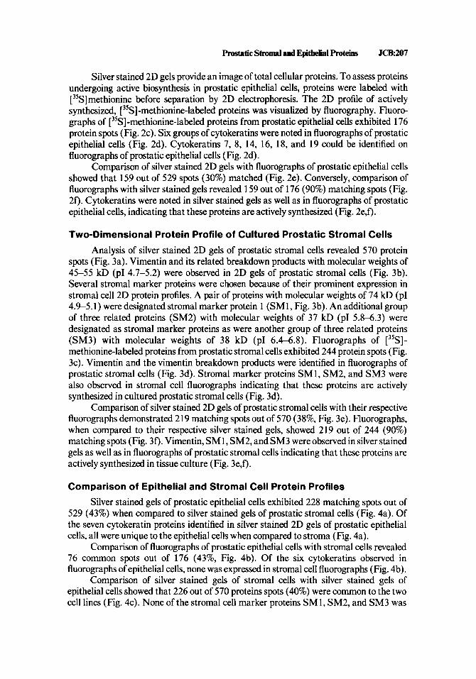
Analysis of silver stained 2D gels of prostatic stromal cells revealed 570 protein spots (Fig. 3a). Vimentin and its related breakdown products with molecular weights of 45-55 kD (PI 4.7-5.2) were observed in 2D gels of prostatic stromal cells (Fig. 3b). Several stromal marker proteins were chosen because of their prominent expression in stromal cell 2D protein profiles. A pair of proteins with molecular weights of 74 kD (PI 4.9-5.1) were designated stromal marker protein 1 (SM1, Fig. 3b). An additional group of three related proteins (SM2) with molecular weights of 37 kD (PI 5.84.3) were designated as stromal marker proteins as were another group of three related proteins (SM3) with molecular weights of 38 kD (PI 6.4-6.8). Fluorographs of [35S]- methionine-labeled proteins from prostatic stromal cells exhibited 244 protein spots (Fig. 3c). Vimentin and the vimentin breakdown products were identified in fluorographs of prostatic stromal cells (Fig. 3d). Stromal marker proteins SM1, SM2, and SM3 were also observed in stromal cell fluorographs indicating that these proteins are actively synthesized in cultured prostatic stromal cells (Fig. 3d).
Comparison of silver stained 2D gels of prostatic stromal cells with their respective fluorographs demonstrated 219 matching spots out of 570 (38%, Fig. 3e). Fluorographs, when compared to their respective silver stained gels, showed 219 out of 244 (90%) matching spots (Fig. 30. Vimentin, SM1, SM2, and SM3 were observed in silver stained gels as well as in fluorographs of prostatic stromal cells indicating that these proteins are actively synthesized in tissue culture (Fig. 3e,f).
Comparison of Epithelial and Stromal Cell Protein Profiles Silver stained gels of prostatic epithelial cells exhibited 228 matching spots out of
529 (43%) when compared to silver stained gels of prostatic stromal cells (Fig. 4a). Of the seven cytokeratin proteins identified in silver stained 2D gels of prostatic epithelial cells, all were unique to the epithelial cells when compared to stroma (Fig. 4a).
Comparison of fluorographs of prostatic epithelial cells with stromal cells revealed 76 common spots out of 176 (43%, Fig. 4b). Of the six cytokeratins observed in fluorographs of epithelial cells, none was expressed in stromal cell fluorographs (Fig. 4b).
Comparison of silver stained gels of stromal cells with silver stained gels of epithelial cells showed that 226 out of 570 proteins spots (40%) were common to the two cell lines (Fig. 4c). None of the stromal cell marker proteins SM1, SM2, and SM3 was
m J C B SherwdetaL
Fig. 3. Two-dimensional protein profiles of cultured stromal cells from hyperplastic human prostate. a: Silver stained 2D gel of isolated prostatic epithelial cells. b Computer-generated image of silver stained 2D profile from prastatic stromal cells. c: fluorograph of 3’S-methionineAabeled stromal cells. d Computer- generated image of stromal cell fluorograph. e: Computer-assisted comparison of silver stained stromal proteins with fluorographs of 35S-methionine-labeled proteins from human prastatic stroma. f: Computer- assisted comparison of stromal fluorographs with silver stained stromal proteins. a, actin; V, vimentin and vimentin breakdown products; 1, stromal marker protein 1 (SMI); 2, stromal marker protein 2 (SM2); 3, stromal marker protein (SM3); 5, area of silver stained gel corresponding with western inmunoblots in Figure 5e. Matching spots (e) and nonmatchmg spots (0) are indicated.

prostatic S t r o d and Epithelial Proteins JCB:u)9
Fig. 4. Comparison of 2D protein profiles of isolated prostatic stromal and epithelial cells. 8: Comparison of silver stained 2D gel of prostatic epithelial cells with silver stained 2D gel of stromal cells. b Comparison of fluorograph of epithelial celk with fluorograph of stromal cells. c: Comparison of silver stained 2D gel of stromal cells with silver-stained gel of epithelial cells. d Comparison of fluorograph of stromal cells with fluorographof epithelialcells. a,actin; 7, K7; 8, K8; 13, K13; 14, K14; 15, K15; 16, K16; 18, K18; 19, K19; V, vimentin and vimentin breakdown products; 1, stromal marker protein SM 1; 2, stromal marker protein SM2; 3, stromal marker protein SM3. Matching spots (e) and nonmatchmg spots (0) are indicated.
noted in 2D gels of epithelial cells. Comparison of fluorographs of prostatic stromal cells with epithelial cells revealed 76 common spots out of 244 (31% Fig. 4d). Of the three stromal marker proteins SM1, SM2, and SM3, none was observed on epithelial cell fluorographs (Fig. 4d).
Analysis of Cytokeratin and Vimentin Expression in Cultured Epithelial and Stromal Cells Using lmmunoprotein Blotting
Expression of the intermediate filament polypeptides cytokeratin and vimentin in primary cultures of prostatic epithelial and stromal cells was confirmed by immunopro- tein blotting of epithelial and stromal cell proteins separated by 2D electrophoresis. Immunoblotting with anti-cytokeratin 4.62 demonstrated the presence of cytokeratins 13, 14, 15, 16, 17, and 19 in 2D gels of prostatic epithelial cells (Fig. 5a). Likewise,

210.JCB Sherwood et aL
Fig. 5. Immunoblot analysis of cytokeratin and vimentin expression in cultured human epithelial and stromal cells from hyperplastic prostate. Immunoblotting of (a) epithelial cells with anti-cytokeratin 4.62, (b) epithelial cells with anti-cytokeratin 8.12, (c) epithelial cells with anti-cytokeratin 8.13, (a) epithelial cells with anti-vimentin, and (e) stromal cells with anti-vimentin. V, vimentin, 5, CK5; 6, CK6; 7, CK7; 8, CK8; 13,CK13; 14,CK14; 15,CK15; 16,CK16; 17,CK17; 18,CK18; 19,CK19.

Prostatic Stromal and Epitllelial Proteim JcB.211
TABLE I. Summary of hot& Identified in 2D Gelr of Cultured Epithelial and Strod Cells From Hyperplastic Human Prostate*
Expression Molecular Isoelectric Protein weight (kD) Point (PH) Epithelium Stroma
Actin Cytokeratins CK5 CK6 CK7 CK8 CK13 CK14 CK15 CK16 CK17 CK18 CK19 Vimentin SMl SM2 SM3
42
58 56 54 52 54 50 50 48 46 45 40
45-55 74 31 38
5.4
7.4 7.8 6.0 6.1 5.1 5.3 4.9 5.1 5.1 5.7 5.2
4.7-5.2 4.9-5.1 5.8-6.3 6.4-6.8
++ ++ ++ ++ ++ ++ ++ ++ ++ ++ ++ ++ + 0 0 0
+ + 0 0 0 0 0 0 0 0 0 0 0
++ ++ ++ ++
*0, no expression; + , minor expression; + + , major expression.
cytokeratins 13, 14, 15, 16, 17, and 19 were observed following immunoblotting with monoclonal anti-cytokeratin 8.12 (Fig. 5b). The proteins exhibiting immunoreactivity with anti-cytokeratins 4.62 and 8.12 are designated in silver stained 2D gels of epithelial cells in area 5 of Figure 2a. Immunoblotting with anti-cytokeratin 8.13, specific for the basic family of cytokeratins, demonstrated cytokeratins 5, 6, 7, and 8 on 2D gels of prostate epithelial cells (Fig. 5c). Cytokeratins 5, 6, 7, and 8 are present in area 5’ of Figure 2a. A low level of immunoreactivity was also observed following immunoblot analysis of epithelial cell proteins with polyclonal anti-vimentin indicating vimentin expression in cultured prostatic epithelial cells (Fig. 5d). Proteins isolated from prostatic epithelial and stromal cells did not exhibit immunoreactivity with monoclonal anticyto- keratin 8.60, which is specific for cytokeratin polypeptides 10 and 1 1 (data not shown).
Cytokeratin expression in cultured prostatic stromal cells was assessed by the use of anti-cytokeratin monoclonals 4.62, 8.12, 8.13, and 8.60. These studies showed that stromal proteins did not exhibit immunoreactivity with the anti-cytokeratins and con- firmed the absence of cytokeratin polypeptides in cultured prostatic stromal cells (data not shown). Immunoblot analysis of stromal proteins separated by 2D electrophoresis with polyclonal anti-vimentin demonstrated strong immunoreactivity with the vimentin antibody and further demonstrated vimentin expression in cultured prostatic stromal cells (Fig. 5e). Vimentin is present in area 5 of the silver stained gel in Figure 3a. A summary of specific proteins that are expressed in cultured prostatic epithelial and stromal cells is provided in Table I.
DISCUSSION
The prostate consists of two major cellular components, stroma and epithelium. Both the stromal and epithelial populations of the prostate are expanded in benign

212JCB Shenvood et aL
prostatic hyperplasia [3]. The present study was undertaken to characterize the protein composition of cultured epithelial and stromal cells from BPH specimens to accomplish two goals: (1) to identify proteins that are specific for prostatic epithelial and stromal cells and can be utilized to distinguish the cell types at the protein level and (2) to characterize specific epithelial and stromal proteins in BPH that can be utilized for comparison with normal and malignant cells of the prostate.
Cytoskeletal elements compose a large portion of the cytoplasmic domain in vertebrate cells. The cytoskeletal components include actin-containing microfilaments [28,29], microtubules containing tubulin [27], and intermediate filaments composed of keratin [27,30,31], vimentin [32], and desmin [33]. The intermediate filament proteins have been shown to be important markers of cellular differentiation and are useful in identifying cells of epithelial and mesenchymal origin.
Cytokeratins are water-insoluble intermediate filament proteins that are structur- ally related to epidermal alpha keratin and present in epithelial cells, but absent in most mesenchymal cells [ 34-36]. Epithelial cells derived from different tissues contain varying combinations of the 19 currently identified cytokeratins and can be character- ized by their specific pattern of cytokeratin expression [27,37,38]. In the present study, cytokeratin components 5,6,7,8, 13, 14, 15, 16, 17, 18, and 19 (K5-Kl9) are identified in cultured benign prostaticepithelial cells. K5, K7, K8, K18, and K19, but not K6, K13, K14, K15, K16, and K17 have been previously observed in normal prostatic epithelium in situ and are common components of diverse types of simple epithelium [27,3942]. K7, K8, K18, and K19 have also been observed to varying degrees in the prostatic tumor cell lines PC3, DU145, and LnCAP [43]. The utility of specific cytokeratin expression to distinguish benign and malignant prostatic epithelial cells is currently undelineated and under investigation in our laboratory. Several tumors have been identified that lose expression of cytokeratin polypeptides that are normally found in benign cells of the same tissue [27]. For example, normal breast epithelium has been shown to express K5, K7, K8, K14, K15, K16, K17, K18, and K19, while ductal carcinomas of the breast generally lack expression of K5 and K 15 [27,44].
Prostate epithelial cells can be divided into basal and luminal components based on morphology and cytokeratin content [35,45,46]. The antibody KA1, raised against sole epidermis, reacts with a topographic epitope shared by cytokeratins K4, K5, and K6 [47]. Basal cells, but not luminal cells, present in prostatic acinar structures react positively with the KA1 antibody in immunohistochemical studies [43]. In the present study, K5 and K6 were observed in 2D gels of prostatic epithelial cells by immunoblot analysis with anti-cytokeratin 8.1 3. This finding suggests that the isolation and cultiva- tion methodology employed in these studies allows for the isolation and cultivation of both luminal and basal elements of prostatic glands. This is in agreement with the findings of Brawer and colleagues, which shows that the cytokeratin antibody 903, which is reactive with the basal layer of prostatic epithelium in vivo, stains cultured benign prostatic epithelial cells [48]. Nagle and colleagues [43] have shown that the established prostatic tumor cell lines PC3, DU145, and LnCAP are not reactive with the KA1 antibody, suggesting the luminal source of these cells. Studies are currently under way to assess the immunoreactivity of antibodies raised to the cytokeratins identified in this report against cultured prostatic tumor cell lines as well as benign and malignant prostatic glandular structures in situ.

Prostatic Stromal and Epithelial Proteins JCB213
The intermediate filament polypeptide vimentin occurs in a variety of nonepithelial cell types and is found most commonly in cells of mesenchymal origin [32]. Vimentin and its related breakdown products [49] were observed in 2D gels of cultured prostatic stromal cells and epithelial cells. The expression of vimentin in cultured prostatic epithelial cells has been previously reported [43]. Three additional stromal marker proteins designated SM1, SM2, and SM3 were selected because of their prominent expression in stromal cells but not epithelial cells. The functional properties of these proteins are currently unknown.
In summary, 2D protein profiles of epithelial and stromal cells of human prostatic origin are reported. Cytokeratins 5,6,7,8, 13, 14, 15, 16, 17, 18, and 19 are present in prostatic epithelial cells. Stromal cells and, to a lesser extent, epithelial cells contain the intermediate filament polypeptide vimentin as well as other distinct marker proteins. These studies document the 2D protein profiles of cultured stromal and epithelial cells derived by percoll gradient centrifugation and selective culture conditions from BPH specimens. The biochemical and cytoskeletal characterization of these cell populations enhances our knowledge of the protein composition of the two major cellular components of the prostate and provides information that can be utilized to distinguish prostatic epithelial and stromal cells as well as compare cells from BPH specimens to those of normal and malignant prostate.
ACKNOWLEDGMENTS
These studies were supported by NIH postdoctoral fellowship DK 08204 and the William 0. Jeffery, I11 fellowship to E.R.S. These studies were also supported by grant DK 39250 from the National Institutes of Health as well as the Lucy and Edwin Kretschmer Fund of Northwestern University Medical School.
REFERENCES
1. Walsh P C In Kimball FA, Buhl AE, Carter DB (eds): “New Approaches to the Study of Benign
2. Franks LM: J Pathol Bacteriol68:617,1954. 3. Grayhack JT, Kozlowski JM: In Gillenwater JY, Grayhack JT, Howards SS, Duckett JW (eds): “Adult
4. Tenniswood M: Prostate 9:375,1986. 5. CunhaGR:Anat Rec 172:179,1972. 6. Cunha GR: Int Rev Cytol47:137,1976. 7. Kozlowski JM, McEwan R, Keer H, Sensibar J, Shenvood E, Lee C, Grayhack JT, Albini A, Martin G:
In Fidler IJ, Nicholson G, (eds): “Tumor Progression and Metastasis.” New York Alan R. Liss, Inc., 1988, pp 189-231.
8. Webber M, Chaproniere-Rickenberg D, Donohoe R: In Barnes DW, Sirbasku DA, Sato GH (eds): “Methods for Serum Free Culture of Cells of the Endocrine System.” New York Alan R. Liss, Inc., 1984, pp 47-61.
Prostatic Hyperplasia.” New York Alan R. Liss, Inc., 1984, pp 1-25.
and Pediatric Urology.” Chicago: Year Book Medical Publishers, 1987, pp 1062-1 125.
9. Cooke DB, Littleton G K Prostate 7:209,1985. 10. McKeehan WL, Adam PS, Rosser M P Cancer Res 44:1998,1984. 11. Kaighn M E In Fischer G, Weiser RJ (eds): “Hormonally Defined Media: A Tool for Cell Biology.”
Heidelberg: Springer-Verlag, 1983, pp 4 18-29. 12. Peehl DM, Stamey T A In Vitro 20981,1984. 13. Peehl DM, Wong ST, Stamey T A In Vitro 24530,1988. 14. Peehl DM, Ham RG: In Vitro 16:526,1980. 15. Chaproniere DM, McKeehan W L Cancer Res 46819,1986.

214JCB Shemood et aL
16. 17. 18. 19. 20.
21. 22. 23. 24.
25. 26. 27. 28. 29. 30.
31. 32. 33. 34. 35. 36. 37. 38. 39. 40. 41. 42. 43. 44. 45. 46. 47.
48. 49.
McKeehan WL, Adams PS, Fast D: In Vitro Cell Dev Biol23:147,1987. Berthois Y, Katzenellenbogen JA, Katzenellenbogen B S Proc Natl Acad Sci 83:2496,1986. Anderson NG, Anderson N L Anal Biochem 85:331,1978. Anderson NL, Anderson NG: Anal Biochem 85:341,1978. Guevera J, Johnston DA, Ranagali LS, Martin BA, Capatillo S, Rodriguez LV: Electrophoresis 3:197, 1982. Lee C, Hu SE, Lok MS, Chen YC, Tseng C C Biotechniques 6216,1988. Gigi-Leitner 0, Geiger B, Levy R, Czernobilslq B: Differentiation 31:191,1986. Gigi-Leitner 0, Geiger B: Cell Motil Cytoskel6:628,1986. Gigi 0, Geiger B, Eshhar Z, Moll R, Schmid E, Winter S, Schiller DL, Franke W W EMBO J 1:1429, 1982. Huszar M, Gigi-Leitner 0, Moll R, Franke W W Differentiation 31:141,1986. Towbin H, Staehelin J, Gordon J: Proc Natl Acad Sci USA 46:4350,1979. Moll R, Franke WW, Schiller DL, Geiger B, Krepler R Cell 31:11,1982. Vandekerkehove J, Weber K Differentiation 1 4 123,1979. Goldman RB, Lazarides G, Pollack R, Weber K Exp Cell Res 90333,1975. Franke WW, Schmid E, Schiller DL, Winter S, Jaransch ED, Moll R, Denk H, Jackson BW, Illmansee K. Cold Spring Harbor Symp Quant Biol4643 1,1982. Tseng SCG, Jarvinen MJ, Nelson WG, Huang J, Woodcock-Mitchell J, Sun T Cell 30:361,1982. Franke WW, Schmid E, Osborn M, Weber K: Proc Natl Acad Sci USA 755034,1978. Lazarides E, Hubbard DB: Proc Natl Acad Sci USA 73:4344,1976. Sun T, Green H: Cell 14469,1978. Schlegal R, Banks-Schlegal S, Pinkus GS: Lab Invest 4291,1980. Sun T, Shih C, Green H: Proc Natl Acad Sci USA 76:2813,1979. Doran TI, Vidrich A, Sun T Cell 221 7,1980. Winter S, Jarasch E, Schmid E, Franke WW, Denk H: Eur J Cell Biol22:371,1980. Moll R, Krepler R, Franke W W Differentiation 23:256,1983. Denk H, Krepler R, Lackingen E, Artlieb U, Franke W W Lab Invest 46584,1982. Fuchs E,Green H: Cell 25:617,1981. Achtstatter T, Moll R, Moore B, Franke WW: J Histochem Cytochem 33:415,1985. Nagle RB, Ahman FR, McDanel KM, Paquin ML, Clark VA, Celniker A. Cancer Res 47:281,1987. Pellegrino MB, Asch BB, Connolly JL, Asch HL: Cancer Res 485831,1988. Barwick K, Mardi A: Lab Invest 48:7A, 1983. Brawer MK, Peehl DM, Stamey T, Bostwick D E Cancer Res 45:3663,1985. Nagle RB, Bocker W, Davis JR, Heid HW, Kaufmann M, Lucas DO, Jarasch E J Histochem Cytochem 34869,1986. Brawer MK, Bostwick DG, Peehl DM, Stamey T A Ann NY Acad Sci 455:729,1986. Czernobilsky B, Moll R, Leppien G, Schweikart G, Franke W W Am J Patholl26:476,1987.