Tailoring hydroxyapatite microspheres by spray-drying ... - OSF
Upload
universitasnegerimakassarCategory
view
3download
0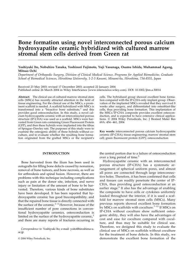
Bone formation using novel interconnected porous calciumhydroxyapatite ceramic hybridized with cultured marrowstromal stem cells derived from Green rat
Yoshiyuki Ito, Nobuhiro Tanaka, Yoshinori Fujimoto, Yuji Yasunaga, Osamu Ishida, Muhammad Agung,Mitsuo OchiDepartment of Orthopedic Surgery, Division of Clinical Medical Science, Programs for Applied Biomedicine, GraduateSchool of Biomedical Sciences, Hiroshima University, 1-2-3 Kasumi, Minami-ku, Hiroshima, 734-8551, Japan
Received 23 May 2003; revised 17 December 2003; accepted 22 January 2004Published online 26 March 2004 in Wiley InterScience (www.interscience.wiley.com). DOI: 10.1002/jbm.a.30014
Abstract: The clinical use of cultured marrow stromal stemcells (MSCs) has recently attracted attention in the field oftissue engineering. For the clinical use of the MSCs, a prom-inent scaffold is needed. A scaffold hybridized with MSCs istransformed into a “bioactive bone substitute,” and thisprovides good osteoconduction. In this study, a novel cal-cium hydroxyapatite ceramic with an interconnected porousstructure (IP-CHA) was used as a scaffold. MSCs were har-vested from Green rats containing Green Fluorescent Protein(GFP), and then these hybrids were implanted into the tibiasof Sprague–Dawley rats. The purposes of this study were toexamine the osteogenic ability of these hybrids without co-culture, and to evaluate whether the resulting bone forma-tion originated from the grafted MSCs or the recipient’s
cells. The hybridized group showed excellent bone forma-tion compared with the IP-CHA-only implant group. Obser-vation of the implanted MSCs revealed that they survived 8weeks after surgery, and differentiated into osteoblast-likecells, thus providing bone formation. This implantation ofthe MSCs/IP-CHA composite provides excellent osteocon-duction, and is expected to have extensive clinical applica-tions. © 2004 Wiley Periodicals, Inc. J Biomed Mater Res69A: 454–461, 2004
Key words: interconnected porous calcium hydroxyapatiteceramic (IP-CHA); tissue engineering; marrow stromal stemcell; green fluorescent protein (GFP); osteoconduction
INTRODUCTION
Bone harvested from the ilium has been used inautografts for filling bone defects caused by nonunion,removal of bone tumors, and bone necrosis, as well asfor arthrodesis and spinal fusion. However, there areproblems with this technique including complicationssuch as pain at the donor site, infection, and nerveinjury or limitation of the amount of bone to be har-vested. Therefore, various kinds of bone substituteshave been developed. It has been reported that hy-droxyapatite ceramic has good biocompatibility, andthat the repaired bone tissue is directly connected withthe surface of the ceramic.1–4 However, because of theinsufficient number of pore connections in conven-tional hydroxyapatite ceramics, osteoconduction islimited on the surface of the hydroxyapatite ceramic,1
and there are many reports of fractures occurring in
the central portion due to a failure of osteoconductionover a long period of time.5
Hydroxyapatite ceramic with an interconnectedporous structure (IP-CHA) has a systematic ar-rangement of spherical uniform pores, and almostall pores are connected through large interconnec-tive holes. Therefore, it has been confirmed that cellsand tissues can readily penetrate the center of IP-CHA, thus providing good osteoconduction at anearlier stage.6 It also has the advantage of enablingthe composite to have cells or cytokines uniformlyloaded throughout the interior, if it is used a scaf-fold for marrow stromal stem cells (MSCs). Manyprevious reports showed excellent bone formationby MSCs on scaffolds with coculture.7–12 If MSCs onIP-CHA without coculture show sufficient osteo-genic ability, they will also have the advantages ofcost and ease for coculture compared with cocul-ture, and thus may be suitable for clinical use.Therefore, we designed this study to evaluate theclinical use of MSCs on scaffolds without coculturefor the treatment of bone defects. In this study, wedemonstrate the excellent bone formation of the
Correspondence to: Yoshiyuki Ito; e-mail: [email protected]
© 2004 Wiley Periodicals, Inc.

MCSs/IP-CHA composite and the origin of the re-sulting bone formation using Green rat MSCs.
MATERIALS AND METHODS
Interconnective porous calcium hydroxyapatiteceramic (IP-CHA)
The IP-CHA used in the current study was researched anddeveloped at the National Institute for Research in InorganicMaterials, and was coresearched and produced by the Depart-ment of Orthopedic Surgery, Osaka University Medical School,Toshiba Ceramics Co., and MMT Co.6 It was produced from aslurry of hydroxyapatite [Ca12(PO4)6(OH)2] by the “foam-geltechnique,” and has a systematic arrangement of sphericaluniform pores. It was sintered at 1200°C with the followingcharacteristics: porosity, 75%; mean porous diameter, 150 �m;interconnective pore hole, 10–90 �m (mean 40 �m); and 90% ormore of the pores are connected with each other throughinterconnective pores (Fig. 1) Mechanically, regardless of theporosity being as high as 75%, the compression strength was10–15 Mpa because of the uniform arrangement of the pores.6
Cylindrical IP-CHA specimens with a diameter of 2 mm and aheight of 4 mm were made for this study.
Green rat
The Green rat [CZ-004 and SD TgN (act-EGFP) OsbCZ-004] developed by the Osaka University Genome Informa-tion Research Center was used in this study. All cells in theGreen rat contain Green Fluorescent Protein (GFP) and theluminescence of these cells can be observed by a fluorescentmicroscope for as long as the cells survive.
MSCs preparation and hybridization to IP-CHA
A femur was harvested from a Green rat (14 weeks ofage), an 18-G syringe needle was inserted once into thefemur, and the bone marrow was flushed into a 50-mLcentrifugal tube using 5 mL of Dulbecco’s minimal essentialmedium containing 10% fetal bovine serum, and was re-peated twice. The collected bone marrow was divided into a100-mm dishes 410 mL each, and was incubated at 37°C in5% CO2. At 2 weeks after incubation, it was subcultured toreach near confluence, and thereafter, subcultured beforereaching confluence (passages 2–5). In these adherent cells, anumber of MSCs were observed.13–17 The adherent cellswere treated with trypsin, a cell suspension was produced,and condensed by centrifugation. These MSCs were sepa-rated to a density of 5 � 105 cells each, and were mixed withgrowth medium, 0.02 mL each (cell density 2.5 � 107/mL).These MSCs mixed solutions were slowly injected into theIP-CHA using a syringe so as to sufficiently penetrate andhybridize to the scaffold. Then, these MSCs/IP-CHA com-posites were implanted without coculture.
Graft of the MSCs/IP-CHA composite
Sprague–Dawley (SD) rats (12 weeks of age, male, mean450 g) were used as the recipients. They were anesthetizedwith intramuscular injections of pentobarbital (Nembutal,Dainippon Pharmaceutical Co. Ltd., Osaka, Japan; 1 mL/kgbody weight), the tibial condyles were exposed, and holeswere made into the cortex of the bone to a depth of 4 mmusing a 2-mm drill. Cylindrical IP-CHA specimens with adiameter of 2 mm and a height of 4 mm, which were hy-bridized with 5 � 105 Green rat cultured MSCs and, withoutcoculturing, implanted into the foramen of the tibia. This
Figure 2. Bone morphological measurements. The densityof the new bone formation in pores was measured in twoareas; the center of the IP-CHA specimen, and the corner ofthe periphery. Each area was 0.72 mm2. HA, hydroxyapatite(IP-CHA). [Color figure can be viewed in the online issue,which is available at www.interscience.wiley.com.]
Figure 1. Scanning electron microscopy. IP-CHA containsnumerous spherical pores of uniform size, and these poresare almost all interconnected.
BONE FORMATION USING CERAMIC 455
cultured MSCs/IP-CHA composite was implanted into theleft tibia as the “MSCs/IP-CHA hybridized group,” and theIP-CHA only was implanted into the right tibia as the “IP-CHA-only implant group.” Both groups consisted of 16 rats.
Immunosuppressive agent
To prevent immunorejection, FK-506 (tacrolimus, Fuji-sawa Pharmaceutical Co, Osaka, Japan) 1 mg/kg was in-jected intramuscularly every day until 2 weeks after surgery.
Histological evaluation
At 1, 2, 4, and 8 weeks after surgery, four rats were sacrificed,and the apatite was removed en masse with the tibia. It wasfixed in 10% paraformaldehyde (Wako Pure Chemical Indus-tries Ltd., Osaka, Japan), and embedded in resin (Technovit8100, Heraeus Kulzer GmbH & Co. KG., Wehrheim, Germany).A nondecalcified section was made, and was observed usinghematoxylin and eosin staining and anti-auto GFP monoclonalantibody immunostaining (Wako Pure Chemical IndustriesLtd., Osaka, Japan). Initially, the GFP luminescence wasplanned to be observed with a fluorescence microscope (Re-search Inverted Microscope, DMIRB, Leica Microsystems, Wet-zlar, Germany). However, because the wavelength of the mar-row cells from SD rats was similar to that of GFP, theimplanted MSCs were observed using anti-auto GFP monoclo-nal antibody immunostaining in this study.
Bone morphological measurement
At 1, 2, 4, and 8 weeks after surgery, the new bone formationwithin the apatite from the four rats was measured. The den-sity of new bone formation in the pores was measured in twoareas of 0.72 mm2 each, using image analysis software (Osteo-plan-2, Carl Zeiss, Thornwood, NY). The measured areas werethe center of the IP-CHA and the corner of the periphery (Fig.2). Descriptive statistics were used to determine the groupmeans and standard deviations. Statistical analysis was donewith an analysis of variance (ANOVA) and the Bonferroni test,and statistical significance was set at p � 0.05.
Biochemical analysis
To examine the differentiation of MSCs into osteoblasts,alkaline phosphatase (ALP) activity and osteocalcin (OC) con-
Figure 3. (a) Fluorescent microscopy, and (b) Anti-autoGFP monoclonal antibody immunostaining. A cross-sectionof the IP-CHA hybridized with MSCs derived from Greenrats before implantation. The MSCs were hybridized uni-formly not only on the surface of the IP-CHA, but also on theinterior of the pores. The MSCs are stuck to the walls of thepores (arrowheads show the MSCs).
Figure 4. At 8 weeks after surgery, lamellar bone, marrowcells, blood cells, and adipocytes were observed through theinterconnective holes in the MSCs/IP-CHA hybridized group.(hematoxylin and eosin staining). HA, hydroxyapatite (IP-CHA); L, lamellar bone; BM, bone marrow cell; B, blood cell; A,adipocyte. [Color figure can be viewed in the online issue,which is available at www.interscience.wiley.com.]
456 ITO ET AL.
tent were investigated. The IP-CHA was harvested at 1, 2, and4 weeks after surgery (four specimens each), and were used forthis analysis. Each construct was freeze-dried for 18 h, crushed,homogenized in distilled water, and centrifuged at 3000 rpmfor 10 min at 4°C. After freeze-drying again, the specimenswere decalcified by 0.5 M ethylenediaminetetraacetic acid, andwere shaken for 18 h. After extraction, the OC content and ALPactivity were measured. The OC content was measured usingan enzyme-linked immunosorbent assay system for the rat(Amersham Japan Co., Tokyo, Japan) and a microplate reader(BIO-RAD, Novapath Co., Tokyo, Japan) at an absorbance of450 nm. ALP activity was measured by disc electrophoresis.The electrophoresis of the polyacrylic-amido disc gel was per-formed, and after the reaction was stopped using an ALP stain,the ALP activity was measured by densitometry (Densitron,Jyoko Co., Osaka, Japan) at an absorbance of 620 nm. Descrip-tive statistics were used to determine the group means andstandard deviations. Statistical analysis was done with anANOVA and the Bonferroni test.
RESULTS
Histological evaluation
MSCs were hybridized uniformly, not only to thesurface of the IP-CHA but also to the interior of the
pores because of their large interconnected holes.MSCs were stuck to the walls of the pores (Fig. 3).
At 8 weeks after implantation, lamellar bone, mar-row cells, blood cells, and adipocytes were observedthrough the interconnective holes in the MSCs/IP-CHA hybridized group (Fig. 4).
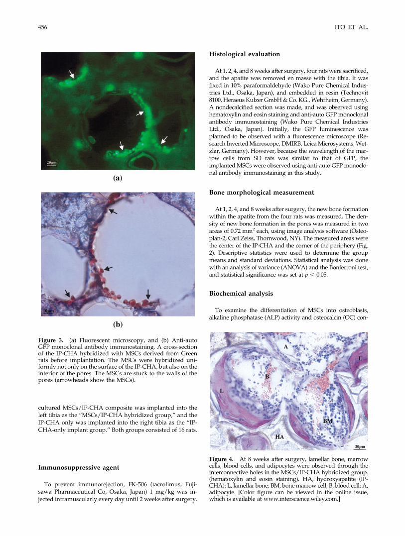
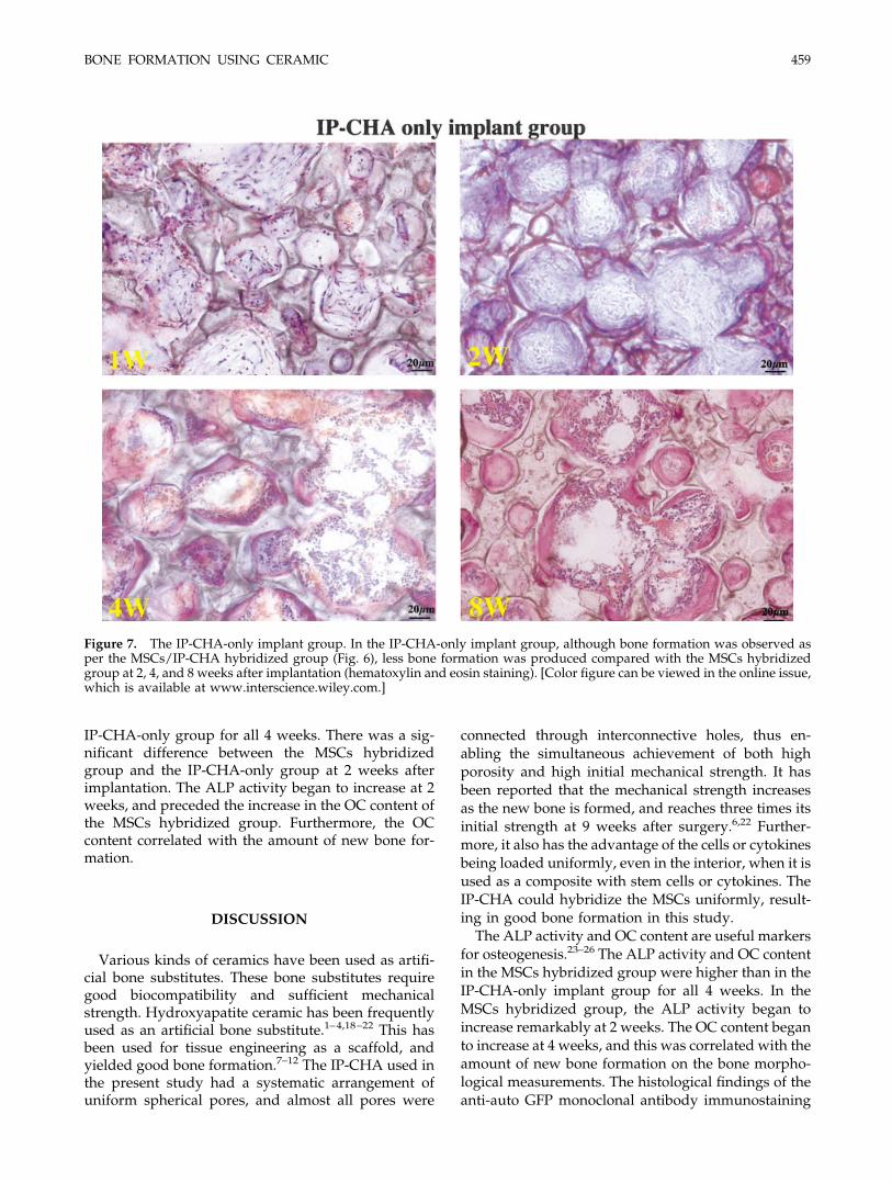
At 1 day after surgery, the surface of implantedIP-CHA was in contact with the SD rat bone marrow,and all pores were filled with blood cells (Fig. 5). At 1week after surgery, penetration of the marrow cellsinto all of the pores was observed in both groups, andthis was achieved through the interconnected holes.At 2 weeks after surgery, bone formation was ob-served in both groups, but more bone was producedin the MSCs hybridized group. This bone formationproceeded from the wall sides of the pores, and oc-curred initially on the surface of the pores. At 4 weeksafter surgery, new bone formation progressed towardthe center of the pores. At 8 weeks after surgery, bonetissue units with blood cells, marrow cells, and adipo-cytes were completed, and many were connectedthrough the interconnective holes in both groups. Inthe IP-CHA-only implant group, although bone for-mation was observed, less bone was produced com-
Figure 5. At 1 day after surgery, the surface of the implanted IP-CHA came into contact with the SD rat bone marrow (b),and all pores were filled with blood cells (c) (hematoxylin and eosin staining). [Color figure can be viewed in the online issue,which is available at www.interscience.wiley.com.]
BONE FORMATION USING CERAMIC 457
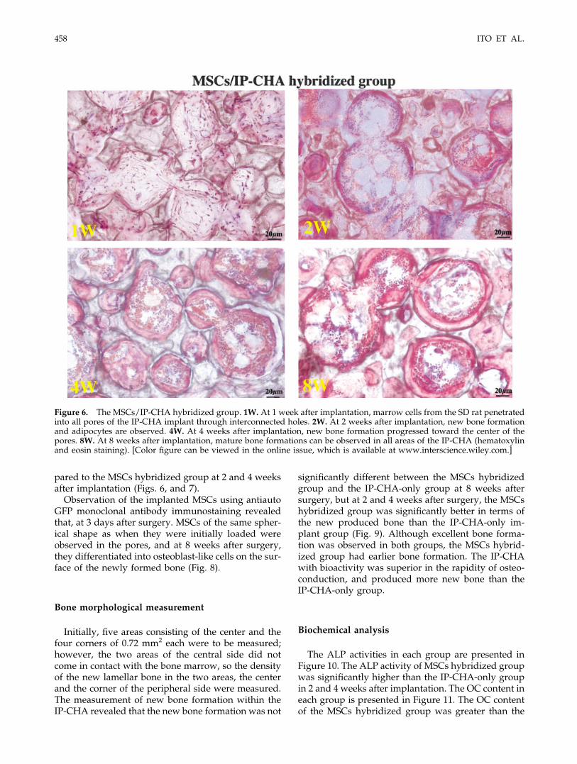
pared to the MSCs hybridized group at 2 and 4 weeksafter implantation (Figs. 6, and 7).
Observation of the implanted MSCs using antiautoGFP monoclonal antibody immunostaining revealedthat, at 3 days after surgery. MSCs of the same spher-ical shape as when they were initially loaded wereobserved in the pores, and at 8 weeks after surgery,they differentiated into osteoblast-like cells on the sur-face of the newly formed bone (Fig. 8).
Bone morphological measurement
Initially, five areas consisting of the center and thefour corners of 0.72 mm2 each were to be measured;however, the two areas of the central side did notcome in contact with the bone marrow, so the densityof the new lamellar bone in the two areas, the centerand the corner of the peripheral side were measured.The measurement of new bone formation within theIP-CHA revealed that the new bone formation was not
significantly different between the MSCs hybridizedgroup and the IP-CHA-only group at 8 weeks aftersurgery, but at 2 and 4 weeks after surgery, the MSCshybridized group was significantly better in terms ofthe new produced bone than the IP-CHA-only im-plant group (Fig. 9). Although excellent bone forma-tion was observed in both groups, the MSCs hybrid-ized group had earlier bone formation. The IP-CHAwith bioactivity was superior in the rapidity of osteo-conduction, and produced more new bone than theIP-CHA-only group.
Biochemical analysis
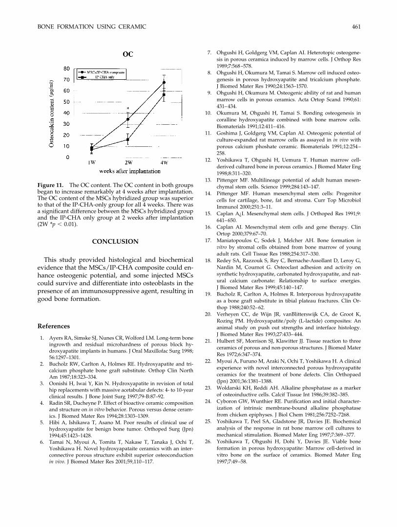
The ALP activities in each group are presented inFigure 10. The ALP activity of MSCs hybridized groupwas significantly higher than the IP-CHA-only groupin 2 and 4 weeks after implantation. The OC content ineach group is presented in Figure 11. The OC contentof the MSCs hybridized group was greater than the
Figure 6. The MSCs/IP-CHA hybridized group. 1W. At 1 week after implantation, marrow cells from the SD rat penetratedinto all pores of the IP-CHA implant through interconnected holes. 2W. At 2 weeks after implantation, new bone formationand adipocytes are observed. 4W. At 4 weeks after implantation, new bone formation progressed toward the center of thepores. 8W. At 8 weeks after implantation, mature bone formations can be observed in all areas of the IP-CHA (hematoxylinand eosin staining). [Color figure can be viewed in the online issue, which is available at www.interscience.wiley.com.]
458 ITO ET AL.
IP-CHA-only group for all 4 weeks. There was a sig-nificant difference between the MSCs hybridizedgroup and the IP-CHA-only group at 2 weeks afterimplantation. The ALP activity began to increase at 2weeks, and preceded the increase in the OC content ofthe MSCs hybridized group. Furthermore, the OCcontent correlated with the amount of new bone for-mation.
DISCUSSION
Various kinds of ceramics have been used as artifi-cial bone substitutes. These bone substitutes requiregood biocompatibility and sufficient mechanicalstrength. Hydroxyapatite ceramic has been frequentlyused as an artificial bone substitute.1–4,18–22 This hasbeen used for tissue engineering as a scaffold, andyielded good bone formation.7–12 The IP-CHA used inthe present study had a systematic arrangement ofuniform spherical pores, and almost all pores were
connected through interconnective holes, thus en-abling the simultaneous achievement of both highporosity and high initial mechanical strength. It hasbeen reported that the mechanical strength increasesas the new bone is formed, and reaches three times itsinitial strength at 9 weeks after surgery.6,22 Further-more, it also has the advantage of the cells or cytokinesbeing loaded uniformly, even in the interior, when it isused as a composite with stem cells or cytokines. TheIP-CHA could hybridize the MSCs uniformly, result-ing in good bone formation in this study.
The ALP activity and OC content are useful markersfor osteogenesis.23–26 The ALP activity and OC contentin the MSCs hybridized group were higher than in theIP-CHA-only implant group for all 4 weeks. In theMSCs hybridized group, the ALP activity began toincrease remarkably at 2 weeks. The OC content beganto increase at 4 weeks, and this was correlated with theamount of new bone formation on the bone morpho-logical measurements. The histological findings of theanti-auto GFP monoclonal antibody immunostaining
Figure 7. The IP-CHA-only implant group. In the IP-CHA-only implant group, although bone formation was observed asper the MSCs/IP-CHA hybridized group (Fig. 6), less bone formation was produced compared with the MSCs hybridizedgroup at 2, 4, and 8 weeks after implantation (hematoxylin and eosin staining). [Color figure can be viewed in the online issue,which is available at www.interscience.wiley.com.]
BONE FORMATION USING CERAMIC 459

confirmed that some MSCs survived for a long periodof time, and differentiated into osteoblast-like cellsand proliferated. Based on these findings, we specu-lated that the implanted MSCs could survive, anddifferentiate into osteoblasts, or release various kindsof cytokines for bone formation in the presence of animmunosuppressive agent. In this study, although theMSCs hybridized group showed excellent bone forma-tion during the early stage after surgery comparedwith the IP-CHA-only implant group, IP-CHA itselfhad excellent osteoconduction, and the IP-CHA-onlyimplant group also showed good bone formation. Thereason may be that the apatite used in this study wassmall, only 2 mm in diameter, which facilitated cell
penetration, including the implant into the bone mar-row. It is expected that differences in bone formationability become greater in the necrotic portion or inenvironments such as large bony defects where cellpenetration is difficult. Although the implantation ofIP-CHA-only can yield excellent osteoconduction forsmall defects, the implantation of MSCs compositemay be desirable for large bone defects or nonunionsites where cell penetration is difficult or where earlierbone union is needed.
Figure 8. Anti-auto GFP monoclonal antibody immuno-staining. (a) Three days after implantation. GFP-immunopo-sitive cells are observed in the pores. The MSCs were thesame spherical shape as when they were initially loaded. (b)Eight weeks after implantation. GFP-immunopositive cellsdifferentiated into osteoblast-like cells on the surface of thenewly formed bone (arrowheads show the GFP-immunopo-sitive cells).
Figure 9. Bone morphological measurements. The new boneformation of the MSCs/IP-CHA hybridized group was supe-rior at 2 or 4 weeks after surgery, but there was no significantdifference at 8 weeks (2W *p � 0.001, 4W **p � 0.01).
Figure 10. The ALP activity. The ALP activity of MSCshybridized group began to increase remarkably at 2 weeksafter implantation. There was a significant different betweenthe MSCs hybridized group and the IP-CHA-only group at2 and 4 weeks after implantation (2W, 4W *p � 0.001).
460 ITO ET AL.
CONCLUSION
This study provided histological and biochemicalevidence that the MSCs/IP-CHA composite could en-hance osteogenic potential, and some injected MSCscould survive and differentiate into osteoblasts in thepresence of an immunosuppressive agent, resulting ingood bone formation.
References
1. Ayers RA, Simske SJ, Nunes CR, Wolford LM. Long-term boneingrowth and residual microhardness of porous block hy-droxyapatite implants in humans. J Oral Maxillofac Surg 1998;56:1297–1301.
2. Bucholz RW, Carlton A, Holmes RE. Hydroxyapatite and tri-calcium phosphate bone graft substitute. Orthop Clin NorthAm 1987;18:323–334.
3. Oonishi H, Iwai Y, Kin N. Hydroxyapatite in revision of totalhip replacements with massive acetabular defects: 4- to 10-yearclinical results. J Bone Joint Surg 1997;79-B:87–92.
4. Radin SR, Ducheyne P. Effect of bioactive ceramic compositionand structure on in vitro behavior. Porous versus dense ceram-ics. J Biomed Mater Res 1994;28:1303–1309.
5. Hibi A, Ishikawa T, Asano M. Poor results of clinical use ofhydroxyapatite for benign bone tumor. Orthoped Surg (Jpn)1994;45:1423–1428.
6. Tamai N, Myoui A, Tomita T, Nakase T, Tanaka J, Ochi T,Yoshikawa H. Novel hydroxyapataite ceramics with an inter-connective porous structure exhibit superior osteoconductionin vivo. J Biomed Mater Res 2001;59,110–117.
7. Ohgushi H, Goldgerg VM, Caplan AI. Heterotopic osteogene-sis in porous ceramica induced by marrow cells. J Orthop Res1989;7:568–578.
8. Ohgushi H, Okumura M, Tamai S. Marrow cell induced osteo-genesis in porous hydroxyapatite and tricalcium phosphate.J Biomed Mater Res 1990;24:1563–1570.
9. Ohgushi H, Okumura M. Osteogenic ability of rat and humanmarrow cells in porous ceramics. Acta Ortop Scand 1990;61:431–434.
10. Okumura M, Ohgushi H, Tamai S. Bonding osteogenesis incoralline hydroxyapatite combined with bone marrow cells.Biomaterials 1991;12:411–416.
11. Goshima J, Goldgerg VM, Caplan AI. Osteogenic potential ofculture-expanded rat marrow cells as assayed in in vivo withporous calcium phoshate ceramic. Biomaterials 1991;12:254–258.
12. Yoshikawa T, Ohgushi H, Uemura T. Human marrow cell-derived cultured bone in porous ceramics. J Biomed Mater Eng1998;8:311–320.
13. Pittenger MF. Multilineage potential of adult human mesen-chymal stem cells. Science 1999;284:143–147.
14. Pittenger MF. Human mesenchymal stem cells: Progenitorcells for cartilage, bone, fat and stroma. Curr Top MicrobiolImmunol 2000;251:3–11.
15. Caplan A¿I. Mesenchymal stem cells. J Orthoped Res 1991;9:641–650.
16. Caplan AI. Mesenchymal stem cells and gene therapy. ClinOrtop 2000;379:67–70.
17. Maniatopoulos C, Sodek J, Melcher AH. Bone formation invitro by stromal cells obtained from bone marrow of youngadult rats. Cell Tissue Res 1988;254:317–330.
18. Redey SA, Razzouk S, Rey C, Bernache-Assollant D, Leroy G,Nardin M, Cournot G. Osteoclast adhesion and activity onsynthetic hydroxyapatite, carbonated hydroxyapatite, and nat-ural calcium carbonate: Relationship to surface energies.J Biomed Mater Res 1999;45:140–147.
19. Bucholz R, Carlton A, Holmes R. Interporous hydroxyapatiteas a bone graft substitute in tibial plateau fractures. Clin Or-thop 1988;240:52–62.
20. Verheyen CC, de Wijn JR, vanBlitterswijk CA, de Groot K,Rozing PM. Hydroxyapatite/poly (L-lactide) composites: Ananimal study on push out strengths and interface histology.J Biomed Mater Res 1993;27:433–444.
21. Hulbert SF, Morrison SJ, Klawitter JJ. Tissue reaction to threeceramics of porous and non-porous structures. J Biomed MaterRes 1972;6:347–374.
22. Myoui A, Furuno M, Araki N, Ochi T, Yoshikawa H. A clinicalexperience with novel interconnected porous hydroxyapatiteceramics for the treatment of bone defects. Clin Orthopaed(Jpn) 2001;36:1381–1388.
23. Woldarski KH, Reddi AH. Alkaline phosphatase as a markerof osteoinductive cells. Calcif Tissue Int 1986;39:382–385.
24. Cyboron GW, Wunthier RE. Purification and initial character-ization of intrinsic membrane-bound alkaline phosphatasefrom chicken epiphyses. J Biol Chem 1981;256:7252–7268.
25. Yoshikawa T, Peel SA, Gladstone JR, Davies JE. Biochemicalanalysis of the response in rat bone marrow cell cultures tomechanical stimulation. Biomed Mater Eng 1997;7:369–377.
26. Yoshikawa T, Ohgushi H, Dohi Y, Davies JE. Viable boneformation in porous hydroxyapatite: Marrow cell-derived invitro bone on the surface of ceramics. Biomed Mater Eng1997;7:49–58.
Figure 11. The OC content. The OC content in both groupsbegan to increase remarkably at 4 weeks after implantation.The OC content of the MSCs hybridized group was superiorto that of the IP-CHA-only group for all 4 weeks. There wasa significant difference between the MSCs hybridized groupand the IP-CHA only group at 2 weeks after implantation(2W *p � 0.01).
BONE FORMATION USING CERAMIC 461
Copyright © 2022 FDOKUMEN