Incorporation of Hydroxyapatite into Glass Ionomer Cement ...
14
materials Article Incorporation of Hydroxyapatite into Glass Ionomer Cement (GIC) Formulated Based on Alumino-Silicate-Fluoride Glass Ceramics from Waste Materials Wan Nurshamimi Wan Jusoh 1 , Khamirul Amin Matori 1,2, *, Mohd Hafiz Mohd Zaid 1 , Norhazlin Zainuddin 3 , Mohammad Zulhasif Ahmad Khiri 2 , Nadia Asyikin Abdul Rahman 1 , Rohaniah Abdul Jalil 1 and Esra Kul 4 Citation: Wan Jusoh, W.N.; Matori, K.A.; Mohd Zaid, M.H.; Zainuddin, N.; Ahmad Khiri, M.Z.; Abdul Rahman, N.A.; Abdul Jalil, R.; Kul, E. Incorporation of Hydroxyapatite into Glass Ionomer Cement (GIC) Formulated Based on Alumino-Silicate-Fluoride Glass Ceramics from Waste Materials. Materials 2021, 14, 954. https:// doi.org/10.3390/ma14040954 Received: 17 October 2020 Accepted: 20 November 2020 Published: 18 February 2021 Publisher’s Note: MDPI stays neutral with regard to jurisdictional claims in published maps and institutional affil- iations. Copyright: © 2021 by the authors. Licensee MDPI, Basel, Switzerland. This article is an open access article distributed under the terms and conditions of the Creative Commons Attribution (CC BY) license (https:// creativecommons.org/licenses/by/ 4.0/). 1 Department of Physics, Faculty of Science, Universiti Putra Malaysia, UPM Serdang, Selangor 43400, Malaysia; [email protected] (W.N.W.J.); [email protected] (M.H.M.Z.); [email protected] (N.A.A.R.); [email protected] (R.A.J.) 2 Material Synthesis and Characterization Laboratory, Institute of Advanced Technology, Universiti Putra Malaysia, UPM Serdang, Selangor 43400, Malaysia; [email protected] 3 Department of Chemistry, Faculty of Science, Universiti Putra Malaysia, UPM Serdang, Selangor 43400, Malaysia; [email protected] 4 Department of Prosthodontics, Faculty of Dentistry, Ataturk University, 25030 Erzurum, Turkey; [email protected] * Correspondence: [email protected]; Tel.: +60-16-267-3321 Abstract: Glass ionomer cement (GIC) is a well-known restorative material applied in dentistry. The present work aims to study the effect of hydroxyapatite (HA) addition into GIC based on physical, mechanical and structural properties. The utilization of waste materials namely clam shell (CS) and soda lime silica (SLS) glass as replacements for the respective CaO and SiO 2 sources in the fabrication of alumino-silicate-fluoride (ASF) glass ceramics powder. GIC was formulated based on ASF glass ceramics, polyacrylic acid (PAA) and deionized water, while 1 wt.% of HA powder was added to enhance the properties of the cement samples. The cement samples were subjected to four different ageing times before being analyzed. In this study, the addition of HA caused an increment in density and compressive strength results along with ageing time. Besides, X-ray Diffraction (XRD) revealed the formation of fluorohydroxyapatite (FHA) phase in HA-added GIC samples and it was confirmed by Fourier Transform Infrared (FTIR) analysis which detected OH-F vibration mode. In addition, needle-like and agglomeration of spherical shapes owned by apatite crystals were observed from Field Emission Scanning Electron Microscopy (FESEM). Based on Energy Dispersive X-ray (EDX) analysis, the detection of chemical elements in the cement samples were originated from chemical compounds used in the preparation of glass ceramics powder and also the polyacid utilized in initiating the reaction of GIC. Keywords: ASF glass ceramics; clam shell; glass ionomer cement; hydroxyapatite; soda lime sil- ica glass 1. Introduction A form of dental restoration and luting material, called glass ionomer cement (GIC), is widely used in dental application. In the early of 70 0 s, Alan Wilson and Brian Kent intro- duced GIC as water-based cement derived from silicate-based glass and polycarboxylate salt [1]. This water-based substance forms essentially when there is a reaction between aqueous polyacrylic acid solution and fluoro-aluminosilicate glass powder [1–3]. GIC has desirable properties which encourage their clinical uses in dentistry such as it provides a direct bonding to tooth structure [4–6], exhibit a good biocompatibility and has the ability to release fluoride which is important as anticariogenic agent in preventing the occurrence of tooth decay [7]. Furthermore, the higher stability of GIC when involved in aqueous environment and also the results of low toxicity when applied to tooth are the Materials 2021, 14, 954. https://doi.org/10.3390/ma14040954 https://www.mdpi.com/journal/materials
-
Upload
khangminh22 -
Category
Documents
-
view
1 -
download
0
Transcript of Incorporation of Hydroxyapatite into Glass Ionomer Cement ...

materials
Article
Incorporation of Hydroxyapatite into Glass Ionomer Cement(GIC) Formulated Based on Alumino-Silicate-Fluoride GlassCeramics from Waste Materials
Wan Nurshamimi Wan Jusoh 1, Khamirul Amin Matori 1,2,*, Mohd Hafiz Mohd Zaid 1 , Norhazlin Zainuddin 3,Mohammad Zulhasif Ahmad Khiri 2, Nadia Asyikin Abdul Rahman 1, Rohaniah Abdul Jalil 1 and Esra Kul 4
�����������������
Citation: Wan Jusoh, W.N.; Matori,
K.A.; Mohd Zaid, M.H.; Zainuddin,
N.; Ahmad Khiri, M.Z.; Abdul
Rahman, N.A.; Abdul Jalil, R.; Kul, E.
Incorporation of Hydroxyapatite into
Glass Ionomer Cement (GIC)
Formulated Based on
Alumino-Silicate-Fluoride Glass
Ceramics from Waste Materials.
Materials 2021, 14, 954. https://
doi.org/10.3390/ma14040954
Received: 17 October 2020
Accepted: 20 November 2020
Published: 18 February 2021
Publisher’s Note: MDPI stays neutral
with regard to jurisdictional claims in
published maps and institutional affil-
iations.
Copyright: © 2021 by the authors.
Licensee MDPI, Basel, Switzerland.
This article is an open access article
distributed under the terms and
conditions of the Creative Commons
Attribution (CC BY) license (https://
creativecommons.org/licenses/by/
4.0/).
1 Department of Physics, Faculty of Science, Universiti Putra Malaysia, UPM Serdang, Selangor 43400,Malaysia; [email protected] (W.N.W.J.); [email protected] (M.H.M.Z.);[email protected] (N.A.A.R.); [email protected] (R.A.J.)
2 Material Synthesis and Characterization Laboratory, Institute of Advanced Technology,Universiti Putra Malaysia, UPM Serdang, Selangor 43400, Malaysia; [email protected]
3 Department of Chemistry, Faculty of Science, Universiti Putra Malaysia, UPM Serdang,Selangor 43400, Malaysia; [email protected]
4 Department of Prosthodontics, Faculty of Dentistry, Ataturk University, 25030 Erzurum, Turkey;[email protected]
* Correspondence: [email protected]; Tel.: +60-16-267-3321
Abstract: Glass ionomer cement (GIC) is a well-known restorative material applied in dentistry.The present work aims to study the effect of hydroxyapatite (HA) addition into GIC based onphysical, mechanical and structural properties. The utilization of waste materials namely clam shell(CS) and soda lime silica (SLS) glass as replacements for the respective CaO and SiO2 sources in thefabrication of alumino-silicate-fluoride (ASF) glass ceramics powder. GIC was formulated based onASF glass ceramics, polyacrylic acid (PAA) and deionized water, while 1 wt.% of HA powder wasadded to enhance the properties of the cement samples. The cement samples were subjected to fourdifferent ageing times before being analyzed. In this study, the addition of HA caused an incrementin density and compressive strength results along with ageing time. Besides, X-ray Diffraction(XRD) revealed the formation of fluorohydroxyapatite (FHA) phase in HA-added GIC samples andit was confirmed by Fourier Transform Infrared (FTIR) analysis which detected OH-F vibrationmode. In addition, needle-like and agglomeration of spherical shapes owned by apatite crystals wereobserved from Field Emission Scanning Electron Microscopy (FESEM). Based on Energy DispersiveX-ray (EDX) analysis, the detection of chemical elements in the cement samples were originated fromchemical compounds used in the preparation of glass ceramics powder and also the polyacid utilizedin initiating the reaction of GIC.
Keywords: ASF glass ceramics; clam shell; glass ionomer cement; hydroxyapatite; soda lime sil-ica glass
1. Introduction
A form of dental restoration and luting material, called glass ionomer cement (GIC),is widely used in dental application. In the early of 70′s, Alan Wilson and Brian Kent intro-duced GIC as water-based cement derived from silicate-based glass and polycarboxylatesalt [1]. This water-based substance forms essentially when there is a reaction betweenaqueous polyacrylic acid solution and fluoro-aluminosilicate glass powder [1–3].
GIC has desirable properties which encourage their clinical uses in dentistry such as itprovides a direct bonding to tooth structure [4–6], exhibit a good biocompatibility and hasthe ability to release fluoride which is important as anticariogenic agent in preventing theoccurrence of tooth decay [7]. Furthermore, the higher stability of GIC when involved inaqueous environment and also the results of low toxicity when applied to tooth are the
Materials 2021, 14, 954. https://doi.org/10.3390/ma14040954 https://www.mdpi.com/journal/materials
Materials 2021, 14, 954 2 of 14
advantages of GIC in dental application. Despite having good properties, there is somelimitation that limits GIC from extensive use in dentistry as filling and restorative material.GIC has been shown to have weak mechanical and physical properties including lowfracture strength and hardness, reduced wear resistance and opaqueness [8]. Besides, GIC isvery brittle and this proved the low physical strength of GIC to be used as dental material.
Therefore, there are many efforts that have been focused on the modification of GIC sothat the physical and mechanical properties of GIC can be enhanced. One of the ways is byincorporating hydroxyapatite into the GIC composition. Hydroxyapatite (HA) is an inor-ganic compound with a constitution equivalent to natural bone mineral and is well knownas an apatite group (a group of mineral phosphate). HA is represented by the chemicalformula of Ca10(PO4)6(OH)2, differ with other apatite compounds by containing hydroxylend-member of the apatite group [4,5]. HA present as a major component of mineral inbone and teeth with calcium-to-phosphate ratio of 1.67 (Ca/P = 1.67) and hexagonal crystalstructure [4,5,8]. According to Ramsden and co-workers, HA has osteoconductive andbioactive properties which make it favorable in orthopedics and dental application [9].
Alumino-silicate-fluoride (ASF) glass ceramics composition used in the formulation ofGIC is believed to have good properties and favorable for dental materials [10–12]. The de-sign of ASF glass ceramics composition is based on the 45S5 bioactive glass system usedby Larry L. Hench which implicated a combination of glass network formers, modifiersand intermediates oxides [13,14]. The use of waste materials like clam shell (CS) and sodalime silica (SLS) glass as replacement of silicate and calcite sources, respectively in thesynthesis of glass ceramics can save the production cost and reduce the environmentalissues regarding the disposal problems. According to previous literatures, the general com-position of SLS glass consists of 70–75 wt.% of SiO2, 12–16 wt.% of Na2O and 10–15 wt.%of CaO [15,16], meanwhile CS contains more than 98% CaC which encouraged it uses asbiomaterial [17].
The key purpose of this work is to investigate the influence of HA addition on thecomposition of glass ceramics in GIC formulation based on physical, mechanical and struc-tural properties. Although some work on the addition of HA into GIC has been performed,a deeper study can be gained especially involving the participation of ASF glass ceramicsas the base silicate powder in the formulation of GIC. ASF glass ceramics composition withthe existence of fluorapatite (FA) had been studied by several researchers [10–12] and theinvolvement of FA crystal phase in the GIC formulation is crucial since it can give effect tothe properties of the resulting cement. Moreover, the utilization of waste materials whichare CS and SLS glass in the fabrication of ASF glass ceramics and subsequently formulatedinto GIC can attract a great interest to researchers since there is limited studies on the useof waste materials in the GIC formulation. However, the GIC formulated based on ASFglass ceramics as base silicate powder was observed to produce inadequate mechanicalstrength which restricted their uses as dental material. Thus, the study on the additionof HA into the GIC formulation can help in improving the properties of resulting GIC.Besides, the effect of ageing time of the cement samples was also investigated in this study.
2. Materials and Experimental Procedure2.1. Materials
Raw materials used as sources of CaO and SiO2 were derived from CS and SLS glass,respectively. Anadara granosa sp. CS were collected from the beachside of Pantai CahayaBulan, Kelantan, Malaysia, while SLS glass which was ‘Jalen’ soy sauce brand obtainedfrom restaurants near to Universiti Putra Malaysia, Selangor, Malaysia.
The glass ceramics component of the cement involved the mixing of raw materialsand chemical components such as CaF2 (R&M Chemicals, Semenyih, Selangor, Malaysia;99.95%), P2O5 (Alfa Aesar, a Johnson Matthey Company, Lancashire, UK; 99.99%) andAl2O3 (Alfa Aesar, a Johnson Matthey Company, UK; 99.5%). Meanwhile, HA powder usedwas reagent grade powder (Sigma Aldrich, a Merck KGaA Company, Darmstadt, Germany;≥90%) with CAS No. 1306-06-5 and molecular weight (MW) ~502.31. The participation

Materials 2021, 14, 954 3 of 14
of dried powder of polyacrylic acid (PAA) with MW ~30,000 obtained from AdvanceHealthcare Ltd., Willenhall, UK and deionized water as a medium of reaction was crucialin the cement formation.
2.2. Preparation of CS and SLS Glass Powder
Preparation of raw materials was started by the cleaning process of collected CSand SLS glass in order to remove dirt from the materials. For CS, calcium oxide (CaO)was obtained from the calcination process of CS at 900 ◦C for 3 h as explained in severalstudies [10–12]. In this process, carbon dioxide (CO2) in the form of gas was removedfrom aragonite and calcite (CaCO3) which were the majority components of the CS. Af-ter that, both CS and SLS glass went through the same process, whereby they were crushed,plunged and sieved before getting ≤45 µm sized CS and SLS glass powder.
2.3. Synthesis of ASF Glass Ceramics Composition
ASF composition was fabricated by mixing 25 wt.% SLS, 20 wt.% CS, 20 wt.% P2O5,20 wt.% Al2O3 and 15 wt.% CaF2 with 30 g of the total weight composition. After the fivecomponents were mixed homogeneously in alumina crucible, they were subjected to amelting process at 1500 ◦C in the furnace for 4 h. Next, the quenching process was doneby pouring the molten glass into water, thus producing frits. Subsequently, the obtainedfrits were manually crushed by using plunger and grinded by using mortar and pestle,which then formed ≤45 µm-sized powder of ASF glass ceramics.
2.4. Formulation of Control and Modified GIC Samples
GIC was formulated by ASF glass ceramics, PAA and deionized water with ratio of3:1:1 (glass ceramics:PAA:water). The setting reaction of cement required the combining ofthe three components on the glass plate for approximately 60 s. Right after 60 s, the cementwas filled and pressed into stainless-steel mold (6 mm × 4 mm) to produce cylindricalshaped pellets and then dried at 37 ◦C for an hour. Next, the cement went through ageingprocess for 1, 7, 14 and 21 days in deionized water before characterized. In this study, a totalof 30 pellets of GIC and 30 pellets of GIC1 samples were formed. GIC sample representedthe GIC without HA addition while GIC1 sample represented GIC with the addition of1 wt.% of HA powder into GIC. The GIC1 samples were prepared by mixing ASF glassceramics with HA powder before formulated into GIC in the proportion of 3 based on theratio as stated previously.
2.5. Characterization of Control and Modified GIC Samples
The cement samples were characterized by density measurement, compressive strengthtest, X-ray Diffraction (XRD), Fourier Transform Infrared (FTIR), Field Emission ScanningElectron Microscopy (FESEM) and Energy Dispersive X-ray (EDX).
2.5.1. Density
The density ($) was evaluated based on Archimedes’ principle at room tempera-ture [18]. The total of 24 samples in pellet form were weighed before being immersedinto distilled water with a density of 1.0 g/cm3 and the test were repeated for three times.After that, the weights of each pellet were taken and resulting density with unit gram percentimeter cubic (g/cm3) were calculated based on formula:
ρsample =Wair
Wair − Wdistilled water× ρdistilled water (1)
where Wair = weight in air, Wdistilled water = weight in distilled water and ρdistilled water = den-sity of distilled water = 1.0 g/cm3.

Materials 2021, 14, 954 4 of 14
2.5.2. Compressive Strength Test
Compressive strength of the samples was determined by using Universal TestingMachine (UTM) from Instron (Instron, Norwood, MA, USA) with a model of 5566. The loadframe applied was up to 10 kN with crosshead of 1 mm/min and the tests were repeatedup to three times to avoid error. The total of 24 samples were prepared in bulk form withcylindrical shape of 6 mm diameter and 10 mm height (6 mm × 4 mm). The compressivestrength (σ) of the sample was calculated by referring to the formula:
σ = 4F/πd2 (2)
where, F = maximum load applied in kN, and d = the average diameter of sample in mm.The results of compressive strength were presented in MPa unit with two decimal places.
2.5.3. X-ray Diffraction (XRD)
Samples in the form of powder were sent for XRD spectroscopy by using a machineof Philips (Model: PW 3040/60) with Cu Kα radiation equipped with 40 kV acceleratingvoltage and 30 mA input current. The range of 2θ value was between 20◦ to 80◦ and the out-come was examined by using X’Pert Highscore (Malvern Panalytical, Worcestershire, UK).
2.5.4. Fourier Transform Infrared (FTIR) Spectroscopy
The powder samples were examined under Perkin Elmer (Model: Spectrum100 series,Perkin Elmer, Shelton, WA, USA) with wavenumber of 400 to 4000 cm−1 and resolution of4 cm−1 for determining chemical bonding existed in the samples.
2.5.5. Field Emission Scanning Electron Microscopy (FESEM)
FESEM characterization was conducted to determine morphology of the samples byusing a machine from NOVA (Model: NANOSEM 230, FEI Company, Hillsboro, OR, USA)FEI brand. In this characterization, the powder samples were sputter-coated with silvercoating before tested by using magnification of ×10,000.
2.5.6. Energy Dispersive X-ray (EDX)
EDX testing was used for the assessment of component composition in samples.The cement samples were prepared in powder form and characterized under FEI NOVANanoSEM 230 (FEI Company, Hillsboro, OR, USA). The EDX results were analyzed intothe weight percentage of elements with two decimal places.
2.5.7. Statistical Analysis
The statistical analysis used for calculation of mean and standard deviation (SD) ofdensity and compressive strength of cement samples was one-way ANOVA model fromExcel 2016. The significance level was set at 0.05. Further statistical analysis by usingTukey-Kramer post hoc test was performed to study the statistical significant differencebetween the samples.
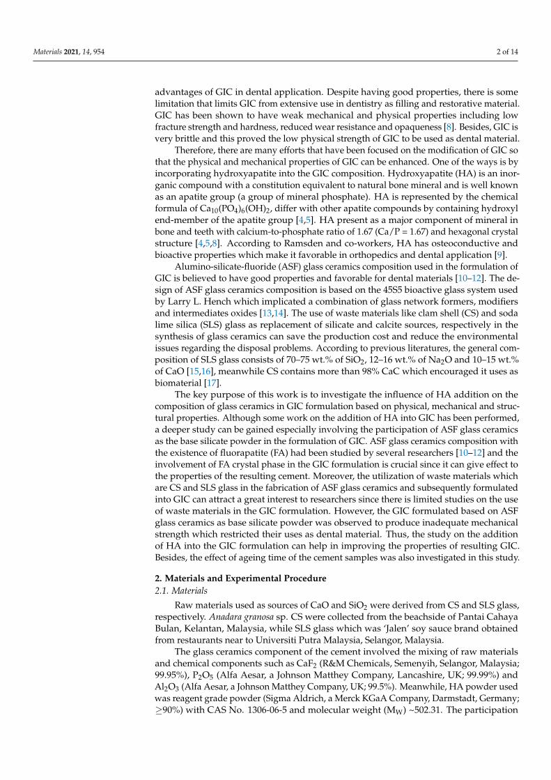
3. Results3.1. Density Analysis
Figure 1 shows the density results of GIC and GIC1 samples at different ageing timewhile Table 1 depicts the statistical data of the density results. According to the figure andtable, the density of the GIC sample at 1 day of ageing time was 1.681 g/cm3. The additionof 1 wt.% of HA powder into GIC formulation represented by the GIC1 sample produceddensity of 1.693 g/cm3 at 1 day of ageing time. The density of cement samples increasedwith the addition of HA powder from 1.681 to 1.693 g/cm3. On the other hand, the densityresults increased as ageing time increased. This trend can be observed in both GIC andGIC1 samples. GIC sample showed the increment in density from 1.681 g/cm3 at 1 day ofageing time to 1.824 g/cm3 at 21 days of ageing time. Meanwhile, GIC1 sample showed an
Materials 2021, 14, 954 5 of 14
increase in density value from 1 day of ageing time with 1.693 g/cm3 to 21 day of ageingtime with 1.839 g/cm3. The highest density results were 1.824 and 1.839 g/cm3 at 21 daysof ageing time for GIC and GIC1 samples, respectively.
Figure 1. Density results of GIC and GIC1 samples at different ageing time (error bars indicatestandard error).
Table 1. Mean density along with the standard deviation (SD) of the GIC and GIC1 samples atdifferent ageing time.
Density of Sample(g/cm3)
Ageing Time
1 Day 7 Days 14 Days 21 Days
GIC 1.681 (0.011) a 1.697 (0.006) a 1.702 (0.015) a 1.824 (0.043) b
GIC1 1.693 (0.015) a 1.738 (0.033) a 1.805 (0.007) b 1.839 (0.016) b
The different superscript letters (example: a and b) for each sample (each row) represent significantdifference between ageing time (p < 0.01). Meanwhile, similar superscript letters (example: a and a)represent no significance difference between ageing time.
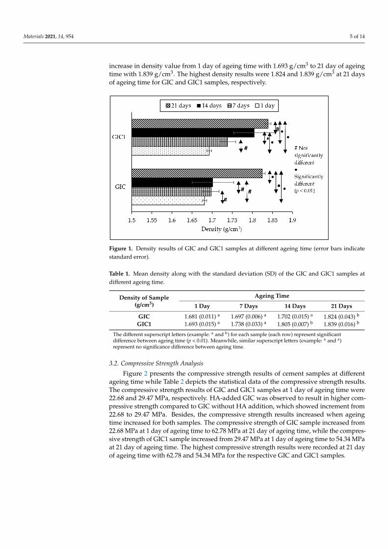
3.2. Compressive Strength Analysis
Figure 2 presents the compressive strength results of cement samples at differentageing time while Table 2 depicts the statistical data of the compressive strength results.The compressive strength results of GIC and GIC1 samples at 1 day of ageing time were22.68 and 29.47 MPa, respectively. HA-added GIC was observed to result in higher com-pressive strength compared to GIC without HA addition, which showed increment from22.68 to 29.47 MPa. Besides, the compressive strength results increased when ageingtime increased for both samples. The compressive strength of GIC sample increased from22.68 MPa at 1 day of ageing time to 62.78 MPa at 21 day of ageing time, while the compres-sive strength of GIC1 sample increased from 29.47 MPa at 1 day of ageing time to 54.34 MPaat 21 day of ageing time. The highest compressive strength results were recorded at 21 dayof ageing time with 62.78 and 54.34 MPa for the respective GIC and GIC1 samples.

Materials 2021, 14, 954 6 of 14
Figure 2. Compressive strength results of GIC and GIC1 samples at different ageing time(error bars indicate standard error).
Table 2. Mean compressive strength along with standard deviation (SD) of GIC and GIC1 samples atdifferent ageing time.
Compressive Strength ofSample (MPa)
Ageing Time
1 Day 7 Days 14 Days 21 Days
GIC 22.68 (0.73) a 31.89 (0.86) b 40.44 (0.58) c 62.78 (0.21) d
GIC1 29.47 (2.17) a 39.17 (1.24) b 45.75 (5.11) c 54.34 (0.63) d
The different superscript letters (example: a and b) for each sample (each row) represent significantdifference between ageing time (p < 0.01). Meanwhile, similar superscript letters (example: a and a)represent no significance difference between ageing time.
3.3. X-ray Diffraction (XRD) Analysis
Figure 3 shows the XRD patterns of GIC and GIC1 samples at different ageing times.According to Figure 3, the formation of fluorapatite (FA; ICDD file No. 98-001-7206) crystalphase was observed in the GIC sample, from 1 day until 21 days of ageing time. For GIC1sample at different ageing times, the addition of HA powder into GIC resulted in thedetection of fluorohydroxyapatite (FHA; ICDD file No. 98-008-0180) crystal phase, as seenin Figure 3. Based on the observation from Figure 3, the highest crystal peak was detectedat the same diffraction angle, which was 2θ = 32◦. No significant difference of XRD patternwas observed from 1, 7, 14 and 21 days of ageing time for both GIC and GIC1 samples.
3.4. Fourier Transform Infrared (FTIR) Analysis
Figure 4 shows the FTIR patterns at different ageing times for the GIC and GIC1samples. Meanwhile, Table 3 depicts the vibrational modes assigned for FTIR spectra thatexisted in the cement samples. By referring to Figure 4, v2 O–P–O bending vibrational modewas detected at ~440 cm−1 while v4 O–P–O bending mode was discovered at ~570 and~600 cm−1. Besides, the existence of the FTIR spectral band at ~1020 cm−1 represented thevibrational mode of v3 asymmetric P–O stretching. The detection of C–O and asymmetricCOOH vibration modes were found at wavenumbers ~1460 and ~1550 cm−1, respectively.A broad and wide peak was discovered at ~3400 cm−1 indicated OH vibration mode.The FTIR pattern of GIC1 sample at different ageing time revealed the similar vibrational

Materials 2021, 14, 954 7 of 14
modes existed in the sample when compared to GIC sample. However, the only differencefound was the existence of OH–F vibration mode at ~3550 cm−1.
Figure 3. XRD patterns of GIC (A) and GIC1 (B) sample with (a) 1 day, (b) 7 days, (c) 14 days, and (d) 21 days of ageing time.
Figure 4. FTIR pattern of GIC (A) and GIC1 (B) sample for (a) 1 day, (b) 7 days, (c) 14 days, and (d) 21 days of ageing time.

Materials 2021, 14, 954 8 of 14
Table 3. Vibrational modes assigned for GIC and GIC1 samples.
Wavenumber (cm−1) Vibrational Mode References
~440 v2 O–P–O bending [19,20]~570, ~600 v4 O–P–O bending [4,21–24]
~1020 v3 asymmetric P-O stretching [19,20]~1460 C–O vibration [21]~1550 Asymmetric COOH [25]~3400 OH vibration [26,27]~3550 OH–F vibration [26,27]
3.5. Field Emission Scanning Electron Microscopy (FESEM) Analysis
Microstructures of GIC and GIC1 samples at different ageing times are shown inFigure 5. The observation of irregular shape of glass ceramics structure with non-uniformparticle distribution was detected in both cement samples, as seen in FESEM images.The formation of needle-like and spherical particles was observed on the surface of glassceramics particles. Besides, no significant difference was detected in the samples whencompared to samples with different ageing time.
3.6. Energy Dispersive X-ray (EDX) Analysis
Table 4 shows the chemical composition of the GIC and GIC1 samples at 1 day ofageing time. The chemical elements existed were oxygen (O), carbon (C), calcium (Ca),aluminum (Al), phosphorus (P), silicon (Si), fluorine (F) and sodium (Na). The mostelement detected in GIC and GIC1 samples was O with the respective 42.54 and 45.20%.Meanwhile, C existed with 22.86 and 20.90%, while Al element was found in the GIC andGIC1 sample with 7.18 and 7.11%, respectively. The detection of P with 5.11 and 5.16%as well as Si element with 4.33 and 3.67% in the respective GIC and GIC1 were recordedfrom EDX analysis. Element F existed as a minor element with 3.87 and 3.91% while Naconsisted at about 1.30 and 1.15% in the respective GIC and GIC1 samples.
Table 4. Chemical composition of GIC and GIC1 samples at 1 day of ageing time.
ElementWeight Percentage (%)
GIC GIC1
O 42.54 45.20C 22.86 20.90Ca 12.81 12.90Al 7.18 7.11P 5.11 5.16Si 4.33 3.67F 3.87 3.91
Na 1.30 1.15
Total 100.00 100.00

Materials 2021, 14, 954 9 of 14
Figure 5. FESEM micrograph under magnification of 10,000× for samples (a) GIC 1 day, (b) GIC 7days, (c) GIC 14 days, (d) GIC 21 days, (e) GIC1 1 day, (f) GIC1 7 days, (g) GIC1 14 days, and (h)GIC1 21 days.
4. Discussion
In this work, density measurement was used to observe the physical properties ofboth GIC and HA-added GIC samples at different ageing time. The density results in

Materials 2021, 14, 954 10 of 14
Figure 1 and Table 1 revealed an increment in density of GIC with the addition of HApowder. This is due to high density of HA compared to ASF glass ceramics samples. In thiscase, the measured density of ASF glass ceramics was 2.561 g/cm3 while the density of HAused in this work was 2.673 g/cm3. According to a study, the increase in the density ofHA-added GIC samples agrees qualitatively with the one, as predicted by the compositionrelation, and might be due to the replacement of high density of HA powders [20]. Thus,the density of HA which has a higher density was proposed to dominate the density ofcement samples. Besides, the density of GIC and GIC1 samples was observed to increase asthe ageing time increased from 1 day to 21 days. The increase in density along with ageingtime is due to the increment in ratio of bound to unbound water [25,28]. The reaction inthe GIC matrix occurs with the presence of water as a medium of setting reaction over aperiod of time. Moreover, water acts as a medium of reaction which helps in maturationand hardening process of cement.
The results of compressive strength test presented in Figure 2 and Table 2 found thatthe addition of HA into GIC resulted in higher compressive strength compared to GIC with-out HA addition. This is due to the presence of crystal phase in the glass structure of glassionomer [23,29]. Metal ions such as Ca2+ are displaced from the added HA, therefore in-creased the participation of ions in acid base reaction. The setting reaction that occurs whenthere is addition of HA into GIC has been explained in few studies [21,30]. Upon additionof HA into GIC, H+ from acid polymer attacks the ceramic particles in the polysalt bridgeformation and cross-linking, thus forming an intermediate layer from the interaction. Theintermediate layer is very resistant to acid and is difficult to break. Therefore, the additionof HA into GIC enhance the mechanical strength of the resulting GIC [23,30,31]. Figure 6explained the schematic diagram of setting reaction of HA added GIC.
Figure 6. Schematic diagram of setting reaction of HA-added GIC.
Besides, the compressive strength of both GIC and GIC1 samples increased as ageingtime increased. The immersion of cements into deionized water as a medium of reaction atdifferent periods of time allows reaction between the GIC matrix to occur. Longer ageingtime seemed to improve the strength of the GIC. Compressive strength results of the cementsamples are related to the density of the samples. The density of GIC samples affects themechanical properties of the set cement [4]. The addition of HA resulted in higher densitycompared to GIC without the HA addition. Such results are in line with the observations ofGoenka et al. [4] who reported that the samples of the HA-added GIC resulted in increased

Materials 2021, 14, 954 11 of 14
hardness due to increased density of the set cement. Besides, some studies reported thatthe addition of certain volume of HA and the role of PAA in cement caused an increase incrosslink density of the cement structure, thus helping to increase the mechanical strengthof the GIC [4,28].
Through Turkey-Kramer post-hoc evaluation, the significant difference in densityand compressive strength of each cement samples when compared between differentageing times was determined and the results were presented in Tables 1 and 2, respec-tively. According to density results in Table 1, the comparison between different ageingtime revealed that not all GIC and GIC1 samples showed significantly different results.Meanwhile, the compressive strength results shown in Table 2 revealed the significantlydifferent results when compared to different ageing time and this situation occurred inboth GIC and GIC1 samples. The increase in ageing time caused the significant increasein compressive strength value. Several studies reported that no significant difference wasobserved in compressive strength between GIC without HA addition and HA-added GICsamples at different ageing time [32,33]. Thereby, it can be concluded that HA additioninto GIC composition did not give any significant effect in compressive strength of theresulting GIC.
XRD patterns in Figure 3 revealed the formation of FA crystal phase in GIC samplesat 1, 7, 14 and 21 days of ageing time. This is due to the utilization of ASF glass ceramics asbase powder in the formulation of GIC. The study of ASF glass ceramics had been done bysome researchers and found that the ASF glass ceramics with similar composition causedthe development of FA crystal phase [10–12]. The addition of a small weight percentageof HA did not affect the XRD pattern except for the detection of FHA phase. Accordingto some studies, the formation of FHA in modified GIC samples of different ageing timeis due to the substitution of hydroxyl ion (OH−) by fluoride ion (F−) that occurred in theapatite composition [28,34]. The reaction of the released fluoride ion with the HA crystalwas summarized in general by Equation (3) [35]. In this case, the substitution of F- ioninto the apatite crystal caused the formation of the FHA crystal phase. FHA crystal wasfound to occur naturally in human bone and teeth. Owing better mechanical and chemicalproperties, FA and FHA have been attracting a great attention in biomedical applicationespecially for dental restoration [36].
Ca10 (PO 4)6(OH)2 + F− → Ca10 (PO 4)6FOH + OH− (3)
FTIR patterns of GIC and GIC1 samples at different ageing times depicted in Figure4 revealed the existence of similar spectral bands. The appearance of the spectral bandat ~400 cm−1 was belonged to double degenerate O–P–O bending mode (v2) indicatedby phosphate group in apatite sample [19,20]. Next, the emergence of triple degenerateO–P–O bending mode (v4) which appeared at both intense peaks at wavenumber ~570 and~600 cm−1. Another occurrence of phosphate chemical bonding was observed at an intensepeak of ~1020 cm−1 which represented asymmetric P–O stretching mode (v3). According torelated literature, more than one distinction site of PO4
3− vibration modes was found inthe FTIR spectral wavenumber of cement sample which proved the formation of apatitephase inside the cement samples [4,21–24].
On the other hand, the carbonate group depicted by C–O vibration mode at wavenum-ber ~1460 cm−1 confirmed the involvement of carbonate precursors in the productionof GIC samples. Besides, Khiri and co-workers explained the formation of asymmetricCOOH band at 1550 cm−1 which originated from the carboxyl group in PAA used in theformulation of GIC [25]. The cross-linking reaction of the carboxylic group was observedin all cement samples due to an active reaction that occurred between PAA and ASF glassceramics. Next, a broad absorption band was discovered at high frequency of wavenumber~3400 cm−1 which indicated the occurrence of OH vibration mode. Jekonovic et al. [26] andMontazeri et al. [27] stated that the occurrence of the vibrational mode is due to stretchingof intermolecular H bonds caused by water absorption. At the same time, the existence ofthis spectral band showed the presence of water during cement preparation [26].

Materials 2021, 14, 954 12 of 14
The only difference spotted in FTIR spectral of GIC1 sample compared to GIC samplewas the existence of hydroxyl group by incorporation of fluorine ion which was representedby OH–F vibration mode at wavenumber ~3550 cm−1, claimed the formation of FHA phasein GIC1 sample. Studies from Eslamia et al. [37] and Jokanovic et al. [27] explained that theband existed at 3550 cm−1 in HA samples due to stretching mode of hydrogen bonded OH−
ion. When hydroxyl groups are partially replaced with fluoride ions in HA, the stretchingmode of OH− can shift to the new band that arises from OH-F bond. In FHA, OH bandappeared at around 3550 cm−1 indicating that the OH–F interaction influences all the OH−
ions present in the samples.FESEM morphology of both GIC and GIC1 samples in Figure 5 showed the irregular
shape of glass ceramics structure with non-uniform particle distribution. The detectionof apatite crystals by the formation of needle-like and spherical particles were observedto disperse on the surface of the glass ceramics particle. This observation is in conjunc-tion with the findings found by Khaghani and co-workers, who investigated the SEMstructure of HA incorporation into GIC composition [29]. According to some literatures,hexagonal structure of apatite was illustrated by rod or needle-like and spherical struc-ture [26,27,38]. Due to high adsorbability, apatite crystals with rod-like morphology haveshown favorable biocompatibility and bioactivity, as the corresponding interactions of Vander Waals are proportional to the large rod surface region [39,40].
EDX analysis was utilized to investigate the chemical composition of cement samplesat 1 day of ageing time and the results were presented in Table 4. Most of the elementsdetected by EDX test originated from chemical compounds used in the preparation ofglass ceramics powder which then used as base powder for GIC formulation, meanwhile Cwas developed from polyacid used in initiating the reaction of GIC. Oxygen existed as themajor element consisted in the cement samples due to consumption of oxide elements inthe fabrication of ASF glass ceramics powder. A study from Rahman et al. [12] explainedthe existence of minor elements such as Na, Si, Al, and other elements make up a ma-jority of SLS glasses, which then utilized in the making of base powder of the cement.In tooth structure, the hardest and highly mineralized substance, called enamel consistsof 96% inorganic matter, 1–2% organic matter and 2–3% water. The majority of inorganicmatter which make up the enamel structure is owned by distinct crystalline phases ofHA. Calcium (Ca) and phosphorus (P) are essential elements composed in HA, and thecomposition of both elements is crucial for the saturation of apatite that improves theprocess of remineralization [10].
5. Conclusions
In the current study, CS and SLS glass had been exploited to fabricate ASF glassceramics which subsequently used in the formulation of GIC. The physical, mechanical andstructural properties of GIC formulated based on ASF glass ceramics as base silicatepowder was enhanced by the addition of HA powder, which revealed the enhancement indensity and compressive strength test along with the formation of FHA crystal phase fromstructural studies. Besides, ageing time also had been observed to improve the propertiesof the resulting GIC. In a nutshell, the improved properties of physical, structural andmechanical encourage the use of HA-added GIC in biomedical application especially fordental restoration.
Author Contributions: W.N.W.J., K.A.M. and M.H.M.Z. take part in planning and conducting thedesign of the study, as well as preparing the manuscript; N.Z., M.Z.A.K. and R.A.J. participated inexperimental procedure, as well as analysis part. N.A.A.R. and E.K. assisted as project conceptualiza-tion and analysed the data of ASF glass ceramics used in the GIC formulation. All authors have readand agreed to the published version of the manuscript.
Funding: This research was funded by Malaysian Ministry of Higher Education (MOHE) underFundamental Research Grant Scheme (FRGS) grant number 5540163 and also Universiti PutraMalaysia under Inisiatif Putra Siswazah (IPS) grant number 9627400.

Materials 2021, 14, 954 13 of 14
Conflicts of Interest: The authors declare no conflict of interest.
References1. Wilson, A.D.; Kent, B.E. A new translucent cement for dentistry, the glass ionomer cement. J. Chem. Technol. Biotechnol. 1971,
21, 313. [CrossRef]2. McLean, J.W.; Gasser, O. Glass-cermet cements. Quintessence Int. 1985, 16, 333–343.3. Moshaverinia, A.; Roohpour, N.; Chee, W.W.; Schricker, S.R. A review of powder modifications in conventional glass-ionomer
dental cements. J. Mater. Chem. 2011, 21, 1319–1328. [CrossRef]4. Goenka, S.; Balu, R.; Kumar, T.S. Effects of nanocrystalline calcium deficient hydroxyapatite incorporation in glass ionomer
cements. J. Mech. Behav. Biomed. Mater. 2012, 7, 69–76. [CrossRef] [PubMed]5. Shiekh, R.A.; Rahman, A.I.; Luddin, N. Modification of glass ionomer cement by incorporating hydroxyapatite-silica nano-powder
composite: Sol-gel synthesis and characterization. Ceram. Int. 2014, 40, 3165–3170. [CrossRef]6. Sharafeddin, F.; Feizi, N. Evaluation of the effect of adding micro-hydroxyapatite and nano-hydroxyapatite on the microleakage
of conventional and resin-modified Glass-ionomer Cl V restorations. J. Clin. Exp. Dent. 2017, 9, e242–e248. [CrossRef]7. Noori, A.J.; Kareem, F.A. Setting time, mechanical and adhesive properties of magnesium oxide nanoparticles modified glass-
ionomer cement. J. Mater. Res. 2019, 9, 1809–1818. [CrossRef]8. Rahman, I.A.; Ghazali, N.A.M.; Bakar, W.Z.W.; Masudi, S.A.M. Modification of glass ionomer cement by incorporating
nanozirconia-hydroxyapatite-silica nano-powder composite by the one-pot technique for hardness and aesthetics improve-ment. Ceram. Int. 2017, 43, 13247–13253. [CrossRef]
9. Ramsden, R.T.; Herdman, R.C.T.; Lye, R.H. Ionomeric bone cement in otoneurological surgery. J. Laryngol. Otol. 1992,106, 949–953. [CrossRef]
10. Abdul Jalil, R.; Amin Matori, K.; Mohd Zaid, M.H.; Zainuddin, N.; Ahmad Khiri, M.Z.; Abdul Rahman, N.A.; Wan Jusoh, W.N.;Kul, E. A study of fluoride-containing bioglass system for dental materials derived from clam shell and soda lime silica glass. J.Spectrosc. 2020, 2020, 1–9. [CrossRef]
11. Jusoh, W.N.W.; Matori, K.A.; Zaid, M.H.M.; Zainuddin, N.; Khiri, M.Z.A.; Rahman, N.A.A.; Jalil, R.A.; Kul, E. Effect of sinteringtemperature on physical and structural properties of Alumino-Silicate-Fluoride glass ceramics fabricated from clam shell andsoda lime silicate glass. Results Phys. 2019, 12, 1909–1914. [CrossRef]
12. Rahman, N.A.A.; Matori, K.A.; Zaid, M.H.M.; Zainuddin, N.; Ab Aziz, S.; Khiri, M.Z.A.; Jalil, R.A.; Jusoh, W.N.W. Fabricationof Alumino-Silicate-Fluoride based bioglass derived from waste clam shell and soda lime silica glasses. Results Phys. 2019,12, 743–747. [CrossRef]
13. Fernandes, H.R.; Gaddam, A.; Rebelo, A.; Brazete, D.; Stan, G.E.; Ferreira, J.M. Bioactive glasses and glass-ceramics for healthcareapplications in bone regeneration and tissue engineering. Materials 2018, 11, 2530. [CrossRef]
14. Henao, J.; Poblano-Salas, C.; Monsalve, M.; Corona-Castuera, J.; Barceinas-Sanchez, O. Bio-active glass coatings manufactured bythermal spray: A status report. J. Mater. Res. Technol. 2019, 8, 4965–4984. [CrossRef]
15. Bauccio, M. ASM Engineered Materials Reference Book; CRC Press LLC: Boca Raton, FL, USA, 1994.16. Pfaender, H.G. Schott Guide to Glass; Springer Science & Business Media: Berlin, Germany, 2012.17. Awang-Hazmi, A.J.; Zuki, A.B.Z.; Noordin, M.M.; Jalila, A.; Norimah, Y. Mineral composition of the cockle (Anadara granosa)
shells of west coast of Peninsular Malaysia and it’s potential as biomaterial for use in bone repair. J. Anim. Vet. Adv. 2007,6, 591–594.
18. Loyd, D. Physics Lab Manual; Cengage Learning: Orlando, FL, USA, 2007.19. Mandal, T.; Mishra, B.K.; Garg, A.; Chaira, D. Optimization of milling parameters for the mechanosynthesis of nanocrystalline
hydroxyapatite. Powder Technol. 2014, 253, 650–656. [CrossRef]20. Zarifah, N.A.; Matori, K.A.; Sidek, H.A.A.; Wahab, Z.A.; Salleh, M.M.; Zainuddin, N.; Khiri, M.Z.A.; Farhana, N.S.; Omar, N.A.S.
Effect of hydroxyapatite reinforced with 45S5 glass on physical, structural and mechanical properties. Procedia Chem. 2016,19, 30–37. [CrossRef]
21. Moshaverinia, A.; Ansari, S.; Moshaverinia, M.; Roohpour, N.; Darr, J.A.; Rehman, I. Effects of incorporation of hydroxya-patite and fluoroapatite nanobioceramics into conventional glass ionomer cements (GIC). Acta Biomater. 2008, 4, 432–440.[CrossRef] [PubMed]
22. Garcia-Contreras, R.; Scougall-Vilchis, R.J.; Contreras-Bulnes, R.; Sakagami, H.; Morales-Luckie, R.A.; Nakajima, H. Mechanical,antibacterial and bond strength properties of nano-titanium-enriched glass ionomer cement. J. Appl. Oral Sci. 2015, 23, 321–328.[CrossRef] [PubMed]
23. Barandehfard, F.; Rad, M.K.; Hosseinnia, A.; Khoshroo, K.; Tahriri, M.; Jazayeri, H.E.; Moharamzadeh, K.; Tayebicgh, L.The addition of synthesized hydroxyapatite and fluorapatite nanoparticles to a glass-ionomer cement for dental restoration andits effects on mechanical properties. Ceram. Int. 2016, 42, 17866–17875. [CrossRef]
24. Alatawi, R.A.; Elsayed, N.H.; Mohamed, W.S. Influence of hydroxyapatite nanoparticles on the properties of glass ionomercement. J. Mater. Res. Technol. 2019, 8, 344–349. [CrossRef]
25. Khiri, M.Z.A.; Matori, K.A.; Zaid, M.H.M.; Abdullah, A.C.; Zainuddin, N.; Jusoh, W.N.W.; Jalil, R.A.; Rahman, N.A.A.; Kul, E.;Wahab, S.A.A.; et al. Soda lime silicate glass and clam shell act as precursor in synthesize calcium fluoroaluminosilicate glass tofabricate glass ionomer cement with different ageing time. J. Mater. Res. Technol. 2020, 9, 6125–6134. [CrossRef]

Materials 2021, 14, 954 14 of 14
26. Montazeri, N.; Jahandideh, R.; Biazar, E. Synthesis of fluorapatite-hydroxyapatite nanoparticles and toxicity investigations. Int. J.Nanomed. 2011, 6, 197–201.
27. Jokanovic, V.; Colovic, B.; Jovic, N.; Babic-Stojic, B.; Jokanovic, B. Mechanochemical and low-temperature synthesis of nanocrys-talline fluorohydroxyapatite/fluorapatite. Int. J. Appl. Ceram. Technol. 2013, 10, 957–969. [CrossRef]
28. Nicholson, J.W. Chemistry of glass-ionomer cements: A review. Biomaterials 1998, 19, 485–494. [CrossRef]29. Khaghani, M.; Alizadeh, S.; Doostmohammadi, A. Influence of incorporating fluoroapatite nanobioceramic on the compressive
strength and bioactivity of glass ionomer cement. J. Dent. Biomater. 2016, 3, 276–283.30. Arita, K.; Yamamoto, A.; Shinonaga, Y.; Harada, K.; Abe, Y.; Nakagawa, K.; Sugiyama, S. Hydroxyapatite particle characteristics
influence the enhancement of the mechanical and chemical properties of conventional restorative glass ionomer cement. Dent.Mater. J. 2011, 30, 672–683. [CrossRef] [PubMed]
31. Bali, P.; Prabhakar, A.R.; Basappa, N. An invitro comparative evaluation of compressive strength and antibacterial activity of con-ventional GIC and hydroxyapatite reinforced GIC in different storage media. J. Clin. Diagn. Res. 2015, 9, ZC51–ZC55. [CrossRef]
32. Yap, A.U.J.; Pek, Y.S.; Kumar, R.A.; Cheang, P.; Khor, K.A. Experimental studies on a new bioactive material: HAIonomer cements.Biomaterials 2002, 23, 955–962. [CrossRef]
33. Shinonaga, Y.; Arita, K.; Nishimura, T.; Chiu, S.Y.; Chiu, H.H.; Abe, Y.; Sonomoto, M.; Harada, K.; Nagaoka, N. Effects ofporous-hydroxyapatite incorporated into glass-ionomer sealants. Dent. Mater. J. 2015, 34, 196–202. [CrossRef]
34. Wei, M.; Evans, J.H.; Bostrom, T.; Grøndahl, L. Synthesis and characterization of hydroxyapatite, fluoride-substituted hydroxyap-atite and fluorapatite. J. Mater. Sci. Mater. Med. 2003, 14, 311–320. [CrossRef] [PubMed]
35. Šupová, M. Substituted hydroxyapatites for biomedical applications: A review. Ceram. Int. 2015, 41, 9203–9231. [CrossRef]36. Pajor, K.; Pajchel, L.; Kolmas, J. Hydroxyapatite and fluorapatite in conservative dentistry and oral implantology—A review.
Materials 2019, 12, 2683–2699. [CrossRef]37. Eslamia, H.; Moztarzadeha, F.; Khoshroob, K.; Ashuria, M.; Tahriria, M. Synthesis and characterization of Ca5(PO4)3(OH)
(0 ≤ x ≤ 1) nanopowders via pH-cycling method as bioceramics. In Proceedings of the 4th International Conference on Nanos-tructures (ICNS4), Kish Island, Iran, 12–14 March 2012.
38. Azami, M.; Jalilifiroozinezhad, S.; Mozafari, M. Calcium fluoride/hydroxyfluorapatite nanocrystals as novel biphasic solidsolution for tooth tissue engineering and regenerative dentistry. Key Eng. Mater. 2012, 493, 626–631. [CrossRef]
39. Sasani, N.; Khadivi, A.H.; Zebarjad, S.M.; Vahdati, K.J. Characterization of rod-like high-purity fluorapatite nanopowdersobtained by sol-gel method. J. Ultrafine Grained Nanostruct. Mater. 2013, 46, 31–37.
40. Zandi, M.; Mirzadeh, H.; Mayer, C.; Urch, H.; Eslaminejad, M.B.; Bagheri, F.; Mivehchi, H. Biocompatibility evaluation ofnano-rod hydroxyapatite/gelatin coated with nano-HAp as a novel scaffold using mesenchymal stem cells. J. Biomed. Mater. Res.A 2010, 92, 1244–1255. [CrossRef]