Phase transformation,microstructuralandmechanicalproperties of...
10
CERAMICS INTERNATIONAL Available online at www.sciencedirect.com Ceramics International 39 (2013) 9835–9844 Phase transformation, microstructural and mechanical properties of hydroxyapatite/alumina nanocomposite scaffolds produced by freeze casting Seyed Mohammad Hossein Ghazanfari, Ali Zamanian n Nanotechnology & Advanced Materials Department, Materials & Energy Research Center, P.O. Box: 13145-1659, Karaj, Iran Received 7 February 2013; received in revised form 25 May 2013; accepted 27 May 2013 Available online 31 May 2013 Abstract Freeze casting is an effective fabrication technique that allows producing scaffolds with variable porosity, pore size, pore orientation and compressive strength. To our knowledge, the present study is the first investigation on the replacement effects of microparticles with nanoparticles in bone scaffolds prepared through the freeze casting method. In this study, the effect of nano-alumina content on phase transformation, microstructural and mechanical properties of hydroxyapatite/nano-alumina (HA/nAl 2 O 3 ) nanocomposite scaffolds, fabricated through the freeze casting method at different cooling rates, has been investigated. In the first stage, slurries with 15 vol% solid loading and different nano-alumina content were prepared. In the next stage, cooling rates of 1 and 4 K/min were applied to synthesize the porous scaffolds, followed by sintering at 1250 and 1350 1C. The characteristics of the initial powders, and phase composition, microstructure, pore size, pore distribution and mechanical strength of the scaffolds were assessed. The porosity of the synthesized scaffolds was in a range of 78–85%, and the compressive strength varied from 0.2 to 4 MPa as a function of nAl 2 O 3 concentration, cooling rate, and sintering temperature. Surprisingly, further addition of nAl 2 O 3 not only affected the microstructural features, but also provided mechanisms to improve the mechanical strength of the scaffolds. & 2013 Elsevier Ltd and Techna Group S.r.l. All rights reserved. Keywords: Freeze casting; Ice templating; Hydroxyapatite; Nano-alumina; Scaffold 1. Introduction Implantation of bone autografts or allografts is a simple strategy to heal large bone defects. However, there are some drawbacks of these strategies which limit their widespread usage, such as extended surgical time and donor site morbidity for autograft, and adverse immune response and pathogen disease transmission for allograft. These problems have guided researchers for the development of bone substitute materials [1–4]. As the most promising bone substitute materials, calcium phosphate (CP) compounds have been widely used clinically due to their similarity to hard tissues. Among different CP ceramics, hydroxyapatite (HA) and tri-calcium phosphate (TCP), or combi- nations of these two materials (biphasic ceramics) have gained special attentions for a wide range of applications such as alveolar ridge augmentation [5], maxillofacial reconstruction [6], orbital implants [7], spine fusion [8] and repair of bone defects [9]. These materials have been clinically used in dense, granular and porous forms [10–12]. It is known that the porous constructs of this class of materials have the possibility of tissue growth, and the capability of being replaced by bone tissue [13–15]. Unlikely, CPs have also some drawbacks such as relatively low mechanical strength (TCPs and biphasic materials) and thermal decomposi- tion during the sintering [16,17]. Many materials such as zirconia [18], alumina [19], silica [20], titania [21] and calcium silicate [22] have been employed to overcome these drawbacks. Gen- erally, porous alumina structures in comparison with porous CPs have better mechanical strength but due to the lack of appropriate cellular responses they cannot form biochemical interfacial bond with the natural tissue [23–25]. Therefore, it is reasonable to reinforce CP porous structures with alumina powder to fabricate highly bioactive nanocomposite scaffolds with a relatively higher mechanical strength. www.elsevier.com/locate/ceramint 0272-8842/$ - see front matter & 2013 Elsevier Ltd and Techna Group S.r.l. All rights reserved. http://dx.doi.org/10.1016/j.ceramint.2013.05.096 n Corresponding author. Tel.: +98 912 3211180; fax: +98 26 36201888. E-mail addresses: [email protected], [email protected] (A. Zamanian).
Transcript of Phase transformation,microstructuralandmechanicalproperties of...

CERAMICSINTERNATIONAL
Available online at www.sciencedirect.com
0272-8842/$ - sehttp://dx.doi.org/
nCorrespondinE-mail addre
zamanian_a@ya
Ceramics International 39 (2013) 9835–9844www.elsevier.com/locate/ceramint
Phase transformation, microstructural and mechanical propertiesof hydroxyapatite/alumina nanocomposite scaffolds produced
by freeze casting
Seyed Mohammad Hossein Ghazanfari, Ali Zamaniann
Nanotechnology & Advanced Materials Department, Materials & Energy Research Center, P.O. Box: 13145-1659, Karaj, Iran
Received 7 February 2013; received in revised form 25 May 2013; accepted 27 May 2013Available online 31 May 2013
Abstract
Freeze casting is an effective fabrication technique that allows producing scaffolds with variable porosity, pore size, pore orientation andcompressive strength. To our knowledge, the present study is the first investigation on the replacement effects of microparticles with nanoparticlesin bone scaffolds prepared through the freeze casting method. In this study, the effect of nano-alumina content on phase transformation,microstructural and mechanical properties of hydroxyapatite/nano-alumina (HA/nAl2O3) nanocomposite scaffolds, fabricated through the freezecasting method at different cooling rates, has been investigated. In the first stage, slurries with 15 vol% solid loading and different nano-aluminacontent were prepared. In the next stage, cooling rates of 1 and 4 K/min were applied to synthesize the porous scaffolds, followed by sintering at1250 and 1350 1C. The characteristics of the initial powders, and phase composition, microstructure, pore size, pore distribution and mechanicalstrength of the scaffolds were assessed. The porosity of the synthesized scaffolds was in a range of 78–85%, and the compressive strength variedfrom 0.2 to 4 MPa as a function of nAl2O3 concentration, cooling rate, and sintering temperature. Surprisingly, further addition of nAl2O3 not onlyaffected the microstructural features, but also provided mechanisms to improve the mechanical strength of the scaffolds.& 2013 Elsevier Ltd and Techna Group S.r.l. All rights reserved.
Keywords: Freeze casting; Ice templating; Hydroxyapatite; Nano-alumina; Scaffold
1. Introduction
Implantation of bone autografts or allografts is a simple strategyto heal large bone defects. However, there are some drawbacks ofthese strategies which limit their widespread usage, such asextended surgical time and donor site morbidity for autograft,and adverse immune response and pathogen disease transmissionfor allograft. These problems have guided researchers for thedevelopment of bone substitute materials [1–4].
As the most promising bone substitute materials, calciumphosphate (CP) compounds have been widely used clinically dueto their similarity to hard tissues. Among different CP ceramics,hydroxyapatite (HA) and tri-calcium phosphate (TCP), or combi-nations of these two materials (biphasic ceramics) have gainedspecial attentions for a wide range of applications such as alveolar
e front matter & 2013 Elsevier Ltd and Techna Group S.r.l. All ri10.1016/j.ceramint.2013.05.096
g author. Tel.: +98 912 3211180; fax: +98 26 36201888.sses: [email protected],hoo.com (A. Zamanian).
ridge augmentation [5], maxillofacial reconstruction [6], orbitalimplants [7], spine fusion [8] and repair of bone defects [9]. Thesematerials have been clinically used in dense, granular and porousforms [10–12]. It is known that the porous constructs of this classof materials have the possibility of tissue growth, and thecapability of being replaced by bone tissue [13–15]. Unlikely,CPs have also some drawbacks such as relatively low mechanicalstrength (TCPs and biphasic materials) and thermal decomposi-tion during the sintering [16,17]. Many materials such as zirconia[18], alumina [19], silica [20], titania [21] and calcium silicate[22] have been employed to overcome these drawbacks. Gen-erally, porous alumina structures in comparison with porous CPshave better mechanical strength but due to the lack of appropriatecellular responses they cannot form biochemical interfacial bondwith the natural tissue [23–25]. Therefore, it is reasonable toreinforce CP porous structures with alumina powder to fabricatehighly bioactive nanocomposite scaffolds with a relatively highermechanical strength.
ghts reserved.

S.M.H. Ghzanfari, A. Zamanian / Ceramics International 39 (2013) 9835–98449836
Different fabrication techniques such as pore-foaming [26],infiltration of compression molded sponge [27], gel-casting [28],slip-casting [29], starch consolidation [30], microwave processing[31] and freeze casting [32,33] can be used for the production ofporous structures. Among these techniques, freeze casting hasgained much attention due to its superior advantages such assimplicity, low shrinkaging in forming process, possibility ofcontrolling the porosity, interconnectivity, relatively goodmechanical strength etc. [14,34,35]. Different additives have beenalso used to improve the properties of HA-based scaffoldsfabricated via the freeze casting method. Recently, Zuo et al.[36] investigated the effect of polyvinyl alcohol (PVA) additionon the morphology of porous HA structures produced by freezecasting. According to their results the PVA-free HA structuresshowed longer lamellar pores. Zhang et al. [37] studied the effectof gelatin addition on the microstructure of freeze cast porous HAstructures. They showed that the viscosity of HA slurry and alsothe linear shrinkage ratio of the sintered samples were enhancedby increasing the gelatin concentration. In another work, Blindowet al. [20] investigated the influence of nano SiO2 addition on HAscaffolds produced via freeze casting. They found that SiO2
addition announced a partial phase transformation of HA toβ-TCP and reduced the shrinkage of the scaffolds after sintering.This interesting study could be completed by investigating themicrostructural changes caused by further addition of nano SiO2
to the HA scaffolds [20].As mentioned above, different compositions, additives and
fabrication techniques can significantly affect the microstruc-tural and mechanical properties of freeze casted scaffolds. Inour previous study, we have demonstrated that the freezecasted HA scaffolds exhibit relatively high mechanical proper-ties [32]. To our knowledge, the influence of adding nano-particles on the microstructural features of scaffolds, preparedby the freeze casting process, has not been fully investigated.The aim of this research is to study the influence of addingnano-alumina (nAl2O3) on the microstructure, mechanicalproperties, shrinkage and phase transformation of HA scaffoldsfabricated by the freeze casting technique.
2. Materials and methods
2.1. Scaffolds fabrication
The HA/nAl2O3 nanocomposite scaffolds with 15 vol%solid concentration have been produced via an unidirectional
Table 1Slurry concentration, cooling rate and sintering temperature of samples.
Sample Code A5C1T2 A5C1T3 A5C4T2 A5C4T3 A10C1T2 A
Micro-HA content(vol%)
95 95 95 95 90
Nano-alumina content(vol%)
5 5 5 5 10
Cooling rate (K/min) 1 1 4 4 1Sintering temperature(1C)
1250 1350 1250 1350 1250 13
freeze casting technique. At first, to prepare stable slurries, asmall amount of dispersant agent (4 wt% of HA/nAl2O3
content) (Dolapix CE 64, Zschimmer & Schwarz, Lahnstein,Germany) has been added to distilled water. Then, differentamounts of HA powder (2196, Merck KGaA, Darmstadt,Germany) and nano-alumina (Nabond, china, purity of 99.9%)have been added bit by bit to suspension, using high speedmagnetic stirring, as shown in Table 1. The mean particle size(d50) and specific surface area were 1.69 μm and 75.81 m2/gfor HA, and 5 nm and 500–600 m2/g for nano-alumina,respectively. All of these data were provided by the manu-facturer. Then, the prepared slurries were ball-milled for 24 hwith zirconia balls. After that, polyvinyl alcohol (PVA,Mw¼15000, Merck, Darmstadt, Germany) was added as abinder at 4 wt% of the HA/nAl2O3 content. Before casting, avacuum oven was used for air bubble removal at a pressure of0.02 MPa for 20 min.The scaffolds were prepared using a custom-made setup as
described previously [32]. Briefly, the slurries were pouredinto a PTFE mold, located on a cold finger, where thetemperature was adjusted using liquid nitrogen and a heaterconnected to a PID controller. The slurries were poured intothe PTFE mold at 5 1C, and different cooling rates wereapplied to solidify the samples (see Table 1). Then, thesolidified samples were removed from the mold carefullyand placed in a freeze dryer (FD-10, Pishtaz EngineeringCo., Tehran, Iran) for 48 h to sublimate the ice crystals. Aftersublimation, the green bodies were sintered at differenttemperatures. All samples were heated to 600 1C at heatingrate of 5 K/min and kept for 24 h at this temperature. Thesamples were then heated to the ultimate sintering tempera-tures at the same heating rate and maintained for 2 h. Thesintering cycles were completed with a cooling rate of 300 K/hdown to room temperature.
2.2. Property characterization
2.2.1. Phase analysisPhase analysis of the sintered HA/nAl2O3 composite scaf-
folds was conducted using a X-ray diffraction (XRD, PhilipsPW3710) with monochromatic Cu-Kα radiation under theoperating conditions of 40 kV and 30 mA. Comparison ofXRD patterns with JCPDS standards was carried out toidentify the crystalline phases.
10C1T3 A10C4T2 A10C4T3 A15C1T2 A15C1T3 A15C4T2 A15C4T3
90 90 90 85 85 85 85
10 10 10 15 15 15 15
1 4 4 1 1 4 450 1250 1350 1250 1350 1250 1350
S.M.H. Ghzanfari, A. Zamanian / Ceramics International 39 (2013) 9835–9844 9837
2.2.2. Microstructural characterizationThe morphology of the nAl2O3 and HA particles was
analyzed by transmission electron microscopy (TEM). For thispurpose, the powders were ultrasonically dispersed in ethanolto form a diluted suspension and then a few droplets weredropped on carbon coated copper grids. The morphology ofthe particles was observed by a TEM instrument (GM200 PEGPhilips), operated at an accelerating voltage of 200 kV. Also,scanning electron microscopy (SEM, Stereoscan S 360-LeicaCambridge, England) was used to characterize the morphologyand the microstructure of the scaffolds. Before scanning, thescaffolds were coated by a thin layer of gold for a betterelectrical conductivity.
Fig. 1. TEM macrograph of (a) HA and (b) nAl2O3 powders.
2.2.3. Porosity and pore size of scaffoldsThe total porosity of the sintered scaffolds (P) was
calculated by density measurement (ratio of weight to totalvolume, ρscaffold) and theoretical density (depending on theamount of HA and nAl2O3, ρsolid) using [38]
P¼ 1−ρscaffold= ρsolid ð1ÞAt least five samples were calculated to obtain the average
value and the standard deviation. Because the HA/nAl2O3
nanocomposites scaffolds were anisotropic, pore sizes weredetermined in both long and short axes. The pore sizes weremeasured using Quantify Image software and five sampleswere studied, with 50 measurements conducted for eachsample.
2.2.4. Shrinkage measurementThe shrinkage volume was calculated using the sample's
volume before and after sintering [39]:
SV ¼ ðV0−V fÞ=V0 ð2Þwhere SV, V0 and Vf are the total shrinkage, initial and finalvolumes, respectively. At least five samples were calculated toobtain the average value and the standard deviation.
2.2.5. Mechanical testingFor the compressive strength measurements, the cylindrical
scaffolds with diameters of 15 mm and heights of 20 mm wereloaded with a cross-head speed of 0.5 mm/min using a screw-driven load frame (Instron 5565, Instron Corp., Canton, MA).The stress responses were monitored for at least five samplesof each group with different HA/nAl2O3 contents to obtain theaverage values and the standard deviations.
3. Results and discussion
3.1. TEM diffraction
Fig. 1 demonstrates the TEM images of HA and nAl2O3
powders. Fig. 1(a) shows the TEM image of HA microparti-cles. As shown in Fig. 1(b), the nAl2O3 grains are extremelysmall and separated from each other.
3.2. Phase analysis
There are two main factors responsible for phase transfor-mation study of HA/nAl2O3 nanocomposites. The first factor isthe content of nAl2O3 in the samples. Fig. 2 (a1–d1) shows theXRD patterns of HA/nAl2O3 nanocomposites with different HA/nAl2O3 contents (and pure HA as the control) sintered at 1250 1C(A5C1T2, A10C1T2, A15C1T2). As shown in Fig. 2 (a1),HA was the only detectable phase and no secondary phasewas found after sintering at 1250 1C. In the sample containing5 vol% nAl2O3 (A5C1T2), the HA phase partially decomposedto TCP. When the volume percent of nAl2O3 increased, thephase transformation from HA to TCP increased and becamecompleted for the sample containing 10 vol% nAl2O3
(A10C1T2). It has been reported that TCP is more biodegrad-able in comparison to HA [40], which is of interest for tissueengineering applications. In all the XRD patterns (A5C1T2,A10C1T2, and A15C1T2), no significant peak related to Al2O3
could be detected. The lack of alumina peaks in the XRDpatterns obviously showed that Al2O3 reacted with HA andformed other phases. Additionally, some other peaks could beidentified in the XRD patterns that belonged to CaAl2O4
(JCPDS#70-0134) and CaAl4O7 (JCPDS#74-1467) whichwere confirmed by other studies [41]. Usually, HA decom-poses into TCP around 1350–1400 1C, as previously describedby the reaction shown in Eq. (3) or in some studies with Eq.(4) [42,43]. However, HA/nAl2O3 nanocomposites are known

Fig. 2. XRD patterns of HA/nAl2O3 nanocomposites sintered at different temperatures ((a) Pure HA at 1250 1C and 1350 1C; (b) A5C1T2 and A5C1T3;(c) A10C1T2 and A10C1T3; and (d) A15C1T2 and A15C1T3).
S.M.H. Ghzanfari, A. Zamanian / Ceramics International 39 (2013) 9835–98449838
to decompose at relatively low temperatures with the formationof calcium aluminates, as shown in Eq. (5):
Ca10(PO4)6(OH)2-3Ca3(PO4)2+CaO+H2O (3)
Ca10(PO4)6(OH)2-2Ca3(PO4)2+Ca4P2O9+H2O (4)
Ca10(PO4)6(OH)2+Al2O3-3Ca3(PO4)2+CaAl2O4+H2O (5)
Also, the formation of calcium aluminates can be related tothe reaction shown in Eq. (6). This means that CaO (calciumoxide), made by HA decomposition, reacted with alumina toform calcium aluminates [41,44,45].
nCaO+mAl2O3-CanAl2mO3m+n (6)
It was observed that increasing the nAl2O3 content in theslurry leaded in diminishing of calcium aluminate with lowcontent of aluminum (CaAl2O4) and instead, formation ofcalcium aluminate with more content of aluminum (CaAl4O7).It is worth mentioning that Eq. (6) could truly confirm theseobservations. The second factor is the sintering temperature.Fig. 2 (a2–d2) shows the XRD patterns of the HA/nAl2O3
nanocomposites with different HA/nAl2O3 contents (and pureHA as the control) sintered at 1350 1C (A5C1T3, A10C1T3,A15C1T3). All the phase transformations of the HA/nAl2O3
nanocomposites in this temperature are the same as thesamples sintered at 1250 1C, only with different intensities.Fig. 2(a) compares the XRD patterns of the pure HA in twodifferent sintering temperatures. As can be seen, HA was theonly phase in both patterns and no secondary phases could befound after sintering in these temperatures. However, accord-ing to Fig. 2(b–d), it is completely obvious that increasing thetemperature accelerates the HA decomposition and calciumaluminates formation in all samples.
3.3. Microstructural characterization
Figs. 3 and 4 show the horizontal and vertical cross sectionSEM micrographs of HA/nAl2O3 composites, respectively,including different nAl2O3 contents at different cooling rates(1 and 4 K/min). As it can be seen, two factors have significanteffects on the microstructure of HA/nAl2O3 nanocompositescaffolds as cooling rate and particle size. The pore size andwall thickness can be adjusted frequently by increasing ordecreasing the cooling rate during the freezing process, aspreviously reported by many researchers [14,34,46]. Figs. 3and 4 obviously show that increasing of cooling rate providessmaller pores and thinner walls. Higher cooling rate means

Fig. 3. Horizontal cross section SEM micrographs of HA/nAl2O3 nanocomposites at different cooling rates ((a) HA/5% nAl2O3, (c) HA/10% nAl2O3 and (e) HA/15%nAl2O3 all at 1 K/min; (b) HA/5% nAl2O3, (d) HA/10% nAl2O3 and (f) HA/15% nAl2O3 all at 4 K/min).
S.M.H. Ghzanfari, A. Zamanian / Ceramics International 39 (2013) 9835–9844 9839
higher interface velocity (ν) and thinner structural wavelength(λ) [average ice crystal thickness+entrapped particles thick-ness, see Fig. 5(a)] [47], and consequently finer lamellaestructure according to
λ� ν−n ð7Þwhere λ is structural wavelength, ν is velocity of ice front, andn depends on particle size [47]. On the one hand, at the coolingrate of 4 K/min, compared to the cooling rate of 1 K/min, thewater in the slurry reaches a supercooled state more quickly,and then higher amounts of ice crystals are formed [34]. Due tothe quick freezing time, the ice crystal growth is repressed andthe smaller crystals are formed. In addition, at the cooling rateof 4 K/min, compared to the cooling rate of 1 K/min, theparticles have less time to migrate and rearrange by diffusionmechanisms that consequently cause formation of thinnerwalls. Generally, the narrower structures could be obtainedby increasing the cooling rate.
As it can be seen in Figs. 3 and 4, the pore size and wallthickness could be attuned by replacing the microsize particleswith the nanosize particles. As previously described [48], thelarger particle size results in finer lamellar structures [Fig. 5(b)],which is in a good agreement with the obtained results about
the composite containing nano- and micro-particles. In fact,increasing the particle size leads to increasing n and νparameters [47,48] and decreasing the structural wavelengthaccording to Eq. (7), and consequently finer lamellar structures.The increasing of ν is due to the larger particles that in this casemeans smaller surface area. This smaller surface area providesfew nucleation sites and simultaneously reduces initial tem-perature for nucleation [48]. So, the system enters in progres-sively supercooled state which causes a faster interfacevelocity. In turn, the larger particles have less time forreordering between the ice crystals, and thereby thinner wallsare produced. According to our previous study, hollow spacesremain between larger particles in comparison with smallerparticles (see Zamanian et al. Fig. 7 Ref. [49]). As shown inFigs. 5(b) and 6, the nanoparticles fill these inter-particle spacesand the walls become denser, that can influence mechanicalstrength.
3.4. Evaluation of mechanical strength
Table 2 shows the values of shrinkage, porosity, compres-sive strength and pore size of all samples. The effectiveparameters for the mechanical strength of the scaffolds can

Fig. 4. Vertical cross section SEM micrographs of HA/nAl2O3 nanocomposites at different cooling rates ((a) HA/5% nAl2O3, (c) HA/10% nAl2O3 and (e) HA/15%nAl2O3 all at 1 K/min; (b) HA/5% nAl2O3, (d) HA/10% nAl2O3 and (f) HA/15% nAl2O3 all at 4 K/min).
S.M.H. Ghzanfari, A. Zamanian / Ceramics International 39 (2013) 9835–98449840
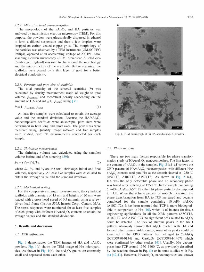
be found in all the processing steps: slurry formulation andpreparation (HA/nAl2O3 percentage), solidification (coolingrate) and sintering. By varying the processing parameters,properties such as porosity and pore size, density and thicknessof the walls, shrinkage percentage and phase composition offinal scaffolds can be controlled [32,34]. Fig. 7 shows poresize, porosity, shrinkage and mechanical strength of thescaffolds as a function of nAl2O3 content. Obviously, the totalporosity value decreases with increasing the nAl2O3 contents.This is due to increasing in volume shrinkage and dense walls,as mentioned above in Section 3.3. Also, it is clear that thecompressive strength of the scaffolds augments with increasingthe nAl2O3 contents [41,45]. The formation of calciumaluminate phases in the TCP matrix as well as decreasingthe porosity can be the reasons of this improvement in thecompressive strength. It is worth mentioning that Al2O3 is oneof the most widely used reinforcement materials for HAcomposites that causes significant increment in the compres-sive strength values as a result of calcium aluminate formation[41,50,51]. As it can be seen in Fig. 7, the pore sizes and alsocompressive strength increased with multiplying nAl2O3 con-tent, while it has been widely stated that compressive strengthdecreases by increasing pore size [46,52].
It is well known that porosity and pore size plays animportant role on initial cell adhesion, cell ingrowth and bloodvascularization. Murphy et al. [53] showed that the increasedsurface area provided by the scaffolds with smaller pore sizemay have a beneficial effect in initial cell adhesion, butultimately the improved cellular infiltration provided byscaffolds with larger pores outweighs this effect. Theyindicated that an early additional peak in cell number couldbe seen in the scaffolds with a mean pore size of 120 μm andthen this early peak disappeared following the cell prolifera-tion. They observed that the scaffolds with pores about 300 μmhad a better cell migration behavior. Thus, different pore sizeranges are required for good vascularization and cell ingrowth.On the other hand both length and width of the pores areimportant for appropriate cell ingrowth. Therefore, as can beseen in Fig. 7, further increase in pore width is desirable for thescaffolds.Another important factor that influences the compressive
strength is cooling rate. As it can be seen in Fig. 8, byincreasing the cooling rate from 1 K/min to 4 K/min, thecompressive strength relatively increased. With an increase inthe cooling rate, the pore size decreased because of the higherinterface velocity (shorter freezing time), and as a result an

Fig. 5. (a) 3D schematic image of ice formation and particles entrapment during the freeze casting process and (b) 2D schematic image of influence of nAl2O3
addition on ice formation, particles entrapment and structural wavelength (λ).
Fig. 6. SEM micrographs of inter-particle spaces filled with nanoparticles((a) HA/5% nAl2O3 and (b) HA/15% nAl2O3). Arrows indicate nanoparticlesin the matrix.
S.M.H. Ghzanfari, A. Zamanian / Ceramics International 39 (2013) 9835–9844 9841
improvement in the compressive strength of the nanocompositescaffolds was observed. Our results related to the relationshipbetween the cooling rate and the compressive strength is in a
good agreement with the reported data by other researchers[32,34].As shown in Fig. 9, the shrinkage and the compressive
strength increased as a function of sintering temperature whilethe sintering temperature had an opposite effect on the totalporosity. According to the literature, when the sinteringtemperature increases from 1250 to 1350 1C, the porosity goesdown and the shrinking value goes up, and as a result thecompressive strength increases [32,34]. In a recent study,Sopyan et al. [54] fabricated HA/alumina scaffolds via theprotein foaming consolidation method and obtained compres-sive strength and porosity values of 0.1 MPa and 45.2%,respectively, after addition of 50% alumina. In another work,An et al. [18] used zirconia to improve the mechanical strengthof HA scaffolds, produced by the polymer sponge method, andobtained a compressive strength of near 3 MPa and porosityaround 80% by even adding 60% zirconia to the HA matrix. Intotal, optimal compressive strengths for this type of scaffoldscan be obtained by controlling the nano-alumina content andcooling rate. Although, more desirable results were achievedcompared to the reported data by other researchers at thenearly same porosity [18,54], these samples are not stillappropriate to be used in load bearing sites [55]. Accordingto the previous studies, there might be some bridges betweenlamellar pores that could improve the mechanical properties ofthe structure [47,56]. However, the formation mechanism andcharacteristics of these bridges have not been completelyunderstood, and there are inadequate data in the availablearticles. The ability to manipulate these bridges would beexpected to achieve engineered scaffold constructs with bettermechanical properties.
Fig. 7. Effect of nAl2O3 content on pore size (a), compressive strength(b), porosity (c) and shrinkage (d) of HA/nAl2O3 nanocomposites.
Table 2The shrinkage, porosity, compressive strength and pore size of samples.
Samplecode
Shrinkage(%)
Porosity(%)
Compressivestrength(MPa)
Pore size—long(lm)
Pore size—short (lm)
A5C1T2 9.01 84.96 0.263 246.55 37.51A5C1T3 39.04 79.36 1.556 218.45 33.2A5C4T2 10.81 84.45 0.268 161.26 22.44A5C4T3 39.33 80.01 2.559 140.87 19.83A10C1T2 32.23 83.81 0.68 286.97 38.19A10C1T3 43.78 79.16 1.547 272.56 36.15A10C4T2 31.46 82.59 0.922 164.62 30.24A10C4T3 44.22 79.06 3.284 154.25 28.25A15C1T2 41.17 79.94 1.365 291.02 52.14A15C1T3 45.48 78.73 2.047 285.07 50.12A15C4T2 40.62 80.37 2.209 201.73 32.96A15C4T3 45.16 78.38 4.007 196.55 31.32
Fig. 8. Effect of cooling rate on pore size (a) and compressive strength (b) ofHA/nAl2O3 nanocomposites.
S.M.H. Ghzanfari, A. Zamanian / Ceramics International 39 (2013) 9835–98449842
4. Conclusions
In this study, the effect of further addition of nano-aluminaparticles to HA-based scaffolds on the microstructural, phasetransformation and mechanical properties as well as sinteringbehavior of porous HA/nAl2O3 scaffolds has been evaluated.The obtained results showed that by increasing the nAl2O3
content and sintering temperature, the decomposition of HA to
TCP was accelerated. In addition, increasing the nAl2O3
content led to increasing the pore size and the compressivestrength of the scaffolds. The formation of calcium aluminatephases caused increasing of the walls' densities and thicknessesas well as decreasing of the porosity, which might be thereason of this improvement in the compressive strength. Bymanipulating the nAl2O3 content, the cooling rate and sinteringtemperature, the scaffolds with total porosities between 78%and 85% and compressive strengths from 0.2 to 4 MPa wereachieved. Finally, the results showed that the pore size,porosity and compressive strength could be attuned by

Fig. 9. Effect of sintering temperature on compressive strength (a), pore size(b), porosity (c) and shrinkage (d) of HA/nAl2O3 nanocomposite scaffolds.
S.M.H. Ghzanfari, A. Zamanian / Ceramics International 39 (2013) 9835–9844 9843
adjusting the ratio of microparticles to nanoparticles content inthe freeze casting process. Since the ice volume, the size andmorphology of the ice crystals can be influenced by usingdifferent additives in the initial suspension, more extensive
studies on the effect of additives such as different anti-freezingagents, binders and dispersants need to be designed for theimprovement of the scaffold properties. The favorable mechan-ical behavior coupled with the ability to modify the micro-structures shows the potential of the freeze casting techniquefor the synthesis of excellent bone tissue engineering scaffolds.
References
[1] K. Arvidson, B.M. Abdallah, L.A. Applegate, N. Baldini, E. Cenni,E.G. Barrena, D. Granchi, M. Kassem, Y.T. Konttinen, K. Mustafa,D.P. Pioletti, T. Sillat, A.F. Wistrand, Bone regeneration and stem cells,Journal of Cellular and Molecular Medicine 15 (4) (2011) 718–746.
[2] M. Yasaei, A. Zamanian, F. Moztarzadeh, M. Ghaffari, M. Mozafari,Characteristics improvement of calcium hydroxide dental cement byhydroxyapatite nanoparticles. Part 1: formulation and microstructure,Biotechnology and Applied Biochemistry, http://dx.doi.org/10.1002/bab.1119, in press.
[3] M. Jafarkhani, A. Fazlali, F. Moztarzadeh, M. Mozafari, Mechanical andstructural properties of polylactide/chitosan scaffolds reinforced withnano calcium phosphate, Iranian Polymer Journal 10 (2012) 713–720.
[4] P. Holzmann, E. Niculescu-Morzsa, H. Zwickl, F. Halbwirth, M. Pichler,M. Matzner, F. Gottsauner-Wolf, S. Nehrer, Investigation of boneallografts representing different steps of the bone bank procedure usingthe CAM-model, Altex 27 (2) (2010) 97–103.
[5] M. Jafarkhani, A. Fazlali, F. Moztarzadeh, Z. Moztarzadeh, M. Mozafari,Fabrication and characterization of PLLA/chitosan/nano calcium phos-phate scaffolds by freeze casting technique, Industrial and EngineeringChemistry Research 51 (2012) 9241–9249.
[6] S.E. Lobo, T.L. Arinzeh, Biphasic calcium phosphate ceramics for boneregeneration and tissue engineering applications, Materials 3 (2) (2010)815–826.
[7] R.Z. LeGeros, Calcium phosphates in oral biology and medicine,Monographs in Oral Science 15 (1991) 1–201.
[8] S. Dorozhkin, Medical application of calcium orthophosphate biocera-mics, BIO 1 (1) (2011) 1–51.
[9] F.D. Burstein, S.R. Cohen, R. Hudgins, W. Boydston, C. Simms, The useof hydroxyapatite cement in secondary craniofacial reconstruction, Plasticand Reconstructive Surgery 104 (5) (1999) 1270–1275.
[10] P. Korovessis, M. Repanti, G. Koureas, Does coralline hydroxyapatiteconduct fusion in instrumented posterior spine fusion?, Studies in HealthTechnology and Informatics 91 (2002) 109–113
[11] S.V. Dorozhkin, Amorphous calcium (ortho)phosphates, Acta Biomater-ialia 6 (12) (2010) 4457–4475.
[12] I. Sato, T. Akizuki, S. Oda, H. Tsuchioka, C. Hayashi, A.A. Takasaki,K. Mizutani, N. Kawakatsu, A. Kinoshita, I. Ishikawa, Y. Izumi,Histological evaluation of alveolar ridge augmentation using injectablecalcium phosphate bone cement in dogs, Journal of Oral Rehabilitation36 (10) (2009) 762–769.
[13] M. Oates, R. Chen, M. Duncan, J.A. Hunt, The angiogenic potential ofthree-dimensional open porous synthetic matrix materials, Biomaterials28 (25) (2007) 3679–3686.
[14] S. Deville, Freeze casting of porous biomaterials: structure, properties andopportunities, Materials 3 (3) (2010) 1913–1927.
[15] A. Hamlekhan, F. Moztarzadeh, M. Mozafari, M. Azami, N. Nezafati,Preparation of laminated poly(ε-caprolactone)-gelatin–hydroxyapatitenanocomposite scaffold bioengineered via compound techniques for bonesubstitution, Biomatter 1 (2011) 1–11.
[16] I. Sopyan, M. Mel, S. Ramesh, K.A. Khalid, Porous hydroxyapatite forartificial bone applications, Science and Technology of AdvancedMaterials 8 (1–2) (2007) 116–123.
[17] F. Baghbani, F. Moztarzadeh, A. Gafari Nazari, A.H. Razavi Kamran,F. Tondnevis, N. Nezafati, M. Gholipourmalekabadi, M. Mozafari,Biological response of biphasic hydroxyapatite/tricalcium phosphatescaffolds intended for low load-bearing orthopaedic applications,Advanced Composites Letters 21 (2012) 16–24.
S.M.H. Ghzanfari, A. Zamanian / Ceramics International 39 (2013) 9835–98449844
[18] S.H. An, T. Matsumoto, H. Miyajima, A. Nakahira, K.H. Kim,S. Imazato, Porous zirconia/hydroxyapatite scaffolds for bone reconstruc-tion, Dental Materials 28 (12) (2012) 1221–1231.
[19] S. Kalmodia, S. Goenka, T. Laha, D. Lahiri, B. Basu, K. Balani,Microstructure, mechanical properties, and in vitro biocompatibility ofspark plasma sintered hydroxyapatite–aluminum oxide–carbon nanotubecomposite, Materials Science and Engineering C 30 (8) (2010)1162–1169.
[20] S. Blindow, M. Pulkin, D. Koch, G. Grathwohl, K. Rezwan, Hydro-xyapatite/SiO2 composites via freeze casting for bone tissue engineering,Advanced Engineering Materials 11 (11) (2009) 875–884.
[21] E. Fidancevska, G. Ruseska, J. Bossert, Y.M. Lin, A.R. Boccaccini,Fabrication and characterization of porous bioceramic composites basedon hydroxyapatite and titania, Materials Chemistry and Physics 103 (1)(2007) 95–100.
[22] S. Sprio, A. Tampieri, G. Celotti, E. Landi, Development of hydro-xyapatite/calcium silicate composites addressed to the design of load-bearing bone scaffolds, Journal of the Mechanical Behavior of Biome-dical Materials 2 (2) (2009) 147–155.
[23] B.-H. Yoon, W.-Y. Choi, H.-E. Kim, J.-H. Kim, Y.-H. Koh, Alignedporous alumina ceramics with high compressive strengths for bone tissueengineering, Scripta Materialia 58 (7) (2008) 537–540.
[24] L.L. Hench, Bioceramics: research and development opportunities,Brazilian Journal of Physics 22 (1992) 70–84.
[25] J.S. Magdeski, The porosity dependence of mechanical properties ofsintered alumina, Journal of the University of Chemical Technology andMetallurgy 45 (2010) 143–148.
[26] K.H. Zuo, Y. Zhang, Y.P. Zeng, D. Jiang, Pore-forming agent inducedmicrostructure evolution of freeze casted hydroxyapatite, CeramicsInternational 37 (1) (2011) 407–410.
[27] S.F. Corbin, X. Zhao-jie, H. Henein, P.S. Apte, Functionally gradedmetal/ceramic composites by tape casting, lamination and infiltration,Materials Science and Engineering A 262 (1–2) (1999) 192–203.
[28] G. Meng, H. Wang, W. Zheng, X. Liu, Preparation of porous ceramics bygelcasting approach, Materials Letters 45 (2000) 224–227.
[29] Y. Zhang, Y. Yokogawa, X. Feng, Y. Tao, Y. Li, Preparation andproperties of bimodal porous apatite ceramics through slip casting usingdifferent hydroxyapatite powders, Ceramics International 36 (1) (2010)107–113.
[30] A. Diaz, S. Hampshire, Characterisation of porous silicon nitridematerials produced with starch, Journal of the European Ceramic Society24 (2) (2004) 413–419.
[31] D.K. Agrawal, Microwave processing of ceramics, Current Opinion inSolid State and Materials Science 3 (1998) 480–485.
[32] S. Farhangdoust, A. Zamanian, M. Yasaei, M. Khorami, The effect ofprocessing parameters and solid concentration on the mechanical andmicrostructural properties of freeze casted macroporous hydroxyapatitescaffolds, Materials Science and Engineering C 33 (1) (2013) 453–460.
[33] N. Arabi, A. Zamanian, Effect of cooling rate and gelatin concentrationon the microstructural and mechanical properties of ice template gelatinscaffolds, Biotechnology and Applied Biochemistry (2013) http://dx.doi.org/10.1002/bab.1120.
[34] S. Deville, E. Saiz, A.P. Tomsia, Freeze casting of hydroxyapatitescaffolds for bone tissue engineering, Biomaterials 27 (32) (2006)5480–5489.
[35] S. Deville, Freeze casting of porous ceramics: a review of currentachievements and issues, Advanced Engineering Materials 10 (3) (2008)155–169.
[36] K.H. Zuo, Y. Zeng, D. Jiang, Effect of cooling rate and polyvinyl alcoholon the morphology of porous hydroxyapatite ceramics, Materials andDesign 31 (6) (2010) 3090–3094.
[37] Y. Zhang, K. Zuo, Y.P. Zeng, Effects of gelatin addition on themicrostructure of freeze cast porous hydroxyapatite ceramics, CeramicsInternational 35 (6) (2009) 2151–2154.
[38] K.K. Mallick, Freeze casting of porous bioactive glass and bioceramics,Journal of the American Ceramic Society 92 (2009) S85–S94.
[39] A. Heunisch, A. Dellert, A. Roosen, Effect of powder, binder and processparameters on anisotropic shrinkage in tape cast ceramic products,Journal of the European Ceramic Society 30 (16) (2010) 3397–3406.
[40] G. Hannink, J.J. Arts, Bioresorbability, porosity and mechanical strengthof bone substitutes: what is optimal for bone regeneration?, Injury,International Journal of the Care of the Injured 42 (Suppl 2) (2011)S22–25.
[41] H. Ji, P.M. Marquis, Preparation and characterization of Al2O3 reinforcedhydroxyapatite, Biomaterials 13 (1992) 744–748.
[42] G. Muralithran, S. Ramesh, The effects of sintering temperature on theproperties of hydroxyapatite, Ceramics International 26 (2000) 221–230.
[43] C.J. Liao, F.H. Lin, K.S. Chen, J.S. Sun, Thermal decomposition andreconstitution of hydroxyapatite in air atmosphere, Biomaterials 20(1999) 1807–1813.
[44] H.W. Kim, Y.H. Koh, S.B. Seo, H.E. Kim, Properties of fluoridatedhydroxyapatite–alumina biological composites densified with addition ofCaF2, Materials Science and Engineering C 23 (4) (2003) 515–521.
[45] Z. Evis, R.H. Doremus, A study of phase stability and mechanicalproperties of hydroxylapatite–nanosize α-alumina composites, MaterialsScience and Engineering C 27 (3) (2007) 421–425.
[46] K. Zhao, Y.F. Tang, Y.S. Qin, J.Q. Wei, Porous hydroxyapatite ceramicsby ice templating: freezing characteristics and mechanical properties,Ceramics International 37 (2) (2011) 635–639.
[47] S. Deville, E. Saiz, A.P. Tomsia, Ice-templated porous alumina structures,Acta Materialia 55 (6) (2007) 1965–1974.
[48] S. Deville, E. Maire, A. Lasalle, A. Bogner, C. Gauthier, J. Leloup,C. Guizard, Influence of particle size on ice nucleation and growth duringthe ice-templating process, Journal of the American Ceramic Society 93(9) (2010) 2507–2510.
[49] A. Zamanian, S. Farhangdoust, M. Yasaei, M. Khorami, M. Hafezi,The effect of particle size on the mechanical and microstructuralproperties of freeze casted macroporous hydroxyapatite scaffolds, Inter-national Journal of Applied Ceramic Technology (2013)http://dx.doi.org/10.1111/ijac.12031.
[50] P.M.M. Huaxia Ji, Sintering behavior of hydroxyapatite reinforced with20 wt% Al2O3, Journal of Materials Science 28 (1993) 1941–1945.
[51] H.Y. Juang, M.H. Hon, Fabrication and mechanical properties ofhydroxyapatite–alumina composites, Materials Science and EngineeringC 2 (1994) 77–81.
[52] G. Tripathi, B. Basu, A porous hydroxyapatite scaffold for bone tissueengineering: physico-mechanical and biological evaluations, CeramicsInternational 38 (1) (2012) 341–349.
[53] C.M. Murphy, M.G. Haugh, F.J. O’Brien, The effect of mean pore sizeon cell attachment, proliferation and migration in collagen–glycosami-noglycan scaffolds for bone tissue engineering, Biomaterials 31 (3)(2010) 461–466.
[54] I. Sopyan, A. Fadli, M. Mel, Porous alumina–hydroxyapatite compositesthrough protein foaming-consolidation method, Journal of the MechanicalBehavior of Biomedical Materials 8 (2012) 86–98.
[55] A.J. Wagoner Johnson, B.A. Herschler, Review of the mechanicalbehavior of CaP and CaP/polymer composites for applications in bonereplacement and repair, Acta biomaterialia 7 (2011) 16–30.
[56] U.G. Wegst, M. Schecter, A.E. Donius, P.M. Hunger, Biomaterials byfreeze casting, Philosophical Transactions of the Royal Society A 368(1917) (2010) 2099–2121.