The Female Reproductive System
17
The Female Reproductive System Paul F. Terranova, Ph.D. 38 CHAPTER 38 T he fertility of the mature human female is cyclic. The release from the ovary of a mature female germ cell or ovum occurs at a distinct phase of the menstrual cycle. The secretion of ovarian steroid hormones, estradiol and prog- esterone, and the subsequent release of an ovum during the menstrual cycle are controlled by cyclic changes in LH and FSH from the pituitary gland, and estradiol and proges- terone from the ovaries. The cyclic changes in steroid hor- mone secretion cause significant changes in the structure and function of the uterus in preparing it for the reception of a fertilized ovum. At different stages of the menstrual cy- cle, progesterone and estradiol exert negative- and posi- tive-feedback effects on the hypothalamus and on pituitary gonadotrophs, generating the cyclic pattern of LH and FSH release characteristic of the female reproductive sys- tem. Since the hormonal events during the menstrual cycle are delicately synchronized, the menstrual cycle can be readily affected by stress and by environmental, psycho- logical, and social factors. The female cycle is characterized by monthly bleeding, resulting from the withdrawal of ovarian steroid hormone support of the uterus, which causes shedding of the super- ficial layers of the uterine lining at the end of each cycle. The first menstrual cycle occurs during puberty. Menstrual ■ AN OVERVIEW OF THE FEMALE REPRODUCTIVE SYSTEM ■ THE HYPOTHALAMIC-PITUITARY AXIS ■ THE FEMALE REPRODUCTIVE ORGANS ■ FOLLICULOGENESIS, STEROIDOGENESIS, ATRESIA, AND MEIOSIS ■ FOLLICLE SELECTION AND OVULATION ■ FORMATION OF THE CORPUS LUTEUM FROM THE POSTOVULATORY FOLLICLE ■ THE MENSTRUAL CYCLE ■ ESTROGEN, PROGESTIN, AND ANDROGEN: TRANSPORT AND METABOLISM ■ PUBERTY ■ MENOPAUSE ■ INFERTILITY CHAPTER OUTLINE 1. Pulses of hypothalamic GnRH regulate the secretion of LH and FSH, which enhance follicular development, steroido- genesis, ovulation, and formation of the corpus luteum. 2. LH and FSH, in coordination with ovarian theca and granu- losa cells, regulate the secretion of follicular estradiol. 3. Ovulation occurs as the result of a positive feedback of fol- licular estradiol on the hypothalamic-pituitary axis that in- duces LH and FSH surges. 4. Follicular development occurs in distinct steps: primordial, primary, secondary, tertiary, and graafian follicle stages. 5. Follicular rupture (ovulation) requires the coordination of appropriately timed LH and FSH surges that induce in- flammatory reactions in the graafian follicle, leading to dissolution at midcycle of the follicular wall by several ovarian enzymes. 6. Follicular atresia results from the withdrawal of go- nadotropin support. 7. The formation of a functional corpus luteum requires the presence of an LH surge, adequate numbers of LH recep- tors, sufficient granulosa cells, and significant proges- terone secretion. 8. The uterine cycle is regulated by estradiol and proges- terone, such that estradiol induces proliferation of the uter- ine endometrium, whereas progesterone induces differen- tiation of the uterine endometrium and the secretion of distinct products. 9. During puberty, the hypothalamus begins to secrete in- creasing quantities of GnRH, which increases LH and FSH secretion, enhances ovarian function, and leads to the first ovulation. 10. Menopause ensues from the loss of numerous oocytes in the ovary and the subsequent failure of follicular develop- ment and estradiol secretion. LH and FSH levels rise from the lack of negative feedback by estradiol. KEY CONCEPTS 667
Transcript of The Female Reproductive System
The Female Reproductive SystemPaul F. Terranova, Ph.D.38
C H A P T E R
38
The fertility of the mature human female is cyclic. Therelease from the ovary of a mature female germ cell or
ovum occurs at a distinct phase of the menstrual cycle. Thesecretion of ovarian steroid hormones, estradiol and prog-esterone, and the subsequent release of an ovum during themenstrual cycle are controlled by cyclic changes in LH andFSH from the pituitary gland, and estradiol and proges-terone from the ovaries. The cyclic changes in steroid hor-mone secretion cause significant changes in the structureand function of the uterus in preparing it for the receptionof a fertilized ovum. At different stages of the menstrual cy-cle, progesterone and estradiol exert negative- and posi-
tive-feedback effects on the hypothalamus and on pituitarygonadotrophs, generating the cyclic pattern of LH andFSH release characteristic of the female reproductive sys-tem. Since the hormonal events during the menstrual cycleare delicately synchronized, the menstrual cycle can bereadily affected by stress and by environmental, psycho-logical, and social factors.
The female cycle is characterized by monthly bleeding,resulting from the withdrawal of ovarian steroid hormonesupport of the uterus, which causes shedding of the super-ficial layers of the uterine lining at the end of each cycle.The first menstrual cycle occurs during puberty. Menstrual
■ AN OVERVIEW OF THE FEMALE REPRODUCTIVE
SYSTEM
■ THE HYPOTHALAMIC-PITUITARY AXIS
■ THE FEMALE REPRODUCTIVE ORGANS
■ FOLLICULOGENESIS, STEROIDOGENESIS, ATRESIA,
AND MEIOSIS
■ FOLLICLE SELECTION AND OVULATION
■ FORMATION OF THE CORPUS LUTEUM FROM THE
POSTOVULATORY FOLLICLE
■ THE MENSTRUAL CYCLE
■ ESTROGEN, PROGESTIN, AND ANDROGEN:
TRANSPORT AND METABOLISM
■ PUBERTY
■ MENOPAUSE
■ INFERTILITY
C H A P T E R O U T L I N E
1. Pulses of hypothalamic GnRH regulate the secretion of LHand FSH, which enhance follicular development, steroido-genesis, ovulation, and formation of the corpus luteum.
2. LH and FSH, in coordination with ovarian theca and granu-losa cells, regulate the secretion of follicular estradiol.
3. Ovulation occurs as the result of a positive feedback of fol-licular estradiol on the hypothalamic-pituitary axis that in-duces LH and FSH surges.
4. Follicular development occurs in distinct steps: primordial,primary, secondary, tertiary, and graafian follicle stages.
5. Follicular rupture (ovulation) requires the coordination ofappropriately timed LH and FSH surges that induce in-flammatory reactions in the graafian follicle, leading todissolution at midcycle of the follicular wall by severalovarian enzymes.
6. Follicular atresia results from the withdrawal of go-nadotropin support.
7. The formation of a functional corpus luteum requires thepresence of an LH surge, adequate numbers of LH recep-tors, sufficient granulosa cells, and significant proges-terone secretion.
8. The uterine cycle is regulated by estradiol and proges-terone, such that estradiol induces proliferation of the uter-ine endometrium, whereas progesterone induces differen-tiation of the uterine endometrium and the secretion ofdistinct products.
9. During puberty, the hypothalamus begins to secrete in-creasing quantities of GnRH, which increases LH and FSHsecretion, enhances ovarian function, and leads to the firstovulation.
10. Menopause ensues from the loss of numerous oocytes inthe ovary and the subsequent failure of follicular develop-ment and estradiol secretion. LH and FSH levels rise fromthe lack of negative feedback by estradiol.
K E Y C O N C E P T S
667

668 PART X REPRODUCTIVE PHYSIOLOGY
cycles are interrupted during pregnancy and lactation andcease at menopause. Menstruation signifies a failure to con-ceive and results from regression of the corpus luteum andsubsequent withdrawal of luteal steroid support of the su-perficial endometrial layer of the uterus.
AN OVERVIEW OF THE FEMALE
REPRODUCTIVE SYSTEM
An overview of the interactions of hormonal factors in fe-male reproduction is shown in Figure 38.1. The femalehormonal system consists of the brain, pituitary, ovaries,and reproductive tract (oviduct, uterus, cervix, and vagina).In the brain, the hypothalamus produces gonadotropin-re-leasing hormone (GnRH), which controls the secretion ofluteinizing hormone (LH) and follicle-stimulating hor-mone (FSH).
The mature ovary has two major functions: the matura-tion of germ cells and steroidogenesis. Each germ cell is ul-timately enclosed within a follicle, a major source of steroidhormones during the menstrual cycle. At ovulation, theovum or egg is released and the ruptured follicle is trans-formed into a corpus luteum, which secretes progesteroneas its main product. FSH is primarily involved in stimulat-ing the growth of ovarian follicles, while LH induces ovu-
lation. Both LH and FSH regulate follicular steroidogenesisand androgen and estradiol secretion, and LH regulates thesecretion of progesterone from the corpus luteum. Ovariansteroids inhibit the secretion of LH and FSH with one ex-ception: Just prior to ovulation (at midcycle), estradiol hasa positive-feedback effect on the hypothalamic-pituitaryaxis and induces significant increases in the secretion ofGnRH, LH, and FSH. The ovary also produces threepolypeptide hormones. Inhibin suppresses the secretion ofFSH. Activin (an inhibin-binding protein) increases the se-cretion of FSH, and follistatin (an activin-binding protein)reduces the secretion of FSH.
Shortly after fertilization, the embryo begins to developplacenta cells, which attach to the uterine lining and unitewith the maternal placental cells. The placenta producesseveral pituitary-like and ovarian steroid-like hormones.These hormones support placental and fetal developmentthroughout pregnancy and have a role in parturition. Themammary glands are also under the control of pituitaryhormones and ovarian steroids, and provide the baby withimmunological protection and nutritional support throughlactation. Lactation is hormonally controlled by prolactin(PRL) from the anterior pituitary, which regulates milkproduction, and oxytocin from the posterior pituitary,which induces milk ejection from the breasts.
THE HYPOTHALAMIC-PITUITARY AXIS
The hypothalamic-pituitary axis has an important role inregulating the menstrual cycle. GnRH, a decapeptide pro-duced in the hypothalamus and released in a pulsatile man-ner, controls the secretion of LH and FSH through a portalvascular system (see Chapter 32). Blockade of the portalsystem reduces the secretion of LH and FSH and leads toovarian atrophy and a reduction in ovarian hormone secre-tion. The secretion of GnRH by the hypothalamus is regu-lated by neurons from other brain regions. Neurotransmit-ters, such as epinephrine and norepinephrine, stimulate thesecretion of GnRH, whereas dopamine and serotonin in-hibit secretion of GnRH. In addition, ovarian steroids andpeptides and hypothalamic neuropeptides can regulate thesecretion of GnRH. GnRH stimulates the pituitary go-nadotrophs to secrete LH and FSH. GnRH binds to high-affinity receptors on the gonadotrophs and stimulates thesecretion of LH and FSH through a phosphoinositide-pro-tein kinase C-mediated pathway (see Chapter 1).
A graph of LH release throughout the female life span isshown in Figure 38.2. During the neonatal period, LH is re-leased at low and steady rates without pulsatility; this pe-riod coincides with lack of development of mature ovarianfollicles and very low to no ovarian estradiol secretion. Pul-satile release begins with the onset of puberty and for sev-eral years is expressed only during sleep; this period coin-cides with increased but asynchronous folliculardevelopment and with increased secretion of ovarian estra-diol. Upon the establishment of regular functional men-strual cycles associated with regular ovulation, LH pulsatil-ity prevails throughout the 24-hour period, changing in amonthly cyclic manner. In postmenopausal women whoseovaries lack sustained follicular development and exhibit
�
�
�
���
AgeEnvironment
Drugs
Hypothalamus
GnRH
Anterior pituitary
PRL
Ovary
Reproductivetract
Secondary sexcharacteristics
BrainCenters
Inhibinactivin
follistatin
��
�
�
�
Dopamine
FSH/LH
�
�
Estradiol,progesterone,
androgen
,,
Regulation of the reproductive tract in the
female. The main reproductive hormones areshown in boxes. Positive and negative regulations are depicted byplus and minus signs.
FIGURE 38.1

low ovarian estradiol secretion, mean circulating LH levelsare high and pulses occur at a high frequency.
THE FEMALE REPRODUCTIVE ORGANS
The female reproductive tract has two major components:the ovaries, which produce the mature ovum and secreteprogestins, androgens, and estrogens; and the ductal sys-tem, which transports ovum, is the place of the union of thesperm and egg, and maintains the developing conceptusuntil delivery. The morphology and function of these struc-
tures change in a cyclic manner under the influence of thereproductive hormones.
The ovaries are in the pelvic portion of the abdominal cav-ity on both sides of the uterus and are anchored by ligaments(Fig. 38.3). An adult ovary weighs 8 to 12 g and consists of anouter cortex and an inner medulla, without a sharp demarca-tion. The cortex is surrounded by a fibrous tissue, the tunicaalbuginea, covered by a single layer of surface epitheliumcontinuous with the mesothelium covering the other organsin the abdominal cavity. The cortex contains oocytes en-closed in follicles of various sizes, corpora lutea, corpora al-bicantia, and stromal cells. The medulla contains connective
CHAPTER 38 The Female Reproductive System 669
Day Night Day Night Day Night Day Night Day Night
Neonatal
Pla
sma
LH c
onc.
Pubertal Tonic Midcycle Postmenopausal
Reproductive
Relative levels of
LH release in hu-
man females throughout life. (Modi-fied from Yen SSC, et al. In: Ferin M, etal., eds. Biorhythms and Human Repro-duction. New York: Wiley, 1974.)
FIGURE 38.2
Ovary
Corpus
Fundus
Endometrium
Myometrium
Cervix
Vagina
Broad ligament
Fimbria
Ampulla
Isthmus
Ovarian ligament
Ovarian vessels
Corpus albicans
Mature corpus luteum
Early corpus luteum
Stroma
Ovulation
Germinal epithelium
Graafian follicle
Early antrum formation
Atretic follicle
Primordial follicle
Primary follicle
Infundibulum
Ovary
OviductUterus
The female reproductive organs. (Modified from Patton BM. Human Embryology. NewYork: McGraw-Hill, 1976.)
FIGURE 38.3

670 PART X REPRODUCTIVE PHYSIOLOGY
and interstitial tissues. Blood vessels, lymphatics, and nervesenter the medulla of the ovary through the hilus.
On the side that ovulates, the oviduct (fallopian tube)receives the ovum immediately after ovulation. Theoviducts are the site of fertilization and provide an envi-ronment for development of the early embryo. Theoviducts are 10 to 15 cm long and composed of sequentialregions called the infundibulum, ampulla, and isthmus.The infundibulum is adjacent to the ovary and opens to theperitoneal cavity. It is trumpet-shaped with finger-like pro-jections called fimbria along its outer border that grasp theovum at the time of follicular rupture. Its thin walls are cov-ered with densely ciliated projections, which facilitateovum uptake and movement through this region. The am-pulla is the site of fertilization. It has a thin musculature andwell-developed mucosal surface. The isthmus is located atthe uterotubal junction and has a narrow lumen surroundedby smooth muscle. It has sphincter-like properties and canserve as a barrier to the passage of germ cells. The oviductstransport the germ cells in two directions: sperm ascend to-ward the ampulla and the zygote descends toward theuterus. This requires coordination between smooth musclecontraction, ciliary movement, and fluid secretion, all ofwhich are under hormonal and neuronal control.
The uterus is situated between the urinary bladder andrectum. On each upper side, an oviduct opens into the uter-ine lumen, and on the lower side, the uterus connects to thevagina. The uterus is composed of two types of tissue. Theouter part is the myometrium, composed of multiple layersof smooth muscle. The inner part, lining the lumen of theuterus, is the endometrium, which contains a deep stromallayer next to the myometrium and a superficial epitheliallayer. The stroma is permeated by spiral arteries and con-tains much connective tissue. The epithelial layer is inter-rupted by uterine glands, which also penetrate the stromallayer and are lined by columnar secretory cells. The uterusprovides an environment for the developing fetus, andeventually, the myometrium will generate rhythmic con-tractions that assist in expelling the fetus at delivery.
The cervix (neck) is a narrow muscular canal that con-nects the vagina and the body (corpus) of the uterus. Itmust dilate in response to hormones to allow the expulsionof the fetus. The cervix has numerous glands with a colum-nar epithelium that produces mucus under the control ofestradiol. As more and more estradiol is produced duringthe follicular phase of the cycle, the cervical mucus changesfrom a scanty viscous material to a profuse watery andhighly elastic substance called spinnbarkeit. The viscosityof the spinnbarkeit can be tested by touching it with a pieceof paper and lifting vertically. The mucus can form a threadup to 6 cm under the influence of elevated estradiol. If adrop of the cervical mucus is placed on a slide and allowedto dry, it will form a typical ferning pattern when under theinfluence of estradiol.
The vagina is well innervated, and has a rich blood sup-ply. It is lined by several layers of epithelium that changehistologically during the menstrual cycle. When estradiollevels are low, as during the prepubertal or post-menopausal periods, the vaginal epithelium is thin and thesecretions are scanty, resulting in a dry and infection-sus-ceptible area. Estradiol induces proliferation and cornifi-
cation (keratinization) of the vaginal epithelium, whereasprogesterone opposes those actions and induces the influxof polymorphonuclear leukocytes into the vaginal fluids.Estradiol also activates vaginal glands that produce lubri-cating fluid during coitus.
FOLLICULOGENESIS, STEROIDOGENESIS,
ATRESIA, AND MEIOSIS
Most follicles in the ovary will undergo atresia. However,some will develop into mature follicles, produce steroids,and ovulate. As follicles mature, oocytes will also mature byentering meiosis, which produces the proper number ofchromosomes in preparation for fertilization.
The Primordial Follicle Contains an
Oocyte Arrested in Meiosis
Female germ cells develop in the embryonic yolk sac andmigrate to the genital ridge where they participate in thedevelopment of the ovary (Table 38.1). Without germcells, the ovary does not develop. The germs cells, calledoogonia, actively divide by mitosis. Oogonia undergo mi-tosis only during the prenatal period. By birth, the ovariescontain a finite number of oocytes, estimated to be about 1million. Most of them will die by a process called atresia. Bypuberty, only 200,000 oocytes remain; by age 30, only26,000 remain; and by the time of menopause, the ovariesare essentially devoid of oocytes.
When oogonia cease the process of mitosis, they are calledoocytes. At that time they enter the meiotic cycle (or meio-sis, to prepare for the production of a haploid ovum), becomearrested in prophase of the first meiotic division, and remainarrested in that phase until they either die or grow into ma-ture oocytes at the time of ovulation. The primordial follicle(Fig. 38.4) is 20 �m in diameter and contains an oocyte,which may or may not be surrounded by a single layer of flat-tened (squamous) pregranulosa cells. When pregranulosacells surround the oocyte, a basement membrane develops,separating the granulosa from the ovarian stroma.
A Graafian Follicle Is the Final Stage of
Follicle Development
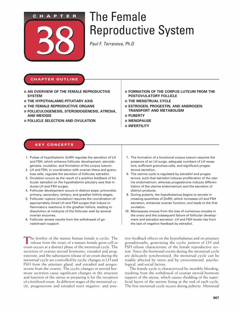
Folliculogenesis (also called follicular development) is theprocess by which follicles develop and mature (see Fig.38.3). Follicles are in one of the following physiologicalstates: resting, growing, degenerating, or ready to ovulate.During each menstrual cycle, the ovaries produce a groupof growing follicles of which most will fail to grow to ma-turity and will undergo follicular atresia (death) at somestage of development. However, one dominant folliclegenerally emerges from the cohort of developing folliclesand it will ovulate, releasing a mature haploid ovum.
Primordial follicles are generally considered the non-growing resting pool of follicles, which gets progressivelydepleted throughout life; by the time of menopause, theovaries are essentially devoid of all follicles. Primordial fol-licles are located in the ovarian cortex (peripheral regionsof the ovary) beneath the tunica albuginea.

Progression from primordial to the next stage of follicu-lar development, the primary stage, occurs at a relativelyconstant rate throughout fetal, juvenile, prepubertal, andadult life. Once primary follicles leave the resting pool,they are committed to further development or atresia. Mostbecome atretic, and typically only one fully developed fol-licle will ovulate. The conversion from primordial to pri-mary follicles is believed to be independent of pituitary go-nadotropins. The exact signal that recruits a follicle from aresting to a growing pool is unknown; it could be pro-grammed by the cell genome or influenced by local ovariangrowth regulators.
The first sign that a primordial follicle is entering thegrowth phase is a morphological change of the flattenedpregranulosa cells into cuboidal granulosa cells. Thecuboidal granulosa cells proliferate to form a single contin-uous layer of cells surrounding the oocyte, which has en-larged from 20 �m in the primordial stage to 140 �m in di-ameter. At this stage, a glassy membrane, the zonapellucida, surrounds the oocyte and serves as means of at-tachment through which the granulosa cells communicatewith the oocyte. This is the primary follicular stage of de-velopment, consisting of one layer of cuboidal granulosacells and a basement membrane.
The follicle continues to grow, mainly through prolifer-ation of its granulosa cells, so that several layers of granu-losa cells exist in the secondary follicular stage of develop-ment (see Fig. 38.4). As the secondary follicle grows deeperinto the cortex, stromal cells, near the basement membrane,begin to differentiate into cell layers called theca internaand theca externa, and a blood supply with lymphatics andnerves forms within the thecal component. The granulosalayer remains avascular.
The theca interna cells become flattened, epithelioid,and steroidogenic. The granulosa cells of secondary follicles
acquire receptors for FSH and start producing smallamounts of estrogen. The theca externa remains fibroblasticand provides structural support to the developing follicle.
Development beyond the primary follicle is go-nadotropin-dependent, begins at puberty, and continues ina cyclic manner throughout the reproductive years. As thefollicle continues to grow, theca layers expand, and fluid-filled spaces or antra begin to develop around the granulosacells. This early antral stage of follicle development is re-ferred to as the tertiary follicular stage (see Fig. 38.4). Thecritical hormone responsible for progression from the pre-antral to the antral stage is FSH. Mitosis of the granulosacells is stimulated by FSH. As the number of granulosa cellsincreases, the production of estrogens, the binding capac-ity for FSH, the size of the follicle, and the volume of thefollicular fluid all increase significantly.
As the antra increase in size, a single, large, coalescedantrum develops, pushing the oocyte to the periphery ofthe follicle and forming a large 2- to 2.5-cm-diametergraafian follicle (preovulatory follicle; see Fig. 38.4). Threedistinct granulosa cell compartments are evident in thegraafian follicle. Granulosa cells surrounding the oocyte arecumulus granulosa cells (collectively called cumulusoophorus). Those cells lining the antral cavity are calledantral granulosa cells and those attached to the basementmembrane are called mural granulosa cells. Mural andantral granulosa cells are more steroidogenically activethan cumulus cells.
In addition to bloodborne hormones, antral follicles havea unique microenvironment in which the follicular fluid con-tains different concentrations of pituitary hormones,steroids, peptides, and growth factors. Some are present inthe follicular fluid at a concentration 100 to 1,000 timeshigher than in the circulation. Table 38.2 lists some parame-ters of human follicles at successive stages of development in
CHAPTER 38 The Female Reproductive System 671
TABLE 38.1 Different Stages in the Development of an Ovum and Follicle
Stage Process Ovum Follicle
Fetal life Migration Primordial germ cellsMitosis Oogonia Primordial follicleFirst meiotic division begins Primary oocyte Primary follicle
Birth Arrest in prophaseGrowth of oocyte and follicle
Puberty Follicular maturation Secondary follicle
Cycle Antral follicle
Ovulation Resumption of meiosis Secondary oocyte Graafian follicleEmission of first polar bodyArrest in metaphase
Corpus luteumFertilization Second meiotic division complete Zygote
Emission of second polar body
Implantation Mitotic divisions EmbryoBlastocyst
Parturition Body Patterning Fetus Corpus albicans
672 PART X REPRODUCTIVE PHYSIOLOGY
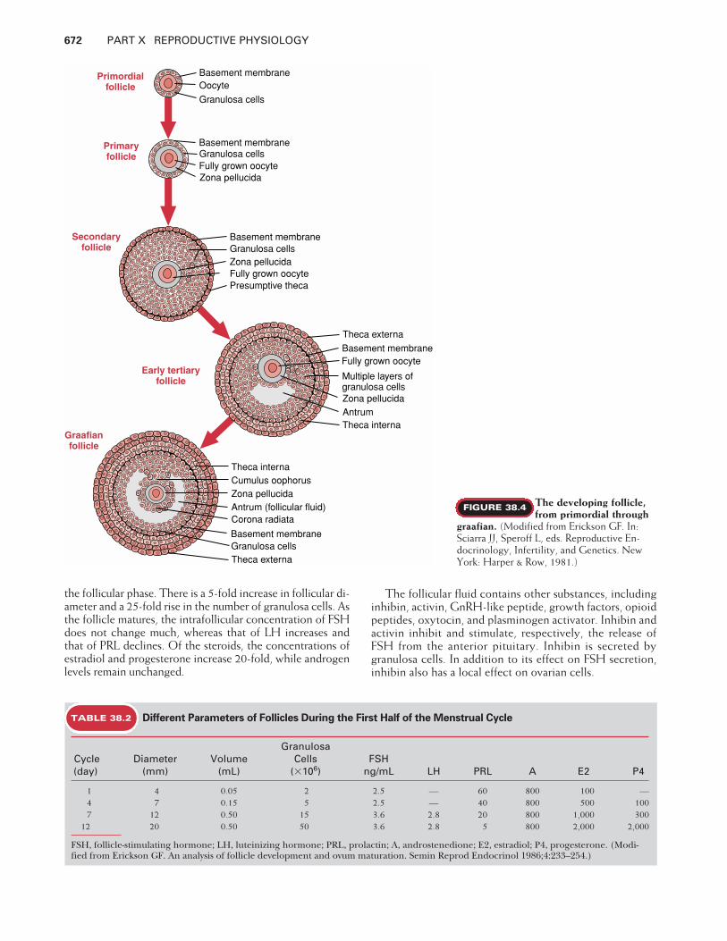
the follicular phase. There is a 5-fold increase in follicular di-ameter and a 25-fold rise in the number of granulosa cells. Asthe follicle matures, the intrafollicular concentration of FSHdoes not change much, whereas that of LH increases andthat of PRL declines. Of the steroids, the concentrations ofestradiol and progesterone increase 20-fold, while androgenlevels remain unchanged.
The follicular fluid contains other substances, includinginhibin, activin, GnRH-like peptide, growth factors, opioidpeptides, oxytocin, and plasminogen activator. Inhibin andactivin inhibit and stimulate, respectively, the release ofFSH from the anterior pituitary. Inhibin is secreted bygranulosa cells. In addition to its effect on FSH secretion,inhibin also has a local effect on ovarian cells.
Primordialfollicle
Primaryfollicle
Early tertiaryfollicle
Secondaryfollicle
Graafianfollicle
Basement membraneOocyte
Granulosa cells
Basement membraneGranulosa cellsZona pellucidaFully grown oocytePresumptive theca
Basement membraneGranulosa cells
Zona pellucidaFully grown oocyte
Theca externa
Basement membraneFully grown oocyte
Multiple layers ofgranulosa cellsZona pellucidaAntrumTheca interna
Theca internaCumulus oophorusZona pellucidaAntrum (follicular fluid)Corona radiata
Basement membraneGranulosa cellsTheca externa
The developing follicle,
from primordial through
graafian. (Modified from Erickson GF. In:Sciarra JJ, Speroff L, eds. Reproductive En-docrinology, Infertility, and Genetics. NewYork: Harper & Row, 1981.)
FIGURE 38.4
TABLE 38.2 Different Parameters of Follicles During the First Half of the Menstrual Cycle
GranulosaCycle Diameter Volume Cells FSH(day) (mm) (mL) (�106) ng/mL LH PRL A E2 P4
1 4 0.05 2 2.5 — 60 800 100 —4 7 0.15 5 2.5 — 40 800 500 1007 12 0.50 15 3.6 2.8 20 800 1,000 300
12 20 0.50 50 3.6 2.8 5 800 2,000 2,000
FSH, follicle-stimulating hormone; LH, luteinizing hormone; PRL, prolactin; A, androstenedione; E2, estradiol; P4, progesterone. (Modi-fied from Erickson GF. An analysis of follicle development and ovum maturation. Semin Reprod Endocrinol 1986;4:233–254.)
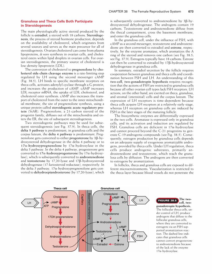
Granulosa and Theca Cells Both Participate
in Steroidogenesis
The main physiologically active steroid produced by thefollicle is estradiol, a steroid with 18 carbons. Steroidoge-nesis, the process of steroid hormone production, dependson the availability of cholesterol, which originates fromseveral sources and serves as the main precursor for all ofsteroidogenesis. Ovarian cholesterol can come from plasmalipoproteins, de novo synthesis in ovarian cells, and choles-terol esters within lipid droplets in ovarian cells. For ovar-ian steroidogenesis, the primary source of cholesterol islow-density lipoprotein (LDL).
The conversion of cholesterol to pregnenolone by cho-lesterol side-chain cleavage enzyme is a rate-limiting stepregulated by LH using the second messenger cAMP (Fig. 38.5). LH binds to specific membrane receptors ontheca cells, activates adenylyl cyclase through a G protein,and increases the production of cAMP. cAMP increasesLDL receptor mRNA, the uptake of LDL cholesterol, andcholesterol ester synthesis. cAMP also increases the trans-port of cholesterol from the outer to the inner mitochondr-ial membrane, the site of pregnenolone synthesis, using aunique protein called steroidogenic acute regulatory pro-tein (StAR). Pregnenolone, a 21-carbon steroid of theprogestin family, diffuses out of the mitochondria and en-ters the ER, the site of subsequent steroidogenesis.
Two steroidogenic pathways may be used for subse-quent steroidogenesis (see Fig. 37.9). In theca cells, thedelta 5 pathway is predominant; in granulosa cells and thecorpus luteum, the delta 4 pathway is predominant. Preg-nenolone gets converted to either progesterone by 3�-hy-droxysteroid dehydrogenase in the delta 4 pathway or to17�-hydroxypregnenolone by 17�-hydroxylase in thedelta 5 pathway. In the delta 4 pathway, progesterone getsconverted to 17�-hydroxyprogesterone (by 17�-hydroxy-lase), which is subsequently converted to androstenedioneand testosterone by 17,20-lyase and 17�-hydroxysteroiddehydrogenase (17-ketosteroid reductase), respectively. Inthe delta 5 pathway, 17�-hydroxypregnenolone gets con-verted to dehydroepiandrosterone (by 17,20-lyase), which
is subsequently converted to androstenedione by 3�-hy-droxysteroid dehydrogenase. The androgens contain 19carbons. Testosterone and androstenedione diffuse fromthe thecal compartment, cross the basement membrane,and enter the granulosa cells.
In the granulosa cell, under the influence of FSH, withcAMP as a second messenger, testosterone and androstene-dione are then converted to estradiol and estrone, respec-tively, by the enzyme aromatase, which aromatizes the Aring of the steroid and removes one carbon (see Fig. 38.5;see Fig. 37.9). Estrogens typically have 18 carbons. Estronecan then be converted to estradiol by 17�-hydroxysteroiddehydrogenase in granulosa cells.
In summary, estradiol secretion by the follicle requirescooperation between granulosa and theca cells and coordi-nation between FSH and LH. An understanding of thistwo-cell, two-gonadotropin hypothesis requires recogni-tion that the actions of FSH are restricted to granulosa cellsbecause all other ovarian cell types lack FSH receptors. LHactions, on the other hand, are exerted on theca, granulosa,and stromal (interstitial) cells and the corpus luteum. Theexpression of LH receptors is time-dependent becausetheca cells acquire LH receptors at a relatively early stage,whereas LH receptors on granulosa cells are induced byFSH in the later stages of the maturing follicle.
The biosynthetic enzymes are differentially expressedin the two cells. Aromatase is expressed only in granulosacells, and its activation and induction are regulated byFSH. Granulosa cells are deficient in 17�-hydroxylaseand cannot proceed beyond the C-21 progestins to gen-erate C-19 androgenic compounds (see Fig. 38.5). Conse-quently, estrogen production by granulosa cells dependson an adequate supply of exogenous aromatizable andro-gens, provided by theca cells. Under LH regulation, thecacells produce androgenic substrates, primarily an-drostenedione and testosterone, which reach the granu-losa cells by diffusion. The androgens are then convertedto estrogens by aromatization.
In follicles, theca and granulosa cells are exposed to dif-ferent microenvironments. Vascularization is restricted tothe theca layer because blood vessels do not penetrate the
CHAPTER 38 The Female Reproductive System 673
Capillary
Granulosa cellTheca cell
FSH
Basem
ent mem
brane
LH
Receptor ATP
cAMP
Receptor
Androstenedione
Testosterone
Cholesterol
Pregnenolone
17OH pregnenolone
Dehydroepiandrosterone
LHATP
cAMPCholesterol
Pregnenolone
Progesterone
Androstenedione
Testosterone
cAMP
Estradiol Estrone
ATP
cAMP
Receptor
�
�
�
�
The two-
cell, two-
gonadotropin hypothesis.
The follicular theca cells, un-der control of LH, produceandrogens that diffuse to thefollicular granulosa cells,where they are converted toestrogens via an FSH-sup-ported aromatization reac-tion. The dashed line indi-cates that granulosa cellscannot convert progesteroneto androstenedione becauseof the lack of the enzyme17�-hydroxylase.
FIGURE 38.5

674 PART X REPRODUCTIVE PHYSIOLOGY
basement membrane. Theca cells, therefore, have better ac-cess to circulating cholesterol, which enters the cells viaLDL receptors. Granulosa cells, on the other hand, prima-rily produce cholesterol from acetate, a less efficientprocess than uptake. In addition, granulosa cells are bathedin follicular fluid and exposed to autocrine, paracrine, andjuxtacrine control by locally produced peptides and growthfactors. “Juxtacrine” describes the interaction of a mem-brane-bound growth factor on one cell with its membrane-bound receptor on an adjacent cell.
FSH acts on granulosa cells by a cAMP-dependentmechanism and produces a broad range of activities, in-cluding increased mitosis and cell proliferation, the stimu-lation of progesterone synthesis, the induction of aro-matase, and increased inhibin synthesis. As the folliclematures, the number of receptors for both gonadotropinsincreases. FSH stimulates the formation of its own recep-tors and induces the appearance of LH receptors. The com-bined activity of the two gonadotropins greatly amplifiesestrogen production.
Androgens are produced by theca and stromal cells.They serve as precursors for estrogen synthesis and alsohave a distinct local action. At low concentrations, andro-gens enhance aromatase activity, promoting estrogen pro-duction. At high concentrations, androgens are convertedby 5�-reductase to a more potent androgen, such as dihy-drotestosterone (DHT). When follicles are overwhelmedby androgens, the intrafollicular androgenic environmentantagonizes granulosa cell proliferation and leads to apop-tosis of the granulosa cells and subsequent follicular atresia.
Follicular Atresia Probably Results From a
Lack of Gonadotropin Support
Follicular atresia, the degeneration of follicles in the ovary,is characterized by the destruction of the oocyte and gran-ulosa cells. Atresia is a continuous process and can occur atany stage of follicular development. During a woman’s life-time approximately 400 to 500 follicles will ovulate; thoseare the only follicles that escape atresia, and they representa small percentage of the 1 to 2 million follicles present atbirth. The cause of follicular atresia is likely due to lack ofgonadotropin support of the growing follicle. For example,at the beginning of the menstrual cycle, several follicles areselected for growth but only one follicle, the dominant fol-licle, will go on to ovulate. Because the dominant folliclehas a preferential blood supply, it gets the most FSH (andLH). Other reasons for the lack of gonadotropin support ofnondominant follicles could be a lack of FSH and LH re-ceptors or the inability of granulosa cells to transduce thegonadotropin signals.
During atresia, granulosa cell nuclei become pyknotic(referring to an apoptotic process characterized by DNAladdering), and/or the oocyte undergoes pseudomatura-tion, characteristic of meiosis. During the early stages ofoocyte death, the nuclear membrane disintegrates, thechromatin condenses, and the chromosomes form ametaphase plate with a spindle; the term pseudomaturation isappropriate because these oocytes are not capable of suc-cessful fertilization. During atresia of follicles containingtheca cells, the theca layer may undergo hyperplasia and
hypertrophy and may remain in the ovary for extended pe-riods of time.
Meiosis Resumes During the Periovulatory Period
All healthy oocytes in the ovary remain arrested in prophaseof the first meiosis. When a graafian follicle is subjected to asurge of gonadotropins (LH and/or FSH), the oocyte withinundergoes the final stages of meiosis, resulting in the pro-duction of a mature gamete. This maturation is accomplishedby two successive cell divisions in which the number of chro-mosomes is reduced, producing haploid gametes. At fertil-ization, the diploid state is restored.
Primary oocytes arrested in meiotic prophase 1 (of thefirst meiosis) have duplicated their centrioles and DNA(4n DNA) so that each chromosome has two identicalchromatids. Crossing over and chromatid exchange occurduring this phase, producing genetic diversity. The re-sumption of meiosis, ending the first meiotic prophaseand beginning of meiotic metaphase 1, is characterized bydisappearance of the nuclear membrane, condensation ofthe chromosomes, nuclear dissolution (germinal vesiclebreakdown), and alignment of the chromosomes on theequator of the spindle. At meiotic anaphase 1, the homol-ogous chromosomes move in opposite directions underthe influence of the retracting meiotic spindle at the cel-lular periphery. At meiotic telophase 1, an unequal divi-sion of the cell cytoplasm yields a large secondary oocyte(2n DNA) and a small, nonfunctional cell, the first polarbody (2n DNA). Each cell contains half the original 4nnumber of chromosomes (only one member of each ho-mologous pair is present, but each chromosome consistsof two unique chromatids).
The secondary oocyte is formed several hours after theinitiation of the LH surge but before ovulation. It rapidlybegins the second meiotic division and proceeds through ashort prophase to become arrested in metaphase. At thisstage, the secondary oocyte is expelled from the graafianfollicle. The second arrest period is relatively short. In re-sponse to penetration by a spermatozoon during fertiliza-tion, meiosis 2 resumes and is rapidly completed. A secondunequal cell division soon follows, producing a small sec-ond polar body (1n DNA) and a large fertilized egg, thezygote (2n DNA, 1n from the mother and 1n from the fa-ther). The first and second polar bodies either degenerateor divide, yielding small nonfunctional cells. If fertilizationdoes not occur, the secondary oocyte begins to degeneratewithin 24 to 48 hours.
FOLLICLE SELECTION AND OVULATION
The number of ovulating eggs is species-specific and is in-fluenced by genetic, nutritional, and environmental factors.In humans, normally only one follicle will ovulate, but mul-tiple ovulations in a single cycle (superovulation) can beinduced by the timed administration of gonadotropins orantiestrogens. The mechanism by which one follicle is se-lected from a cohort of growing follicles is poorly under-stood. It occurs during the first few days of the cycle, im-mediately after the onset of menstruation. Once selected,

the follicle begins to grow and differentiate at an exponen-tial rate and becomes the dominant follicle.
In parallel with the growth of the dominant follicle, therest of the preantral follicles undergo atresia. Two main fac-tors contribute to atresia in the nonselected follicles. One isthe suppression of plasma FSH in response to increased estra-diol secretion by the dominant follicle. The decline in FSHsupport decreases aromatase activity and estradiol produc-tion and interrupts granulosa cell proliferation in those non-dominant follicles. The dominant follicle is protected from afall in circulating FSH levels because it has a healthy bloodsupply, FSH accumulated in the follicular fluid, and an in-creased density of FSH receptors on its granulosa cells. An-other factor in selection is the accumulation of atretogenicandrogens, such as DHT, in the nonselected follicles. Theincrease in DHT changes the intrafollicular ratio of estrogento androgen and antagonizes the actions of FSH.
As the dominant follicle grows, vascularization of thetheca layer increases. On day 9 or 10 of the cycle, the vascu-larity of the dominant follicle is twice that of the other antralfollicles, permitting a more efficient delivery of cholesterolto theca cells and better exposure to circulating go-nadotropins. At this time, the main source of circulatingestradiol is the dominant follicle. Since estradiol is the pri-mary regulator of LH and FSH secretion by positive and neg-ative feedback, the dominant follicle ultimately determinesits own fate.
The midcycle LH surge occurs as a result of rising levelsof circulating estradiol, and it causes multiple changes in thedominant follicle, which occur within a relatively short time.These include the resumption of meiosis in the oocyte (as al-ready discussed); granulosa cell differentiation and transfor-mation into luteal cells; the activation of proteolytic en-zymes that degrade the follicle wall and surrounding tissues;increased production of prostaglandins, histamine, and otherlocal factors that cause localized hyperemia; and an increasein progesterone secretion. Within 30 to 36 hours after theonset of the LH surge, this coordinated series of biochemicaland morphological events culminates in follicular ruptureand ovulation. The midcycle FSH surge is not essential forovulation because an injection of either LH or human chori-onic gonadotropin (hCG) before the endogenous go-nadotropin surge can induce normal ovulation. However,only follicles that have been adequately primed with FSHwill ovulate because they contain sufficient numbers of LHreceptors for ovulation and subsequent luteinization.
Four ovarian proteins are essential for ovulation: the prog-esterone receptor, the cyclooxygenase enzyme (which con-verts arachidonic acid to prostaglandins), cyclin D2 (a cellcycle regulator), and a transcription factor called C/EBP�(CCAAT/enhancer binding protein). The mechanisms bywhich these proteins interact to regulate follicular rupture arelargely unknown. However, mice with specific disruption ofgenes for any of these proteins fail to ovulate, and these pro-teins are likely to have a functional role in human ovulation.
The earliest responses of the ovary to the midcycle LHsurge are the release of vasodilatory substances, such as his-tamine, bradykinin, and prostaglandins, which mediate in-creased ovarian and follicular blood flow. The highly vascu-larized dominant follicle becomes hyperemic and edematousand swells to a size of at least 20 to 25 mm in diameter. There
is also an increased production of follicular fluid, disaggrega-tion of granulosa cells, and detachment of the oocyte-cumu-lus complex from the follicular wall, moving it to the centralportion of the follicle. The basement membrane separatingtheca cells from granulosa cells begins to disintegrate, gran-ulosa cells begin to undergo luteinization, and blood vesselsbegin to penetrate the granulosa cell compartment.
Just prior to follicular rupture, the follicular wall thins bycellular deterioration and bulges at a specific site called thestigma, the point on the follicle that actually ruptures. Asovulation approaches, the follicle enlarges and protrudesfrom the surface of the ovary at the stigma. In response to theLH surge, plasminogen activator is produced by theca andgranulosa cells of the dominant follicle and converts plas-minogen to plasmin. Plasmin is a proteolytic enzyme thatacts directly on the follicular wall and stimulates the produc-tion of collagenase, an enzyme that digests the connectivetissue matrix. The thinning and increased distensibility of thewall facilitates the rupture of the follicle. The extrusion of theoocyte-cumulus complex is aided by smooth muscle con-traction. At the time of rupture, the oocyte-cumulus complexand follicular fluid are ejected from the follicle.
The LH surge triggers the resumption of the first meiosis.Up to this point, the primary oocyte has been protected byunknown factors within the follicle from premature cell divi-sion. The LH surge also causes transient changes in plasmaestradiol and a prolonged increase in plasma progesteroneconcentrations. Within a couple of hours after the initiationof the LH surge, the production of progesterone, androgens,and estrogens begins to increase. Progesterone, actingthrough the progesterone receptor on granulosa cells, pro-motes ovulation by releasing mediators that increase the dis-tensibility of the follicular wall and enhance the activity ofproteolytic enzymes. As LH levels reach their peak, plasmaestradiol levels plunge because of down-regulation by LH ofFSH receptors on granulosa cells and the inhibition of gran-ulosa cell aromatase. Eventually, LH receptors on luteinizinggranulosa cells escape the down-regulation, and proges-terone production increases.
FORMATION OF THE CORPUS LUTEUM FROM
THE POSTOVULATORY FOLLICLE
In response to the LH and FSH surges and after ovulation,the wall of the graafian follicle collapses and becomes con-voluted, blood vessels course through the luteinizing gran-ulosa and theca cell layers, and the antral cavity fills withblood. The granulosa cells begin to cease their proliferationand begin to undergo hypertrophy and produce proges-terone as their main secretory product. The ruptured follicledevelops a rich blood supply and forms a solid structurecalled the corpus luteum (yellow body). The mature corpusluteum develops as the result of numerous biochemical andmorphological changes, collectively referred to as luteiniza-tion. The granulosa cells and theca cells in the corpus lu-teum are called granulosa-lutein cells and theca-luteincells, respectively.
Continued stimulation by LH is needed to ensure mor-phological integrity (healthy luteal cells) and functionality(progesterone secretion). If pregnancy does not occur, the
CHAPTER 38 The Female Reproductive System 675
676 PART X REPRODUCTIVE PHYSIOLOGY
corpus luteum regresses, a process called luteolysis or lutealregression. Luteolysis occurs as a result of apoptosis andnecrosis of the luteal cells. After degeneration, theluteinized cells are replaced by fibrous tissue, creating anonfunctional structure, the corpus albicans. Therefore, thecorpus luteum is a transient endocrine structure formed fromthe postovulatory follicle. It serves as the main source of cir-culating steroids during the luteal (postovulatory) phase ofthe cycle and is essential for maintaining pregnancy duringthe first trimester (see Case Study) as well as maintainingmenstrual cycles of normal length.
The process of luteinization begins before ovulation. Af-ter acquiring a high concentration of LH receptors, granu-losa cells respond to the LH surge by undergoing morpho-logical and biochemical transformation. This changeinvolves cell enlargement (hypertrophy) and the develop-ment of smooth ER and lipid inclusions, typical of steroid-secreting cells. Unlike the nonvascular granulosa cells in thefollicle, luteal cells have a rich blood supply. Invasion bycapillaries starts immediately after the LH surge and is facil-itated by the dissolution of the basement membrane be-tween theca and granulosa cells. Peak vascularization isreached 7 to 8 days after ovulation.
Differentiated theca and stroma cells, as well as granulosacells, are incorporated into the corpus luteum, and all threeclasses of steroids—androgens, estrogens, and progestins—are synthesized. Although some progesterone is secretedbefore ovulation, peak progesterone production is reached 6to 8 days after the LH surge. The life span of the corpus lu-teum is limited. Unless pregnancy occurs, it degenerateswithin about 13 days after ovulation. During the menstrualcycle, the function of the corpus luteum is maintained by
LH; therefore, LH is referred to as a luteotropic hormone.Lack of LH can lead to luteal insufficiency (see Clinical Fo-cus Box 38.1).
Regression of the corpus luteum at the end of the cycle isnot understood. Luteal regression is thought to be inducedby locally produced luteolytic agents that inhibit LH action.Several ovarian hormones, such as estrogen, oxytocin,prostaglandins, and GnRH, have been proposed, but theirrole as luteolysins is controversial. The corpus luteum is res-cued from degeneration in the late luteal phase by the actionof human chorionic gonadotropin (hCG), an LH-like hor-mone that is produced by the embryonic trophoblast duringthe implantation phase (see Chapter 39). This hormonebinds the LH receptor and increases cAMP and proges-terone secretion.
THE MENSTRUAL CYCLE
Under normal conditions, ovulation occurs at timed inter-vals. Sexual intercourse may occur at any time during the cy-cle, but fertilization occurs only during the postovulatoryperiod. Once pregnancy occurs, ovulation ceases, and afterparturition, lactation also inhibits ovulation. The first men-strual cycle occurs in adolescence, usually around age 12.The initial period of bleeding is called the menarche. Thefirst few cycles are usually irregular and anovulatory, as theresult of delayed maturation of the positive feedback byestradiol on a hypothalamus that fails to secrete significantGnRH. During puberty, LH secretion occurs more duringperiods of sleep than during periods of being awake, result-ing in a diurnal cycle.
CLINICAL FOCUS BOX 38.1
Luteal Insufficiency
Occasionally, the corpus luteum will not produce sufficientprogesterone to maintain pregnancy during its very earlystages. Initial signs of early spontaneous pregnancy termi-nation include pelvic cramping and the detection of blood,similar to indications of menstruation. If the corpus luteumis truly deficient, then fertilization may occur around the ide-alized day 14 (ovulation), pregnancy will terminate duringthe deficient luteal phase, and menses will start on sched-ule. Without measuring levels of hCG, the pregnancy detec-tion hormone, the woman would not know that she is preg-nant because of the continuation of regular menstrualcycles. Luteal insufficiency is a common cause of infertil-ity. Women are advised to see their physician if pregnancydoes not result after 6 months of unprotected intercourse.
Analysis of the regulation of progesterone secretion bythe corpus luteum provides insights into this clinical prob-lem. There are several reasons for luteal insufficiency.First, the number of luteinized granulosa cells in the corpusluteum may be insufficient because of the ovulation of asmall follicle or the premature ovulation of a follicle thatwas not fully developed. Second, the number of LH recep-tors on the luteinized granulosa cells in the graafian follicleand developing corpus luteum may be insufficient. LH re-
ceptors mediate the action of LH, which stimulates prog-esterone secretion. An insufficient number of LH receptorscould be due to insufficient priming of the developing fol-licle with FSH. It is well known that FSH increases the num-ber of LH receptors in the follicle. Third, the LH surge couldhave been inadequate in inducing full luteinization of thecorpus luteum, yet there was sufficient LH to induce ovu-lation. It has been estimated that only 10% of the LH surgeis required for ovulation, but the amount required for fullluteinization and adequate progesterone secretion tomaintain pregnancy is not known.
If progesterone values are low in consecutive cycles atthe midluteal phase and do not match endometrial biop-sies, exogenous progesterone may be administered inorder to prevent early pregnancy termination during afertile cycle. Other options include the induction of follic-ular development and ovulation with clomiphene andhCG. This treatment would likely produce a large,healthy, estrogen-secreting graafian follicle with suffi-cient LH receptors for luteinization. The exogenous hCGis given to supplement the endogenous LH surge and toensure full stimulation of the graafian follicle, ovulation,adequate progesterone, and luteinization of the develop-ing corpus luteum.

The average menstrual cycle length in adult women is 28days, with a range of 25 to 35 days. The interval from ovu-lation to the onset of menstruation is relatively constant, av-eraging 14 days in most women and is dictated by the fixedlife span of the corpus luteum. In contrast, the interval fromthe onset of menses to ovulation (the follicular phase) ismore variable and accounts for differences in cycle lengthsamong ovulating women.
The menstrual cycle is divided into four phases (Fig. 38.6). The menstrual phase, also called menses ormenstruation, is the bleeding phase and lasts about 5 days.The ovarian follicular phase lasts about 10 to 16 days; folli-cle development occurs, estradiol secretion increases, andthe uterine endometrium undergoes proliferation in re-sponse to rising estrogen levels. The ovulatory phase lasts24 to 48 hours, and the luteal phase lasts 14 days. In theluteal phase, progesterone is produced, and the en-dometrium secretes numerous proteins in preparation forimplantation of an embryo.
The cycles become irregular as menopause approachesaround age 50, and cycles cease thereafter. During the re-productive years, menstrual cycling is interrupted by con-
ception and lactation and is subjected to modulation byphysiological, psychological, and social factors.
The Menstrual Cycle Requires Synchrony
Among the Ovary, Brain, and Pituitary
The menstrual cycle requires several coordinated elements:hypothalamic control of pituitary function, ovarian follicu-lar and luteal changes, and positive and negative feedbackof ovarian hormones at the hypothalamic-pituitary axis.We have discussed separately the mechanisms that regulatethe synthesis and release of the reproductive hormones;now we put them together in terms of sequence and inter-action. For this purpose, we use a hypothetical cycle of 28days (see Fig. 38.6), divided into four phases as follows:menstrual (days 0 to 5), follicular (days 0 to 13), ovulatory(days 13 to 14), and luteal (days 14 to 28).
During menstruation, estrogen, progesterone, and in-hibin levels are very low as a result of the luteal regressionthat has just occurred and the low estrogen synthesis by im-mature follicles. The plasma FSH levels are high while LH
CHAPTER 38 The Female Reproductive System 677
Menses
FSH
LH
50
40
30
20
10
0
(mIU
/mL)
LH peak
20
10
1
0
Pro
gest
eron
e (n
g/m
L)
Est
radi
ol (
pg/m
L)
PE2β
17-OH P
300
200
100
0 2 4
Fol
licle
dia
met
er(m
m)
20
10
Day:
Phase: Menstrual
50
40
30
20
10
0
(mIU
/mL)
2
1
17-Hydroxyprogesterone
(ng/mL)
6 8 10 12 14 20 22 24 26 28
20
Follicular
Ovulation
Day of menstrual cycle
Luteal
Corpus luteum
diameter (m
m)
Ovulatory
Lutealregression
16 18
10
Hormonal and ovarian events during the menstrual cycle. P, progesterone; E2�, estra-diol; 17-OH P, 17-hydroxyprogesterone.
FIGURE 38.6
678 PART X REPRODUCTIVE PHYSIOLOGY
levels are low in response to the removal of negative feed-back by estrogen, progesterone, and inhibin. A few dayslater, however, LH levels slowly begin to rise. FSH acts ona cohort of follicles recruited 20 to 25 days earlier from aresting pool of smaller follicles. The follicles on days 3 to 5average 4 to 6 mm in diameter, and they are stimulated byFSH to grow into the preantral stages. In response to FSH,the granulosa cells proliferate, aromatase activity increases,and plasma estradiol levels rise slightly between days 3 and7. The designated dominant follicle is selected betweendays 5 and 7, and increases in size and steroidogenic activ-ity. Between days 8 and 10, plasma estradiol levels risesharply, reaching peak levels above 200 pg/mL on day 12,the day before the LH surge.
During the early follicular phase, LH pulsatility is of lowamplitude and high frequency (about every hour). Coin-ciding pulses of GnRH are released about every hour. Asestradiol levels rise, the pulse frequency in GnRH furtherincreases, without a change in amplitude. The mean plasmaLH level increases and further supports follicular steroido-genesis, especially since FSH has increased the number ofLH receptors on growing follicles. During the midfollicularto late follicular phase, rising estradiol and inhibin from thedominant follicle suppress FSH release. The decline inFSH, together with an accumulation of nonaromatizableandrogens, induces atresia in the nonselected follicles. Thedominant follicle is saved by virtue of its high density ofFSH receptors, the accumulation of FSH in its follicularfluid (see Table 38.2), and the acquisition of LH receptorsby the granulosa cells.
The midcycle surge of LH is rather short (24 to 36hours) and is an example of positive feedback. For the LHsurge to occur, estradiol must be maintained at a criticalconcentration (about 200 pg/mL) for a sufficient duration(36 to 48 hours) prior to the surge. Any reduction of theestradiol rise or a rise that is too small or too short elimi-nates or reduces the LH surge. In addition, in the presenceof elevated progesterone, high concentrations of estradioldo not induce an LH surge. Paradoxically, although it ex-erts negative feedback on LH release most of the time, pos-itive feedback by estradiol is required to generate the mid-cycle surge.
Estrogen exerts its effects directly on the anterior pitu-itary, with GnRH playing a permissive, albeit mandatory,role. This concept is derived from experiments in monkeyswhose medial basal hypothalamus, including the GnRH-producing neurons, was destroyed by lesioning, resulting ina marked decrease in plasma LH levels. The administrationof exogenous GnRH at a fixed frequency restored LH re-lease. When estradiol was given at an optimal concentrationfor an appropriate time, an LH surge was generated, in spiteof maintaining steady and unchanging pulses of GnRH.
The mechanism that transforms estradiol from a nega-tive to a positive regulator of LH release is unknown. Onefactor involves an increase in the number of GnRH recep-tors on the gonadotrophs, increasing pituitary responsive-ness to GnRH. Another factor is the conversion of a stor-age pool of LH (perhaps within a subpopulation ofgonadotrophs) to a readily releasable pool. Estrogen mayalso increase GnRH release, serving as a fine-tuning or fail-safe mechanism. A small but distinct rise in progesterone
occurs before the LH surge. This rise is important for aug-menting the LH surge and, together with estradiol, pro-motes a concomitant surge in FSH. There are indicationsthat the midcycle FSH surge is important for inducingenough LH receptors on granulosa cells for luteinization,stimulating plasminogen activator for follicular rupture,and activating a cohort of follicles destined to develop inthe next cycle.
The LH surge reduces the concentration of 17�-hy-droxylase and subsequently decreases androstenedioneproduction by the dominant follicle. Estradiol levels de-cline, 17-hydroxyprogesterone increases, and progesteronelevels plateau. The prolonged exposure to high LH levelsduring the surge down-regulates the ovarian LH receptors,accounting for the immediate postovulatory suppression ofestradiol. As the corpus luteum matures, it increases prog-esterone production and reinitiates estradiol secretion.Both reach high plasma concentrations on days 20 to 23,about 1 week after ovulation.
During the luteal phase, circulating FSH levels are sup-pressed by the elevated steroids. The LH pulse frequency isreduced during the early luteal phase, but the amplitude ishigher than that during the follicular phase. LH is impor-tant at this time for maintaining the function of the corpusluteum and sustaining steroid production. In the late lutealphase, both LH pulse frequency and amplitude are reducedby a progesterone-dependent, opioid-mediated suppres-sion of the GnRH pulse generator.
After the demise of the corpus luteum on days 24 to 26,estradiol and progesterone levels plunge, causing thewithdrawal of support of the uterine endometrium, culmi-nating within 2 to 3 days in menstruation. The reductionin ovarian steroids acts centrally to remove feedback inhi-bition. The FSH level begins to rise and a new cycle is ini-tiated.
Estradiol and Progesterone Influence Cyclic
Changes in the Reproductive Tract
The female reproductive tract undergoes cyclic alterationsin response to the changing levels of ovarian steroids. Themost notable changes occur in the function and histologyof the oviduct and uterine endometrium, the compositionof cervical mucus, and the cytology of the vagina (Fig. 38.7). At the time of ovulation, there is also a small butdetectable rise in basal body temperature, caused by prog-esterone. All of the above parameters are clinically usefulfor diagnosing menstrual dysfunction and infertility.
The oviduct is a muscular tube lined internally with a cil-iated, secretory, columnar epithelium with a deeper stromaltissue. Fertilization occurs in the oviduct, after which thezygote enters the uterus; therefore, the oviduct is involvedin transport of the gametes and provides a site for fertiliza-tion and early embryonic development. Estrogens maintainthe ciliated nature of the epithelium, and ovariectomycauses a loss of the cilia. Estrogens also increase the motil-ity of the oviducts. Exogenous estrogen given around thetime of fertilization can cause premature expulsion of thefertilized egg, whereas extremely high doses of estrogencan cause “tube locking,” the entrapment of the fertilized

egg and an ectopic pregnancy. Progesterone opposes theseactions of estrogen.
The endometrium (also called uterine mucosa) is com-posed of a superficial layer of epithelial cells and an under-lying stromal layer. The epithelial layer contains glandsthat penetrate the stromal layer. The glands are lined by asecretory columnar epithelium.
The endometrial cycle consists of four phases. The pro-liferative phase coincides with the midfollicular to late fol-licular phase of the menstrual cycle. Under the influence ofthe rising plasma estradiol concentration, the stromal andepithelial layers of the uterine endometrium undergo hy-perplasia and hypertrophy and increase in size and thick-ness. The endometrial glands elongate and are lined withcolumnar epithelium. The endometrium becomes vascular-ized, and more spiral arteries, a rich blood supply to this re-gion, develop. Estradiol also induces the formation ofprogesterone receptors and increases myometrial excitabil-ity and contractility.
The secretory phase begins on the day of ovulation andcoincides with the early to midluteal phase of the menstrualcycle. The endometrium contains numerous progesterone
receptors. Under the combined action of progesterone andestrogen, the endometrial glands become coiled, storeglycogen, and secrete large amounts of carbohydrate-richmucus. The stroma increases in vascularity and becomesedematous, and the spiral arteries become tortuous (see Fig. 38.7). Peak secretory activity, edema formation, andoverall thickness of the endometrium are reached on days 6to 8 after ovulation in preparation for implantation of theblastocyst. Progesterone antagonizes the effect of estrogen on the myometrium and reduces spontaneous myometrialcontractions.
The ischemic phase, generally not depicted graphically,occurs immediately before the menses and is initiated bythe declining levels of progesterone and estradiol caused byregression of the corpus luteum. Necrotic changes andabundant apoptosis occur in the secretory epithelium as itcollapses. The arteries constrict, reducing the blood supplyto the superficial endometrium. Leukocytes andmacrophages invade the stroma and begin to phagocytosethe ischemic tissue. Leukocytes persist in large numbersthroughout menstruation, providing resistance against in-fection to the denuded endometrial surface.
CHAPTER 38 The Female Reproductive System 679
Estradiol
Progesterone
Basal body temperature
Vaginal cornification and pyknotic index
Cervical mucusferning
Deg
rees
(F
)P
lasm
a le
vel
99
98
97
100
50
0
3�
2�
1�
0
3
2
1
0
4
Artery
Proliferative phase Secretory phase
Days 0 4 8 12 16 20 24 28
Ovulation
EndometriumGland
Glycogen vacuoles
mm
32
Menses Menses
Cyclic changes in the uterus,
cervix, vagina, and body tempera-
ture in relationship to estradiol, progesterone, and
ovulation during the menstrual cycle. (Modifiedfrom Odell WD. The reproductive system in women.In: Degroot LJ, et al, eds. Endocrinology. Vol 3. NewYork: Grune & Stratton, 1979.)
FIGURE 38.7

680 PART X REPRODUCTIVE PHYSIOLOGY
Desquamation and sloughing of the entire functionallayer of the endometrium occurs during the menstrualphase (menses). The mechanism leading to necrosis is onlypartly understood. The reduction in steroids destabilizeslysosomal membranes in endometrial cells, resulting in theliberation of proteolytic enzymes and increased productionof vasoconstrictor prostaglandins (e.g., PGF2� ). Theprostaglandins induce vasospasm of the spiral arteries, andthe proteolytic enzymes digest the tissue. Eventually, theblood vessels rupture and blood is released, together withcellular debris. The endometrial tissue is expelled throughthe cervix and vagina, with blood from the ruptured arter-ies. The menstrual flow lasts 4 to 5 days and averages 30 to50 mL in volume. It does not clot because of the presenceof fibrinolysin, but the spiral arteries constrict, resulting ina reduction in bleeding.
Changes in the properties of the cervical mucus promotethe survival and transport of sperm and, thus, can be im-portant for normal fertility. The cervical mucus undergoescyclic changes in composition and volume. During the fol-licular phase, estrogen increases the quantity, alkalinity,viscosity, and elasticity of the mucus. The cervical musclesrelax, and the epithelium becomes secretory in response toestrogen. By the time of ovulation, elasticity of the mucusor spinnbarkeit is greatest. Sperm can readily pass throughthe estrogen-dominated mucus. With progesterone risingeither after ovulation, during pregnancy, or with low-doseprogestogen administration during the cycle, the quantityand elasticity of the mucus decline; it becomes thicker (lowspinnbarkeit) and does not form a ferning pattern whendried on a microscope slide. With these conditions, themucus provides better protection against infections andsperm do not easily pass through.
The vaginal epithelium proliferates under the influenceof estrogen. Basophilic cells predominate early in the fol-licular phase. The columnar epithelium becomes cornified(keratinized) under the influence of estrogen and reachesits peak in the periovulatory period. During the postovula-tory period, progesterone induces the formation of thickmucus, the epithelium becomes infiltrated with leukocytes,and cornification decreases (see Fig. 38.7).
ESTROGEN, PROGESTIN, AND ANDROGEN:
TRANSPORT AND METABOLISM
The principal sex steroids in the female are estrogen, prog-estin, and androgen. Three estrogens are present in signif-icant quantities—estradiol, estrone, and estriol. Estradiol isthe most abundant and is 12 and 80 times more potent thanestrone and estriol, respectively. Much of estrone is derivedfrom peripheral conversion of either androstenedione orestradiol (see Fig 37.9). During pregnancy, large quantitiesof estriol are produced from dehydroepiandrosterone sul-fate after 16�-hydroxylation by the fetoplacental unit (seeChapter 39). Most estrogens are bound to either albumin(�60%) with a low affinity or to sex hormone-bindingglobulin (SHBG) (�40%) with high affinity. Estrogens aremetabolized in the liver through oxidation or conversion toglucuronides or sulfates. The metabolites are then ex-creted in the urine.
The most important progestin is progesterone. It is se-creted in significant amounts during the luteal phase of themenstrual cycle. During pregnancy, the corpus luteum se-cretes progesterone throughout the first trimester, and theplacenta continues progesterone production until parturi-tion. Small amounts of 17-hydroxyprogesterone are se-creted along with progesterone. Progesterone bindsequally to albumin and to a plasma protein called corticos-teroid-binding protein (transcortin). Progesterone is me-tabolized in the liver to pregnanediol and, subsequently,excreted in the urine as a glucuronide conjugate.
Circulating androgens in the female originate from theovaries and adrenals and from peripheral conversion. An-drostenedione and dehydroepiandrosterone (DHEA) orig-inate from the adrenal cortex (see Chapter 34), and ovariantheca and stroma cells. Peripheral conversion from an-drostenedione provides an additional source of testos-terone. Testosterone can also be converted in peripheraltissues to dihydrotestosterone (DHT) by 5�-reductase.However, the primary biologically active androgen inwomen is testosterone. Androgens bind primarily to SHBGand bind to albumin by about half as much. Androgens arealso metabolized to water-soluble forms by oxidation, sul-fation, or glucuronidation and excreted in the urine.
PUBERTY
During the prepubertal period, the hypothalamic-pituitary-ovarian axis becomes activated—an event known as go-nadarche—and gonadotropins increase in the circulationand stimulate ovarian estrogen secretion. The increase in go-nadotropins is a direct result of increased secretion of GnRH.Factors stimulating the secretion of GnRH include gluta-mate, norepinephrine, and neuropeptide Y emanating fromsynaptic inputs to GnRH-producing neurons. In addition, adecrease in �-aminobutyric acid (GABA), an inhibitor ofGnRH secretion, may occur at this time. It is also known thatthe response of the pituitary to GnRH increases at the timeof puberty. Collectively, numerous factors control the rise inovarian estradiol secretion that triggers the development ofphysical characteristics of sexual maturation.
Estradiol induces the development of secondary sexcharacteristics, including the breasts and reproductivetract, and increased fat in the hips. Estrogens also regulatethe growth spurt at puberty, induce closure of the epi-physes, have a positive effect in maintaining bone forma-tion, and can antagonize the degrading actions ofparathyroid hormone on bone. Therefore, estrogens havea positive effect on bone maintenance, and later in life, ex-ogenous estrogens oppose the osteoporosis often associ-ated with menopause.
As mentioned earlier, the first menstruation is calledmenarche and occurs around age 12. The first ovulationdoes not occur until 6 to 9 months after menarche be-cause the hypothalamic-pituitary axis is not fully respon-sive to the feedback effects of estrogen. During the pu-bertal period, the development of breasts, under theinfluence of estrogen, is known as thelarche. At this time,the appearance of axillary and pubic hair occurs, a devel-opment known as pubarche, controlled by adrenal an-
drogens. The adrenals begin to produce significantamounts of androgens (dehydroepiandrosterone and an-drostenedione) 4 to 5 years prior to menarche, and thisevent is called adrenarche. The adrenal androgens are re-sponsible in part for pubarche. Adrenarche is independ-ent of gonadarche.
MENOPAUSE
Menopause is the time after which the final menses occurs.It is associated with the cessation of ovarian function andreproductive cycles. Generally, menstrual cycles and bleed-ing become irregular, and the cycles become shorter fromthe lack of follicular development (shortened follicularphases). The ovaries atrophy and are characterized by thepresence of few, if any, healthy follicles.
The decline in ovarian function is associated with a de-crease in estrogen secretion and a concomitant increase inLH and FSH, which is characteristic of menopausal women(Table 38.3). It is used as a diagnostic tool. The elevatedLH stimulates ovarian stroma cells to continue producingandrostenedione. Estrone, derived almost entirely from theperipheral conversion of adrenal and ovarian androstene-dione, becomes the dominant estrogen (see Fig. 37.9). Be-cause the ratio of estrogens to androgens decreases, somewomen exhibit hirsutism, which results from androgen ex-cess. The lack of estrogen causes atrophic changes in thebreasts and reproductive tract, accompanied by vaginaldryness, which often causes pain and irritation. Similarchanges in the urinary tract may give rise to urinary distur-bances. The epidermal layer of the skin becomes thinnerand less elastic.
Hot flashes, as a result of the loss of vasomotor tone, os-teoporosis, and an increased risk of cardiovascular disease arenot uncommon. Hot flashes are associated with episodic in-creases in upper body and skin temperature, peripheral va-sodilation, and sweating. They occur concurrently with LHpulses but are not caused by the gonadotropins because theyare evident in hypophysectomized women. Hot flashes, con-sisting of episodes of sudden warmth and sweating, reflecttemporary disturbances in the hypothalamic thermoregula-tory centers, which are somehow linked to the GnRH pulsegenerator.
Osteoporosis increases the risk of hip fractures and es-trogen replacement therapy reduces the risk. Estrogen an-tagonizes the effects of PTH on bone but enhances its ef-fect on kidney, i.e., it stimulates retention of calcium.Estrogen also promotes the intestinal absorption of calcium
through 1,25-dihydroxyvitamin D3.Menopausal symptoms are often treated with hormone
replacement therapy (HRT), which includes estrogens andprogestins. HRT is not an uncommon treatment to improvethe quality of life. In some patients, treatment with estro-gen can cause adverse effects, such as vaginal bleeding,nausea, and headache. Estrogen therapy is contraindicatedin cases of existing reproductive tract carcinomas or hyper-tension and other cardiovascular disease. The prevailingopinion is that the benefit of treating postmenopausalwomen with estrogens for limited periods outweighs anyrisk of developing breast or endometrial carcinomas.
INFERTILITY
One of five women in the United States will be affected byinfertility. A thorough understanding of female endocrinol-ogy, anatomy, and physiology are critical to gaining in-sights into solving this major health problem. Infertility canbe caused by several factors. Environmental factors, disor-ders of the central nervous system, hypothalamic disease,pituitary disorders, and ovarian abnormalities can interferewith follicular development and/or ovulation. If a normalovulation occurs, structural, pathological, and/or endocrineproblems associated with the oviduct and/or uterus can pre-vent fertilization, impede the transport or implantation ofthe embryo, and, ultimately, interfere with the establish-ment or maintenance of pregnancy.
Amenorrhea Is Caused by Endocrine Disruption
Menstrual cycle disorders can be divided into two cate-gories: amenorrhea, the absence of menstruation, andoligomenorrhea, infrequent or irregular menstruation. Pri-mary amenorrhea is a condition in which menstruation hasnever occurred. An example is Turner’s syndrome, alsocalled gonadal dysgenesis, a congenital abnormality causedby a nondisjunction of one of the X chromosomes, resultingin a 45 X0 chromosomal karyotype. Because the two X chro-mosomes are necessary for normal ovarian development,women with this condition have rudimentary gonads and donot have a normal puberty. Because of ovarian steroid defi-ciency (lack of estrogen), secondary sex characteristics re-main prepubertal, and plasma LH and FSH are elevated.Other abnormalities include short stature, a webbed neck, acoarctation of the aorta, and renal disorders.
Another congenital form of primary amenorrhea is hy-pogonadotropism with anosmia, similar to Kallmann’s syn-
CHAPTER 38 The Female Reproductive System 681
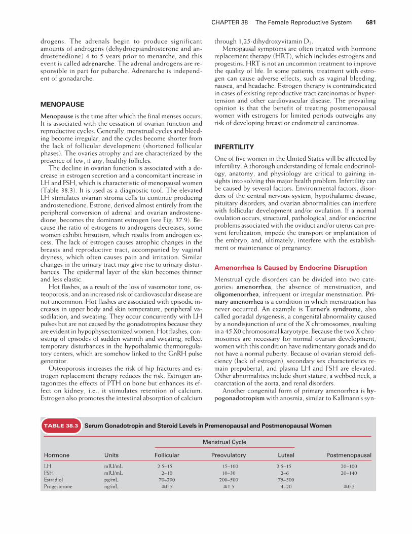
TABLE 38.3 Serum Gonadotropin and Steroid Levels in Premenopausal and Postmenopausal Women
Menstrual Cycle
Hormone Units Follicular Preovulatory Luteal Postmenopausal
LH mIU/mL 2.5–15 15–100 2.5–15 20–100FSH mIU/mL 2–10 10–30 2–6 20–140Estradiol pg/mL 70–200 200–500 75–300Progesterone ng/mL 0.5 1.5 4–20 0.5

682 PART X REPRODUCTIVE PHYSIOLOGY
drome in males (see Chapter 37). Patients do not progressthrough normal puberty and have low and nonpulsatile LHand FSH levels. However, they can have normal stature,female karyotype, and anosmia. The disorder is caused by afailure of olfactory lobe development and GnRH defi-ciency. Primary amenorrhea can also be caused by a con-genital malformation of reproductive tract structures origi-nating from the müllerian duct, including the absence orobstruction of the uterus, cervix, or upper vagina.
Secondary amenorrhea is the cessation of menstrua-tion for longer than 6 months. Pregnancy, lactation, andmenopause are common physiological causes of second-ary amenorrhea. Other causes are premature ovarian fail-ure, polycystic ovarian syndrome, hyperprolactinemia,and hypopituitarism.
Premature ovarian failure is characterized by amenor-rhea, low estrogen levels, and high gonadotropin (LH andFSH) levels before age 40. The symptoms are similar tothose of menopause, including hot flashes and an in-creased risk of osteoporosis. The etiology is variable, in-cluding chromosomal abnormalities; lesions resultingfrom irradiation, chemotherapy, or viral infections; andautoimmune conditions.
Polycystic ovarian syndrome, also called Stein-Leven-thal syndrome, is a heterogeneous group of disorders char-acterized by amenorrhea or anovulatory bleeding, an ele-vated LH/FSH ratio, high androgen levels, hirsutism, andobesity. Although the etiology is unknown, the syndromemay be initiated by excessive adrenal androgen production,during puberty or following stress, that deranges the hypo-thalamic-pituitary axis secretion of LH. Androgens are con-verted peripherally to estrogens and stimulate LH release.Excess LH, in turn, increases ovarian stromal and thecal an-drogen production, resulting in impaired follicular matura-tion. The LH-stimulated ovaries are enlarged and containmany small follicles and hyperplastic and luteinized thecacells (the site of LH receptors). The elevated plasma an-drogen levels cause hirsutism, increased activity of seba-ceous glands, and clitoral hypertrophy, which are signs ofvirilization in females.
Hyperprolactinemia is also a cause of secondary amen-orrhea. Galactorrhea, a persistent milk-like discharge fromthe nipple in nonlactating individuals, is a frequent symp-tom and is due to the excess prolactin (PRL). The etiologyof hyperprolactinemia is variable. Pituitary prolactinomasaccount for about 50% of cases. Other causes are hypo-thalamic disorders, trauma to the pituitary stalk, and psy-chotropic medications, all of which are associated with areduction in dopamine release, resulting in an increasedPRL secretion. Hypothyroidism, chronic renal failure, andhepatic cirrhosis are additional causes of hyperprolactine-mia. In some forms of hypothyroidism, increased hypo-thalamic thyrotropin-releasing hormone (TRH) is thoughtto contribute to excess PRL secretion, as experimental stud-
ies reveal that exogenous TRH increases the secretion ofPRL. The mechanism by which elevated PRL levels sup-press ovulation is not entirely clear. It has been postulatedthat PRL may inhibit GnRH release, reduce LH secretion inresponse to GnRH stimulation, and act directly at the levelof the ovary by inhibiting the action of LH and FSH on fol-licle development.
Oligomenorrhea can be caused by excessive exerciseand by nutritional, psychological, and social factors.Anorexia nervosa, a severe behavioral disorder associatedwith the lack of food intake, is characterized by extrememalnutrition and endocrine changes secondary to psycho-logical and nutritional disturbances. About 30% of patientsdevelop amenorrhea that is not alleviated by weight gain.Strenuous exercise, especially by competitive athletes anddancers, frequently causes menstrual irregularities. Twomain factors are thought to be responsible: a low level ofbody fat, and the effect of stress itself through endorphinsthat are known to inhibit the secretion of LH. Other typesof stress, such as relocation, college examinations, generalillness, and job-related pressures, have been known to in-duce some forms of oligomenorrhea.
Female Infertility Is Caused by
Endocrine Malfunction and Abnormalities
in the Reproductive Tract
The diagnosis and treatment of amenorrhea present a chal-lenging problem. The amenorrhea must first be classified asprimary or secondary, and menopause, pregnancy, and lac-tation must be excluded. The next step is to determinewhether the disorder originates in one of the following ar-eas: the hypothalamus and central nervous system, the an-terior pituitary, the ovary, and/or the reproductive tract.
Several treatments can alleviate infertility problems; forexample, some success has been achieved in hypothalamicdisease with pulsatile administration of GnRH. When hy-pogonadotropism is the cause of infertility, sequential ad-ministration of FSH and hCG is a common treatment forinducing ovulation, although the risk of ovarian hyperstim-ulation and multiple ovulations is increased. Hyperpro-lactinemia can be treated surgically by removing the pitu-itary adenoma containing numerous lactotrophs(prolactin-secreting cells). It can also be treated pharmaco-logically with bromocriptine, a dopaminergic agonist thatreduces the size and number of the lactotrophs and PRL se-cretion. Treatment with clomiphene, an antiestrogen thatbinds to and blocks estrogen receptors, can induce ovula-tion in women with endogenous estrogens in the normalrange. Clomiphene reduces the negative feedback effectsof estrogen and thus increases endogenous FSH and LH se-cretion. When reproductive tract lesions are the cause ofinfertility, corrective surgery or in vitro fertilization is thetreatment of choice.

CHAPTER 38 The Female Reproductive System 683
DIRECTIONS: Each of the numbereditems or incomplete statements in thissection is followed by answers or bycompletions of the statement. Select theONE lettered answer or completion that isBEST in each case.
1. Estradiol synthesis in the graafianfollicle involves(A) Activation of LH-stimulatedgranulosa production of androgen(B) Stimulation of aromatase in thegranulosa cell by FSH(C) Decreased secretion ofprogesterone from the corpus luteum,resulting in increased LH(D) Inhibition of the LH surge duringthe preovulatory period(E) Synergy between FSH andprogesterone
2. Granulosa cells do not produceestradiol from cholesterol because theydo not have an active(A) 17�-Hydroxylase(B) Aromatase(C) 5�-Reductase(D) Sulfatase(E) Steroidogenic acute regulatoryprotein
3. A clinical sign indicating the onset ofthe menopause is(A) The onset of menses near age 50(B) An increase in plasma FSH levels(C) An excessive presence of corporalutea(D) An increased number of cornifiedcells in the vagina(E) Regular menstrual cycles
4. Increased progesterone during thepostovulatory period is associated with(A) Proliferation of the uterineendometrium(B) Enhanced development of graafianfollicles(C) Luteal regression(D) An increase in basal bodytemperature by 0.5 to 1.0C
(E) Increased secretion of FSH5. The theca interna cells of the graafian
follicle are distinguished by(A) Their capacity to produceandrogens from cholesterol(B) The lack of cholesterol side-chaincleavage enzyme(C) Aromatization of testosterone toestradiol(D) The lack of a blood supply(E) The production of inhibin
6. Disruption of the hypothalamic-pituitary portal system will lead to(A) High circulating levels of PRL, lowlevels of LH and FSH, and ovarianatrophy(B) Enhanced follicular development asa result of increased circulating levelsof PRL(C) Ovulation, followed by increasedcirculating levels of progesterone(D) A reduction of ovarian inhibinlevels, followed by increasedcirculating FSH(E) Excessive androgen production bythe ovaries
7. Inhibin is an ovarian hormone that(A) Inhibits the secretion of LH andPRL(B) Is produced by granulosa cells andinhibits the secretion of FSH(C) Only has local ovarian effects andno effect on the secretion of FSH(D) Has two forms, A and B, with thesame � subunits but distinct � subunits(E) Binds activin and increases FSHsecretion
8. Spinnbarkeit formation is induced by(A) Secretory endometrium(B) Progesterone action on the uterus(C) Androgen production from theovaries(D) Estrogen action on the vaginalsecretions(E) Prolactin secretion
9. Successful fertilization is most likely tooccur when the oocyte is in
(A) The oviduct and has entered thesecond meiotic division(B) The uterus and has completed thefirst meiotic division(C) Metaphase of mitosis(D) The graafian follicle, which thenenters the oviduct(E) The uterus, extruding the secondpolar body and implanting
10.The enzyme, 5�-reductase isresponsible for(A) Conversion of cholesterol topregnenolone and enhancingsteroidogenesis(B) Conversion of testosterone todihydrotestosterone(C) Aromatization of testosterone toestradiol(D) Increasing the synthesis of LH(E) Female secondary sexcharacteristics
SUGGESTED READING
Carr BR, Blackwell RE. Textbook of Re-productive Medicine. Norwalk, CT:Appleton & Lange, 1998.
Griffin JE, Ojeda, SR. Textbook of En-docrine Physiology. 4th Ed. New York:Oxford University Press, 2000.
Johnson MH, Everitt BJ. Essential Repro-duction. Oxford: Blackwell Science,2000.
Kettyle WM, Arky RA. Endocrine Patho-physiology. Philadelphia: Lippincott-Raven, 1998.
Van Voorhis BJ. Follicular development.In: Knobil E, Neill JD, eds. The En-cyclopedia of Reproduction. NewYork: Academic Press, 1999;376–389.
Van Voorhis BJ. Follicular steroidogenesis.In: Knobil E, Neill JD, eds. The Ency-clopedia of Reproduction. New York:Academic Press, 1999;389–395.
Yen SSC, Jaffe RB, Barbieri RL. Reproduc-tive Endocrinology. 4th Ed. Philadel-phia: WB Saunders, 1999.
R E V I E W Q U E S T I O N S