Development of the female Reproductive System - BSOG
60
Development of the female Reproductive System Dr. Susheela Rani
-
Upload
khangminh22 -
Category
Documents
-
view
0 -
download
0
Transcript of Development of the female Reproductive System - BSOG

Development of the female Reproductive System
Dr. Susheela Rani

Genital System
•Gonads
•Internal genitals
•External genitals

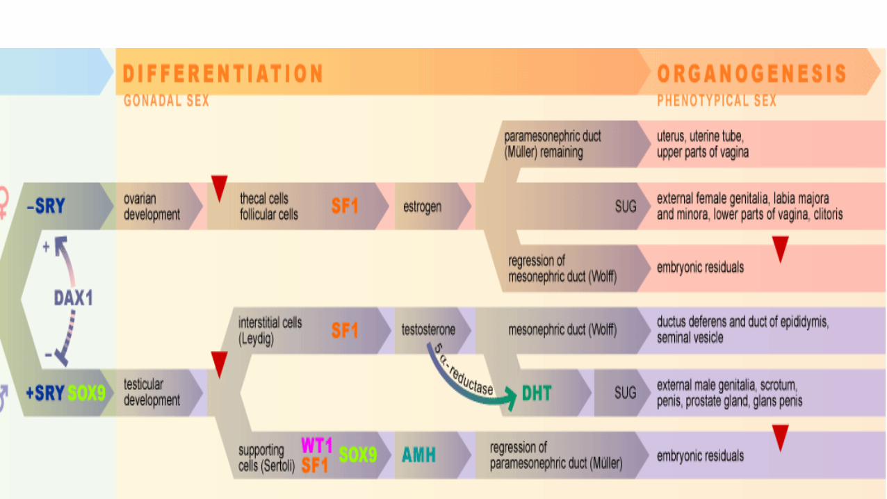
Determining sex – chronology of events
Genetic sex•Determined
at fertilization
Gonadal sex •6th week
Phenotypic sex
BehaviouralSex
•Differentiation of Psyche - Preoptic and Median region of Hypothalamus

Genetic Sex
Genetic sex of an embryo is determined at the time of fertilization,
depending on whether the spermatocyte carries an X or a Y chromosome.

The ‘Master’ Gene that determines Gender
• SRY (Sex determining Region Y gene)
• Has a testis-determining effect on the indifferent gonads.
• Small gene (a single exon)
• Localized on the shorter arm of the Y chromosome (Yp)
• Gets expressed in the gonadal cells
• Controls a whole number of further genes on the autosomes as well as on the X chromosome.
• Causes development of Testes

• Pseudo autosomal regions PAR1 and PAR 2 – Yellow
• Heterochromatin – redundant DNA sequences – Pink
• SRY – Region for Sex Determining Gene- Dark red
• ZFY , Y linked Zinc Finger Protein – Orange
• Spermatogenesis Genes in long arm – Azoospermia
factor AZF
• Telomeres – green
• Centromeres - Blue


It is not the number of gonosomes that is decisive for the gender, but rather the presence or absence of the Y-chromosome

Aneuploidy and Euploidy of GonosomesKaryotype Phenotypic
GenderGonad Syndrome Fate
45, XO Female Ovaries Turner’s Atrophy of Ovaries in the fetus
45, YO ------ ----- ----- Absence of X chromosome is lethal
46, XX Female Ovaries Normal Woman
Normal Development
47, XXX Female Ovaries Normal fertility
Normal Development
46, XY Male Testes Normal Man Normal Development
47, XXY Male Testes Klinefelter’s Small TestisAspermatogenesis
47, XYY Male Testes Normal fertility
Normal Development

Gender by Ultrasound
Earliest gestational age that fetal gender may reliably be determined
CRL ≥ 60 mm (gestational age ≥ 12+2)

Development of the Gonads

Primordial Germ Cells
• Primordial germ cells originate in the epiblast
• 3rd week – PGC migrate and reside among the endodermal cells in the wall of the yolk sac close to allantois.
• 4th week, - Migrate by amoeboid movements along the dorsal mesentery of the hind gut
• 5th week – Reach the genital ridge
• 6th week – Reach the primitive gonads


Development of the GonadsThe Urogenital ridge arises from the
Intermediate mesoderm
It consists of:
Nephrogenic cord - Urinary apparatus
Genital ridge – Gonads
The genital ridge extends from the
upper thorax to claoca.
Middle part develops into gonads
Cranial and caudal parts form the
Gubernaculum

In the female, the upper gubernaculum forms the suspensory ligament of ovary. The lower gubernaculum extends from the lower pole of the ovary as the ovarian ligament and then to the uterine tube angle, where it continues as the round ligament of uterus

Indifferent Gonad
• Shortly before and during arrival of PGC, the epithelium of the genital ridge proliferate, penetrate the underlying mesenchyme.
• They form the primitive sex cords
• In both male and female embryos, these cords are connected to surface epithelium and it is impossible to differentiate between the male and female gonad.
• This Gonad has a Cortex and Medulla

Differentiation of Ovaries
The differentiation of the ovaries happens at 8weeks (later than
that of the testes.
Primitive Ovary has 2 regions
•Cortex, containing all the elements of the parenchyma
•Medulla, which shares the elements of the stroma with the
cortex.

Development of Stroma• In female embryos with an
XX sex chromosome complement and no Y chromosome, primitive sex cords dissociate into irregular cell clusters These clusters, containing groups of primitive germ cells, occupy the medullary part of the ovary. Later, they disappear and are replaced by a vascular stroma that forms the ovarian medulla
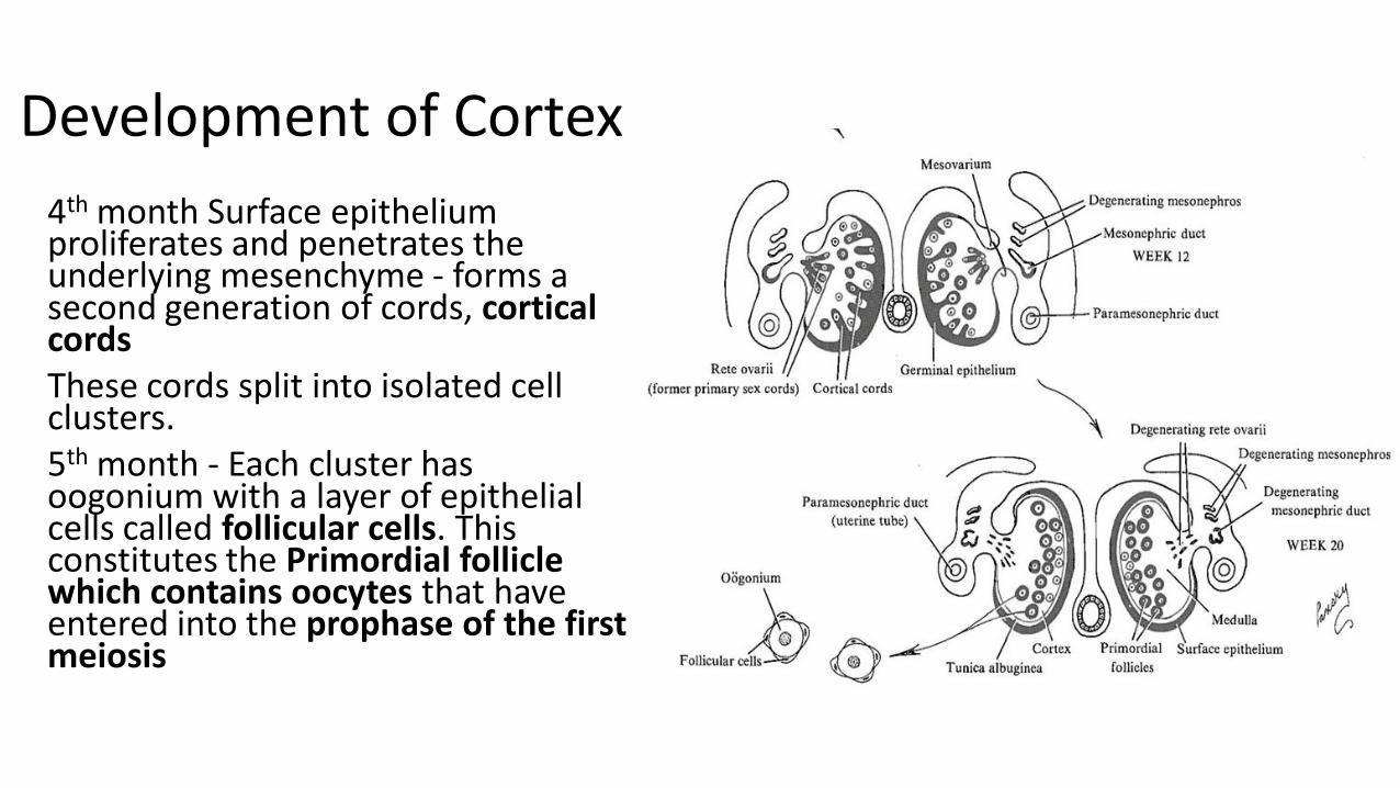
Development of Cortex
4th month Surface epithelium proliferates and penetrates the underlying mesenchyme - forms a second generation of cords, cortical cordsThese cords split into isolated cell clusters. 5th month - Each cluster has oogonium with a layer of epithelial cells called follicular cells. This constitutes the Primordial follicle which contains oocytes that have entered into the prophase of the first meiosis

Germ cells in Ovary
• 70 L at 20 weeks
• 3 L at birth
• 30,000 at Puberty
• 300 are used from Puberty to Menopause

Internal genital Organs

Formation of uterus – 7th to 8th week
• Up to the 7th week two canal systems on each side exist in both sexes. In the 8th week the paramesonephric ducts (Müller) fuse in the lower portion after they have crossed medially on both sides of the mesonephric duct (Wolff).

Formation of Uterus after 8 weeks
• Formation of the utero-vaginal canal through fusion of the lower section of the two paramesonephric ducts
• From the upper section - on both sides - arise the fallopian tubes with their ampullae.

End of 3rd month
• The separating wall dissolves in the uterus and the vagina.
• The uterus lengthens and is subsequently canalized.
• Out of the lower section arises the upper part of the vagina. It joins with the vaginal lamina, which arises from the urogenital sinus and forms the lower portion of the vagina.

Female sex organs – 7th week
• The utero-vaginal canal comes up against the urogenital sinus and forms the sinu-vaginal eminence.

Female sex organs – 12th week
• This sinu-vaginal eminence becomes thicker due to epithelial proliferation. This also leads to a epithelial proliferation in the SUG epithelium. Together they form the vaginal plate.

Female sex organs – 3rd month
• Canalization of the vaginal plate

Female sex organs- 5th month
Vaginal canal is completely canalized, but the lumen is separated from the SUG by the hymen.

Female sex organs – 9th month
• Normally, the hymen tears open at the time of birth. The uterus and the vagina then have a connection to the vaginal vestibule.

The Exgternal Genitalia

Indifferent Ext Genitalia – 6th week
• In front, the cloacal membrane is delimited by a swelling, the future genital tubercle, that at the rear continues in the two cloacal folds.

Indifferent Ext Genitalia – 7th week
• Urorectal septum subdivides the cloacal membrane into urogenital and anal membranes.

Indifferent Ext Genitalia – 9th week
From the cloacal folds around the urogenital orifice arise the urethral folds and, from the one around the anal orifice, the anal folds. Outside the urethral folds a further prominence arises on both sides, the genital swelling.
It is important to remember that the morphology of the external genitalia in both sexes is very similar up to the 9th week.

Differentiated female genitalia – 10th week
• In the female, the genital tubercle lengthens only a little and shrinks again while forming the clitoris.
• The urethral folds do not fuse and the urogenital sinus remains wide open.

Differentiated female genitalia – 12th -14th wk
• The urethral folds do not fuse –the labia minora arise from them.
• From the genital swellings arise the labia majora. They fuse only in the rear part and form the posterior labial commissure that is continued towards the rear by the perineum.
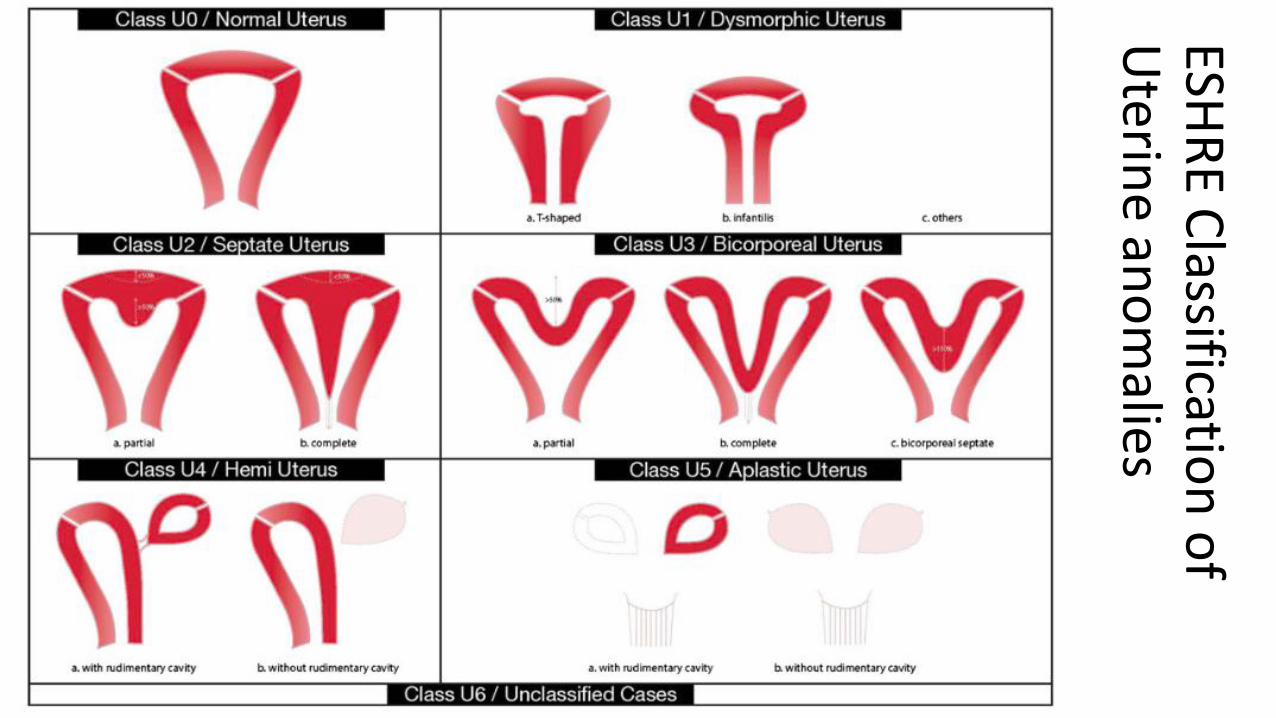
ESHR
E Classificatio
n o
f U
terine an
om
alies


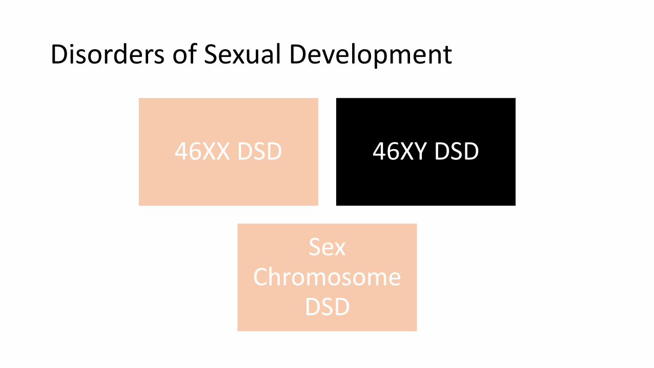
Disorders of Sexual Development
46XX DSD 46XY DSD
Sex Chromosome
DSD

Disorders of sexual development classification

Disorders of Sexual Development
46XX DSD 46XY DSD
Sex Chromosome
DSD

46XX DSD
Disorders of Gonadal Devpt.
Androgen Excess
Other disorders
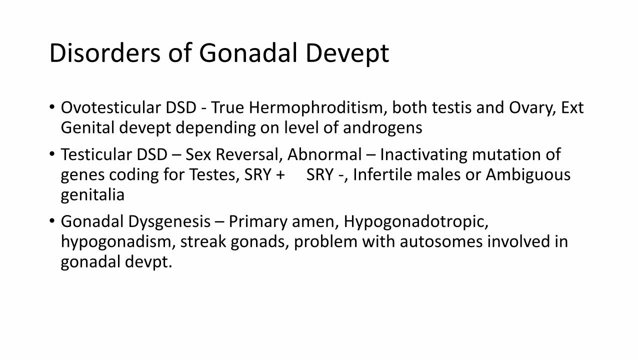
Disorders of Gonadal Devept
• Ovotesticular DSD - True Hermophroditism, both testis and Ovary, Ext Genital devept depending on level of androgens
• Testicular DSD – Sex Reversal, Abnormal – Inactivating mutation of genes coding for Testes, SRY + SRY -, Infertile males or Ambiguous genitalia
• Gonadal Dysgenesis – Primary amen, Hypogonadotropic, hypogonadism, streak gonads, problem with autosomes involved in gonadal devpt.

46XX DSD
Disorders of Gonadal Devpt.
Androgen Excess
Other disorders

Others
• Maternal Origin – Drugs, Pregnancy Luteoma, Theca lutein cysts
• Fetal origin – CAH – 21 Hydroxylase and others etc
• Feto Placental origin – Aromatase / P450 oxidoreductase deficiency
Androgen Excess
• MRKH
• MURCS Association

Virilization of Ext. GenitaliaPartial Complete
Disorders of Sexual Development
46XX DSD 46XY DSD
Sex Chromosome
DSD

46XY DSD
Disordes of Testicular devpt
Disorders of Androgen
synthesis / Action

Disorders of Testicular Devpt
• Complete Gonadal Dysgenesis – Swyer syndrome, female despite Y,
Streak gonads, No AMH, No Androgens, Primary Amen at Pubery
• Partial Gonadal dysgenesis
• Testicular regression – Normal devpt, torsion of testis in IU life,
regression , absent testes at birth

Disorders of Androgen synthesis/ action
• Androgen synthesis defect
• LH Receptor defect
• AIS
• 5a reductase deficiency
• Disorders of AMH – Male with Hernia uterine Inguinale
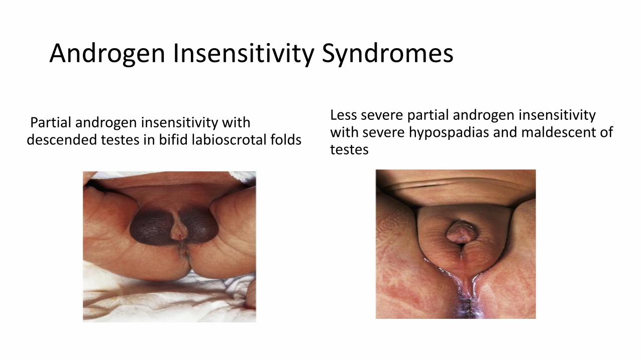
Androgen Insensitivity Syndromes
Partial androgen insensitivity with descended testes in bifid labioscrotal folds
Less severe partial androgen insensitivity with severe hypospadias and maldescent of testes

Hernia Uterine Inguinale

Disorders of Sexual Development
46XX DSD 46XY DSD
Sex Chromosome
DSD

Others - Disorders of Sex Chromosome
• 45,X0 – Turner’s and variants
• 47,XXY – Kilnefelters and variants
• 45,X, 46,XXy MGD
• 46,XX/46, XY Chromosomal Ovotesticular DSD


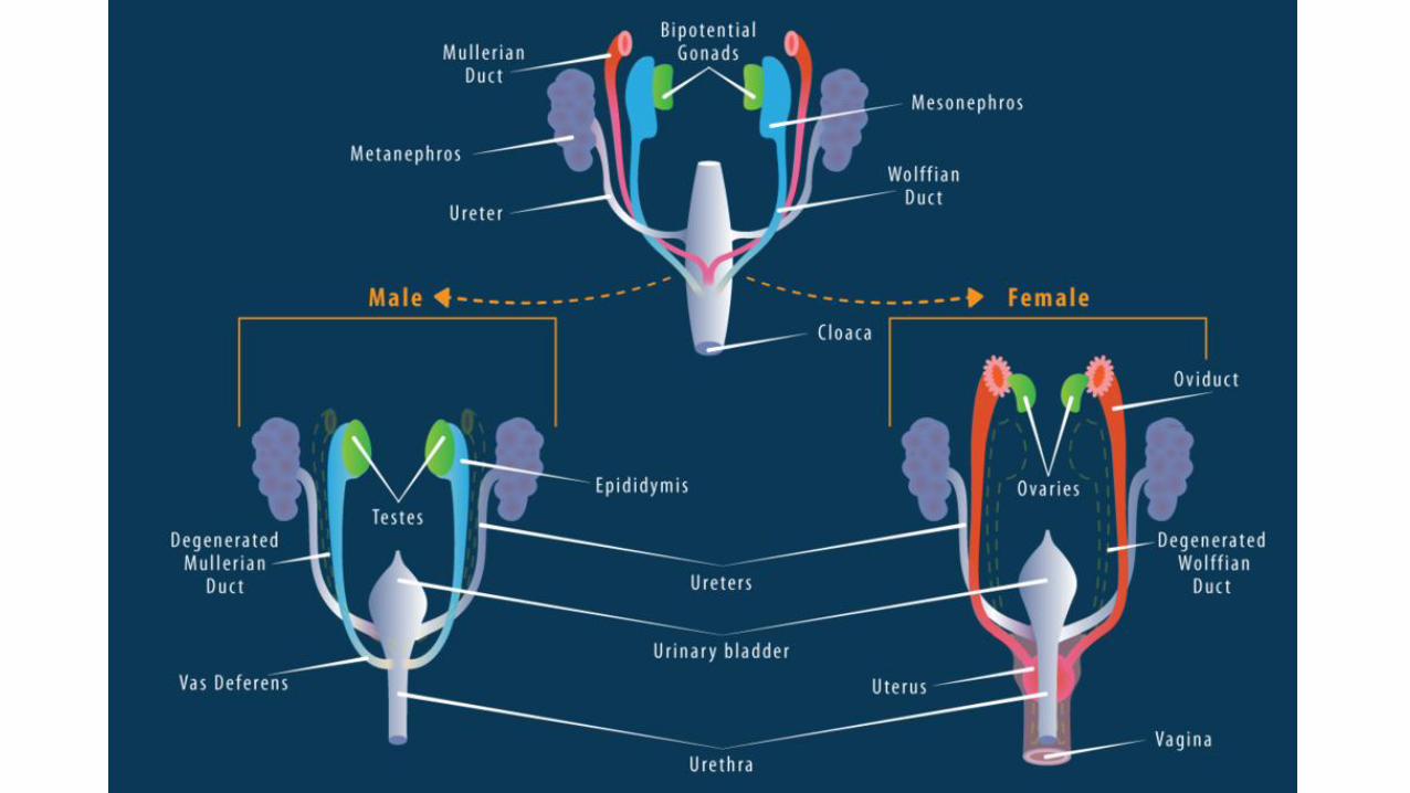

Differentiation of female sex organs
• 7th week – differentiation begins
• Mesonephric duct and its tubules atrophy
• Embryonic remnants of the mesonephric duct remain in the form of the epoöphoron, the paroöphoron at the level of the mesovarium, and a row of small cysts of Gartner.Paramesonephric duct develops further
• Upper nonfused portions – Fallopian tubes
• Lower fused portion – Uterovaginal Canal
• The septum in between disappears at the end of the 3rd month.