
THE REPRODUCTIVE SYSTEM
23
Transcript of THE REPRODUCTIVE SYSTEM


456 ✦ CHAPTER TWENTY THREE
◗ ReproductionThe chapters in this unit deal with what is certainlyone of the most interesting and mysterious attributesof life: the ability to reproduce. The simplest forms oflife, one-celled organisms, usually need no partner toreproduce; they simply divide by themselves. Thisform of reproduction is known as asexual (nonsexual)reproduction.
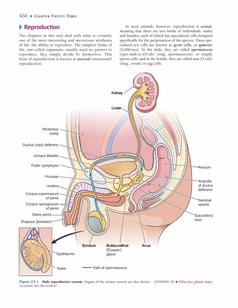
Figure 23-1 Male reproductive system. Organs of the urinary system are also shown. ZOOMING IN ✦ What four glands emptysecretions into the urethra?
Peritonealcavity
Urethra
Corpus cavernosumof penis
Corpus spongiosumof penis
Pubic symphysis
Urinary bladder
Ductus (vas) deferens
Glans penis
Prepuce (foreskin)
Epididymis
Testis
Ampullaof ductusdeferens
Seminalvesicle
Prostate
Rectum
(Cowper)gland
Ejaculatoryduct
Path of spermatozoa
In most animals, however, reproduction is sexual,meaning that there are two kinds of individuals, malesand females, each of which has specialized cells designedspecifically for the perpetuation of the species. These spe-cialized sex cells are known as germ cells, or gametes(GAM-etes). In the male, they are called spermatozoa(sper-mah-to-ZO-ah) (sing. spermatozoon), or simplysperm cells, and in the female, they are called ova (O-vah)(sing., ovum) or egg cells.

THE MALE AND FEMALE REPRODUCTIVE SYSTEM ✦ 457
◗ The Male Reproductive SystemThe male reproductive system, like that of the female,may be divided into two groups of organs: primary andaccessory (see Fig. 23-1).
◗ The primary organs are the gonads (GO-nads), or sexglands; they produce the germ cells and manufacturehormones. The male gonad is the testis. (In compari-son, the female gonad is the ovary, as explained below.)
◗ The accessory organs include a series of ducts thattransport the germ cells as well as various exocrineglands.
The TestesThe male gonads, the testes (TES-teze) (sing. testis) arelocated outside the body proper, suspended between thethighs in a sac called the scrotum (SKRO-tum). The testesare oval organs measuring about 4.0 cm (1.5 inches) inlength and about 2.5 cm (1 inch) in each of the other twodimensions. During embryonic life, each testis developsfrom tissue near the kidney.
A month or two before birth, the testis normally de-scends (moves downward) through the inguinal (ING-gwih-nal) canal in the abdominal wall into the scrotum.Each testis then remains suspended by a spermatic cord(Fig. 23-2) that extends through the inguinal canal. Thiscord contains blood vessels, lymphatic vessels, nerves,and the tube (ductus deferens) that transports spermato-zoa away from the testis. The gland must descend com-pletely if it is to function normally; to produce spermato-zoa, the testis must be kept at the temperature of thescrotum, which is several degrees lower than that of theabdominal cavity.
Internal Structure Most of the specialized tissue ofthe testis consists of tiny coiled seminiferous (seh-mih-NIF-er-us) tubules. Primitive cells in the walls of thesetubules develop into mature spermatozoa, aided byneighboring cells called sustentacular (sus-ten-TAK-u-lar) (Sertoli) cells. These so-called “nurse” cells nourishand protect the developing spermatozoa. They also se-
crete a protein that binds testosterone in the seminiferoustubules.
Specialized interstitial (in-ter-STISH-al) cells that se-crete the male sex hormone testosterone (tes-TOS-teh-rone) are located between the seminiferous tubules. Fig-ure 23-3 is a microscopic view of the testis incross-section, showing the seminiferous tubules, intersti-tial cells, and developing spermatozoa.
Testosterone After its secretion, testosterone is ab-sorbed directly into the bloodstream. This hormone hasthree functions:
23
Checkpoint 23-1: What is the process of cell division thathalves the chromosome number in a cell to produce a gamete?
MeiosisGametes are characterized by having half as many chro-mosomes as are found in any other body cell. Duringtheir formation, they go through a special process of celldivision, called meiosis (mi-O-sis), which halves thenumber of chromosomes. In humans, meiosis reduces thechromosome number in a cell from 46 to 23. The role ofmeiosis in reproduction is explained in more detail inChapter 25.
Figure 23-2 Structure of the testis. The epididymis andspermatic cord are also shown. ZOOMING IN ✦ What duct re-ceives secretions from the epididymis?
Body ofepididymis
Seminiferoustubule
Capsule
Septum
Testis
Lobule
Head ofepididymis
Nerve
Artery
Vein
Spermatic cord
Ductus (vas)deferens
Figure 23-3 Microscopic view of the testis. (Courtesy ofDana Morse Bittus and B. J. Cohen.)
Seminiferous tubule
Interstitial cellsPrimitivespermatozoa
Maturespermatozoa

458 ✦ CHAPTER TWENTY THREE
◗ Development and maintenance of the reproductivestructures
◗ Development of spermatozoa◗ Development of secondary sex characteristics, traits
that characterize males and females but are not directlyconcerned with reproduction. In males, these traits in-clude a deeper voice, broader shoulders, narrower hips,a greater percentage of muscle tissue, and more bodyhair than are found in females.
The Spermatozoa Spermatozoa are tiny individualcells illustrated in Figure 23-4. They are so small that atleast 200 million are contained in the average ejaculation(release of semen). After puberty, sperm cells are manu-factured continuously in the seminiferous tubules of thetestes.
The spermatozoon has an oval head that is largely anucleus containing chromosomes. The acrosome (AK-ro-some), which covers the head like a cap, contains en-zymes that help the sperm cell to penetrate the ovum.
Whiplike movements of the tail (flagellum) propel thesperm through the female reproductive system to theovum. The cell’s middle region (midpiece) contains manymitochondria that provide energy for movement.
Accessory OrgansThe system of ducts that transports the spermatozoa be-gins with tubules inside the testis itself. From thesetubules, the cells collect in a greatly coiled tube called theepididymis (ep-ih-DID-ih-mis), which is 6 meters (20feet) long and is located on the surface of the testis insidethe scrotal sac (see Fig. 23-2). While they are temporarilystored in the epididymis, the sperm cells mature and be-come motile, able to move or “swim” by themselves.
The epididymis finally extends upward as the ductusdeferens (DEF-er-enz), also called the vas deferens. Thistube, contained in the spermatic cord, continues throughthe inguinal canal into the abdominal cavity. Here, it sep-arates from the remainder of the spermatic cord andcurves behind the urinary bladder. The ductus deferensthen joins with the duct of the seminal vesicle (VES-ih-kl) on the same side to form the ejaculatory (e-JAK-u-lah-to-re) duct. The right and left ejaculatory ducts travelthrough the body of the prostate gland and then emptyinto the urethra.
Figure 23-4 Diagram of a human spermatozoon. Majorstructural features are shown. ZOOMING IN ✦ What or-ganelles provide energy for sperm cell motility?
Head
Neck
Midpiece
Tail (flagellum)
Acrosome
Nucleus
Mitochondria
Checkpoint 23-2: What is the male gonad?
Checkpoint 23-3: What is the main male sex hormone?
Checkpoint 23-4: What is the male sex cell (gamete) called?
Checkpoint 23-5: What is the order in which sperm cells travelthrough the ducts of the male reproductive system?
SemenSemen (SE-men) (meaning “seed”) is the mixture ofsperm cells and various secretions that is expelled fromthe body. It is a sticky fluid with a milky appearance. ThepH is in the alkaline range of 7.2 to 7.8. The secretions insemen serve several functions:
◗ Nourish the spermatozoa◗ Transport the spermatozoa◗ Neutralize the acidity of the male urethra and the fe-
male vaginal tract◗ Lubricate the reproductive tract during sexual inter-
course◗ Prevent infection with antibacterial enzymes and anti-
bodies
The glands discussed next contribute secretions to thesemen (see Fig. 23-1).
The Seminal Vesicles The seminal vesicles aretwisted muscular tubes with many small outpouchings.They are about 7.5 cm (3 inches) long and are attached tothe connective tissue at the posterior of the urinary blad-der. The glandular lining produces a thick, yellow, alka-line secretion containing large quantities of simple sugarand other substances that provide nourishment for thesperm. The seminal fluid makes up a large part of thesemen’s volume.
The Prostate Gland The prostate gland lies immedi-ately inferior to the urinary bladder, where it surroundsthe first part of the urethra. Ducts from the prostate carry

THE MALE AND FEMALE REPRODUCTIVE SYSTEM ✦ 459
its secretions into the urethra. The thin, alkaline prostaticsecretion helps neutralize the acidity of the vaginal tractand enhance the motility of the spermatozoa. Theprostate gland is also supplied with muscular tissue,which, upon signals from the nervous system, contractsto aid in the expulsion of the semen from the body.
Bulbourethral Glands The bulbourethral (bul-bo-u-RE-thral) glands, also called Cowper glands, are a pair ofpea-sized organs located in the pelvic floor just inferior tothe prostate gland. They secrete mucus to lubricate theurethra and tip of the penis during sexual stimulation.The ducts of these glands extend about 2.5 cm (1 inch)from each side and empty into the urethra before it ex-tends into the penis.
Other very small glands secrete mucus into the ure-thra as it passes through the penis.
sum enlarges to form the glans penis, which is coveredwith a loose fold of skin, the prepuce (PRE-puse), com-monly called the foreskin. It is the end of the foreskin thatis removed in a circumcision (sir-kum-SIZH-un), a sur-gery frequently performed on male babies for religious orcultural reasons. Experts disagree on the medical value ofcircumcision with regard to improved cleanliness anddisease prevention.
The penis and scrotum together make up the externalgenitalia of the male.
Ejaculation Ejaculation (e-jak-u-LA-shun) is the force-ful expulsion of semen through the urethra to the out-side. The process is initiated by reflex centers in thespinal cord that stimulate smooth muscle contraction inthe prostate. This is followed by contraction of skeletalmuscle in the pelvic floor, which provides the forceneeded for expulsion. During ejaculation, the involuntarysphincter at the base of the bladder closes to prevent therelease of urine.
A male typically ejaculates 2 to 5 mL of semen con-taining 50 to 150 million sperm cells per mL. Out of themillions of spermatozoa in an ejaculation, only one, ifany, can fertilize an ovum. The remainder of the cells livefrom only a few hours up to a maximum of 3 days.
23
Figure 23-5 Cross-section of the penis. ZOOMING IN ✦
What subdivision of the penis contains the urethra?
Corporacavernosa
Dorsal veins
Dorsal nerve
Dorsal artery
Centralartery
Connective tissue
Subcutaneoustissue
Skin
Urethra
Corpus spongiosum
Checkpoint 23-6: What glands, aside from the testis, contributesecretions to semen?
The Urethra and PenisThe male urethra, as discussed in Chapter 22, serves thedual purpose of conveying urine from the bladder andcarrying the reproductive cells with their accompanyingsecretions to the outside. The ejection of semen into thereceiving canal (vagina) of the female is made possible bythe erection, or stiffening and enlargement, of the penis,through which the longest part of the urethra extends.The penis is made of spongy tissue containing manyblood spaces that are relatively empty when the organ isflaccid but fill with blood and distend when the penis iserect. This tissue is subdivided into three segments, eachcalled a corpus (body) (Fig. 23-5). A single, ventrally lo-cated corpus spongiosum contains the urethra. On eitherside is a larger corpus cavernosum (pl., corpora caver-nosa). At the distal end of the penis, the corpus spongio-
Checkpoint 23-7: What are the main subdivisions of a sperma-tozoon?
◗ Hormonal Control of MaleReproductionThe activities of the testes are under the control of twohormones produced by the anterior pituitary. These hor-mones are named for their activity in female reproduction(described later), although they are chemically the samein both males and females.
◗ Follicle-stimulating hormone (FSH) stimulates thesustentacular (Sertoli) cells and promotes the forma-tion of spermatozoa.
◗ Luteinizing hormone (LH) stimulates the interstitialcells between the seminiferous tubules to producetestosterone, which is also needed for sperm cell devel-opment. An older name for this hormone, which de-scribes its action in males, is interstitial cell–stimulatinghormone (ICSH).
Starting at puberty, the hypothalamus begins to se-crete hormones that trigger the release of FSH and LH.These hormones are secreted continuously in the male.
The activity of the hypothalamus is in turn regulatedby a negative feedback mechanism involving testosterone.As the blood level of testosterone increases, the hypothal-amus secretes less releasing hormone; as the level oftestosterone decreases, the hypothalamus secretes morereleasing hormone (see Fig. 12-3 in Chap. 12).
460 ✦ CHAPTER TWENTY THREE
A male may be intentionally sterilized by an operationcalled a vasectomy (vah-SEK-to-me). In this procedure, aportion of the ductus deferens on each side is removed,and the cut end is closed to keep spermatozoa fromreaching the urethra. The tiny sperm cells are simply re-absorbed. A man who has had a vasectomy retains theability to produce hormones and all other seminal secre-tions as well as the ability to perform the sex act, but nofertilization can occur.
Structural DisordersCryptorchidism (kript-OR-kid-izm) is a disorder charac-terized by failure of the testis to descend into the scrotum.Unless corrected in childhood, this condition results insterility. Undescended testes are also particularly subjectto tumor formation. Most testes that are undescended atbirth descend spontaneously by 1 year of age. Surgicalcorrection is the usual remedy in the remaining cases.
Torsion of the testis is a twisting of the spermaticcord that results from rotation of the testis (Fig. 23-6).This turning may occur during descent of the testis orlater in life, most commonly between the ages of 8 to 18years. The condition is accompanied by acute pain,swelling and shortening of the spermatic cord. It requiresemergency surgery to correct the defect, and may involveremoval the testis (orchiectomy). Torson of the testis is adevelopmental disorder that often affects both glands, sothe other testis must be examined to determine whetheror not preventive surgery is needed.
Hernia (HER-ne-ah), or rupture, refers to the abnor-mal protrusion of an organ or organ part through the wallof the cavity in which it is normally contained (Fig. 23-7). Hernias most often occur where there is a weak area
Checkpoint 23-8: What two pituitary hormones regulate bothmale and female reproduction?
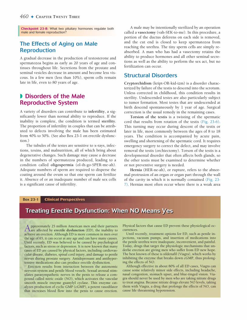
Approximately 25 million American men and their partnersare affected by erectile dysfunction (ED), the inability to
achieve an erection. Although ED is more common in men overthe age of 65, it can occur at any age and can have many causes.Until recently, ED was believed to be caused by psychologicalfactors, such as stress or depression. It is now known that manycases of ED are caused by physical factors, including cardiovas-cular disease, diabetes, spinal cord injury, and damage to penilenerves during prostate surgery. Antidepressant and antihyper-tensive medications also can produce erectile dysfunction.
Erection results from interaction between the autonomicnervous system and penile blood vessels. Sexual arousal stim-ulates parasympathetic nerves in the penis to release a com-pound called nitric oxide (NO), which activates the vascularsmooth muscle enzyme guanylyl cyclase. This enzyme cat-alyzes production of cyclic GMP (cGMP), a potent vasodilatorthat increases blood flow into the penis to cause erection.
Physical factors that cause ED prevent these physiological oc-currences.
Until recently, treatment options for ED, such as penile in-jections, vacuum pumps, and insertion of medications intothe penile urethra were inadequate, inconvenient, and painful.Today, drugs that target the physiologic mechanisms that un-derlie erection are giving men who suffer from ED new hope.The best known of these is sildenafil (Viagra), which works byinhibiting the enzyme that breaks down cGMP, thus prolong-ing the effects of NO.
Although effective in about 80% of all ED cases, Viagra cancause some relatively minor side effects, including headache,nasal congestion, stomach upset, and blue-tinged vision. Via-gra should never be used by men who are taking nitrate drugsto treat angina. Because nitrate drugs elevate NO levels, takingthem with Viagra, a drug that prolongs the effects of NO, cancause life threatening hypotension.
Treating Erectile Dysfunction: When NO Means Yes
Box 23-1 Clinical Perspectives
Treating Erectile Dysfunction: When NO Means Yes
The Effects of Aging on MaleReproductionA gradual decrease in the production of testosterone andspermatozoa begins as early as 20 years of age and con-tinues throughout life. Secretions from the prostate andseminal vesicles decrease in amount and become less vis-cous. In a few men (less than 10%), sperm cells remainlate in life, even to 80 years of age.
◗ Disorders of the MaleReproductive SystemA variety of disorders can contribute to infertility, a sig-nificantly lower than normal ability to reproduce. If theinability is complete, the condition is termed sterility.The proportion of infertility in couples that can be attrib-uted to defects involving the male has been estimatedfrom 40% to 50%. (See also Box 23-1 on erectile dysfunc-tion.)
The tubules of the testes are sensitive to x-rays, infec-tions, toxins, and malnutrition, all of which bring aboutdegenerative changes. Such damage may cause a decreasein the numbers of spermatozoa produced, leading to acondition called oligospermia (ol-ih-go-SPER-me-ah).Adequate numbers of sperm are required to disperse thecoating around the ovum so that one sperm can fertilizeit. Absence of or an inadequate number of male sex cellsis a significant cause of infertility.

THE MALE AND FEMALE REPRODUCTIVE SYSTEM ✦ 461
in the abdominal wall, at the inguinal canal for example.In this region, during development, the testis pushes itsway through the muscles and connective tissues of theabdominal wall, carrying with it the blood vessels andother structures that form the spermatic cord.
Normally, in the adult, the inguinal area is fairly wellreinforced with connective tissue, and there is no directconnection between the abdominal cavity and the scrotalsac. As in other regions where an opening permits struc-ture’s passage through the abdominal wall, however, thisarea constitutes a weak place where a hernia may occur.
Phimosis (fi-MO-sis) is a tightness of the foreskin(prepuce), so that it cannot be drawn back. Phimosis may
be remedied by circumcision, in which part or all of theforeskin is surgically removed.
InfectionsSexually transmitted infections (STI), formerly known assexually transmitted diseases (STD) or venereal diseases(VD), are spread through sexual contact in both malesand females. They most commonly involve chlamydial in-fections and gonococcal infections (gonorrhea). In males,these diseases are manifested by a discharge from the ure-thra, which may be accompanied by burning and pain, es-pecially during urination. The infection may travel alongthe mucous membrane into the prostate gland and theepididymis; if both sides are affected and enough scar tis-sue is formed to destroy the tubules, sterility may result.
Another common STI is a persistent infection calledgenital herpes. Caused by a virus, this disorder is charac-terized by fluid-filled vesicles (blisters) on and around thegenital organs.
The sexually transmitted disease syphilis is caused bya spirochete (Treponema pallidum). Because syphilisspreads quickly in the bloodstream, it is regarded as a sys-temic disorder (see Appendix 5, Table 1). The genital ul-cers caused by syphilis increase the chances of infectionwith the AIDS virus. HIV itself is considered an STI be-cause of its most common route of spread. (See Box 23-2on lowering risks for STIs.)
Epididymitis Organisms from an STI or urinary tractinfection (UTI) may travel through the ducts of the re-productive system to the epididymis. A congenital mal-formation in the urinary tract can predispose to epi-didymitis (ep-ih-did-ih-MI-tis), and infection may alsobe carried to the organ systemically by blood or lymph.Treatment includes an antibiotic along with bed rest andsupport of the scrotum to promote lymphatic drainage.
Prostatitis The usual cause of prostatic inflammation isbacterial infection secondary to an ascending urinary tractinfection. A variety of intestinal organisms and bacteriafrom other sources may be involved, but E. coli is the mostcommon. Treatment with antibiotics usually clears the in-fection, but tests to diagnose the source of infection maybe needed if the condition persists. Other possible causesof prostatitis (pros-tah-TI-tis) are bladder-neck obstruc-tion that forces urine into the prostate, and autoimmunity.
Orchitis Orchitis (or-KI-Is) is inflammation of thetestis, which also may follow from infection of the urinaryor reproductive tract. The testes also may be involved inmumps, a viral infection of the parotid salivary gland. Amumps infection during or after puberty may result in in-flammation of the testes, which could lead to sterility.
23
Figure 23-6 Torsion of the testis. (A) Normal. (B) Torsion.The testis rotates, twisting the spermatic cord. (Reprinted withpermission from LifeART Pediatrics 1 [CD-ROM]. Baltimore:Lippincott Williams & Wilkins, 2000.)
Spermatic cord
Epididymis
Testis
A B
Figure 23-7 Inguinal hernia. (A) Normal. (B) Hernia. Theintestine protrudes through a weakness in the abdominal wall atthe inguinal canal. (Reprinted with permission from Cohen BJ.Medical Terminology. 4th ed. Philadelphia: Lippincott Williams& Wilkins, 2004.)
Peritoneum
A B
Testis
Smallintestine
Inguinal canal
Checkpoint 23-9: What are some infectious diseases of the re-productive tract?

462 ✦ CHAPTER TWENTY THREE
TumorsTumors of the prostate may be benign or malignant. Bothtypes cause such pressure on the urethra that urinationbecomes difficult. Back pressure may destroy kidney tis-sue and may lead to stasis of urine in the bladder with aresulting susceptibility to infection. Men with benignprostate enlargement, known as benign prostatic hyper-plasia (BPH), may respond to medication to shrink theprostate. An herbal remedy that may help to slowprogress of BPH is an extract of berries of the saw pal-metto, a low-growing palm tree. If urinary function isthreatened, however, surgery is performed to reduce theobstruction. Most commonly, surgery is performedthrough the urethra, in a transurethral prostatectomy(TURP). Newer methods include use of laser and ultra-sound to destroy excess tissue or placement of a stent towiden the urethra.
Prostatic cancer is the most common cancer of malesin the U.S., especially among men older than 50 years ofage. Other risk factors, in addition to age, are race, familyhistory and certain environmental agents. A high fat dietmay increase risk by promoting production of male sexhormones. Prostatic cancer is frequently detected as anodule during rectal examination. Early detection has im-proved with annual blood tests for prostate-specific anti-gen (PSA). This protein increases in cases of prostate can-cer, although it may increase in other prostatic disordersas well. Depending on the age of the patient and the na-ture of the cancer, the course of treatment may includesurveillance, radiation therapy, surgery or hormone treat-ments.
Testicular cancer affects young to middle-aged adults.Almost all testicular cancers arise in the germ cells, and atumor can metastasize through the lymphatic system atan early stage of development. Early detection with regu-
lar testicular self-examination (TSE), however, improvesthe chances for effective treatment. The 5-year survivalrate for this form of cancer is now greater than 95%.Often fertility can be preserved, although sperm bankingis an option for men about to undergo treatment for tes-ticular cancer.
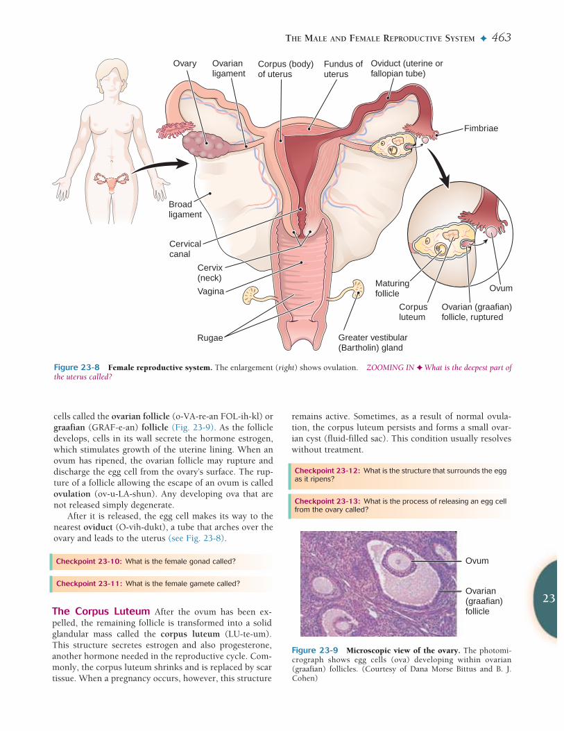
◗ The Female ReproductiveSystemThe female gonads are the ovaries (O-vah-reze), wherethe female sex cells, or ova, are formed (Fig. 23-8). Theremainder of the female reproductive tract consists of anorgan (uterus) to hold and nourish a developing infant,various passageways, and the external genital organs.
The OvariesThe ovary is a small, somewhat flattened oval body meas-uring about 4 cm (1.6 inches) in length, 2 cm (0.8 inch)in width, and 1 cm (0.4 inch) in depth. Like the testes,the ovaries descend, but only as far as the pelvic portionof the abdomen. Here, they are held in place by ligaments,including the broad ligament, the ovarian ligament, andothers, that attach them to the uterus and the body wall.
The Ova and OvulationThe outer layer of the ovary is made of a single layer ofepithelium. Beneath this layer, the female gametes, theova, are produced. The ovaries of a newborn female con-tain a large number of potential ova. Each month duringthe reproductive years, several ripen, but usually only oneis released.
The complicated process of maturation, or “ripening,”of an ovum takes place in a small fluid-filled cluster of
Sexually transmitted infections (STIs) such as chlamydia,gonorrhea, genital herpes, HIV, and syphilis are some of the
most common infectious diseases in the United States, affect-ing more than 13 million men and women each year. Thesediseases are associated with complications such as pelvic in-flammatory disease, epididymitis, infertility, liver failure, neu-rological disorders, cancer, and AIDS. Women are more likelyto contract STIs than are men. The same mechanisms thattransport sperm cells through the female reproductive tractalso move infectious organisms. The surest way to preventSTIs is to avoid sexual contact with others. If you are sexuallyactive, the following techniques can lower your risks:
◗ Maintain a monogamous sexual relationship with an un-infected partner.
◗ Correctly and consistently use a condom. While not100% effective, condoms greatly reduce the risk of con-tracting an STI.
◗ Avoid having sex during menstruation. Women may bemore infectious as well as more susceptible to certainSTIs during this time.
◗ Avoid contact with body fluids such as blood, semen, andvaginal fluids, all of which may harbor infectious organ-isms.
◗ Urinate and wash the genitals after sex. This may help re-move infectious organisms before they cause disease.
◗ Have regular checkups for STIs. Most of the time STIscause no symptoms, particularly in women.
Sexually Transmitted Infections: Lowering Your Risks
Box 23-2 • Health Maintenance
Sexually Transmitted Infections: Lowering Your Risks
THE MALE AND FEMALE REPRODUCTIVE SYSTEM ✦ 463
cells called the ovarian follicle (o-VA-re-an FOL-ih-kl) orgraafian (GRAF-e-an) follicle (Fig. 23-9). As the follicledevelops, cells in its wall secrete the hormone estrogen,which stimulates growth of the uterine lining. When anovum has ripened, the ovarian follicle may rupture anddischarge the egg cell from the ovary’s surface. The rup-ture of a follicle allowing the escape of an ovum is calledovulation (ov-u-LA-shun). Any developing ova that arenot released simply degenerate.
After it is released, the egg cell makes its way to thenearest oviduct (O-vih-dukt), a tube that arches over theovary and leads to the uterus (see Fig. 23-8).
23
Figure 23-8 Female reproductive system. The enlargement (right) shows ovulation. ZOOMING IN ✦ What is the deepest part ofthe uterus called?
Ovary Oviduct (uterine or fallopian tube)
Fimbriae
Ovarianligament
Broadligament
Corpus (body) of uterus
Fundus ofuterus
Cervicalcanal
Cervix(neck)
Vagina
Rugae Greater vestibular(Bartholin) gland
Ovum
Ovarian (graafian)follicle, ruptured
Maturingfollicle
Corpusluteum
Figure 23-9 Microscopic view of the ovary. The photomi-crograph shows egg cells (ova) developing within ovarian(graafian) follicles. (Courtesy of Dana Morse Bittus and B. J.Cohen)
Ovum
Ovarian(graafian)follicle
Checkpoint 23-10: What is the female gonad called?
Checkpoint 23-11: What is the female gamete called?
The Corpus Luteum After the ovum has been ex-pelled, the remaining follicle is transformed into a solidglandular mass called the corpus luteum (LU-te-um).This structure secretes estrogen and also progesterone,another hormone needed in the reproductive cycle. Com-monly, the corpus luteum shrinks and is replaced by scartissue. When a pregnancy occurs, however, this structure
remains active. Sometimes, as a result of normal ovula-tion, the corpus luteum persists and forms a small ovar-ian cyst (fluid-filled sac). This condition usually resolveswithout treatment.
Checkpoint 23-12: What is the structure that surrounds the eggas it ripens?
Checkpoint 23-13: What is the process of releasing an egg cellfrom the ovary called?

464 ✦ CHAPTER TWENTY THREE
Accessory OrgansThe accessory organs in the female are the oviducts, theuterus, the vagina, the greater vestibular glands, and thevulva and perineum.
The Oviducts The tubes that transport the ova in thefemale reproductive system, the oviducts, are also knownas uterine (U-ter-in) tubes, or fallopian (fah-LO-pe-an)tubes. Each is a small, muscular structure, nearly 12.5 cm(5 inches) long, extending from a point near the ovary tothe uterus (womb). There is no direct connection be-tween the ovary and this tube. The ovum is swept into theoviduct by a current in the peritoneal fluid produced bythe small, fringelike extensions called fimbriae (FIM-bre-e) that are located at the edge of the tube’s opening intothe abdomen (see Fig. 23-8)
Unlike the sperm cell, the ovum cannot move by it-self. Its progress through the oviduct toward the uterusdepends on the sweeping action of cilia in the tube’s lin-ing and on peristalsis of the tube. It takes about 5 days foran ovum to reach the uterus from the ovary.
floor between the bladder and the rectum. The wider upperregion of the uterus is called the corpus, or body; thelower, narrower region is the cervix (SER-viks), or neck.The small, rounded region above the level of the tubal en-trances is known as the fundus (FUN-dus) (see Fig. 23-8).
The broad ligaments support the uterus, extendingfrom each side of the organ to the lateral body wall. Alongwith the uterus, these two portions of peritoneum form apartition dividing the female pelvis into anterior and pos-terior areas. The ovaries are suspended from the broadligaments, and the oviducts lie within the upper borders.Blood vessels that supply these organs are found betweenthe layers of the broad ligament (see Fig. 23-8).
The muscular wall of the uterus is called the my-ometrium (mi-o-ME-tre-um) (Fig. 23-10). The lining ofthe uterus is a specialized epithelium known as en-dometrium (en-do-ME-tre-um). This inner layer changesduring the menstrual cycle, first preparing to nourish afertilized egg, then breaking down if no fertilization oc-curs to be released as the menstrual flow. The cavity insidethe uterus is shaped somewhat like a capital T, but it is ca-pable of changing shape and dilating as a fetus develops.
Checkpoint 23-14: What does the follicle become after ovula-tion?
The Uterus The oviducts lead to the uterus (U-ter-us),an organ in which a fetus can develop to maturity. Theuterus is a pear-shaped, muscular organ about 7.5 cm (3inches) long, 5 cm (2 inches) wide, and 2.5 cm (1 inch)deep. (The organ is typically larger in women who haveborne children and smaller in postmenopausal women).The superior portion rests on the upper surface of the uri-nary bladder; the inferior portion is embedded in the pelvic
Figure 23-10 The uterus as seen under the microscope. The photomicrographs show the myometrium and endometrium andillustrate the changes that occur in the endometrium during the menstrual cycle. (A) Proliferative phase (first part of cycle). (B) Se-cretory phase (second part of cycle). (Reprinted with permission from Cormack DH. Essential Histology. 2nd ed. Philadelphia: Lip-pincott Williams & Wilkins, 2001.) ZOOMING IN ✦ In which part of the menstrual cycle is the endometrium most highly developed?
Endometrium
Glands
Myometrium Myometrium
Glands
A B
Endometrium
Checkpoint 23-15: In what organ does a fetus develop?
The Vagina The cervix leads to the vagina (vah-JI-nah),the distal part of the birth canal, which opens to the outsideof the body. The vagina is a muscular tube about 7.5 cm (3inches) long connecting the uterine cavity with the outside.The cervix dips into the superior portion of the vaginaforming a circular recess known as the fornix (FOR-niks).The deepest area of the fornix, located behind the cervix, isthe posterior fornix (Fig. 23-11). This recess in the poste-

THE MALE AND FEMALE REPRODUCTIVE SYSTEM ✦ 465
rior vagina lies adjacent to the most inferior portion of theperitoneal cavity, a narrow passage between the uterus andthe rectum named the cul-de-sac (from the French meaning“bottom of the sack”). This area is also known as therectouterine pouch or the pouch of Douglas. A rather thinlayer of tissue separates the posterior fornix from this re-gion, so that abscesses or tumors in the peritoneal cavitycan sometimes be detected by vaginal examination.
The lining of the vagina is a wrinkled mucous mem-brane similar to that found in the stomach. The folds(rugae) permit enlargement so that childbirth usuallydoes not tear the lining. In addition to being a part of thebirth canal, the vagina is the organ that receives the penisduring sexual intercourse. A fold of membrane called thehymen (HI-men) may sometimes be found at or near thevaginal (VAJ-ih-nal) canal opening (see Fig. 23-12).
The Greater Vestibular Glands Just superior and lat-eral to the vaginal opening are the two mucus-producing
23
Figure 23-11 Female reproductive system (sagittal section). This view shows the relationship of the reproductive organs to eachother and to other structures in the pelvic cavity. ZOOMING IN ✦ Which has the more anterior opening, the vagina or the urethra?
Figure 23-12 External parts of the female reproductivesystem. Related structures are also shown.
Mons pubis
Labium majus
Labiumminus
Hymen
Anus Obstetricalperineum
ClitorisUrethralorifice
Vaginalorifice

466 ✦ CHAPTER TWENTY THREE
greater vestibular (ves-TIB-u-lar) (Bartholin) glands (seeFig. 23-8). These glands secrete into an area near thevaginal opening known as the vestibule. Like the Cowperglands in males, these glands provide lubrication duringintercourse. If a gland becomes infected, a surgical inci-sion may be needed to reduce swelling and promotedrainage.
The Vulva and the Perineum The external parts ofthe female reproductive system form the vulva (VUL-vah), which includes two pairs of lips, or labia (LA-be-ah); the clitoris (KLIT-o-ris), which is a small organ ofgreat sensitivity; and related structures. Although the en-tire pelvic floor in both the male and female (see Fig. 8-15 in Chapter 8) is properly called the perineum (per-ih-NE-um), those who care for pregnant women usuallyrefer to the limited area between the vaginal opening andthe anus as the perineum or obstetrical perineum.
◗ The Menstrual CycleIn the female, as in the male, reproductive function iscontrolled by pituitary hormones that are regulated bythe hypothalamus. Female activity differs, however, inthat it is cyclic; it shows regular patterns of increases anddecreases in hormone levels. These changes are regulatedby hormonal feedback. The typical length of the men-strual cycle varies between 22 and 45 days, but 28 days istaken as an average, with the first day of menstrual flowbeing considered the first day of the cycle (Fig. 23-13).
Beginning of the CycleAt the start of each cycle, under the influence of pituitaryFSH, several follicles, each containing an ovum, begin todevelop in the ovary. Usually, only one of these follicleswill ultimately release an ovum from the ovary in a singlemonth. The follicle produces increasing amounts of es-trogen as it matures (see Fig. 23-13). (Estrogen is the termused for a group of related hormones, the most active ofwhich is estradiol.) The estrogen is carried in the blood-stream to the uterus, where it starts preparing the en-dometrium for a possible pregnancy. This preparation in-cludes thickening of the endometrium and elongation ofthe glands that produce the uterine secretion. Estrogen inthe blood also acts as a feedback messenger to inhibit therelease of FSH and stimulate the release of LH from thepituitary (see Fig. 12-3 in Chap. 12). (Note that there isan unexplained rise in FSH at the time of ovulation, asshown in Fig. 23-13.)
OvulationIn an average 28-day cycle, ovulation occurs on day 14and is followed two weeks later by the start of the men-strual flow. However, an ovum can be released any timefrom day 7 to 21, thus accounting for the variation in the
length of normal cycles. About 1 day before ovulation,there is an LH surge, a sharp rise of LH in the blood. Thishormone causes ovulation and transforms the rupturedfollicle into the corpus luteum, which produces some es-trogen and large amounts of progesterone. Under the in-fluence of these hormones, the endometrium continues tothicken, and the glands and blood vessels increase in size.The rising levels of estrogen and progesterone feed backto inhibit the release of FSH and LH from the pituitary.During this time, the ovum makes its journey to theuterus by way of the oviduct. If the ovum is not fertilizedwhile passing through the uterine tube, it dies within 2 to3 days and then disintegrates.
During each menstrual cycle, changes occur in boththe ovary and the uterus (see Fig. 23-13). The time beforeovulation is described as the follicular phase in the ovary,because it encompasses development of the ovarian folli-cle. The uterus during this same time is in a proliferativephase, marked by growth of the endometrium. After ovu-lation, the ovary is in a luteal phase, with conversion ofthe follicle to the corpus luteum, and the uterus is de-scribed as being in a secretory phase, based on activity ofthe endometrial glands.
Figure 23-13 The menstrual cycle. Changes in hormones,the ovary, and the uterus are shown during a typical 28-day men-strual cycle with ovulation on day 14. (Pituitary hormones areshown with dashed lines, ovarian hormones with solid lines.)ZOOMING IN ✦ What hormone peaks closest to ovulation?
Hor
mon
es LHFSH
Menstrualphase
Proliferativephase
Follicular phase Luteal phase
Secretoryphase
Ova
ryU
teru
s
0 Days 5 10 14(Ovulation)
20 25 28
Estrogen Progesterone

THE MALE AND FEMALE REPRODUCTIVE SYSTEM ✦ 467
The Menstrual PhaseIf fertilization does not occur, the corpus luteum degen-erates, and the levels of estrogen and progesterone de-crease. Without the hormones to support growth, the en-dometrium degenerates. Small hemorrhages appear inthis tissue, producing the bloody discharge known asmenstrual flow, or menses (MEN-seze). Bits of en-dometrium break away and accompany the blood flowduring this period of menstruation (men-stru-A-shun).The average duration of menstruation is 2 to 6 days.
Even before the menstrual flow ceases, the en-dometrium begins to repair itself through the growth ofnew cells. The low levels of estrogen and progesteroneallow the release of FSH from the anterior pituitary. FSHcauses new follicles to begin to ripen within the ovaries,and the cycle begins anew.
The activity of ovarian hormones as negative feedbackmessengers is the basis of hormonal methods of contra-ception (birth control). Estrogen and progesterone in-hibit the release of FSH and LH from the pituitary, re-sulting in a menstrual period but no ovulation.
◗ MenopauseMenopause (MEN-o-pawz) is the period during whichmenstruation ceases altogether. It ordinarily occurs grad-ually between the ages of 45 and 55 years and is causedby a normal decline in ovarian function. The ovary be-comes chiefly scar tissue and no longer produces ripe fol-licles or appreciable amounts of estrogen. Eventually, theuterus, oviducts, vagina, and vulva all become somewhatatrophied and the vaginal mucosa becomes thinner, dryerand more sensitive.
Menopause is an entirely normal condition, but its onsetsometimes brings about effects that are temporarily disturb-ing. The decrease in estrogen levels can cause nervous symp-toms, such as anxiety, insomnia, and “hot flashes.”
Hormone Replacement TherapyPhysicians may prescribe hormone replacement therapy(HRT) to relieve the discomforts associated withmenopause. This medication is usually a combination ofestrogen with a synthetic progesterone (progestin), whichis included to prevent overgrowth of the endometriumand the risk of endometrial cancer. Early assumptionsabout the role of estrogen in preventing heart attackshave been disproved by carefully controlled studies, atleast with regard to the most commonly prescribed formof HRT. The hormone therapy did lower the incidence ofcolorectal cancer and hip fractures, a sign of osteoporosis.Studies are continuing with estrogen alone, generally pre-
scribed for women who have undergone a hysterectomyand do not have a uterus.
In addition to an increased risk of breast cancer, HRTalso carries a risk of thrombosis and embolism, which ishighest among women who smoke. All HRT risks increasewith the duration of therapy. Therefore, treatment shouldbe given for a short time and at the lowest effective dose.Women with a history or family history of breast cancer orcirculatory problems should not take HRT.
23
Checkpoint 23-16: What are the two hormones produced inthe ovaries?
Checkpoint 23-17: What is the definition of menopause?
◗ Birth ControlBirth control is most commonly achieved by contracep-tion, which is the use of artificial methods to prevent fer-tilization of the ovum. Birth control measures that pre-vent implantation of the fertilized ovum are alsoconsidered contraceptives, although technically they donot prevent conception and are more accurately calledabortifacients (ah-bor-tih-FA-shents) (agents that causeabortion). Some of the birth control methods act by bothmechanisms. Table 23-1 presents a brief description ofthe main contraceptive methods currently in use alongwith some advantages and disadvantages of each. The listis given in rough order of decreasing effectiveness. Unlessspecifically mentioned as doing so, a given method doesnot prevent the transmission of STIs
The various hormonal methods of birth control basi-cally differ in how they administer the hormones. Theemergency contraceptive pill (ECP) is a synthetic proges-terone (progestin) taken within 72 hours after inter-course, usually in two doses 12 hours apart. It reduces therisk of pregnancy following unprotected intercourse. Thisso-called “morning after pill” is intended for emergencyuse and not as a regular birth control method. Birth con-trol hormones can also be implanted as capsules underthe skin of the upper arm. This method is highly effectiveand lasts for 3-5 years, but the capsules must be im-planted and removed by a health professional, and theyhave been difficult to remove in some cases. (See Box 23-3 on hormonal contraception for men.)
The female condom is a sheath that fits into thevagina. It does protect against STIs, but is not very con-venient to use. Researchers have also done trials with amale contraceptive pill, but none is on the market as yet.Mefipristone (RU 486) is a drug taken after conception toterminate an early pregnancy. It blocks the action ofprogesterone, causing the uterus to shed its lining and re-lease the fertilized egg. It must be combined with admin-istration of prostaglandins to expel the uterine tissue.Mefipristone is not in widespread use in the U.S., but ithas been used in other countries.
Checkpoint 23-18: What is the definition of contraception?

468 ✦ CHAPTER TWENTY THREE
Main Methods of Birth Control Currently in UseTable 23•1
METHOD DESCRIPTION ADVANTAGES DISADVANTAGES
SurgicalVasectomy/tubal
ligation
HormonalBirth control pills
Birth control shot
Birth control patch
Birth control ring
BarrierMale condom
Diaphragm (withspermicide)
Contraceptivesponge (withspermicide)
Intrauterinedevice (IUD)
OtherSpermicide
Fertility awareness
Cutting and tying of tubescarrying gametes
Estrogen and progestin orprogestin alone taken orallyto prevent ovulation
Injection of synthetic proges-terone every 3 months toprevent ovulation
Adhesive patch placed on bodythat administers estrogen andprogestin through the skin;left on for 3 weeks and re-moved for a fourth week
Flexible ring inserted intovagina that releases hor-mones internally; left inplace for three weeks and re-moved for a fourth week.
Sheath that fits over erect penisand prevents release ofsemen
Rubber cap that fits over cervixand prevents entrance ofsperm
Soft, disposable foam disk con-taining spermicide, which ismoistened with water and in-serted into vagina
Metal or plastic device insertedinto uterus through vagina;prevents fertilization and im-plantation by release of cop-per or birth control hor-mones
Chemicals used to kill sperm;best when used in combina-tion with a barrier method
Abstinence during fertile partof cycle as determined bymenstrual history, basal bodytemperature, or quality ofcervical mucus
Nearly 100% effective; involvesno chemical or mechanicaldevices
Highly effective; requires nolast-minute preparation
Highly effective; lasts for 3 to 4months
Protects long-term; less chanceof incorrect use; no last-minute preparation
Long-lasting, highly effective;no last minute preparation
Easily available; does not effectphysiology; protects againstsexually transmitted disease(STI)
Does not affect physiology;some protection against STI;no side effects
Protects against pregnancy for24 hours; non-hormonal;some STI protection; avail-able without prescription;inexpensive
Highly effective for 5–10 yearsdepending on type; re-versible; no last-minutepreparation
Available without prescription;inexpensive; does not affectphysiology; some protectionagainst STI
Does not affect physiology; ac-cepted by certain religions
Not usually reversible: raresurgical complications
Alters physiology; return tofertility may be delayed. Risk ofcardiovascular disease in olderwomen who smoke or havehypertension.
Alters physiology; same possibleside effects as birth control pill;also possible menstrual irregu-larity, amenorrhea
Alters physiology; same possibleside effects as birth control pill
Possible infections, irritation;same possible side effects asbirth control pill
Must be applied just before inter-course; may slip or tear
Must be inserted beforeintercourse and left in place for6 hours; requires fitting byphysician
85–90% effective depending onproper use; skin irritation
Must be introduced and removedby health professional; heavymenstrual bleeding
Local irritation; must be used justbefore intercourse
High failure rate; requires carefulrecord keeping

THE MALE AND FEMALE REPRODUCTIVE SYSTEM ✦ 469
◗ Disorders of the FemaleReproductive SystemFemale reproductive disorders include menstrual distur-bances, various forms of tumors, and infections, any ofwhich can contribute to infertility.
Menstrual DisordersAbsence of menstrual flow is known as amenorrhea (ah-men-o-RE-ah). This condition can be symptomatic of in-sufficient hormone secretion or congenital abnormality ofthe reproductive organs. Stress and other psychologicalfactors often play a part in cessation of the menstrualflow. For example, any significant change in a woman’sgeneral state of health or change in her living habits, suchas a shift in working hours, can interfere with menstrua-tion. Very low body weight with a low percentage of bodyfat can lead to amenorrhea by reducing estrogen synthe-sis, as may occur in athletes who over-train without eat-ing enough and in women who are starving or have eat-ing disorders.
Dysmenorrhea (dis-men-o-RE-ah) means painful ordifficult menstruation. In young women, this may be dueto immaturity of the uterus. Dysmenorrhea is frequentlyassociated with cycles in which ovulation has occurred.Often, the pain can be relieved by drugs that blockprostaglandins, because some prostaglandins are knownto cause painful uterine contractions.
In many cases, women have been completely relievedof menstrual cramps by their first pregnancies. Appar-ently, enlargement of the cervical opening remedies thecondition. Artificial dilation of the cervical opening mayalleviate dysmenorrhea for several months. Often, such
health measures as sufficient rest, a well-balanced diet,and appropriate exercise remedy the disorder. In cases ofdysmenorrhea, the application of heat over the abdomenusually relieves the pain, just as it may ease other types ofmuscular cramps.
Premenstrual syndrome (PMS), also called premen-strual tension, is a condition in which nervousness, irri-tability, and depression precede the menstrual period. Itis thought to be due to fluid retention in various tissues,including the brain. Sometimes, a low-salt diet and ap-propriate medication for 2 weeks before the menses pre-vent this disorder. This treatment may also avert dys-menorrhea.
Abnormal uterine bleeding includes excessive men-strual flow, too-frequent menstruation, and nonmen-strual bleeding. Any of these may cause serious anemiasand deserve careful medical attention. Nonmenstrualbleeding may be an indication of a tumor, possibly can-cer.
Benign and Malignant TumorsFibroids, which are more correctly called myomas, arecommon tumors of the uterus. Studies indicate that about50% of women who reach the age of 50 have one or moreof these growths in the walls of the uterus. Often, thesetumors are small, and usually they remain benign andproduce no symptoms. They develop between pubertyand menopause and ordinarily stop growing after awoman has reached the age of 50 years. In some cases,these growths interfere with pregnancy, and in a patientyounger than 40 years of age, a surgeon may simply re-move the tumor and leave the uterus fairly intact. Normalpregnancies have occurred after such surgery.
23
At present, sexually active men have few effective optionsfor contraception, the most reliable being condoms or va-
sectomy. While condoms have the additional benefit of pre-venting sexually transmitted infections, their failure rate forpreventing pregnancy is about 10%. With a failure rate ofabout 1%, vasectomies are much more reliable but are suitableonly for couples who do not want children or are finished hav-ing children. A new option is on the horizon, however—malehormonal contraception.
Like female contraception, the male version of “the pill”works by suppressing the release of gonadotropin releasinghormone (GnRH) from the hypothalamus. This, in turn,blocks the pituitary’s release of luteinizing hormone (LH) andfollicle stimulating hormone (FSH), both of which play an im-portant role in spermatogenesis.
Several therapies that decrease LH and FSH production in menare under investigation. One method already in clinical trials uses
high levels of testosterone to negatively feed back to the hypo-thalamus and suppress GnRH secretion. Currently, this methodrequires regular testosterone injections, which makes it impracti-cal for general use. In addition, doses of testosterone high enoughto stop spermatogenesis may be associated with side effects suchas acne, weight gain, mood changes, and increased risk of car-diovascular disease. More practical methods of drug delivery,such as pills, transdermal patches, and implants are being devel-oped and may be effective at lower testosterone doses.
Another promising method uses the female hormone prog-esterone to block GnRH production. While this method sup-presses spermatogenesis more effectively than does testos-terone alone, it also suppresses normal testosteroneproduction. Thus, the progesterone must be combined withtestosterone to prevent the loss of secondary sex characteris-tics. Investigation is still underway to determine the best wayto deliver such combination therapy.
Hormonal Contraception: New Options for Men
Box 23-3 Hot Topics
Hormonal Contraception: New Options for Men

470 ✦ CHAPTER TWENTY THREE
Fibroids may become so large that pressure on adja-cent structures causes grave disorders. In some cases, in-vasion of blood vessels near the uterine cavity causes se-rious hemorrhages. For these and other reasons, it may benecessary to remove the entire uterus or a large part of it.Surgical removal of the uterus is called a hysterectomy(his-ter-EK-to-me).
Breast Cancer Cancer of the breast is the most com-monly occurring malignant disease in women. The riskfactors in breast cancer are age past 40, family history ofbreast cancer, and factors that increase exposure to estro-gen, such as early onset of menstruation, late menopause,late or no pregnancies, long-term HRT, and obesity (fatcells produce estrogen). Mutations in two genes (BRCA1and BRCA2) are responsible for hereditary forms of breastcancer, which make up only about 8% of all cases. Thesesame genetic mutations are associated with an increasedrisk of ovarian cancer.
The tumor is usually a painless, movable mass that isoften noticed by a woman and all too frequently ignored.In recent years, however, there has been increasing em-phasis on the importance of regular breast self-examina-tion (BSE). (Most breast lumps are discovered by womenthemselves.) Any lump, no matter how small, should bereported to a physician immediately. The mammogram, aradiographic study of the breast, has improved the detec-tion of early breast cancer. Guidelines recommend regu-lar mammograms after the age of 40 years unless there isa history of breast cancer in the family, in which case ear-lier studies are recommended. Suspicious areas requirefurther study by ultrasonography or biopsy, either a nee-dle aspiration, removal of a core of tissue or excision ofthe lump.
Breast cancer treatment consists of surgery with fol-low-up therapy of radiation, chemotherapy, or both. Sur-gical treatment by removal of the lump (“lumpectomy”)or a segment of the breast is most common. Removal ofthe entire breast and dissection of the lymph nodes in theaxilla (armpit) is called modified radical mastectomy(mas-TEK-to-me). The extent of tumor spread throughthe lymph nodes is an important factor in prognosis. In asentinel lymph node biopsy, the first (sentinel) lymphnodes to receive lymph from the tumor are identified andtested for cancerous cells. Treatment is based on howmuch spread has occurred (see Box 16-2). Treatment ofbreast cancer is often followed by administration of drugsto block estrogen receptors in breast tissue or drugs thatinhibit tumor growth factors.
Note that the incidence of the various types of cancershould not be confused with the death rates for each type.Owing to education of the public and increasingly bettermethods of diagnosis and treatment, some forms of can-cer have a higher cure rate than others. For example,breast cancer appears much more often in women thanlung cancer, but more women now die each year fromlung cancer than from breast cancer.
Endometrial Cancer The most common cancer ofthe female reproductive tract is cancer of the en-dometrium (the lining of the uterus). This type of cancerusually affects women during or after menopause. It isseen most frequently in women who have been exposedto high levels of estrogen, which causes overgrowth of theendometrium. This group includes those who have re-ceived estrogen therapy unopposed by progesterone,those who have had few or no pregnancies, and the obese.Symptoms include an abnormal discharge or irregularbleeding; later there is cramping and pelvic pain. Thistype of cancer is diagnosed by endometrial biopsy. Theusual methods of treatment include surgery and irradia-tion. Endometrial cancer grows slowly in its beginningstages, so early, aggressive treatment usually saves a pa-tient’s life.
Ovarian Cancer Ovarian cancer is the second mostcommon reproductive tract cancer in the female, usuallyoccurring in women between the ages of 40 and 65 years.It is a leading cause of cancer deaths in women. Althoughmost ovarian cysts are not malignant, they should alwaysbe investigated for possible malignant change. Ovariancancer is highly curable if treated before it has spread toother organs. However, these malignancies tend toprogress rapidly, and they are difficult to detect becausesymptoms are vague, there are few recognized risk fac-tors, and at present there is no screening test.
Cervical Cancer Cancer of the cervix is linked to in-fection with human papilloma virus (HPV), which causesgenital warts and is spread through sexual contact. Thus,cervical cancer can be considered a sexually transmitteddisease. Risk factors for the disease are related to expo-sure to HPV, such as early age of sexual activity and mul-tiple sex partners. Certain strains of the virus are found incervical carcinomas and precancerous cervical cells.
Early detection is often possible because the cancer de-velops slowly from atypical cervical cells. The decline in thedeath rate from cervical cancer is directly related to use ofthe Papanicolaou (pap-ah-nik-o-LAH-o) test, also knownas the Pap test or Pap smear. The Pap smear is a microscopicexamination of cells obtained from cervical scrapings andswabs of the cervical canal. All women should be encour-aged to have this test every year. Even girls younger than 18years of age should be tested if they are sexually active.Guidelines now recommend less frequent testing after nor-mal results are obtained in three annual tests.
InfectionsInfections that affect the male reproductive system alsoinfect the female genital organs (Fig. 23-14), althoughthese diseases may be less apparent in women. The mostcommon STIs in women are chlamydial infections, gon-orrhea, HIV and genital herpes, caused by herpes simplexvirus (HSV). Syphilis also occurs in women and can be

THE MALE AND FEMALE REPRODUCTIVE SYSTEM ✦ 471
passed through the placenta from mother to fetus, caus-ing stillbirth or birth of an infected infant.
The incidence of genital warts, caused by human pa-pillomavirus, (HPV) has increased in recent years. Theseinfections have been linked to cancer of the reproductivetract, especially as noted, cancer of the uterine cervix.
Salpingitis (sal-pin-JI-tis) means inflammation of anytube, but usually refers to disease of the uterine tubes(oviducts). Most uterine tube infections are caused bygonococci or by the bacterium Chlamydia trachomatis,but other bacteria may be the cause. Salpingitis may causesterility by obstructing the tubes, thus preventing the pas-sage of ova.
Pelvic inflammatory disease (PID) is due to exten-sion of infections from the reproductive organs into thepelvic cavity, and it often involves the peritoneum. (Seethe red arrow pathway in Fig. 23-14.) Gonococcus orchlamydia is usually the initial cause of infection, butmost cases of PID involve multiple organisms.
InfertilityInfertility is much more difficult to diagnose and evaluatein the female than in the male. Whereas a microscopicexamination of properly collected semen may be enoughto determine the presence of abnormal or too few sperm
23
Figure 23-14 Pathway of infection. Disease organisms can travel from outside to the peritoneum and into the urinary system.

472 ✦ CHAPTER TWENTY THREE
cells in the male, no such simple study can be made in thefemale. Infertility in women, as in men, may be relative orabsolute. Causes of female infertility include infections,endocrine disorders, psychogenic factors, and abnormali-ties in the structure and function of the reproductive or-
gans themselves. In all cases of apparent infertility, themale partner should be investigated first because the pro-cedures for determining lack of fertility in the male aremuch simpler and less costly than those in the female, aswell as being essential for the evaluation.
Word Anatomy
Medical terms are built from standardized word parts (prefixes, roots, and suffixes). Learning the meanings of these parts can help youremember words and interpret unfamiliar terms.
WORD PART MEANING EXAMPLE
The Male Reproductive Systemsemin/o semen, seed Sperm cells are produced in the seminiferous tubules.test/o testis The hormone testosterone is produced in the testis.acr/o extreme end The acrosome covers the head of a sperm cell.fer to carry The ductus deferens carries spermatozoa away from (de-) the
testis.circum- around A cut is made around the glans to remove part of the foreskin in
a circumcision.
Disorders of the Male Reproductive Systemolig/o- few, deficiency Oligospermia is a deficiency in the numbers of spermatozoa pro-
duced.crypt/o- hidden Cryptorchidism refers to an undescended testis (orchid/o).orchid/o, orchi/o testis Orchiectomy is removal of the testis.
The Female Reproductive Systemov/o, ov/i egg An ovum is an egg cell.ovar, ovari/o ovary The ovarian follicle encloses a maturing ovum.metr/o uterus The myometrium is the muscular (my/o) layer of the uterus.rect/o rectum The rectouterine pouch is between the uterus and rectum.
Disorders of the Female Reproductive Systemmen/o uterine bleeding; menses Amenorrhea is absence of menstrual flowhyster/o uterus Hysterectomy is surgical removal of the uterus.mamm/o breast, mammary gland A mammogram is radiographic study of the breast.mast/o breast A mastectomy is surgical removal of the breast.salping/o tube Salpingitis is inflammation of a tube, such as the oviduct.
Summary
I. ReproductionA. Meiosis—reduces chromosome number from 46 to 23
1. Gametes (sex cells)a. Spermatozoa (sperm cells)—maleb. Ova (egg cells)—female
II. Male reproductive system1. Primary organs—gonads2. Accessory organs—ducts and exocrine glands
A. Testes1. Scrotum—sac that holds the testes2. Inguinal canal—channel through which testis descends3. Internal structure
a. Seminiferous tubules—tubes in which sperm cellsare produced(1) Sustentacular (Sertoli) cells—aid in develop-
ment of spermatozoa
b. Interstitial cells (between tubules)—secrete hor-mones(1) Testosterone
(a) Maintains reproductive structures(b) Promotes development of secondary sex characteristics4. Spermatozoa
a. Head—contains chromosomesb. Acrosome—covers head; has enzymes to help pene-
tration of ovumc. Midpiece—contains mitochondriad. Tail (flagellum)—propels sperm
B. Ducts1. Epididymis—stores spermatozoa until ejaculation2. Ductus (vas) deferens—conducts sperm cells through
spermatic cord3. Ejaculatory duct—empties into urethra

THE MALE AND FEMALE REPRODUCTIVE SYSTEM ✦ 473
C. Semen1. Functions
a. Nourish spermatozoab. Transport spermatozoac. Neutralize male urethra and vaginal tractd. Lubricate reproductive tract during intercoursee. Prevent infection
2. Glandsa. Seminal vesiclesb. Prostate—around first portion of urethrac. Bulbourethral (Cowper) glands
D. Urethra and penis1. Urethra
a. Conveys urine and semen through penis2. Penis
a. Structure1. Corpus spongiosum—central; contains urethra2. Corpora cavernosa—lateral3. Glans—distal enlargement of corpus spongiosum4. Prepuce—foreskin
b. Erection—stiffening and enlargement of penisc. Ejaculation—forceful expulsion of semen
III. Hormonal control of male reproduction1. Pituitary hormones
a. FSH (follicle stimulating hormone)(1) Stimulates Sertoli cells(2) Promotes formation of spermatozoa
b. LH (luteinizing hormone)(1) Stimulates interstitial cells to produce testos-
terone(2) Also called ICSH (interstitial cell–stimulating
hormone)B. Effects of aging on male reproduction
1. Decline in testosterone, spermatozoa, and semen
IV. Disorders of the male reproductivesystem
A. Infertility—lower than normal ability to reproduceB. Structural disorders
1. Cryptorchidism—failure of testis to descend2. Torsion of the testis—twisting of spermatic cord3. Inguinal hernia4. Phimosis—tightness of the foreskin
C. Infections1. Sexually transmitted infections (STI)2. Epididymitis—inflammation of the epididymis3. Prostatitis—inflammation of the prostate4. Orchitis—inflammation of the testis
D. Tumors1. Tumors of the prostate
a. Benign prostatic hyperplasia (BPH)2. Cancer of the testis.V. Female reproductive system
A. Ovaries—gonads in which ova form1. Ova and ovulation
a. Egg ripens in graafian follicleb. Ovulation—release of ovum from ovary
2. Corpus luteuma. Remainder of follicle in ovary
b. Continues to function if egg fertilizedc. Disintegrates if egg not fertilized
B. Oviducts (uterine tubes, fallopian tubes)1. Fimbriae—fringelike extensions that sweep egg into
oviductC. Uterus
1. Holds developing fetus2. Supported by broad ligament3. Endometrium—lining of uterus4. Myometrium—muscle layer5. Cervix—narrow, lower part
D. Vagina1. Tube connecting uterus to outside2. Hymen—fold of membrane over vaginal opening3. Greater vestibular (Bartholin) glands—secrete mucus
E. Vulva and perineum1. Vulva—external genitalia
a. Labia—two sets of folds (majora, minora)b. Clitoris—organ of great sensitivity
2. Perineum—pelvic floora. In obstetrics—area between vagina and anus
VI. Menstrual cycle—average 28 daysA. Beginning of the cycle
1. FSH stimulates follicle- follicular phase2. Follicle secretes estrogen3. Estrogen thickens lining of uterus—proliferative phase
B. Ovulation1. LH surge 1 day before2. Corpus luteum produces progesterone—luteal phase3. Progesterone continues growth of endometrium—secre-
tory phase4. Ovum disintegrates if not fertilized
C. Menstrual phase (menstruation)1. If egg not fertilized, corpus luteum degenerates2. Lining of uterus breaks down releasing menses
VII. Menopause—period during whichmenstruation stops
A. Hormone replacement therapy (HRT)1. Reduces adverse symptoms of menopause2. Risks of HRT—cardiovasculara disorders, breast cancer
VIII. Birth controlA. Contraception—use of artificial methods to prevent fertil-
ization or implantation of fertilized eggB. Methods—surgery, hormonal, barrier, IUD, spermicides,
fertility awareness
IX. Disorders of the female reproductivesystem
A. Menstrual disorders1. Amenorrhea—absence of menstrual flow2. Dysmenorrhea—painful or difficult menstruation3. Premenstrual syndrome4. Abnormal uterine bleeding
B. Benign and malignant tumors1. Fibroids (myomas)—common tumors of uterus2. Breast cancer
a. Mammogram—radiographic study of the breast
23

Building UnderstandingFill in the blanks1. Gametes go through a special process of cell divisioncalled ______.2. Spermatozoa begin their development in tiny coiled______.3. An ovum matures in a small fluid-filled cluster of cellscalled the ______.
4. Failure of the testis to descend into the scrotum resultsin the disorder ______.5. Surgical removal of the uterus is called a(n) ______.
474 ✦ CHAPTER TWENTY THREE
b. Mastectomy—removal of breast or breast tissue3. Endometrial cancer—cancer of uterine lining4. Ovarian cancer5. Cervical cancer- due to human papilloma virus (HPV)
infectiona. Pap test (smear) for cervical cancer
D. Infections1. Sexually transmitted diseases2. Genital warts—caused by HPV3. Salpingitis—inflammation of uterine tubes4. Pelvic inflammatory disease (PID)
E. Infertility
Questions for Study and Review
MatchingMatch each numbered item with the most closely related lettered item.___ 6. A hormone released by the pituitary that promotes follicular
development in the ovary___ 7. A hormone released by developing follicles that promotes thickening of
the endometrium___ 8. A hormone released by the pituitary that stimulates ovulation___ 9. A hormone released by the corpus luteum that promotes thickening of
the endometrium
a. follicle stimulating hormoneb. estrogenc. luteinizing hormoned. progesterone
Multiple choice___ 10. A month or two before birth, the testis travels
from the abdominal cavity to the scrotumthrough thea. spermatic cordb. inguinal canalc. seminiferous tubuled. vas deferens
___ 11. Enzymes that help the sperm cell to penetratethe ovum are found in thea. acrosomeb. headc. midpieced. flagellum
___ 12. Inflammation of the testis is calleda. phimosisb. epididymitisc. prostatitisd. orchitis
___ 13. The uterus and ovaries are supported bythea. uterine tubesb. broad ligamentsc. fimbriaed. fornix
___ 14. The area between the vaginal opening and theanus is referred to as thea. vestibuleb. vulva
c. hymend. perineum
___ 15. The most common site of cancer in the femalereproductive tract is thea. endometriumb. myometriumc. ovariesd. cervix
Understanding Concepts16. Compare and contrast the following terms:
a. asexual reproduction and sexual reproductionb. spermatozoa and ovac. sustentacular cell and interstitial celld. ovarian follicle and corpus luteume. myometrium and endometrium
17. Trace the pathway of sperm from the site of produc-tion to the urethra.18. Describe the components of semen, their sites of pro-duction, and their functions.19. List the hormones that control male reproductionand state their functions.20. Trace the pathway of an ovum from the site of pro-duction to the site of implantation.21. Beginning with the first day of the menstrual flow,describe the events of one complete cycle, including therole of the hormones involved.
THE MALE AND FEMALE REPRODUCTIVE SYSTEM ✦ 475
23
22. Define contraception. Describe methods of contracep-tion that involve (1) barriers; (2) chemicals; (3) hor-mones; (4) prevention of implantation.23. Compare and contrast the following disorders:
a. epididymitis and prostatitisb. benign prostatic hyperplasia and prostatic cancerc. amenorrhea and dysmenorrhead. fibroids and endometrial cancere. ovarian cancer and cervical cancer
Conceptual Thinking24. Theoretically, it is possible for a brain-dead man toejaculate. What anatomical and physiological featuremakes this possible?25. Nicole, a middle-aged mother of three, is consideringa tubal ligation, a contraceptive procedure that involvescutting the uterine tubes. Nicole is worried that thismight cause her to enter early menopause. Should she beworried?