The Impact of Dietary Organic and Transgenic Soy on the Reproductive System of Female Adult Rat
8
THE ANATOMICAL RECORD 292:587–594 (2009) The Impact of Dietary Organic and Transgenic Soy on the Reproductive System of Female Adult Rat FLA ´ VIA BITTENCOURT BRASIL, 1 LAVI ´ NIA LEAL SOARES, 2 TATIANE SILVA FARIA, 1 GILSON TELLES BOAVENTURA, 2 FRANCISCO JOSE ´ BARCELLOS SAMPAIO, 1 AND CRISTIANE FONTE RAMOS 1* 1 Urogenital Research Unit, State University of Rio de Janeiro, Rio de Janeiro, Brazil 2 Laboratory of Experimental Nutrition, Fluminense Federal University, Rio de Janeiro, Brazil ABSTRACT The goal of this article was to compare the effects of a prolonged use of organic and transgenic soy on the lipid profile and ovary and uterus morphology. Wistar rats were fed three different diets from weaning until sacrifice at 15 months of age. The three diets were: casein-based diet con- trol group (CG), organic soy-based diet group (OSG), or transgenic soy- based diet group (GMSG). There were no differences in food consumption or in the diet isoflavone components among the groups. Compared with the CG diet, both the OSG and GMSG diets were associated with signifi- cant reductions in body weight, serum triglycerides, and cholesterol (P < 0.05) (CG ¼ 406 23.1; 104.3 13.2; 119.9 7.3 GMSG ¼ 368 17.6; 60.3 4.6; 83.3 5.7 OSG ¼ 389 23.5; 72.3 12.5; 95.5 8.0, respec- tively). The volume density of endometrial glandular epithelium was greater in the GMSG group (29.5 7.17, P < 0.001) when compared with the CG (18.5 7.4) and OSG (20.3 10.6) groups. The length density of endometrial glandular epithelium was shorter in both GMSG (567.6 41.1) and OSG (514.8 144.5) diets compared with the CG (P < 0.05) diet. GMSG also resulted in reduced follicle number and increased corpus luteum number compared to the OSG or CG diets (P < 0.05). In sum- mary, both GMSG and OSG diets resulted in decreased body weight and lower serum triglyceride and cholesterol levels, and alterations in uterine and ovarian morphology were also observed. The prolonged use of soy- based diets and their relation to reproductive health warrants further investigation. Anat Rec, 292:587–594, 2009. V V C 2009 Wiley-Liss, Inc. Key words: transgenic soy; endometrium; ovary; estradiol; cholesterol Concerns have recently been raised regarding poten- tial risks with soy protein formulae, in particular regarding their high phytoestrogenic isoflavone content. The main consumers for soy consumption include infants with severe lactose intolerance, glactosemia, dietary pro- tein allergy, and infants of vegetarian parents (Turck, 2007). Soyfood has also been used to improve cardiovas- cular disease risk factors (Anthony et al., 1996; Bairey et al., 2006; Kohno et al., 2006) and to reduce risk, de- velopment, or incidence of breast cancer (Jin and Mac Donald, 2002; Liu et al., 2005). *Correspondence to: Cristiane Fonte Ramos, Urogenital Research Unit – UERJ, Av. 28 de Setembro, 87 – fundos – FCM – terreo, 20551-030, Rio de Janeiro, RJ, Brazil.. Fax: þ55 21 2587-6121. E-mail: [email protected] Received 20 April 2008; Accepted 26 November 2008 DOI 10.1002/ar.20878 Published online in Wiley InterScience (www.interscience.wiley. com). V V C 2009 WILEY-LISS, INC.
-
Upload
independent -
Category
Documents
-
view
3 -
download
0
Transcript of The Impact of Dietary Organic and Transgenic Soy on the Reproductive System of Female Adult Rat

THE ANATOMICAL RECORD 292:587–594 (2009)
The Impact of Dietary Organic andTransgenic Soy on the Reproductive
System of Female Adult RatFLAVIA BITTENCOURT BRASIL,1 LAVINIA LEAL SOARES,2
TATIANE SILVA FARIA,1 GILSON TELLES BOAVENTURA,2
FRANCISCO JOSE BARCELLOS SAMPAIO,1 AND CRISTIANE FONTE RAMOS1*
1Urogenital Research Unit, State University of Rio de Janeiro, Rio de Janeiro, Brazil2Laboratory of Experimental Nutrition, Fluminense Federal University,
Rio de Janeiro, Brazil
ABSTRACTThe goal of this article was to compare the effects of a prolonged use
of organic and transgenic soy on the lipid profile and ovary and uterusmorphology. Wistar rats were fed three different diets from weaning untilsacrifice at 15 months of age. The three diets were: casein-based diet con-trol group (CG), organic soy-based diet group (OSG), or transgenic soy-based diet group (GMSG). There were no differences in food consumptionor in the diet isoflavone components among the groups. Compared withthe CG diet, both the OSG and GMSG diets were associated with signifi-cant reductions in body weight, serum triglycerides, and cholesterol (P <0.05) (CG ¼ 406 � 23.1; 104.3 � 13.2; 119.9 � 7.3 GMSG ¼ 368 � 17.6;60.3 � 4.6; 83.3 � 5.7 OSG ¼ 389 � 23.5; 72.3 � 12.5; 95.5 � 8.0, respec-tively). The volume density of endometrial glandular epithelium wasgreater in the GMSG group (29.5 � 7.17, P < 0.001) when compared withthe CG (18.5 � 7.4) and OSG (20.3 � 10.6) groups. The length density ofendometrial glandular epithelium was shorter in both GMSG (567.6 �41.1) and OSG (514.8 � 144.5) diets compared with the CG (P < 0.05)diet. GMSG also resulted in reduced follicle number and increased corpusluteum number compared to the OSG or CG diets (P < 0.05). In sum-mary, both GMSG and OSG diets resulted in decreased body weight andlower serum triglyceride and cholesterol levels, and alterations in uterineand ovarian morphology were also observed. The prolonged use of soy-based diets and their relation to reproductive health warrants furtherinvestigation. Anat Rec, 292:587–594, 2009. VVC 2009 Wiley-Liss, Inc.
Keywords: transgenic soy; endometrium; ovary; estradiol;cholesterol
Concerns have recently been raised regarding poten-tial risks with soy protein formulae, in particularregarding their high phytoestrogenic isoflavone content.The main consumers for soy consumption include infantswith severe lactose intolerance, glactosemia, dietary pro-tein allergy, and infants of vegetarian parents (Turck,2007). Soyfood has also been used to improve cardiovas-cular disease risk factors (Anthony et al., 1996; Baireyet al., 2006; Kohno et al., 2006) and to reduce risk, de-velopment, or incidence of breast cancer (Jin and MacDonald, 2002; Liu et al., 2005).
*Correspondence to: Cristiane Fonte Ramos, UrogenitalResearch Unit – UERJ, Av. 28 de Setembro, 87 – fundos – FCM– terreo, 20551-030, Rio de Janeiro, RJ, Brazil.. Fax: þ55 212587-6121. E-mail: [email protected]
Received 20 April 2008; Accepted 26 November 2008
DOI 10.1002/ar.20878Published online in Wiley InterScience (www.interscience.wiley.com).
VVC 2009 WILEY-LISS, INC.

Several studies have demonstrated a relationshipbetween soy consumption and uterine and ovarian mor-phology and function. The majority report exposure tosoy or any soy derivative during neonatal or adult lifecan cause abnormal estrous cycles, altered ovarian func-tion, early reproductive senescence, subfertility/infertil-ity, and uterotropic effects. These findings lead toquestions about the safety of soy-based food consumptionby women of reproductive age (Jefferson et al., 2002,2005, 2006; Michael et al., 2006; Piotrowska et al., 2006;Rachon et al., 2007a,b).
Increased dietary soy consumption has lead to the de-velopment of transgenically produced soy to increaseproduction and reduce associated cost (Rott et al., 2004).Transgenic soy is a genetically modified organism towhich three foreign genes are added, one of them from avirus and the others from a bacterium found in soil.This modification provides the soy plant with resistanceto glyphosate herbicides used to destroy weeds that com-pete with the crop.
On the other hand, organic soy is grown in an ecologi-cal manner, without chemical products that would con-taminate or modify the product. Organically producedsoy, however, results in a significant loss in productivityand increased cost (Magkos et al., 2003).
On the basis of the concern about the use of geneti-cally modified food on health and the lack of data on theeffects of transgenic soy on the reproductive female sys-tem, the goal of this article was to compare the effects ofa prolonged use of organic and transgenic soy on thelipid profile in serum, and ovary and uterus morphology.
MATERIAL AND METHODSAnimals
The handling of the animals was approved by the Ani-mal Care and Use Committee of State University of Riode Janeiro, which based their analysis on the Guide forthe Care and Use of Laboratory Animals (Bayne, 1996),and the study design was approved by the local EthicalCommittee for the care and use of laboratory animals.
The biological assay was conducted on 24 female Wis-tar rats from the Laboratory of Experimental Nutrition(LABNE) of the Department of Nutrition and Dietetics,Nutrition College, Fluminense Federal University. Therats were divided into three groups of eight animalseach, which received the experimental diets, as follows:control group (CG) fed a casein-based diet, organic soygroup (OSG) fed an organic soy-based diet supplementedwith 0.3 g cysteine, and a genetically modified soy group(GMSG) receiving a transgenic soy-based diet. As recom-
mended by the American Institute of Nutrition-93, cys-tine was added to the OSG diet as a metionineprecursor. However, because of concerns about geneticmodification, we decided to not make an additionalmanipulation of the transgenic soy-based diet, so no cys-teine was added to this diet.
During the studies, the rats were kept in polypropyl-ene cages, in an environment with controlled tempera-ture at 22�C and a 12-h light/dark period. Water anddiets were offered ad libitum. Food consumption and ani-mal weight were recorded daily.
To evaluate the prolonged use of soy by two genera-tions, the animals used in this study were the offspringof parents (preceding generation) who also received thesame diet throughout their lives. The animals were fedthe above diets exclusively, from weaning until theywere 15 months old. At the end of this period, theanimals were euthanized under thiopental anesthesia(0.10 mL/100 g body weight), blood collection was madethrough cardiac puncture, and serum stored at �20�C todetermine 17b-estradiol, cholesterol, and triglyceride se-rum levels. The left ovary and horn of the uterus werecarefully removed, weighed, and fragmented accordingthe Ortrip method (Mandarim-de-Lacerda, 2003). Thematerial obtained was fixed in formalin (pH 7.2) andprocessed following the routine histological proceduresfor embedment in paraffin. Sections of 5 lm of thicknesswere stained by the hematoxylin and eosin for the analy-sis of the integrity of the specimens and exclusion of thesamples that had artifacts.
Diets
Transgenic soy was supplied by Jasmine IntegralFoods (Curitiba, PR, Brazil) and organic soy was sup-plied by Bunge Foods (Porto Alegre, RS, Brazil). Thesoybeans were processed as described in Soares et al.(2005) to minimize the antinutritional factors, and thenthe beans were used as the protein source for diet prepa-ration. All diets were prepared in the LABNE accordingto the rules of the Committee on Laboratory AnimalDiets, 1979, modified according to the recommendationsof the American Institute of Nutrition-93 (Reeves et al.,1993) and the chemical composition are shown in Table1. The ingredients of the diets were homogenized in anindustrial mixer with boiling water. The mass obtainedwas transformed into tablets, which were dried in a ven-tilated oven at 60�C for 24 h, properly identified andstored refrigeration until the time for use.
The isoflavone content was determined as described byKlump et al. (2001). Briefly, samples of organic andtransgenic soy were extracted at 65�C with methanol-
TABLE 1. Chemical composition of diets
CG GMSG OSG
Water % 2.61 � 0.51 2.72 � 0.06 2.17 � 0.01Fat % 7.88 � 0.12 8.53 � 0.14 8.17 � 0.11Minerals % 1.67 � 0.01 2.11 � 0.08 2.14 � 0.06Protein % 10.95 � 0.43 12.96 � 1.18 11.05 � 0.31Fiber % 4.78 � 0.75 4.88 � 0.43 5.01 � 0.36Carbohydrate % 73.87 69.66 69.45Calories/100 g 1,522.766 kJ 1,522.766 kJ 1,522.766 kJ
CG, control group; GMSG, genetically modified soy group; OSG, organic soy group. Results arepresent as mean � standard deviation of three determinations.
588 BRASIL ET AL.

water (80 þ 20), saponified with dilute sodium hydroxidesolution, and analyzed by reversed phase liquid chroma-tography, with UV detection at 260 nm. The data wereanalyzed for individual isoflavone components, subtotalsof daidzin, daidzein, glycitin, genistin, and genistein(Table 2).
Stereological Parameters
Sections of 5 lm of thickness were stained with Gomo-ris’ Trichrome (Bradbury and Rae, 1996). The M42 mul-tipurpose test-system was used to quantify theendometrial compartment of the uterus (Mandarim-de-Lacerda, 2003). From each uterus, five different sectionswere selected from five fragments. Then, five randomfields were evaluated from each section at 200� finalmagnification. Therefore, there were 25 test areas fromeach uterus. The stereological parameters analyzedwere: (1) Volumetric density (Vv) of the glandular epi-thelium and (2) Length density (Lv) of the endometrialglands (Lv ¼ 2 QA (mm/mm3), where QA is the numberof the glandular profiles in the test area).
Morphologic Classification of Follicles
Sections stained by hematoxylin and eosin (Bradburyand Rae, 1996) were taken at intervals of 50 lm to avoidthe same follicle being counted twice. The 5 lm sectionswere digitized using a video camera coupled to a lightmicroscope with 400� final magnification for primordialfollicles and 100� for primary, preantral, antral, andGraafian follicles, and corpus luteum. Follicle types inovarian cross-sections were defined as follows. Primaryfollicles comprised an oocyte surrounded by a singlelayer of cuboidal granulosa cells. Preantral follicles com-
prised an oocyte surrounded by two or more layers ofgranulosa cells with no antrum. Antral follicles were dis-tinguished by the presence of an antrum within thegranulosa cell layers enclosing the oocyte (Cheng et al.,2002).
Biochemical Analysis
Cholesterol and triglycerides serum concentrationwere determined by a cholorimetric method (Bioclin,Belo Horizonte, MG, Brazil). 17b-Estradiol serum con-centration was determined by radioimunoassay, using acommercial kit (Solid Phase Component System, INCPharmaceuticals). The sensitivity of the kit was 0.13 pg/dl, and the intra and inter-assay variation coefficientswere of 5.5% and 5.3%, respectively.
Statistical Analysis
The data are reported as mean � standard deviationof eight animals. Statistical significance of experimentalobservations was determined by ANOVA, followed byNewman Keuls pos þ hoc test. The level of significancewas set at P < 0.05 (Sokal and Rohlf, 1995).
RESULTS
The chemical composition of diets is shown in Table 1.The data related to the total and individual isoflavonecomponents of diets as daidzein, genistein, daidzin, glici-tin, and genistin are shown in Table 2. There was no sig-nificant difference in the isoflavone components of diets.The food consumption per 100 g of body weight was thesame among the groups.
Table 3 shows the body and organs weights, choles-terol, triglycerides, and estradiol serum levels of all
TABLE 3. Body, ovary, and uterus weights, ovary and uterus relative weight,cholesterol, triglycerides, and estradiol serum levels
CG GMSG OSGMean � SD Mean � SD Mean � SD
Body weight (g) 406 � 23.1a 368 � 17.6b 389 � 23.5a
Ovary weight (g) 0.1 � 0.03a 0.1 � 0.02a 0.1 � 0.02a
Ovary relative weight(mg tissue/g body weight)
0.02 � 0.01a 0.03 � 0.01a 0.02 � 0.01a
Uterus weight (g) 0.5 � 0.1a 0.5 � 0.1a 0.4 � 0.1a
Uterus relative weight(mg tissue/g body weight)
0.1 � 0.03a 0.1 � 0.03a 0.1 � 0.04a
Cholesterol (mg/dL) 119.9 � 7.3a 83.3 � 5.7b 95.5 � 8.0b
Triglycerides (mg/dL) 104.3 � 13.2a 60.3 � 4.6b 72.3 � 12.5b
Estradiol (pg/dL) 149.3 � 1.0a 94.7 � 15.4b 102 � 6.1b
CG, control group; GMSG, genetically modified soy group; OSG, organic soy group.Values are given as mean � standard deviation of eight animals. Different superscript letter inthe same row means statistically significant differences.
TABLE 2. Total and individual isoflavone components of diets (mg/g diet)
Groups Total Isofl (mg/g) Daidzein (mg/g) Genistein (mg/g) Daidzin (mg/g) Glicitin (mg/g) Genistin (mg/g)
GMSG 0.396 � 0.03 0.032 � 0.003 0.038 � 0.003 0.063 � 0.002 0.014 � 0.002 0.249 � 0.05OSG 0.384 � 0.04 0.030 � 0.004 0.034 � 0.002 0.067 � 0.005 0.018 � 0.001 0.235 � 0.06
GMSG, genetically modified soy group; OSG, organic soy group. Data are reported as mean � standard deviation of eightanimals. The CG (control group) did not contain isoflavones.
SOY AND REPRODUCTIVE SYSTEM OF ADULT RAT 589
groups. Both OSG and GMSG groups had lower bodyweight when compared with CG, but this reduction wassignificant only in the GMSG (P < 0.05). There was nosignificant difference in the ovary or uterus absolute andrelative weights (mg of tissue/g body weight) for theGMSG and OSG compared with the CG. Both GMSGand OSG groups demonstrated lower serum level of es-tradiol P < 0.01) than the CG. In relation to lipid profile,both GMSG and OSG groups demonstrated lower trigli-ceryde (P < 0.05) and cholesterol (CG vs. OSG ¼ P <0.05; CG vs. GMSG ¼ P < 0.01) serum levels thanthe CG.
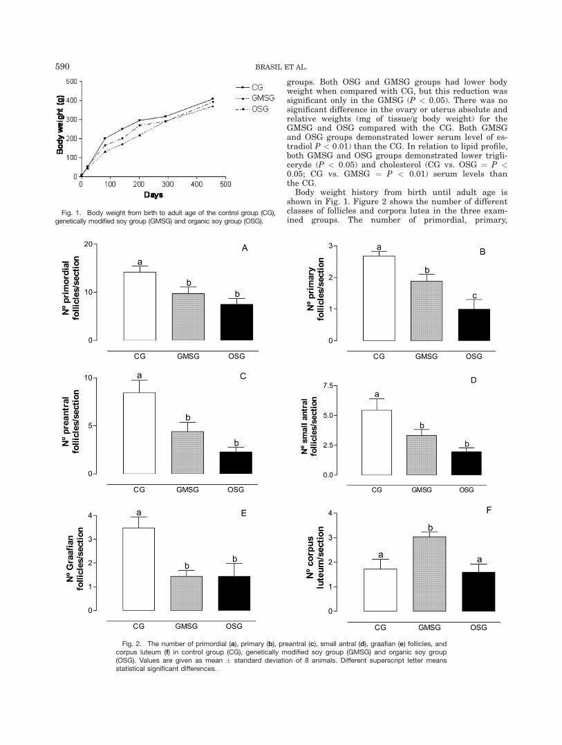
Body weight history from birth until adult age isshown in Fig. 1. Figure 2 shows the number of differentclasses of follicles and corpora lutea in the three exam-ined groups. The number of primordial, primary,
Fig. 1. Body weight from birth to adult age of the control group (CG),genetically modified soy group (GMSG) and organic soy group (OSG).
Fig. 2. The number of primordial (a), primary (b), preantral (c), small antral (d), graafian (e) follicles, andcorpus luteum (f) in control group (CG), genetically modified soy group (GMSG) and organic soy group(OSG). Values are given as mean � standard deviation of 8 animals. Different superscript letter meansstatistical significant differences.
590 BRASIL ET AL.

preantral, small antral, and Graafian follicles was signif-icantly reduced in both GMSG (P < 0.05) and OSG (P <0.01) groups when compared with the CG. The numberof corpus luteum was significantly (P < 0.05) increasedonly in the GMSG when compared with the CG. Thenumber of primary follicles and corpus luteum was sig-nificantly different (P < 0.05) between the GMSG andOSG groups.
The morphometric analysis of the uterus showed thatvolumetric density of epithelium was significantly higherin the GMSG compared with both CG (P < 0.01) andOSG (P < 0.05) (Fig. 3). Both soy groups presented a sig-nificant reduction in the length density of endometrialglands (P < 0.01) when compared with the CG (Fig. 4).
Figure 5 show histological sections of ovary (A, B, andC) in CG, GMSG and OSG groups, respectively. Primaryfollicles consist of an oocyte surrounded by a single layerof cuboidal granulosa cells. The preantral follicles pres-ent a central oocyte surrounded by several layers ofgranulosa cells and bounded by thecal cells, which forma fibrous theca externa and an inner theca interna withno antrum. In antral follicles, fluid appeared betweenthe granulosa cells, and the drops coalesced to form fol-licular fluid within the follicular antrum. In Graafianfollicles, the follicular antrum is clearly developed, leav-ing the oocyte surrounded by a distinct and denser layerof granulosa cells, the cumulus oophorus. The corpusluteum is formed by luteal cells and abundantcapillaries.
Figure 6 show histological sections of uterus (A, B,and C) in CG, GMSG, and OSG groups, respectively.The endometrial and glandular epithelium, stroma, andmyometrium can be observed in the photomicrographs.
DISCUSSION
Soyfood has been reported to have beneficial effectsincluding improving the lipid profile (Simons et al.,2000; Dent et al., 2001; Gardner et al., 2001; Kanget al., 2002; Engelman et al., 2005; Ho et al., 2007), bonemetabolism (Marini et al., 2007; Ma et al., 2007), cancerdevelopment (Jin and MacDonald, 2002; Liu et al.,2005), without having any effects on the uterus and
ovary in postmenopausal women or menopausal animalsmodels (Bahr et al., 2005; Castillo et al., 2006; Kaari etal., 2006; Wood et al., 2006; Marini et al., 2007). How-ever, if soyfood is used for neonatal, young, or adult ani-mals at a reproductive age, it can cause adverse effectsrelated to the reproductive organs (Jefferson et al., 2002,2005, 2006; Michael et al., 2006; Piotrowska et al., 2006;Rachon et al., 2007a,b). On the basis of the soy consump-tion increment, this study was designed to compare theeffects of a prolonged use of organic and transgenic soyon the lipid profile and ovary and uterus morphology.
Although the use of genetically modified food is stillquestionable, there is no evidence that genetic modifica-tion through biotechnology will impose immediate signif-icant risks as food allergen sources beyond that of ourdaily dietary intake of foods from crop plants (Helm,2003) or beyond other methodologies widely accepted inthe food industry (Lack, 2002). Also, there is no evidencesuggesting that recombinant DNA would be processed inthe gut in any manner different from endogenous feed-ingested genetic material (Jennings et al., 2003; Sharmaet al., 2006). The data presented here show that theeffects of organic and transgenic soy consumption werevery similar, except those observed in the corpora luteaand volumetric density of glandular epithelium.
In agreement with the literature, both organic andtransgenic soy reduced the body weight (Demonty et al.,2002; Rachon et al., 2007a) and estradiol serum levels(Lu et al., 1996; Nagata et al., 1997; Duncan et al., 1999;Wood et al., 2007). Both soy treatments also improvedthe lipid profile by reducing cholesterol and triglyceridesserum levels (Simons et al., 2000; Dent et al., 2001;Gardner et al., 2001; Kang et al., 2002; Engelman et al.,2005; Ho et al., 2007). Probably the reduction in estra-diol serum levels reflects the capacity of isoflavones tobind the estrogen receptor and blocking the actions ofendogenous estrogens (Lissin and Cooke, 2000). Thealterations presented here were more marked in thetransgenic group, which showed the lowest body weightand cholesterol and triglycerides levels.
Also, corroborating previous results (Jefferson et al.,2002, 2005, 2006; Michael et al., 2006; Piotrowska et al.,2006; Rachon et al., 2007a,b), both transgenic and
Fig. 3. Volumetric density of glandular epithelium of the controlgroup (CG), genetically modified soy group (GMSG) and organic soygroup (OSG). Values are given as mean � standard deviation of eightanimals. Different superscript letter means statistical significantdifferences.
Fig. 4. Length density of endometrial glands of the control group(CG), genetically modified soy group (GMSG) and organic soy group(OSG). Values are given as mean � standard deviation of eightanimals. Different superscript letter means statistical significantdifferences.
SOY AND REPRODUCTIVE SYSTEM OF ADULT RAT 591
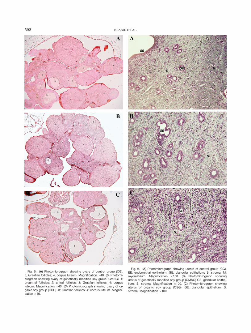
Fig. 6. (A) Photomicrograph showing uterus of control group (CG).EE, endometrial epithelium; GE, glandular epithelium; S, stroma; M,myometrium. Magnification �100. (B) Photomicrograph showinguterus of genetically modified soy group (GMSG) GE, glandular epithe-lium; S, stroma. Magnification �100. (C) Photomicrograph showinguterus of organic soy group (OSG). GE, glandular epithelium; S,stroma. Magnification �100.
Fig. 5. (A) Photomicrograph showing ovary of control group (CG).3, Graafian follicles; 4, corpus luteum. Magnification �40. (B) Photomi-crograph showing ovary of genetically modified soy group (GMSG). 1:preantral follicles; 2: antral follicles; 3: Graafian follicles; 4: corpusluteum. Magnification �40. (C) Photomicrograph showing ovary of or-ganic soy group (OSG). 3: Graafian follicles; 4: corpus luteum. Magnifi-cation �40.
592 BRASIL ET AL.

organic soy had an adverse effect on some parameters ofthe uterus and ovary morphology. The number of thegrowing follicles was significantly reduced in both soy-treated groups, in spite of normal corpus luteum numberin the organic group. In relation to the uterus, both soy-treated groups exhibited a reduction in the length den-sity of the glands, while the volumetric density of the ep-ithelium was unaltered in the organic group. These datasuggest that the transgenic and organic soy may havespecific effects in the reproductive system.
The isoflavone content of both transgenic and organicsoy was evaluated and no significant difference in theindividual components or in food consumption wasfound. So, at this moment we may assume that the dif-ferences related to the transgenic and organic soy arenot related to isoflavone, but it can probably be relatedto the small differences in fat, sugar and especially pro-tein or amino acids diet content among the three groups.
In summary, both transgenic and organic-derived soydiets improved the lipid profile and reduced body weight;however, alterations in uterine and ovarian morphologywere also found in animals with prolonged exposure tothese diets. The prolonged use of soy-based diets andtheir relation to reproductive health warrants furtherinvestigation.
LITERATURE CITED
Anthony MS, Clarkson TB, Hughes CL, Jr, Morgan TM, Burke GL.1996. Soybean isoflavones improve cardiovascular risk factorswithout affecting the reproductive system of peripubertal rhesusmonkeys. J Nutr 126:43–50.
Bahr JM, Nakai M, Rivera A, Walsh J, Evans GL, Lotinun S,Turner RT, Black M, Jeffery EH. 2005. Dietary soy protein andisoflavones: minimal beneficial effects on bone and no effect onthe reproductive tract of sexually mature ovariectomizedSprague-Dawley rats. Menopause 12:165–173.
Bairey Merz CN, Johnson BD, Braunstein GD, Pepine CJ, Reis SE,Paul-Labrador M, Hale G, Sharaf BL, Bittner V, Sopko G, KelseySF. 2006. Phytoestrogens and lipoproteins in women. J Clin Endo-crinol Metab 91:2209–2213.
Bayne K. 1996. Revised guide for the care and use of laboratoryanimals available. Physiologist 39:208–211.
Bradbury P, Rae K. 1996. Connective tissues and stains. In: Ban-croft JD, Stevens A, editors. Theory and practice of histologicaltechniques, 4th ed. New York: Churchill Livingstone. p 113–138.
Castillo C, Salazar V, Ariznavarreta C, Vara E, Tresguerres JA.2006. Effect of isoflavone administration on age-related hepato-cyte changes in old ovariectomized femal Wistar rats. Phytomedi-cine 13:468–476.
Cheng G WZ, Makinen S, Makela S, Saji S, Warner M, GustafssonJA, Hovatta O. 2002. A Role for the androgen receptor in follicu-lar atresia of estrogen receptor beta knockout mouse ovary. BiolReprod 66:77–84.
Demonty I, Lamarche B, Deshaies Y, Jacques H. 2002. Role of soyisoflavones in the hypotriglyceridemic effect of soy protein in therat. J Nutr Biochem 13:671–677.
Dent SB, Peterson CT, Brace LD, Swain JH, Reddy MB, HansonKB, Robinson JG, Alekel DL. 2001. Soy protein intake by perime-nopausal women does not affect circulating lipids and lipoproteinsor coagulation and fibrinolytic factors. J Nutr 131:2280–2287.
Duncan AM, Underhill KE, Xu X, Lavalleur J, Phipps WR, KurzerMS. 1999. Modest hormonal effects of soy isoflavones in post-menopausal women. J Clin Endocrinol Metab 84:3479–3484.
Engelman HM, Alekel DL, Hanson LN, Kanthasamy AG, ReddyMB. 2005. Blood lipid and oxidative stress responses to soy pro-tein with isoflavones and phytic acid in postmenopausal women.Am J Clin Nutr 81:590–596.
Gardner CD, Newell KA, Cherin R, Haskell WL. 2001. The effect ofsoy protein with or without isoflavones relative to milk protein onplasma lipids in hypercholesterolemic postmenopausal women.Am J Clin Nutr 73:728–735.
Helm RM. 2003. Food biotechnology: is this good or bad? Implica-tions to allergic diseases. Ann Allergy Asthma Immunol 90:90–98.
Ho SC, Chen YM, Ho SS, Woo JL. 2007. Soy isoflavone supplemen-tation and fasting serum glucose and lipid profile among post-menopausal Chinese women: A double-blind, randomized,placebo-controlled trial. Menopause 14:905–912.
Jefferson W, Newbold R, Padilla-Banks E, Pepling M. 2006. Neona-tal genistein treatment alters ovarian differentiation in themouse: inhibition of oocyte nest breakdown and increased oocytesurvival. Biol Reprod 74:161–168.
Jefferson WN, Padilla-Banks E, Newbold RR. 2005. Adverse effectson female development and reproduction in CD-1 mice followingneonatal exposure to the phytoestrogen genistein at environmen-tally relevant doses. Biol Reprod 73:798–806.
Jefferson WN, Couse JF, Padilla-Banks E, Korach KS, Newbold RR.2002. Neonatal exposure to genistein induces estrogen receptor(ER)alpha expression and multioocyte follicles in the maturingmouse ovary: evidence for ERbeta-mediated and nonestrogenicactions. Biol Reprod 67:1285–1296.
Jennings JC, Kolwyck DC, Kays SB, Whetsell AJ, Surber JB, Crom-well GL, Lirette RP, Glenn KC. 2003 Determining whether trans-genic and endogenous plant DNA and transgenic protein aredetectable in muscle from swine fed Roundup Ready soybeanmeal. J Anim Sci 81:1447–1455.
Jin Z, MacDonald RS. 2002 Soy isoflavones increase latencyof spontaneous mammary tumors in mice. J Nutr 132:3186–3190.
Kaari C, Haidar MA, Junior JM, Nunes MG, Quadros LG, Kemp C,Stavale JN, Baracat EC. 2006. Randomized clinical trial compar-ing conjugated equine estrogens and isoflavones in postmeno-pausal women: a pilot study. Maturitas 53:49–58.
Kang HJ, Ansbacher R, Hammoud MM. 2002. Use of alternativeand complementary medicine in menopause. Int J GynaecolObstet 79:195–207.
Kohno M, Hirotsuka M, Kito M, Matsuzawa Y. 2006. Decreases inserum triacylglycerol and visceral fat mediated by dietary soy-bean beta-conglycinin. J Atheroscler Thromb 13:247–255.
Klump SP, Allred MC, MacDonald JL, Ballam JM. 2001. Determi-nation of isoflavones in soy and selected foods containing soy byextraction, saponification, and liquid chromatography: collabora-tive study. J AOAC Int 84:1865–1883.
Lack G. 2002. Clinical risk assessment of GM foods. Toxicol Lett127:337–340.
Lissin LW, Cooke JP. 2000. Phytoestrogens and cardiovascularhealth. J Am Coll Cardiol 35:1403–1410.
Liu B, Edgerton S, Yang X, Kim A, Ordonez-Ercan D, Mason T,Alvarez K, McKimmey C, Liu N, Thor A. 2005. Low-dose dietaryphytoestrogen abrogates tamoxifen-associated mammary tumorprevention. Cancer Res 65:879–886.
Lu LJ, Anderson KE, Grady JJ, Nagamani M. 1996. Effects of soyaconsumption for one month on steroid hormones in premeno-pausal women: implications for breast cancer risk reduction. Can-cer Epidemiol Biomarkers Prev 5:63–70.
Ma DF, Qin LQ, Wang PY, Katoh R. 2008. Soy isoflavone intakeinhibits bone resorption and stimulates bone formation in meno-pausal women: meta-analysis of randomized controlled trials. EurJ Clin Nutr 62:155–161.
Magkos F, Arvaniti F, Zampelas A. 2003. Putting the safety of or-ganic Food into perspective. Nutr Res Rev 16:241–252.
Mandarim-de-Lacerda CA. 2003. Stereological tools in biomedicalresearch. An Acad Bras Cienc 75:469–486.
Marini H, Minutoli L, Polito F, Bitto A, Altavilla D, Atteriano M,Gaudio A, Mazzaferro S, Frisina A, Frisina N, Lubrano C,Bonaiuto M, D’Anna R, Cannata ML, Corrado F, Adamo EB, Wil-son S, Squadrito F. 2007. Effects of the phytoestrogen genisteinon bone metabolism in osteopenic postmenopausal women: arandomized trial. Ann Intern Med 146:839–847.
SOY AND REPRODUCTIVE SYSTEM OF ADULT RAT 593

Michael McClain R, Wolz E, Davidovich A, Pfannkuch F, EdwardsJA, Bausch J. 2006. Acute, subchronic and chronic safety studieswith genistein in rats. Food Chem Toxicol 44:56–80.
Nagata C, Kabuto M, Kurisu Y, Shimizu H. 1997. Decreased serum es-tradiol concentration associated with high dietary intake of soy prod-ucts in premenopausal Japanese women. Nutr Cancer 29:228–233.
Piotrowska KK, Woclawek-Potocka I, Bah MM, Piskula MK, Pilaw-ski W, Bober A, Skarzynski DJ. 2006. Phytoestrogens and theirmetabolites inhibit the sensitivity of the bovine corpus luteum toluteotropic factors. J Reprod Dev 52:33–41.
Rachon D, Vortherms T, Seidlova-Wuttke D, Wuttke W. 2007a.Effects of dietary equol on body weight gain, intra-abdominal fataccumulation, plasma lipids, and glucose tolerance in ovariectom-ized Sprague-Dawley rats. Menopause 14:925–932.
Rachon D, Vortherms T, Seidlova-Wuttke D, Wuttke W. 2007b. Die-tary daidzein and puerarin do not affect pituitary LH expressionbut exert uterotropic effects in ovariectomized rats. Maturitas57:161–170.
Reeves PG, Nielsen FH, Fahey GC. 1993. AIN-93 purified diets forlaboratory rodents: final report of the American Institute ofNutrition Ad Hoc Writing Committee on the reformulation of theAIN-76 rodent diet. J Nutr 123:1939–1951.
Rott ME, Lawrence TS, Wall EM, Green MJ. 2004. Detection andquantification of roundup ready soy in foods by conventional and
real-time polymerase chain reaction. J Agric Food Chem 52:5223–5232.
Sharma R, Damgaard D, Alexander TW, Dugan ME, Aalhus JL,Stanford K, McAllister TA. 2006. Detection of transgenic and en-dogenous plant DNA in digesta and tissues of sheep and pigs fedRoundup Ready canola meal. J Agric Food Chem 54:1699–1709.
Simons LA Konigsmark Mv, Simons J, Celermajer DS. 2000. Phy-toestrogens do not influence lipoprotein levels or endothelial func-tion in healthy, postmenopausal women. Am J Cardiol 85:1297–1301.
Soares LL, Lucas AM, Boaventura GT. 2005. Can organic andtransgenic soy be used as a substitute for animal protein by rats?Braz J Med Biol Res 38:583–586.
Sokal RR, Rohlf FJ. 1995. Biometry: the principles and practice ofstatistics in biological research. New York: WH Freeman. p 887.
Turck D. 2007. Soy protein for infant feeding: what do we know?Curr Opin Clin Nutr Metab Care 10:360–365.
Wood CE, Appt SE, Clarkson TB, Franke AA, Lees CJ, Doerge DR,Cline JM. 2006. Effects of high-dose soy isoflavones and equol onreproductive tissues in female cynomolgus monkeys. Biol Reprod75:477–486.
Wood CE, Register TC, Cline JM. 2007. Soy isoflavonoid effects onendogenous estrogen metabolism in postmenopausal female mon-keys. Carcinogenesis 28:801–808.
594 BRASIL ET AL.