Dietary and Non-Dietary Phytochemicals and Cancer - Unglue.it
158
Dietary and Non-Dietary Phytochemicals and Cancer Carmela Fimognari www.mdpi.com/journal/toxins Edited by Printed Edition of the Special Issue Published in Toxins toxins
-
Upload
khangminh22 -
Category
Documents
-
view
0 -
download
0
Transcript of Dietary and Non-Dietary Phytochemicals and Cancer - Unglue.it
Dietary and Non-Dietary Phytochemicals and Cancer
Carmela Fimognari
www.mdpi.com/journal/toxins
Edited by
Printed Edition of the Special Issue Published in Toxins
toxins

Dietary and Non-Dietary
Phytochemicals and Cancer
Special Issue Editor Carmela Fimognari
Special Issue Editor
Carmela Fimognari
University of Bologna
Italy
Editorial Office
MDPI AG
St. Alban-Anlage 66
Basel, Switzerland
This edition is a reprint of the Special Issue published online in the open access
journal Toxins (ISSN 2072-6651) from 2016–2017 (available at:
http://www.mdpi.com/journal/toxins/special_issues/phytochemicals_cancer).
For citation purposes, cite each article independently as indicated on the article
page online and as indicated below:
Author 1; Author 2; Author 3 etc. Article title. Journal Name. Year.
Article number/page range.
ISBN 978-3-03842-378-2 (Pbk)
ISBN 978-3-03842-379-9 (PDF)
Articles in this volume are Open Access and distributed under the Creative Commons Attribution
license (CC BY), which allows users to download, copy and build upon published articles even for
commercial purposes, as long as the author and publisher are properly credited, which
ensures maximum dissemination and a wider impact of our publications. The book taken as a whole is
© 2017 MDPI, Basel, Switzerland, distributed under the terms and conditions of the Creative
Commons license CC BY-NC-ND (http://creativecommons.org/licenses/by-nc-nd/4.0/).
iii
Table of Contents
About the Guest Editor ................................................................................................................................. v
Preface to “Dietary and Non-Dietary Phytochemicals and Cancer” ..................................................... vii
Jisun Oh, Lynn Hlatky, Yong-Seob Jeong and Dohoon Kim
Therapeutic Effectiveness of Anticancer Phytochemicals on Cancer Stem Cells
Reprinted from: Toxins 2016, 8(7), 199; doi: 10.3390/toxins8070199
http://www.mdpi.com/2072-6651/8/7/199 .................................................................................................. 1
Kuan-Ta Lu, Bing-Yen Wang, Wan-Yu Chi, Ju Chang-Chien, Jiann-Jou Yang, Hsueh-Te Lee,
Yew-Min Tzeng and Wen-Wei Chang
Ovatodiolide Inhibits Breast Cancer Stem/Progenitor Cells through SMURF2-Mediated
Downregulation of Hsp27
Reprinted from: Toxins 2016, 8(5), 127; doi: 10.3390/toxins8050127
http://www.mdpi.com/2072-6651/8/5/127 .................................................................................................. 12
Tariq Ismail 1, Cinzia Calcabrini, Anna Rita Diaz, Carmela Fimognari, Eleonora Turrini,
Elena Catanzaro, Saeed Akhtar and Piero Sestili
Ellagitannins in Cancer Chemoprevention and Therapy
Reprinted from: Toxins 2016, 8(5), 151; doi: 10.3390/toxins8050151
http://www.mdpi.com/2072-6651/8/5/151 .................................................................................................. 23
Mohd Farhan, Husain Yar Khan, Mohammad Oves, Ahmed Al-Harrasi, Nida Rehmani,
Hussain Arif, Sheikh Mumtaz Hadi and Aamir Ahmad
Cancer Therapy by Catechins Involves Redox Cycling of Copper Ions and Generation of Reactive
Oxygen Species
Reprinted from: Toxins 2016, 8(2), 37; doi: 10.3390/toxins8020037
http://www.mdpi.com/2072-6651/8/2/37 .................................................................................................... 45
Chung-Yi Chen, Ching-Yu Yen, Hui-Ru Wang, Hui-Ping Yang, Jen-Yang Tang,
Hurng-Wern Huang, Shih-Hsien Hsu and Hsueh-Wei Chang
Tenuifolide B from Cinnamomum tenuifolium Stem Selectively Inhibits Proliferation of Oral Cancer
Cells via Apoptosis, ROS Generation, Mitochondrial Depolarization, and DNA Damage
Reprinted from: Toxins 2016, 8(11), 319; doi: 10.3390/toxins8110319
http://www.mdpi.com/2072-6651/8/11/319 ................................................................................................ 61
Thao T. Nguyen, Marie-Odile Parat, Mark P. Hodson, Jenny Pan, Paul N. Shaw and
Amitha K. Hewavitharana
Chemical Characterization and in Vitro Cytotoxicity on Squamous Cell Carcinoma Cells of
Carica Papaya Leaf Extracts
Reprinted from: Toxins 2016, 8(1), 7; doi: 10.3390/toxins8010007
http://www.mdpi.com/2072-6651/8/1/7 ...................................................................................................... 75
Eleonora Turrini, Cinzia Calcabrini, Piero Sestili, Elena Catanzaro, Elena de Gianni,
Anna Rita Diaz, Patrizia Hrelia, Massimo Tacchini, Alessandra Guerrini, Barbara Canonico,
Stefano Papa, Giovanni Valdrè and Carmela Fimognari
Withania somnifera Induces Cytotoxic and Cytostatic Effects on Human T Leukemia Cells
Reprinted from: Toxins 2016, 8(5), 147; doi: 10.3390/toxins8050147
http://www.mdpi.com/2072-6651/8/5/147 .................................................................................................. 86

iv
Estefanía Burgos-Morón, José Manuel Calderón-Montaño, Manuel Luis Orta,
Emilio Guillén-Mancina, Santiago Mateos and Miguel López-Lázaro
Cells Deficient in the Fanconi Anemia Protein FANCD2 are Hypersensitive to the Cytotoxicity and
DNA Damage Induced by Coffee and Caffeic Acid
Reprinted from: Toxins 2016, 8(7), 211; doi: 10.3390/toxins8070211
http://www.mdpi.com/2072-6651/8/7/211 .................................................................................................. 101
Geum-A. Lee, Kyung-A. Hwang and Kyung-Chul Choi
Roles of Dietary Phytoestrogens on the Regulation of Epithelial-Mesenchymal Transition in Diverse
Cancer Metastasis
Reprinted from: Toxins 2016, 8(6), 162; doi: 10.3390/toxins8060162
http://www.mdpi.com/2072-6651/8/6/162 .................................................................................................. 110
Anna Kakehashi, Midori Yoshida, Yoshiyuki Tago, Naomi Ishii, Takahiro Okuno, Min Gi
and Hideki Wanibuchi
Pueraria mirifica Exerts Estrogenic Effects in the Mammary Gland and Uterus and Promotes
Mammary Carcinogenesis in Donryu Rats
Reprinted from: Toxins 2016, 8(11), 275; doi: 10.3390/toxins8110275
http://www.mdpi.com/2072-6651/8/11/275 ................................................................................................ 127

v
About the Guest Editor
Carmela Fimognari earned her PhD in Toxicology in 1997 from the University of Bologna, Italy. After training at the GSF-National Research Centre for Environment and Health, Munich, Germany, she focused her research on cancer chemoprevention and therapy by natural compounds and synthetic analogues with cytostatic and cell death-inducing potential. As an example, she and her co-workers investigated isothiocyanates and anthocyanins, known to exhibit anti-inflammatory and anti-cancer activities. She directs the Laboratory for Molecular and Cellular Toxicology at the Department for Life Quality Studies of the University of Bologna. She was appointed associate Professor of Toxicology at the Faculty of Pharmacy of the University of Bologna in 2014. She wrote over 90 publications on international journals and book chapters.


toxins
Review
Introduction to the Toxins Special Issue on Dietaryand Non-Dietary Phytochemicals and Cancer
Carmela Fimognari
Department for Life Quality Studies, Alma Mater Studiorum-University of Bologna, Corso d’Augusto 237,Rimini 47921, Italy; [email protected]
Academic Editor: Nilgun E. TumerReceived: 15 December 2016; Accepted: 21 December 2016; Published: 28 December 2016
The role of many phytochemicals in the modulation of the carcinogenesis process has been welldocumented by combining in vitro and animal studies, as well as epidemiological evidence. Whenacting in synergy, phytochemicals exert potential anti-cancer properties, and much progress has beenmade in defining their many biological activities at the molecular level. However, an interesting featurein the field of phytochemicals and cancer is the role of some phytochemicals in promoting cancerdevelopment. This Special Issue of Toxins aims to provide a comprehensive look at the contribution ofdietary and non-dietary phytochemicals to cancer development and at the molecular mechanisms bywhich phytochemicals inhibit or promote cancer.
Cancer stem cells represent a small subset of tumor cells endowed with uncontrolled proliferativecapacity and indefinite potential for self-renewal that drive tumorigenesis. Considering the potentialof cancer stem cells in the initial development of cancer, resistance to therapy and metastasis, theyhave become a critical target for the identification and development of new approaches to fight cancer.Oh et al. present an overview of phytochemicals targeting signaling pathways involved in stemnessmaintenance and survival of cancer stem cells [1]. Some examples include cyclopamine from the cornlily, curcumin from turmeric, and piperine from black and long peppers, sulforaphane from cruciferousvegetables, the soy isoflavone genistein, and blueberry polyphenols. Lu et al. [2] report the effect ofovatodiolide—a macrocyclic diterpenoid compound isolated from Anisomeles indica—of blocking theself-renewal capability of breast cancer stem cells and downregulating the expression of stemnessgenes on human breast cancer stem cells.
The detailed review by Ismail et al. [3] focuses on ellagitannins, a class of phytochemicals widelyinvestigated for their chemopreventive and anticancer activities. With the aim of delineating andpredicting their actual clinical potential, the authors present the dietary sources, the pharmacokinetics,and the evidence on the chemopreventive efficacy and the anticancer activity of ellagitannins.The chemopreventive effects of ellagitannins are linked to their antioxidant and anti-inflammatoryproperties. Their anticancer activity is imputable to different mechanisms, including inhibition ofpro-inflammatory pathway, cell-cycle arrest ability, and proapoptotic properties. The pleiotropic natureof the mechanisms behind the anticancer activity of ellegitannins is also demonstrated by their abilityto block angiogenesis and inhibit endothelial cell growth. However, as the authors point out, orallyadministered ellagitannins are characterized by a limited bioavailability. Their widely recognizedanticancer efficacy may lead to the adoption of different administration routes to overcome their lowbioavailability, such as the intravenous route. However, their toxicological profile after intravenousadministration is lacking.
This Special Issue also features several research papers reporting selective effects ofphytochemicals against cancer cells, for which follow brief synopses.
The article by Farhan et al. [4] highlights mechanistic studies which provide insights intothe cytotoxic and anticancer activity of catechins. Catechin, epicatechin, epigallocatechin, andepigallocatechin-3-gallate—the four major constituents of green tea—exert a prooxidant action
Toxins 2017, 9, 12 vii www.mdpi.com/journal/toxins
Toxins 2017, 9, 12
through redox recycling of copper ions, and induce cellular DNA breakage in human peripherallymphocytes. The presence of copper ions boosts their genotoxic effect. Catechins (especiallyepigallocatechin-3-gallate) have antiproliferative activity on breast cancer cells. Copper levels are muchhigher in cancer cells than in non-transformed cells. Thus, cancer cells would be more susceptibleto redox cycling between copper ions and catechins to generate reactive oxygen species and thusDNA breakage. Accordingly, the authors demonstrate that normal breast epithelial cells are quiteresistant to the treatment with catechins, but their culture in medium enriched with copper make themmore susceptible to catechins’ antiproliferative effect. Taking the results presented in the paper intoaccount, catechins exert their preferential prooxidant cytotoxic effect against cancer cells. Chen et al. [5]investigate the effect of tenuifolide B—derived from the stems of Cinnamomum tenuifolium—on viability,cycle progression, apoptosis, reactive oxygen species production, mitochondrial depolarization, andDNA damage in transformed and non-transformed oral cells. Tenuifolide B reduces the viability ofcancer cells through the induction of apoptotic cell death. The production of reactive oxygen speciesand the induction of DNA damage are probably involved in its cytotoxic effect. Of note, the effectsof tenuifolide B seem to be selective for cancer cells. Indeed, its effects on normal oral cells are muchless pronounced than on cancer cells. Nguyen et al. [6] show that papaya leaves are a potential sourceof anticancer compounds. They actually contain flavonoids or flavonoid glycosides, particularlycompounds from the kaempferol and quercetin families. Moreover, aqueous and ethanolic extracts ofleaves reduce the survival of human oral squamous cell carcinoma cells. Interestingly, the two fractionswith acidic pH are selectively cytotoxic towards the cancer cells, and the phenolic and flavonoidcontent positively correlates with the differential effect on cell viability.
However, it is worth noting that some phytochemicals can have an ambivalent character(especially in the possible context of high dose therapeutic applications), and favor cancer development.Some papers of this Special Issue deal with this important aspect.
Turrini et al. [7] provide new insights into the anticancer mechanisms of the root extract ofWithania somnifera, a plant used in Indian traditional medicine. The article presents the cytotoxic andcytostatic effects of the extract on human leukemia cells and suggests the role of reactive oxygenspecies in its cytotoxic activity. Of note, Withania induces the expression of specific molecules such ascalreticulin, Hsp-70, and Hsp-90 on cancer cells, thus boosting the immunogenic profile of tumor cellsand stimulating the innate immune system response. In order to elaborate a preliminary risk/benefitprofile, the authors analyze the genotoxicity of the extract through the quantification of histone H2A.Xphosphorylation (γ-H2A.X), a biomarker of double-strand DNA breaks. The extract is found to begenotoxic. Bearing in mind that DNA damage plays a well-established role in cancer initiation andposes serious risks for human safety [8], the genotoxicity of Withania should be carefully examined foran accurate prediction of its risk–benefit profile.
Burgos-Morón et al. [9] examine the genotoxicity of caffeic acid and a commercial lyophilizedcoffee extract in cells deficient in the critical DNA repair protein Fanconi anemia D2 and demonstratethat this kind of cell is hypersensitive to the DNA damage induced by caffeic acid and coffee comparedto non-deficient cells. These results suggest that coffee and caffeic acid may increase the risk of cancer,particularly in people with germline or sporadic mutations in the DNA repair protein Fanconi anemiaD2. Taking into account that caffeic acid accumulates in the urinary bladder, the risk of bladdercancer development may be particularly high. The authors also discuss the key role that caffeicacid and other coffee constituents—such as chlorogenic acid and hydroquinone, endowed with anantioxidant activity at low concentrations and a pro-oxidant activity at higher concentrations—mayhave in cancer development.
An overview of the effects of some phytoestrogens on cancer progression is presented byLee et al. [10]. Genistein, resveratrol, kaempferol, and 3,3′-diindolylmethane were extensively studiedfor their anticancer effects and as alternatives for hormone replacement therapy. In particular, they caninhibit the epithelial–mesenchymal transition, which plays a key role in cancer migration, invasion,and metastasis, and modulate the signaling pathways and the expression of epithelial–mesenchymal
viii
Toxins 2017, 9, 12
transition-related markers, such as TGF-β and PI3K/Akt/mTOR/NF-κB. Nevertheless, phytoestrogenslike genistein and resveratrol can have a biphasic effect and lead to cancer cell growth at lowerconcentrations and to inhibition of cancer cell growth at higher concentrations.
Kakehashi et al. [11] explore the estrogenic effects in the mammary gland and uterus andthe carcinogenetic activity of a diet containing Pueraria mirifica powder in female rats. To thisend, they use different experimental strategies: Pueraria administered to ovariectomized animalsat doses of 0.03%, 0.3%, and 3% in a phytoestrogen-low diet for 2 weeks; a 4 week applicationto non-operated rats at a dose of 3% after 7,12-dimethylbenz[a]anthracene cancer initiation;postpubertal administration of 0.3% to 5-week-old non-operated animals for 36 weeks followinginitiation of mammary and endometrial carcinogenesis with 7,12-dimethylbenz[a]anthracene andN-ethyl-N′-nitro-N-nitrosoguanidine, respectively. In the first experimental setting, Pueraria increaseduterus weight; in the second one, Pueraria stimulated cell proliferation in the mammary gland; in thethird experimental model, it boosted mammary adenocarcinoma incidence. These data raise veryimportant questions on the safety of long-term exposure to phytoestrogens with regard to effects onthe mammary gland and endometrium. Different products containing Pueraria mirifica are widelyavailable in the USA and Japan. Despite the data on its positive health effects, including increasinghair growth, improving appetite, and providing relief for ailments like osteoporosis and even cancer, itevokes an estrogen-like effect that should be considered to better understand its risk–benefit profile.More research has to be performed to better define the relationship between the hazardous andchemopreventive effects of phytoestrogens.
I hope that this Special Issue will provide readers a better understanding of the mechanism ofaction of phytochemicals in modulating the carcinogenetic process. These aspects have advancedparticularly far in recent years, and are extremely useful for the definition of efficient preventive ortherapeutic strategies against cancer. I would also like to thank all authors contributing to this SpecialIssue in Toxins for their commitment and time, and our reviewers for their expert input and criticalevaluation of the papers.
References
1. Oh, J.; Hlatky, L.; Jeong, Y.S.; Kim, D. Therapeutic effectiveness of anticancer phytochemicals on cancer stemcells. Toxins 2016, 8, 199. [CrossRef] [PubMed]
2. Lu, K.T.; Wang, B.Y.; Chi, W.Y.; Chang-Chien, J.; Yang, J.J.; Lee, H.T.; Tzeng, Y.M.; Chang, W.W. Ovatodiolideinhibits breast cancer stem/progenitor cells through SMURF2-mediated downregulation of Hsp27. Toxins2016, 8, 127. [CrossRef] [PubMed]
3. Ismail, T.; Calcabrini, C.; Diaz, A.R.; Fimognari, C.; Turrini, E.; Catanzaro, E.; Akhtar, S.; Sestili, P.Ellagitannins in cancer chemoprevention and therapy. Toxins 2016, 8, 151. [CrossRef] [PubMed]
4. Farhan, M.; Khan, H.Y.; Oves, M.; Al-Harrasi, A.; Rehmani, N.; Arif, H.; Hadi, S.M.; Ahmad, A. Cancertherapy by catechins involves redox cycling of copper ions and generation of reactive oxygen species. Toxins2016, 8, 37. [CrossRef] [PubMed]
5. Chen, C.Y.; Yen, C.Y.; Wang, H.R.; Yang, H.P.; Tang, J.Y.; Huang, H.W.; Hsu, S.H.; Chang, H.W. Tenuifolide Bfrom Cinnamomum tenuifolium stem selectively inhibits proliferation of oral cancer cells via apoptosis, ROSgeneration, mitochondrial depolarization, and DNA damage. Toxins 2016, 8, 319. [CrossRef] [PubMed]
6. Nguyen, T.T.; Parat, M.O.; Hodson, M.P.; Pan, J.; Shaw, P.N.; Hewavitharana, A.K. Chemical characterizationand in vitro cytotoxicity on squamous cell carcinoma cells of Carica papaya leaf extracts. Toxins 2016, 8, 7.[CrossRef] [PubMed]
7. Turrini, E.; Calcabrini, C.; Sestili, P.; Catanzaro, E.; De Gianni, E.; Diaz, A.R.; Hrelia, P.; Tacchini, M.;Guerrini, A.; Canonico, B.; et al. Withania somnifera induces cytotoxic and cytostatic effects on human Tleukemia cells. Toxins 2016, 8, 147. [CrossRef] [PubMed]
8. Turrini, E.; Ferruzzi, L.; Fimognari, C. Natural compounds to overcome cancer chemoresistance: Toxicologicaland clinical issues. Expert Opin. Drug Metab. Toxicol. 2014, 10, 1677–1690. [CrossRef] [PubMed]
ix

Toxins 2017, 9, 12
9. Burgos-Morón, E.; Calderón-Montaño, J.M.; Orta, M.L.; Guillén-Mancina, E.; Mateos, S.; López-Lázaro, M.Cells deficient in the Fanconi anemia protein FANCD2 are hypersensitive to the cytotoxicity and DNAdamage induced by coffee and caffeic acid. Toxins 2016, 8, 211. [CrossRef] [PubMed]
10. Lee, G.A.; Hwang, K.A.; Choi, K.C. Roles of dietary phytoestrogens on the regulation ofepithelial-mesenchymal transition in diverse cancer metastasis. Toxins 2016, 8, 162. [CrossRef] [PubMed]
11. Kakehashi, A.; Yoshida, M.; Tago, Y.; Ishii, N.; Okuno, T.; Gi, M.; Wanibuchi, H. Pueraria mirifica exertsestrogenic effects in the mammary gland and uterus and promotes mammary carcinogenesis in Donryu rats.Toxins 2016, 8, 275. [CrossRef] [PubMed]
© 2016 by the author. Licensee MDPI, Basel, Switzerland. This article is an open accessarticle distributed under the terms and conditions of the Creative Commons Attribution(CC BY) license (http://creativecommons.org/licenses/by/4.0/).
x


toxins
Review
Therapeutic Effectiveness of AnticancerPhytochemicals on Cancer Stem Cells
Jisun Oh 1,*, Lynn Hlatky 2, Yong-Seob Jeong 3 and Dohoon Kim 4,*1 School of Food Science and Biotechnology (BK21 Plus), Kyungpook National University, Daegu 41566, Korea2 Center of Cancer Systems Biology, Tufts University School of Medicine, Boston, MA 02135, USA;
[email protected] Department of Food Science and Technology, Chonbuk National University, Jeonju 54896, Korea;
[email protected] Department of Integrative Physiology and Pathobiology, Tufts University School of Medicine, Boston,
MA 02111, USA* Correspondence: [email protected] (J.O.); [email protected] (D.K.); Tel.: +82-53-950-5752 (J.O.);
+1-617-519-3530 (D.K.)
Academic Editor: Carmela FimognariReceived: 29 March 2016; Accepted: 23 June 2016; Published: 30 June 2016
Abstract: Understanding how to target cancer stem cells (CSCs) may provide helpful insights forthe development of therapeutic or preventive strategies against cancers. Dietary phytochemicalswith anticancer properties are promising candidates and have selective impact on CSCs. This reviewsummarizes the influence of phytochemicals on heterogeneous cancer cell populations as well as onspecific targeting of CSCs.
Keywords: cancer; cancer stem cells; anticancer; phytochemicals; polyphenols
1. Introduction
While cancer cells are heterogeneous in their tumorigenic potential, a small subset of tumorcells—cancer stem cells (CSCs)—have uniquely high potency for initiating tumorigenesis. These CSCsare postulated to proliferate with unlimited potential, exhibit high resistance to therapy, and have theability to fuel tumor regrowth post-treatment. Considering the potential of CSCs in both the initialdevelopment of cancer and in post-treatment regrowth, they have become a critical focus for thedevelopment of new therapeutic strategies. Numerous studies have demonstrated that phytochemicalsthat have antioxidative properties have anticancer effects. This review summarizes the influence ofphytochemicals on cancer cell populations, highlighting the importance of those known to selectivelytarget CSCs and discussing their mechanisms of action.
2. Cancer Stem Cells
Cancer cells within a tumor consist of various clonal subpopulations, thereby exhibitingheterogeneity across many properties, such as genetic variations, marker expression, and proliferativeand metastatic potential, and sensitivity to drugs [1]. Considering this heterogeneity of cancer cells, twomodels have been proposed regarding the origin of tumorigenesis: (i) assembly of diverse cancer clones(referred to as the “stochastic model”) and (ii) generation of multiple subclones from a single clone(referred to as the “hierarchical model”) [2,3]. In the stochastic model, most cancer cells are capable ofproliferating extensively and forming new tumors in cooperation with intrinsic and extrinsic factors.According to this model, tumorigenesis occurs randomly from somatic cells undergoing transformation.In the hierarchical model, on the other hand, only a distinct subpopulation of cancer cells, CSCs, hasthe ability to extensively proliferate and initiate tumor formation and growth. According to this model,
Toxins 2016, 8, 199 1 www.mdpi.com/journal/toxins
Toxins 2016, 8, 199
tumorigenesis originates from the CSCs which can be enriched based on unique cellular features [4].This means only the CSCs possess the cellular capacity to replenish the tumor population. It hasrecently been shown that cancer non-stem and cancer stem cells may plastically interconvert underparticular conditions [5]. However, this does not diminish the fact that the achievement of CSC status,either naturally or by cellular plasticity, is necessary and sufficient for tumorigenicity.
Current treatment approaches in cancer are grounded in the need to kill the majority of cancercells, based on the stochastic model. However, in many instances where such efforts have not beensuccessful in the treatment of solid cancers, it may be time to refocus our thinking around the hierarchymodel in trying to explain resistance to anticancer therapeutics and tumor recurrence [6].
2.1. Cancer Stem Cell Hypothesis
The hierarchical model for tumorigenesis maintains that CSCs are the origin of tumor formation,metastasis, and relapse. In the past two decades, conclusive evidence has demonstrated the existenceof CSCs. In 1997, Bonnet and Dick reported a subset of cells—leukemic stem cells—that was isolatedfrom the blood of acute myeloid leukemia, originating from normal hematopoietic stem cells andcapable of self-renewing and differentiating into leukemic blasts in immunocompromised mice [7].This study suggested that the hematopoietic stem cells may be susceptible to leukemic transformationand progression, and was presumably responsible for the hierarchical organization of the leukemicclone. Subsequently, CSCs from solid human tumors, such as breast cancer [8,9], prostate cancer [10]and brain tumors [11], were also isolated and identified on the basis of their tumorigenic capability andcell surface antigen expression. With accumulating evidence for the existence of CSCs within a myriadof other solid tumors [12], the CSC hypothesis has been strongly considered to be a fundamentalunderpinning of cancer biology that should be considered in thinking about the development ofeffective cancer therapeutic strategies.
2.2. Cellular Properties of Cancer Stem Cells
Like normal tissue stem cells, CSCs are capable of self-renewal and differentiation into cancerprogenitors or mature cancer cells. CSCs can repopulate clonally by cell division (symmetric orasymmetric) or uncontrolled proliferation [13]. Thus, it is thought that CSCs may derive eitherfrom normal stem cells that undergo genetic or epigenetic alterations, or from cancer cells (not fullydifferentiated; cancer progenitor cells) that acquire the potential for unrestrained proliferation [3,14,15].Although the exact cellular origin of CSCs may be a critical issue in cancer research, it is still unresolved.
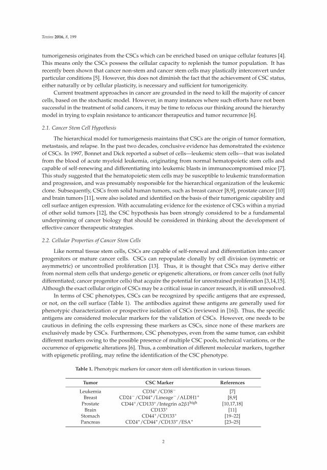
In terms of CSC phenotypes, CSCs can be recognized by specific antigens that are expressed,or not, on the cell surface (Table 1). The antibodies against these antigens are generally used forphenotypic characterization or prospective isolation of CSCs (reviewed in [16]). Thus, the specificantigens are considered molecular markers for the validation of CSCs. However, one needs to becautious in defining the cells expressing these markers as CSCs, since none of these markers areexclusively made by CSCs. Furthermore, CSC phenotypes, even from the same tumor, can exhibitdifferent markers owing to the possible presence of multiple CSC pools, technical variations, or theoccurrence of epigenetic alterations [6]. Thus, a combination of different molecular markers, togetherwith epigenetic profiling, may refine the identification of the CSC phenotype.
Table 1. Phenotypic markers for cancer stem cell identification in various tissues.
Tumor CSC Marker References
Leukemia CD34+/CD38´ [7]Breast CD24´/CD44+/Lineage´/ALDH1+ [8,9]
Prostate CD44+/CD133+/Integrin α2β1high [10,17,18]Brain CD133+ [11]
Stomach CD44+/CD133+ [19–22]Pancreas CD24+/CD44+/CD133+/ESA+ [23–25]
2
Toxins 2016, 8, 199
Table 1. Cont.
Tumor CSC Marker References
Colon CD44+/CD133+/ALDH1+ [26,27]Ovary CD133+/ALDH1+ [28,29]Lung CD133+ [30–32]Liver CD90+ [33–35]
CSC: cancer stem cell; CD24: heat stable antigen; CD34: hematopoietic progenitor cell antigen; CD38: cyclicADP ribose hydrolase; CD44: hyaluronate receptor; CD90: Thy-1; CD133: prominin-1; ALDH1: aldehydedehydrogenase 1A1; ESA: epithelial surface antigen.
Furthermore, tumor tissues composed of malignant cells, including CSCs, reside in theperivascular niche, which is the milieu that nourishes cancer cells and consists of vasculature,hematopoietic cells, inflammatory cells, and myofibroblasts. Although the niche is not indispensablefor the sustainment of all types of cancer, mutual interactions between CSCs and the microenvironmentare known to profoundly influence cellular properties, such as cell fate and secretory profiles, forcertain types of cancer cells [36–38].
3. Anticancer Phytochemicals Targeting CSCs
CSCs are believed to be responsible for the initial formation and growth of cancer, as well asthe relapse of cancer after treatment, due to the fact that CSCs are more resistant to conventionaltherapeutic treatment than differentiated cancer cells [39]. Thus, it would have important implicationsfor cancer prevention and further therapy if treatment could specifically target CSCs while avoidingdamage to normal stem cells. Based on their unique features [40,41] and dynamics [42] (reviewedin [6]), CSCs can be targeted by several strategies, such as inhibition of self-renewal, induction ofdifferentiation into mature cancer cells, and sensitization to anticancer agents.
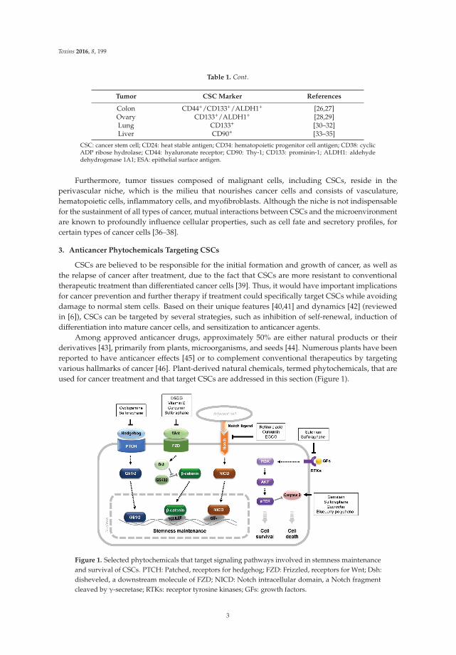
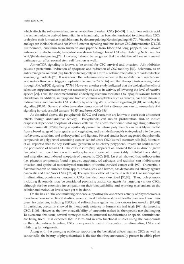
Among approved anticancer drugs, approximately 50% are either natural products or theirderivatives [43], primarily from plants, microorganisms, and seeds [44]. Numerous plants have beenreported to have anticancer effects [45] or to complement conventional therapeutics by targetingvarious hallmarks of cancer [46]. Plant-derived natural chemicals, termed phytochemicals, that areused for cancer treatment and that target CSCs are addressed in this section (Figure 1).
Figure 1. Selected phytochemicals that target signaling pathways involved in stemness maintenanceand survival of CSCs. PTCH: Patched, receptors for hedgehog; FZD: Frizzled, receptors for Wnt; Dsh:disheveled, a downstream molecule of FZD; NICD: Notch intracellular domain, a Notch fragmentcleaved by γ-secretase; RTKs: receptor tyrosine kinases; GFs: growth factors.
3
Toxins 2016, 8, 199
3.1. Anticancer Phytochemicals
A large number of phytochemicals, i.e., chemical compounds produced from plants, includingvegetables, fruits, and grains, have been reported to possess anticancer properties and are promotedfor cancer prevention and treatment [44,45,47–49] (Table 2). Phytochemicals have been shown tointerfere with stabilization of the microtubule structure, thereby inhibiting mitosis and cancer cellpropagation. Vincristine and vinblastine, isolated from the leaves of Madagascar periwinkle, were thefirst phytochemicals to be used clinically in combination with other anticancer agents in lymphomas,leukemias, and breast and lung cancers. Paclitaxel (Taxol), which was originally discovered in the barkof the Pacific yew tree, is one of the most effective and widely used phytochemical compound againstbreast and ovarian cancers [50,51].
Table 2. Examples of plant-derived anticancer phytochemicals.
Function Phytochemicals Plant Derived from
Interference of microtubule stabilizationVincristine, vinblastine Madagascar periwinkle
Paclitaxel Pacific yew tree
Limitation of cell proliferation Epigallocatechin-3-gallate Camellia sinensisCurcumin Turmeric
Disruption of chromatin structureβ-lapachone Lapacho plantcamptothecin Camptotheca
podophyllotoxin Mayapple plant
Another group of phytochemicals, known as polyphenols, has been shown to have free-radicalscavenging activity, working like antioxidants. Epigallocatechin-3-gallate (EGCG), a polyphenolfrom the leaves of Camellia sinensis (processed to green tea), has been used effectively against breastcancer [47]. EGCG was demonstrated to limit cancer cell proliferation by reducing DNA methylationthrough the inhibition of DNA methyltransferase together with reactivation of the silenced tumorsuppressor genes. Curcumin (diferuloylmethane), a polyphenol isolated from the rhizome of theturmeric plant, has also shown therapeutic efficacy on numerous disorders, including cancer [52].Curcumin is reported to inhibit NF-κB signaling that triggers the intracellular inflammatory responseas well as cell-cycle-associated genes. By arresting the cell cycle and inducing apoptosis through therelaying pathways, curcumin interferes with angiogenesis and reduces tumor invasion.
An additional group of anticancer phytochemicals functions as inhibitors of topoisomerase I orII, which are the nuclear enzymes that control DNA supercoiling, eliminate tangles in the chromatinstructure, and allow DNA to be replicated and transcribed. Thus, topoisomerase inhibitors can actas anticancer agents by inducing a delay of the cell cycle, followed by cell death [44]. β-Lapachonefrom the bark of the lapacho plant [53], camptothecin from the bark/stem of Camptotheca (the Chinesehappy tree), and podophyllotoxin from the root of the Mayapple plant are examples of phytochemicalsinhibiting topoisomerases in cancer cells [54,55].
3.2. Phytochemicals Targeting CSCs
Several phytochemicals have been reported to intervene in signaling pathways critical forstemness maintenance of CSCs or to modulate the CSC phenotype [56,57]. The hedgehog,Wnt/β-catenin, and Notch-mediated signaling pathways play important roles in CSC self-renewaland differentiation [58]. Considering that tumorigenesis might be derived from CSCs in whichthese pathways are aberrantly regulated, the signaling molecules in these pathways may be ofparticular interest for targeting CSCs [59]. Multiple studies have demonstrated that cancer cell growthcan be suppressed by specific inhibitors of these pathways [60,61]. Specific phytochemicals havebeen reported to influence these signaling pathways. Cyclopamine, initially found in the corn lily(Veratrum californicum), targets hedgehog signaling [62–65]. EGCG inhibits Wnt/β-catenin signaling,
4
Toxins 2016, 8, 199
which affects the self-renewal and invasive abilities of certain CSCs [66–68]. In addition, retinoic acid,the active molecule derived from vitamin A in animals, has been demonstrated to differentiate CSCsor deplete their formation in glioblastoma by downregulating Notch signaling [69,70]. Vitamin D or itsanalogs can inhibit Notch and/or Wnt/β-catenin signaling and thus induce CSC differentiation [71,72].Furthermore, curcumin from turmeric and piperine from black and long peppers, well-knownanticancer phytochemicals, have also been shown to target breast CSCs by inhibiting Notch and/orWnt/β-catenin signaling [73]. However, it should be recognized that the inhibition of these self-renewalpathways can affect normal stem cell function as well.
Akt/mTOR signaling is known to be critical for CSC survival and invasion. Akt inhibitioncauses a preferential induction of apoptosis and reduction of CSC motility [57]. Selenium, as ananticarcinogenic nutrient [74], functions biologically in a form of selenoproteins that are oxidoreductasescavenging oxidants [75]. It was shown that selenium involvement in the modulation of arachidonicacid metabolism could trigger apoptosis of leukemia CSCs [76], and that the apoptosis was regulatedthrough Akt/mTOR signaling [77,78]. However, another study indicated that the biological benefits ofselenium supplementation may not necessarily be due to its activity of lowering the level of reactivespecies [79]. Thus, the exact mechanisms underlying selenium-mediated CSC apoptosis awaits furtherelucidation. In addition, sulforaphane from cruciferous vegetables, such as broccoli, has been shown toreduce breast and pancreatic CSC viability by affecting Wnt/β-catenin signaling [80,81] or hedgehogsignaling [82,83]. Several studies have also demonstrated that sulforaphane can downregulate Aktsignaling in various solid cancers [84,85] and breast CSCs [86].
As described above, the polyphenols EGCG and curcumin are known to exert their anticancereffects though antioxidative activity. Polyphenols can inhibit proliferation and/or inducecaspase-3-dependent apoptosis of cancer cells via the above-mentioned vital signaling pathwaysor their cross-talk [87,88]. Being ubiquitously present in nature, polyphenols can be richly extractedfrom a broad range of fruits, grains, and vegetables, and include flavonoids (categorized into flavones,isoflavones, catechins, and anthocyanins) and lignans. Several studies have suggested that phenoliccompounds or polyphenol-containing extracts can influence CSCs as well as cancer cells [89]. Montaleset al. reported that the soy isoflavone genistein or blueberry polyphenol treatment could reducethe population of breast CSC-like cells in vitro [90]. Appari et al. showed that a mixture of greentea catechins in combination with sulforaphane and quercetin remarkably inhibited the viabilityand migration and induced apoptosis of pancreatic CSCs [91]. Lu et al. showed that anthocyanins(i.e., phenolic compounds found in grapes, eggplants, red cabbages, and radishes) can inhibit cancerinvasion and epithelial-mesenchymal transition of uterine cervical cancer cells [92]. Quercetin, aflavonol that can be enriched from apples, onions, teas, and berries, has demonstrated efficacy againstpancreatic and head/neck CSCs [93,94]. The synergistic effect of quercetin with EGCG or sulforaphanein eliminating prostate or pancreatic CSCs has also been described [95,96]. Thus, polyphenols,including flavonoids, may be considered promising anticancer agents for targeting various CSCs,although further extensive investigation on their bioavailability and working mechanisms at thecellular and molecular levels have yet to be done.
On the basis of the accumulating evidence supporting the anticancer activity of phytochemicals,there have been some clinical studies. Recent clinical trials have shown the effectiveness of curcumin,green tea catechins, including EGCG, and sulforaphane against various cancers (reviewed in [97,98]).In particular, curcumin showed its therapeutic potency in human clinical trials [99] via targetingCSCs [100]. However, the low bioavailability of curcumin makes its therapeutic use challenging.To overcome this issue, several strategies such as structural modifications or special formulationsare being tried. It is expected that in vitro and in vivo functional studies using the compoundsor their derivatives targeting CSCs may provide useful information on eliminating CSCs andinhibiting tumorigenesis.
Along with the emerging evidence supporting the beneficial effects against CSCs as well ascancer cells, the beauty of phytochemicals is the fact that they are naturally present in edible plant
5
Toxins 2016, 8, 199
materials. This warrants the assurance of safety for ingestion. In addition, certain phytochemicalssensitize CSCs to conventional chemotherapeutic agents by interfering with the key signaling pathwaysfor cell survival, stemness maintenance of CSCs, or both. Thus, synergistic effects are expectedwhen the CSC-targeting phytochemicals and chemotherapeutic drugs are used combinatorially [98].However, rigorous examinations are required to test the potential adverse effects, such as thecounteraction of phytochemicals against chemotherapeutic drugs and the additive toxicity ofphytochemicals [101].
4. Summary and Conclusions
Tumors comprise of phenotypically and functionally heterogeneous cells. Based on this feature,two models have been established regarding tumorigenesis: the stochastic model and the hierarchicalmodel. The latter model postulates a hierarchical organization of diverse populations of cells andpremises the presence of CSCs that account for the sustainment of tumorigenesis. Thus, control ofCSCs may be a necessary first step in an effective strategy for cancer treatment. Dietary phytochemicalscan exert influence at all stages of cancer development. Since some of the phytochemicals are alreadyknown to affect CSC viability and fate, extensive and intensive studies of these compounds shouldprovide insight into their pharmaceutical efficacy for cancer prevention and therapy.
Acknowledgments: This study was supported by the Basic Science Research Program through the NationalResearch Foundation of Korea funded by the Ministry of Education (Grant No. NRF-2013R1A1A2013362).
Conflicts of Interest: The authors declare no conflict of interest.
References
1. Heppner, G.H.; Miller, B.E. Tumor heterogeneity: Biological implications and therapeutic consequences.Cancer Metastasis Rev. 1983, 2, 5–23. [CrossRef] [PubMed]
2. Dick, J.E. Looking ahead in cancer stem cell research. Nat. Biotechnol. 2009, 27, 44–46. [CrossRef] [PubMed]3. Reya, T.; Morrison, S.J.; Clarke, M.F.; Weissman, I.L. Stem cells, cancer, and cancer stem cells. Nature 2001,
414, 105–111. [CrossRef] [PubMed]4. Dick, J.E. Stem cell concepts renew cancer research. Blood 2008, 112, 4793–4807. [CrossRef] [PubMed]5. Cabrera, M.C.; Hollingsworth, R.E.; Hurt, E.M. Cancer stem cell plasticity and tumor hierarchy. World J.
Stem Cells 2015, 7, 27–36. [CrossRef] [PubMed]6. Visvader, J.E.; Lindeman, G.J. Cancer stem cells: Current status and evolving complexities. Cell Stem Cell
2012, 10, 717–728. [CrossRef] [PubMed]7. Bonnet, D.; Dick, J.E. Human acute myeloid leukemia is organized as a hierarchy that originates from a
primitive hematopoietic cell. Nat. Med. 1997, 3, 730–737. [CrossRef] [PubMed]8. Al-Hajj, M.; Wicha, M.S.; Benito-Hernandez, A.; Morrison, S.J.; Clarke, M.F. Prospective identification of
tumorigenic breast cancer cells. Proc. Natl. Acad. Sci. USA 2003, 100, 3983–3988. [CrossRef] [PubMed]9. Ginestier, C.; Hur, M.H.; Charafe-Jauffret, E.; Monville, F.; Dutcher, J.; Brown, M.; Jacquemier, J.; Viens, P.;
Kleer, C.G.; Liu, S.; et al. ALDH1 is a marker of normal and malignant human mammary stem cells and apredictor of poor clinical outcome. Cell Stem Cell 2007, 1, 555–567. [CrossRef] [PubMed]
10. Collins, A.T.; Berry, P.A.; Hyde, C.; Stower, M.J.; Maitland, N.J. Prospective identification of tumorigenicprostate cancer stem cells. Cancer Res. 2005, 65, 10946–10951. [CrossRef] [PubMed]
11. Singh, S.K.; Hawkins, C.; Clarke, I.D.; Squire, J.A.; Bayani, J.; Hide, T.; Henkelman, R.M.; Cusimano, M.D.;Dirks, P.B. Identification of human brain tumour initiating cells. Nature 2004, 432, 396–401. [CrossRef][PubMed]
12. Visvader, J.E.; Lindeman, G.J. Cancer stem cells in solid tumours: Accumulating evidence and unresolvedquestions. Nat. Rev. Cancer 2008, 8, 755–768. [CrossRef] [PubMed]
13. Li, Y.; Wicha, M.S.; Schwartz, S.J.; Sun, D. Implications of cancer stem cell theory for cancer chemopreventionby natural dietary compounds. J. Nutr. Biochem. 2011, 22, 799–806. [CrossRef] [PubMed]
14. Takebe, N.; Ivy, S.P. Controversies in cancer stem cells: Targeting embryonic signaling pathways.Clin. Cancer Res. 2010, 16, 3106–3112. [CrossRef] [PubMed]
6
Toxins 2016, 8, 199
15. Eaves, C.J. Cancer stem cells: Here, there, everywhere? Nature 2008, 456, 581–582. [CrossRef] [PubMed]16. Kreso, A.; Dick, J.E. Evolution of the cancer stem cell model. Cell Stem Cell 2014, 14, 275–291. [CrossRef]
[PubMed]17. Vander Griend, D.J.; Karthaus, W.L.; Dalrymple, S.; Meeker, A.; DeMarzo, A.M.; Isaacs, J.T. The role of CD133
in normal human prostate stem cells and malignant cancer-initiating cells. Cancer Res. 2008, 68, 9703–9711.[CrossRef] [PubMed]
18. Williamson, S.C.; Hepburn, A.C.; Wilson, L.; Coffey, K.; Ryan-Munden, C.A.; Pal, D.; Leung, H.Y.;Robson, C.N.; Heer, R. Human α2 β1
HI CD133+VE epithelial prostate stem cells express low levels ofactive androgen receptor. PLoS One 2012, 7, e48944. [CrossRef] [PubMed]
19. Wang, T.; Ong, C.W.; Shi, J.; Srivastava, S.; Yan, B.; Cheng, C.L.; Yong, W.P.; Chan, S.L.; Yeoh, K.G.;Iacopetta, B.; et al. Sequential expression of putative stem cell markers in gastric carcinogenesis. Br. J. Cancer2011, 105, 658–665. [CrossRef] [PubMed]
20. Zhang, C.; Li, C.; He, F.; Cai, Y.; Yang, H. Identification of CD44+CD24+ gastric cancer stem cells. J. CancerRes. Clin. Oncol. 2011, 137, 1679–1686. [CrossRef] [PubMed]
21. Chen, W.; Zhang, X.; Chu, C.; Cheung, W.L.; Ng, L.; Lam, S.; Chow, A.; Lau, T.; Chen, M.; Li, Y.; et al.Identification of CD44+ cancer stem cells in human gastric cancer. Hepatogastroenterology 2013, 60, 949–954.[PubMed]
22. Chen, S.; Hou, J.H.; Feng, X.Y.; Zhang, X.S.; Zhou, Z.W.; Yun, J.P.; Chen, Y.B.; Cai, M.Y. Clinicopathologicsignificance of putative stem cell marker, CD44 and CD133, in human gastric carcinoma. J. Surg. Oncol. 2013,107, 799–806. [CrossRef] [PubMed]
23. Hermann, P.C.; Huber, S.L.; Herrler, T.; Aicher, A.; Ellwart, J.W.; Guba, M.; Bruns, C.J.; Heeschen, C. Distinctpopulations of cancer stem cells determine tumor growth and metastatic activity in human pancreatic cancer.Cell Stem Cell 2007, 1, 313–323. [CrossRef] [PubMed]
24. Fitzgerald, T.L.; McCubrey, J.A. Pancreatic cancer stem cells: Association with cell surface markers, prognosis,resistance, metastasis and treatment. Adv. Biol. Regul. 2014, 56, 45–50. [CrossRef] [PubMed]
25. Li, C.; Heidt, D.G.; Dalerba, P.; Burant, C.F.; Zhang, L.; Adsay, V.; Wicha, M.; Clarke, M.F.; Simeone, D.M.Identification of pancreatic cancer stem cells. Cancer. Res. 2007, 67, 1030–1037. [CrossRef] [PubMed]
26. Ricci-Vitiani, L.; Lombardi, D.G.; Pilozzi, E.; Biffoni, M.; Todaro, M.; Peschle, C.; De Maria, R. Identificationand expansion of human colon-cancer-initiating cells. Nature 2007, 445, 111–115. [CrossRef] [PubMed]
27. O’Brien, C.A.; Pollett, A.; Gallinger, S.; Dick, J.E. A human colon cancer cell capable of initiating tumourgrowth in immunodeficient mice. Nature 2007, 445, 106–110. [CrossRef] [PubMed]
28. Silva, I.A.; Bai, S.; McLean, K.; Yang, K.; Griffith, K.; Thomas, D.; Ginestier, C.; Johnston, C.; Kueck, A.;Reynolds, R.K.; et al. Aldehyde dehydrogenase in combination with CD133 defines angiogenic ovariancancer stem cells that portend poor patient survival. Cancer Res. 2011, 71, 3991–4001. [CrossRef] [PubMed]
29. Kryczek, I.; Liu, S.; Roh, M.; Vatan, L.; Szeliga, W.; Wei, S.; Banerjee, M.; Mao, Y.; Kotarski, J.; Wicha, M.S.;et al. Expression of aldehyde dehydrogenase and CD133 defines ovarian cancer stem cells. Int. J. Cancer2012, 130, 29–39. [CrossRef] [PubMed]
30. Eramo, A.; Lotti, F.; Sette, G.; Pilozzi, E.; Biffoni, M.; Di Virgilio, A.; Conticello, C.; Ruco, L.; Peschle, C.; DeMaria, R. Identification and expansion of the tumorigenic lung cancer stem cell population. Cell Death Differ.2008, 15, 504–514. [CrossRef] [PubMed]
31. Bertolini, G.; Roz, L.; Perego, P.; Tortoreto, M.; Fontanella, E.; Gatti, L.; Pratesi, G.; Fabbri, A.; Andriani, F.;Tinelli, S.; et al. Highly tumorigenic lung cancer CD133+ cells display stem-like features and are spared bycisplatin treatment. Proc. Natl. Acad. Sci. USA 2009, 106, 16281–16286. [CrossRef] [PubMed]
32. Tirino, V.; Camerlingo, R.; Franco, R.; Malanga, D.; La Rocca, A.; Viglietto, G.; Rocco, G.; Pirozzi, G. The roleof CD133 in the identification and characterisation of tumour-initiating cells in non-small-cell lung cancer.Eur. J. Cardiothorac. Surg. 2009, 36, 446–453. [CrossRef] [PubMed]
33. Yang, Z.F.; Ho, D.W.; Ng, M.N.; Lau, C.K.; Yu, W.C.; Ngai, P.; Chu, P.W.; Lam, C.T.; Poon, R.T.; Fan, S.T.Significance of CD90+ cancer stem cells in human liver cancer. Cancer Cell 2008, 13, 153–166. [CrossRef][PubMed]
34. Yang, Z.F.; Ngai, P.; Ho, D.W.; Yu, W.C.; Ng, M.N.; Lau, C.K.; Li, M.L.; Tam, K.H.; Lam, C.T.; Poon, R.T.; et al.Identification of local and circulating cancer stem cells in human liver cancer. Hepatology 2008, 47, 919–928.[CrossRef] [PubMed]
7
Toxins 2016, 8, 199
35. Tomuleasa, C.; Soritau, O.; Rus-Ciuca, D.; Pop, T.; Todea, D.; Mosteanu, O.; Pintea, B.; Foris, V.; Susman, S.;Kacso, G.; et al. Isolation and characterization of hepatic cancer cells with stem-like properties fromhepatocellular carcinoma. J. Gastrointestin. Liver Dis. 2010, 19, 61–67. [PubMed]
36. Gilbertson, R.J.; Rich, J.N. Making a tumour’s bed: Glioblastoma stem cells and the vascular niche.Nat. Rev. Cancer 2007, 7, 733–736. [CrossRef] [PubMed]
37. Kelly, P.N.; Dakic, A.; Adams, J.M.; Nutt, S.L.; Strasser, A. Tumor growth need not be driven by rare cancerstem cells. Science 2007, 317, 337. [CrossRef] [PubMed]
38. Vermeulen, L.; De Sousa, E.M.F.; van der Heijden, M.; Cameron, K.; de Jong, J.H.; Borovski, T.; Tuynman, J.B.;Todaro, M.; Merz, C.; Rodermond, H.; et al. Wnt activity defines colon cancer stem cells and is regulated bythe microenvironment. Nat. Cell. Biol. 2010, 12, 468–476. [CrossRef] [PubMed]
39. Creighton, C.J.; Li, X.; Landis, M.; Dixon, J.M.; Neumeister, V.M.; Sjolund, A.; Rimm, D.L.; Wong, H.;Rodriguez, A.; Herschkowitz, J.I.; et al. Residual breast cancers after conventional therapy displaymesenchymal as well as tumor-initiating features. Proc. Natl. Acad. Sci. USA 2009, 106, 13820–13825.[CrossRef] [PubMed]
40. Saito, Y.; Uchida, N.; Tanaka, S.; Suzuki, N.; Tomizawa-Murasawa, M.; Sone, A.; Najima, Y.; Takagi, S.;Aoki, Y.; Wake, A.; et al. Induction of cell cycle entry eliminates human leukemia stem cells in a mousemodel of AML. Nat. Biotechnol. 2010, 28, 275–280. [PubMed]
41. Lathia, J.D.; Hitomi, M.; Gallagher, J.; Gadani, S.P.; Adkins, J.; Vasanji, A.; Liu, L.; Eyler, C.E.; Heddleston, J.M.;Wu, Q.; et al. Distribution of CD133 reveals glioma stem cells self-renew through symmetric and asymmetriccell divisions. Cell Death Dis. 2011, 2, e200. [CrossRef] [PubMed]
42. Fornari, C.; Beccuti, M.; Lanzardo, S.; Conti, L.; Balbo, G.; Cavallo, F.; Calogero, R.A.; Cordero, F. Amathematical-biological joint effort to investigate the tumor-initiating ability of Cancer Stem Cells. PLoS ONE2014, 9, e106193. [CrossRef] [PubMed]
43. Newman, D.J.; Cragg, G.M. Natural products as sources of new drugs over the 30 years from 1981 to 2010.J. Nat. Prod. 2012, 75, 311–335. [CrossRef] [PubMed]
44. Nobili, S.; Lippi, D.; Witort, E.; Donnini, M.; Bausi, L.; Mini, E.; Capaccioli, S. Natural compounds for cancertreatment and prevention. Pharmacol. Res. 2009, 59, 365–378. [CrossRef] [PubMed]
45. Graham, J.G.; Quinn, M.L.; Fabricant, D.S.; Farnsworth, N.R. Plants used against cancer - an extension of thework of Jonathan Hartwell. J. Ethnopharmacol. 2000, 73, 347–377. [CrossRef]
46. Bishayee, A.; Block, K. A broad-spectrum integrative design for cancer prevention and therapy: The challengeahead. Semin. Cancer Biol. 2015, 35, S1–S4. [CrossRef] [PubMed]
47. Amin, A.; Gali-Muhtasib, H.; Ocker, M.; Schneider-Stock, R. Overview of major classes of plant-derivedanticancer drugs. Int. J. Biomed. Sci. 2009, 5, 1–11. [PubMed]
48. Diederich, M.; Cerella, C. Non-canonical programmed cell death mechanisms triggered by naturalcompounds. Semin. Cancer Biol. 2016. [CrossRef] [PubMed]
49. Shanmugam, M.K.; Lee, J.H.; Chai, E.Z.; Kanchi, M.M.; Kar, S.; Arfuso, F.; Dharmarajan, A.; Kumar, A.P.;Ramar, P.S.; Looi, C.Y.; et al. Cancer prevention and therapy through the modulation of transcription factorsby bioactive natural compounds. Semin. Cancer Biol. 2016. [CrossRef] [PubMed]
50. Wani, M.C.; Taylor, H.L.; Wall, M.E.; Coggon, P.; McPhail, A.T. Plant antitumor agents. VI. The isolation andstructure of taxol, a novel antileukemic and antitumor agent from Taxus brevifolia. J. Am. Chem. Soc. 1971,93, 2325–2327. [CrossRef] [PubMed]
51. Schiff, P.B.; Fant, J.; Horwitz, S.B. Promotion of microtubule assembly in vitro by taxol. Nature 1979, 277,665–667. [CrossRef] [PubMed]
52. Sa, G.; Das, T. Anti cancer effects of curcumin: Cycle of life and death. Cell Div. 2008, 3. [CrossRef] [PubMed]53. Li, Y.Z.; Li, C.J.; Pinto, A.V.; Pardee, A.B. Release of mitochondrial cytochrome C in both apoptosis and
necrosis induced by beta-lapachone in human carcinoma cells. Mol. Med. 1999, 5, 232–239. [PubMed]54. Pommier, Y. Topoisomerase I inhibitors: Camptothecins and beyond. Nat. Rev. Cancer 2006, 6, 789–802.
[CrossRef] [PubMed]55. Hartmann, J.T.; Lipp, H.P. Camptothecin and podophyllotoxin derivatives: Inhibitors of topoisomerase I
and II - mechanisms of action, pharmacokinetics and toxicity profile. Drug Saf. 2006, 29, 209–230. [CrossRef][PubMed]
56. Kim, D.H.; Surh, Y.J. Chemopreventive and therapeutic ootential of phytochemicals targeting cancer stemcells. Curr. Pharmacol. Rep. 2015, 1, 302–311. [CrossRef]
8
Toxins 2016, 8, 199
57. Dandawate, P.; Padhye, S.; Ahmad, A.; Sarkar, F.H. Novel strategies targeting cancer stem cells throughphytochemicals and their analogs. Drug Deliv. Transl. Res. 2013, 3, 165–182. [CrossRef] [PubMed]
58. Kim, Y.S.; Farrar, W.; Colburn, N.H.; Milner, J.A. Cancer stem cells: Potential target for bioactive foodcomponents. J. Nutr. Biochem. 2012, 23, 691–698. [CrossRef] [PubMed]
59. Liu, S.; Dontu, G.; Wicha, M.S. Mammary stem cells, self-renewal pathways, and carcinogenesis.Breast. Cancer Res. 2005, 7, 86–95. [CrossRef] [PubMed]
60. Kubo, M.; Nakamura, M.; Tasaki, A.; Yamanaka, N.; Nakashima, H.; Nomura, M.; Kuroki, S.; Katano, M.Hedgehog signaling pathway is a new therapeutic target for patients with breast cancer. Cancer Res. 2004, 64,6071–6074. [CrossRef] [PubMed]
61. Romer, J.T.; Kimura, H.; Magdaleno, S.; Sasai, K.; Fuller, C.; Baines, H.; Connelly, M.; Stewart, C.F.;Gould, S.; Rubin, L.L.; et al. Suppression of the Shh pathway using a small molecule inhibitor eliminatesmedulloblastoma in Ptc1+/-p53-/- mice. Cancer Cell 2004, 6, 229–240. [CrossRef] [PubMed]
62. Berman, D.M.; Karhadkar, S.S.; Hallahan, A.R.; Pritchard, J.I.; Eberhart, C.G.; Watkins, D.N.; Chen, J.K.;Cooper, M.K.; Taipale, J.; Olson, J.M.; et al. Medulloblastoma growth inhibition by hedgehog pathwayblockade. Science 2002, 297, 1559–1561. [CrossRef] [PubMed]
63. Liu, S.; Dontu, G.; Mantle, I.D.; Patel, S.; Ahn, N.S.; Jackson, K.W.; Suri, P.; Wicha, M.S. Hedgehog signalingand Bmi-1 regulate self-renewal of normal and malignant human mammary stem cells. Cancer Res. 2006, 66,6063–6071. [CrossRef] [PubMed]
64. Feldmann, G.; Dhara, S.; Fendrich, V.; Bedja, D.; Beaty, R.; Mullendore, M.; Karikari, C.; Alvarez, H.;Iacobuzio-Donahue, C.; Jimeno, A.; et al. Blockade of hedgehog signaling inhibits pancreatic cancer invasionand metastases: A new paradigm for combination therapy in solid cancers. Cancer Res. 2007, 67, 2187–2196.[CrossRef] [PubMed]
65. Peacock, C.D.; Wang, Q.; Gesell, G.S.; Corcoran-Schwartz, I.M.; Jones, E.; Kim, J.; Devereux, W.L.; Rhodes, J.T.;Huff, C.A.; Beachy, P.A.; et al. Hedgehog signaling maintains a tumor stem cell compartment in multiplemyeloma. Proc. Natl. Acad. Sci. USA 2007, 104, 4048–4053. [CrossRef] [PubMed]
66. Lee, S.H.; Nam, H.J.; Kang, H.J.; Kwon, H.W.; Lim, Y.C. Epigallocatechin-3-gallate attenuates head and neckcancer stem cell traits through suppression of Notch pathway. Eur. J. Cancer 2013, 49, 3210–3218. [CrossRef][PubMed]
67. Mineva, N.D.; Paulson, K.E.; Naber, S.P.; Yee, A.S.; Sonenshein, G.E. Epigallocatechin-3-gallate inhibitsstem-like inflammatory breast cancer cells. PLoS ONE 2013, 8, e73464. [CrossRef] [PubMed]
68. Lin, C.H.; Shen, Y.A.; Hung, P.H.; Yu, Y.B.; Chen, Y.J. Epigallocathechin gallate, polyphenol present in greentea, inhibits stem-like characteristics and epithelial-mesenchymal transition in nasopharyngeal cancer celllines. BMC Complement. Altern. Med. 2012, 12. [CrossRef] [PubMed]
69. Clarke, N.; Germain, P.; Altucci, L.; Gronemeyer, H. Retinoids: Potential in cancer prevention and therapy.Expert Rev. Mol. Med. 2004, 6, 1–23. [CrossRef] [PubMed]
70. Ying, M.; Wang, S.; Sang, Y.; Sun, P.; Lal, B.; Goodwin, C.R.; Guerrero-Cazares, H.; Quinones-Hinojosa, A.;Laterra, J.; Xia, S. Regulation of glioblastoma stem cells by retinoic acid: Role for Notch pathway inhibition.Oncogene 2011, 30, 3454–3467. [CrossRef] [PubMed]
71. Palmer, H.G.; Gonzalez-Sancho, J.M.; Espada, J.; Berciano, M.T.; Puig, I.; Baulida, J.; Quintanilla, M.; Cano, A.;de Herreros, A.G.; Lafarga, M.; et al. Vitamin D3 promotes the differentiation of colon carcinoma cells bythe induction of E-cadherin and the inhibition of beta-catenin signaling. J. Cell Biol. 2001, 154, 369–387.[CrossRef] [PubMed]
72. Garcia, J.J.; Lopez-Pingarron, L.; Almeida-Souza, P.; Tres, A.; Escudero, P.; Garcia-Gil, F.A.; Tan, D.X.;Reiter, R.J.; Ramirez, J.M.; Bernal-Perez, M. Protective effects of melatonin in reducing oxidative stress andin preserving the fluidity of biological membranes: A review. J. Pineal Res. 2014, 56, 225–237. [CrossRef][PubMed]
73. Kakarala, M.; Brenner, D.E.; Korkaya, H.; Cheng, C.; Tazi, K.; Ginestier, C.; Liu, S.; Dontu, G.; Wicha, M.S.Targeting breast stem cells with the cancer preventive compounds curcumin and piperine. Breast CancerRes. Treat. 2010, 122, 777–785. [CrossRef] [PubMed]
74. Zeng, H.; Combs, G.F., Jr. Selenium as an anticancer nutrient: Roles in cell proliferation and tumor cellinvasion. J. Nutr. Biochem. 2008, 19, 1–7. [CrossRef] [PubMed]
75. Hatfield, D.L.; Tsuji, P.A.; Carlson, B.A.; Gladyshev, V.N. Selenium and selenocysteine: Roles in cancer,health, and development. Trends Biochem. Sci. 2014, 39, 112–120. [CrossRef] [PubMed]
9
Toxins 2016, 8, 199
76. Gandhi, U.H.; Kaushal, N.; Hegde, S.; Finch, E.R.; Kudva, A.K.; Kennett, M.J.; Jordan, C.T.; Paulson, R.F.;Prabhu, K.S. Selenium suppresses leukemia through the action of endogenous eicosanoids. Cancer Res. 2014,74, 3890–3901. [CrossRef] [PubMed]
77. Sanmartin, C.; Plano, D.; Sharma, A.K.; Palop, J.A. Selenium compounds, apoptosis and other types of celldeath: An overview for cancer therapy. Int. J. Mol. Sci. 2012, 13, 9649–9672. [CrossRef] [PubMed]
78. Kim, I.; He, Y.Y. Targeting the AMP-Activated Protein Kinase for Cancer Prevention and Therapy. Front. Oncol.2013, 3. [CrossRef] [PubMed]
79. Li, F.; Lutz, P.B.; Pepelyayeva, Y.; Arner, E.S.; Bayse, C.A.; Rozovsky, S. Redox active motifs in selenoproteins.Proc. Natl. Acad. Sci. USA 2014, 111, 6976–6981. [CrossRef] [PubMed]
80. Li, Y.; Zhang, T.; Korkaya, H.; Liu, S.; Lee, H.F.; Newman, B.; Yu, Y.; Clouthier, S.G.; Schwartz, S.J.; Wicha, M.S.;et al. Sulforaphane, a dietary component of broccoli/broccoli sprouts, inhibits breast cancer stem cells.Clin. Cancer Res. 2010, 16, 2580–2590. [CrossRef] [PubMed]
81. Kallifatidis, G.; Rausch, V.; Baumann, B.; Apel, A.; Beckermann, B.M.; Groth, A.; Mattern, J.; Li, Z.; Kolb, A.;Moldenhauer, G.; et al. Sulforaphane targets pancreatic tumour-initiating cells by NF-kappaB-inducedantiapoptotic signalling. Gut 2009, 58, 949–963. [CrossRef] [PubMed]
82. Filipe, P.; Morliere, P.; Silva, J.N.; Maziere, J.C.; Patterson, L.K.; Freitas, J.P.; Santus, R. Plasma lipoproteinsas mediators of the oxidative stress induced by UV light in human skin: A review of biochemical andbiophysical studies on mechanisms of apolipoprotein alteration, lipid peroxidation, and associated skin cellresponses. Oxidative Med. Cell. Longevity 2013, 2013, 285825. [CrossRef] [PubMed]
83. Rodova, M.; Fu, J.; Watkins, D.N.; Srivastava, R.K.; Shankar, S. Sonic hedgehog signaling inhibition providesopportunities for targeted therapy by sulforaphane in regulating pancreatic cancer stem cell self-renewal.PLoS ONE 2012, 7, e46083. [CrossRef] [PubMed]
84. Chaudhuri, D.; Orsulic, S.; Ashok, B.T. Antiproliferative activity of sulforaphane in Akt-overexpressingovarian cancer cells. Mol. Cancer Ther. 2007, 6, 334–345. [CrossRef] [PubMed]
85. Shankar, S.; Ganapathy, S.; Srivastava, R.K. Sulforaphane enhances the therapeutic potential of TRAILin prostate cancer orthotopic model through regulation of apoptosis, metastasis, and angiogenesis.Clin. Cancer Res. 2008, 14, 6855–6866. [CrossRef] [PubMed]
86. Korkaya, H.; Paulson, A.; Charafe-Jauffret, E.; Ginestier, C.; Brown, M.; Dutcher, J.; Clouthier, S.G.; Wicha, M.S.Regulation of mammary stem/progenitor cells by PTEN/Akt/beta-catenin signaling. PLoS Biol. 2009, 7,e1000121. [CrossRef] [PubMed]
87. Fresco, P.; Borges, F.; Diniz, C.; Marques, M.P. New insights on the anticancer properties of dietarypolyphenols. Med. Res. Rev. 2006, 26, 747–766. [CrossRef] [PubMed]
88. Ramos, S. Cancer chemoprevention and chemotherapy: Dietary polyphenols and signalling pathways.Mol. Nutr. Food Res. 2008, 52, 507–526. [CrossRef] [PubMed]
89. Sak, K.; Everaus, H. Role of Flavonoids in Future Anticancer Therapy by Eliminating the Cancer Stem Cells.Curr. Stem Cell Res. Ther. 2015, 10, 271–282. [CrossRef] [PubMed]
90. Montales, M.T.; Rahal, O.M.; Kang, J.; Rogers, T.J.; Prior, R.L.; Wu, X.; Simmen, R.C. Repressionof mammosphere formation of human breast cancer cells by soy isoflavone genistein and blueberrypolyphenolic acids suggests diet-mediated targeting of cancer stem-like/progenitor cells. Carcinogenesis2012, 33, 652–660. [CrossRef] [PubMed]
91. Appari, M.; Babu, K.R.; Kaczorowski, A.; Gross, W.; Herr, I. Sulforaphane, quercetin and catechinscomplement each other in elimination of advanced pancreatic cancer by miR-let-7 induction and K-rasinhibition. Int. J. Oncol. 2014, 45, 1391–1400. [CrossRef] [PubMed]
92. Lu, J.N.; Lee, W.S.; Yun, J.W.; Kim, M.J.; Kim, H.J.; Kim, D.C.; Jeong, J.H.; Choi, Y.H.; Kim, G.S.; Ryu, C.H.;et al. Anthocyanins from Vitis coignetiae Pulliat Inhibit Cancer Invasion and Epithelial-MesenchymalTransition, but These Effects Can Be Attenuated by Tumor Necrosis Factor in Human Uterine CervicalCancer HeLa Cells. Evid. Based Complement. Altern. Med. 2013, 2013, 503043. [CrossRef] [PubMed]
93. Chang, W.W.; Hu, F.W.; Yu, C.C.; Wang, H.H.; Feng, H.P.; Lan, C.; Tsai, L.L.; Chang, Y.C. Quercetin inelimination of tumor initiating stem-like and mesenchymal transformation property in head and neck cancer.Head Neck 2013, 35, 413–419. [CrossRef] [PubMed]
94. Zhou, W.; Kallifatidis, G.; Baumann, B.; Rausch, V.; Mattern, J.; Gladkich, J.; Giese, N.; Moldenhauer, G.;Wirth, T.; Buchler, M.W.; et al. Dietary polyphenol quercetin targets pancreatic cancer stem cells. Int. J. Oncol.2010, 37, 551–561. [PubMed]
10
Toxins 2016, 8, 199
95. Srivastava, R.K.; Tang, S.N.; Zhu, W.; Meeker, D.; Shankar, S. Sulforaphane synergizes with quercetin toinhibit self-renewal capacity of pancreatic cancer stem cells. Front. Biosci. 2011, 3, 515–528. [CrossRef]
96. Tang, S.N.; Singh, C.; Nall, D.; Meeker, D.; Shankar, S.; Srivastava, R.K. The dietary bioflavonoid quercetinsynergizes with epigallocathechin gallate (EGCG) to inhibit prostate cancer stem cell characteristics, invasion,migration and epithelial-mesenchymal transition. J. Mol. Signal. 2010, 5, 14. [CrossRef] [PubMed]
97. Hosseini, A.; Ghorbani, A. Cancer therapy with phytochemicals: Evidence from clinical studies.Avicenna J. Phytomed. 2015, 5, 84–97. [PubMed]
98. Scarpa, E.S.; Ninfali, P. Phytochemicals as Innovative Therapeutic Tools against Cancer Stem Cells. Int. J.Mol. Sci. 2015, 16, 15727–15742. [CrossRef] [PubMed]
99. Hatcher, H.; Planalp, R.; Cho, J.; Torti, F.M.; Torti, S.V. Curcumin: From ancient medicine to current clinicaltrials. Cell. Mol. Life Sci. 2008, 65, 1631–1652. [CrossRef] [PubMed]
100. Li, Y.; Zhang, T. Targeting cancer stem cells by curcumin and clinical applications. Cancer Lett. 2014, 346,197–205. [CrossRef] [PubMed]
101. Sparreboom, A.; Cox, M.C.; Acharya, M.R.; Figg, W.D. Herbal remedies in the United States: Potentialadverse interactions with anticancer agents. J. Clin. Oncol. 2004, 22, 2489–2503. [CrossRef] [PubMed]
© 2016 by the authors; licensee MDPI, Basel, Switzerland. This article is an open accessarticle distributed under the terms and conditions of the Creative Commons Attribution(CC-BY) license (http://creativecommons.org/licenses/by/4.0/).
11
toxins
Article
Ovatodiolide Inhibits Breast Cancer Stem/ProgenitorCells through SMURF2-Mediated Downregulationof Hsp27
Kuan-Ta Lu 1, Bing-Yen Wang 2,3,4, Wan-Yu Chi 5, Ju Chang-Chien 5,6, Jiann-Jou Yang 5,7,Hsueh-Te Lee 8, Yew-Min Tzeng 9,10,* and Wen-Wei Chang 5,7,*
1 Department of Anesthesiology, Changhua Christian Hospital, Changhua 500, Taiwan; [email protected] Division of Thoracic Surgery, Department of Surgery, Changhua Christian Hospital, Changhua 500, Taiwan;
[email protected] School of Medicine, Kaohsiung Medical University, Kaohsiung 807, Taiwan4 Institute of Genomics and Bioinformatics, National Chung Hsing University, Taichung 402, Taiwan5 School of Biomedical Sciences, Chung Shan Medical University, Taichung 40201, Taiwan;
[email protected] (W.-Y.C.); [email protected] (J.C.-C.); [email protected] (J.-J.Y.)6 Institute of Microbiology & Immunology, Chung Shan Medical University, Taichung 40201, Taiwan7 Department of Medical Research, Chung Shan Medical University Hospital, Taichung 40201, Taiwan8 Institute of Anatomy and Cell Biology, School of Medicine, National Yang Ming University, Taipei 11221,
Taiwan; [email protected] Center for General Education, National Taitung University, Taitung 95092, Taiwan10 Department of Appiled Chemistry, Chaoyang University of Technology, Taichung 41349, Taiwan* Correspondence: [email protected] (Y.-M.T.); [email protected] (W.-W.C.);
Tel.: +886-89-517-300 (Y.-M.T.); +886-4-2473-0022 (ext.12317) (W.-W.C.)
Academic Editor: Carmela FimognariReceived: 8 March 2016; Accepted: 20 April 2016; Published: 28 April 2016
Abstract: Cancer stem/progenitor cells (CSCs) are a subpopulation of cancer cells involved intumor initiation, resistance to therapy and metastasis. Targeting CSCs has been considered as thekey for successful cancer therapy. Ovatodiolide (Ova) is a macrocyclic diterpenoid compoundisolated from Anisomeles indica (L.) Kuntze with anti-cancer activity. Here we used two humanbreast cancer cell lines (AS-B145 and BT-474) to examine the effect of Ova on breast CSCs. We firstdiscovered that Ova displayed an anti-proliferation activity in these two breast cancer cells. Ova alsoinhibited the self-renewal capability of breast CSCs (BCSCs) which was determined by mammosphereassay. Ova dose-dependently downregulated the expression of stemness genes, octamer-bindingtranscription factor 4 (Oct4) and Nanog, as well as heat shock protein 27 (Hsp27), but upregulatedSMAD ubiquitin regulatory factor 2 (SMURF2) in mammosphere cells derived from AS-B145 orBT-474. Overexpression of Hsp27 or knockdown of SMURF2 in AS-B145 cells diminished thetherapeutic effect of ovatodiolide in the suppression of mammosphere formation. In summary, ourdata reveal that Ova displays an anti-CSC activity through SMURF2-mediated downregulation ofHsp27. Ova could be further developed as an anti-CSC agent in the treatment of breast cancer.
Keywords: ovatodiolide; cancer stem/progenitor cells; Hsp27; SMURF2
1. Introduction
Cancer stem/progenitor cells (CSCs) have been described for decades and these particularcancer cells have been reported to be involved in tumor initiation, resistance to chemotherapy orradiotherapy, and metastasis [1,2]. Breast CSCs were first identified by Al-Hajj et al. with the marker ofCD24-CD44+ [3]. Ginestier et al. later reported that breast cancer cells with high intracellular aldehydedehydrogenase (ALDH) activity also represented the population of BCSCs [4]. In addition to cell
Toxins 2016, 8, 127 12 www.mdpi.com/journal/toxins
Toxins 2016, 8, 127
surface markers or intracellular enzyme activity, BCSCs could be enriched with a cultivation methodof the mammosphere, a clump of cancer cells with stem/progenitor cell properties [5]. The drugscreening results from tumorsphere assay have been reported to be more translatable than those fromthe 2-dimensional adherent condition [6–9]. Targeting CSCs is considered as a key for successfultreatment in cancer [2,10].
Heat shock proteins (Hsps) are a group of stress-induced proteins with a molecular chaperonefunction to maintain or correct the structure of intracellular proteins [11]. Several Hsps have beenreported to be overexpressed in cancers, such as Hsp90 and Hsp27 [12]. Hsp27 belongs to smallHsps and its high expression in breast cancer tissues has been reported to be associated with lymphnode metastasis [13]. We previously discovered that Hsp27 was upregulated in ALDH+ BCSCs [14].Knockdown of Hsp27 in ALDH+ BCSCs resulted in the inhibition of epithelial-mesenchymal transition(EMT) and tumorigenicity [14]. We also demonstrated that the phosphorylation of Hsp27 was involvedin the epidermal growth factor (EGF)-induced vasculogenic mimicry activity of BCSCs [15]. Agentsthat display the activity in Hsp27 inhibition are potentially being developed as anti-breast cancer drugs.
Ovatodiolide (Ova) is a macrocyclic diterpenoid compound extracted from Anisomeles indica(L.) Kuntze [16] with activities of anti-inflammation [17], anti–Helicobacter pylori [18], dermatologicalwhitening [19], and anti-neoplasm [20–23]. Here we report that Ova displays an anti-CSC activity inbreast cancer. Ova dose-dependently suppressed the self-renewal property of BCSCs and inhibitedthe expression of stemness genes, such as octamer-binding transcription factor 4 (Oct4) and Nanog.We further demonstrated that the anti-BCSC activity of Ova was mediated by the downregulation ofHsp27 through the induction of SMAD-specific E3 ubiquitin protein ligase 2 (SMURF2).
2. Results
2.1. Ovatodiolide Inhibited Self-Renewal Capability of BCSCs
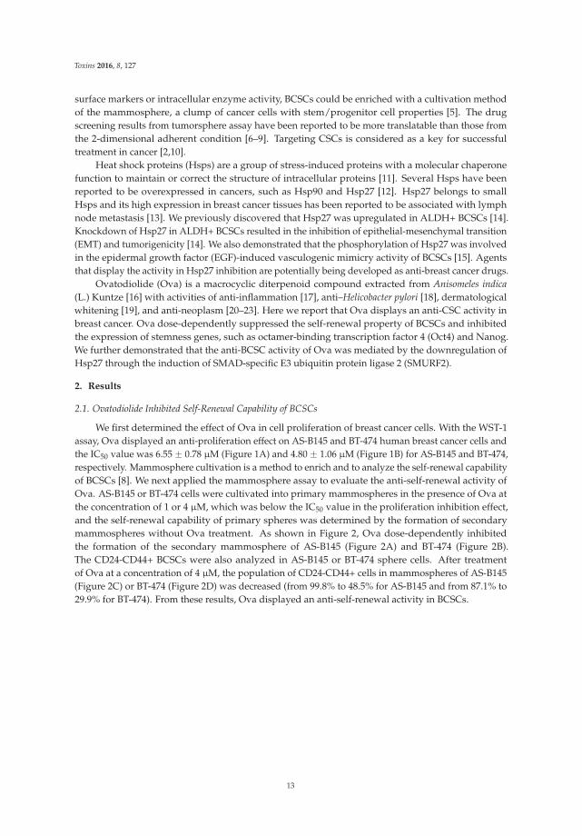
We first determined the effect of Ova in cell proliferation of breast cancer cells. With the WST-1assay, Ova displayed an anti-proliferation effect on AS-B145 and BT-474 human breast cancer cells andthe IC50 value was 6.55 ˘ 0.78 μM (Figure 1A) and 4.80 ˘ 1.06 μM (Figure 1B) for AS-B145 and BT-474,respectively. Mammosphere cultivation is a method to enrich and to analyze the self-renewal capabilityof BCSCs [8]. We next applied the mammosphere assay to evaluate the anti-self-renewal activity ofOva. AS-B145 or BT-474 cells were cultivated into primary mammospheres in the presence of Ova atthe concentration of 1 or 4 μM, which was below the IC50 value in the proliferation inhibition effect,and the self-renewal capability of primary spheres was determined by the formation of secondarymammospheres without Ova treatment. As shown in Figure 2, Ova dose-dependently inhibitedthe formation of the secondary mammosphere of AS-B145 (Figure 2A) and BT-474 (Figure 2B).The CD24-CD44+ BCSCs were also analyzed in AS-B145 or BT-474 sphere cells. After treatmentof Ova at a concentration of 4 μM, the population of CD24-CD44+ cells in mammospheres of AS-B145(Figure 2C) or BT-474 (Figure 2D) was decreased (from 99.8% to 48.5% for AS-B145 and from 87.1% to29.9% for BT-474). From these results, Ova displayed an anti-self-renewal activity in BCSCs.
13
Toxins 2016, 8, 127
Figure 1. The cytotoxic effect of ovatodiolide in human breast cancer cells. AS-B145 (A) or BT-474(B) cells were seeded in a 96-well plate and treated with a different concentration of ovatodiolide(0, 1.625, 3.125, 6.25, 12.5, 25, 50, 100 μM) for 72 h (n = 4 for each concentration). Cell proliferation wasdetermined by WST-1 reagent and the IC50 value was calculated by GraFit software. The experimentswere repeated two times and results from a representative experiment were presented.
Figure 2. Ovatodiolide suppresses the self-renewal property of BCSCs. AS-B145 (A) or BT-474 (B) cellswere seeded into ultralow attachment in a six-well plate under 0.1% DMSO or different concentrationsof ovatodiolide (1 or 4 μM) for seven days and the formed primary mammospheres were collectedand dissociated into a single cell suspension. The same number of dissociated primary sphere cellswas used to evaluate the effect of ovatodiolide on the self-renewal property of BCSCs by secondarymammosphere formation without treatment of ovatodiolide (n = 3 for each treatment). The experimentswere repeated two times and results from a representative experiment were presented. Data werepresented as relative percentage of DMSO control. Scale bar = 50 μm. *, p < 0.05; **, p < 0.01.The mammosphere cells of AS-B145 (C) or BT-474 (D) were harvested and dissociated into a single-cellsuspension. CD24-CD44+ cells were analyzed by flow cytometry.
14
Toxins 2016, 8, 127
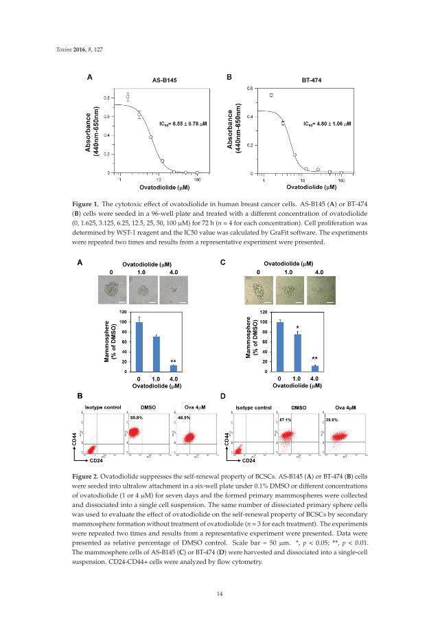
2.2. Ovatodiolide Downregulated the Expression of Stemness Genes and Hsp27 but UpregulatedSMURF2 Expression
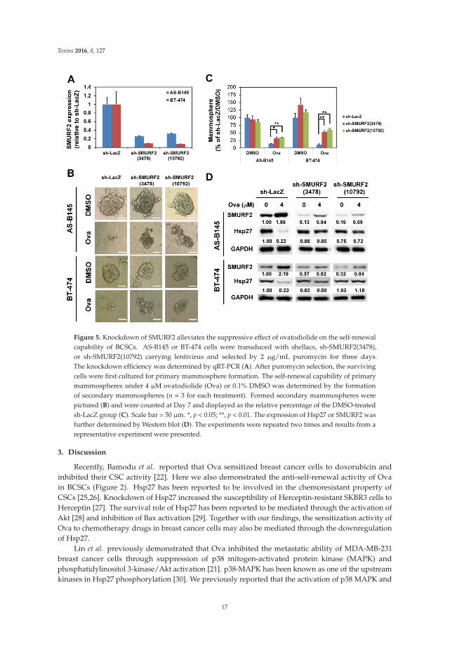
We next examined the effect of Ova on the expression of stemness genes. With Western blotanalysis, Ova dose-dependently inhibited the expression of Oct4 (Figure 3C) and Nanog (Figure 3D) inmammosphere cells derived from AS-B145 (Figure 3A) or BT-474 (Figure 3B) and the inhibitoryeffect was significantly observed at a concentration of 4 μM. We previously demonstrated thatHsp27 regulated the self-renewal and tumorigenicity of BCSCs through modulating EMT and NF-κBactivity [14]. The effect of Ova in Hsp27 expression in mammosphere cells was examined. As shownin Figure 3, Ova dose-dependently downregulated Hsp27 expression (Figure 3E) in mammospherecells derived from AS-B145 (Figure 3A) or BT-474 (Figure 3B). However, the mRNA expression ofHsp27 in mammosphere cells of AS-B145 or BT-474 was not inhibited by Ova treatment (Figure S1).A previous report indicated that SMURF2 mediated the ubiquitin-dependent degradation of Hsp27 inA549 lung cancer cells [24]. We further examined the expression of SMURF2 in mammosphere cellsafter Ova treatment and results revealed that Ova dose-dependently upregulated SMURF2 expression(Figure 3F) in AS-B145 (Figure 3A) or BT-474 (Figure 3B).
Figure 3. The change of protein expression in ovatodiolide-treated BCSCs. BCSCs were first enrichedby primary mammosphere cultivation from AS-B145 (A) or BT-474 (B), dissociated into a single-cellsuspension, and treated with different concentrations of ovatodiolide (0, 1, 4 μM) for 72 h (n = 2 foreach treatment). The expressions of Oct4, Nanog, Hsp27 and SMURF2 were determined by Westernblot. The quantification results of Oct4 (C), Nanog (D), Hsp27 (E) and SMURF2 (F) were determinedby Image J software. The experiments were repeated three times and results from two representativeexperiments were used for quantifications. * p < 0.05; ** p < 0.01.
2.3. Overexpression of Hsp27 or Knockdown of SMURF2 Alleviated the Inhibitory Effect of Ovatodiolide
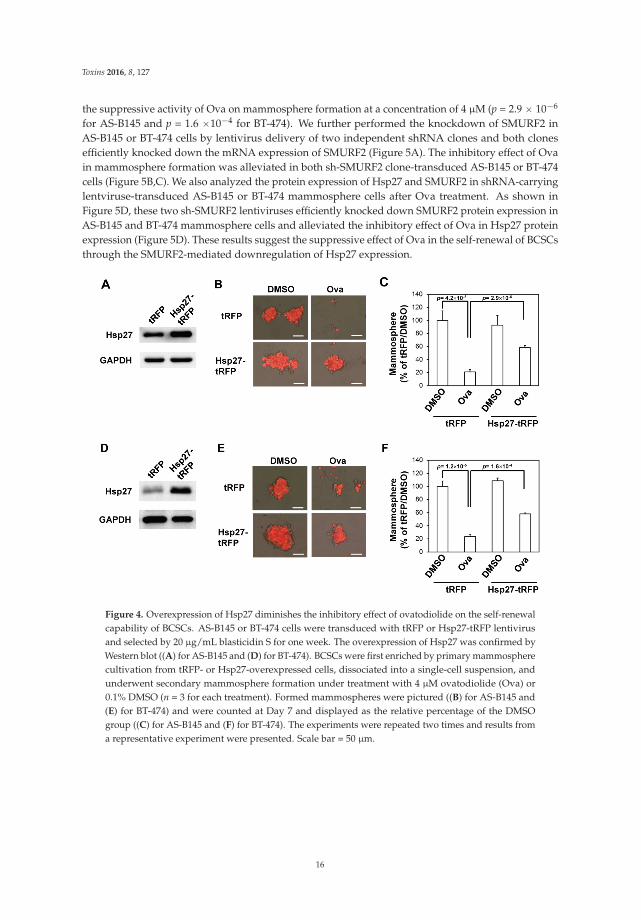
We next examined if overexpression of Hsp27 could alleviate the inhibitory effect of Ova on themammosphere formation capability of AS-B145 cells. Overexpression of Hsp27 in AS-B145 or BT-474cells was performed by lentivirus-mediated gene delivery and confirmed by Western blot (Figure 4A).Exogenous Hsp27 expression in AS-B145 (Figure 4B,C) or BT-474 (Figure 4D,E) significantly diminished
15
Toxins 2016, 8, 127
the suppressive activity of Ova on mammosphere formation at a concentration of 4 μM (p = 2.9 ˆ 10´6
for AS-B145 and p = 1.6 ˆ10´4 for BT-474). We further performed the knockdown of SMURF2 inAS-B145 or BT-474 cells by lentivirus delivery of two independent shRNA clones and both clonesefficiently knocked down the mRNA expression of SMURF2 (Figure 5A). The inhibitory effect of Ovain mammosphere formation was alleviated in both sh-SMURF2 clone-transduced AS-B145 or BT-474cells (Figure 5B,C). We also analyzed the protein expression of Hsp27 and SMURF2 in shRNA-carryinglentviruse-transduced AS-B145 or BT-474 mammosphere cells after Ova treatment. As shown inFigure 5D, these two sh-SMURF2 lentiviruses efficiently knocked down SMURF2 protein expression inAS-B145 and BT-474 mammosphere cells and alleviated the inhibitory effect of Ova in Hsp27 proteinexpression (Figure 5D). These results suggest the suppressive effect of Ova in the self-renewal of BCSCsthrough the SMURF2-mediated downregulation of Hsp27 expression.
Figure 4. Overexpression of Hsp27 diminishes the inhibitory effect of ovatodiolide on the self-renewalcapability of BCSCs. AS-B145 or BT-474 cells were transduced with tRFP or Hsp27-tRFP lentivirusand selected by 20 μg/mL blasticidin S for one week. The overexpression of Hsp27 was confirmed byWestern blot ((A) for AS-B145 and (D) for BT-474). BCSCs were first enriched by primary mammospherecultivation from tRFP- or Hsp27-overexpressed cells, dissociated into a single-cell suspension, andunderwent secondary mammosphere formation under treatment with 4 μM ovatodiolide (Ova) or0.1% DMSO (n = 3 for each treatment). Formed mammospheres were pictured ((B) for AS-B145 and(E) for BT-474) and were counted at Day 7 and displayed as the relative percentage of the DMSOgroup ((C) for AS-B145 and (F) for BT-474). The experiments were repeated two times and results froma representative experiment were presented. Scale bar = 50 μm.
16
Toxins 2016, 8, 127
Figure 5. Knockdown of SMURF2 alleviates the suppressive effect of ovatodiolide on the self-renewalcapability of BCSCs. AS-B145 or BT-474 cells were transduced with shellacs, sh-SMURF2(3478),or sh-SMURF2(10792) carrying lentivirus and selected by 2 μg/mL puromycin for three days.The knockdown efficiency was determined by qRT-PCR (A). After puromycin selection, the survivingcells were first cultured for primary mammosphere formation. The self-renewal capability of primarymammospheres under 4 μM ovatodiolide (Ova) or 0.1% DMSO was determined by the formationof secondary mammospheres (n = 3 for each treatment). Formed secondary mammospheres werepictured (B) and were counted at Day 7 and displayed as the relative percentage of the DMSO-treatedsh-LacZ group (C). Scale bar = 50 μm. *, p < 0.05; **, p < 0.01. The expression of Hsp27 or SMURF2 wasfurther determined by Western blot (D). The experiments were repeated two times and results from arepresentative experiment were presented.
3. Discussion
Recently, Bamodu et al. reported that Ova sensitized breast cancer cells to doxorubicin andinhibited their CSC activity [22]. Here we also demonstrated the anti-self-renewal activity of Ovain BCSCs (Figure 2). Hsp27 has been reported to be involved in the chemoresistant property ofCSCs [25,26]. Knockdown of Hsp27 increased the susceptibility of Herceptin-resistant SKBR3 cells toHerceptin [27]. The survival role of Hsp27 has been reported to be mediated through the activation ofAkt [28] and inhibition of Bax activation [29]. Together with our findings, the sensitization activity ofOva to chemotherapy drugs in breast cancer cells may also be mediated through the downregulationof Hsp27.
Lin et al. previously demonstrated that Ova inhibited the metastatic ability of MDA-MB-231breast cancer cells through suppression of p38 mitogen-activated protein kinase (MAPK) andphosphatidylinositol 3-kinase/Akt activation [21]. p38-MAPK has been known as one of the upstreamkinases in Hsp27 phosphorylation [30]. We previously reported that the activation of p38 MAPK and
17
Toxins 2016, 8, 127
Hsp27 phosphorylation was upregulated in BCSCs [14]. The knockdown of Hsp27 in BCSCs resulted inthe suppression of the EMT features [14]. Hsp27 has been demonstrated to be involved in transforminggrowth factor β–induced EMT through the maintenance of Snail expression [31]. The emergence ofCSCs could be a result of EMT [32]. Here we reported the inhibitory activity of Ova on the Hsp27expression of BCSCs (Figure 3). The anti-CSC activity of Ova may be mediated through its inhibitoryactivity in EMT but remains to be further investigated. Ho et al. reported that Ova inhibited β-cateninsignaling in renal cell carcinoma [23]. Hsp27 has been reported to interact with cytoplasmic β-cateninin breast cancer cells and clinical specimens [33]. It is worth investigating the effect of Ova on theHsp27–β-catenin interaction in BCSCs.
4. Conclusions
In conclusion, we observed that Ova displayed anti-self-renewal activity in the mammopshereformation of human breast cancer cells as well as lead to the downregulation of stemness genessuch as Oct4 and Nanog. The anti-BCSC activity of Ova was mediated through SMURF2-mediateddownegulation of Hsp27. These findings reveal the potential of Ova in breast cancer treatment.
5. Materials and Methods
5.1. Reagents and Antibodies
Ovatodiolide was prepared as the previous report [17]. WST-1 cell proliferation reagent waspurchased from Roche Life Science (Indianapolis, IN, USA). Polyclonal rabbit anti-Oct4 (CST-2750) oranti-Nanog (CST-3580) antibodies were purchased from Cell Signaling Technology, Inc. (Danvers, MA,USA). Polyclonal rabbit anti-Hsp27 antibody (ADI-SPA-803) was purchased from Enzo Life Sciences,Inc. (Farmingdale, NY, USA). Polyclonal rabbit anti-SMURF2 antibody (GTX110487) was purchasedfrom GeneTex International Corporation (Hsinchu City, Taiwan). Peroxidase conjugated ployclonalgoat anti-rabbit IgG secondary antibody (AP132P) was purchased from Merck Millipore (Temecula,CA, USA).
5.2. Cell Culture and Cytotoxicity Assay
AS-B145 human breast cancer cells were established from a female breast cancer specimen asour previous report [14,34] and maintained in MEMα (Gibco, Invitrogen Corporation, Carlsbad, CA,USA) supplemented with 10% fetal bovine serum (FBS, Gibco) and 5 μg/mL insulin (Sigma-Aldrich,St. Louis, MO, USA) at 37 ˝C, 5% CO2 incubator. BT-474 human breast cancer cells were obtainedfrom The Bioresource Collection and Research Center in Taiwan (Hsinchu City, Taiwan) and culturedin DMEM/F12 (Gibco) supplemented with 10% FBS. For determination of cytotoxic effect of Ova,AS-B145 or BT-474 cells were seeded as 2 ˆ 104 cells/well in 96-well plates in presence of differentconcentration of Ova and cultured at 37 ˝C for 72 h. The proliferation/survival of cells was determinedby adding WST-1 reagent and measured the absorbance of 440 and 650 nm wavelength. IC50 valueswere calculated by GraFit software (version 7, Erithacus Software Ltd., West Sussex, UK, 2012).
5.3. Mammosphere Cultivation
For primary mammosphere formation, AS-B145 or BT-474 cells were suspended as1 ˆ 104/well/2 mL and cultured in ultralow attachment 6-well-plates (Corning, Lowell, MA, USA)in DMEM/F12 medium supplemented with 0.4% bovine serum albumin (Sigma), 20 ng/mL EGF(PeproTech, Rocky Hill, NJ, USA) , 20 ng/mL basic fibroblast growth factor (Sino Biological Inc., Beijing,China), 4 μg/mL heparin (Sigma-Aldrich), 5 μg/mL insulin, 1 μM hydrocortisone (Sigma-Aldrich),and 1X B27 supplement (Gibco). After 7 days, primary mammospheres with diameter larger than50 μm were collected with 70 μm cell strainer (BD Biosciences, San Jose, CA, USA) and dissociated intosingle cell suspension with HyQTase (GE Healthcare Life Sciences HyClone Laboratories, Logan, UT,USA) for secondary mammosphere formation at a cell density as 2500 cells/well/2mL for further seven
18
Toxins 2016, 8, 127
days. The form secondary mammospheres were pictured and counted with an inverted microscopy(AE30, Motic Electric Group Co., Ltd., Xiamen, China).
5.4. Analysis of CD24-CD44+ Cells
Mammosphere cells were dissociated into single cell suspension with HyQTase solution. 1 ˆ 105
dissociated cells were stained by anti-human CD24-PE (Cat. No. 555428, BD Biosciences) andanti-human CD44-APC (Cat. No. 559942, BD Biosciences) conjugated antibodies in staining buffer(PBS containing 1% FBS and 0.05% NaN3) on ice for 30 min. PE conjugated mouse IgG2a κ (Cat.No. 555574, BD Biosciences) and APC conjugated mouse IgG2b κ (Cat. No. 555745, BD Biosciences)antibodies were used as isotype controls. After washing with 2 ml PBS/0.01% NaN3, the fluorescencesignals of stained cells were acquired with FACSAriaTM flow cytometer (BD Biosciences) and the datawere further analyzed by WinMDI software (version 2.9, The Scripps Research Institute, La Jolla, CA,USA, 2000).
5.5. Western Blot
Mammospheres were collected by centrifugation and lysed in M-PER Mammalian ProteinExtraction Reagent (Pirece Thermo Fisher Scientific Inc., Waltham, MA, USA). Then 20 μg of totalprotein were separated by SDS-PAGE and transferred onto PVDF membrane (Immobilon-P, MerckMillipore). The membrane was then blocked with 5% skimmed milk (Sigma-Aldrich, St. Louis, MO,USA) dissolved in Tris buffered slaine (Sigma-Aldrich) containing 0.05% Tween-20 (Sigma-Aldrich)(TBS-T) at room temperature for 1 h followed by incubation with primary antibodies at 4 ˝C overnight.After washing with TBS-T, the membrane was then incubated with peroxidase conjugated secondaryantibodies (PerkinElmer, Waltham, MA, USA) at room temperature for one hour. The signals weredeveloped by ECL-plus chemiluminescence substrate (PerkinElmer) and captured using a LuminescentImage Analyzer (Fusion SOLO, Vilber Lourmat, Marne-la-Vallée, France). The band intensity wasquantified using ImageJ software (version 1.48a, NIH, Bethesda. MA, USA, 2013).
5.6. Lentivirus Production and Transduction
Hsp27 cDNA was amplified from pDsRed-Hsp27 [14] and cloned into pLAS5w.Pbsd-L-tRFPlentiviral plasmid (obtained from the National RNAi Core Facility at the Institute of Molecular Biology,Academia Sinica, Taipei, Taiwan) with following primers:
AfeI-Hsp27-F: 5’-AATAGCGCTATGACCGAGCGCCGCGTCCCC-3’Hsp27-EcoRI-R: 5’-CGCGAATTCTTACTTGGCGGCAGTCTCATC-3’
The amplified DNA fragments and pLAS5w.Pbsd-L-tRFP plasmid were digested with AfeIand EcoRI restriction enzymes followed by ligation and transformation into Stbl3 competent cells(Life Technologies, Carlsbad, CA, USA). Lentiviral shRNA plasmids (TRCN00023122 for sh-LacZ,TRCN0000003478 for sh-SMURF2(3478), or TRCN0000010792 for sh-SMURF2(10792)) were alsoobtained from the National RNAi Core Facility. For lentivirus production, individual lentiviralplasmid was mixed with pCMV-ΔR8.91 and pMD.G plasmids as a ratio of 1:0.9:0.1 and transfected into293T cells by GenJetTM Transfection Reagent (SignaGen Laboratories, Ijamsville, MD, USA). Cells weretransduced with lentivirus as MOI = 1 in presence of 8 μg/mL polybrene (Sigma-Aldrich) for 24 hand selected by blasticidin S (for Hsp27) or puromycin (for sh-LacZ or sh-SMURF2 clones) (TOKU-ECompany, Bellingham, WA, USA).
5.7. Quantitative Reverse Transcription Polymerase Chain Reaction (qRT-PCR)
Total RNA was extracted by Quick-RNATM MiniPrep Kit (Zymo Research Corporation, Irvine,CA, USA). Then 1 μg of extracted RNA was used for cDNA synthesis with random hexamersprovided in RevertAid First Strand cDNA Synthesis Kit (Thermo Fisher Scientific Inc., Waltham,
19
Toxins 2016, 8, 127
MA, USA) followed by SYBR Green-based qPCR reaction (SYBR® FAST qPCR Kit, Kapa Biosystems,Inc., Wilmington, MA, USA). The cycling conditions were as follows: 50 ˝C for 2 min, 95 ˝C for 10 min,followed by 40 cycles of 95 ˝C for 10 sec and 60 ˝C for 1 min. The end-point used in the real-timequantification was calculated by the StepOne software (v2.2.2, Applied Biosystmes, Carlsbad, CA,USA, 2011), and the threshold cycle number (Ct value) for each analyzed sample was calculated.The primer sets used in this study were listed as followed:
SMURF2Forward: 5’-TAGCCCTGGCAGACCTCTTA-3’Reverse: 5’- AATACACCTGGCCTTGTTGC-3’MRPL19 (internal control)Forward: 5’- GGGATTTGCATTCAGAGATCAG-3’Reverse: 5’- GGAAGGGCATCTCGTAAG-3’qPCR data were analyzed as previous described [35].
5.8. Statistical Analysis
Quantitative data were presented as the mean ˘ SD. The comparisons between two groupswere analyzed with Student’s t-test. The comparisons among multiple groups (more than three)were analyzed with one-way ANOVA and performed post-hoc test with Tukey Multiple Comparisonanalysis. A p-value of less that 0.05 was considered significantly different.
Supplementary Materials: The following are available online at www.mdpi.com/2072-6651/8/5/127/s1,Figure S1: Ovatodiolide did not inhibit Hsp27 mRNA expression in BCSCs. BCSCs were enriched by primarymammosphere cultivation from AS-B145 or BT-474 breast cancer cells and dissociated into single cell suspensionfollowed by treated with ovatodilide (1 or 4 mM) or 0.1% DMSO for 48 h. Total RNA were than extracted and theHsp27 mRNA expression was determined by SYBR Green-based qRT-PCR method.
Acknowledgments: This work is supported by the Ministry of Science and Technology of Taiwan (grantNo. MOST 103-2113-M-324-001-MY2 (Y.-M.T.) and MOST 103-2314-B-040-015-MY3 (W.-W.C.)) and theinter-institutional research grant between Changhua Christian Hospital and Chung Medical University (grantNo. CSMU-CCH-104-03 (W.-W.C. and K.-T.L.)).
Author Contributions: J.-J.Y., W.-W.C. and Y.-M.Y. conceived and designed the experiments; W.-Y.C. and J.C.-C.performed the experiments; K.-T.L., W.-Y.C. and W.-W.C. analyzed the data; K.-T.L., B.-Y.W. and Y.-M.T. contributedreagents/materials; H.-T.L., W.-W.C. and Y.-M.T. wrote the paper.
Conflicts of Interest: The authors declare no conflict of interest.
Abbreviations
The following abbreviations are used in this manuscript:
CSCs Cancer stem/progenitor cellsHsp27 heat shock protein 27Ova OvatiodiolideOct4 POU class 5 homeobox 1Nanog Nanog homeoboxSMURF2 SMAD ubiquitin regulatory factor 2
References
1. Ajani, J.A.; Song, S.; Hochster, H.S.; Steinberg, I.B. Cancer stem cells: The promise and the potential.Semin. Oncol. 2015, 42 (Suppl. 1), S3–S17. [CrossRef] [PubMed]
2. Maccalli, C.; de Maria, R. Cancer stem cells: Perspectives for therapeutic targeting. Cancer Immunol.Immunother. CII 2015, 64, 91–97. [CrossRef] [PubMed]
3. Al-Hajj, M.; Wicha, M.S.; Benito-Hernandez, A.; Morrison, S.J.; Clarke, M.F. Prospective identification oftumorigenic breast cancer cells. Proc. Natl. Acad. Sci. USA 2003, 100, 3983–3988. [CrossRef] [PubMed]
20
Toxins 2016, 8, 127
4. Ginestier, C.; Hur, M.H.; Charafe-Jauffret, E.; Monville, F.; Dutcher, J.; Brown, M.; Jacquemier, J.; Viens, P.;Kleer, C.G.; Liu, S.; et al. Aldh1 is a marker of normal and malignant human mammary stem cells and apredictor of poor clinical outcome. Cell Stem Cell 2007, 1, 555–567. [CrossRef] [PubMed]
5. Ponti, D.; Costa, A.; Zaffaroni, N.; Pratesi, G.; Petrangolini, G.; Coradini, D.; Pilotti, S.; Pierotti, M.A.;Daidone, M.G. Isolation and in vitro propagation of tumorigenic breast cancer cells with stem/progenitorcell properties. Cancer Res. 2005, 65, 5506–5511. [CrossRef]
6. Hongisto, V.; Jernstrom, S.; Fey, V.; Mpindi, J.P.; Kleivi Sahlberg, K.; Kallioniemi, O.; Perala, M.High-throughput 3d screening reveals differences in drug sensitivities between culture models of JIMT1breast cancer cells. PLoS ONE 2013, 8, e77232. [CrossRef]
7. Kim, S.; Alexander, C.M. Tumorsphere assay provides more accurate prediction of in vivo responses tochemotherapeutics. Biotechnol. Lett. 2014, 36, 481–488. [CrossRef] [PubMed]
8. Lee, C.H.; Yu, C.C.; Wang, B.Y.; Chang, W.W. Tumorsphere as an effective in vitro platform for screeninganti-cancer stem cell drugs. Oncotarget 2016, 7, 1215–1226.
9. Morrison, B.J.; Hastie, M.L.; Grewal, Y.S.; Bruce, Z.C.; Schmidt, C.; Reynolds, B.A.; Gorman, J.J.; Lopez, J.A.Proteomic comparison of mcf-7 tumoursphere and monolayer cultures. PLoS ONE 2012, 7, e52692. [CrossRef]
10. Bouvard, C.; Barefield, C.; Zhu, S. Cancer stem cells as a target population for drug discovery.Future Med. Chem. 2014, 6, 1567–1585. [CrossRef]
11. Powers, M.V.; Workman, P. Inhibitors of the heat shock response: Biology and pharmacology. FEBS Lett.2007, 581, 3758–3769. [CrossRef]
12. Soo, E.T.; Yip, G.W.; Lwin, Z.M.; Kumar, S.D.; Bay, B.H. Heat shock proteins as novel therapeutic targets incancer. In Vivo 2008, 22, 311–315. [PubMed]
13. Kaigorodova, E.V.; Zavyalova, M.V.; Bogatyuk, M.V.; Tarabanovskaya, N.A.; Slonimskaya, E.M.;Perelmuter, V.M. Relationship between the expression of phosphorylated heat shock protein beta-1 withlymph node metastases of breast cancer. Cancer Biomark 2015, 15, 143–150. [PubMed]
14. Wei, L.; Liu, T.T.; Wang, H.H.; Hong, H.M.; Yu, A.L.; Feng, H.P.; Chang, W.W. Hsp27 participates in themaintenance of breast cancer stem cells through regulation of epithelial-mesenchymal transition and nuclearfactor-kappab. Breast Cancer Res. BCR 2011, 13, R101. [CrossRef]
15. Lee, C.H.; Wu, Y.T.; Hsieh, H.C.; Yu, Y.; Yu, A.L.; Chang, W.W. Epidermal growth factor/heat shock protein27 pathway regulates vasculogenic mimicry activity of breast cancer stem/progenitor cells. Biochimie 2014,104, 117–126. [CrossRef]
16. Arisawa, M.; Nimura, M.; Fujita, A.; Hayashi, T.; Morita, N.; Koshimura, S. Biological active macrocyclicditerpenoids from chinese drug “fang feng cao”; II. Derivatives of ovatodiolids and their cytotoxity.Planta Med. 1986, 4, 297–299. [CrossRef]
17. Rao, Y.K.; Chen, Y.C.; Fang, S.H.; Lai, C.H.; Geethangili, M.; Lee, C.C.; Tzeng, Y.M. Ovatodiolide inhibits thematuration of allergen-induced bone marrow-derived dendritic cells and induction of TH2 cell differentiation.Int. Immunopharmacol. 2013, 17, 617–624. [CrossRef]
18. Lien, H.M.; Wang, C.Y.; Chang, H.Y.; Huang, C.L.; Peng, M.T.; Sing, Y.T.; Chen, C.C.; Lai, C.H. Bioevaluationof anisomeles indica extracts and their inhibitory effects on helicobacter pylori-mediated inflammation.J. Ethnopharmacol. 2013, 145, 397–401. [CrossRef]
19. Huang, H.C.; Lien, H.M.; Ke, H.J.; Chang, L.L.; Chen, C.C.; Chang, T.M. Antioxidative characteristics ofanisomeles indica extract and inhibitory effect of ovatodiolide on melanogenesis. Int. J. Mol. Sci. 2012, 13,6220–6235. [CrossRef] [PubMed]
20. Liao, Y.F.; Rao, Y.K.; Tzeng, Y.M. Aqueous extract of anisomeles indica and its purified compound exertsanti-metastatic activity through inhibition of nf-kappab/ap-1-dependent MMP-9 activation in human breastcancer MCF-7 cells. Food Chem. Toxicol. 2012, 50, 2930–2936. [CrossRef] [PubMed]
21. Lin, K.L.; Tsai, P.C.; Hsieh, C.Y.; Chang, L.S.; Lin, S.R. Antimetastatic effect and mechanism of ovatodiolidein MDA-MB-231 human breast cancer cells. Chemico-Biol. Interact. 2011, 194, 148–158. [CrossRef] [PubMed]
22. Bamodu, O.A.; Huang, W.C.; Tzeng, D.T.; Wu, A.; Wang, L.S.; Yeh, C.T.; Chao, T.Y. Ovatodiolide sensitizesaggressive breast cancer cells to doxorubicin, eliminates their cancer stem cell-like phenotype, and reducesdoxorubicin-associated toxicity. Cancer Lett. 2015, 364, 125–134. [CrossRef] [PubMed]
23. Ho, J.Y.; Hsu, R.J.; Wu, C.L.; Chang, W.L.; Cha, T.L.; Yu, D.S.; Yu, C.P. Ovatodiolide targets β-cateninsignaling in suppressing tumorigenesis and overcoming drug resistance in renal cell carcinoma. Evid. BasedComplement. Altern. Med. 2013, 2013, 161628. [CrossRef] [PubMed]
21
Toxins 2016, 8, 127
24. Sun, Y.; Zhou, M.; Fu, D.; Xu, B.; Fang, T.; Ma, Y.; Chen, J.; Zhang, J. Ubiquitination of heat shock protein 27is mediated by its interaction with smad ubiquitination regulatory factor 2 in a549 cells. Exp. Lung Res. 2011,37, 568–573. [CrossRef]
25. Lee, C.H.; Hong, H.M.; Chang, Y.Y.; Chang, W.W. Inhibition of heat shock protein (hsp) 27 potentiates thesuppressive effect of hsp90 inhibitors in targeting breast cancer stem-like cells. Biochimie 2012, 94, 1382–1389.[CrossRef]
26. Hsu, H.S.; Lin, J.H.; Huang, W.C.; Hsu, T.W.; Su, K.; Chiou, S.H.; Tsai, Y.T.; Hung, S.C. Chemoresistance oflung cancer stemlike cells depends on activation of hsp27. Cancer 2011, 117, 1516–1528. [CrossRef]
27. Kang, S.H.; Kang, K.W.; Kim, K.H.; Kwon, B.; Kim, S.K.; Lee, H.Y.; Kong, S.Y.; Lee, E.S.; Jang, S.G.; Yoo, B.C.Upregulated hsp27 in human breast cancer cells reduces herceptin susceptibility by increasing her2 proteinstability. BMC Cancer 2008, 8, 286. [CrossRef]
28. Wu, R.; Kausar, H.; Johnson, P.; Montoya-Durango, D.E.; Merchant, M.; Rane, M.J. Hsp27 regulatesakt activation and polymorphonuclear leukocyte apoptosis by scaffolding MK2 to akt signal complex.J. Biol. Chem. 2007, 282, 21598–21608. [CrossRef]
29. Havasi, A.; Li, Z.; Wang, Z.; Martin, J.L.; Botla, V.; Ruchalski, K.; Schwartz, J.H.; Borkan, S.C. Hsp27 inhibitsbax activation and apoptosis via a phosphatidylinositol 3-kinase-dependent mechanism. J. Biol. Chem. 2008,283, 12305–12313. [CrossRef]
30. Kostenko, S.; Moens, U. Heat shock protein 27 phosphorylation: Kinases, phosphatases, functions andpathology. Cell. Mol. Life Sci. CMLS 2009, 66, 3289–3307. [CrossRef]
31. Wettstein, G.; Bellaye, P.S.; Kolb, M.; Hammann, A.; Crestani, B.; Soler, P.; Marchal-Somme, J.; Hazoume, A.;Gauldie, J.; Gunther, A.; et al. Inhibition of hsp27 blocks fibrosis development and emt features by promotingsnail degradation. FASEB J. 2013, 27, 1549–1560. [CrossRef]
32. Singh, A.; Settleman, J. Emt, cancer stem cells and drug resistance: An emerging axis of evil in the war oncancer. Oncogene 2010, 29, 4741–4751. [CrossRef]
33. Fanelli, M.A.; Montt-Guevara, M.; Diblasi, A.M.; Gago, F.E.; Tello, O.; Cuello-Carrion, F.D.; Callegari, E.;Bausero, M.A.; Ciocca, D.R. P-cadherin and beta-catenin are useful prognostic markers in breast cancerpatients; beta-catenin interacts with heat shock protein hsp27. Cell Stress Chaperones 2008, 13, 207–220.[CrossRef] [PubMed]
34. Chang, W.W.; Lee, C.H.; Lee, P.; Lin, J.; Hsu, C.W.; Hung, J.T.; Lin, J.J.; Yu, J.C.; Shao, L.E.; Yu, J.; et al.Expression of globo H and SSEA3 in breast cancer stem cells and the involvement of fucosyl transferases 1and 2 in globo H synthesis. Proc. Natl. Acad. Sci. USA 2008, 105, 11667–11672. [CrossRef] [PubMed]
35. Chang, Y.C.; Lin, C.W.; Yu, C.C.; Wang, B.Y.; Huang, Y.H.; Hsieh, Y.C.; Kuo, Y.L.; Chang, W.W. Resveratrolsuppresses myofibroblast activity of human buccal mucosal fibroblasts through the epigenetic inhibition ofzeb1 expression. Oncotarget 2016. [CrossRef]
© 2016 by the authors; licensee MDPI, Basel, Switzerland. This article is an open accessarticle distributed under the terms and conditions of the Creative Commons Attribution(CC-BY) license (http://creativecommons.org/licenses/by/4.0/).
22
toxins
Review
Ellagitannins in Cancer Chemopreventionand Therapy
Tariq Ismail 1, Cinzia Calcabrini 2,3, Anna Rita Diaz 2, Carmela Fimognari 3, Eleonora Turrini 3,Elena Catanzaro 3, Saeed Akhtar 1 and Piero Sestili 2,*
1 Institute of Food Science & Nutrition, Faculty of Agricultural Sciences and Technology,Bahauddin Zakariya University, Bosan Road, Multan 60800, Punjab, Pakistan;[email protected] (T.I.); [email protected] (S.A.)
2 Department of Biomolecular Sciences, University of Urbino Carlo Bo, Via I Maggetti 26, 61029 Urbino (PU),Italy; [email protected]
3 Department for Life Quality Studies, Alma Mater Studiorum-University of Bologna, Corso d'Augusto 237,47921 Rimini (RN), Italy; [email protected] (C.C.); [email protected] (C.F.);[email protected] (E.T.); [email protected] (E.C.)
* Correspondence: [email protected]; Tel.: +39-(0)-722-303-414
Academic Editor: Jia-You FangReceived: 31 March 2016; Accepted: 9 May 2016; Published: 13 May 2016
Abstract: It is universally accepted that diets rich in fruit and vegetables lead to reduction in therisk of common forms of cancer and are useful in cancer prevention. Indeed edible vegetablesand fruits contain a wide variety of phytochemicals with proven antioxidant, anti-carcinogenic,and chemopreventive activity; moreover, some of these phytochemicals also display directantiproliferative activity towards tumor cells, with the additional advantage of high tolerabilityand low toxicity. The most important dietary phytochemicals are isothiocyanates, ellagitannins (ET),polyphenols, indoles, flavonoids, retinoids, tocopherols. Among this very wide panel of compounds,ET represent an important class of phytochemicals which are being increasingly investigated fortheir chemopreventive and anticancer activities. This article reviews the chemistry, the dietarysources, the pharmacokinetics, the evidence on chemopreventive efficacy and the anticancer activityof ET with regard to the most sensitive tumors, as well as the mechanisms underlying theirclinically-valuable properties.
Keywords: ellagitannins; phytochemicals; cancer; chemoprevention; cancer therapy; safety
1. Introduction
Despite the enormous efforts of the scientific and medical community, cancer still represents thesecond leading cause of death and is nearly becoming the leading one in the elderly [1]. It is estimatedthat, due to demographic changes alone, in the next 15 years the number of new cancer cases willincrease by 70% worldwide [2].
The lack of effective diagnostic tools for early detection of several tumors, the limited treatmentoptions for patients with advanced stages of cancer, and the onset of multiple drug resistance favorpoor prognosis and high mortality rates. The significant, but still unsatisfactory, improvement ofsurvival, the severe toxicity profile, and the high costs characterizing many current anticancer therapiesclearly show that a threshold in terms of clinical benefit and patients’ tolerance has been reached.Thus, the identification and development of innovative, preventive as well as therapeutic strategies tocontrast cancer-associated morbidity and mortality are urgently needed.
Epidemiological, preclinical, and clinical studies have generally concluded that a diet richin phytochemicals can reduce the risk of cancer [2,3]. Due to their pleiotropism which includesantioxidant, anti-inflammatory, and antiproliferative activities as well as modulatory effects on
Toxins 2016, 8, 151 23 www.mdpi.com/journal/toxins
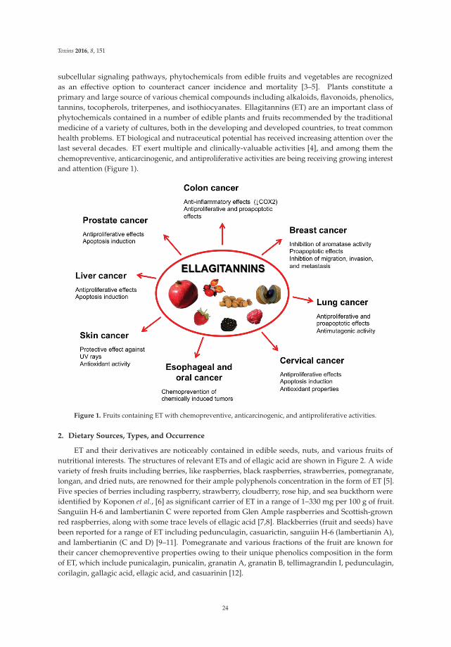
Toxins 2016, 8, 151
subcellular signaling pathways, phytochemicals from edible fruits and vegetables are recognizedas an effective option to counteract cancer incidence and mortality [3–5]. Plants constitute aprimary and large source of various chemical compounds including alkaloids, flavonoids, phenolics,tannins, tocopherols, triterpenes, and isothiocyanates. Ellagitannins (ET) are an important class ofphytochemicals contained in a number of edible plants and fruits recommended by the traditionalmedicine of a variety of cultures, both in the developing and developed countries, to treat commonhealth problems. ET biological and nutraceutical potential has received increasing attention over thelast several decades. ET exert multiple and clinically-valuable activities [4], and among them thechemopreventive, anticarcinogenic, and antiproliferative activities are being receiving growing interestand attention (Figure 1).
Figure 1. Fruits containing ET with chemopreventive, anticarcinogenic, and antiproliferative activities.
2. Dietary Sources, Types, and Occurrence
ET and their derivatives are noticeably contained in edible seeds, nuts, and various fruits ofnutritional interests. The structures of relevant ETs and of ellagic acid are shown in Figure 2. A widevariety of fresh fruits including berries, like raspberries, black raspberries, strawberries, pomegranate,longan, and dried nuts, are renowned for their ample polyphenols concentration in the form of ET [5].Five species of berries including raspberry, strawberry, cloudberry, rose hip, and sea buckthorn wereidentified by Koponen et al., [6] as significant carrier of ET in a range of 1–330 mg per 100 g of fruit.Sanguiin H-6 and lambertianin C were reported from Glen Ample raspberries and Scottish-grownred raspberries, along with some trace levels of ellagic acid [7,8]. Blackberries (fruit and seeds) havebeen reported for a range of ET including pedunculagin, casuarictin, sanguiin H-6 (lambertianin A),and lambertianin (C and D) [9–11]. Pomegranate and various fractions of the fruit are known fortheir cancer chemopreventive properties owing to their unique phenolics composition in the formof ET, which include punicalagin, punicalin, granatin A, granatin B, tellimagrandin I, pedunculagin,corilagin, gallagic acid, ellagic acid, and casuarinin [12].
24
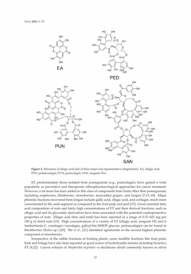
Toxins 2016, 8, 151
Figure 2. Structures of ellagic acid and of three major and representative ellagitannins. EA, ellagic acid;PED, pedunculagin; PUN, punicalagin; SAN, sanguiin H-6.
ET, predominately those isolated from pomegranate (e.g., punicalagin), have gained a widepopularity as preventive and therapeutic ethnopharmacological approaches for cancer treatment.However, a lot more has been added to this class of compounds from fruits other than pomegranate,including raspberries, blueberries, strawberries, muscadine grapes, and longan [7,13–18]. Majorphenolic fractions recovered from longan include gallic acid, ellagic acid, and corilagin, much moreconcentrated in the seed segment as compared to the fruit pulp and peel [17]. Good essential fattyacid composition of nuts and fairly high concentrations of ET and their derived fractions, such asellagic acid and its glycosidic derivatives have been associated with the potential cardioprotectiveproperties of nuts. Ellagic acid (free and total) has been reported in a range of 0.37–823 mg per100 g of dried nuts [19]. High concentrations of a variety of ET (ellagic acid, sanguiin H2 and 6,lambertianin C, castalagin/vescalagin, galloyl-bis-HHDP glucose, pedunculagin) can be found inblackberries (Rubus sp.) [20]. Shi et al., [21] identified agrimoniin as the second highest phenoliccompound of strawberries.
Irrespective of the edible fractions of fruiting plants, some inedible fractions like fruit peels,bark and foliage have also been reported as good source of hydrolysable tannins including bioactiveET [4,22]. Leaves extracts of Shepherdia argentea—a deciduous shrub commonly known as silver
25
Toxins 2016, 8, 151
buffaloberry—were reported as a good reserve of gluconic acid core carrying the potential anti-HIVnovel ET, such as hippophaenin A, shephagenin A and shephagenin B [23].
3. Ellagitannins—Classification and Chemistry
Tannins are unique secondary metabolites of plant phenolics with relatively higher molecularweight (300–30,000 Da) and bear the ability to generate complexes with some macromolecules, likeproteins and carbohydrates [24]. Chemistry and nomenclature of the tannins is complicated by virtueof the frequent changes which parallel the advancement in this very specific field [25]. Taking intoaccount different definitions of tannins [26,27], these compounds may be referred as either galloylesters and their derivatives (ET, gallotannins, and complex tannins), or the oligomeric and polymericproanthocyanidins (condensed tannins). In a broader perspective, tannins may be classified mostsatisfactorily and unambiguously on the basis of structural configuration and/or solubility [28]. C–Ccoupling of galloyl units in absence of glycosidically-linked catechin make ETs structurally differentfrom the condensed tannins that are characterized by monomeric catechin linkages (C4–C8 or C4–C6)to generate oligomeric likewise polymeric proanthocyanidins [27]. Gallotannins and ETs constitute amajor group of tannins i.e., hydrolysable tannins that are well known for their properties to hydrolyzeinto hexahydroxydiphenol (HHDP) or gallic acid moieties. Gallotannins are the gallic acid derivativescarrying ě six gallyol groups and might further be characterized on account of one or more than onedigalloyl group [29].
ETs (hydrolysable tannins) on their hydrolysis yield gallic acid and ellagic acid from thecompounds carrying gallyol groups and HHDP groups, respectively [28]. In vitro digestion modelsdeclare ETs to remain stable under the normal physiological condition of the stomach [30]. However,ETs hydrolysis to free ellagic acid or their degradation may proceed in the small intestine at neutral toalkaline pH [31]. Biologically, condensed tannins and gallotannins are thought to deliver relativelyhigher protein precipitation properties as compare to the ETs and hence are considered potentialantinutritional compounds from the class of plants polyphenolics [32]. Gallotannins and condensedtannins have also been reported as oxidatively least active tannins as compared to the ETs and on thesame time gallotannins and condensed tannins have also been found to reduce pro-oxidant propertiesof ETs [33,34].
3.1. Simple Ellagitannins
ET (M.W. 300–20,000 Da) are non-nitrogenous compounds with at least two C–C coupledgalloyl units with no glycosidically-bonded catechin unit [3,35]. ET are derivatives of1,2,3,4,6-penta-O-galloyl-β-D-glucopyranose (PGG). Structurally, ET are esters of carbohydrates and orcyclitols and also include metabolic compounds derived from oxidative cleavage of either condensedor hydrolysable tannins [27,35,36]. The presence of hexahydroxydiphenol (HHDP) in a glucopyranosering in addition to acyl units and certain HHDP metabolites such as dehydrohexahydroxydiphenol(DHHDP), valoneoyl and chebuloyl groups constitute simple ET. Tellimagrandin I and II, pedunulagin,casuarictin, and chebulagic acid originate from the specific orientation and number of acyl groupson glucose units. Variation in HHDP group originates by linking (C–C or C–O) one or more galloylgroups to HHDP unit.
Structural diversity of ET has been reported to correlate with their carrier-plants’ taxonomy andevolutionary hierarchy [37]. More often, monomeric ET or oligomeric ET constitute the major tanniccomponent of plant species. The monomeric compounds of the group include tellimagrandins I and II,pedunculagin, casuarictin, and potentillin. Type I hydrolysable tannins (i.e., gallotannins) carryingHHDP in stable conformation at either the 2,3 or 4,6 position on a D-glucopyranose may be referred toas a simple ET [38–40]. Geraniin, a type III ET, is another example of monomeric simple ET carryinga DHHDP unit linked to D-gluopyranose of HHDP unit via 1C4 conformation. Dimers of ET aregenerated by intermolecular oxidative coupling/condensation of simple ET.
26
Toxins 2016, 8, 151
3.2. Glycosidic Ellagitannins
Chemically, the C-glycosidic linkage of ET is established via intermolecular bonds betweentwo monomeric units, one carrying anomeric carbon while the second one galloyl or HHDPgroup [3,35,41]. Most recently C-glycosidic ET including granadinin, vescalagin, methylvescalagin,castalagin, stachyurin, and casuarinin have been reported from the peel and seed fraction ofcamu-camu, a fruiting tree of Amazon rainforest [42]. Woody fractions of various fruits, particularlythe nuts and berries, have also been observed to hold novel C-glycosidic ET (e.g., castalagin andvescalagin). Castacrenins D and F are two other forms of C-glycosidic ET isolated from the woodyfraction of Japanese chestnut and carry gallic acid/ellagic acid moieties [43]. Treating vescalaginwith Lentinula edodes generates quercusnins A and B that may be referred as fungal metabolites ofC-glycosidic ET [44]. Castacrenins D and F isolated from chestnut wood may generate oxidativemetabolites, namely castacrenins E and G, by replacing pyrogallol rings of C-glycosidic ET withcyclopentone rings [43]. Rhoipteleanins H, I, and J were reported as novel C-glycosidic ET isolatedfrom the fruit and bark fractions of Rhoiptelea chiliantha. Structural configuration of rhoipteleaninsH revealed the presence of cyclopentenone carboxy moieties that are generated by oxidation andrearrangement of C-glycosidic ET aromatic ring [45].
Condensate of C-glycosidic ET is another subclass of hydrolysable tannins, which includesrhoipteleanin J produced by the intermolecular condensation (C–C or C–O) of monomeric C-glycosidicET followed by oxidation of aromatic rings of ET [45]. Wine aged in oak wood barrels is often reportedto carry oak ET, particularly the condensation products of monomeric C-glycosidic ET. The studiesinfer C-glycosidic ET to play a significant role in modulation of organoleptic features of wine aged inoak wood barrels [46].
4. Ellagitannins Pharmacokinetics
A precise knowledge of phytochemicals’ pharmacokinetics is very important to exploit their healthbenefits, as well as the effects of their metabolites [47]. In vivo, ET, instead of being absorbed directlyinto the blood stream, are physiologically hydrolyzed to ellagic acid, which is further metabolizedto biologically-active and bioavailable derivatives, i.e., urolithins, by the activity of microbiota ingastrointestinal (GI) tract [5,48]. The biological properties of ET, such as free radical scavenging,further depend on their metabolic transformation inside gut. ET recovered from pomegranate juicemay be metabolically converted by gut microbiota to urolithin A, B, C, D, 8-O-methylurolithin A,8,9-di-O-methylurolithin C, and 8,9-di-O-methylurolithin D, and some of these metabolites displayhigher antioxidant activity than the parental tannins themselves. For instance, urolithin C and Dshow an antioxidant capacity—as determined in a cell-based assay—which is 10- to 50-fold higheras compared to punicalagin, punicalin, ellagic acid, and gallic acid [49]. This finding suggeststhat intestinal transformation products of ET are likely to play a central role for the antioxidantproperties at least inside the GI tract. Significant differences in urolithins’ profiles in individualhuman subjects feed on raspberries—a renowned source of ET—have been attributed to gut microflora,whose variations on an inter-individual basis affect their capacity of hydrolyzing ET and subsequentmetabolite synthesis [48,50]. The interaction of gut microbiota composition and the host endogenousexcretory system is also likely to play a further role in the observed inter-individual variability [51].ET are highly stable under the acidic environment of stomach, and retain their composition withoutbeing hydrolyzed to simpler compounds when exposed to various gastric enzymes. Consequently thecomplex structure of ET impedes their gastric absorption: however, the stomach might serve as thefirst site of absorption of free ellagic acid and pre-hydrolyzed forms of ET.
Contrary to stomach, the neutral or alkaline environments of duodenum and small intestines,characterized by pH values ranging from 7.1 to 8.4, allow ET hydrolyzation [31,41]. In humans, ET arerapidly absorbed and metabolized, as documented by [18,52]: following ingestion of pomegranate juice(at a dose containing 25 mg of ellagic acid and 318 mg of ET), ellagic acid can be found in plasma for upto 4 h while, at later times, it is no more detectable. In contrast, another study reported that no ellagic
27
Toxins 2016, 8, 151
acid could be detected in plasma during the 4 h following the juice intake [53], a discrepancy whichhas been attributed to inter-individual variability [54]. Ellagic acid is converted by catechol-O-methyltransferase to dimethylellagic acid, which is then glucuronidated and excreted [52].
Finally, the microbiologically metabolized fraction of ET, i.e., urolithins, is further incorporated toenterohepatic circulation system [18,53,55,56].
5. Ellagitannins for Tumor Chemoprevention and Therapy
The development of novel mechanism-based chemopreventive and antitumor approaches to fightcancer through the use of dietary substances which humans can easily accept has become an importantgoal. Along this line, ET have received increasing attention over the last two decades.
Similarly to other anticancer phytochemicals, ET display chemopreventive and chemotherapeuticactivities [3]. The chemopreventive activity of ET and derivatives, such as ellagic acid has beenprimarily associated with their antioxidant capacity, that varies with the degree of hydroxylation [5,57]and depends from both a direct radical scavenging and iron chelation activity.
The well-known anti-inflammatory capacity represents another important feature of ETchemopreventive and antitumor activity [55], that being persisting inflammation involved both asa causative and a facilitating factor in carcinogenesis and cancer development [58]. For example,pomegranate ET inhibit pro-inflammatory pathways including, but not limited to, the NF-κBpathway, whose activation leads to immune reactions, inflammation, and the transcription of genesinvolved in cell survival, such as Bclx and inhibitors of apoptosis. Constitutive activation of NF-κBhas been observed in prostate cancers, where it sustains chronic inflammation and promotes thedevelopment of high-grade prostate cancer. With respect to inflammation, it is worth noting that,similarly to many polyphenols, the antioxidant activity of ET participates to an “anti-inflammatoryloop” with other mechanisms, since it lowers the levels of radicals which otherwise would act aspro-inflammatory stimuli.
The direct antiproliferative effects of ET have been attributed to multiple mechanisms (see the nextsubchapters) including the cell cycle arrest capacity and the properties enabling cancer cells to followapoptosis through the mitochondrial route and self-destruction after replication [59–62]. In addition todirectly targeting tumor cell survival, the cytotoxic/cytostatic activities of ET might also concur withthe chemopreventive potential, since they prevent tumor cells from converting into more malignantphenotypes and from replicating.
A study on 1,3-di-O-galloyl-4,6-(s)-HHDP-β-D-glucopyranose (an ET from Balanophorajaponica MAKINO) points to the complexity and multiplicity of the mechanisms contributingto the anticancer activity of ET, i.e., the same complexity and multiplicity characterizing alsoother classes of phytochemicals. Indeed, the antiproliferative activity of 1,3-di-O-galloyl-4,6-(s)-HHDP-β-D-glucopyranose in human Hep-G2 liver cancer cells was also associated to an alteredregulation of 25 miRNAs including the let-7 family members miR-370, miR-373, and miR-526b,identified as likely targets with roles in cell proliferation and differentiation [63]. The fact that in cellculture systems combinations of ET or of ET and other phytochemicals present in plant or fruit extractsare more cytotoxic than any single ET [64], is suggestive of the multifactorial effects, chemical synergy,and multiplicity of the mechanisms behind their antitumor activity. To this regard, the capacity of someET to inhibit angiogenesis, a fundamental event accompanying tumor growth, both in in vitro andin vivo prostate cancer models [65], and to reduce endothelial cell growth through binding to vascularendothelial growth factor receptors [66] represents a further and significant antitumor mechanism.
In analogy to other polyphenols, ET could also be utilized to increase the sensitivity of tumorcells to standard chemotherapeutic drugs [67], with the aim of obtaining an increase of their antitumorefficacy along with a reduction of their doses and, consequently, of their severe adverse effects whichoften represent a limiting factor for the prosecution of the therapeutic regimens.
As a premise to the literature data discussed in the next paragraphs, it is important noting that,since ET are not absorbed systemically after oral administration as such [48], the studies where ET
28

Toxins 2016, 8, 151
extracts were given to cultured cancer cells are unlikely to be predictive of the effects which couldbe attained after oral ingestion in vivo. Rather these data could be representative of intravenouslyadministered ET, but the toxicology of this administration route is not known.
The next sections of the review will discuss more in depth the ET anticancer mechanisms andproperties emerging from in vivo and in vitro studies on a panel of tumors or tumor cells which appearas potentially sensitive targets for these phytochemicals.
5.1. Prostate Cancer
Prostate cancer is the second leading cancer-associated death risk factor among U.S. males [68].Phytochemicals originating from various food sources slow down the progression of prostate cancer,whereas a majority of other nutrients are reported to be non-effective in either preventing or curingprostate cancer [69]. Evidence-based findings support the consolidated role of fruits, vegetables, andvarious culinary herbs of different cultures in averting various forms of cancers, but relatively weakand inconsistent relationships have been presented so far for prostate cancer [70,71]. Somehowmore promising seem to be the edible fruits containing high amounts of ET, which have beenextensively tested in vivo for their prostate cancer inhibitory properties. As it has been shown inanimal models, higher concentrations of ET are recorded in prostate and colon tissues as compared tothe others [72]. Pomegranate holds one of the highest concentration of ET [55]. Antitumor activitiesof pomegranate fruit juice, peel extracts, and seed oil have been reported against prostate cancercells [73]. Dose-dependent anti-proliferative and pro-apoptotic effects of pomegranate fruit extracts(10–100 μg/mL) have been documented against aggressive human prostate cancer cells (PC3) [74]:induction of pro-apoptotic mediators (Bax and Bak), downregulation of Bcl-2 and Bcl-XL, and reducedexpression of cyclin-dependent kinases 2, 4, 6, and cyclins D1, D2, and E have been identified as themechanisms responsible for these effects.
Pomegranate extract inhibited proliferation of endothelial (HUVEC) and prostate (LNCaP) cancercells; the extract also reduced LNCaP prostate cancer xenograft size, tumor vessel density, VEGFpeptide levels and HIF-α expression after four weeks of treatment in severe combined immunodeficientmice [65].
Oenothein B, a macrocyclic ET, and quercetin-3-O-glucuronide from Epilobium sp. herbs—usedin traditional medicine to treat benign prostatic hyperplasia and prostatic adenoma—have beenproven to strongly inhibit the proliferation of human prostate cancer cells [75]. Hibiscus sabdariffa leafextracts, which contain high amounts of ellagic acid, have been reported to inhibit the growth [76]and the expressions of metastasis-related molecular proteins [75] of LNCaP cells via activation ofthe mitochondrial pathway and suppression of the Akt/NF-kB signaling pathway, respectively.Terminalia chebula—a common ayurvedic ethnic drug of the Indian subcontinent—has been recognizedfor its potential biological and pharmacological properties [77]. Chebulinic acid is the predominant andmore characteristic ET among the various constituents of chebula fruit (T. chebula). Methanolic extract(70%) of T. chebula fruits was shown to inhibit proliferation and induce cell death in PC3 prostate cancercells as well as in PNT1A non-tumorigenic human prostate cells in a dose-dependent manner [78].At low concentrations, the extract promoted initiation of apoptotic cell death, while at higher dosesnecrosis was the predominant type of cell demise; chebulinic acid, tannic acid, and ellagic acid werethe most cytotoxic phenolics, and are likely responsible for the antitumor activity of T. chebula fruitextracts [78].
5.2. Colon Cancer
Cancer statistics, as reported from Centers for Disease Control and Prevention, rate colon canceras the fourth highest death factor in USA [68]. It is widely accepted that herbal sources may providetherapeutically relevant compounds for the management of colorectal cancers. In this regard, it isworth noting that World Health Organization estimates over 80% of the entire world population relyon biomolecules with broad ethnopharmacological properties as a primary health care solution [79].
29

Toxins 2016, 8, 151
The strict correlation between chronic inflammation, malignant transformation and developmentof colorectal cancer is widely recognized [80]. Indeed, non-steroidal anti-inflammatory drugshave proven to be effective in preventing the formation of colorectal tumors and their malignanttransformation in both preclinical and clinical studies [81]. However, unwanted, sometimes severe oreven fatal, side effects (ulceration, renal toxicity, gastric bleeding) represent a major limitation for theuse of these synthetic drugs: in search of alternative therapeutic options, exploration and utilization ofnatural biomolecules as anti-inflammatory formulations are in progress [82]. Various phytochemicalsmodulate inflammatory cell signaling in colon cancer: among them, pomegranate ET (i.e., punicalaginand ellagic acid) have been shown to suppress cyclooxygenase-2 (COX-2) protein expression in humancolon cancer (HT-29) cells [83]. Exposing HT-29 cells to 50 mg/L of powdered pomegranate juice,total pomegranate ET, or punicalagin reduces the expression of COX-2 protein by 79%, 55%, and48%, respectively, and inhibits production of pro-inflammatory prostaglandins [83]. Another studyconducted by Kasimsetty et al., [84] reported that pomegranate ET and their metabolites, i.e., urolithinsA and C, inhibit HT-29 cells proliferation via G0/G1 and G2/M arrest, followed by induction ofapoptosis. Interestingly, urolithins display advantageous pharmacokinetics over other agents, in thatthey tend to persist in the colon through enterohepatic circulation. Scarce information is available onthe mechanistic role of ET and their metabolites, mainly urolithins, in colon cancer chemoprevention.Sharma et al., [85] showed that fruit ET and their metabolites inhibit canonical Wnt signaling pathway,which is involved in the development of the majority (~90%) of colon cancers. In this light, ET andtheir colon-derived metabolites may be most relevant in relation to cancer prevention rather thantreatment. To this regard, it is important noting that the concentrations of ET and their metabolitessuch as ellagic acid or urolithin A resulting in a 50% inhibition of Wnt signaling in 293T human coloncancer cells are comparable with those nutritionally attainable after regular consumption of ET-richfruits or beverages [85].
5.3. Breast Cancer
Breast cancer is the most prevalent, spontaneous hormone-associated malignancy in women andis the most common gender-related cause of death around the globe [86,87]. Estrogen is the majorstimulating factor of breast cancer cells’ proliferation and tumor cells’ growth. Upregulation of growthhormone receptors in breast malignant cells, as compared to the normal breast tissue, points to thekey role of the pituitary, as well as the growth hormones, in the development of breast cancer inhumans [88].
Complementary and alternative medicines in the form of bioactive fractions and raw decoctionsof herbs, edible and inedible segments of various fruits and vegetables, are under assessment fortheir potential in treating breast cancer [89]. Pomegranate, its juice, and other fractions of the fruitare the richest source of high-molecular-weight ET, in particular punicalagin, as compared to anyother known and commonly-consumed fruit [55]. Estrogen-induced expression of peptides growthfactors is the major concern in the development and growth of estrogen-responsive mammary cancer:inhibition of this circuitry is the rationale for the use of antiestrogens and aromatase inhibitors to treatthese types of breast cancer [90,91]. Pomegranate ET-derived compounds have been shown to blockendogenous estrogen synthesis by inhibiting aromatase activity. Polyphenol-rich fractions derived fromfermented juice, aqueous pericarp extract and cold-pressed or supercritical CO2-extracted seed oil ofpomegranate (Wonderful cultivar) have been reported to inhibit aromatase and 17-beta-hydroxysteroiddehydrogenase type 1 (a key determinant of the increase in estradiol/estrone ratio) activities [92].The same authors found that the polyphenol-rich fractions from fermented juice and pericarpinhibited the viability of MCF-7 estrogen-dependent tumor cells to a higher extent as comparedto estrogen-independent MB-MDA-231 cells; interestingly, normal human breast epithelial cells(MCF-10A) were far less sensitive to the inhibitory effect of polyphenol-rich fractions. Among someother fruits, the ripened fruit and seeds of Syzygium cumini (commonly known as jamun in Indiansubcontinent culture) and Eugenia jambolana have also been reported as good reservoir of ellagic
30
Toxins 2016, 8, 151
acid/ET which, in addition to anthocyanins, can exhibit anti-proliferative properties against variouscancer cells [93]. Accordingly, and in strict analogy with the study by Kim et al., [92], Jamun fruitextracts have been shown to inhibit over-expressing aromatase and estrogen-dependent MCF-7arocell proliferation (IC50 27 μg/mL) more effectively as compared to estrogen receptor-negativeMDA-MB-231 (IC50 40 μg/mL) breast cancer cells [94]. Pro-apoptotic effects were observed(200 μg/mL) against both MCF-7aro and MDA-MB-231 breast cancer cells, but not toward the normalMCF-10A breast cells.
Upregulation of the phosphoinositide-3 kinase (PI3K)/Akt signaling pathway is a common featurein most human cancers, including breast cancer. Targeting the PI3K pathway with small moleculeinhibitors has been studied for therapeutic purposes, and inhibitors such as GDC-0941 or GDC-0980have entered preclinical trials [95].
Cistaceae family—rock rose family—has been traditionally used in Mediterranean cultures sinceancient times. Aqueous extracts recovered from the leaves of C. populifolius, which contain highamounts of punicalagin and other ET, have been shown to be cytotoxic against HER 2-dependent(MCF 7/HER2) and -independent (JIMT-1) human breast cancer cells [96]. Since JIMT-1 cells arerepresentative of trastuzumab-resistant cells, C. populifolius extracts may be important in the treatmentof breast tumors insensitive to this targeted drug.
Finally, oenothein B has proven to exert in vitro inhibitory properties against mammary ascitestumors (MM2) cells and Meth-2 solid tumors by releasing interlukin-1 and interlukin-1β-likecytokines [97].
5.4. Oral, Esophageal, and Gastric Cancers
Enzinger and Mayer [98] in their report published in the New England Journal of Medicineindicated esophageal cancer as the deadliest and least-studied type of cancer, with relatively smalladvancements in diagnosis and treatment over a three decades period. Among other etiological factorsof esophageal cancer, inhalation of cigarette smoke is the most obnoxious one in exposing esophagealmucosa to potential carcinogens (i.e., nitrosamines) [99]. Fruits, particularly berries, are a goodsource of antioxidant including vitamins, anthocyanins, ET, and a wide range of phenolic acids [100].Consumption of fruits and vegetables has been linked with lower risks of gastrointestinal tract cancerdevelopment. This is one of the reasons that prompted researchers to exploit the nutraceutical potentialof berries and their biomolecules as chemopreventive food and dietary supplements [101].
As demonstrated by Yoshida et al., [23], high molecular weight oligomeric ET (eucarpaninsand elaeagnatins) and macrocyclic dimers including camelliin B, oentothein B, and woodfordinC have cytotoxic properties and induce apoptosis through a pro-oxidant mechanism in tumorcells of oral squamous cell carcinoma (HSC-2, HSG) to a higher extent as compared to normalfibroblasts. These ET are contained in high amounts in flowering plants of Myrtaceae and Elaeagnaceaefamily. Black raspberries possess conspicuous quantities of anthocyanins and ET that make themrational candidates for a preventive and therapeutic approach against certain GI tract cancers [102].Previous studies by Mandal and Stoner [103] and Daniel and Stoner [104] demonstrated that ellagicacid (4 g/kg b.w.) significantly decreased (~60%) the number of N-nitrosomethylbenzylamine(NBMA)-induced esophageal tumors in rats. Latter work by Stoner and Morse [105] confirmedthe potent anti-tumorigenic property of ellagic acid in rats exposed to NMBA and tobacco nitrosaminesthrough the inhibition of cytochrome P450, which is responsible for the metabolic activation ofthese carcinogens. Another study by Stoner et al., [100] showed that a lyophilized mix of berries(black raspberries, blackberries, and strawberries) inhibits tumor initiation and progression viadownregulation of COX-2 and inducible nitric oxide synthase, events leading to reduced prostaglandinproduction and nitrate/nitrite levels in the esophagus, respectively.
In a more recent study [106], NBMA-treated rats fed 5%–10% freeze-dried black raspberriesshowed fewer hyperplastic and dysplastic esophageal lesions, reduced tumor incidence (~54%),multiplicity (~62%), and proliferation as compared to NBMA control rats; more interestingly, it was
31
Toxins 2016, 8, 151
shown that black raspberries modulate the expression of a panel of genes and proteins involved in thelate stages of NMBA-induced rat esophageal tumorigenesis, such as genes involved in carbohydrateand lipid metabolism, cell proliferation and death, inflammation, and proteins involved in cell-celladhesion, cell proliferation, apoptosis, inflammation, angiogenesis, and both COX and lipoxygenasepathways of arachidonic acid metabolism.
However, the question of which is the relative contribution of ET and anthocyanins to theabove chemopreventive activity of berries in esophageal cancer is still open. Indeed, a study byWang et al., [107] reported that different berries suppress NMBA-induced tumorigenesis irrespectiveof their ET and anthocyanin content. This finding suggests that also other components of the activepreparations of berries, such as lignans and fibers, contribute to the whole chemopreventive capacity,which does not necessarily coincide with the simple sum of the intrinsic activity of each activeconstituent, but rather depends on positive (or negative) interactions occurring at specific proportions.
Gemin A and B, two ET from Geum japonicum Thunb., were found to exert mild cytotoxic effectson human BGC-823 gastric cancer cells [108].
As to oral cancer, Zhang et al., reported that strawberry crude extracts or their isolated componentsincluding ellagic acid were toxic toward human oral CAL-27 and KB tumor cells [109]; ellagic acidalone (50–200 μM) exhibited selective cytotoxicity against HSC-2 oral carcinoma cells [110].
Lyophilized strawberries (LS), which carry 42.9% ET and their derivatives and 48.8% anthocyanins,have been referred as an effective option to prevent oral carcinogenesis: indeed a diet containing 5%LS reduced the number of 7,12-dimethylbenz[a]anthracene (DMBA)-induced cheek pouch tumorsin hamsters inhibiting Ras/Raf/ERK-dependent cell proliferation, VEGF-dependent angiogenesis,5-LOX/LTB4 pathway, and prevented oxidative damage [111]; LS was also found to modulate thegenetic signature related to DMBA-induced tumor development, such as p13Arf, p16, p53, andBcl-2 [112].
In the same experimental model of hamster buccal pouch carcinoma, it was demonstrated thatdietary supplementation of ellagic acid (up to 0.4%) modulated the expression profiles of 37 genesinvolved in DMBA-induced oral carcinogenesis [113], blocked the development of carcinomas bysuppression of Wnt/β-catenin signaling associated with the inactivation of NF-κB and modulationof key components of the mitochondrial apoptotic network [114], and prevented angiogenesisby abrogating hypoxia-driven PI3K/Akt/mTOR, MAPK, and VEGF/VEGFR2 signaling pathways.These effects were mediated by the suppression of histone deacetylase 6 and HIF-1α responses [115].
By virtue of these properties, LS and its major component ellagic acid are considered among themost important and attractive nutraceutical tools for the prevention of oral cancer [116].
5.5. Liver Cancer
Primary liver cancer is, globally, the sixth most frequent cancer, and the second leading cause ofcancer death, with a 17% five year survival rate; the leading cause of liver cancer is cirrhosis due toeither hepatitis B, hepatitis C, or alcohol [117].
PGG, a major component of Paeonia suffruticosa ANDREWS and from Rhus chinensis Mill, wasfound to exhibit in vitro antiproliferative activity on human SK-HEP-1 hepatocellular carcinomacells [118]. The growth-inhibitory effect was related to the ability to cause a G0/G1-phase arrest andto suppress the activation of NF-κB, likely via an IκB-mediated mechanism. PGG was also shown toinduce atypical senescence-like S-phase arrest in HepG2 and Huh-7 human hepatocarcinoma cells atsub-lethal doses, increased senescence-associated β-galactosidase activity, and loss of proliferativecapacity, through a mechanism involving intracellular generation of oxygen free radicals [119].No evidence of necrosis or apoptosis was noticed in this study. Interestingly, a more recent report fromthe same group showed that autophagy was involved in the PGG-induced senescence-like growtharrest, and that activation of MAPK8/9/10 (mitogen-activated protein kinase 8/9/10/c-Jun N-terminalkinases) was an essential upstream signal for autophagy to occur [120]; interestingly, these in vitroresults were also validated in vivo in a xenograft mouse model of human HepG2 liver cancer.
32
Toxins 2016, 8, 151
Intraperitoneal administration of corilagin from Phyllanthus urinaria was found to significantlyreduce the in vivo growth of xenografted Hep3B hepatocellular carcinoma cells in athymic nudemice with no adverse effects on liver [121]. Corilagin inhibited the growth of normal or tumorhepatic cells with remarkably different IC50s: indeed the values for normal Chang-liver cells vs. thehepatocarcinoma cell lines Bel7402 and SMMC7721 were 131.4 vs. 24.5 and 23.4 μM, respectively [122].The antiproliferative effect in SMMC7721 cells was causally associated with arrest at the G2/M phaseby the activation of phospho-p53-p21(Cip1)-cdc2/cyclin. Furthermore, a 47.3% growth inhibition wasrecorded in hepatocarcinoma MHCC97-H cells xenografted in Balb/c mice intraperitoneally treatedwith 30 mg/kg b.w. corilagin for five weeks.
In a parallel, but different direction, corilagin was found to enhance the cytotoxicity of thereference antitumor drugs cisplatin and doxorubicin on Hep3B hepatoma cells at nutritionally-attainableconcentrations [67]. The association of corilagin with low dosages of standard anticancer drugs suchas cisplatin or doxorubicin could increment their anticancer effect, enhance their cytotoxic activitytoward multi-drug resistant cells, and reduce their toxicity.
Thonningianin A from Thonningia sanguinea inhibited the proliferation of HepG-2 humanhepatocellular carcinoma cells [123]. Thonningianin A induced caspase-dependent apoptotic celldeath, accompanied by an increase in the sub-G1 cell population and DNA fragmentation. Severalmechanisms contributing to the antitumor effects were identified: thonningianin A disrupted themitochondrial membrane potential promoting an increased generation of reactive oxygen species,downregulated the Bcl-xL mRNA expression, induced cell-cycle arrest by changing the cyclin D1 andCDK4 mRNA expression levels. Furthermore, thonningianin A significantly downregulated the NF-κBcell survival pathway concomitantly with the upregulation of the expression level of phosphorylatedP38 and downregulation of the expression level of phosphorylated ERK.
5.6. Cervical Cancer
Cervical cancer has long remained a leading cause of malignancies-related death in women fromUnited States of America. However, the number of cervical cancer patients and associated death tollhas significantly decreased since last few decades, probably due to the regular Human PapillomaVirus (HPV) screening [124]. Apart from other risk factors, strong association exists between cervicalcancers and HPV infection, and HPVs are indicated as central etiological factor in incidents of cervicalcancer, globally [125]. Ramasamy et al., [126] found that Phyllanthus watsonii extract induced apoptosisin HPV-transformed CaSki epidermoid cervical carcinoma cells, and attributed to the high ellagiccontent its cytotoxic effect. Raspberry extracts naturally enriched with ET inhibit proliferation ofcervical cancer cells (HeLa) in a dose-dependent manner [127]. The study further reported the boundET-enriched fraction of raspberry extracts as more effective (IC50 = 13 μg/mL) than the unboundanthocyanin-enriched fraction (IC50 = 67 μg/mL).
Hydrolysable tannins improve dysfunctional gap junctions communication, which are involvedin carcinogenesis. Tellimagrandin I and chebulinic acid restore dysfunctional gap junctions in HeLacells. In vitro exposure of HeLa cells to tellimagrandin I inhibits their proliferation as well as theirsubstrate-independent growth [128].
Camelliin B, the hydrolysable tannin isolated from a non-edible plant (i.e., Gordonia axillaris orfried eggplant), is another example of phytochemical useful for cervical cancer treatment. Camelliin Bisolated from G. axillaris inhibited the growth of HeLa cells with an IC50 of 46.3 μg/mL as compared tothe IC50 of 108.0 μg/mL observed in normal cervical fibroblasts [129]. The study showed that camelliinB induces chromatin condensation, a hallmark of apoptosis. Furthermore, camelliin B also exhibitedDNA fragmentation properties and inhibited the DNA repair-associated enzyme poly (ADP-ribose)polymerase in HeLa cells. Walnut extracts rich in tellimagrandin I and II induce cytotoxic effectsin human HeLa cancer cells by reducing mitochondrial respiration and promoting apoptosis [130].Ellagic acid was shown to induce G1 arrest via induction of p21 and apoptosis in CaSki human cervixcarcinoma cells [59].
33
Toxins 2016, 8, 151
The elevated risk of cervical cancer in cigarette smokers is thought to depend on the increasedmutations in cervical cells caused by the persistence of smoke habit-associated DNA damage in thepresence of HPV infection. Importantly, ellagic acid significantly attenuates cigarette smoke-inducedDNA damage in HPV16-transformed human ECT1/E6 E7 ectocervical cells [131], an effect which islikely to derive from ellagic acid antioxidant and free-radical scavenging activity and that furthersupport its chemopreventive potential.
5.7. Lung Cancer
Lung cancer is the most prevalent cancer worldwide [68]. The prognosis of lung cancer patientsis still poor, and while it is not the most frequently diagnosed cancer in the United States, it is by farthe leading cause of cancer-related deaths in the US and also worldwide. Therefore, advances in thetreatment of lung cancer are urgently needed.
Although the relative importance of its major constituents, ET and anthocyanins, was notaddressed, pomegranate extracts have been found to exert antiproliferative and chemopreventiveactivities against lung cancer in vitro and in animal models [132,133]. Other reports suggest a specificand important role for ET in the pomegranate extract activity against this type of malignancy, inboth in vitro and animal experimental settings. In a study focusing on purified ellagic acid andpunicalagin, Zahin et al., [134] demonstrated that these two compounds were antimutagenic, preventedthe formation of benzo[a]pyrene-induced DNA adducts, and were antiproliferative in non-small celllung cancer A549 and H1299 lung cancer cells. It is worth noting that punicalagin, using the sametoxicity tetrazolium assay, had been shown to be far less antiproliferative toward the same A549 cellline [135] as compared to the data reported by Zahin et al, [134]. This apparent discrepancy, whichpoints to the importance of standardizing the experimental settings in this kind of studies, is likely todepend on the post-treatment incubation times before determining cell viability: in the first study, cellviability was determined at 24 h [121], while in the second one at 48 h [122], a time which allows amore accurate estimate of the growth inhibitory activity. Kuo et al., [136] found that the ET casuarininfrom the bark of Terminalia arjuna induced apoptosis in human breast adenocarcinoma MCF-7 cellsand in A549 cells by blocking cell-cycle progression in the G0/G1 phase.
Similarly, jamun (Syzygium cumini L.) seeds and pulp hydrolyzed extracts have been reported toexert antiproliferative activity in A549 cells, which has been associated with the presence of ellagicacid [93].
5.8. Skin Cancer
Prolonged exposure of skin to UV radiation is causally linked to several pathological conditions,including photo-aging and photocarcinogenesis. UV damage is partly attributable to increased skinreactive oxygen species generation. Pomegranate fruit extract, which contains very high amounts ofET, has been shown to exert a significant protective effect against UV rays insult and pathologicalconsequences. Orally-administered pomegranate extract containing 90% ellagic acid, by virtue of itsantioxidant activity, has been shown to inhibit skin pigmentation induced by exposure to UV radiationin brown guinea pigs [137]; under the same conditions, the extract decreased melanocyte proliferationand melanin synthesis via inhibition of tyrosinase activity to a degree comparable to that of arbutin, anestablished tyrosinase inhibitor.
Several studies have confirmed the ability of standardized pomegranate extract and pomegranateET (500–10,000 mg/L) to inhibit free radical generation in UVA- and UVB-irradiated human skin,thus protecting it from DNA fragmentation, skin burns, and pigmentation, and finally decreasingthe risk of malignant transformation [4]. Various mechanisms involved include reduction ofDNA damage, prevention of UVB-caused matrix metalloproteinases induction, inhibition of matrixmetalloproteinases 2 and 9 activity, and decrease in UVB-induced c-Fos protein expression and c-Junphosphorylation [138].
34
Toxins 2016, 8, 151
Animal studies further confirmed the chemopreventive and anticancer activity of ET-richpomegranate extract: in a UVB initiation-promotion protocol, SKH-1 hairless mice receiving oralpomegranate extract supplementation showed reduced tumor incidence, prolonged latency periods oftumor appearance, and lower tumor body burden compared to that of unsupplemented UVB-irradiatedcontrol animals [139].
6. Risks and Safe Consumption Levels
In contrast with the widely accepted notion that ET, similarly to other phytochemicals, arehealth-promoting, chemopreventive, and therapeutically-valuable compounds, data emerged fromsome studies raised the question of the safety of their consumption [140]. In general, tannins may betoxic to cells and tissues because of their protein precipitation, enzymes inhibition, and mineral bindingproperties [140,141]. Furthermore, it was reported that pomegranate hydroalcoholic extract exertsmutagenic, genotoxic and clastogenic effects in a panel of in vitro and in vivo assays [142]. In Chinesehamster B14 cells, ellagic acid and gallic acid caused the production of DNA single-strand breaks withno relation to the concentration used, cytotoxic effects and increased lipid bilayer fluidity, an eventwhich the authors suggested as contributing to DNA single-strand breakage [143]. However, theseresults are controversial and contradicted by studies demonstrating the lack of mutagenicity of ellagicacid in similar experimental settings [144] and by the hundreds of reports on the DNA protectiveactivity of polyphenols, including ET, against established genotoxic agents.
A study conducted by Filippich et al., [145] linked the generation of lesions on mice liver, earlyand severe liver necrosis, to punicalagin. However, an update on punicalagin risk assessmentrevealed neither hepatotoxic nor nephrotoxic effects following sub-chronic oral exposure (6% daily) toSprague–Dawley rats [146].
ET have been reported to act as α-glucosidase inhibitors and, thus, proposed as adjunctive agentsin type-2 diabetes management [147]: a caveat has been associated with this property since the dietaryintake of any α-glucosidase inhibitor in normal circumstances might generate risks of carbohydratemalabsorption, gastrointestinal discomfort, flatulence, and diarrhea, such as for acarbose [148,149].However, to the best of our knowledge, there is no report of such side effects causally linked toingestion of ET-rich food and fruits.
ET, alongside the condensed tannins, could be considered as antinutritional in animal diets dueto their ability of interacting with protein and inhibiting certain enzymes. Antinutritional effects havebeen reported in animal models, where diet carrying tannins at dosages higher than 10 g/kg b.w.affected animal growth and digestive capacity [150]. However, levels ě10 g/kg b.w. are unlikely to beattained using standard nutritional regimens; furthermore, a study conducted for risk assessment ofchestnut hydrolysable tannins included in lamb diet revealed the lack of any toxic response in terms ofweight gain, protein conversion efficiency, and histopathological features [151].
To date, incomplete information is available on toxicity and risk assessment of individual ET.However, the no observed effect levels (NOEL) and no observed adverse effect levels (NOAEL)as determined in some reports are unlikely to portray dietary consumption-associated toxicity.For example, a 90-day sub-chronic toxicity study performed in F344 rats showed that ellagic acid NOELwas 3011 mg/kg b.w./day for males and the NOAEL and NOEL in females were 3254 mg/kg b.w./dayand ď778 mg/kg b.w./day, respectively, and there were no obvious histopathological changes in anyof the groups [152]. A 90-day sub-chronic study showed that the LD50 of a standardized pomegranatefruit extract containing 30% punicalagin in Wistar rats was >5 g/kg b.w., with no visible sign of toxicityin terms of feed consumption, weight gain, ophthalmic, and pathological evaluation [153].
Dietary intake of ET varies among cultures, communities and region as has been evidentlydocumented in studies from different countries [6,154]. A global report on the dietary consumption ofphytonutrients reveals that peoples from Western Europe have maximum ellagic acid consumptiontrends in both genders (7.6 mg/day in males and 7.9 mg/day in females). Berries accountapproximately for 90% of the daily ellagic acid intake [154]. A few reports on the nutritional habits of
35
Toxins 2016, 8, 151
German and Finnish communities indicate that consumption of berries provides up to 5 mg and 12 mgET per day, respectively [6,155,156]. Correlating ET consumption trends from various dietary sourceswith the so far identified NOEL or NOAEL for these biomolecules undoubtedly indicate that ET posenegligible threats to the safety and health security of the consumers, consolidating the notion that ET,either in individual or composite form, can potentially be exploited as health-promoting and potentialchemopreventive phytonutrients.
As a final consideration, it could be speculated that an increasing use of ET as anticanceragents could pave the way to the adoption of administration routes different from oral one, suchas the intravenous administration: such a route, however, would need to be characterized from thetoxicological point of view since this kind of data is still lacking.
7. Concluding Remarks
The increasing awareness and knowledge of the capacity of plant-derived compounds to modifycell transformation and cancer cell growth suggest that they could serve as new tools for eitherpreventive and therapeutic interventions. Today, ET are recognized as a class of phytochemicalscharacterized by a strong potential for development as chemopreventive, and possibly as therapeutic,agents against various human cancers. This could have a direct clinical and translational relevance tocancer patients if consumption of ET-rich fruits and vegetables will unequivocally prove to contrast theprocess of carcinogenesis and tumor growth, with positive outcomes in terms of survival and qualityof life of the patient. To this end, future research should be addressed to define the actual clinicalpotential of ET through specific studies such as the determination of the systemic bioavailability fromeither food sources or concentrated formulations, the optimal period of administration and dosing,the toxicity and side effects (if any), the anticancer activity. The effects of single ET and of rationalcombinations of different ET should also be addressed. A multidisciplinary and coordinated approachwill be needed and will involve basic research investigations, epidemiological and preclinical studiesincluding the effect of combining ET with conventional antineoplastic drugs.
Author Contributions: All the authors contributed to the writing and revision of this review article.
Conflicts of Interest: The authors declare no conflict of interest.
References
1. Siegel, R.; Ward, E.; Brawley, O.; Jemal, A. The impact of eliminating socioeconomic and racial disparities onpremature cancer deaths. CA Cancer J. Clin. 2011, 61, 212–236.
2. Durko, L.; Malecka-Panas, E. Lifestyle modifications and colorectal cancer. Curr. Colorectal. Cancer Rep. 2014,10, 45–54. [CrossRef] [PubMed]
3. Quideau, S.; Deffieux, D.; Douat-Casassus, C.; Pouysegu, L. Plant polyphenols: Chemical properties,biological activities, and synthesis. Angew. Chem. Int. Ed. Engl. 2011, 50, 586–621. [CrossRef] [PubMed]
4. Ismail, T.; Sestili, P.; Akhtar, S. Pomegranate peel and fruit extracts: A review of potential anti-inflammatoryand anti-infective effects. J. Ethnopharmacol. 2012, 143, 397–405. [CrossRef] [PubMed]
5. Landete, J. Ellagitannins, ellagic acid and their derived metabolites: A review about source, metabolism,functions and health. Food Res. Int. 2011, 44, 1150–1160. [CrossRef]
6. Koponen, J.M.; Happonen, A.M.; Mattila, P.H.; Torronen, A.R. Contents of anthocyanins and ellagitannins inselected foods consumed in Finland. J. Agric. Food Chem. 2007, 55, 1612–1619. [CrossRef] [PubMed]
7. Mullen, W.; McGinn, J.; Lean, M.E.; MacLean, M.R.; Gardner, P.; Duthie, G.G.; Yokota, T.; Crozier, A.Ellagitannins, flavonoids, and other phenolics in red raspberries and their contribution to antioxidantcapacity and vasorelaxation properties. J. Agric. Food Chem. 2002, 50, 5191–5196. [CrossRef] [PubMed]
8. Mullen, W.; Stewart, A.J.; Lean, M.E.; Gardner, P.; Duthie, G.G.; Crozier, A. Effect of freezing and storageon the phenolics, ellagitannins, flavonoids, and antioxidant capacity of red raspberries. J. Agric. Food Chem.2002, 50, 5197–5201. [CrossRef] [PubMed]
36
Toxins 2016, 8, 151
9. Gancel, A.L.; Feneuil, A.; Acosta, O.; Perez, A.M.; Vaillant, F. Impact of industrial processing and storageon major polyphenols and the antioxidant capacity of tropical highland blackberry (Rubus adenotrichus).Food Res. Int. 2011, 44, 2243–2251. [CrossRef]
10. Hager, T.J.; Howard, L.R.; Liyanage, R.; Lay, J.O.; Prior, R.L. Ellagitannin composition of blackberry asdetermined by HPLC-ESI-MS and MALDI-TOF-MS. J. Agric. Food Chem. 2008, 56, 661–669. [CrossRef][PubMed]
11. Mertz, C.; Cheynier, V.; Gunata, Z.; Brat, P. Analysis of phenolic compounds in two blackberry species(Rubus glaucus and Rubus adenotrichus) by high-performance liquid chromatography with diode arraydetection and electrospray ion trap mass spectrometry. J. Agric. Food Chem. 2007, 55, 8616–8624. [CrossRef][PubMed]
12. Lansky, E.P.; Newman, R.A. Punica granatum (pomegranate) and its potential for prevention and treatmentof inflammation and cancer. J. Ethnopharmacol. 2007, 109, 177–206. [CrossRef] [PubMed]
13. Beekwilder, J.; Jonker, H.; Meesters, P.; Hall, R.D.; van der Meer, I.M.; de Vos, C.H.R. Antioxidants inraspberry: On-line analysis links antioxidant activity to a diversity of individual metabolites. J. Agric.Food Chem. 2005, 53, 3313–3320. [CrossRef] [PubMed]
14. Lee, J.H.; Johnson, J.V.; Talcott, S.T. Identification of ellagic acid conjugates and other polyphenolics inmuscadine grapes by HPLC-ESI-MS. J. Agric. Food Chem. 2005, 53, 6003–6010. [CrossRef] [PubMed]
15. Maatta-Riihinen, K.R.; Kamal-Eldin, A.; Torronen, A.R. Identification and quantification of phenoliccompounds in berries of Fragaria and Rubus species (family Rosaceae). J. Agric. Food Chem. 2004, 52,6178–6187. [CrossRef] [PubMed]
16. Mullen, W.; Yokota, T.; Lean, M.E.; Crozier, A. Analysis of ellagitannins and conjugates of ellagic acid andquercetin in raspberry fruits by LC-MSn. Phytochemistry 2003, 64, 617–624. [CrossRef]
17. Rangkadilok, N.; Worasuttayangkurn, L.; Bennett, R.N.; Satayavivad, J. Identification and quantification ofpolyphenolic compounds in longan (Euphoria longana Lam.) fruit. J. Agric. Food Chem. 2005, 53, 1387–1392.[CrossRef] [PubMed]
18. Seeram, N.P.; Lee, R.; Heber, D. Bioavailability of ellagic acid in human plasma after consumption ofellagitannins from pomegranate (Punica granatum L.) juice. Clin. Chim. Acta 2004, 348, 63–68. [CrossRef][PubMed]
19. Abe, L.T.; Lajolo, F.M.; Genovese, M.I. Comparison of phenol content and antioxidant capacity of nuts.Food Sci. Technol. (Camp.) 2010, 30, 254–259. [CrossRef]
20. Kaume, L.; Howard, L.R.; Devareddy, L. The blackberry fruit: A review on its composition and chemistry,metabolism and bioavailability, and health benefits. J. Agric. Food Chem. 2012, 60, 5716–5727. [CrossRef][PubMed]
21. Shi, N.; Clinton, S.K.; Liu, Z.; Wang, Y.; Riedl, K.M.; Schwartz, S.J.; Zhang, X.; Pan, Z.; Chen, T. Strawberryphytochemicals inhibit azoxymethane/dextran sodium sulfate-induced colorectal carcinogenesis in Crj:CD-1 mice. Nutrients 2015, 7, 1696–1715. [CrossRef] [PubMed]
22. Yoshida, T.; Ito, H.; Hatano, T.; Kurata, M.; Nakanishi, T.; Inada, A.; Murata, H.; Inatomi, Y.; Matsuura, N.;Ono, K.; et al. New hydrolyzable tannins, Shephagenins A and B, from shepherdia argentea as HIV-1 reversetranscriptase inhibitors. Chem. Pharm. Bull. 1996, 44, 1436–1439. [CrossRef] [PubMed]
23. Yoshida, T.; Hatano, T.; Ito, H. Chemistry and function of vegetable polyphenols with high molecular weights.BioFactors 2000, 13, 121–125. [CrossRef] [PubMed]
24. Harborne, J.B. Plant Phenolics; Academic Press Ltd.: Chicago, IL, USA, 1983; Volume 1.25. Haslam, E. Plant Polyphenols: Vegetable Tannins Revisited; CUP Archive: Cambridge, UK, 1989.26. Haslam, E.; Cai, Y. Plant polyphenols (vegetable tannins): Gallic acid metabolism. Nat. Prod. Rep. 1994, 11,
41–66. [CrossRef] [PubMed]27. Khanbabaee, K.; van Ree, T. Tannins: Classification and definition. Nat. Prod. Rep. 2001, 18, 641–649.28. Salminen, J.P.; Karonen, M. Chemical ecology of tannins and other phenolics: We need a change in approach.
Funct. Ecol. 2011, 25, 325–338. [CrossRef]29. Gross, G.G. From lignins to tannins: Forty years of enzyme studies on the biosynthesis of phenolic
compounds. Phytochemistry 2008, 69, 3018–3031. [CrossRef] [PubMed]30. Larrosa, M.; García-Conesa, M.T.; Espín, J.C.; Tomás-Barberán, F.A. Bioavailability and Metabolism of Ellagic
Acid and Ellagitannins; CRC Press: Boca Raton, FL, USA, 2012.
37
Toxins 2016, 8, 151
31. Larrosa, M.; Tomas-Barberan, F.A.; Espin, J.C. The dietary hydrolysable tannin punicalagin releases ellagicacid that induces apoptosis in human colon adenocarcinoma Caco-2 cells by using the mitochondrialpathway. J. Nutr. Biochem. 2006, 17, 611–625. [CrossRef] [PubMed]
32. Kilkowski, W.J.; Gross, G.G. Color reaction of hydrolyzable tannins with bradford reagent, coomassiebrilliant blue. Phytochemistry 1999, 51, 363–366. [CrossRef]
33. Barbehenn, R.V.; Jones, C.P.; Hagerman, A.E.; Karonen, M.; Salminen, J.P. Ellagitannins have greateroxidative activities than condensed tannins and galloyl glucoses at high pH: Potential impact on caterpillars.J. Chem. Ecol. 2006, 32, 2253–2267. [CrossRef] [PubMed]
34. Barbehenn, R.V.; Jones, C.P.; Karonen, M.; Salminen, J.P. Tannin composition affects the oxidative activities oftree leaves. J. Chem. Ecol. 2006, 32, 2235–2251. [CrossRef] [PubMed]
35. Quideau, S. Chemistry and Biology of Ellagitannins: An Underestimated Class of Bioactive Plant Polyphenols;World Scientific: Hackensack, NJ, USA, 2009.
36. Okuda, T.; Yoshida, T.; Hatano, T. Hydrolyzable tannins and related polyphenols. In Fortschritte der ChemieOrganischer Naturstoffe/Progress in the Chemistry of Organic Natural Products; Springer: Vienna, Austria, 1995;pp. 1–117.
37. Okuda, T.; Yoshida, T.; Hatano, T. Correlation of oxidative transformations of hydrolyzable tannins andplant evolution. Phytochemistry 2000, 55, 513–529. [CrossRef]
38. Okuda, T.; Yoshida, T.; Ashida, M.; Yazaki, K. Tannis of Casuarina and Stachyurus species. Part 1. Structuresof pendunculagin, casuarictin, strictinin, casuarinin, casuariin, and stachyurin. J. Chem. Soc. Perkin Trans.1983, 1, 1765–1772. [CrossRef]
39. Okuda, T.; Yoshida, T.; Kuwahara, M.; Memon, M.U.; Shingu, T. Agrimoniin and potentillin, an ellagitannindimer and monomer having an α-glucose core. J. Chem. Soc. Chem. Commun. 1982, 163–164. [CrossRef]
40. Okuda, T.; Yoshida, T.; Kuwahara, M.; Memon, M.U.; Shingu, T. Tannins of rosaceous medicinal plants. I.Structures of potentillin, agrimonic acids A and B, and agrimoniin, a dimeric ellagitannin. Chem. Pharm. Bull.1984, 32, 2165–2173. [CrossRef]
41. Lipinska, L.; Klewicka, E.; Sójka, M. Structure, occurrence and biological activity of ellagitannins: A generalreview. Acta Sci. Pol. Technol. Aliment. 2014, 13, 289–299. [CrossRef] [PubMed]
42. Kaneshima, T.; Myoda, T.; Nakata, M.; Fujimori, T.; Toeda, K.; Nishizawa, M. Antioxidant activity ofC-glycosidic ellagitannins from the seeds and peel of camu-camu (Myrciaria dubia). LWT Food Sci. Technol.2016, 69, 76–81. [CrossRef]
43. Tanaka, T.; Ueda, N.; Shinohara, H.; Nonaka, G.-I.; Kouno, I. Four new C-glycosidic ellagitannins, castacreninsDG, from Japanese chestnut wood (castanea crenata SIEB. Et ZUCC.). Chem. Pharm. Bull. 1997, 45, 1751–1755.[CrossRef]
44. Omar, M.; Matsuo, Y.; Maeda, H.; Saito, Y.; Tanaka, T. New metabolites of C-glycosidic ellagitannin fromJapanese oak sapwood. Org Lett. 2014, 16, 1378–1381. [CrossRef] [PubMed]
45. Jiang, Z.-H.; Tanaka, T.; Kouno, I. Three novel C-glycosidic ellagitannins, Rhoipteleanins H, I, and J, fromRhoiptelea c hiliantha. J. Nat. Prod. 1999, 62, 425–429. [CrossRef] [PubMed]
46. Quideau, S.; Jourdes, M.; Lefeuvre, D.; Montaudon, D.; Saucier, C.; Glories, Y.; Pardon, P.; Pourquier, P. Thechemistry of wine polyphenolic C-glycosidic ellagitannins targeting human topoisomerase II. Chemistry2005, 11, 6503–6513. [CrossRef] [PubMed]
47. Clifford, M.N.; Scalbert, A. Ellagitannins—Nature, occurrence and dietary burden. J. Sci. Food Agric. 2000,80, 1118–1125. [CrossRef]
48. Garcia-Munoz, C.; Vaillant, F. Metabolic fate of ellagitannins: Implications for health, and researchperspectives for innovative functional foods. Crit. Rev. Food Sci. Nutr. 2014, 54, 1584–1598. [CrossRef][PubMed]
49. Bialonska, D.; Kasimsetty, S.G.; Khan, S.I.; Ferreira, D. Urolithins, intestinal microbial metabolites ofpomegranate ellagitannins, exhibit potent antioxidant activity in a cell-based assay. J. Agric. Food Chem. 2009,57, 10181–10186. [CrossRef] [PubMed]
50. González-Barrio, R.O.; Borges, G.; Mullen, W.; Crozier, A. Bioavailability of anthocyanins and ellagitanninsfollowing consumption of raspberries by healthy humans and subjects with an ileostomy. J. Agric. Food Chem.2010, 58, 3933–3939. [CrossRef] [PubMed]
38
Toxins 2016, 8, 151
51. Garcia-Munoz, C.; Hernàndez, L.; Pèrez, A.; Vaillant, F. Diversity of urinary excretion patterns of mainellagitannins’ colonic metabolites after ingestion of tropical highland blackberry (Rubus adenotrichus) juice.Food Res. Int. 2014, 55, 161–169. [CrossRef]
52. Seeram, N.P.; Lee, R.; Scheuller, H.S.; Heber, D. Identification of phenolic compounds in strawberries byliquid chromatography electrospray ionization mass spectroscopy. Food Chem. 2006, 97, 1–11. [CrossRef]
53. Cerdá, B.; Espín, J.C.; Parra, S.; Martínez, P.; Tomás-Barberán, F.A. The potent in vitro antioxidantellagitannins from pomegranate juice are metabolised into bioavailable but poor antioxidanthydroxy-6H-dibenzopyran-6-one derivatives by the colonic microflora of healthy humans. Eur. J. Nutr. 2004,43, 205–220. [CrossRef] [PubMed]
54. Larrosa, M.; Garcia-Conesa, M.T.; Espin, J.C.; Tomas-Barberan, F.A. Ellagitannins, ellagic acid and vascularhealth. Mol. Asp. Med. 2010, 31, 513–539. [CrossRef] [PubMed]
55. Heber, D. Multitargeted therapy of cancer by ellagitannins. Cancer Lett. 2008, 269, 262–268. [CrossRef][PubMed]
56. Tomás-Barberán, F.A.; García-Villalba, R.; González-Sarrías, A.; Selma, M.V.; Espín, J.C. Ellagic acidmetabolism by human gut microbiota: Consistent observation of three urolithin phenotypes in interventiontrials, independent of food source, age, and health status. J. Agric. Food Chem. 2014, 62, 6535–6538. [CrossRef][PubMed]
57. Nicoli, M.; Anese, M.; Parpinel, M. Influence of processing on the antioxidant properties of fruit andvegetables. Trends Food Sci. Technol. 1999, 10, 94–100. [CrossRef]
58. Balkwill, F.; Coussens, L.M. Cancer: An inflammatory link. Nature 2004, 431, 405–406. [CrossRef] [PubMed]59. Narayanan, B.A.; Geoffroy, O.; Willingham, M.C.; Re, G.G.; Nixon, D.W. p53/p21(WAF1/CIP1) expression
and its possible role in G1 arrest and apoptosis in ellagic acid treated cancer cells. Cancer Lett. 1999, 136,215–221. [CrossRef]
60. Vanella, L.; di Giacomo, C.; Acquaviva, R.; Barbagallo, I.; Cardile, V.; Kim, D.H.; Abraham, N.G.; Sorrenti, V.Apoptotic markers in a prostate cancer cell line: Effect of ellagic acid. Oncol. Rep. 2013, 30, 2804–2810.[PubMed]
61. Vicinanza, R.; Zhang, Y.; Henning, S.M.; Heber, D. Pomegranate juice metabolites, ellagic acid and urolithina, synergistically inhibit androgen-independent prostate cancer cell growth via distinct effects on cell cyclecontrol and apoptosis. Evid. Based Complement. Altern. Med. 2013, 2013, 1–12. [CrossRef] [PubMed]
62. Chen, H.-S.; Bai, M.-H.; Zhang, T.; Li, G.-D.; Liu, M. Ellagic acid induces cell cycle arrest and apoptosisthrough TGF-β/Smad3 signaling pathway in human breast cancer MCF-7 cells. Int. J. Oncol. 2015, 46,1730–1738. [CrossRef]
63. Wen, X.Y.; Wu, S.Y.; Li, Z.Q.; Liu, Z.Q.; Zhang, J.J.; Wang, G.F.; Jiang, Z.H.; Wu, S.G. Ellagitannin (BJA3121),an anti-proliferative natural polyphenol compound, can regulate the expression of miRNAs in HepG2 cancercells. Phytother. Res. 2009, 23, 778–784. [CrossRef] [PubMed]
64. Seeram, N.P.; Adams, L.S.; Henning, S.M.; Niu, Y.; Zhang, Y.; Nair, M.G.; Heber, D. In vitro antiproliferative,apoptotic and antioxidant activities of punicalagin, ellagic acid and a total pomegranate tannin extract areenhanced in combination with other polyphenols as found in pomegranate juice. J. Nutr. Biochem. 2005, 16,360–367. [CrossRef] [PubMed]
65. Sartippour, M.R.; Seeram, N.P.; Rao, J.Y.; Moro, A.; Harris, D.M.; Henning, S.M.; Firouzi, A.; Rettig, M.B.;Aronson, W.J.; Pantuck, A.J. Ellagitannin-rich pomegranate extract inhibits angiogenesis in prostate cancerin vitro and in vivo. Int. J. Oncol. 2008, 32, 475–480. [CrossRef] [PubMed]
66. Lee, S.-J.; Lee, H.-K. Sanguiin H-6 blocks endothelial cell growth through inhibition of VEGF binding toVEGF receptor. Arch. Pharmacal. Res. 2005, 28, 1270–1274. [CrossRef]
67. Gambari, R.; Hau, D.K.P.; Wong, W.Y.; Chui, C.H. Sensitization of Hep3B hepatoma cells to cisplatin anddoxorubicin by corilagin. Phytotherapy Res. 2014, 28, 781–783. [CrossRef] [PubMed]
68. CDC. 2012 Top Ten Cancers. Available online: https://nccd.cdc.gov/uscs/toptencancers.aspx (accessed on29 January 2016).
69. Masko, E.M.; Allott, E.H.; Freedland, S.J. The relationship between nutrition and prostate cancer: Is morealways better? Eur. Urol. 2013, 63, 810–820. [CrossRef] [PubMed]
70. Cohen, J.H.; Kristal, A.R.; Stanford, J.L. Fruit and vegetable intakes and prostate cancer risk.J. Natl. Cancer Inst. 2000, 92, 61–68. [CrossRef] [PubMed]
39
Toxins 2016, 8, 151
71. Kolonel, L.N.; Hankin, J.H.; Whittemore, A.S.; Wu, A.H.; Gallagher, R.P.; Wilkens, L.R.; John, E.M.;Howe, G.R.; Dreon, D.M.; West, D.W.; et al. Vegetables, fruits, legumes and prostate cancer: A multiethniccase-control study. Cancer Epidemiol. Biomark. Prev. 2000, 9, 795–804.
72. Seeram, N.P.; Aronson, W.J.; Zhang, Y.; Henning, S.M.; Moro, A.; Lee, R.-P.; Sartippour, M.; Harris, D.M.;Rettig, M.; Suchard, M.A. Pomegranate ellagitannin-derived metabolites inhibit prostate cancer growth andlocalize to the mouse prostate gland. J. Agric. Food Chem. 2007, 55, 7732–7737. [CrossRef] [PubMed]
73. Albrecht, M.; Jiang, W.; Kumi-Diaka, J.; Lansky, E.P.; Gommersall, L.M.; Patel, A.; Mansel, R.E.; Neeman, I.;Geldof, A.A.; Campbell, M.J. Pomegranate extracts potently suppress proliferation, xenograft growth, andinvasion of human prostate cancer cells. J. Med. Food 2004, 7, 274–283. [CrossRef] [PubMed]
74. Malik, A.; Afaq, F.; Sarfaraz, S.; Adhami, V.M.; Syed, D.N.; Mukhtar, H. Pomegranate fruit juice forchemoprevention and chemotherapy of prostatesystemic antioxidant propo cancer. Proc. Natl. Acad.Sci. USA 2005, 102, 14813–14818. [CrossRef] [PubMed]
75. Stolarczyk, M.; Piwowarski, J.P.; Granica, S.; Stefanska, J.; Naruszewicz, M.; Kiss, A.K. Extracts from Epilobiumsp. Herbs, their components and gut microbiota metabolites of epilobium ellagitannins, urolithins, inhibithormone-dependent prostate cancer cells-(lNCaP) proliferation and PSA secretion. Phytother. Res. 2013, 27,1842–1848. [CrossRef] [PubMed]
76. Stolarczyk, M.; Naruszewicz, M.; Kiss, A.K. Extracts from Epilobium sp. Herbs induce apoptosis in humanhormone-dependent prostate cancer cells by activating the mitochondrial pathway. J. Pharm. Pharmacol.2013, 65, 1044–1054. [CrossRef] [PubMed]
77. Walia, H.; Arora, S. Terminalia chebula—A pharmacognistic account. J. Med. Plant Res. 2013, 7, 1351–1361.78. Saleem, A.; Husheem, M.; Harkonen, P.; Pihlaja, K. Inhibition of cancer cell growth by crude extract and the
phenolics of Terminalia chebula retz. Fruit. J. Ethnopharmacol. 2002, 81, 327–336. [CrossRef]79. Calixto, J.B. Twenty-five years of research on medicinal plants in Latin America: A personal view.
J. Ethnopharmacol. 2005, 100, 131–134. [CrossRef] [PubMed]80. Eberhart, C.E.; Coffey, R.J.; Radhika, A.; Giardiello, F.M.; Ferrenbach, S.; Dubois, R.N. Up-regulation of
cyclooxygenase 2 gene expression in human colorectal adenomas and adenocarcinomas. Gastroenterology1994, 107, 1183–1188. [PubMed]
81. Fajardo, A.M.; Piazza, G.A. Chemoprevention in gastrointestinal physiology and disease. Anti-inflammatoryapproaches for colorectal cancer chemoprevention. Am. J. Physiol. Gastrointest. Liver Physiol. 2015, 309,G59–G70. [CrossRef] [PubMed]
82. Madka, V.; Rao, C.V. Anti-inflammatory phytochemicals for chemoprevention of colon cancer. Curr. CancerDrug Targets 2013, 13, 542–557. [CrossRef] [PubMed]
83. Adams, L.S.; Seeram, N.P.; Aggarwal, B.B.; Takada, Y.; Sand, D.; Heber, D. Pomegranate juice, totalpomegranate ellagitannins, and punicalagin suppress inflammatory cell signaling in colon cancer cells.J. Agric. Food Chem. 2006, 54, 980–985. [CrossRef] [PubMed]
84. Kasimsetty, S.G.; Bialonska, D.; Reddy, M.K.; Ma, G.; Khan, S.I.; Ferreira, D. Colon cancer chemopreventiveactivities of pomegranate ellagitannins and urolithins. J. Agric. Food Chem. 2010, 58, 2180–2187. [CrossRef][PubMed]
85. Sharma, M.; Li, L.; Celver, J.; Killian, C.; Kovoor, A.; Seeram, N.P. Effects of fruit ellagitannin extracts, ellagicacid, and their colonic metabolite, urolithin a, on Wnt signaling. J. Agric. Food Chem. 2009, 58, 3965–3969.[CrossRef] [PubMed]
86. CDC. Breast Cancer Statistics. Available online: http://www.cdc.gov/cancer/breast/statistics/ (accessedon 2 February 2016).
87. Russo, I.H.; Russo, J. Role of hormones in mammary cancer initiation and progression. J. Mammary GlandBiol. Neoplasia 1998, 3, 49–61. [CrossRef] [PubMed]
88. Gebre-Medhin, M.; Kindblom, L.-G.; Wennbo, H.; Törnell, J.; Meis-Kindblom, J.M. Growth hormone receptoris expressed in human breast cancer. Am. J. Pathol. 2001, 158, 1217–1222. [CrossRef]
89. Chen, Z.; Gu, K.; Zheng, Y.; Zheng, W.; Lu, W.; Shu, X.O. The use of complementary and alternative medicineamong Chinese women with breast cancer. J. Altern. Complement. Med. 2008, 14, 1049–1055. [CrossRef][PubMed]
90. Brodie, A.; Sabnis, G.; Jelovac, D. Aromatase and breast cancer. J. Steroid Biochem. Mol. Biol. 2006, 102, 97–102.[CrossRef] [PubMed]
91. Chen, S. Aromatase and breast cancer. Front. Biosci. 1998, 3, d922–d933. [CrossRef] [PubMed]
40
Toxins 2016, 8, 151
92. Kim, N.D.; Mehta, R.; Yu, W.; Neeman, I.; Livney, T.; Amichay, A.; Poirier, D.; Nicholls, P.; Kirby, A.; Jiang, W.Chemopreventive and adjuvant therapeutic potential of pomegranate (Punica granatum) for human breastcancer. Breast Cancer Res. Treat. 2002, 71, 203–217. [CrossRef] [PubMed]
93. Aqil, F.; Gupta, A.; Munagala, R.; Jeyabalan, J.; Kausar, H.; Sharma, R.J.; Singh, I.P.; Gupta, R.C. Antioxidantand antiproliferative activities of anthocyanin/ellagitannin-enriched extracts from Syzygium cumini L.(Jamun, the Indian Blackberry). Nutr. Cancer 2012, 64, 428–438. [CrossRef] [PubMed]
94. Li, L.; Adams, L.S.; Chen, S.; Killian, C.; Ahmed, A.; Seeram, N.P. Eugenia jambolana lam. Berry extractinhibits growth and induces apoptosis of human breast cancer but not non-tumorigenic breast cells.J. Agric. Food Chem. 2009, 57, 826–831. [CrossRef] [PubMed]
95. Shi, L.; Gao, X.; Li, X.; Jiang, N.; Luo, F.; Gu, C.; Chen, M.; Cheng, H.; Liu, P. Ellagic acid enhances the efficacyof PI3K inhibitor GDC-0941 in breast cancer cells. Curr. Mol. Med. 2015, 15, 478–486. [CrossRef] [PubMed]
96. Barrajón-Catalán, E.; Fernández-Arroyo, S.; Saura, D.; Guillén, E.; Fernández-Gutiérrez, A.;Segura-Carretero, A.; Micol, V. Cistaceae aqueous extracts containing ellagitannins show antioxidant andantimicrobial capacity, and cytotoxic activity against human cancer cells. Food Chem. Toxicol. 2010, 48,2273–2282. [CrossRef] [PubMed]
97. Miyamoto, K.I.; Nomura, M.; Sasakura, M.; Matsui, E.; Koshiura, R.; Murayama, T.; Furukawa, T.; Hatano, T.;Yoshida, T.; Okuda, T. Antitumor activity of oenothein B, a unique macrocyclic ellagitannin. Jpn. J. CancerRes. Gann 1993, 84, 99–103. [CrossRef] [PubMed]
98. Enzinger, P.C.; Mayer, R.J. Esophageal cancer. N. Engl. J. Med. 2003, 349, 2241–2252. [CrossRef] [PubMed]99. De Stefani, E.; Barrios, E.; Fierro, L. Black (air-cured) and blond (flue-cured) tobacco and cancer risk. III:
Oesophageal cancer. Eur. J. Cancer 1993, 29A, 763–766. [CrossRef]100. Stoner, G.D.; Chen, T.; Kresty, L.A.; Aziz, R.M.; Reinemann, T.; Nines, R. Protection against esophageal
cancer in rodents with lyophilized berries: Potential mechanisms. Nutr. Cancer 2006, 54, 33–46. [CrossRef][PubMed]
101. Kresty, L.A.; Morse, M.A.; Morgan, C.; Carlton, P.S.; Lu, J.; Gupta, A.; Blackwood, M.; Stoner, G.D.Chemoprevention of esophageal tumorigenesis by dietary administration of lyophilized black raspberries.Cancer Res. 2001, 61, 6112–6119. [PubMed]
102. Bishayee, A.; Haskell, Y.; Do, C.; Siveen, K.S.; Mohandas, N.; Sethi, G.; Stoner, G.D. Potential benefits ofedible berries in the management of aerodigestive and gastrointestinal tract cancers: Preclinical and clinicalevidence. Crit. Rev. Food Sci. Nutr. 2015. in press. [CrossRef] [PubMed]
103. Mandal, S.; Stoner, G.D. Inhibition of N-nitrosobenzylmethylamine-induced esophageal tumorigenesis inrats by ellagic acid. Carcinogenesis 1990, 11, 55–61. [CrossRef] [PubMed]
104. Daniel, E.M.; Stoner, G.D. The effects of ellagic acid and 13-cis-retinoic acid on N-nitrosobenzylmethylamine-induced esophageal tumorigenesis in rats. Cancer Lett. 1991, 56, 117–124. [CrossRef]
105. Stoner, G.D.; Morse, M.A. Isothiocyanates and plant polyphenols as inhibitors of lung and esophageal cancer.Cancer Lett. 1997, 114, 113–119. [CrossRef]
106. Wang, L.S.; Dombkowski, A.A.; Seguin, C.; Rocha, C.; Cukovic, D.; Mukundan, A.; Henry, C.; Stoner, G.D.Mechanistic basis for the chemopreventive effects of black raspberries at a late stage of rat esophagealcarcinogenesis. Mol. Carcinog. 2011, 50, 291–300. [CrossRef] [PubMed]
107. Wang, L.S.; Hecht, S.; Carmella, S.; Seguin, C.; Rocha, C.; Yu, N.; Stoner, K.; Chiu, S.; Stoner, G. Berryellagitannins may not be sufficient for prevention of tumors in the rodent esophagus. J. Agric. Food Chem.2010, 58, 3992–3995. [CrossRef] [PubMed]
108. Liu, H.; Li, J.; Zhao, W.; Bao, L.; Song, X.; Xia, Y.; Wang, X.; Zhang, C.; Wang, X.; Yao, X. Fatty acid synthaseinhibitors from Geum japonicum Thunb. var. chinense. Chem. Biodivers. 2009, 6, 402–410. [CrossRef] [PubMed]
109. Zhang, Y.; Seeram, N.P.; Lee, R.; Feng, L.; Heber, D. Isolation and identification of strawberry phenolicswith antioxidant and human cancer cell antiproliferative properties. J. Agric. Food Chem. 2008, 56, 670–675.[CrossRef] [PubMed]
110. Weisburg, J.H.; Schuck, A.G.; Reiss, S.E.; Wolf, B.J.; Fertel, S.R.; Zuckerbraun, H.L.; Babich, H. Ellagic acid, adietary polyphenol, selectively cytotoxic to HSC-2 oral carcinoma cells. Anticancer Res. 2013, 33, 1829–1836.[PubMed]
111. Zhu, X.; Xiong, L.; Zhang, X.; Shi, N.; Zhang, Y.; Ke, J.; Sun, Z.; Chen, T. Lyophilized strawberriesprevent 7, 12-dimethylbenz [α] anthracene (DMBA)-induced oral squamous cell carcinogenesis in hamsters.J. Funct. Foods 2015, 15, 476–486. [CrossRef]
41
Toxins 2016, 8, 151
112. Casto, B.C.; Knobloch, T.J.; Galioto, R.L.; Yu, Z.; Accurso, B.T.; Warner, B.M. Chemoprevention of oral cancerby lyophilized strawberries. Anticancer Res. 2013, 33, 4757–4766. [PubMed]
113. Priyadarsini, R.V.; Kumar, N.; Khan, I.; Thiyagarajan, P.; Kondaiah, P.; Nagini, S. Gene expression signature ofDMBA-induced hamster buccal pouch carcinomas: Modulation by chlorophyllin and ellagic acid. PLoS ONE2012, 7, e34628. [CrossRef] [PubMed]
114. Anitha, P.; Priyadarsini, R.V.; Kavitha, K.; Thiyagarajan, P.; Nagini, S. Ellagic acid coordinately attenuatesWnt/β-catenin and NF-κb signaling pathways to induce intrinsic apoptosis in an animal model of oraloncogenesis. Eur. J. Nutr. 2013, 52, 75–84. [CrossRef] [PubMed]
115. Kowshik, J.; Giri, H.; Kranthi Kiran Kishore, T.; Kesavan, R.; Naik Vankudavath, R.; Bhanuprakash Reddy, G.;Dixit, M.; Nagini, S. Ellagic acid inhibits VEGF/VEGFR2, PI3K/Akt and MAPK signaling cascades in thehamster cheek pouch carcinogenesis model. Anti-Cancer Agents Med. Chem. 2014, 14, 1249–1260. [CrossRef]
116. Ding, Y.; Yao, H.; Yao, Y.; Fai, L.Y.; Zhang, Z. Protection of dietary polyphenols against oral cancer. Nutrients2013, 5, 2173–2191. [CrossRef] [PubMed]
117. Naghavi, M.; Wang, H.; Lozano, R.; Davis, A.; Liang, X.; Zhou, M.; Vollset, S.E.; Ozgoren, A.A.; Abdalla, S.;Abd-Allah, F. Global, regional, and national age-sex specific all-cause and cause-specific mortality for 240causes of death, 1990–2013: A systematic analysis for the global burden of disease study 2013. Lancet 2015,385, 117–171.
118. Oh, G.-S.; Pae, H.-O.; Oh, H.; Hong, S.-G.; Kim, I.-K.; Chai, K.-Y.; Yun, Y.-G.; Kwon, T.-O.; Chung, H.-T. In vitroanti-proliferative effect of 1,2,3,4,6-penta-O-galloyl-beta-D-glucose on human hepatocellular carcinoma cellline, SK-HEP-1 cells. Cancer Lett. 2001, 174, 17–24. [CrossRef]
119. Yin, S.; Dong, Y.; Li, J.; Lü, J.; Hu, H. Penta-1,2,3,4,6-O-galloyl-beta-D-glucose induces senescence-liketerminal S-phase arrest in human hepatoma and breast cancer cells. Mol. Carcinog. 2011, 50, 592–600.[CrossRef] [PubMed]
120. Dong, Y.; Yin, S.; Jiang, C.; Luo, X.; Guo, X.; Zhao, C.; Fan, L.; Meng, Y.; Lu, J.; Song, X. Involvement ofautophagy induction in penta-1,2,3,4,6-O-galloyl-β-D-glucose-induced senescence-like growth arrest inhuman cancer cells. Autophagy 2014, 10, 296–310. [CrossRef] [PubMed]
121. Hau, D.K.-P.; Zhu, G.-Y.; Leung, A.K.-M.; Wong, R.S.-M.; Cheng, G.Y.-M.; Lai, P.B.; Tong, S.-W.; Lau, F.-Y.;Chan, K.-W.; Wong, W.-Y.; et al. In vivo anti-tumour activity of corilagin on Hep3B hepatocellular carcinoma.Phytomedicine 2010, 18, 11–15. [CrossRef] [PubMed]
122. Ming, Y.; Zheng, Z.; Chen, L.; Zheng, G.; Liu, S.; Yu, Y.; Tong, Q. Corilagin inhibits hepatocellular carcinomacell proliferation by inducing G2/M phase arrest. Cell Biol. Int. 2013, 37, 1046–1054. [CrossRef] [PubMed]
123. Zhang, T.-T.; Yang, L.; Jiang, J.-G. Effects of thonningianin A in natural foods on apoptosis and cell cyclearrest of HepG-2 human hepatocellular carcinoma cells. Food Funct. 2015, 6, 2588–2597. [CrossRef] [PubMed]
124. CDC. Cervical Cancer Statistics. Available online: http://www.cdc.gov/cancer/cervical/statistics/(accessed on 21 March 2016).
125. Bosch, F.X.; Manos, M.M.; Munoz, N.; Sherman, M.; Jansen, A.M.; Peto, J.; Schiffman, M.H.; Moreno, V.;Kurman, R.; Shah, K.V.; et al. Prevalence of human papillomavirus in cervical cancer: A worldwideperspective. J. Natl. Cancer Inst. 1995, 87, 796–802. [CrossRef] [PubMed]
126. Ramasamy, S.; Abdul Wahab, N.; Zainal Abidin, N.; Manickam, S.; Zakaria, Z. Growth inhibition of humangynecologic and colon cancer cells by phyllanthus watsonii through apoptosis induction. PLoS ONE 2012,7, e34793.
127. Ross, H.A.; McDougall, G.J.; Stewart, D. Antiproliferative activity is predominantly associated withellagitannins in raspberry extracts. Phytochemistry 2007, 68, 218–228. [CrossRef] [PubMed]
128. Yi, Z.C.; Liu, Y.Z.; Li, H.X.; Yin, Y.; Zhuang, F.Y.; Fan, Y.B.; Wang, Z. Tellimagrandin I enhances gap junctionalcommunication and attenuates the tumor phenotype of human cervical carcinoma HeLa cells in vitro.Cancer Lett. 2006, 242, 77–87. [CrossRef] [PubMed]
129. Wang, C.C.; Chen, L.G.; Yang, L.L. Camelliin B induced apoptosis in HeLa cell line. Toxicology 2001, 168,231–240. [CrossRef]
130. Le, V.; Esposito, D.; Grace, M.H.; Ha, D.; Pham, A.; Bortolazzo, A.; Bevens, Z.; Kim, J.; Okuda, R.;Komarnytsky, S.; et al. Cytotoxic effects of ellagitannins isolated from walnuts in human cancer cells.Nutr. Cancer 2014, 66, 1304–1314. [CrossRef] [PubMed]
42
Toxins 2016, 8, 151
131. Moktar, A.; Ravoori, S.; Vadhanam, M.V.; Gairola, C.G.; Gupta, R.C. Cigarette smoke-induced DNA damageand repair detected by the comet assay in HPV-transformed cervical cells. Int. J. Oncol. 2009, 35, 1297–1304.[PubMed]
132. Khan, N.; Afaq, F.; Kweon, M.-H.; Kim, K.; Mukhtar, H. Oral consumption of pomegranate fruit extractinhibits growth and progression of primary lung tumors in mice. Cancer Res. 2007, 67, 3475–3482. [CrossRef][PubMed]
133. Khan, N.; Hadi, N.; Afaq, F.; Syed, D.N.; Kweon, M.H.; Mukhtar, H. Pomegranate fruit extract inhibitsprosurvival pathways in human A549 lung carcinoma cells and tumor growth in athymic nude mice.Carcinogenesis 2007, 28, 163–173. [CrossRef] [PubMed]
134. Zahin, M.; Ahmad, I.; Gupta, R.C.; Aqil, F. Punicalagin and ellagic acid demonstrate antimutagenic activityand inhibition of benzo [a] pyrene induced DNA adducts. BioMed Res. Int. 2014, 2014. [CrossRef] [PubMed]
135. Kulkarni, A.P.; Mahal, H.; Kapoor, S.; Aradhya, S. In vitro studies on the binding, antioxidant, and cytotoxicactions of punicalagin. J. Agric. Food chem. 2007, 55, 1491–1500. [CrossRef] [PubMed]
136. Kuo, P.-L.; Hsu, Y.-L.; Lin, T.-C.; Lin, L.-T.; Chang, J.-K.; Lin, C.-C. Casuarinin from the bark of Terminaliaarjuna induces apoptosis and cell cycle arrest in human breast adenocarcinoma MCF-7 cells. Planta Med.2005, 71, 237–243. [CrossRef] [PubMed]
137. Yoshimura, M.; Watanabe, Y.; Kasai, K.; Yamakoshi, J.; Koga, T. Inhibitory effect of an ellagic acid-richpomegranate extract on tyrosinase activity and ultraviolet-induced pigmentation. Biosci. Biotechnol. Biochem.2005, 69, 2368–2373. [CrossRef] [PubMed]
138. Afaq, F.; Zaid, M.A.; Khan, N.; Dreher, M.; Mukhtar, H. Protective effect of pomegranate-derived products onUVB-mediated damage in human reconstituted skin. Exp. Dermatol. 2009, 18, 553–561. [CrossRef] [PubMed]
139. Afaq, F.; Zaid, M.; Khan, N.; Syed, D.; Yun, J.-M.; Sarfaraz, S.; Suh, Y.; Mukhtar, H. Inhibitory effectof oral feeding of pomegranate fruit extract on UVB-induced skin carcinogenesis in SKH-1 hairlessmice. In Proceedings of the 99th AACR Annual Meeting, San Diego, CA, USA, 12–16 April 2008; AACRPublications: Philadelphia, PA, USA; San Diego, CA, USA; p. 1246.
140. Chung, K.-T.; Wei, C.-I.; Johnson, M.G. Are tannins a double-edged sword in biology and health? Trends FoodSci. Technol. 1998, 9, 168–175. [CrossRef]
141. Mennen, L.I.; Walker, R.; Bennetau-Pelissero, C.; Scalbert, A. Risks and safety of polyphenol consumption.Am. J. Clin. Nutr. 2005, 81, 326S–329S. [PubMed]
142. Sánchez-Lamar, A.; Fonseca, G.; Fuentes, J.L.; Cozzi, R.; Cundari, E.; Fiore, M.; Ricordy, R.; Perticone, P.;Degrassi, F.; de Salvia, R. Assessment of the genotoxic risk of Punica granatum L.(Punicaceae) whole fruitextracts. J. Ethnopharmacol. 2008, 115, 416–422. [CrossRef] [PubMed]
143. Labieniec, M.; Gabryelak, T. Effects of tannins on Chinese hamster cell line B14. Mutat. Res. Genet. Toxicol.Environ. Mutagen. 2003, 539, 127–135. [CrossRef]
144. Chen, S.C.; Chung, K.T. Mutagenicity and antimutagenicity studies of tannic acid and its related compounds.Food Chem. Toxicol. 2000, 38, 1–5. [CrossRef]
145. Filippich, L.J.; Zhu, J.; Oelrichs, P.; Alsalami, M.T.; Doig, A.J.; Cao, G.R.; English, P.B. Hepatotoxic andnephrotoxic principles in terminalia oblongata. Res. Vet. Sci. 1991, 50, 170–177. [CrossRef]
146. Cerdá, B.; Cerón, J.J.; Tomás-Barberán, F.A.; Espín, J.C. Repeated oral administration of high doses of thepomegranate ellagitannin punicalagin to rats for 37 days is not toxic. J. Agric. Food Chem. 2003, 51, 3493–3501.[CrossRef] [PubMed]
147. McDougall, G.J.; Shpiro, F.; Dobson, P.; Smith, P.; Blake, A.; Stewart, D. Different polyphenolic componentsof soft fruits inhibit α-amylase and α-glucosidase. J. Agric. Food Chem. 2005, 53, 2760–2766. [CrossRef][PubMed]
148. Godbout, A.; Chiasson, J.L. Who should benefit from the use of alpha-glucosidase inhibitors?Curr. Diabetes Rep. 2007, 7, 333–339. [CrossRef]
149. Li, H.; Tanaka, T.; Zhang, Y.-J.; Yang, C.-R.; Kouno, I. Rubusuaviins A–F, monomeric and oligomericellagitannins from Chinese sweet tea and their α-amylase inhibitory activity. Chem. Pharm. Bull. 2007, 55,1325–1331. [CrossRef] [PubMed]
150. Santos-Buelga, C.; Scalbert, A. Proanthocyanidins and tannin-like compounds—Nature, occurrence, dietaryintake and effects on nutrition and health. J. Sci. Food Agric. 2000, 80, 1094–1117. [CrossRef]
43
Toxins 2016, 8, 151
151. Frutos, P.; Raso, M.; Hervás, G.; Mantecón, Á.R.; Pérez, V.; Giráldez, F.J. Is there any detrimental effect whena chestnut hydrolysable tannin extract is included in the diet of finishing lambs? Anim. Res. 2004, 53, 127–136.[CrossRef]
152. Tasaki, M.; Umemura, T.; Maeda, M.; Ishii, Y.; Okamura, T.; Inoue, T.; Kuroiwa, Y.; Hirose, M.; Nishikawa, A.Safety assessment of ellagic acid, a food additive, in a subchronic toxicity study using F344 rats.Food Chem. Toxicol. 2008, 46, 1119–1124. [CrossRef] [PubMed]
153. Patel, C.; Dadhaniya, P.; Hingorani, L.; Soni, M. Safety assessment of pomegranate fruit extract: Acute andsubchronic toxicity studies. Food Chem. Toxicol. 2008, 46, 2728–2735. [CrossRef] [PubMed]
154. Murphy, M.M.; Barraj, L.M.; Spungen, J.H.; Herman, D.R.; Randolph, R.K. Global assessment of selectphytonutrient intakes by level of fruit and vegetable consumption. Br. J. Nutr. 2014, 112, 1004–1018.[CrossRef] [PubMed]
155. Ovaskainen, M.-L.; Törrönen, R.; Koponen, J.M.; Sinkko, H.; Hellström, J.; Reinivuo, H.; Mattila, P. Dietaryintake and major food sources of polyphenols in Finnish adults. J. Nutr. 2008, 138, 562–566. [PubMed]
156. Radtke, J.; Linseisen, J.; Wolfram, G. Phenolic acid intake of adults in a Bavarian subgroup of the nationalfood consumption survey. Z. Ernahrungswiss. 1998, 37, 190–197. [CrossRef] [PubMed]
© 2016 by the authors; licensee MDPI, Basel, Switzerland. This article is an open accessarticle distributed under the terms and conditions of the Creative Commons Attribution(CC-BY) license (http://creativecommons.org/licenses/by/4.0/).
44
toxins
Article
Cancer Therapy by Catechins Involves RedoxCycling of Copper Ions and Generation ofReactive Oxygenspecies
Mohd Farhan 1, Husain Yar Khan 2, Mohammad Oves 3, Ahmed Al-Harrasi 2, Nida Rehmani 1,Hussain Arif 1, Sheikh Mumtaz Hadi 1,* and Aamir Ahmad 4,*
1 Department of Biochemistry, Faculty of Life Sciences, AMU, Aligarh 202002, India;[email protected] (M.F.); [email protected] (N.R.); [email protected] (H.A.)
2 UoN Chair of Oman’s Medicinal Plants and Marine Natural Products, University of Nizwa, Birkat Al Mauz,PO Box 33, Postal Code 616, Nizwa, Oman; [email protected] (H.Y.K.);[email protected] (A.A.-H.)
3 Center of Excellence in Environmental Studies, King Abdulaziz University, Jeddah 21589, Saudia Arabia;[email protected]
4 Karmanos Cancer Institute and Wayne State School of Medicine, Detroit, MI 48201, USA* Correspondence: [email protected] (A.A.); [email protected] (S.M.H.);
Tel.: +1-313-576-8315 (A.A.); +91-983-726-6761 (S.M.H.); Fax: +1-313-576-8389 (A.A.)
Academic Editor: Carmela FimognariReceived: 2 January 2016; Accepted: 26 January 2016; Published: 4 February 2016
Abstract: Catechins, the dietary phytochemicals present in green tea and other beverages, areconsidered to be potent inducers of apoptosis and cytotoxicity to cancer cells. While it is believedthat the antioxidant properties of catechins and related dietary agents may contribute to lowering therisk of cancer induction by impeding oxidative injury to DNA, these properties cannot accountfor apoptosis induction and chemotherapeutic observations. Catechin (C), epicatechin (EC),epigallocatechin (EGC) and epigallocatechin-3-gallate (EGCG) are the four major constituents ofgreen tea. In this article, using human peripheral lymphocytes and comet assay, we show that C,EC, EGC and EGCG cause cellular DNA breakage and can alternatively switch to a prooxidantaction in the presence of transition metals such as copper. The cellular DNA breakage was foundto be significantly enhanced in the presence of copper ions. Catechins were found to be effective inproviding protection against oxidative stress induced by tertbutylhydroperoxide, as measured byoxidative DNA breakage in lymphocytes. The prooxidant action of catechins involved productionof hydroxyl radicals through redox recycling of copper ions. We also determined that catechins,particularly EGCG, inhibit proliferation of breast cancer cell line MDA-MB-231 leading to a prooxidantcell death. Since it is well established that tissue, cellular and serum copper levels are considerablyelevated in various malignancies, cancer cells would be more subject to redox cycling between copperions and catechins to generate reactive oxygen species (ROS) responsible for DNA breakage. Sucha copper dependent prooxidant cytotoxic mechanism better explains the anticancer activity andpreferential cytotoxicity of dietary phytochemicals against cancer cells.
Keywords: catechins; prooxidant; anticancer; copper; DNA breakage; reactive oxygen species;epicatechin; epigallocatechin; epigallocatechin-3-gallate
1. Introduction
In recent years, there has been an increasing interest in understanding the potential of cancerchemopreventive properties of plant derived polyphenolic compounds. Epidemiological evidencesuggests that a high consumption of dietary products derived from plant sources among certain
Toxins 2016, 8, 37 45 www.mdpi.com/journal/toxins
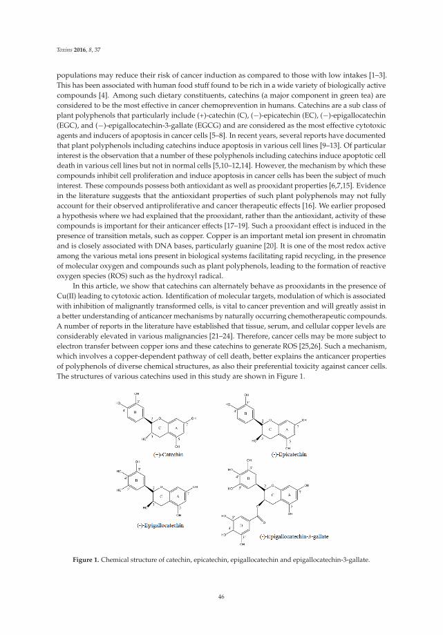
Toxins 2016, 8, 37
populations may reduce their risk of cancer induction as compared to those with low intakes [1–3].This has been associated with human food stuff found to be rich in a wide variety of biologically activecompounds [4]. Among such dietary constituents, catechins (a major component in green tea) areconsidered to be the most effective in cancer chemoprevention in humans. Catechins are a sub class ofplant polyphenols that particularly include (+)-catechin (C), (´)-epicatechin (EC), (´)-epigallocatechin(EGC), and (´)-epigallocatechin-3-gallate (EGCG) and are considered as the most effective cytotoxicagents and inducers of apoptosis in cancer cells [5–8]. In recent years, several reports have documentedthat plant polyphenols including catechins induce apoptosis in various cell lines [9–13]. Of particularinterest is the observation that a number of these polyphenols including catechins induce apoptotic celldeath in various cell lines but not in normal cells [5,10–12,14]. However, the mechanism by which thesecompounds inhibit cell proliferation and induce apoptosis in cancer cells has been the subject of muchinterest. These compounds possess both antioxidant as well as prooxidant properties [6,7,15]. Evidencein the literature suggests that the antioxidant properties of such plant polyphenols may not fullyaccount for their observed antiproliferative and cancer therapeutic effects [16]. We earlier proposeda hypothesis where we had explained that the prooxidant, rather than the antioxidant, activity of thesecompounds is important for their anticancer effects [17–19]. Such a prooxidant effect is induced in thepresence of transition metals, such as copper. Copper is an important metal ion present in chromatinand is closely associated with DNA bases, particularly guanine [20]. It is one of the most redox activeamong the various metal ions present in biological systems facilitating rapid recycling, in the presenceof molecular oxygen and compounds such as plant polyphenols, leading to the formation of reactiveoxygen species (ROS) such as the hydroxyl radical.
In this article, we show that catechins can alternately behave as prooxidants in the presence ofCu(II) leading to cytotoxic action. Identification of molecular targets, modulation of which is associatedwith inhibition of malignantly transformed cells, is vital to cancer prevention and will greatly assist ina better understanding of anticancer mechanisms by naturally occurring chemotherapeutic compounds.A number of reports in the literature have established that tissue, serum, and cellular copper levels areconsiderably elevated in various malignancies [21–24]. Therefore, cancer cells may be more subject toelectron transfer between copper ions and these catechins to generate ROS [25,26]. Such a mechanism,which involves a copper-dependent pathway of cell death, better explains the anticancer propertiesof polyphenols of diverse chemical structures, as also their preferential toxicity against cancer cells.The structures of various catechins used in this study are shown in Figure 1.
Figure 1. Chemical structure of catechin, epicatechin, epigallocatechin and epigallocatechin-3-gallate.
46
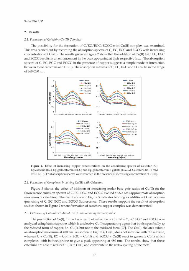
Toxins 2016, 8, 37
2. Results
2.1. Formation of Catechins-Cu(II) Complex
The possibility for the formation of C/EC/EGC/EGCG with Cu(II) complex was examined.This was carried out by recording the absorption spectra of C, EC, EGC and EGCG with increasingconcentrations of Cu(II). The results given in Figure 2 show that the addition of Cu(II) to C, EC, EGCand EGCG results in an enhancement in the peak appearing at their respective λmax. The absorptionspectra of C, EC, EGC and EGCG in the presence of copper suggests a simple mode of interactionbetween these catechins and Cu(II). The absorption maxima of C, EC, EGC and EGCG lie in the rangeof 260–280 nm.
Figure 2. Effect of increasing copper concentrations on the absorbance spectra of Catechin (C),Epicatechin (EC), Epigallocatechin (EGC) and Epigallocatechin-3-gallate (EGCG). Catechins (in 10 mMTris-HCl, pH 7.5) absorption spectra were recorded in the presence of increasing concentration of Cu(II).
2.2. Formation of Complexes Involving Cu(II) with Catechins
Figure 3 shows the effect of addition of increasing molar base pair ratios of Cu(II) on thefluorescence emission spectra of C, EC, EGC and EGCG excited at 273 nm (approximate absorptionmaximum of catechins). The result shown in Figure 3 indicates binding as addition of Cu(II) causesquenching of C, EC, EGC and EGCG fluorescence. These results support the result of absorptionstudies shown in Figure 2 where formation of catechins-copper complex was demonstrated.
2.3. Detection of Catechins Induced Cu(I) Production by Bathocuproine
The production of Cu(I), formed as a result of reduction of Cu(II) by C, EC, EGC and EGCG, wasanalyzed using bathocuproine which is a selective Cu(I) sequestering agent that binds specifically tothe reduced form of copper, i.e., Cu(I), but not to the oxidized form [27]. The Cu(I)-chelates exhibitan absorption maximum at 480 nm. As shown in Figure 4, Cu(II) does not interfere with the maxima,whereas C + Cu(II), EC + Cu(II), EGC + Cu(II) and EGCG + Cu(II) react to generate Cu(I) whichcomplexes with bathocuproine to give a peak appearing at 480 nm. The results show that thesecatechins are able to reduce Cu(II) to Cu(I) and contribute to the redox cycling of the metal.
47
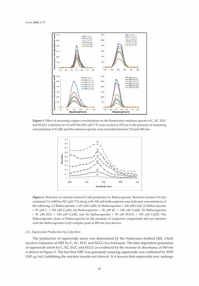
Toxins 2016, 8, 37
Figure 3. Effect of increasing copper concentrations on the fluorescence emission spectra of C, EC, EGCand EGCG. Catechins (in 10 mM Tris-HCl, pH 7.5) were excited at 273 nm in the presence of increasingconcentration of Cu(II) and the emission spectra were recorded between 510 and 580 nm.
Figure 4. Detection of catechin induced Cu(I) production by Bathocuproine. Reaction mixture (3.0 mL)contained 3.0 mMTris-HCl (pH 7.5) along with 300 μM bathocuproine and indicated concentrations ofthe following: (1) Bathocuproine + 100 μM Cu(II); (2) Bathocuproine + 100 μM Cu(I); (3) Bathocuproine+ 50 μM C + 100 μM Cu(II); (4) Bathocuproine + 50 μM EC + 100 μM Cu(II); (5) Bathocuproine+ 50 μM EGC + 100 μM Cu(II); and (6) Bathocuproine + 50 μM EGCG + 100 μM Cu(II). TheBathocuproine alone or Bathocuproine in the presence of respective compounds did not interferewith the Bathocuproine-Cu(I) complex peak at 480 nm (not shown).
2.4. Superoxide Production by Catechins
The production of superoxide anion was determined by the Nakayama method [28], whichinvolves reduction of NBT by C, EC, EGC and EGCG to a formazan. The time dependent generationof superoxide anion by C, EC, EGC and EGCG as evidenced by the increase in absorbance at 560 nmis shown in Figure 5. The fact that NBT was genuinely assaying superoxide was confirmed by SOD(100 μg/mL) inhibiting the reaction (results not shown). It is known that superoxide may undergo
48
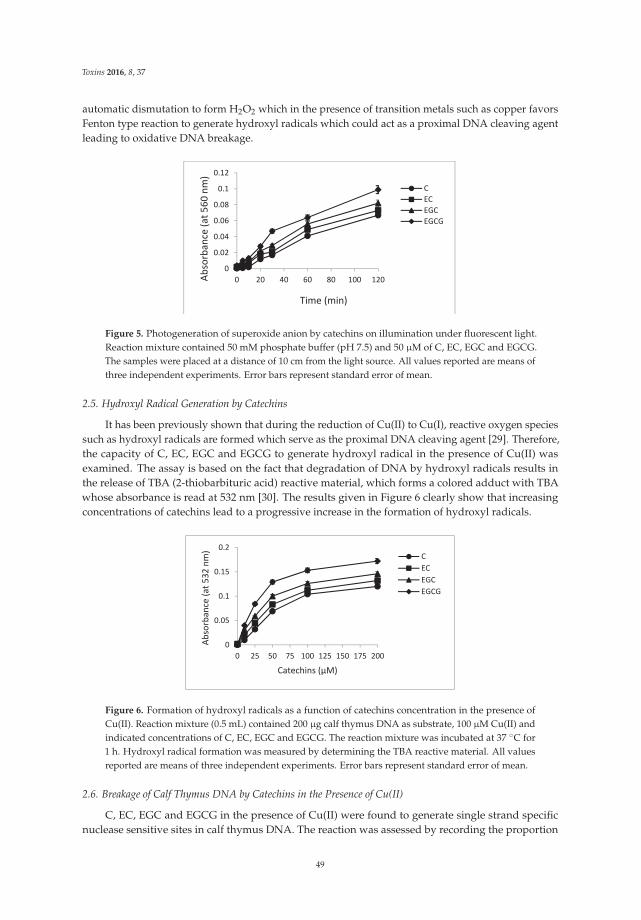
Toxins 2016, 8, 37
automatic dismutation to form H2O2 which in the presence of transition metals such as copper favorsFenton type reaction to generate hydroxyl radicals which could act as a proximal DNA cleaving agentleading to oxidative DNA breakage.
0
0.02
0.04
0.06
0.08
0.1
0.12
0 20 40 60 80 100 120Absorbance
(at5
60nm
)
Time (min)
CECEGCEGCG
Figure 5. Photogeneration of superoxide anion by catechins on illumination under fluorescent light.Reaction mixture contained 50 mM phosphate buffer (pH 7.5) and 50 μM of C, EC, EGC and EGCG.The samples were placed at a distance of 10 cm from the light source. All values reported are means ofthree independent experiments. Error bars represent standard error of mean.
2.5. Hydroxyl Radical Generation by Catechins
It has been previously shown that during the reduction of Cu(II) to Cu(I), reactive oxygen speciessuch as hydroxyl radicals are formed which serve as the proximal DNA cleaving agent [29]. Therefore,the capacity of C, EC, EGC and EGCG to generate hydroxyl radical in the presence of Cu(II) wasexamined. The assay is based on the fact that degradation of DNA by hydroxyl radicals results inthe release of TBA (2-thiobarbituric acid) reactive material, which forms a colored adduct with TBAwhose absorbance is read at 532 nm [30]. The results given in Figure 6 clearly show that increasingconcentrations of catechins lead to a progressive increase in the formation of hydroxyl radicals.
0
0.05
0.1
0.15
0.2
0 25 50 75 100 125 150 175 200
Absorbance
(at5
32nm
)
Catechins (μM)
CECEGCEGCG
Figure 6. Formation of hydroxyl radicals as a function of catechins concentration in the presence ofCu(II). Reaction mixture (0.5 mL) contained 200 μg calf thymus DNA as substrate, 100 μM Cu(II) andindicated concentrations of C, EC, EGC and EGCG. The reaction mixture was incubated at 37 ˝C for1 h. Hydroxyl radical formation was measured by determining the TBA reactive material. All valuesreported are means of three independent experiments. Error bars represent standard error of mean.
2.6. Breakage of Calf Thymus DNA by Catechins in the Presence of Cu(II)
C, EC, EGC and EGCG in the presence of Cu(II) were found to generate single strand specificnuclease sensitive sites in calf thymus DNA. The reaction was assessed by recording the proportion
49
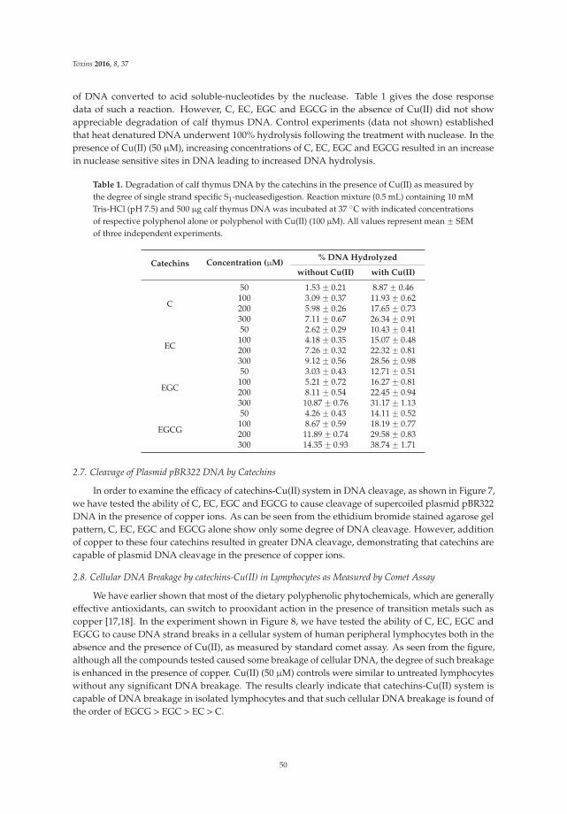
Toxins 2016, 8, 37
of DNA converted to acid soluble-nucleotides by the nuclease. Table 1 gives the dose responsedata of such a reaction. However, C, EC, EGC and EGCG in the absence of Cu(II) did not showappreciable degradation of calf thymus DNA. Control experiments (data not shown) establishedthat heat denatured DNA underwent 100% hydrolysis following the treatment with nuclease. In thepresence of Cu(II) (50 μM), increasing concentrations of C, EC, EGC and EGCG resulted in an increasein nuclease sensitive sites in DNA leading to increased DNA hydrolysis.
Table 1. Degradation of calf thymus DNA by the catechins in the presence of Cu(II) as measured bythe degree of single strand specific S1-nucleasedigestion. Reaction mixture (0.5 mL) containing 10 mMTris-HCl (pH 7.5) and 500 μg calf thymus DNA was incubated at 37 ˝C with indicated concentrationsof respective polyphenol alone or polyphenol with Cu(II) (100 μM). All values represent mean ˘ SEMof three independent experiments.
Catechins Concentration (μM)% DNA Hydrolyzed
without Cu(II) with Cu(II)
C
50 1.53 ˘ 0.21 8.87 ˘ 0.46100 3.09 ˘ 0.37 11.93 ˘ 0.62200 5.98 ˘ 0.26 17.65 ˘ 0.73300 7.11 ˘ 0.67 26.34 ˘ 0.91
EC
50 2.62 ˘ 0.29 10.43 ˘ 0.41100 4.18 ˘ 0.35 15.07 ˘ 0.48200 7.26 ˘ 0.32 22.32 ˘ 0.81300 9.12 ˘ 0.56 28.56 ˘ 0.98
EGC
50 3.03 ˘ 0.43 12.71 ˘ 0.51100 5.21 ˘ 0.72 16.27 ˘ 0.81200 8.11 ˘ 0.54 22.45 ˘ 0.94300 10.87 ˘ 0.76 31.17 ˘ 1.13
EGCG
50 4.26 ˘ 0.43 14.11 ˘ 0.52100 8.67 ˘ 0.59 18.19 ˘ 0.77200 11.89 ˘ 0.74 29.58 ˘ 0.83300 14.35 ˘ 0.93 38.74 ˘ 1.71
2.7. Cleavage of Plasmid pBR322 DNA by Catechins
In order to examine the efficacy of catechins-Cu(II) system in DNA cleavage, as shown in Figure 7,we have tested the ability of C, EC, EGC and EGCG to cause cleavage of supercoiled plasmid pBR322DNA in the presence of copper ions. As can be seen from the ethidium bromide stained agarose gelpattern, C, EC, EGC and EGCG alone show only some degree of DNA cleavage. However, additionof copper to these four catechins resulted in greater DNA cleavage, demonstrating that catechins arecapable of plasmid DNA cleavage in the presence of copper ions.
2.8. Cellular DNA Breakage by catechins-Cu(II) in Lymphocytes as Measured by Comet Assay
We have earlier shown that most of the dietary polyphenolic phytochemicals, which are generallyeffective antioxidants, can switch to prooxidant action in the presence of transition metals such ascopper [17,18]. In the experiment shown in Figure 8, we have tested the ability of C, EC, EGC andEGCG to cause DNA strand breaks in a cellular system of human peripheral lymphocytes both in theabsence and the presence of Cu(II), as measured by standard comet assay. As seen from the figure,although all the compounds tested caused some breakage of cellular DNA, the degree of such breakageis enhanced in the presence of copper. Cu(II) (50 μM) controls were similar to untreated lymphocyteswithout any significant DNA breakage. The results clearly indicate that catechins-Cu(II) system iscapable of DNA breakage in isolated lymphocytes and that such cellular DNA breakage is found ofthe order of EGCG > EGC > EC > C.
50
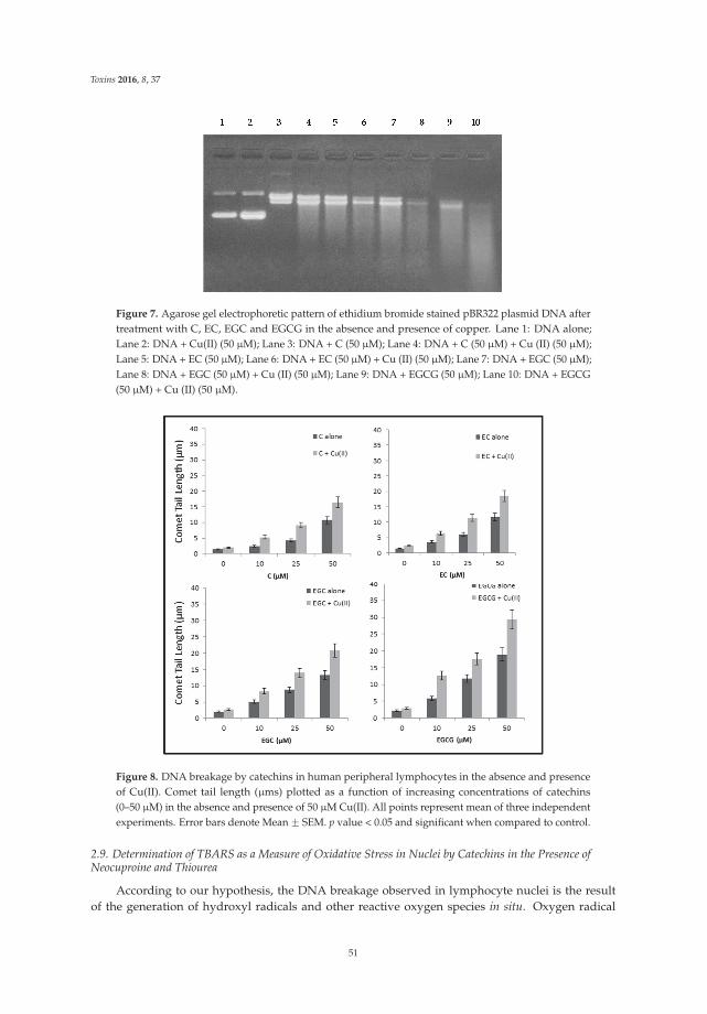
Toxins 2016, 8, 37
Figure 7. Agarose gel electrophoretic pattern of ethidium bromide stained pBR322 plasmid DNA aftertreatment with C, EC, EGC and EGCG in the absence and presence of copper. Lane 1: DNA alone;Lane 2: DNA + Cu(II) (50 μM); Lane 3: DNA + C (50 μM); Lane 4: DNA + C (50 μM) + Cu (II) (50 μM);Lane 5: DNA + EC (50 μM); Lane 6: DNA + EC (50 μM) + Cu (II) (50 μM); Lane 7: DNA + EGC (50 μM);Lane 8: DNA + EGC (50 μM) + Cu (II) (50 μM); Lane 9: DNA + EGCG (50 μM); Lane 10: DNA + EGCG(50 μM) + Cu (II) (50 μM).
Figure 8. DNA breakage by catechins in human peripheral lymphocytes in the absence and presenceof Cu(II). Comet tail length (μms) plotted as a function of increasing concentrations of catechins(0–50 μM) in the absence and presence of 50 μM Cu(II). All points represent mean of three independentexperiments. Error bars denote Mean ˘ SEM. p value < 0.05 and significant when compared to control.
2.9. Determination of TBARS as a Measure of Oxidative Stress in Nuclei by Catechins in the Presence ofNeocuproine and Thiourea
According to our hypothesis, the DNA breakage observed in lymphocyte nuclei is the resultof the generation of hydroxyl radicals and other reactive oxygen species in situ. Oxygen radical
51
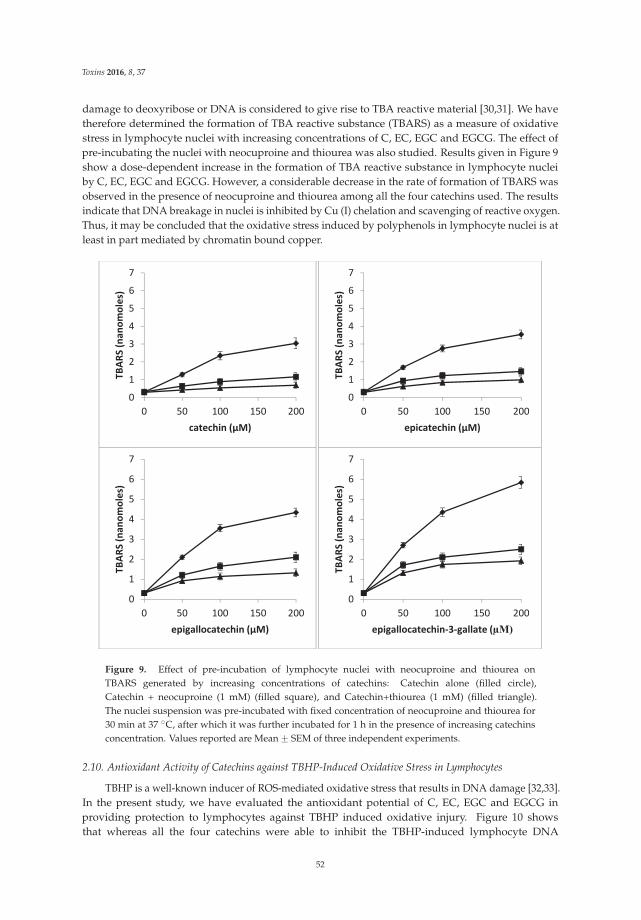
Toxins 2016, 8, 37
damage to deoxyribose or DNA is considered to give rise to TBA reactive material [30,31]. We havetherefore determined the formation of TBA reactive substance (TBARS) as a measure of oxidativestress in lymphocyte nuclei with increasing concentrations of C, EC, EGC and EGCG. The effect ofpre-incubating the nuclei with neocuproine and thiourea was also studied. Results given in Figure 9show a dose-dependent increase in the formation of TBA reactive substance in lymphocyte nucleiby C, EC, EGC and EGCG. However, a considerable decrease in the rate of formation of TBARS wasobserved in the presence of neocuproine and thiourea among all the four catechins used. The resultsindicate that DNA breakage in nuclei is inhibited by Cu (I) chelation and scavenging of reactive oxygen.Thus, it may be concluded that the oxidative stress induced by polyphenols in lymphocyte nuclei is atleast in part mediated by chromatin bound copper.
0
1
2
3
4
5
6
7
0 50 100 150 200
TBAR
S(nan
omoles)
catechin (μM)
0
1
2
3
4
5
6
7
0 50 100 150 200
TBAR
S(nan
omoles)
epicatechin (μM)
0
1
2
3
4
5
6
7
0 50 100 150 200
TBAR
S(nan
omoles)
epigallocatechin (μM)
0
1
2
3
4
5
6
7
0 50 100 150 200
TBAR
S(nan
omoles)
epigallocatechin 3 gallate (μM)
Figure 9. Effect of pre-incubation of lymphocyte nuclei with neocuproine and thiourea onTBARS generated by increasing concentrations of catechins: Catechin alone (filled circle),Catechin + neocuproine (1 mM) (filled square), and Catechin+thiourea (1 mM) (filled triangle).The nuclei suspension was pre-incubated with fixed concentration of neocuproine and thiourea for30 min at 37 ˝C, after which it was further incubated for 1 h in the presence of increasing catechinsconcentration. Values reported are Mean ˘ SEM of three independent experiments.
2.10. Antioxidant Activity of Catechins against TBHP-Induced Oxidative Stress in Lymphocytes
TBHP is a well-known inducer of ROS-mediated oxidative stress that results in DNA damage [32,33].In the present study, we have evaluated the antioxidant potential of C, EC, EGC and EGCG inproviding protection to lymphocytes against TBHP induced oxidative injury. Figure 10 showsthat whereas all the four catechins were able to inhibit the TBHP-induced lymphocyte DNA
52
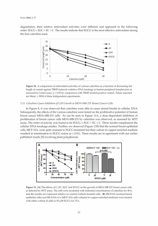
Toxins 2016, 8, 37
degradation, their relative antioxidant activities were different and appeared in the followingorder: EGCG > EGC > EC > C. The results indicate that EGCG is the most effective antioxidant amongthe four catechins used.
0
5
10
15
20
25
30
35
0 10 20 30
Comet
taillength
(μm)
Catechins (μM)
C
EC
EGC
EGCG
Figure 10. A comparison of antioxidant activities of various catechins as a function of decreasing taillength of comets against TBHP-induced oxidative DNA breakage in human peripheral lymphocytes asassessed by Comet assay. p < 0.05 by comparison with TBHP-treated positive control. Values reportedare Mean ˘ SEM of three independent experiments.
2.11. Catechins Cause Inhibition of Cell Growth in MDA-MB-231 Breast Cancer Cells
In Figure 8, it was observed that catechins were able to cause strand breaks in cellular DNA.Subsequently, the effects of the various catechins were tested on the proliferative potential of humanbreast cancer MDA-MB-231 cells. As can be seen in Figure 11A, a dose-dependent inhibition ofproliferation of breast cancer cells MDA-MB-231 by catechins was observed, as assessed by MTTassay. The order of activity was found to be EGCG > EGC > EC > C. These results complement thecellular DNA breakage studies. Further, we observed (Figure 11B) that the normal breast epithelialcells, MCF-10A, were quite resistant to EGCG treatment but their culture in copper-enriched mediumresulted in sensitization to EGCG action (p < 0.01). These results are in agreement with our earlierpublished results [5] involving plant polyphenols.
Figure 11. (A) The effects of C, EC, EGC and EGCG on the growth of MDA-MB-231 breast cancer cellsas detected by MTT assay. The cells were incubated with indicated concentrations of catechins for 48 h,and the results are expressed relative to control (vehicle-treated) cells. (B) MCF10A (normal breastepithelial cells) and MCF10A+Cu (MCF-10A cells cultured in copper-enriched medium) were treatedwith either vehicle (0 μM) or 50 μM EGCG for 72 h.
53
Toxins 2016, 8, 37
3. Discussion
Studies mainly on anticancer mechanisms of plant polyphenols involve the induction of cell cyclearrest and modulation of transcription factors that lead to anti-neoplastic effects [10,34]. In light of theabove findings in our laboratory and those of many others in the literature, it may be concluded that theplant polyphenols, particularly present in dietary agents, possessing anticancer and apoptosis-inducingactivities are able to mobilize endogenous copper ions, possibly the copper bound to chromatin.Essentially, this would be an alternative, non-enzymatic, and copper-dependent pathway for thecytotoxic action of anticancer agents that are capable of mobilizing and reducing endogenous copper.As such, this would be independent of Fas and mitochondria mediated programmed cell deaths. It isconceivable that such a mechanism may also lead to internucleosomal DNA breakage (a hallmarkof apoptosis), as internucleosomal spacer DNA would be relatively more susceptible to cleavage byROS. Indeed, such a common mechanism better explains the anticancer effects of dietary molecules(i.e., catechins) studied above with diverse chemical structures as also their preferential cytotoxicitytoward cancer cells. The generation of hydroxyl radicals in the proximity of DNA is well establishedas a cause of strand scission. It is generally recognized that such a reaction with DNA is precededby the association of a ligand with DNA followed by the formation of hydroxyl radicals at that site.The location of the redox-active metal is of utmost importance because the hydroxyl radical, owing to itsextreme reactivity, interacts exclusively in the vicinity of the bound metal [35]. Copper ions are knownto interact with both DNA phosphates and the bases through coordination binding [36]. Further, copperis also present in chromatin and is closely associated with DNA bases, particularly guanine [20]. Directinteraction of catechins with the DNA bound copper ions in a ternary complex and localized generationof non-diffusible hydroxyl radicals is a likely mechanism involved in catechin/Cu(II)-induced DNAcleavage. It has already been reported by us that the number of galloyl moieties present in catechinsplayan important role in cellular DNA breakage and catechins on reducing copper lead to the formation of“oxidized species” of the compounds. [26]. It may be presumed that the concentration of metals such ascopper in cells is a decisive factor in driving antioxidant property of catechins toward their prooxidantaction. As already mentioned, cancer cells are known to contain elevated levels of copper [37] andtherefore may be more subject to electron transfer with catechins to generate ROS [25,26]. In normalcells, there exists a balance between the free radical generation and the antioxidant defense [38].However, it has been clearly documented that tumor cells are under persistent oxidative stress andhave an altered antioxidant system [39] and thus further ROS stress in malignant cells reachinga threshold level could result in apoptosis [37]. These observations further suggest that neoplastic cellsmay be more vulnerable to oxidative stress because they function with a heightened basal level of ROSbecause of increased rate of growth and metabolism [40]. Thus, in cancer cells, an enhanced exposureto ROS generated through the antioxidant/catechin-induced redox activity of endogenous copper canoverwhelm the cells antioxidant capacity, leading to irreversible damage and apoptosis.
Beyond the preclinical findings, catechins such as EGCG show little promise as potentchemopreventive agents in the clinical settings, mainly owing to their inefficient systemic deliveryand poor bioavailability. Catechins, like other polyphenols, are rapidly metabolized in vivo, resultingin a short systemic half-life and low plasma concentrations in the free form. For instance, it has beenobserved that the peak plasma concentrations reached up to 1.3 μM and 3.1 μM in healthy volunteersreceiving 687.5 mg EGCG and 663.5 mg of ECG, respectively [41]. In this context, it needs to beemphasized that the significance of our work lies not so much in the potential therapeutic action ofcatechins against cancer, but in establishing a principle, namely that it is possible to mobilize elevatedlevels of endogenous copper in cancer cells by catechins to promote a pro-oxidant cell death. Oncesuch a principle is established, catechins can serve as lead compounds to synthesize/formulate novel,anticancer drugs with superior bioavailability and extended systemic half-life. Presumably, suchanticancer drugs would have a better therapeutic impact than the catechins per se.
Nevertheless, a previous study from our lab has shown that oral administration of copper torats can induce a copper overload in their lymphocytes, rendering such isolated lymphocytes more
54
Toxins 2016, 8, 37
susceptible to EGCG-induced pro-oxidant cellular DNA breakage [42]. This suggests that in cancercells, where there are considerably higher levels of copper, the concentration of catechins requiredto elicit such a pro-oxidant cell death mechanism would be significantly lower. Therefore, based onthese observations, it is clear that the pro-oxidant action of polyphenols is physiologically feasibleprovided an appropriate microenvironment with elevated copper levels is present, as is the scenario incancer cells.
Further, it should be noted that in real life situation, catechins like EGCG are only one of the severalpolyphenols consumed as part of the diet. Since various other polyphenols present in diet, such asflavonoids and tannins are also active as pro-oxidants [17], their cumulative plasma concentration,bioavailability and anticancer effect should be much greater than a single polyphenol alone. In fact,a combination of EGCG with luteolin, has been found to be more effective than either of the polyphenolalone in inducing apoptosis in cancer cell lines in vitro and inhibition of tumor growth in nude mousexenograft model [43].
4. Materials and Methods
4.1. Chemicals, Reagents and Cell Line
(+)-Catechin, (´)-epicatechin, (´)-epigallocatechin, (´)-epigallocatechin-3-gallate, calf thymusDNA, cupric chloride, neocuproine, thiourea, agarose, low melting point agarose, RPMI 1640, TritonX-100, Trypan blue, Histopaque1077, and phosphate buffered saline (PBS) Ca2+ and Mg2+ free werepurchased from Sigma (St. Louis, MO, USA). All other chemicals were of analytical grade. Freshsolutions of C, EC, EGC and EGCG were prepared as a stock of 3.0 mM in double distilled water(ddH2O) before use as a stock of 1 mM solution. Upon addition to reaction mixtures, in the presence ofbuffers mentioned and at concentrations used, all the catechins used remained in solution. Thevolumes of stock solution added did not lead to any appreciable change in the pH of reactionmixtures. Breast cancer cell line MDA-MB-231 was maintained in DMEM (Invitrogen, Carlsbad,CA, USA) growth media. The medium was supplemented with 10% foetal bovine serum (FBS) and 1%antimycotic antibiotic (Invitrogen, Carlsbad, CA, USA). Normal breast epithelial cells MCF10A werecultured in DMEM/F12 (Invitrogen) supplemented with 5% horse serum, 20 ng/mL EGF, 0.5 μg/mLhydrocortisone, 0.1 μg/mL cholera toxin, 10 μg/mL insulin, 100 units/mL penicillin and 100 μg/mLstreptomycin. Cells were cultured in a 5% CO2-humidified atmosphere at 37 ˝C. MCF10A+Cu cellsare MCF10A cells cultured with additional supplementation of 25 μM CuCl2 for at least 4 weeks.A 5 mg/mL stock solution of 3-(4,5-dimethylthiazol-2-yl)-2,5-diphenyltetrazolium bromide (MTT) wasprepared in PBS.
4.2. Absorbance Studies
The effect of increasing concentrations of Cu(II) on absorption spectra of C, EC, EGC and EGCGwas observed. The reaction mixture (3.0 mL) contained 10 mMTris-HCl (pH 7.5), 50 μM C, EC, EGC,EGCG and increasing concentrations of Cu(II). The spectra were recorded immediately after additionof all components.
4.3. Flourescence Studies
The fluorescence studies were performed on a Shimadzu spectrofluorometer RF-5310 PC (Kyoto,Japan) equipped with a plotter and a calculator. C, EC, EGC and EGCG were excited at their absorptionmaxima (λmax) of 273 nm (approximate absorption maximum of catechins). Emission spectra wererecorded in the wavelength range shown in figures.
4.4. Detection of Cu(II) Reduction
The selective sequestering agent bathocuproine was employed to detect reduction of Cu(II)to Cu(I) by recording the formation of bathocuproine-Cu(I) complex which absorbs maximally at
55
Toxins 2016, 8, 37
480 nm. The reaction mixture (3.0 mL) contained 3.0 mM Tris-HCl (pH 7.5), fixed concentrations(100 μM) of Cu(II) and Cu(I) (for positive control), bathocuproine (300 μM) and of catechins (C, EC,EGC and EGCG) (50 μM). The reaction was started by adding Cu(II) and the spectra were recordedimmediately afterwards.
4.5. Detection of Superoxide Anion Generation
Superoxide (O2´) was detected by the reduction of nitroblue tetrazolium (NBT) essentially as
described by Nakayama et al. [28]. A typical assay mixture contained 50 mM sodium phosphate buffer(pH 7.5), 33 μM NBT, 100 μM EDTA and 0.06% triton X-100 in a total volume of 3.0 mL. The reactionwas started by the addition of catechins (C/EC/EGC/EGCG). After mixing, absorbance was recordedat 560 nm at different time intervals, against a blank, which did not contain the compound.
4.6. Detection of Hydroxyl Radical Generation
In order to compare the hydroxyl radical production by increasing concentrations of C, EC,EGC and EGCG in the presence of 100 μM Cu(II), the method of Quinlan and Gutteridge [30] wasfollowed. Calf thymus DNA (200 μg) was used as a substrate and the malondialdehyde generatedfrom deoxyribose radicals was assayed by recording the absorbance at 532 nm.
4.7. Degradation of Calf Thymus DNA
Single strand specific digestion was performed as described by Wani and Hadi [44]. Reactionmixtures (0.5 mL) contained 10 mM Tris-HCl (pH 7.5), 500 μg of calf thymus DNA and varying amountsof C, EC, EGC, EGCG and cupric chloride (50 μM). All solutions were sterilized before use. Incubationwas performed at 37 ˝C for one hour. The assay determines the acid soluble nucleotides released fromDNA as a result of enzyme digestion. Reaction mixture in a total volume of 1.0 mL contained 40 mMTris-HCl (pH 7.5), 1 mM Magnesium Chloride, water and enzyme. The reaction mixture was incubatedat 37 ˝C for 2 h. The reaction was stopped by adding 0.2 mL bovine serum albumin (10 mg/mL)and 1.0 mL of 14% perchloric acid (chilled). The tubes were immediately transferred to 0 ˝C for45 min before centrifugation at 2500 rpm for 10 min at room temperature to remove undigested DNAand precipitated protein. Acid soluble deoxyribonucleotides were determined in the supernatant,colorimetrically, using the diphenylamine method [45]. To a 1.0 mL aliquot, 2.0 mL diphenyl reagent(freshly prepared by dissolving 1 gram of recrystallized diphenylamine in 100 mL glacial acetic acidand 2.75 mL of concentrated H2SO4) was added. The tubes were heated in a boiling water bath for30 min. The intensity of blue color was read at 600 nm.
4.8. Treatment of pBR322 DNA
Reaction mixture (30 μL) contained 10 mMTris-HCl (pH 7.5), 0.5 μg of plasmid DNA and othercomponents as indicated in legends. Incubation was performed at 37 ˝C for 2 h. After incubation, 10 μLof solution containing 40 mM EDTA, 0.05% bromophenol blue (tracking dye) and 50% (v/v) glycerolwas added and the solution was subjected to electrophoresis in submarine 1% agarose gel. The gel wasstained with ethidium bromide (0.5 μg/mL), viewed and photographed on a UV-transilluminator.
4.9. Isolation of Lymphocytes
Heparinized blood samples (2 mL) from a single, healthy, non-smoking donor was obtained byvenepuncture and diluted suitably in Ca2+ and Mg2+ free PBS. Lymphocytes were isolated from bloodusing Histopaque 1077 (Sigma Diagnostics, St Louis, MS, USA), and the cells were finally suspendedin RPMI 1640.
56
Toxins 2016, 8, 37
4.10. Viability Assessment of Lymphocytes
The lymphocytes were checked for their viability before the start and after the end of the reactionusing Trypan Blue Exclusion Test by Pool-Zobel et al. [46]. The viability of the cells was found to begreater than 93%.
4.11. Comet Assay
Comet assay was performed under alkaline conditions essentially according to the procedure ofSingh et al. [47] with slight modifications. Fully frosted microscopic slides precoated with 1.0% normalmelting agarose at about 50 ˝C (dissolved in Ca2+ and Mg2+ free PBS) were used. Approximately10,000 cells were mixed with 75 μL of 2.0% LMPA to form a cell suspension and pipetted over thefirst layer and covered immediately by a coverslip. The agarose layer was allowed to solidify byplacing the slides on a flat tray and keeping it on ice for 10 min. The coverslips were removed anda third layer of 0.5% LMPA (75 μL) was pipetted and coverslips placed over it and kept on ice for5 min for proper solidification of layer. The coverslips were removed and the slides were immersedin cold lysing solution containing 2.5 M NaCl, 100 mM EDTA, 10 mM Tris, pH 10, and 1% TritonX-100 added just prior to use for a minimum of 1 h at 4 ˝C. After lysis DNA was allowed to unwindfor 30 min in alkaline electrophoretic solution consisting of 300 mM NaOH, 1 mM EDTA, pH > 13.Electrophoresis was performed at 4 ˝C in a field strength of 0.7 V/cm and 300 mA current. The slideswere then neutralized with cold 0.4 M Tris, pH 7.5, stained with 75 μL Ethidium Bromide (20 μg/mL)and covered with a coverslip. The slides were placed in a humidified chamber to prevent drying of thegel and analyzed the same day. Slides were scored using an image analysis system (Komet 5.5, KineticImaging, Liverpool, UK) attached to a Olympus (CX41) fluorescent microscope and a COHU 4910(equipped with a 510–560 nm excitation and 590 nm barrier filters) integrated CC camera. Cometswere scored at 100ˆ magnification. Images from 50 cells (25 from each replicate slide) were analyzed.The parameter taken to assess lymphocytes DNA damage was tail length (migration of DNA from thenucleus, μm) and was automatically generated by Komet 5.5 image analysis system.
Treatment of intact lymphocytes with four catechins (C, EC, EGC and EGCG) and the subsequentComet assay was performed essentially as described earlier by Azmi et al. [48]. For antioxidantstudy [49], the cells were preincubated with polyphenols in eppendorf tubes in a reaction volume of1.0 mL. After the preincubation (for 30 min at 37 ˝C), the reaction mixture was centrifuged at 4000 rpm,the supernatant was discarded and the pelleted lymphocytes were resuspended in 100 μL of PBS (Ca2+
and Mg2+ free) and layered for further treatment with TBHP (50 μM). The incubation period was30 min at 37 ˝C in dark. The other conditions remained the same as described above.
4.12. Determination of TBARS
Thiobarbituric acid reactive substance was determined according to the method ofRamanathan et al. [50]. A cell suspension (1 ˆ 105/mL) was incubated with C, EC, EGC and EGCG(0–200 μM) at 37 ˝C for 1 h and then centrifuged at 1000 rpm. In some experiments the cells werepre-incubated with fixed concentrations of neocuproine and thiourea. The cell pellet was washedtwice with phosphate buffered saline (Ca2+ and Mg2+ free) and suspended in 0.1 N NaOH. This cellsuspension (1.4 mL) was further treated with 10% TCA and 0.6 M TBA (2-thiobarbituric acid) in boilingwater bath for 10 min. The absorbance was read at 532 nm and converted into nmoles of TBA reactivesubstance using the molar extinction coefficient (1.56 ˆ 9 ˆ 105 M´1¨ cm´1).
4.13. Cell Growth Inhibition Studies by MTT Assay
MDA-MB-231 cells were seeded at a density of 1 ˆ 104 cells per well in 96-well microtiter cultureplates. After overnight incubation, normal growth medium was removed and replaced with eitherfresh medium (untreated control) or different concentrations of respective catechins in growth medium.After the desired time of incubation (24 or 48 h), MTT solution was added to each well (0.1 mg/mL in
57
Toxins 2016, 8, 37
DMEM) and incubated further for 4 hours at 37 ˝C. Upon termination, the supernatant was aspiratedand the MTT formazan, formed by metabolically viable cells, was dissolved in a solubilisation solutioncontaining DMSO (100 μL) by mixing for 5 min on a gyratory shaker. The absorbance was measuredat 540 nm (reference wavelength 690 nm) on an Ultra Multifunctional Microplate Reader (Bio-Rad,Hercules, CA, USA). Absorbance of control (without treatment) was considered as 100% cell survival.Each treatment had four replicate wells and the mean values were plotted.
4.14. Statistics
The statistical analysis was performed as described by Tice et al. [51] and is expressed asmean ˘ SEM/SD of three independent experiments. A student’s t-test was used for examiningstatistically significant differences. Analysis of variance was performed using ANOVA. p values < 0.05were considered statistically significant.
Acknowledgments: Acknowledgments: Authors are thankful to BSR, UGC, New Delhi, for providing financialassistance for the research work. We are also thankful to the Department of Biochemistry, AMU, Aligarh forproviding us the necessary facilities.
Author Contributions: Author Contributions: MF, HYK, MO, AAH, NR and HA performed experiments. MFanalyzed data and drafted manuscript. AA helped in the manuscript draft. SMH conceptualized the study,provided facilities and oversaw the project.
Conflicts of Interest: Conflicts of Interest: The authors declare no conflict of interest.
References
1. Adlercreutz, C.H.; Goldin, B.R.; Gorbach, S.L.; Hockerstedt, K.A.; Watanabe, S.; Hamalainen, E.K.;Markkanen, M.H.; Makela, T.H.; Wahala, K.T.; Adlercreutz, T. Soybean phytoestrogen intake and cancer risk.J. Nutr. 1995, 125, 757S–770S. [PubMed]
2. Park, O.J.; Surh, Y.J. Chemopreventive potential of epigallocatechin gallate and genistein: Evidence fromepidemiological and laboratory studies. Toxicol. Lett. 2004, 150, 43–56. [CrossRef] [PubMed]
3. Barnes, S.; Peterson, G.; Grubbs, C.; Setchell, K. Potential role of dietary isoflavones in the prevention ofcancer. Adv. Exp. Med. Biol. 1994, 354, 135–147. [PubMed]
4. Surh, Y.J. Cancer chemoprevention with dietary phytochemicals. Nat. Rev. Cancer 2003, 3, 768–780. [CrossRef][PubMed]
5. Khan, H.Y.; Zubair, H.; Faisal, M.; Ullah, M.F.; Farhan, M.; Sarkar, F.H.; Ahmad, A.; Hadi, S.M. Plantpolyphenol induced cell death in human cancer cells involves mobilization of intracellular copper ions andreactive oxygen species generation: A mechanism for cancer chemopreventive action. Mol. Nutr. Food Res.2014, 58, 437–446. [CrossRef] [PubMed]
6. Yang, G.Y.; Liao, J.; Kim, K.; Yurkow, E.J.; Yang, C.S. Inhibition of growth and induction of apoptosis inhuman cancer cell lines by tea polyphenols. Carcinogenesis 1998, 19, 611–616. [CrossRef] [PubMed]
7. Shamim, U.; Hanif, S.; Ullah, M.F.; Azmi, A.S.; Bhat, S.H.; Hadi, S.M. Plant polyphenols mobilize nuclearcopper in human peripheral lymphocytes leading to oxidatively generated DNA breakage: Implications foran anticancer mechanism. Free Radic. Res. 2008, 42, 764–772. [CrossRef] [PubMed]
8. Ullah, M.F.; Ahmad, A.; Khan, H.Y.; Zubair, H.; Sarkar, F.H.; Hadi, S.M. The prooxidant action of dietaryantioxidants leading to cellular DNA breakage and anticancer effects: Implications for chemotherapeuticaction against cancer. Cell Biochem. Biophys. 2013, 67, 431–438. [CrossRef] [PubMed]
9. Clement, M.V.; Hirpara, J.L.; Chawdhury, S.H.; Pervaiz, S. Chemopreventive agent resveratrol, a naturalproduct derived from grapes, triggers cd95 signaling-dependent apoptosis in human tumor cells. Blood 1998,92, 996–1002. [PubMed]
10. Kuo, M.L.; Huang, T.S.; Lin, J.K. Curcumin, an antioxidant and anti-tumor promoter, induces apoptosis inhuman leukemia cells. Biochim. Biophys. Acta 1996, 1317, 95–100. [CrossRef]
11. Chang, K.L.; Cheng, H.L.; Huang, L.W.; Hsieh, B.S.; Hu, Y.C.; Chih, T.T.; Shyu, H.W.; Su, S.J. Combinedeffects of terazosin and genistein on a metastatic, hormone-independent human prostate cancer cell line.Cancer Lett. 2009, 276, 14–20. [CrossRef] [PubMed]
58

Toxins 2016, 8, 37
12. Gupta, S.; Hastak, K.; Ahmad, N.; Lewin, J.S.; Mukhtar, H. Inhibition of prostate carcinogenesis in trampmice by oral infusion of green tea polyphenols. Proc. Natl. Acad. Sci. USA 2001, 98, 10350–10355. [CrossRef][PubMed]
13. Orsolic, N.; Knezevic, A.; Sver, L.; Terzic, S.; Hackenberger, B.K.; Basic, I. Influence of honey bee products ontransplantable murine tumours. Vet. Comp. Oncol. 2003, 1, 216–226. [CrossRef] [PubMed]
14. Chen, Z.P.; Schell, J.B.; Ho, C.T.; Chen, K.Y. Green tea epigallocatechin gallate shows a pronounced growthinhibitory effect on cancerous cells but not on their normal counterparts. Cancer Lett. 1998, 129, 173–179.[CrossRef]
15. Said Ahmad, M.; Fazal, F.; Rahman, A.; Hadi, S.M.; Parish, J.H. Activities of flavonoids for the cleavage ofDNA in the presence of cu(ii): Correlation with generation of active oxygen species. Carcinogenesis 1992,13, 605–608. [CrossRef] [PubMed]
16. Gali, H.U.; Perchellet, E.M.; Klish, D.S.; Johnson, J.M.; Perchellet, J.P. Hydrolyzable tannins:Potent inhibitors of hydroperoxide production and tumor promotion in mouse skin treated with12-o-tetradecanoylphorbol-13-acetate in vivo. Int. J. Cancer 1992, 51, 425–432. [CrossRef] [PubMed]
17. Hadi, S.M.; Asad, S.F.; Singh, S.; Ahmad, A. Putative mechanism for anticancer and apoptosis-inducingproperties of plant-derived polyphenolic compounds. IUBMB Life 2000, 50, 167–171. [PubMed]
18. Hadi, S.M.; Bhat, S.H.; Azmi, A.S.; Hanif, S.; Shamim, U.; Ullah, M.F. Oxidative breakage of cellular DNA byplant polyphenols: A putative mechanism for anticancer properties. Semin. Cancer Biol. 2007, 17, 370–376.[CrossRef] [PubMed]
19. Bhat, S.H.; Azmi, A.S.; Hanif, S.; Hadi, S.M. Ascorbic acid mobilizes endogenous copper in human peripherallymphocytes leading to oxidative DNA breakage: A putative mechanism for anticancer properties. Int. J.Biochem. Cell Biol. 2006, 38, 2074–2081. [CrossRef] [PubMed]
20. Kagawa, T.F.; Geierstanger, B.H.; Wang, A.H.; Ho, P.S. Covalent modification of guanine bases indouble-stranded DNA. The 1.2-A Z-DNA structure of d(CGCGCG) in the presence of CuCl2. J. Biol. Chem.1991, 266, 20175–20184. [PubMed]
21. Ebadi, M.; Swanson, S. The status of zinc, copper, and metallothionein in cancer patients. Prog. Clin. Biol. Res.1988, 259, 161–175. [PubMed]
22. Margalioth, E.J.; Udassin, R.; Cohen, C.; Maor, J.; Anteby, S.O.; Schenker, J.G. Serum copper level ingynecologic malignancies. Am. J. Obstet. Gynecol. 1987, 157, 93–96. [CrossRef]
23. Yoshida, D.; Ikeda, Y.; Nakazawa, S. Quantitative analysis of copper, zinc and copper/zinc ratio in selectedhuman brain tumors. J. Neuro-Oncol. 1993, 16, 109–115. [CrossRef]
24. Ebara, M.; Fukuda, H.; Hatano, R.; Saisho, H.; Nagato, Y.; Suzuki, K.; Nakajima, K.; Yukawa, M.; Kondo, F.;Nakayama, A.; et al. Relationship between copper, zinc and metallothionein in hepatocellular carcinoma andits surrounding liver parenchyma. J. Hepatol. 2000, 33, 415–422. [CrossRef]
25. Zheng, L.F.; Wei, Q.Y.; Cai, Y.J.; Fang, J.G.; Zhou, B.; Yang, L.; Liu, Z.L. DNA damage induced by resveratroland its synthetic analogues in the presence of Cu (II) ions: Mechanism and structure-activity relationship.Free Radic. Biol. Med. 2006, 41, 1807–1816. [CrossRef] [PubMed]
26. Farhan, M.; Zafar, A.; Chibber, S.; Khan, H.Y.; Arif, H.; Hadi, S.M. Mobilization of copper ions in humanperipheral lymphocytes by catechins leading to oxidative DNA breakage: A structure activity study.Arch. Biochem. Biophys. 2015, 580, 31–40. [CrossRef] [PubMed]
27. Simpson, J.A.; Narita, S.; Gieseg, S.; Gebicki, S.; Gebicki, J.M.; Dean, R.T. Long-lived reactive species onfree-radical-damaged proteins. Biochem. J. 1992, 282, 621–624. [CrossRef] [PubMed]
28. Nakayama, T.; Kimura, T.; Kodama, M.; Nagata, C. Generation of hydrogen peroxide and superoxideanion from active metabolites of naphthylamines and aminoazo dyes: Its possible role in carcinogenesis.Carcinogenesis 1983, 4, 765–769. [CrossRef] [PubMed]
29. Rahman, A.; Shahabuddin; Hadi, S.M.; Parish, J.H.; Ainley, K. Strand scission in DNA induced by quercetinand Cu(II): Role of Cu(I) and oxygen free radicals. Carcinogenesis 1989, 10, 1833–1839. [CrossRef] [PubMed]
30. Quinlan, G.J.; Gutteridge, J.M. Oxygen radical damage to DNA by rifamycin sv and copper ions.Biochem. Pharmacol. 1987, 36, 3629–3633. [CrossRef]
31. Smith, C.; Halliwell, B.; Aruoma, O.I. Protection by albumin against the pro-oxidant actions of phenolicdietary components. Food Chem. Toxicol. 1992, 30, 483–489. [CrossRef]
59
Toxins 2016, 8, 37
32. Dubuisson, M.L.; de Wergifosse, B.; Trouet, A.; Baguet, F.; Marchand-Brynaert, J.; Rees, J.F. Antioxidativeproperties of natural coelenterazine and synthetic methyl coelenterazine in rat hepatocytes subjected totert-butyl hydroperoxide-induced oxidative stress. Biochem. Pharmacol. 2000, 60, 471–478. [CrossRef]
33. Suzuki, Y.; Apostolova, M.D.; Cherian, M.G. Astrocyte cultures from transgenic mice to study the role ofmetallothionein in cytotoxicity of tert-butyl hydroperoxide. Toxicology 2000, 145, 51–62. [CrossRef]
34. Ahmad, N.; Feyes, D.K.; Nieminen, A.L.; Agarwal, R.; Mukhtar, H. Green tea constituentepigallocatechin-3-gallate and induction of apoptosis and cell cycle arrest in human carcinoma cells. J. Natl.Cancer Inst. 1997, 89, 1881–1886. [CrossRef] [PubMed]
35. Chevion, M. A site-specific mechanism for free radical induced biological damage: The essential role ofredox-active transition metals. Free Radic. Biol. Med. 1988, 5, 27–37. [CrossRef]
36. Zimmer, C.; Luck, G.; Fritzsche, H.; Triebel, H. DNA-copper (II) complex and the DNA conformation.Biopolymers 1971, 10, 441–463. [CrossRef] [PubMed]
37. Gupte, A.; Mumper, R.J. Elevated copper and oxidative stress in cancer cells as a target for cancer treatment.Cancer Treat. Rev. 2009, 35, 32–46. [CrossRef] [PubMed]
38. Devi, G.S.; Prasad, M.H.; Saraswathi, I.; Raghu, D.; Rao, D.N.; Reddy, P.P. Free radicals antioxidant enzymesand lipid peroxidation in different types of leukemias. Clin. Chim. Acta 2000, 293, 53–62. [CrossRef]
39. Oberley, T.D.; Oberley, L.W. Antioxidant enzyme levels in cancer. Histol. Histopathol. 1997, 12, 525–535.[PubMed]
40. Kong, Q.; Beel, J.A.; Lillehei, K.O. A threshold concept for cancer therapy. Med. Hypotheses 2000, 55, 29–35.[CrossRef] [PubMed]
41. Van Amelsvoort, J.M.; Van Hof, K.H.; Mathot, J.N.; Mulder, T.P.; Wiersma, A.; Tijburg, L.B. Plasmaconcentrations of individual tea catechins after a single oral dose in humans. Xenobiotica: Fate ForeignCompd. Biol. Syst. 2001, 31, 891–901. [CrossRef] [PubMed]
42. Khan, H.Y.; Zubair, H.; Ullah, M.F.; Ahmad, A.; Hadi, S.M. Oral administration of copper to rats leadsto increased lymphocyte cellular DNA degradation by dietary polyphenols: Implications for a cancerpreventive mechanism. Biometals 2011, 24, 1169–1178. [CrossRef] [PubMed]
43. Amin, A.R.; Wang, D.; Zhang, H.; Peng, S.; Shin, H.J.; Brandes, J.C.; Tighiouart, M.; Khuri, F.R.;Chen, Z.G.; Shin, D.M. Enhanced anti-tumor activity by the combination of the natural compounds(´)-epigallocatechin-3-gallate and luteolin: Potential role of p53. J. Biol. Chem. 2010, 285, 34557–34565.[CrossRef] [PubMed]
44. Wani, A.A.; Hadi, S.M. Partial purification and properties of an endonuclease from germinating pea seedsspecific for single-stranded DNA. Arch. Biochem. Biophys. 1979, 196, 138–146. [CrossRef]
45. Schneider, W.C. Determination of nucleic acids in tissues by pentose analysis. Methods Enzymol 1957,3, 880–884.
46. Pool-Zobel, B.L.; Guigas, C.; Klein, R.; Neudecker, C.; Renner, H.W.; Schmezer, P. Assessment of genotoxiceffects by lindane. Food Chem. Toxicol. 1993, 31, 271–283. [CrossRef]
47. Singh, N.P.; McCoy, M.T.; Tice, R.R.; Schneider, E.L. A simple technique for quantitation of low levels ofDNA damage in individual cells. Exp. Cell Res. 1988, 175, 184–191. [CrossRef]
48. Azmi, A.S.; Bhat, S.H.; Hadi, S.M. Resveratrol-Cu(II) induced DNA breakage in human peripherallymphocytes: Implications for anticancer properties. FEBS Lett. 2005, 579, 3131–3135. [CrossRef] [PubMed]
49. Kanupriya; Dipti, P.; Sharma, S.K.; Sairam, M.; Ilavazhagan, G.; Sawhney, R.C.; Banerjee, P.K. Flavonoidsprotect u-937 macrophages against tert-butylhydroperoxide induced oxidative injury. Food Chem. Toxicol.2006, 44, 1024–1030. [CrossRef] [PubMed]
50. Ramanathan, R.; Das, N.P.; Tan, C.H. Effects of gamma-linolenic acid, flavonoids, and vitamins on cytotoxicityand lipid peroxidation. Free Radic. Biol. Med. 1994, 16, 43–48. [CrossRef]
51. Tice, R.R.; Strauss, G.H. The single cell gel electrophoresis/comet assay: A potential tool for detectingradiation-induced DNA damage in humans. Stem Cells 1995, 13 (Suppl. 1), 207–214. [PubMed]
© 2016 by the authors; licensee MDPI, Basel, Switzerland. This article is an open accessarticle distributed under the terms and conditions of the Creative Commons Attribution(CC-BY) license (http://creativecommons.org/licenses/by/4.0/).
60
toxins
Article
Tenuifolide B from Cinnamomum tenuifolium StemSelectively Inhibits Proliferation of Oral Cancer Cellsvia Apoptosis, ROS Generation, MitochondrialDepolarization, and DNA Damage
Chung-Yi Chen 1,†, Ching-Yu Yen 2,3,†, Hui-Ru Wang 4, Hui-Ping Yang 5, Jen-Yang Tang 6,7,8,Hurng-Wern Huang 9, Shih-Hsien Hsu 5,* and Hsueh-Wei Chang 4,10,11,12,*
1 Department of Nutrition and Health Sciences, School of Medical and Health Sciences, Fooyin University,Kaohsiung 83102, Taiwan; [email protected]
2 Department of Oral and Maxillofacial Surgery Chi-Mei Medical Center, Tainan 71004, Taiwan;[email protected]
3 School of Dentistry, Taipei Medical University, Taipei 11031, Taiwan4 Department of Biomedical Science and Environmental Biology, Kaohsiung Medical University,
Kaohsiung 80708, Taiwan; [email protected] Graduate Institute of Medicine, College of Medicine, Kaohsiung Medical University, Kaohsiung 80708,
Taiwan; [email protected] Department of Radiation Oncology, Faculty of Medicine, College of Medicine,
Kaohsiung Medical University, Kaohsiung 80708, Taiwan; [email protected] Department of Radiation Oncology, Kaohsiung Medical University Hospital, Kaohsiung 80708, Taiwan8 Department of Radiation Oncology, Kaohsiung Municipal Ta-Tung Hospital, Kaohsiung 80145, Taiwan9 Institute of Biomedical Science, National Sun Yat-Sen University, Kaohsiung 80424, Taiwan;
[email protected] Institute of Medical Science and Technology, National Sun Yat-sen University, Kaohsiung 80424, Taiwan11 Cancer Center, Translational Research Center, Kaohsiung Medical University Hospital,
Kaohsiung Medical University, Kaohsiung 80708, Taiwan12 Center for Research Resources and Development of Kaohsiung Medical University,
Kaohsiung 80708, Taiwan* Correspondence: [email protected] (S.-H.H.); [email protected] (H.-W.C.);
Tel.: +886-7-312-1101 (ext. 6353) (S.-H.H.); +886-7-312-1101 (ext. 2691) (H.-W.C.);Fax: +886-7-312-0701 (S.-H.H.); +886-7-312-5339 (H.-W.C.)
† These authors contributed equally to this work.
Academic Editor: Carmela FimognariReceived: 15 January 2016; Accepted: 19 October 2016; Published: 5 November 2016
Abstract: The development of drugs that selectively kill oral cancer cells but are less harmful tonormal cells still provide several challenges. In this study, the antioral cancer effects of tenuifolideB (TFB), extracted from the stem of the plant Cinnamomum tenuifolium are evaluated in terms oftheir effects on cancer cell viability, cell cycle analysis, apoptosis, oxidative stress, and DNA damage.Cell viability of oral cancer cells (Ca9-22 and CAL 27) was found to be significantly inhibited byTFB in a dose-responsive manner in terms of ATP assay, yielding IC50 = 4.67 and 7.05 μM (24 h),but are less lethal to normal oral cells (HGF-1). Dose-responsive increases in subG1 populationsas well as the intensities of flow cytometry-based annexin V/propidium iodide (PI) analysis andpancaspase activity suggested that apoptosis was inducible by TFB in these two types of oral cancercells. Pretreatment with the apoptosis inhibitor (Z-VAD-FMK) reduced the annexin V intensity ofthese two TFB-treated oral cancer cells, suggesting that TFB induced apoptosis-mediated cell deathto oral cancer cells. Cleaved-poly (ADP-ribose) polymerase (PARP) and cleaved-caspases 3, 8, and 9were upregulated in these two TFB-treated oral cancer cells over time but less harmful for normaloral HGF-1 cells. Dose-responsive and time-dependent increases in reactive oxygen species (ROS)and decreases in mitochondrial membrane potential (MitoMP) in these two TFB-treated oral cancer
Toxins 2016, 8, 319 61 www.mdpi.com/journal/toxins
Toxins 2016, 8, 319
cells suggest that TFB may generate oxidative stress as measured by flow cytometry. N-acetylcysteine(NAC) pretreatment reduced the TFB-induced ROS generation and further validated that ROS wasrelevant to TFB-induced cell death. Both flow cytometry and Western blotting demonstrated thatthe DNA double strand marker γH2AX dose-responsively increased in TFB-treated Ca9-22 cellsand time-dependently increased in two TFB-treated oral cancer cells. Taken together, we infer thatTFB can selectively inhibit cell proliferation of oral cancer cells through apoptosis, ROS generation,mitochondrial membrane depolarization, and DNA damage.
Keywords: selective killing; oral cancer; natural product; apoptosis; ROS; DNA damage
1. Introduction
Oral cancer is the sixth most common type of cancer globally [1] and is especially prevalent inareas that feature a high frequency of betel nut, alcohol, and cigarette consumption [2–4]. Oral canceris likely ignored by patients in early stages and is commonly detected at a later stage. Late detection,combined with poor chemotherapy outcomes, leads to high morbidity and mortality rates of oralcancer [5]. While several drugs have proven effective at killing cancer cells, they are also toxic tonormal tissue cells, and the need for selective antioral cancer drugs remains urgent.
A growing number of studies have reported that natural products are potent resources foranticancer drug discovery [6–11]. Many bioactive extracts and isolated compounds have been extractedfrom the bark of Cinnamomum of the Formosan Lauraceous family (C. zeylanicum and C. cassia) [12],leaves (C. wilsonii [13], C. kotoense [14–17], C. subavenium [18]), stems (C. subavenium [19,20]),and heartwood and roots (C. osmophloeum [21]). These findings indicate the antiproliferative effectof Cinnamomum plants for several types of cancer, such as that of the colon [12,13,17], lung [14,16],liver [15,21], breast [17], prostate [18,20], melanoma [19], and bladder [20]. However, the selectivekilling effect of Cinnamomum plants on oral cancer cells remains undetermined.
To try to discover new compounds from other Cinnamomum plants, we extracted material fromC. tenuifolium Sugimoto form. nervosum (Meissn.) Hara [22], an evergreen form of the Lauraceaeplant family grown on Orchid Island of Taiwan. Methanol extracts were used to identify a newbenzodioxocinone, benzodioxocinone (2,3-dihydro-6,6-dimethylbenzo-[b][1,5]dioxocin-4(6H)-one),from the leaves of C. tenuifolium [23]. The benzodioxocinone showed mild levels of cytotoxicity forhuman oral cancer (OC2), with an IC50 value of 107.7 μM after 24 h of treatment.
Alternatively, we previously used the stems of C. tenuifolium [22] to identify several novelcompounds, including tenuifolide A, isotenuifolide A, tenuifolide B (TFB), secotenuifolide A,and tenuifolin, along with some known compounds. Secotenuifolide A was found to provide thebest antiproliferative effect against two human prostate cancer cells (DU145 and LNCaP) with IC50
values < 7 μM after 24 h of treatment. For TFB (3-(1-methoxyeicosyl)-5-methylene-5H-furan-2-one),its IC50 values were 246 and 22.2 μM for DU145 and LNCaP cancer cells after 24 h of treatment [22].However, the biological effect of TFB against the oral cancer cells was not addressed as yet.
The current study first evaluates the possible selectively antiproliferactive effect and themechanism of C. tenuifolium stem-derived TFB on oral cancer cells by analyzing cell viability, cell cycleprogression, apoptosis, reactive oxygen species (ROS) induction, mitochondrial depolarization,and DNA damage.
2. Results
2.1. Cell Viability and ATP Cellular Content
ATP content has been widely used to measure cell viability [24,25]. Figure 1 shows the ATP assayof cell viability after 24 h of treatment with TFB (0, 5, 10, and 15 μM). The viability of TFB-treated oral
62
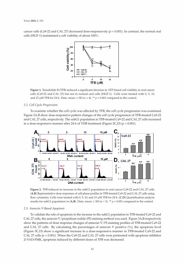
Toxins 2016, 8, 319
cancer cells (Ca9-22 and CAL 27) decreased dose-responsively (p < 0.001). In contrast, the normal oralcells (HGF-1) maintained a cell viability of about 100%.
Figure 1. Tenuifolide B (TFB) induced a significant decrease in ATP-based cell viability in oral cancercells (Ca9-22 and CAL 27) but not in normal oral cells (HGF-1). Cells were treated with 0, 5, 10,and 15 μM TFB for 24 h. Data: mean ± SD (n = 4). ** p < 0.001 compared to the control.
2.2. Cell Cycle Progression
To examine whether the cell cycle was affected by TFB, the cell cycle progression was examined.Figure 2A,B show dose-responsive pattern changes of the cell cycle progression of TFB-treated Ca9-22and CAL 27 cells, respectively. The subG1 population in TFB-treated Ca9-22 and CAL 27 cells increasedin a dose-responsive manner after 24 h of THB treatment (Figure 2C,D) (p < 0.001).
Figure 2. TFB induced an increase in the subG1 population in oral cancer Ca9-22 and CAL 27 cells.(A,B) Representative dose responses of cell phase profiles in TFB-treated Ca9-22 and CAL 27 cells usingflow cytometry. Cells were treated with 0, 5, 10, and 15 μM TFB for 24 h. (C,D) Quantification analysisresults for subG1 population in (A,B). Data: mean ± SD (n = 3). ** p < 0.001 compared to the control.
2.3. Annexin V-Based Apoptosis
To validate the role of apoptosis in the increase in the subG1 population in TFB-treated Ca9-22 andCAL 27 cells, the annexin V/propidium iodide (PI) staining method was used. Figure 3A,B respectivelyshow the patterns of dose response changes of annexin V/PI staining profiles of TFB-treated Ca9-22and CAL 27 cells. By calculating the percentages of annexin V positive (%), the apoptosis level(Figure 3C,D) show a significant increase in a dose-responsive manner in TFB-treated Ca9-22 andCAL 27 cells (p < 0.001). When the Ca9-22 and CAL 27 cells were pretreated with apoptosis inhibitorZ-VAD-FMK, apoptosis induced by different doses of TFB was decreased.
63
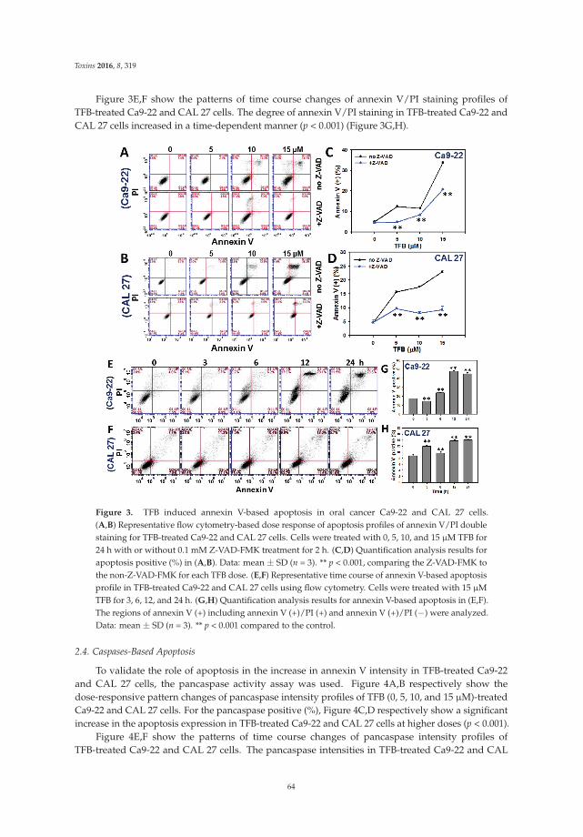
Toxins 2016, 8, 319
Figure 3E,F show the patterns of time course changes of annexin V/PI staining profiles ofTFB-treated Ca9-22 and CAL 27 cells. The degree of annexin V/PI staining in TFB-treated Ca9-22 andCAL 27 cells increased in a time-dependent manner (p < 0.001) (Figure 3G,H).
Figure 3. TFB induced annexin V-based apoptosis in oral cancer Ca9-22 and CAL 27 cells.(A,B) Representative flow cytometry-based dose response of apoptosis profiles of annexin V/PI doublestaining for TFB-treated Ca9-22 and CAL 27 cells. Cells were treated with 0, 5, 10, and 15 μM TFB for24 h with or without 0.1 mM Z-VAD-FMK treatment for 2 h. (C,D) Quantification analysis results forapoptosis positive (%) in (A,B). Data: mean ± SD (n = 3). ** p < 0.001, comparing the Z-VAD-FMK tothe non-Z-VAD-FMK for each TFB dose. (E,F) Representative time course of annexin V-based apoptosisprofile in TFB-treated Ca9-22 and CAL 27 cells using flow cytometry. Cells were treated with 15 μMTFB for 3, 6, 12, and 24 h. (G,H) Quantification analysis results for annexin V-based apoptosis in (E,F).The regions of annexin V (+) including annexin V (+)/PI (+) and annexin V (+)/PI (−) were analyzed.Data: mean ± SD (n = 3). ** p < 0.001 compared to the control.
2.4. Caspases-Based Apoptosis
To validate the role of apoptosis in the increase in annexin V intensity in TFB-treated Ca9-22and CAL 27 cells, the pancaspase activity assay was used. Figure 4A,B respectively show thedose-responsive pattern changes of pancaspase intensity profiles of TFB (0, 5, 10, and 15 μM)-treatedCa9-22 and CAL 27 cells. For the pancaspase positive (%), Figure 4C,D respectively show a significantincrease in the apoptosis expression in TFB-treated Ca9-22 and CAL 27 cells at higher doses (p < 0.001).
Figure 4E,F show the patterns of time course changes of pancaspase intensity profiles ofTFB-treated Ca9-22 and CAL 27 cells. The pancaspase intensities in TFB-treated Ca9-22 and CAL
64
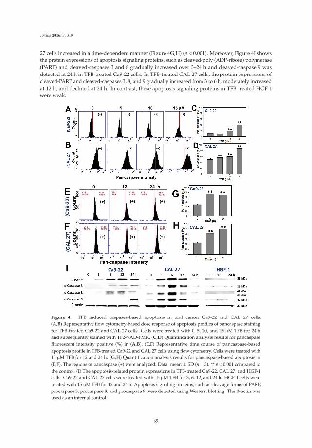
Toxins 2016, 8, 319
27 cells increased in a time-dependent manner (Figure 4G,H) (p < 0.001). Moreover, Figure 4I showsthe protein expressions of apoptosis signaling proteins, such as cleaved-poly (ADP-ribose) polymerase(PARP) and cleaved-caspases 3 and 8 gradually increased over 3–24 h and cleaved-caspase 9 wasdetected at 24 h in TFB-treated Ca9-22 cells. In TFB-treated CAL 27 cells, the protein expressions ofcleaved-PARP and cleaved-caspases 3, 8, and 9 gradually increased from 3 to 6 h, moderately increasedat 12 h, and declined at 24 h. In contrast, these apoptosis signaling proteins in TFB-treated HGF-1were weak.
Figure 4. TFB induced caspases-based apoptosis in oral cancer Ca9-22 and CAL 27 cells.(A,B) Representative flow cytometry-based dose response of apoptosis profiles of pancaspase stainingfor TFB-treated Ca9-22 and CAL 27 cells. Cells were treated with 0, 5, 10, and 15 μM TFB for 24 hand subsequently stained with TF2-VAD-FMK. (C,D) Quantification analysis results for pancaspasefluorescent intensity positive (%) in (A,B). (E,F) Representative time course of pancaspase-basedapoptosis profile in TFB-treated Ca9-22 and CAL 27 cells using flow cytometry. Cells were treated with15 μM TFB for 12 and 24 h. (G,H) Quantification analysis results for pancaspase-based apoptosis in(E,F). The regions of pancaspase (+) were analyzed. Data: mean ± SD (n = 3). ** p < 0.001 compared tothe control. (I) The apoptosis-related protein expressions in TFB-treated Ca9-22, CAL 27, and HGF-1cells. Ca9-22 and CAL 27 cells were treated with 15 μM TFB for 3, 6, 12, and 24 h. HGF-1 cells weretreated with 15 μM TFB for 12 and 24 h. Apoptosis signaling proteins, such as cleavage forms of PARP,procaspase 3, procaspase 8, and procaspase 9 were detected using Western blotting. The β-actin wasused as an internal control.
65
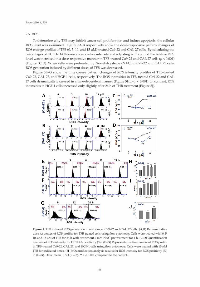
Toxins 2016, 8, 319
2.5. ROS
To determine why TFB may inhibit cancer cell proliferation and induce apoptosis, the cellularROS level was examined. Figure 5A,B respectively show the dose-responsive pattern changes ofROS change profiles of TFB (0, 5, 10, and 15 μM)-treated Ca9-22 and CAL 27 cells. By calculating thepercentages of DCFH-DA fluorescence-positive intensity and adjusting with control, the relative ROSlevel was increased in a dose-responsive manner in TFB-treated Ca9-22 and CAL 27 cells (p < 0.001)(Figure 5C,D). When cells were pretreated by N-acetylcysteine (NAC) in Ca9-22 and CAL 27 cells,ROS generation induced by different doses of TFB was decreased.
Figure 5E–G show the time course pattern changes of ROS intensity profiles of TFB-treatedCa9-22, CAL 27, and HGF-1 cells, respectively. The ROS intensities in TFB-treated Ca9-22 and CAL27 cells dramatically increased in a time-dependent manner (Figure 5H,I) (p < 0.001). In contrast, ROSintensities in HGF-1 cells increased only slightly after 24 h of THB treatment (Figure 5J).
Figure 5. TFB induced ROS generation in oral cancer Ca9-22 and CAL 27 cells. (A,B) Representativedose responses of ROS profiles for TFB-treated cells using flow cytometry. Cells were treated with 0, 5,10, and 15 μM of TFB for 24 h with or without 2 mM NAC pretreatment for 1 h. (C,D) Quantificationanalysis of ROS intensity for DCFD-A positivity (%). (E–G) Representative time course of ROS profilein TFB-treated Ca9-22, CAL 27, and HGF-1 cells using flow cytometry. Cells were treated with 15 μMTFB for indicated times. (H–J) Quantification analysis results for ROS intensity for ROS positivity (%)in (E–G). Data: mean ± SD (n = 3). ** p < 0.001 compared to the control.
66
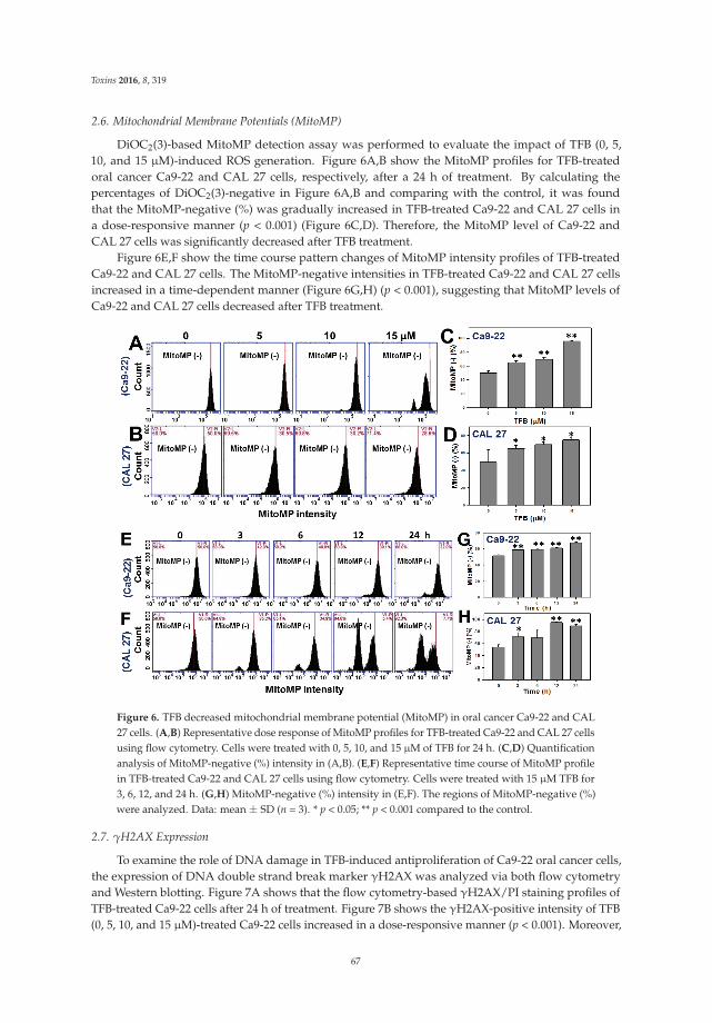
Toxins 2016, 8, 319
2.6. Mitochondrial Membrane Potentials (MitoMP)
DiOC2(3)-based MitoMP detection assay was performed to evaluate the impact of TFB (0, 5,10, and 15 μM)-induced ROS generation. Figure 6A,B show the MitoMP profiles for TFB-treatedoral cancer Ca9-22 and CAL 27 cells, respectively, after a 24 h of treatment. By calculating thepercentages of DiOC2(3)-negative in Figure 6A,B and comparing with the control, it was foundthat the MitoMP-negative (%) was gradually increased in TFB-treated Ca9-22 and CAL 27 cells ina dose-responsive manner (p < 0.001) (Figure 6C,D). Therefore, the MitoMP level of Ca9-22 andCAL 27 cells was significantly decreased after TFB treatment.
Figure 6E,F show the time course pattern changes of MitoMP intensity profiles of TFB-treatedCa9-22 and CAL 27 cells. The MitoMP-negative intensities in TFB-treated Ca9-22 and CAL 27 cellsincreased in a time-dependent manner (Figure 6G,H) (p < 0.001), suggesting that MitoMP levels ofCa9-22 and CAL 27 cells decreased after TFB treatment.
Figure 6. TFB decreased mitochondrial membrane potential (MitoMP) in oral cancer Ca9-22 and CAL27 cells. (A,B) Representative dose response of MitoMP profiles for TFB-treated Ca9-22 and CAL 27 cellsusing flow cytometry. Cells were treated with 0, 5, 10, and 15 μM of TFB for 24 h. (C,D) Quantificationanalysis of MitoMP-negative (%) intensity in (A,B). (E,F) Representative time course of MitoMP profilein TFB-treated Ca9-22 and CAL 27 cells using flow cytometry. Cells were treated with 15 μM TFB for3, 6, 12, and 24 h. (G,H) MitoMP-negative (%) intensity in (E,F). The regions of MitoMP-negative (%)were analyzed. Data: mean ± SD (n = 3). * p < 0.05; ** p < 0.001 compared to the control.
2.7. γH2AX Expression
To examine the role of DNA damage in TFB-induced antiproliferation of Ca9-22 oral cancer cells,the expression of DNA double strand break marker γH2AX was analyzed via both flow cytometryand Western blotting. Figure 7A shows that the flow cytometry-based γH2AX/PI staining profiles ofTFB-treated Ca9-22 cells after 24 h of treatment. Figure 7B shows the γH2AX-positive intensity of TFB(0, 5, 10, and 15 μM)-treated Ca9-22 cells increased in a dose-responsive manner (p < 0.001). Moreover,
67
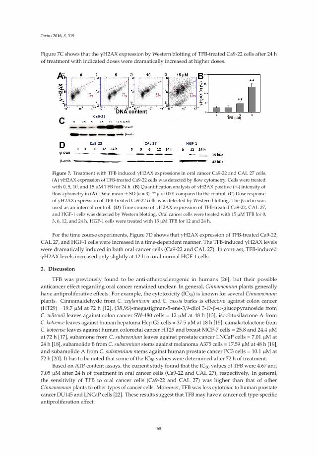
Toxins 2016, 8, 319
Figure 7C shows that the γH2AX expression by Western blotting of TFB-treated Ca9-22 cells after 24 hof treatment with indicated doses were dramatically increased at higher doses.
Figure 7. Treatment with TFB induced γH2AX expressions in oral cancer Ca9-22 and CAL 27 cells.(A) γH2AX expression of TFB-treated Ca9-22 cells was detected by flow cytometry. Cells were treatedwith 0, 5, 10, and 15 μM TFB for 24 h. (B) Quantification analysis of γH2AX positive (%) intensity offlow cytometry in (A). Data: mean ± SD (n = 3). ** p < 0.001 compared to the control. (C) Dose responseof γH2AX expression of TFB-treated Ca9-22 cells was detected by Western blotting. The β-actin wasused as an internal control. (D) Time course of γH2AX expression of TFB-treated Ca9-22, CAL 27,and HGF-1 cells was detected by Western blotting. Oral cancer cells were treated with 15 μM TFB for 0,3, 6, 12, and 24 h. HGF-1 cells were treated with 15 μM TFB for 12 and 24 h.
For the time course experiments, Figure 7D shows that γH2AX expression of TFB-treated Ca9-22,CAL 27, and HGF-1 cells were increased in a time-dependent manner. The TFB-induced γH2AX levelswere dramatically induced in both oral cancer cells (Ca9-22 and CAL 27). In contrast, TFB-inducedγH2AX levels increased only slightly at 12 h in oral normal HGF-1 cells.
3. Discussion
TFB was previously found to be anti-atherosclerogenic in humans [26], but their possibleanticancer effect regarding oral cancer remained unclear. In general, Cinnamomum plants generallyhave antiproliferative effects. For example, the cytotoxicity (IC50) is known for several Cinnamomumplants. Cinnamaldehyde from C. zeylanicum and C. cassia barks is effective against colon cancer(HT29) = 19.7 μM at 72 h [12], (3R,9S)-megastigman-5-ene-3,9-diol 3-O-β-D-glucopyranoside fromC. wilsonii leaves against colon cancer SW-480 cells = 12 μM at 48 h [13], isoobtusilactone A fromC. kotoense leaves against human hepatoma Hep G2 cells = 37.5 μM at 18 h [15], cinnakotolactone fromC. kotoense leaves against human colorectal cancer HT29 and breast MCF-7 cells = 25.8 and 24.4 μMat 72 h [17], subamone from C. subavenium leaves against prostate cancer LNCaP cells = 7.01 μM at24 h [18], subamolide B from C. subavenium stems against melanoma A375 cells = 17.59 μM at 48 h [19],and subamolide A from C. subavenium stems against human prostate cancer PC3 cells = 10.1 μM at72 h [20]. It has to be noted that some of the IC50 values were determined after 72 h of treatment.
Based on ATP content assays, the current study found that the IC50 values of TFB were 4.67 and7.05 μM after 24 h of treatment in oral cancer cells (Ca9-22 and CAL 27), respectively. In general,the sensitivity of TFB to oral cancer cells (Ca9-22 and CAL 27) was higher than that of otherCinnamomum plants to other types of cancer cells. Moreover, TFB was less cytotoxic to human prostatecancer DU145 and LNCaP cells [22]. These results suggest that TFB may have a cancer cell type-specificantiproliferation effect.
68
Toxins 2016, 8, 319
Following 24 h of treatment, the cytotoxicity (IC50) of taxol in human prostate cancer DU145 andLNCaP cells is 4.84 and 6.32 μM, respectively [22]. The IC50 of cisplatin in oral cancer Ca9-22 cellsis 10.2 μM (data not shown). Therefore, our developed TFB (IC50 = 4.67 and 7.05 μM) has similarsensitivity to these clinical drugs in oral cancer cells (Ca9-22 and CAL 27). Moreover, we found thatTFB is less harmful to normal oral HGF-1 cells (Figure 1), suggesting that TFB selectively kills oralcancer cells and may prevent side effects of oral cancer therapy. Similarly, Cinnamomum stem barkextract has been reported to selectively kill other types of cancer cells. In Cinnamomum burmanniiBlume stem bark extract after 24 h of treatment, the IC50 values of nasopharyngeal carcinoma cells(HK1 and C666-1) were 108.32 and 224.32 μg/mL, respectively, whereas the IC50 of immortalizedhuman skin keratinocyte HaCaT cells was 320.29 μg/mL [27]. However, our study only tested onenormal oral cell line, and further study is needed to confirm these findings, including more normaloral cell lines to further show the lack of possible side-effects of TFB.
Secotenuifolide A, also isolated from the same material of this study (C. tenuifolium stems),has been reported to inhibit cell proliferation, increase the subG1 population, induce apoptosis andROS generation, and decrease mitochondrial membrane potential in human prostate cancer cells,DU145 [22]. Secotenuifolide A also exhibited a release of cytochrome c from mitochondria and theactivation of caspase-9/caspase-3 [22]. Similarly, the TFB from C. tenuifolium stems showed the sameeffect of oxidative stress (ROS induction and MitoMP depletion) on oral cancer Ca9-22 and CAL27 cells (Figures 5 and 6). In contrast, the TFB-induced ROS generation in HGF-1 cells was onlyslightly induced. These results suggest that TFB exhibited selective ROS induction in oral cancer cells(Ca9-22 and CAL 27), but less induction in HGF-1 cells. NAC pretreatment experiments (Figure 5A,B)validated that ROS was relevant to TFB-induced cell death because the ROS generation in the twotypes of oral cancer cells was reduced by NAC pretreatment.
Annexin V and pancaspase results (Figures 3 and 4) support that TFB is apoptosis-inducible inoral cancer cells. Moreover, the TFB-induced apoptosis was reduced by Z-VAD-FMK pretreatment(Figure 3A,B), suggesting that apoptosis was involved in selective killing by TFB. However, the roleof apoptosis signaling in TFB-induced apoptosis was not addressed specifically. Caspases 8 and 9involved in intrinsic and extrinsic apoptotic pathways, respectively. Both converge in activating theexecutioner caspases 3 and 7 [28]. PARP is also involved in apoptosis [29,30]. To address the role ofapoptosis signaling, we found that both TFB-treated oral cancer cells (Ca9–22 and CAL 27) inducedactivation of PARP and caspases 3, 8, and 9 by cleavage. However, these oral cancer cells displayed adifferential expression of these TFB-induced apoptosis proteins. For example, cleaved-caspase 8 wasmainly or early induced during 3–12 h in TFB-treated Ca9-22 cells, but cleaved-caspase 9 inductionshowed a later response at 24 h (Figure 4I). For CAL 27 cells, cleaved-PARP and cleaved-caspases 3,8 and 9 were upregulated early after 3 h. These caspases peaked at 6 h, gradually declining by 24 h.Similarly, other drug-induced apoptosis also showed a similar tendency for cleaved-caspase expression.For example, cleaved-caspase 3 increased at 6–12 h and declined at 24 h in 0.2 μM staurosporine-treatedhuman endothelial cornea cells [31]. After treatment of 0.2 μM staurosporine, cleaved-caspase 3 wasalso increased at 2–8 h and declined at 12–24 h for human cervical cancer HeLa cells. Cleaved-caspase3 increased at 0.5–1 h and declined at 2–24 h in cervical cancer C-33A cells [32].
Mounting evidence demonstrated that ROS generation and mitochondrial membranedepolarization may lead to DNA damage [33–38] and apoptosis [38–41] in drug-treated cancercells. Accordingly, TFB also showed a correlation between oxidative stress and DNA damage inits antiproliferative and apoptotic effects of two oral cancer cells (Ca9-22 and CAL 27) in our study.
4. Conclusions
TFB treatment induces apoptosis, ROS generation, mitochondrial depolarization, and DNAdamage, which ultimately results in the antiproliferation of oral cancer Ca9-22 cells. This study alsoshows that TFB selectively kills the two oral cancer cell lines tested here and opts for its application inanti-oral cancer therapies.
69
Toxins 2016, 8, 319
5. Materials and Methods
5.1. Drug Information and Oral Cancer and Normal Cell Lines
The TFB (C26H46O3; MW: 406.3447; [3-(1-methoxyeicosyl)-5-methylene-5H-furan-2-one]) waspurified from methanol extracts of C. tenuifolium stem as previously described [22]. It was dissolved indimethyl sulfoxide (DMSO) for drug treatments. The oral cancer cell lines Ca9-22 [42] and CAL 27 [43]were incubated in a mixed medium with Dulbecco’s Modified Eagle Medium (DMEM) and F12 (Gibco,Grand Island, NY, USA) (3:2), 10% fetal bovine serum, antibiotics (penicillin and streptomycin), andothers under a humidified atmosphere with 5% CO2 at 37 ◦C. The normal oral cells (human gingivalfibroblasts, HGF-1) were incubated in a DMEM medium with a similar supplement with 1 mMpyruvate as described above.
5.2. Measurement of Cell Viability—Cellular ATP Content
Cellular ATP level was determined by using the ATP-lite Luminescence ATP Detection AssaySystem (PerkinElmer Life Sciences, Boston, MA, USA) according to the manufacturer’s instructionswith a slight modification [44]. Briefly, Ca9-22 cells were plated at 4000 cells/well in 96-well plates.After seeding overnight, cells were treated with vehicle control (DMSO) or with TFB at indicatedconcentrations (5, 10, and 15 μM) for 24 h. After removing the medium solution, 100 μL of serum-freemedium and 50 μL of a mammalian cell lysis solution was added per well of a microplate with orbitalshaking at 100 rpm for 5 min. Then, 100 μL of the cell lysates/well was transferred to white 96-wellplates and reacted with 50 μL of substrate solution (D-Luciferin and luciferase) under orbital shakingat 100 rpm for 5 min and then left to stand in darkness for a further 10 min. Finally, the luminescencewas assayed using a microplate luminometer (CentroPRO LB 962, Berthold, ND, USA).
5.3. Measurement of Cell Cycle Progression
The cellular DNA was stained with PI as previously described [45]. Cells were plated at3 × 105 cells/2 mL cell culture medium on a 6-well plate. Briefly, cells were added with vehicle(DMSO only) or TFB. After collection for 70% ethanol fixation overnight, the centrifuged cell pelletswere resuspended in 1 mL of PBS containing 50 μg/mL PI for 15 min at room temperature in darkness.Subsequently, these samples were examined using a FACSCalibur flow cytometer (Becton-Dickinson,Mansfield, MA, USA) (excitation: 488 nm and emission: 617 nm) and BD Accuri C6 software.
5.4. Measurement of Apoptosis by Annexin V Staining
Annexin V (Strong Biotech Corporation, Taipei, Taiwan)/PI (Sigma, St. Louis, MO, USA)double-staining for apoptosis analysis was performed as previously described [46]. Cells were platedat 3 × 10⁵ cells/2 mL cell culture medium on a 6-well plate with or without 0.1 mM Z-VAD-FMKpretreatments for 2 h (Selleckchem.com; Houston, TX, USA). Briefly, cells were added with vehicleor TFB. The cells were then resuspended in the binding buffer containing 5 μg/mL of annexinV-fluorescein isothiocyanate and 50 μg/mL of PI and examined with a BD Accuri C6 flow cytometer(Becton-Dickinson, Mansfield, MA, USA) (excitation: 488 nm and emission: 525 nm and 617 nm forFITC and PI, respectively) and BD Accuri C6 software.
5.5. Measurement of Apoptosis by Caspase Activity
Apoptosis was also measured by caspase activation [47]. The generic caspase activity assay kit(Fluorometric-Green; ab112130) (Abcam, Cambridge, UK) was used to detect the activity of caspase-1,-3, -4, -5, -6, -7, -8, and -9 as described [42]. Briefly, cells were seeded as 3 × 10⁵ cells per well in 6-wellplates with a 2 mL medium for overnight. The cells were then treated with vehicle or TFB. Subsequently,cells were incubated at 37 ◦C, 5% CO2 for 2 h with 2 μL of 500X TF2-VAD-FMK. After PBS washing,
70
Toxins 2016, 8, 319
cells were resuspended in 0.5 mL of an assay buffer for immediate flow cytometry measurement(BD Accuri™ C6; Becton-Dickinson).
The apoptosis signaling expressions were further measured via Western blotting. 30 μg proteinlysates were resolved in 10% SDS-PAGE. After electrotransferring, the nonspecific bindings of PDVFmembranes (Pall Corporation, Port Washington, NY, USA) were blocked with 5% non-fat milk inTris-buffered saline with Tween-20 and incubated with primary antibodies (the cleaved caspase-8(Asp391) (18C8) rabbit mAb and the rabbit mAb in the apoptosis antibody sampler kit (cleaved PARP(Asp214) (D64E10) XP®; cleaved caspase-3 (Asp175) (5A1E); cleaved caspase-9 (Asp330) (D2D4) fromCell Signalling Technology, Inc., Danvers, MA, USA, β-actin (#GTX629630, GeneTex Inc.)) under1:10000 dilution as well as their matched secondary antibody. The WesternBright™ ECL HRP substrate(#K-12045-D50, Advansta, Menlo Park, CA, USA) was chosen for signal amplification.
5.6. Measurement of Intracellular ROS
2′,7′-Dichlorodihydrofluorescein diacetate (DCFH-DA) (Sigma Chemical Co., St. Louis, MO,USA) was used to detect intracellular ROS as previously described [33]. Cells were plated at3 × 105 cells/2 mL cell culture medium on a 6 cm dish. Briefly, cells were added with vehicle orTFB with or without 2 mM NAC pretreatment for 1 h (Sigma; St. Louis, MO, USA). After the collectionand PBS washing, cells were treated with 0.1 μM DCFH-DA in serum-free medium for 30 min at 37 ◦Cin darkness. Cells were resuspended in PBS after centrifugation and examined with a BD Accuri C6flow cytometer (excitation: 488 nm and emission: 525 nm) and BD Accuri C6 software.
5.7. Measurement of MitoMP
A MitoProbe™ DiOC2(3) assay kit (Invitrogen, San Diego, CA, USA) was used to detectmitochondrial membrane potential (MitoMP) as described previously [34]. Cells were plated at3 × 105 cells/2 mL cell culture medium on a 6-well plate. Briefly, cells were added with vehicle or withTFB. The TFB-treated cells were washed in 1 mL of PBS/well, provided with 1 mL of medium/well,loaded with 10 μL of 10 μM DiOC2(3), and left to stand at 37 ◦C in 5% CO2 for 20–30 min. Afterharvesting and washing, cells were resuspended in PBS and examined immediately using a FACSCaliburflow cytometer (excitation: 488 nm and emission: 525 nm) and BD Accuri C6 software.
5.8. Measurement of DNA Damage by γH2AX Expression
DNA double strand breaks were detected by both flow cytometry [48] and Western blotting [35]as described previously. For flow cytometry, TFB-treated cells were fixed, washed, and incubatedat 4 ◦C for 1 h in 2 μg/mL of p-Histone H2AX (Ser 139) (γH2AX) monoclonal antibody (sc-101696;Santa Cruz Biotechnology, Santa Cruz, CA, USA). After washing, cells were suspended for 1 h ina secondary antibody (Jackson Laboratory, Bar Harbor, ME, USA) for 30 min at room temperature.Finally, the cells were resuspended in 20 μg/mL of PI for flow cytometry analysis (BD Accuri™ C6;Becton-Dickinson).
For Western blotting of γH2AX expression, 30 μg protein lysates were resolved in 10%SDS-PAGE. Except when p-Histone H2AX (Santa Cruz Biotechnology) was chosen for the primaryantibody, procedures were the same as those employing Western blotting for apoptosis proteins,mentioned above.
5.9. Statistical Analysis
All data are shown as mean ± SD. The significant differences between test and control wereanalyzed with a Student t-test.
Acknowledgments: This work was partly supported by funds of the Ministry of Science and Technology(NSC 101-2320-B-037-049, MOST 103-2320-B-037-008, MOST 103-2320-B-242-001, and MOST 104-2320-B-037-013-MY3),the Chimei-KMU jointed project (105CM-KMU-06), the National Sun Yat-sen University-KMU Joint Research Project(#NSYSUKMU 105-P0322), the Kaohsiung Municipal Ta-Tung Hospital (kmtth-104-003), the Kaohsiung Medical
71

Toxins 2016, 8, 319
University “Aim for the Top Universities Grant, grant No. KMU-TP104PR02”, and the Health and Welfare Surchargeof Tobacco Products, Ministry of Health and Welfare, Taiwan, Republic of China (MOHW105-TDU-B-212-134007).We also thank Hans-Uwe Dahms for the help of English editing.
Author Contributions: Hui-Ru Wang and Hui-Ping Yang carried out the experiments. Ching-Yu Yen, Jen-YangTang, Hurng-Wern Huang, and Shih-Hsien Hsu analyzed the data. Chung-Yi Chen, Ching-Yu Yen, and Hsueh-WeiChang conceived and designed the study. Chung-Yi Chen, Shih-Hsien Hsu, and Hsueh-Wei Chang wrote andrevised the manuscript.
Conflicts of Interest: The authors declare no conflict of interest.
References
1. Warnakulasuriya, S. Global epidemiology of oral and oropharyngeal cancer. Oral Oncol. 2009, 45, 309–316.[CrossRef] [PubMed]
2. Ko, Y.C.; Huang, Y.L.; Lee, C.H.; Chen, M.J.; Lin, L.M.; Tsai, C.C. Betel quid chewing, cigarette smoking andalcohol consumption related to oral cancer in Taiwan. J. Oral Pathol. Med. 1995, 24, 450–453. [CrossRef][PubMed]
3. Lee, C.H.; Ko, Y.C.; Huang, H.L.; Chao, Y.Y.; Tsai, C.C.; Shieh, T.Y.; Lin, L.M. The precancer risk of betel quidchewing, tobacco use and alcohol consumption in oral leukoplakia and oral submucous fibrosis in southernTaiwan. Br. J. Cancer 2003, 88, 366–372. [CrossRef] [PubMed]
4. Chiang, S.L.; Lee, C.P.; Chang, J.G.; Lee, C.H.; Yeh, K.T.; Tsai, Y.S.; Chen, M.K.; Chen, C.H.; Ko, Y.C. Combinedeffects of differentiation factor 15 and substance use of alcohol, betel quid and cigarette on risk of head andneck cancer. Head Neck Oncol. 2013, 5, 23.
5. Myoung, H.; Hong, S.P.; Yun, P.Y.; Lee, J.H.; Kim, M.J. Anti-cancer effect of genistein in oral squamous cellcarcinoma with respect to angiogenesis and in vitro invasion. Cancer Sci. 2003, 94, 215–220. [CrossRef][PubMed]
6. Chen, B.H.; Chang, H.W.; Huang, H.M.; Chong, I.W.; Chen, J.S.; Chen, C.Y.; Wang, H.M. (−)-Anonaineinduces DNA damage and inhibits growth and migration of human lung carcinoma h1299 cells. J. Agric.Food Chem. 2011, 59, 2284–2290. [CrossRef] [PubMed]
7. Wang, H.M.; Cheng, K.C.; Lin, C.J.; Hsu, S.W.; Fang, W.C.; Hsu, T.F.; Chiu, C.C.; Chang, H.W.; Hsu, C.H.;Lee, A.Y. Obtusilactone A and (−)-sesamin induce apoptosis in human lung cancer cells by inhibitingmitochondrial Lon protease and activating DNA damage checkpoints. Cancer Sci. 2010, 101, 2612–2620.[CrossRef] [PubMed]
8. Hseu, Y.C.; Wu, C.R.; Chang, H.W.; Kumar, K.J.; Lin, M.K.; Chen, C.S.; Cho, H.J.; Huang, C.Y.;Lee, H.Z.; Hsieh, W.T.; et al. Inhibitory effects of Physalis angulata on tumor metastasis and angiogenesis.J. Ethnopharmacol. 2011, 135, 762–771. [CrossRef] [PubMed]
9. Russo, M.; Spagnuolo, C.; Tedesco, I.; Russo, G.L. Phytochemicals in cancer prevention and therapy: Truth ordare? Toxins 2010, 2, 517–551. [CrossRef] [PubMed]
10. Melchini, A.; Traka, M.H. Biological profile of erucin: A new promising anticancer agent from cruciferousvegetables. Toxins 2010, 2, 593–612. [CrossRef] [PubMed]
11. Yadav, V.R.; Prasad, S.; Sung, B.; Kannappan, R.; Aggarwal, B.B. Targeting inflammatory pathways bytriterpenoids for prevention and treatment of cancer. Toxins 2010, 2, 2428–2466. [CrossRef] [PubMed]
12. Cabello, C.M.; Bair, W.B., 3rd; Lamore, S.D.; Ley, S.; Bause, A.S.; Azimian, S.; Wondrak, G.T.The cinnamon-derived Michael acceptor cinnamic aldehyde impairs melanoma cell proliferation,invasiveness, and tumor growth. Free Radic. Biol. Med. 2009, 46, 220–231. [CrossRef] [PubMed]
13. Shu, P.; Wei, X.; Xue, Y.; Li, W.; Zhang, J.; Xiang, M.; Zhang, M.; Luo, Z.; Li, Y.; Yao, G.; et al. Wilsonols A-L,megastigmane sesquiterpenoids from the leaves of Cinnamomum wilsonii. J. Nat. Prod. 2013, 76, 1303–1312.[CrossRef] [PubMed]
14. Chen, C.Y.; Chen, C.H.; Lo, Y.C.; Wu, B.N.; Wang, H.M.; Lo, W.L.; Yen, C.M.; Lin, R.J. Anticancer activityof isoobtusilactone A from Cinnamomum kotoense: Involvement of apoptosis, cell-cycle dysregulation,mitochondria regulation, and reactive oxygen species. J. Nat. Prod. 2008, 71, 933–940. [CrossRef] [PubMed]
15. Chen, C.Y.; Liu, T.Z.; Chen, C.H.; Wu, C.C.; Cheng, J.T.; Yiin, S.J.; Shih, M.K.; Wu, M.J.; Chern, C.L.Isoobtusilactone A-induced apoptosis in human hepatoma Hep G2 cells is mediated via increased NADPHoxidase-derived reactive oxygen species (ROS) production and the mitochondria-associated apoptoticmechanisms. Food Chem. Toxicol. 2007, 45, 1268–1276. [CrossRef] [PubMed]
72

Toxins 2016, 8, 319
16. Chen, C.Y.; Hsu, Y.L.; Chen, Y.Y.; Hung, J.Y.; Huang, M.S.; Kuo, P.L. Isokotomolide A, a new butanolideextracted from the leaves of Cinnamomum kotoense, arrests cell cycle progression and induces apoptosisthrough the induction of p53/p21 and the initiation of mitochondrial system in human non-small cell lungcancer A549 cells. Eur. J. Pharmacol. 2007, 574, 94–102. [PubMed]
17. Yang, S.S.; Hou, W.C.; Huang, L.W.; Lee, T.H. A new gamma-lactone from the leaves of Cinnamomum kotoense.Nat. Prod. Res. 2006, 20, 1246–1250. [CrossRef] [PubMed]
18. Lin, R.J.; Lo, W.L.; Wang, Y.D.; Chen, C.Y. A novel cytotoxic monoterpenoid from the leaves ofCinnamomum subavenium. Nat. Prod. Res. 2008, 22, 1055–1059. [CrossRef] [PubMed]
19. Yang, S.Y.; Wang, H.M.; Wu, T.W.; Chen, Y.J.; Shieh, J.J.; Lin, J.H.; Ho, T.F.; Luo, R.J.; Chen, C.Y.; Chang, C.C.Subamolide B isolated from medicinal plant Cinnamomum subavenium induces cytotoxicity in humancutaneous squamous cell carcinoma cells through mitochondrial and CHOP-dependent cell death pathways.Evid Based Complement. Altern. Med. 2013, 2013, 630415. [CrossRef] [PubMed]
20. Liu, C.H.; Chen, C.Y.; Huang, A.M.; Li, J.H. Subamolide A, a component isolated fromCinnamomum subavenium, induces apoptosis mediated by mitochondria-dependent, p53 and ERK1/2pathways in human urothelial carcinoma cell line NTUB1. J. Ethnopharmacol. 2011, 137, 503–511. [CrossRef][PubMed]
21. Chen, T.H.; Huang, Y.H.; Lin, J.J.; Liau, B.C.; Wang, S.Y.; Wu, Y.C.; Jong, T.T. Cytotoxic lignan esters fromCinnamomum osmophloeum. Planta Med. 2010, 76, 613–619. [CrossRef] [PubMed]
22. Lin, R.J.; Cheng, M.J.; Huang, J.C.; Lo, W.L.; Yeh, Y.T.; Yen, C.M.; Lu, C.M.; Chen, C.Y. Cytotoxic compoundsfrom the stems of Cinnamomum tenuifolium. J. Nat. Prod. 2009, 72, 1816–1824. [CrossRef] [PubMed]
23. Chen, H.L.; Kuo, S.Y.; Li, Y.P.; Kang, Y.F.; Yeh, Y.T.; Huang, J.C.; Chen, C.Y. A new benzodioxocinone fromthe leaves of Cinnamomum tenuifolium. Nat. Prod. Res. 2012, 26, 1881–1886. [CrossRef] [PubMed]
24. Tam, K.F.; Ng, T.Y.; Liu, S.S.; Tsang, P.C.; Kwong, P.W.; Ngan, H.Y. Potential application of the ATP cellviability assay in the measurement of intrinsic radiosensitivity in cervical cancer. Gynecol. Oncol. 2005, 96,765–770. [CrossRef] [PubMed]
25. Lu, X.; Errington, J.; Chen, V.J.; Curtin, N.J.; Boddy, A.V.; Newell, D.R. Cellular ATP depletion by LY309887as a predictor of growth inhibition in human tumor cell lines. Clin. Cancer Res. 2000, 6, 271–277. [PubMed]
26. Dong, H.P.; Wu, H.M.; Chen, S.J.; Chen, C.Y. The effect of butanolides from Cinnamomum tenuifolium onplatelet aggregation. Molecules 2013, 18, 11836–11841. [CrossRef] [PubMed]
27. Daker, M.; Lin, V.Y.; Akowuah, G.A.; Yam, M.F.; Ahmad, M. Inhibitory effects of Cinnamomum burmanniiBlume stem bark extract and trans-cinnamaldehyde on nasopharyngeal carcinoma cells; synergism withcisplatin. Exp. Ther. Med. 2013, 5, 1701–1709. [CrossRef] [PubMed]
28. Tait, S.W.; Green, D.R. Mitochondria and cell death: Outer membrane permeabilization and beyond. Nat. Rev.Mol. Cell Biol. 2010, 11, 621–632. [CrossRef] [PubMed]
29. Chang, Y.T.; Huang, C.Y.; Li, K.T.; Li, R.N.; Liaw, C.C.; Wu, S.H.; Liu, J.R.; Sheu, J.H.; Chang, H.W.Sinuleptolide inhibits proliferation of oral cancer Ca9–22 cells involving apoptosis, oxidative stress, and DNAdamage. Arch. Oral Biol. 2016, 66, 147–154. [CrossRef] [PubMed]
30. Yen, Y.H.; Farooqi, A.A.; Li, K.T.; Butt, G.; Tang, J.Y.; Wu, C.Y.; Cheng, Y.B.; Hou, M.F.; Chang, H.W.Methanolic extracts of Solieria robusta inhibits proliferation of oral cancer Ca9–22 cells via apoptosis andoxidative stress. Molecules 2014, 19, 18721–18732. [CrossRef] [PubMed]
31. Thuret, G.; Chiquet, C.; Herrag, S.; Dumollard, J.M.; Boudard, D.; Bednarz, J.; Campos, L.; Gain, P.Mechanisms of staurosporine induced apoptosis in a human corneal endothelial cell line. Br. J. Ophthalmol.2003, 87, 346–352. [CrossRef] [PubMed]
32. Nicolier, M.; Decrion-Barthod, A.Z.; Launay, S.; Pretet, J.L.; Mougin, C. Spatiotemporal activation ofcaspase-dependent and -independent pathways in staurosporine-induced apoptosis of p53wt and p53mthuman cervical carcinoma cells. Biol. Cell 2009, 101, 455–467. [CrossRef] [PubMed]
33. Yeh, C.C.; Yang, J.I.; Lee, J.C.; Tseng, C.N.; Chan, Y.C.; Hseu, Y.C.; Tang, J.Y.; Chuang, L.Y.; Huang, H.W.;Chang, F.R.; et al. Anti-proliferative effect of methanolic extract of Gracilaria tenuistipitata on oral cancercells involves apoptosis, DNA damage, and oxidative stress. BMC Complement. Altern. Med. 2012, 12, 142.[CrossRef] [PubMed]
34. Yen, C.Y.; Chiu, C.C.; Haung, R.W.; Yeh, C.C.; Huang, K.J.; Chang, K.F.; Hseu, Y.C.; Chang, F.R.; Chang, H.W.;Wu, Y.C. Antiproliferative effects of goniothalamin on Ca9–22 oral cancer cells through apoptosis, DNAdamage and ROS induction. Mutat. Res. 2012, 747, 253–258. [CrossRef] [PubMed]
73
Toxins 2016, 8, 319
35. Chiu, C.C.; Haung, J.W.; Chang, F.R.; Huang, K.J.; Huang, H.M.; Huang, H.W.; Chou, C.K.; Wu, Y.C.;Chang, H.W. Golden berry-derived 4beta-hydroxywithanolide E for selectively killing oral cancer cells bygenerating ROS, DNA damage, and apoptotic pathways. PLoS ONE 2013, 8, e64739. [CrossRef] [PubMed]
36. Guo, J.; Zhao, W.; Hao, W.; Ren, G.; Lu, J.; Chen, X. Cucurbitacin B induces DNA damage, G2/M phasearrest, and apoptosis mediated by reactive oxygen species (ROS) in leukemia K562 cells. Anticancer AgentsMed. Chem. 2014, 14, 1146–1153. [CrossRef] [PubMed]
37. Ni, C.H.; Yu, C.S.; Lu, H.F.; Yang, J.S.; Huang, H.Y.; Chen, P.Y.; Wu, S.H.; Ip, S.W.; Chiang, S.Y.; Lin, J.G.; et al.Chrysophanol-induced cell death (necrosis) in human lung cancer A549 cells is mediated through increasingreactive oxygen species and decreasing the level of mitochondrial membrane potential. Environ. Toxicol.2014, 29, 740–749. [CrossRef] [PubMed]
38. Hseu, Y.C.; Lee, M.S.; Wu, C.R.; Cho, H.J.; Lin, K.Y.; Lai, G.H.; Wang, S.Y.; Kuo, Y.H.; Kumar, K.J.; Yang, H.L.The chalcone flavokawain B induces G2/M cell-cycle arrest and apoptosis in human oral carcinoma HSC-3cells through the intracellular ROS generation and downregulation of the Akt/p38 MAPK signaling pathway.J. Agric. Food Chem. 2012, 60, 2385–2397. [CrossRef] [PubMed]
39. Huang, F.J.; Hsuuw, Y.D.; Chan, W.H. Characterization of apoptosis induced by emodin and relatedregulatory mechanisms in human neuroblastoma cells. Int. J. Mol. Sci. 2013, 14, 20139–20156. [CrossRef][PubMed]
40. Shih, H.C.; El-Shazly, M.; Juan, Y.S.; Chang, C.Y.; Su, J.H.; Chen, Y.C.; Shih, S.P.; Chen, H.M.; Wu, Y.C.;Lu, M.C. Cracking the cytotoxicity code: Apoptotic induction of 10-acetylirciformonin B is mediated throughROS generation and mitochondrial dysfunction. Mar. Drugs 2014, 12, 3072–3090. [CrossRef] [PubMed]
41. Thangam, R.; Senthilkumar, D.; Suresh, V.; Sathuvan, M.; Sivasubramanian, S.; Pazhanichamy, K.;Gorlagunta, P.K.; Kannan, S.; Gunasekaran, P.; Rengasamy, R.; et al. Induction of ROS-dependentmitochondria-mediated intrinsic apoptosis in MDA-MB-231 cells by glycoprotein from Codium decorticatum.J. Agric. Food Chem. 2014, 62, 3410–3421. [CrossRef] [PubMed]
42. Yeh, C.C.; Tseng, C.N.; Yang, J.I.; Huang, H.W.; Fang, Y.; Tang, J.Y.; Chang, F.R.; Chang, H.W. Antiproliferationand induction of apoptosis in Ca9–22 oral cancer cells by ethanolic extract of Gracilaria tenuistipitata. Molecules2012, 17, 10916–10927. [CrossRef] [PubMed]
43. Jiang, L.; Ji, N.; Zhou, Y.; Li, J.; Liu, X.; Wang, Z.; Chen, Q.; Zeng, X. CAL 27 is an oral adenosquamouscarcinoma cell line. Oral Oncol. 2009, 45, e204–e207. [CrossRef] [PubMed]
44. Wei, J.; Stebbins, J.L.; Kitada, S.; Dash, R.; Zhai, D.; Placzek, W.J.; Wu, B.; Rega, M.F.; Zhang, Z.; Barile, E.; et al.An optically pure apogossypolone derivative as potent pan-active inhibitor of anti-apoptotic bcl-2 familyproteins. Front. Oncol. 2011, 1, 28. [CrossRef] [PubMed]
45. Chiu, C.C.; Chang, H.W.; Chuang, D.W.; Chang, F.R.; Chang, Y.C.; Cheng, Y.S.; Tsai, M.T.; Chen, W.Y.;Lee, S.S.; Wang, C.K.; et al. Fern plant-derived protoapigenone leads to DNA damage, apoptosis, andG(2)/m arrest in lung cancer cell line H1299. DNA Cell Biol. 2009, 28, 501–506. [CrossRef] [PubMed]
46. Chiu, C.C.; Liu, P.L.; Huang, K.J.; Wang, H.M.; Chang, K.F.; Chou, C.K.; Chang, F.R.; Chong, I.W.; Fang, K.;Chen, J.S.; et al. Goniothalamin inhibits growth of human lung cancer cells through DNA damage, apoptosis,and reduced migration ability. J. Agric. Food Chem. 2011, 59, 4288–4293. [CrossRef] [PubMed]
47. Kaufmann, S.H.; Lee, S.H.; Meng, X.W.; Loegering, D.A.; Kottke, T.J.; Henzing, A.J.; Ruchaud, S.;Samejima, K.; Earnshaw, W.C. Apoptosis-associated caspase activation assays. Methods 2008, 44, 262–272.[CrossRef] [PubMed]
48. Yen, C.Y.; Hou, M.F.; Yang, Z.W.; Tang, J.Y.; Li, K.T.; Huang, H.W.; Huang, Y.H.; Lee, S.Y.; Fu, T.F.;Hsieh, C.Y.; et al. Concentration effects of grape seed extracts in anti-oral cancer cells involving differentialapoptosis, oxidative stress, and DNA damage. BMC Complement. Altern. Med. 2015, 15, 94. [CrossRef][PubMed]
© 2016 by the authors; licensee MDPI, Basel, Switzerland. This article is an open accessarticle distributed under the terms and conditions of the Creative Commons Attribution(CC-BY) license (http://creativecommons.org/licenses/by/4.0/).
74
Article
Chemical Characterization and in Vitro Cytotoxicityon Squamous Cell Carcinoma Cells of Carica PapayaLeaf ExtractsThao T. Nguyen 1, Marie-Odile Parat 1, Mark P. Hodson 1,2, Jenny Pan 1, Paul N. Shaw 1 andAmitha K. Hewavitharana 1,*
1 School of Pharmacy, The University of Queensland, Brisbane, QLD 4072, Australia;[email protected] (T.T.N.); [email protected] (M.-O.P.); [email protected] (M.P.H.);[email protected] (J.P.); [email protected] (P.N.S.)
2 Metabolomics Australia, Australian Institute for Bioengineering and Nanotechnology,The University of Queensland, Brisbane, QLD 4072, Australia
* Correspondence: [email protected]; Tel.: +61-7-334-61898; Fax: +61-7-334-61999
Academic Editor: Carmela FimognariReceived: 26 October 2015; Accepted: 18 December 2015; Published: 24 December 2015
Abstract: In traditional medicine, Carica papaya leaf has been used for a wide range of therapeuticapplications including skin diseases and cancer. In this study, we investigated the in vitro cytotoxicityof aqueous and ethanolic extracts of Carica papaya leaves on the human oral squamous cell carcinomaSCC25 cell line in parallel with non-cancerous human keratinocyte HaCaT cells. Two out of fourextracts showed a significantly selective effect towards the cancer cells and were found to containhigh levels of phenolic and flavonoid compounds. The chromatographic and mass spectrometricprofiles of the extracts obtained with Ultra High Performance Liquid Chromatography-QuadrupoleTime of Flight-Mass Spectrometry were used to tentatively identify the bioactive compounds usingcomparative analysis. The principal compounds identified were flavonoids or flavonoid glycosides,particularly compounds from the kaempferol and quercetin families, of which several have previouslybeen reported to possess anticancer activities. These results confirm that papaya leaf is a potentialsource of anticancer compounds and warrant further scientific investigation to validate the traditionaluse of papaya leaf to treat cancer.
Keywords: Carica papaya; cytotoxicity; mass spectrometry; cancer; flavonoids; chromatography
1. Introduction
A book entitled “The most wonderful tree in the world—the papaw tree (Carica papaia)”, publishedsome 100 years ago, contains many anecdotes relating to the cure of breast, liver or rectal cancer after“treatment” with Carica papaya preparations [1]. Subsequent reports have been published in variousmedia that have detailed “the healing capabilities of an old Australian Aboriginal remedy—boiledextract of pawpaw leaves—against cancer” [2] and several other anecdotes relating “cancer cure”following consumption of various preparations of papaya plant [3–6].
Recently, we undertook a comprehensive literature review [7] and found that research providingscientific evidence for the effectiveness of Carica papaya in the treatment and prevention of cancer waslimited. However, in contrast to the limited number of studies that have been done to evaluate theeffects of papaya extracts on cancer, the abundance in Carica papaya of phytochemicals with reportedanticancer activities, such as carotenoids (in fruits and seeds), alkaloids (in leaves), phenolics (in fruits,leaves, shoots) and glucosinolates (in seeds and fruits), suggests that there are opportunities for newresearch to evaluate the anticancer potential of this medicinal plant [7].
Toxins 2016, 8, 7 75 www.mdpi.com/journal/toxins
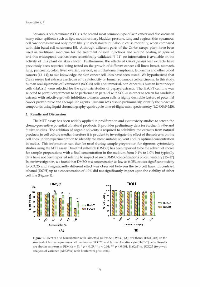
Toxins 2016, 8, 7
Squamous cell carcinoma (SCC) is the second most common type of skin cancer and also occurs inmany other epithelia such as lips, mouth, urinary bladder, prostate, lung and vagina. Skin squamouscell carcinomas are not only more likely to metastasize but also to cause mortality, when comparedwith skin basal cell carcinoma [8]. Although different parts of the Carica papaya plant have beenused as traditional medicine for the treatment of skin infections and wound healing in general,and this widespread use has been scientifically validated [9–11], no information is available on theactivity of this plant on skin cancer. Furthermore, the effects of Carica papaya leaf extracts havepreviously been reported being tested on the growth of different cancer cell lines: breast, stomach,lung, pancreatic, colon, liver, ovarian, cervical, neuroblastoma, lymphoma, leukaemia and other bloodcancers [12–14]; to our knowledge, no skin cancer cell lines have been tested. We hypothesized thatCarica papaya leaf extracts exerted in vitro cytotoxicity on human squamous cell carcinoma. In this study,human oral squamous cell carcinoma (SCC25) cells and immortal, non-cancerous human keratinocytecells (HaCaT) were selected for the cytotoxic studies of papaya extracts. The HaCaT cell line wasselected to permit experiments to be performed in parallel with SCC25 in order to screen for candidateextracts with selective growth inhibition towards cancer cells, a highly desirable feature of potentialcancer preventative and therapeutic agents. Our aim was also to preliminarily identify the bioactivecompounds using liquid chromatography-quadrupole time-of-flight-mass spectrometry (LC-QToF-MS).
2. Results and Discussion
The MTT assay has been widely applied in proliferation and cytotoxicity studies to screen thechemo-preventive potential of natural products. It provides preliminary data for further in vitro andin vivo studies. The addition of organic solvents is required to solubilize the extracts from naturalproducts in cell culture media; therefore it is prudent to investigate the effect of the solvents on thecell lines under experimentation to identify the most suitable solvent and its optimal concentrationin media. This information can then be used during sample preparation for rigorous cytotoxicitystudies using the MTT assay. Dimethyl sulfoxide (DMSO) has been reported to be the solvent of choicefor sample preparations with a final concentration in the medium from 0.1% to 1.0% but typicallydata have not been reported relating to impact of such DMSO concentrations on cell viability [15–17].In our investigation, we found that DMSO at a concentration as low as 0.05% causes significant toxicityto SCC25 and a significantly different effect was observed between the two cell lines. In contrast,ethanol (EtOH) up to a concentration of 1.0% did not significantly impact upon the viability of eithercell line (Figure 1).
Figure 1. Effect of a 48-h incubation with Dimethyl sulfoxide (DMSO) (A); or Ethanol (EtOH) (B) on thesurvival of human squamous cell carcinoma (SCC25) and human keratinocyte (HaCaT) cells. Resultsare shown as mean ˘ SEM (n = 3). * p < 0.05, ** p < 0.01; *** p < 0.001, HaCaT vs. SCC25 (two-wayanalysis of variance (ANOVA) with Bonferroni post-tests).
76
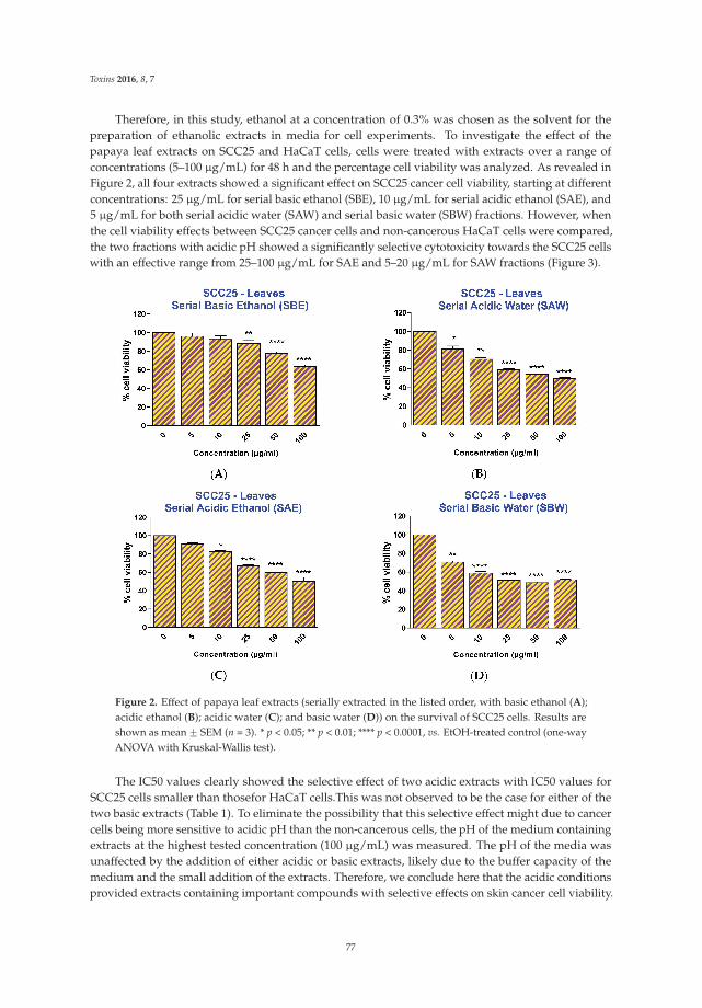
Toxins 2016, 8, 7
Therefore, in this study, ethanol at a concentration of 0.3% was chosen as the solvent for thepreparation of ethanolic extracts in media for cell experiments. To investigate the effect of thepapaya leaf extracts on SCC25 and HaCaT cells, cells were treated with extracts over a range ofconcentrations (5–100 μg/mL) for 48 h and the percentage cell viability was analyzed. As revealed inFigure 2, all four extracts showed a significant effect on SCC25 cancer cell viability, starting at differentconcentrations: 25 μg/mL for serial basic ethanol (SBE), 10 μg/mL for serial acidic ethanol (SAE), and5 μg/mL for both serial acidic water (SAW) and serial basic water (SBW) fractions. However, whenthe cell viability effects between SCC25 cancer cells and non-cancerous HaCaT cells were compared,the two fractions with acidic pH showed a significantly selective cytotoxicity towards the SCC25 cellswith an effective range from 25–100 μg/mL for SAE and 5–20 μg/mL for SAW fractions (Figure 3).
Figure 2. Effect of papaya leaf extracts (serially extracted in the listed order, with basic ethanol (A);acidic ethanol (B); acidic water (C); and basic water (D)) on the survival of SCC25 cells. Results areshown as mean ˘ SEM (n = 3). * p < 0.05; ** p < 0.01; **** p < 0.0001, vs. EtOH-treated control (one-wayANOVA with Kruskal-Wallis test).
The IC50 values clearly showed the selective effect of two acidic extracts with IC50 values forSCC25 cells smaller than thosefor HaCaT cells.This was not observed to be the case for either of thetwo basic extracts (Table 1). To eliminate the possibility that this selective effect might due to cancercells being more sensitive to acidic pH than the non-cancerous cells, the pH of the medium containingextracts at the highest tested concentration (100 μg/mL) was measured. The pH of the media wasunaffected by the addition of either acidic or basic extracts, likely due to the buffer capacity of themedium and the small addition of the extracts. Therefore, we conclude here that the acidic conditionsprovided extracts containing important compounds with selective effects on skin cancer cell viability.
77
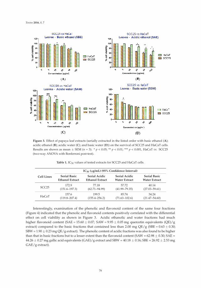
Toxins 2016, 8, 7
Figure 3. Effect of papaya leaf extracts (serially extracted in the listed order with basic ethanol (A);acidic ethanol (B); acidic water (C); and basic water (D)) on the survival of SCC25 and HaCaT cells.Results are shown as mean ˘ SEM (n = 3). * p < 0.05; ** p < 0.01; *** p < 0.001, HaCaT vs. SCC25(two-way ANOVA with Bonferroni post-test).
Table 1. IC50 values of tested extracts for SCC25 and HaCaT cells.
Cell Lines
IC50 (μg/mL) (95% Confidence Interval)
Serial BasicEthanol Extract
Serial AcidicEthanol Extract
Serial AcidicWater Extract
Serial BasicWater Extract
SCC25172.9 77.18 57.72 40.14
(151.6–197.3) (62.71–94.99) (41.99–79.35) (27.03–59.61)
HaCaT157.6 199.5 85.74 34.24
(119.8–207.4) (155.4–256.2) (71.63–102.6) (21.47–54.60)
Interestingly, examination of the phenolic and flavonoid content of the same four fractions(Figure 4) indicated that the phenolic and flavonoid contents positively correlated with the differentialeffect on cell viability as shown in Figure 3. Acidic ethanolic and water fractions had muchhigher flavonoid content (SAE = 15.60 ˘ 0.07; SAW = 9.95 ˘ 0.05 mg quercetin equivalents (QE)/gextract) compared to the basic fractions that contained less than 2.00 mg QE/g (SBE = 0.63 ˘ 0.30;SBW = 1.90 ˘ 0.23 mg QE/g extract). The phenolic content of acidic fractions was also found to be higherthan that in basic fractions but to a lesser extent than the flavonoid content (SAW = 62.98 ˘ 0.30; SAE =44.26 ˘ 0.27 mg gallic acid equivalents (GAE)/g extract and SBW = 40.18 ˘ 0.16; SBE = 26.92 ˘ 2.53 mgGAE/g extract).
78
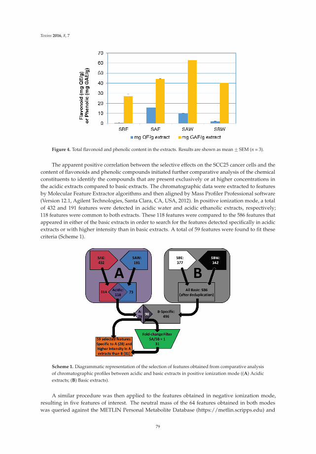
Toxins 2016, 8, 7
Figure 4. Total flavonoid and phenolic content in the extracts. Results are shown as mean ˘ SEM (n = 3).
The apparent positive correlation between the selective effects on the SCC25 cancer cells and thecontent of flavonoids and phenolic compounds initiated further comparative analysis of the chemicalconstituents to identify the compounds that are present exclusively or at higher concentrations inthe acidic extracts compared to basic extracts. The chromatographic data were extracted to featuresby Molecular Feature Extractor algorithms and then aligned by Mass Profiler Professional software(Version 12.1, Agilent Technologies, Santa Clara, CA, USA, 2012). In positive ionization mode, a totalof 432 and 191 features were detected in acidic water and acidic ethanolic extracts, respectively;118 features were common to both extracts. These 118 features were compared to the 586 features thatappeared in either of the basic extracts in order to search for the features detected specifically in acidicextracts or with higher intensity than in basic extracts. A total of 59 features were found to fit thesecriteria (Scheme 1).
Scheme 1. Diagrammatic representation of the selection of features obtained from comparative analysisof chromatographic profiles between acidic and basic extracts in positive ionization mode ((A) Acidicextracts; (B) Basic extracts).
A similar procedure was then applied to the features obtained in negative ionization mode,resulting in five features of interest. The neutral mass of the 64 features obtained in both modeswas queried against the METLIN Personal Metabolite Database (https://metlin.scripps.edu) and
79

Toxins 2016, 8, 7
the customized NPACT database (http://crdd.osdd.net/raghava/npact) to match to compoundsusing a mass tolerance window of ď10 ppm. The METLIN database contains 64,092 structures ofendogenous and exogenous metabolites (as of June 2015) whereas the customized NPACT databaseconsists of 1,574 entries of plant-derived natural compounds that exhibit anti-cancerous activity [18].The METLIN-based search resulted in several candidates (a total of 880 hits for 64 features) anddirected further investigation with MS fragmentation spectra to confirm the identities of bioactivecompounds in the extracts (Table 2).
Table 2. Tentative identification of compounds which appear in acidic extracts exclusively or at greaterextent than in basic extracts.
Formula ExperimentalMass Error (ppm) * Number of Candidates
from Metlin DatabasePutative Compounds from
NPACT Database
C4H5NO3 115.0269 0 1 -C5H7NO3 129.0428 1 7 -
C14H25NO3 255.1835 0 1 -C14H25NO3 255.1846 4 1 -
C15H10O6 286.0486 3 28 Fisetin, Kaempferol, Luteolin,Scullarein, Tetrahydroxyflavone
C18H33NO2 295.252 2 1 -C15H10O7 302.0436 3 22 Quercetin; Morin;Viscidulin I
C18H19NO3 307.2158 3 4 -C22H43NO 337.335 1 3 -C22H41NO2 351.3145 2 1 -C21H30O10 442.1843 0 1 -
C21H20O11 448.1009 0 74 Kaempferol β-D-glucopyranoside,Luteolin β-D-glucopyranoside
C21H20O12 464.0958 0 56 Myricetin 3-O-rhamnosideC20H26O13 474.1373 0 1 -
C28H46N2O4 474.346 0 1 -C28H46N2O4 474.3467 1 1 -C28H46N2O4 474.3468 2 1 -C28H48N2O4 476.3624 2 1 -C30H48N2O4 500.3617 0 1 -C26H55O8P 526.3631 0 1 -C33H50O5 526.3631 5 1 -
C34H56N2O4 556.4245 0 1 -C29H44O12 584.2809 4 2 -C34H48O8 584.3322 4 3 -C27H30O15 594.158 0 120 -C27H30O16 610.153 0 109 RutinC27H30O16 610.1531 0 109 -C28H32O16 624.1686 0 78 -C28H32O16 624.1689 0 78 -
C34H65O13P 712.4155 1 1 -C33H40O19 740.2154 1 33 -C33H40O20 756.2107 0 81 -C33H40O21 772.2069 0 56 -C46H77O9P 804.5319 1 1 -
* Error (ppm): the difference between experimental mass and theoretical mass of compound.
Among the tentatively identified metabolites from the METLIN database, the most remarkablewere the flavonoids or flavonoid glycosides (kaempferol, quercetin, rutin, manghaslin, nicotiflorin,clitorin, quercetin 7-galactoside, myricetin 7-rhamnoside, luteolin 3,7-diglucoside). These correlatedwith the results of the total flavonoid content in acidic extracts compared to basic extracts.Furthermore, the search within NPACT database revealed several compounds with previouslyreported anti-cancer activities including fisetin, kaempferol, luteolin, scullarein, tetrahydroxyflavone,quercetin, morin, viscidulin I, kaempferol β-D-glucopyranoside, luteolin β-D-glucopyranoside,myricetin 3-O-rhamnoside, and rutin [18]. Kaempferol and quercetin have been detected andquantified in Carica papaya leaves by gas chromatography-mass spectrometry analysis with quantities
80
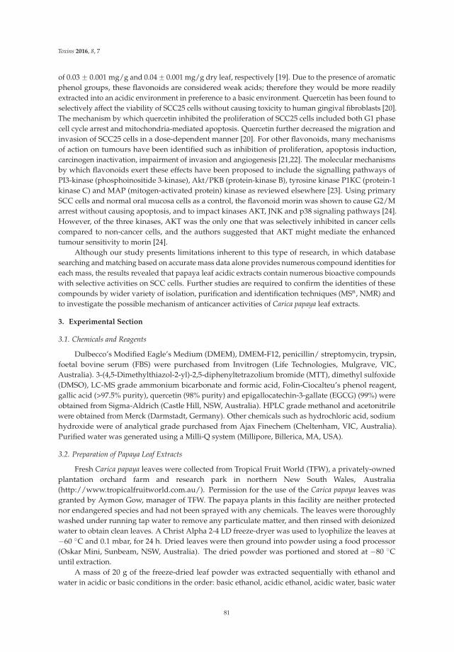
Toxins 2016, 8, 7
of 0.03 ˘ 0.001 mg/g and 0.04 ˘ 0.001 mg/g dry leaf, respectively [19]. Due to the presence of aromaticphenol groups, these flavonoids are considered weak acids; therefore they would be more readilyextracted into an acidic environment in preference to a basic environment. Quercetin has been found toselectively affect the viability of SCC25 cells without causing toxicity to human gingival fibroblasts [20].The mechanism by which quercetin inhibited the proliferation of SCC25 cells included both G1 phasecell cycle arrest and mitochondria-mediated apoptosis. Quercetin further decreased the migration andinvasion of SCC25 cells in a dose-dependent manner [20]. For other flavonoids, many mechanismsof action on tumours have been identified such as inhibition of proliferation, apoptosis induction,carcinogen inactivation, impairment of invasion and angiogenesis [21,22]. The molecular mechanismsby which flavonoids exert these effects have been proposed to include the signalling pathways ofPI3-kinase (phosphoinositide 3-kinase), Akt/PKB (protein-kinase B), tyrosine kinase P1KC (protein-1kinase C) and MAP (mitogen-activated protein) kinase as reviewed elsewhere [23]. Using primarySCC cells and normal oral mucosa cells as a control, the flavonoid morin was shown to cause G2/Marrest without causing apoptosis, and to impact kinases AKT, JNK and p38 signaling pathways [24].However, of the three kinases, AKT was the only one that was selectively inhibited in cancer cellscompared to non-cancer cells, and the authors suggested that AKT might mediate the enhancedtumour sensitivity to morin [24].
Although our study presents limitations inherent to this type of research, in which databasesearching and matching based on accurate mass data alone provides numerous compound identities foreach mass, the results revealed that papaya leaf acidic extracts contain numerous bioactive compoundswith selective activities on SCC cells. Further studies are required to confirm the identities of thesecompounds by wider variety of isolation, purification and identification techniques (MSn, NMR) andto investigate the possible mechanism of anticancer activities of Carica papaya leaf extracts.
3. Experimental Section
3.1. Chemicals and Reagents
Dulbecco’s Modified Eagle’s Medium (DMEM), DMEM-F12, penicillin/ streptomycin, trypsin,foetal bovine serum (FBS) were purchased from Invitrogen (Life Technologies, Mulgrave, VIC,Australia). 3-(4,5-Dimethylthiazol-2-yl)-2,5-diphenyltetrazolium bromide (MTT), dimethyl sulfoxide(DMSO), LC-MS grade ammonium bicarbonate and formic acid, Folin-Ciocalteu’s phenol reagent,gallic acid (>97.5% purity), quercetin (98% purity) and epigallocatechin-3-gallate (EGCG) (99%) wereobtained from Sigma-Aldrich (Castle Hill, NSW, Australia). HPLC grade methanol and acetonitrilewere obtained from Merck (Darmstadt, Germany). Other chemicals such as hydrochloric acid, sodiumhydroxide were of analytical grade purchased from Ajax Finechem (Cheltenham, VIC, Australia).Purified water was generated using a Milli-Q system (Millipore, Billerica, MA, USA).
3.2. Preparation of Papaya Leaf Extracts
Fresh Carica papaya leaves were collected from Tropical Fruit World (TFW), a privately-ownedplantation orchard farm and research park in northern New South Wales, Australia(http://www.tropicalfruitworld.com.au/). Permission for the use of the Carica papaya leaves wasgranted by Aymon Gow, manager of TFW. The papaya plants in this facility are neither protectednor endangered species and had not been sprayed with any chemicals. The leaves were thoroughlywashed under running tap water to remove any particulate matter, and then rinsed with deionizedwater to obtain clean leaves. A Christ Alpha 2-4 LD freeze-dryer was used to lyophilize the leaves at´60 ˝C and 0.1 mbar, for 24 h. Dried leaves were then ground into powder using a food processor(Oskar Mini, Sunbeam, NSW, Australia). The dried powder was portioned and stored at ´80 ˝Cuntil extraction.
A mass of 20 g of the freeze-dried leaf powder was extracted sequentially with ethanol andwater in acidic or basic conditions in the order: basic ethanol, acidic ethanol, acidic water, basic water
81
Toxins 2016, 8, 7
(2 ˆ 200 mL for each solvent; pH was maintained at pH 1–2 or 10–11 during the extraction process byadjusting with either 10% hydrochloric acid or 5 M sodium hydroxide solution). The obtained ethanolicextracts were concentrated at 30 ˝C and 120 rpm under vacuum by a rotary evaporator (IKA�RV 10,IKA-Werke, Staufen im Breisgau, Germany). The resulting concentrates of ethanolic extracts and waterextracts were lyophilised using the freeze-dryer. All lyophilised fractions (Basic ethanol: SBE, Acidicethanol: SAE, Acidic water: SAW, Basic water: SBW) were stored at ´80 ˝C prior to analysis.
3.3. Cell Culture Conditions
SCC25 cells (ATCC� CRL-1628™, Manassas, VA, USA) were maintained in DMEM/F12 mediumsupplemented with 10% v/v heat-inactivated foetal bovine serum, 1% penicillin-streptomycin and0.4 μg/mL hydrocortisone. HaCaT cells (a generous gift from Professor Fusenig) [25] were propagatedin DMEM medium supplemented with 10% foetal bovine serum and 1% penicillin-streptomycin.The cells were grown in a humidified incubator at 37 ˝C in a 5% CO2 atmosphere. Cells werepassaged every 3 days and cultures were allowed to reach 70%–90% confluence before experimentswere performed.
3.4. Cell Viability Assays
The effect of extracts on SCC25 and HaCaT viability was evaluated using the colorimetrictetrazolium dye procedure commonly referred to as the 3-(4,5-dimethylthiazol-2-yl)-2,5-diphenyltetrazolium bromide (MTT) assay developed by Mosmann with minor modification [26].SCC25 or HaCaT cells were plated into 96-well plates at densities of 6 ˆ 103 cells per well in 100 μLof DMEM/F12 (10% serum) and 3 ˆ 103 cells well in 100 μL of DMEM (10% serum), respectively.Cells were incubated at 37 ˝C for 24 h and were subsequently treated for 48 h with 0.5% serummedium containing increasing concentrations (5–100 μg dry mass/mL) of extracts in ethanol (0.3% finalconcentration in medium). Control cells were exposed to an equivalent volume of ethanol (0.3% finalconcentration). Cells were then incubated in 100 μL of MTT-containing medium (0.2 mg/mL MTT in0.5% serum medium) at 37 ˝C for an additional two hours. The medium was then removed and theformazan crystals trapped in cells were dissolved in 100 μL of DMSO by gentle shaking for 20 minon an orbital shaker. Absorbance of the solubilized product was measured at 595 nm using an Imarkplate reader (BioRad, Hercules, CA, USA). The absorbance of cells exposed to medium containing0.3% ethanol only was taken as 100% cell viability (i.e., the control). The results are expressed as percentof the viability of control cells ˘ standard error of the mean (SEM) from 4–8 parallel determinationsin three independent experiments (n = 3). Dose-effect analysis on SCC25 cells was performed byone-way analysis of variance (ANOVA) with Kruskal-Wallis test (as the data were non-parametric).Differences between the SCC25 and HaCaT cell lines, and interaction between cell line and extracteffects were analysed by two-way ANOVA with Bonferroni post-tests. All statistical analyses werecarried out using GraphPad Prism 6.0 (GraphPad Software Inc., San Diego, CA, USA, 2014).
3.5. Determination of Total Phenolic Content
Total phenolic content of ethanol and water extracts was determined using the Folin-Ciocalteuassay as described previously [27] with minor modifications. Briefly, 0.5 mL of diluted extract wasmixed with 2.5 mL of freshly prepared Folin-Ciocalteu’s phenol reagent, followed by the additionof 2 mL of 7.5% Na2CO3. The mixture was vortex mixed for 2 min and left in the dark at roomtemperature for 30 min. The absorbance was then measured at the maximum wavelength of 758 nmagainst a blank comprising 0.5 mL diluted extract, 2.5 mL water and 2 mL of 7.5% Na2CO3. Gallic acidwas used as the standard, and results were expressed as gallic acid equivalents (GAE) in mg/g of dryweight of each extract.
82
Toxins 2016, 8, 7
3.6. Determination of Total Flavonoid Content
Total flavonoid content of the extracts was determined using a colorimetric assay developedpreviously [27]. Diluted extract was mixed with 2% AlCl3 in ethanol in equal volume and absorbancewas measured after 15 min at 425 nm, against the blank sample consisting of equal volume of diluteextract and ethanol without AlCl3. Quercetin was used as the standard, and results were expressed asquercetin equivalents (QE) in mg/g of the dry weight of each extract.
3.7. UHPLC-ToF-MS Analysis
Chromatographic analysis of compounds in papaya leaf extracts was performed on an Agilent1290 UHPLC system (Agilent Technologies, Santa Clara, CA, USA). Chromatographic separation wasachieved on a 2.1 ˆ 150 mm, 3.5 μm ECLIPSE PLUS C18 analytical column (Agilent) with guardprotection. Mobile phase A was purified water containing 5 mM ammonium bicarbonate and 0.1%formic acid, adjusted to pH 7.0 ˘ 0.1 and mobile phase B was 95% acetonitrile and 5% water (v/v)containing 5 mM ammonium bicarbonate and 0.1% formic acid. The following gradient elution wasadopted: 10% to 80% B for the first 42 min; 80% to 90% B from 42 to 45 min; 90% to 100% B from46 to 48 min; held at 100% B from 48 to 50 min; returned to 10% B over the next 2 min, and the columnre-equilibrated with 10% B for 10 min prior to the next injection. Thus the total chromatographic runtime was 60 min. A flow rate of 0.2 mL/min was applied and 20 μL of sample was injected.
Each extract was run in triplicate in both positive and negative ionization mode.Mass spectrometric detection was performed on an Agilent 6520 high-resolution accurate massquadrupole time-of-flight (Q-ToF) mass spectrometer equipped with a multimode source in bothElectrospray Ionisation (ESI) and Atmospheric Pressure Chemical Ionization (APCI). Mass spectrawere controlled using MassHunter acquisition software (Version B.02.01 SP3, Agilent Technologies,Santa Clara, CA, USA, 2010). The mass spectrometer was operated in the range of m/z 100–1700, ata scan rate of 0.8 cycles/second under the following conditions: capillary voltage 2500 V, nebulizerpressure 30 psi, drying gas flow 5.0 L/min, gas temperature 300 ˝C, fragmenting voltage 175 V,skimmer voltage 65 V. To ensure the desired mass accuracy of recorded ions, continuous internalcalibration was performed during analysis with the use of reference ions—m/z 121.050873 (protonatedpurine) and m/z 922.009798 (protonated hexakis) in positive mode; in negative mode, ions with m/z119.0362 (deprotonated purine) and m/z 966.000725 (formate adduct of hexakis) were used to correctfor scan to scan variations.
3.8. MS Data Analysis
Data analysis was performed using Agilent MassHunter Qualitative software (Version B.05.00,Agilent Technologies, Santa Clara, CA, USA, 2012) with Molecular Feature Extractor (MFE) algorithmsin concert with Mass Profiler Professional software (Version 12.1, Agilent Technologies, Santa Clara, CA,USA, 2012) to align features from the chromatograms of all samples from four extracts of papaya leaves.The following cut-off settings were employed: minimum peak filters of 500 counts, peak spacingtolerance of 0.0025 m/z plus 7.0 ppm, assigned charge states limited to a maximum of two, minimumcompound filters of 3000 counts. The Molecular Feature Generator algorithm was utilised to generateputative molecular formulae from the following common elements C, H, N, O, P and S. Compoundidentification was carried out by using a Personal Compound Database Library (PCDL) (Agilent, SantaClara, CA, USA) with the METLIN Personal Metabolite Database and a customized PCDL databasewhich was compiled using the PCDL platform with compounds obtained from Naturally occurringPlant-based Anticancerous Compound-Activity-Target Database (NPACT) [18].
Acknowledgments: We would like to thank Aymon Gow and staff at Tropical Fruit World, New South Wales,Australia for their generosity in providing Carica papaya leaves for this study. Thao T. Nguyen is supported byEndeavour Postgraduate Award from Australian Government.
83
Toxins 2016, 8, 7
Author Contributions: Amitha K. Hewavitharana, Marie-Odile Parat, Mark P. Hodson and Paul N. Shaw conceivedand designed the experiments; Thao T. Nguyen and Jenny Pan performed the experiments; Thao T. Nguyen,Amitha K. Hewavitharana, Marie-Odile Parat, Mark P. Hodson analyzed the data; Amitha K. Hewavitharana,Marie-Odile Parat, Mark P. Hodson and Paul N. Shaw contributed reagents/materials/analysis tools;Thao T. Nguyen wrote the paper; Amitha K. Hewavitharana, Marie-Odile Parat, Mark P. Hodson and Paul N. Shawcritically reviewed the manuscript. All authors read and approved the final manuscript.
Conflicts of Interest: The authors declare no conflict of interest.
References
1. Lucas, T.P. The Most Wonderful Tree in the World, the Papaw Tree (Carica Papaia); Carter-Watson: Brisbane, QLD,Australia, 1914.
2. Tietze, H.W. Papaya the Medicine Tree, 3rd ed.; Harald Tietze Publishing: Bermagui, NSW, Australia, 2003.3. Gallo, J.N. Papaya Leaf Tea for Cancer—Promoting, Researching, and Growing Papaya Leaf Tea. Available
online: http://papayaleavesforcancer.com/ (accessed on 1 July 2015).4. Dillan, J. Could Papaya Leaf be a Cancer Treatment? Available online: http://superfoodprofiles.com/
papaya-leaves-cancer-treatment (accessed on 1 July 2015).5. Clarks, D.H. Papaya leaf: The Anti-Cancer Treatment. Available online: http://www.huldaclarkzappers.
com/php2/papayaleaf.php (accessed on 1 July 2015).6. Barrett, M. Papaya Leaf Extract Is a Powerful Cancer Fighter. Available online: http://naturalsociety.com/
papaya-leaf-extract-is-a-powerful-cancer-fighter/ (accessed on 1 July 2015).7. Nguyen, T.T.T.; Shaw, P.N.; Parat, M.O.; Hewavitharana, A.K. Anticancer activity of Carica papaya: A review.
Mol. Nutr. Food Res. 2013, 57, 153–164. [CrossRef] [PubMed]8. Clayman, G.L.; Lee, J.J.; Holsinger, F.C.; Zhou, X.; Duvic, M.; El-Naggar, A.K.; Prieto, V.G.; Altamirano, E.;
Tucker, S.L.; Strom, S.S. Mortality risk from squamous cell skin cancer. J. Clin. Oncol. 2005, 23, 759–765.[CrossRef] [PubMed]
9. Hewitt, H.; Whittle, S.; Lopez, S.; Bailey, E.; Weaver, S. Topical use of papaya in chronic skin ulcer therapy injamaica. West Indian Med. J. 2000, 49, 32–33. [PubMed]
10. Mikhal’chik, E.V.; Ivanova, A.V.; Anurov, M.V.; Titkova, S.M.; Pen’kov, L.Y.; Kharaeva, Z.F.; Korkina, L.G.Wound-healing effect of papaya-based preparation in experimental thermal trauma. Bull. Exp. Biol. Med.2004, 137, 560–562. [CrossRef] [PubMed]
11. Nayak, B.S.; Pereira, L.P.; Maharaj, D. Wound healing activity of Carica papaya L. In experimentally induceddiabetic rats. Indian J. Exp. Biol. 2007, 45, 739–743. [PubMed]
12. Morimoto, C.; Dang, N.H. Compositions for cancer prevention, treatment, or amelioration comprisingpapaya extract. US Patent 20080069907, 20 March 2008.
13. Otsuki, N.; Dang, N.H.; Kumagai, E.; Kondo, A.; Iwata, S.; Morimoto, C. Aqueous extract of Carica papayaleaves exhibits anti-tumor activity and immunomodulatory effects. J. Ethnopharmacol. 2010, 127, 760–767.[CrossRef]
14. Rumiyati, S.A. Effect of the protein fraction of Carica papaya L. Leaves on the expressions of P53 and BCL-2in breast cancer cells line. Maj. Farm. Indones. 2006, 17, 170–176.
15. Kaneshiro, T.; Suzui, M.; Takamatsu, R.; Murakami, A.; Ohigashi, H.; Fujino, T.; Yoshimi, N. Growthinhibitory activities of crude extracts obtained from herbal plants in the ryukyu islands on several humancolon carcinoma cell lines. Asian Pac. J. Cancer Prev. 2005, 6, 353–358. [PubMed]
16. Manosroi, J.; Dhumtanom, P.; Manosroi, A. Anti-proliferative activity of essential oil extracted from thaimedicinal plants on KB and P388 cell lines. Cancer Lett. 2006, 235, 114–120. [CrossRef] [PubMed]
17. Wang, G.H.; Chou, T.H.; Lin, R.J.; Sheu, J.H.; Wang, S.H.; Liang, C.H. Cytotoxic effect of the genus sinulariaextracts on human SCC25 and HaCaT cells. J. Toxicol. 2008, 2009. [CrossRef]
18. Mangal, M.; Sagar, P.; Singh, H.; Raghava, G.P.; Agarwal, S.M. Npact: Naturally occurring plant-basedanti-cancer compound-activity-target database. Nucl. Acids Res. 2013, 41, D1124–D1129. [CrossRef][PubMed]
19. Canini, A.; Alesiani, D.; D’Arcangelo, G.; Tagliatesta, P. Gas chromatography-mass spectrometry analysis ofphenolic compounds from Carica papaya L. Leaf. J. Food Compos. Anal. 2007, 20, 584–590. [CrossRef]
20. Chen, S.F.; Nien, S.; Wu, C.H.; Liu, C.L.; Chang, Y.C.; Lin, Y.S. Reappraisal of the anticancer efficacy ofquercetin in oral cancer cells. J. Chin. Med. Assoc. 2013, 76, 146–152. [CrossRef] [PubMed]
84
Toxins 2016, 8, 7
21. Kanadaswami, C.; Lee, L.T.; Lee, P.P.H.; Hwang, J.J.; Ke, F.C.; Huang, Y.T.; Lee, M.T. The antitumor activitiesof flavonoids. In Vivo 2005, 19, 895–909.
22. Chahar, M.K.; Sharma, N.; Dobhal, M.P.; Joshi, Y.C. Flavonoids: A versatile source of anticancer drugs.Pharmacogn. Rev. 2011, 5, 1–12. [PubMed]
23. Hertzogi, D.; Tica, O. Molecular mechanism underlying the anticancerous action of flavonoids. Curr. HealthSci. J. 2012, 38, 145–149.
24. Brown, J.; O’Prey, J.; Harrison, P. Enhanced sensitivity of human oral tumours to the flavonol, morin, duringcancer progression: Involvement of the Akt and stress kinase pathways. Carcinogenesis 2003, 24, 171–177.[CrossRef] [PubMed]
25. Boukamp, P.; Petrussevska, R.T.; Breitkreutz, D.; Hornung, J.; Markham, A.; Fusenig, N.E. Normalkeratinization in a spontaneously immortalized aneuploid human keratinocyte cell line. J. Cell Biol. 1988,106, 761–771. [CrossRef] [PubMed]
26. Mosmann, T. Rapid colorimetric assay for cellular growth and survival—Application to proliferation andcytotoxicity assays. J. Immunol. Methods 1983, 65, 55–63. [CrossRef]
27. Maisarah, A.; Amira, B.; Asmah, R.; Fauziah, O. Antioxidant analysis of different parts of Carica papaya.Int. Food Res. J. 2013, 20, 1043–1048.
© 2015 by the authors; licensee MDPI, Basel, Switzerland. This article is an open accessarticle distributed under the terms and conditions of the Creative Commons Attribution(CC-BY) license (http://creativecommons.org/licenses/by/4.0/).
85
toxins
Article
Withania somnifera Induces Cytotoxic and CytostaticEffects on Human T Leukemia CellsEleonora Turrini 1, Cinzia Calcabrini 1, Piero Sestili 2, Elena Catanzaro 1, Elena de Gianni 1,Anna Rita Diaz 2, Patrizia Hrelia 3, Massimo Tacchini 4, Alessandra Guerrini 4,Barbara Canonico 2, Stefano Papa 2, Giovanni Valdrè 5 and Carmela Fimognari 1,*
1 Department for Life Quality Studies, Alma Mater Studiorum-University of Bologna, Corso d’Augusto 237,47921 Rimini, Italy; [email protected] (E.T.); [email protected] (C.C.);[email protected] (E.C.); [email protected] (E.d.G.)
2 Department of Biomolecular Sciences, University of Urbino Carlo Bo, Via Saffi 2, 61029 Urbino, Italy;[email protected] (P.S.); [email protected] (A.R.D.); [email protected] (B.C.);[email protected] (S.P.)
3 Department of Pharmacy and Biotechnology, Alma Mater Studiorum-University of Bologna, Via Irnerio 48,40126 Bologna, Italy; [email protected]
4 Department of Life Sciences and Biotechnology (SVeB)-LT Terra & Acqua Tech RU, University of Ferrara,Corso Ercole I d’Este 32, I-44121 Ferrara, Italy; [email protected] (M.T.); [email protected] (A.G.)
5 Department of Biological, Geological and Environmental Sciences (BiGeA),Alma Mater Studiorum-University of Bologna, Piazza di Porta S. Donato 1, 40126 Bologna, Italy;[email protected]
* Correspondence: [email protected]; Tel.: +39-0541-434-658
Academic Editor: Tzi Bun NGReceived: 30 December 2015; Accepted: 9 May 2016; Published: 12 May 2016
Abstract: Cancer chemotherapy is characterized by an elevated intrinsic toxicity and the developmentof drug resistance. Thus, there is a compelling need for new intervention strategies with an improvedtherapeutic profile. Immunogenic cell death (ICD) represents an innovative anticancer strategy wheredying cancer cells release damage-associated molecular patterns promoting tumor-specific immuneresponses. The roots of Withania somnifera (W. somnifera) are used in the Indian traditional medicine fortheir anti-inflammatory, immunomodulating, neuroprotective, and anticancer activities. The presentstudy is designed to explore the antileukemic activity of the dimethyl sulfoxide extract obtained fromthe roots of W. somnifera (WE). We studied its cytostatic and cytotoxic activity, its ability to induce ICD,and its genotoxic potential on a human T-lymphoblastoid cell line by using different flow cytometricassays. Our results show that WE has a significant cytotoxic and cytostatic potential, and induces ICD.Its proapoptotic mechanism involves intracellular Ca2+ accumulation and the generation of reactiveoxygen species. In our experimental conditions, the extract possesses a genotoxic potential. Since theuse of Withania is suggested in different contexts including anti-infertility and osteoarthritis care, itsgenotoxicity should be carefully considered for an accurate assessment of its risk–benefit profile.
Keywords: Withania somnifera; apoptosis; cell cycle; leukemia; oxidative stress; immunogenic celldeath; genotoxicity
1. Introduction
Cancer causes millions of deaths every year. Only in 2012, cancer deaths have reached 8.2 million.In 2030, 12.6 million cancer deaths have been estimated [1].
Cancer originates from multiple alterations induced by a direct interaction between toxic agentsand DNA. This triggers gene and chromosome mutations. Altered expressions of oncogenes andtumor suppressor genes are found in different cancer types. The consequence is an uncontrollableproliferation mediated by growth signals released from the tumor cells themselves, resistance against
Toxins 2016, 8, 147 86 www.mdpi.com/journal/toxins
Toxins 2016, 8, 147
antigrowth signals, and inhibition of apoptosis. Furthermore, cancer expansion is helped by the releaseof various growth factors which lead to the formation of new blood vessels that provide nutrients andoxygen thus favoring cancer spread and metastasis dissemination [2]. Despite the progress made inanticancer research, traditional cytotoxic chemotherapy continues to serve as the basis for the currentstandard therapeutic regimen. This is characterized by an elevated intrinsic toxicity, mainly due to itspoor selectivity for cancer cells. Furthermore, cancer cells’ ability to develop drug resistance representsa major problem in anticancer therapy [3]. Thus, there is a compelling need for new interventionstrategies with an improved therapeutic profile. Cancer cells create a favorable microenvironmentallowing them to survive, proliferate, and counteract immunosurveillance. A promising anticancerstrategy could be represented by the use of cytotoxic drugs that are not only able to induce tumor celldeath, but also promote tumor-specific immune responses, potentially preventing tumor progressionand relapse [4]. Very recent studies have introduced the concept of immunogenic cell death (ICD),a modality of cell death where, after the exposure to some cytotoxic agents, dying cancer cells releaseendogenous damage-associated molecular patterns (DAMPs) including calreticulin, heat shock protein(Hsp)-70 and Hsp-90 recognized by antigen-presenting cells such as dendritic cells (DCs). This isfollowed by T-cell-mediated adaptive immunity [5].
Natural products represent a rich source of biologically active compounds that can be able tointeract simultaneously with different targets involved in cell growth, cell differentiation, and apoptosisregulation [6]. In 2015, the Food and Drug Administration (FDA) released an updated guidanceon botanical drug development. Unlike drugs that are constituted by a single active ingredient,botanical drugs have a heterogeneous nature, which may lead to uncertainty in relation to their activeconstituents. The number of botanical products submitted to the FDA is particularly high in theoncological area [7]. The first botanical drug in this area is Polyphenon E, a standardized extractobtained from the leaves of green tea (Camellia sinensis), approved by the FDA in 2007 for treatment ofgenital warts linked to human papilloma viruses. The well-defined make-up, standardization, andcheap cost make Polyphenon E a very interesting candidate for human clinical studies. PolyphenonE is currently in several trials as a chemopreventive and chemotherapeutic agent against chroniclymphocytic leukemia, bladder and lung cancers (phase II), and in breast cancer (phase I) [8–10].
Many recent studies focus on the potential anticancer effect of crude extracts from plants usedin traditional medicine and their isolated compounds. The roots of Withania somnifera (W. somnifera),a plant originating from Asia and South Africa [11], are used in the Indian traditional medicine [12].A wide range of biological activities is reported for W. somnifera including anti-inflammatory [13],immunomodulating [14], neuroprotective [15], and anticancer activities [16]. The present studyis designed to explore the antileukemic activity of the DMSO extract obtained from the roots ofW. somnifera (WE). Particular emphasis is given to the role of reactive oxygen species (ROS) in itsanticancer effect. With the aim to extend the potential clinical impact of Withania, we investigated itsability to induce ICD and assessed on a preliminary basis the risk–benefit profile associated with theuse of this plant through the analysis of its genotoxic potential.
2. Results
2.1. WE Contains Withaferin A (WFA), Whitanolide A (WDA), Withanolide B in Trace Amount
We detected and quantified WFA and WDA (Table 1), which are among the most representativemarkers of Withania somnifera [17]. WFA was also described as highly soluble in DMSO, confirmingour results. Withanolide B was instead under the Limit Of Quantification (LOQ = 4.36 ˘ 0.65 μg/mL)and withanone undetectable.
87
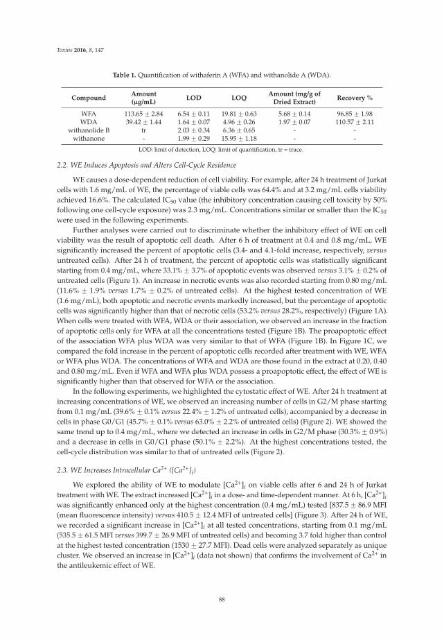
Toxins 2016, 8, 147
Table 1. Quantification of withaferin A (WFA) and withanolide A (WDA).
Compound Amount(μg/mL) LOD LOQ Amount (mg/g of
Dried Extract) Recovery %
WFA 113.65 ˘ 2.84 6.54 ˘ 0.11 19.81 ˘ 0.63 5.68 ˘ 0.14 96.85 ˘ 1.98WDA 39.42 ˘ 1.44 1.64 ˘ 0.07 4.96 ˘ 0.26 1.97 ˘ 0.07 110.57 ˘ 2.11
withanolide B tr 2.03 ˘ 0.34 6.36 ˘ 0.65 - -withanone - 1.99 ˘ 0.29 15.95 ˘ 1.18 - -
LOD: limit of detection, LOQ: limit of quantification, tr = trace.
2.2. WE Induces Apoptosis and Alters Cell-Cycle Residence
WE causes a dose-dependent reduction of cell viability. For example, after 24 h treatment of Jurkatcells with 1.6 mg/mL of WE, the percentage of viable cells was 64.4% and at 3.2 mg/mL cells viabilityachieved 16.6%. The calculated IC50 value (the inhibitory concentration causing cell toxicity by 50%following one cell-cycle exposure) was 2.3 mg/mL. Concentrations similar or smaller than the IC50
were used in the following experiments.Further analyses were carried out to discriminate whether the inhibitory effect of WE on cell
viability was the result of apoptotic cell death. After 6 h of treatment at 0.4 and 0.8 mg/mL, WEsignificantly increased the percent of apoptotic cells (3.4- and 4.1-fold increase, respectively, versusuntreated cells). After 24 h of treatment, the percent of apoptotic cells was statistically significantstarting from 0.4 mg/mL, where 33.1% ˘ 3.7% of apoptotic events was observed versus 3.1% ˘ 0.2% ofuntreated cells (Figure 1). An increase in necrotic events was also recorded starting from 0.80 mg/mL(11.6% ˘ 1.9% versus 1.7% ˘ 0.2% of untreated cells). At the highest tested concentration of WE(1.6 mg/mL), both apoptotic and necrotic events markedly increased, but the percentage of apoptoticcells was significantly higher than that of necrotic cells (53.2% versus 28.2%, respectively) (Figure 1A).When cells were treated with WFA, WDA or their association, we observed an increase in the fractionof apoptotic cells only for WFA at all the concentrations tested (Figure 1B). The proapoptotic effectof the association WFA plus WDA was very similar to that of WFA (Figure 1B). In Figure 1C, wecompared the fold increase in the percent of apoptotic cells recorded after treatment with WE, WFAor WFA plus WDA. The concentrations of WFA and WDA are those found in the extract at 0.20, 0.40and 0.80 mg/mL. Even if WFA and WFA plus WDA possess a proapoptotic effect, the effect of WE issignificantly higher than that observed for WFA or the association.
In the following experiments, we highlighted the cytostatic effect of WE. After 24 h treatment atincreasing concentrations of WE, we observed an increasing number of cells in G2/M phase startingfrom 0.1 mg/mL (39.6% ˘ 0.1% versus 22.4% ˘ 1.2% of untreated cells), accompanied by a decrease incells in phase G0/G1 (45.7% ˘ 0.1% versus 63.0% ˘ 2.2% of untreated cells) (Figure 2). WE showed thesame trend up to 0.4 mg/mL, where we detected an increase in cells in G2/M phase (30.3% ˘ 0.9%)and a decrease in cells in G0/G1 phase (50.1% ˘ 2.2%). At the highest concentrations tested, thecell-cycle distribution was similar to that of untreated cells (Figure 2).
2.3. WE Increases Intracellular Ca2+ ([Ca2+]i)
We explored the ability of WE to modulate [Ca2+]i on viable cells after 6 and 24 h of Jurkattreatment with WE. The extract increased [Ca2+]i in a dose- and time-dependent manner. At 6 h, [Ca2+]iwas significantly enhanced only at the highest concentration (0.4 mg/mL) tested [837.5 ˘ 86.9 MFI(mean fluorescence intensity) versus 410.5 ˘ 12.4 MFI of untreated cells] (Figure 3). After 24 h of WE,we recorded a significant increase in [Ca2+]i at all tested concentrations, starting from 0.1 mg/mL(535.5 ˘ 61.5 MFI versus 399.7 ˘ 26.9 MFI of untreated cells) and becoming 3.7 fold higher than controlat the highest tested concentration (1530 ˘ 27.7 MFI). Dead cells were analyzed separately as uniquecluster. We observed an increase in [Ca2+]i (data not shown) that confirms the involvement of Ca2+ inthe antileukemic effect of WE.
88
Toxins 2016, 8, 147
Figure 1. Percentage of viable, necrotic and apoptotic cells after 24 h treatment of Jurkat cells withincreasing concentrations of: DMSO extract obtained from the roots of W. somnifera (WE) (A); andwithaferin A (WFA), withanolide A (WDA) or WFA plus WDA (B). Fold increase in the percent ofapoptotic cells after treatment with different concentrations of WE, WFA, or WFA plus WDA (C).* p < 0.05; ** p < 0.01; *** p < 0.001 versus untreated cells.
0.00 0.05 0.10 0.25 0.40 0.80 1.000
20
40
60
80
G0-G1SG2-M
*** ****** *
***** ***
WE concentrations (mg/mL)
% c
ells
Figure 2. Cell-cycle distribution following 24 h treatment of Jurkat with increasing concentrations ofWE. * p < 0.05; ** p < 0.01; *** p < 0.001 versus untreated cells.
2.4. WE Induces Oxidative Stress
WE extract increased ROS production in a dose-dependent manner in Jurkat cells (Figure 4A).Most of the ROS were generated between 3 and 6 h of incubation. After 6 h treatment with WE,0.8 and 1.6 mg/mL of WE led to ROS levels similar or even higher than those promoted by a mildlytoxic dose of H2O2 (0.1 mM for 15 min), included as a positive control. After longer times of treatment(18 and 24 h), ROS generation reached a plateau (data not shown). N-acetylcysteine (NAC) ando-phenantroline (o-Phe) significantly inhibited the ROS generation induced by WE (0.8 mg/mL for6 h), while rotenone (Rot) was unable to afford a protective effect (Figure 4B).
89

Toxins 2016, 8, 147
Figure 3. Fraction of living cells with increased [Ca2+]i following 6 and 24 h exposure to increasingconcentrations of WE. * p < 0.05; ** p < 0.01; *** p < 0.001 versus untreated cells.
Figure 4. Reactive oxygen species (ROS) generation in WE-treated cells: (A) Jurkat exposed toincreasing concentrations of WE for 1 h, 3 h or 6 h. Cells treated with H2O2 0.1 mM for 15 minrepresent the positive control (dotted line parallel to x-axis). (B) Cells were treated for 6 h with WE0.8 mg/mL in the absence or presence of o-phenanthroline (o-Phe, 10 μM), rotenone (Rot, 2 μM) orN-acetylcysteine (NAC, 10 mM). Cells treated with H2O2 0.1 mM for 15 min represent the positivecontrol. * p < 0.05, ** p < 0.01, *** p < 0.001 versus control, and ˝˝˝ p < 0.001 versus WE.
2.5. Co-Treatment of Cells with WE and NAC Significantly Decreases WE-Induced Apoptosis
Because of the crucial role of ROS in the bioactivity of WE, we investigated whether the alterationof the redox state induced by NAC treatment could play a role in the apoptosis induced by WE.We observed a significant decrease in the WE-induced apoptotic events following 24 h of Jurkatco-treatment with WE plus NAC (10 mM). The WE-induced apoptotic events were significantlyreduced from 35% after 0.40 mg/mL of WE to 10% after WE plus NAC and from 38% after 0.80 mg/mLof WE to 12% after WE plus NAC (Figure 5).
0.40 0.800
20
40
60WEWE + NAC
* **
WE concentrations (mg/mL)
% a
popt
otic
cel
ls
Figure 5. Apoptotic events after 24 h of Jurkat treatment with WE in the absence and presence ofN-acetyl cysteine (NAC) (10 mM). * p < 0.05; ** p < 0.01 versus WE.
90
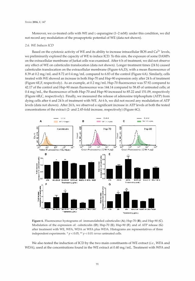
Toxins 2016, 8, 147
Moreover, we co-treated cells with WE and L-asparagine (1–2 mM): under this condition, we didnot record any modulation of the proapoptotic potential of WE (data not shown).
2.6. WE Induces ICD
Based on the cytotoxic activity of WE and its ability to increase intracellular ROS and Ca2+ levels,we preliminarily explored the capacity of WE to induce ICD. To this aim, the exposure of some DAMPson the extracellular membrane of Jurkat cells was examined. After 6 h of treatment, we did not observeany effect of WE on calreticulin translocation (data not shown). Longer treatment times (24 h) causedcalreticulin translocation on the extracellular membrane (Figure 6A,D), with a mean fluorescence of8.39 at 0.2 mg/mL and 8.72 at 0.4 mg/mL compared to 6.83 of the control (Figure 6A). Similarly, cellstreated with WE showed an increase in both Hsp-70 and Hsp-90 expression only after 24 h of treatment(Figure 6E,F, respectively). As an example, at 0.2 mg/mL Hsp-70 fluorescence was 57.92 compared to42.17 of the control and Hsp-90 mean fluorescence was 144.14 compared to 58.45 of untreated cells; at0.4 mg/mL, the fluorescence of both Hsp-70 and Hsp-90 increased to 85.22 and 151.09, respectively(Figure 6B,C, respectively). Finally, we measured the release of adenosine triphosphate (ATP) fromdying cells after 6 and 24 h of treatment with WE. At 6 h, we did not record any modulation of ATPlevels (data not shown). After 24 h, we observed a significant increase in ATP levels at both the testedconcentrations of the extract (2- and 2.45-fold increase, respectively) (Figure 6G).
Figure 6. Fluorescence hystograms of: immunolabeled calreticulin (A); Hsp-70 (B); and Hsp-90 (C).Modulation of the expression of: calreticulin (D); Hsp-70 (E); Hsp-90 (F); and of ATP release (G)after treatment with WE, WFA, WDA or WFA plus WDA. Histograms are representatives of threeindependent experiments. * p < 0.05, ** p < 0.01 versus untreated cells.
We also tested the induction of ICD by the two main constituents of WE extract (i.e., WFA andWDA), used at the concentrations found in the WE extract at 0.40 mg/mL. Treatment with WFA and
91
Toxins 2016, 8, 147
WDA alone or in association for 24 h did not cause a statistically significant modulation of calreticulintranslocation, Hsp-70 and Hsp-90 expression or ATP release (Figure 6D–G).
2.7. WE Induces DNA Damage
To evaluate the ability of WE to induce DNA damage, H2A.X phosphorylation was analyzed.H2A.X phosphorylation at Ser 139 represents a sensitive marker for DNA strand breakage [18]. WEinduced a dose-dependent increase in H2A.X phosphorylation, which was eight times higher thanuntreated cells at the highest tested concentration (0.80 mg/mL). This increase was similar to thephosphorylation induced by etoposide 10 μM, used as positive control (Figure 7).
0.00
Etoposide
0.20
0.40
0.80
0
5
10
15***
***
WE concentrations (mg/mL)
P-H
2A.X
Figure 7. Relative expression of phosphorylated H2A.X (P-H2A.X) induced by WE in Jurkat cells after6 h of treatment. Etoposide (10 μM) was used as positive control. *** p < 0.001 versus untreated cells.
3. Discussion
In this study, we demonstrated the in vitro antileukemic effect of the root extract of W. somnifera in aT-lymphoblastoid cell line. The high-performance liquid chromatography (HPLC) analysis performedon WE revealed the presence of WFA, WDA and to a lesser extent withanolide B. Withanolides, inparticular WFA and its acetyl derivative, are highly bioactive and show anticancer activity [19–21].Of note, its dihydroderivative is not active, thus suggesting that an unsaturated lactone moiety inring A of WFA is important for its biological activity. In our experimental settings, WE significantlyinduced apoptosis in a remarkable proportion of cells. Moreover, it blocked cell proliferation throughan accumulation of cells in the G2/M phase starting from the lowest tested concentrations. Cell-cycledysregulation represents a hallmark of cancer [22] and targeting the checkpoint signaling pathway,which usually leads to an arrest at G1/S or G2/M boundaries, is an effective therapeutic strategy [23].Our results confirm the antiproliferative and proapoptotic effect reported for withanolides and fora methanolic crude Withania leaf extract in different leukemia cell lines [24,25]. However, the IC50
calculated for the above mentioned methanolic leaf extract was much lower than that calculated in ourstudy. The difference could be imputable to the different part of plant (root versus leaf) used and/orthe method of extraction (the methanolic leaf extract was subjected to a sequential solvent extraction,which progressively concentrated the active components of Withania leaves).
Different mechanisms can be involved in the proapoptotic activity of our WE. Numerous studiesreported that intracellular Ca2+ mobilization plays a crucial role in apoptosis [26,27] and that calciumionophores exhibit proapoptotic activity [28]. Since WE exhibited a marked proapoptotic ability, wemeasured [Ca2+]i and demonstrated that further to the treatment of Jurkat cells with WE, [Ca2+]isignificantly increased. The underlying mechanisms need to be explored, however we can hypothesizethat the proapoptotic mechanism of WE involves intracellular Ca2+ accumulation.
92
Toxins 2016, 8, 147
Fruits of W. somnifera contain different enzymes including L-asparaginase, which catalyzesthe conversion of the aminoacid asparagine to aspartate and ammonia. Through this mechanism,L-asparaginase depletes the cellular levels of asparagine and induces the death of leukemic cellsthat are unable to synthesize asparagine. L-asparaginase exhibits cytotoxic effects on patient-derivedleukemic blasts [29]. A specific phytochemical analysis should be performed to detect the presenceof L-asparaginase in our extract. However, in our experimental conditions, the co-treatment of cellswith WE plus L-asparagine did not affect the proapoptotic potential of WE. The latter finding maysuggest that our extract does not contain L-asparaginase. Thus, it is conceivable that the presence ofL-asparaginase does not play a critical role in the cytotoxic activity of WE.
As already mentioned, the main component of our WE is WFA. Many studies have demonstratedthe role of WFA inducing oxidative stress as an anticancer strategy on different tumor cell lines, suchas prostate cancer, breast cancer, pancreatic cancer, leukemia, and melanoma [30–34]. ROS-mediatedapoptosis by WFA was shown to depend on both intrinsic and extrinsic pathways. Mitochondrialmembrane potential loss, release of cytochrome c and translocation of Bax, as well as increase incaspase-8 activity were observed together with the decrease of Bid, as crosstalk between intrinsic andextrinsic pathways [32]. Accordingly, we demonstrated that WE increased intracellular ROS levels.The remarkable inhibition of the proapoptotic potential of WE after co-treatment with NAC confirmsthe key role of ROS production in the apoptosis induced by WE. Similar data were obtained in bothestrogen receptor (ER) positive- and ER negative-breast cancer cell lines, where the apoptotic effect ofWFA was blunted by the presence of antioxidants [30].
Notably, selectivity by WFA towards cancer cells was observed in pancreatic and breast cancercells, as compared to normal human fibroblasts and normal human mammary epithelial cell line [30,34].ROS levels in cancer cells are close to the threshold and a ROS-mediated apoptotic mechanismrepresents an established indicator of cancer selectivity for an anticancer compound [35].
WE extract caused a dose-dependent ROS generation in Jurkat cells. This finding is in agreementwith previous studies reporting the ROS-generating ability of a similar WE extract [32] and some of itscomponents such as WFA [32,33,36–38]. ROS generation induced by WE extract reached a plateau after6 h of incubation. This suggests that the extract rapidly induces a pro-oxidative status in intoxicatedcells. Similar results were obtained by Malik [32], who found a significant ROS increase following1 to 3 h of exposure to WFA [38]. The co-incubation of cells with WE extract and NAC, an establishedROS scavenger, quenched the ROS generation induced by WE. This finding is in conformity withprevious data that reported that NAC attenuates Withania-induced ROS production in several celllines [32,36–38]. Co-incubation with o-Phe, an iron chelator that breaks Fenton reaction and stopsROS generation [39], attenuated ROS generation to a similar extent as NAC. These data strengthenthe notion that WE causes the cellular formation of ROS. Since analyses performed on melanoma celllines treated with WFA recorded mitochondrial ROS generation [33], we investigated whether themitochondrial respiratory chain is involved in this process. Rot, a prototypical Complex I inhibitor [40],did not affect WE-induced ROS production, suggesting that this complex is not involved in this processand that further studies will be needed to individuate the exact site of ROS production.
ROS production and endoplasmic reticulum (ER) stress are critical event promoting ICD, whichis also associated with the expression and/or release of DAMPs [41]. For example, calreticulin is aDAMP usually located on the lumen of ER and translocated on the extracellular membrane in caseof ER stress [41]. ER regulates many cellular events including [Ca2+]i levels. Alterations in Ca2+
homeostasis causes ER stress [42]. In our experimental settings, WE treatment causes ROS productionand increases [Ca2+]i levels. Accordingly, to the best of our knowledge, we demonstrated for thefirst time the ability of Withania to induce ICD starting from the lowest tested concentrations, asindicated by the up-regulation of calreticulin, Hsp-70, Hsp-90, and ATP release. The expression ofthese molecules increases the immunogenic profile of tumor cells, thus promoting the innate immunesystem response [43]. However, WFA and WDA that represent the two main constituents of our WEextract did not induce ICD either alone or in association. Recent evidence shows that WFA does not
93
Toxins 2016, 8, 147
alter the expression of Hsp-90 either in lymphoma or in pancreatic cells, and inhibits Hsp-90 with anATP-independent mechanism [44–46]. The anticancer activity of WFA depends on the inhibition ofcritical kinases and cell-cycle regulators controlled by Hsp-90 [44,45]. Hsp-90 is a molecular chaperoneinvolved in regulating protein folding and modulating a number of oncogenic client proteins playinga critical role in oncogenesis and cancer progression. Hsp-90 depends upon different co-chaperonesfor its function. WFA blocks the association of Hsp-90 to Cdc37, i.e., its co-chaperone, thus acting as apotent Hsp-90-client modulating agent [46]. The different activity of the two withanolides and WEcould be imputable to the complex nature of WE. In other words, the combined effects of the bioactivemolecules of the extract could differently influence DAMPs’ expression.
An immunostimulatory activity of an aqueous/alcoholic (1:1) root extract of Withania has beenreported on BALB/c mice and on ex vivo and in vitro macrophages [47]. The immunostimolatoryactivity of Withania together with the induction of ICD represents a promising strategy for thegeneration of a tumor-specific response.
Finally, we analyzed the genotoxic potential of WE. Our results showed that the treatment ofJurkat cells with WE significantly boosts H2A.X phosphorylation, which is an index of the ability ofa compound to interact with DNA thus triggering a genotoxic lesion. However, some recent in vivostudies reported the lack of genotoxicity of one of the most important constituents of Withania, i.e., WFA,and demonstrated its ability to provide protection against the 7,12-dimethylbenz(a)anthracene-inducedgenotoxicity [48,49]. As with the different behavior of WE and withanolides in ICD induction, thedifferent genotoxic profile of WE and WFA could be due to the matrix effects. Genotoxic studies oncomplex products of natural origin are usually performed on single phytochemicals rather than onthe product in its complexity. The matrix effect can cause an incomplete release of a key constituentfrom the vegetal matrix or modulate its bioavailability. This means that the use of toxicity dataconcerning the pure phytochemical are unsuited for the purposes of assessing the risk derived fromthe use of the same phytochemical within the complex vegetal matrix [2]. The use of Withania issuggested in different contexts including naturopathic care for anxiety [50], anti-infertility care [51],and osteoarthritis care [52]. Its genotoxicity should be carefully considered for an accurate assessmentof its risk–benefit profile. It is important to note that the H2A.X phosphorylation test used in our studyis able to detect only premutational, thus reparable DNA lesions. For this reason, further experimentsare needed to define the net and actual mutagenic effect of the lesions caused by WE and to directlyrelate the DNA damage to the mutagenic effect.
4. Experimental Section
4.1. WE Preparation
Withania somnifera roots were collected during the balsamic period (summer) and authenticatedby Dr. Paolo Scartezzini, Maharishi Ayurveda Product Ltd., Noida, India. The quality control wasperformed by Vedic Herbs s.r.l. (Caldiero, VR, Italy), which gifted us with a sample of root powder(voucher #12/11). The extract was prepared by mixing 10 g of Withania root powder with 100 mLof DMSO. The extract was vortexed for 15 min at room temperature and centrifuged to discard anyinsoluble part. The experiments and the HPLC analysis were performed using this stock solutionof 100 mg/mL.
4.2. HPLC Analysis
WE was subjected to RP-HPLC-DAD analysis to identify and quantify the main phytomarkers.The reference compounds WFA, WDA, withanolide B, and withanone were purchased fromExtrasynthese, Lyon, France. WFA and WDA were used as external standards to set up andcalculate appropriate calibration curves. The analyses were performed using a Jasco modular HPLC(model PU 2089, Jasco Corporation, Tokyo, Japan,) coupled to a diode array apparatus (MD 2010 Plus)linked to an injection valve with a 20 μL sampler loop. The column used was a Kinetex XB-C18 (5 μm,
94
Toxins 2016, 8, 147
15 cm ˆ 0.46 cm) with a flow rate of 0.6 mL/min. The analyses were performed at 25 ˝C with mobilephase and gradient chosen according to literature [17].
Following chromatogram recording, sample peaks were identified by comparing their ultraviolet(UV) spectra and retention time with those of the pure standards. Dedicated Jasco software(PDA version 1.5, Jasco Corporation, 2004) was used to calculate peak area by integration.
4.3. Validation
The individual stock solutions of each phytomarkers were prepared in ethanol or acetonitrile. Thecalibration curves of the considered compounds were prepared within different range: 500–50 μg/mLfor WFA, and 100–10 μg/mL for WDA. Each calibration solution was injected into HPLC in triplicate.The calibration graphs were provided by the regression analysis of the peak area of the analytes versusthe related concentrations. The analysis of the extract was performed under the same experimentalconditions. The obtained calibration graphs allowed the determination of the concentration of thephytomarkers inside the extract.
Limit Of Detection (LOD) and LOQ were calculated following the approach based on the standarddeviation of the response and the slope for WFA and WDA, on signal and noise ratio for withanolide Band withanone, as presented in the “Note for guidance on validation of analytical procedures: text andmethodology”, European Medicine Agency ICH Topic Q2 (R1). The accuracy was reported as percentof recovery and was estimated by adding known amount of analyte in the studied sample.
4.4. Cell Cultures
Human T-lymphoblastoid cells (Jurkat) were provided from LGC standards (LGC Group,Middlesex, UK). Cells were grown in suspension in Roswell Park Memorial Institute (RPMI) 1640supplemented with 10% heat-inactivated bovine serum, 1% penicillin/streptomycin solution, and 1%L-glutamine solution (all obtained from Biochrom, Merck Millipore, Darmstadt, Germany). Cells wereincubated at 37 ˝C with 5% CO2. To maintain exponential growth, the cultures were diluted to neverexceed the maximum suggested density of 3 ˆ 106 cells/mL.
4.5. Cell Treatment
Cells were treated with increasing concentrations of WE (0.0–1.6 mg/mL) for 1, 3, 6 or 24 h,according to the experimental requirements, or with WFA, WDA or WFA plus WDA for 24 h. WFAand WDA were tested at the concentrations found in the WE extract at 0.2, 0.4 and 0.8 mg/mL:0.23–0.92 μg/mL for WFA; 0.08–0.32 μg/mL for WDA. Etoposide 10 μM and hydrogen peroxide0.1 mM were used as positive controls. In some experiments, a co-treatment of WE with NAC orL-asparagine was performed.
4.6. Analysis of Cell Viability and Induction of Apoptosis
To determine cells’ viability, Guava ViaCount Reagent (Merck Millipore, Darmstadt, Germany)was used according to manufacturer’s instructions. Briefly, cells were appropriately diluted with thereagent containing 7-amino-actinomycin D (7-AAD) and incubated at room temperature in the darkfor 5 min before detection with flow cytometer. Furthermore, to discriminate between necrotic andapoptotic events, Guava Nexin Reagent (Merck Millipore) was used. Through the use of 7-AAD andannexin V-phycoerythrin, the assay allows the discrimination of apoptotic and necrotic events. Cellswere incubated with the reagent for 20 min at room temperature in the dark and then analyzed via flowcytometry. IC50 was calculated by interpolation from dose–response curve. Concentrations ď IC50
were used in the subsequent experiments.
95
Toxins 2016, 8, 147
4.7. Cell-Cycle Analysis
After treatment with WE for 24 h, cells were fixed with 70% ice-cold ethanol and, after washing,suspended in 200 μL of Guava Cell Cycle Reagent (Merck Millipore), containing propidium iodide.At the end of incubation at room temperature for 30 min in the dark, samples were analyzed viaflow cytometry.
4.8. Measurement of [Ca2+]i
After WE treatment for 6 or 24 h, [Ca2+]i was analyzed by using Fura Red™, AM (Thermo FisherScientific, Carlsbad, CA, USA), according to manufacturer’s instructions. Briefly, after treatment, cellswere incubated with the dye that freely permeates the cytoplasmic membrane but, once inside thecells, is hydrolyzed by the intracellular esterases and trapped into the cells. The fluorescence of thismolecule is enhanced once it binds Ca2+. To determine the optimal concentration of dye, a titration ofFura RedTM, AM was performed by loading Jurkat cells with a range of concentrations recommendedby the manufacturer (1–10 μM). The exposure of cells to Fura Red can cause cell death [53]. Thus, theuse of the lowest concentration of Fura Red is recommended. Following this experimental phase, theconcentration of 1 μM was adopted.
To detect intracellular calcium levels, Jurkat cells were incubated at 37 ˝C for 30 min in PBSwithout calcium and magnesium. This buffer condition allows to detect the intracellular calcium storesand exclude the secondary increase in [Ca2+]i due to Ca2+ entry [54]. Moreover, the removal of externalCa2+ reduces the non-specific fluctuations in [Ca2+]i normally observed during the first 20–30 s ofsample acquisition via flow cytometry. Results are expressed as MFI.
4.9. Detection of ROS Production
ROS production was determined after 1, 3, 6, 18 or 24 h of WE treatment by using the probedihydrorhodamine (DHR, 10 μM) [55], which was added during the last 15 min of incubation.Hydrogen peroxide was used as positive control. Additionally, cells were pre- treated for 30 min withRot (2 μM) or o-Phe (10 μM) and co-treated for 6 h with WE (0.8 mg/mL). In some experiments, cellswere co-treated for 6 h with WE (0.8 mg/mL) plus NAC (10 mM). After three washing in PBS, cellularfluorescence was imaged using a Leica DMLB/DFC300F fluorescence microscope (Leica Microsystems,Wetzlar, Germany) equipped with an Olympus ColorviewIIIu CCD camera (Polyphoto, Milan, Italy).Fluorescence images (100 cells per sample from randomly selected fields) were digitally acquiredand processed for fluorescence determination at the single cell level on a personal computer usingthe public domain program, Image J. Mean fluorescence values were determined by averaging thefluorescence of at least 100 cells/treatment condition/experiment.
4.10. Analysis of Calreticulin Translocation, Hsp-70 and Hsp-90 Expression, and ATP Release
After 6 or 24 h of treatment, cells were washed and incubated with phycoerythrin-labeledcalreticulin antibody (1:100, Abcam, San Francisco, CA, USA) or isotope-matched negative control(isotypic mouse IgG1 K Alexa Fluor 488®) (eBioscience, San Diego, CA, USA).
To analyze Hsp-70 and Hsp-90 expression, cells were incubated with an anti-Hsp-70 or anti-Hsp-90antibody (1:100, Abcam, for both antibodies) and, after washing, incubated with fluoresceinisothyocianate-labeled secondary antibody (1:100, Sigma, Merck Millipore, Darmstadt, Germany)or the isotype control. Mean fluorescence was detected via flow cytometry.
The kit ATPLite™ 1step (Perkin Elmer, Waltham, MA, USA) was used for the detection ofATP extracellular concentration. Jurkat cells were seeded and treated with WE in Hank's BalancedSalt Solution (HBSS) or complete medium for 6 and 24 h, respectively. At the end of incubation,supernatants were collected and treated with 100 μL of ATPLite 1step reagent containing luciferaseand D-luciferin. After shaking for 2 min at 700 rpm using the orbital microplate shaker 711/+
96
Toxins 2016, 8, 147
(Asal srl, Florence, Italy), luminescence of the samples was measured in a 96-well black plate usingthe microplate reader Victor X3 (Perkin Elmer).
4.11. DNA Damage Analysis
Phosphorylation of histone P-H2A.X was used as marker of WE genotoxic potential. After, 6 hof treatment with WE, cells were fixed, permeabilized and incubated for 30 min in the dark at roomtemperature with an anti-P-H2A.X-Alexa Fluor® antibody (Merck Millipore). Etoposide 10 μM wasused as positive control. Samples were analyzed via flow cytometry.
4.12. Flow Cytometry
EasyCyte 5HT (Merck Millipore) was used to perform all flow cytometric analyses, with theexception of the measurements of [Ca2+]i performed by using a FACSCanto II (BD Bioscience, FranklinLakes, NJ, USA). For each sample, approximately 5000 events were evaluated.
4.13. Statistical Analysis
All results are expressed as mean ˘ SEM of at least three independent experiments. Differencesbetween treatments were assessed by t test or one-way ANOVA and Dunnet or Bonferroni was usedas post-tests. All statistical analyses were performed using GraphPad InStat 5.0 version (GraphPadPrism, San Diego, CA, USA, 2007). p < 0.05 was considered significant.
Acknowledgments: This work was supported by FARB—Finanziamenti di Ateneo alla Ricerca di base2012 (RFBO124222).
Author Contributions: C.F. and P.S. conceived and designed the experiments; E.T., C.C., E.C., E.d.G., M.T., A.R.D.,and B.C. performed the experiments; C.F., E.T., C.C., S.P., A.G., and M.T. analyzed the data; and C.F., E.T., C.C.,P.S., G.V., A.G., and P.H. wrote the paper.
Conflicts of Interest: The authors declare no conflict of interest.
References
1. Global Status Report on Noncommunicable Diseases 2014. Available online: http://www.who.int/nmh/publications/ncd-status-report-2014/en/ (accessed on 22 December 2015).
2. Fimognari, C.; Ferruzzi, L.; Turrini, E.; Carulli, G.; Lenzi, M.; Hrelia, P.; Cantelli-Forti, G. Metabolic andtoxicological considerations of botanicals in anticancer therapy. Expert Opin. Drug Metab. Toxicol. 2012, 8,819–832. [CrossRef] [PubMed]
3. Rivera, E.; Gomez, H. Chemotherapy resistance in metastatic breast cancer: The evolving role of ixabepilone.Breast Cancer Res. 2010, 12 (Suppl. 2), 1–12. [CrossRef] [PubMed]
4. Kroemer, G.; Galluzzi, L.; Kepp, O.; Zitvogel, L. Immunogenic cell death in cancer therapy. Annu. Rev.Immunol. 2013, 31, 51–72. [CrossRef] [PubMed]
5. Hou, W.; Zhang, Q.; Yan, Z.; Chen, R.; Zeh, H.J., III; Kang, R.; Lotze, M.T.; Tang, D. Strange attractors: DAMPsand autophagy link tumor cell death and immunity. Cell Death Dis. 2013, 4, e966. [CrossRef] [PubMed]
6. Newman, D.J.; Cragg, G.M.; Snader, K.M. Natural products as sources of new drugs over the period1981–2002. J. Nat. Prod. 2003, 66, 1022–1037. [CrossRef] [PubMed]
7. Chen, S.T.; Dou, J.; Temple, R.; Agarwal, R.; Wu, K.M.; Walker, S. New therapies from old medicines.Nat. Biotechnol. 2008, 26, 1077–1083. [CrossRef] [PubMed]
8. Newman, D.J.; Cragg, G.M. Natural products as sources of new drugs over the 30 years from 1981 to 2010.J. Nat. Prod. 2012, 75, 311–335. [CrossRef] [PubMed]
9. Nance, C.L. Clinical efficacy trials with natural products and herbal medicines. In Phytotherapies: Efficacy,Safety and Regulation; Ramzan, I., Ed.; John Wiley & Sons Inc.: Hoboken, NJ, USA, 2015; pp. 65–88.
10. Shanafelt, T.D.; Call, T.G.; Zent, C.S.; Leis, J.F.; LaPlant, B.; Bowen, D.A.; Roos, M.; Laumann, K.; Ghosh, A.K.;Lesnick, C.; et al. Phase 2 trial of daily, oral Polyphenon E in patients with asymptomatic, Rai stage 0 to IIchronic lymphocytic leukemia. Cancer 2013, 119, 363–370. [CrossRef] [PubMed]
97
Toxins 2016, 8, 147
11. Kulkarni, S.K.; Dhir, A. Withania somnifera: An Indian ginseng. Prog. Neuropsychopharmacol. Biol. Psychiatry2008, 32, 1093–1105. [CrossRef] [PubMed]
12. Masevhe, N.A.; McGaw, L.J.; Eloff, J.N. The traditional use of plants to manage candidiasis and relatedinfections in Venda, South Africa. J. Ethnopharmacol. 2015, 168, 364–372. [CrossRef] [PubMed]
13. Khanna, D.; Sethi, G.; Ahn, K.S.; Pandey, M.K.; Kunnumakkara, A.B.; Sung, B.; Aggarwal, A.; Aggarwal, B.B.Natural products as a gold mine for arthritis treatment. Curr. Opin. Pharmacol. 2007, 7, 344–351. [CrossRef][PubMed]
14. Nosalova, G.; Fleskova, D.; Jurecek, L.; Sadlonova, V.; Ray, B. Herbal polysaccharides and cough reflex.Respir. Physiol. Neurobiol. 2013, 187, 47–51. [CrossRef] [PubMed]
15. Jain, S.; Shukla, S.D.; Sharma, K.; Bhatnagar, M. Neuroprotective effects of Withania somnifera Dunn.in hippocampal sub-regions of female albino rat. Phytother. Res. 2001, 15, 544–548. [CrossRef] [PubMed]
16. Rai, M.; Jogee, P.S.; Agarkar, G.; Santos, C.A. Anticancer activities of Withania somnifera: Current research,formulations, and future perspectives. Pharm. Biol. 2016, 54, 189–197. [CrossRef] [PubMed]
17. Chaurasiya, N.D.; Uniyal, G.C.; Lal, P.; Misra, L.; Sangwan, N.S.; Tuli, R.; Sangwan, R.S. Analysis ofwithanolides in root and leaf of Withania somnifera by HPLC with photodiode array and evaporative lightscattering detection. Phytochem. Anal. 2008, 19, 148–154. [CrossRef] [PubMed]
18. Sharma, A.; Singh, K.; Almasan, A. Histone H2AX phosphorylation: A marker for DNA damage.Methods Mol. Biol. 2012, 920, 613–626. [PubMed]
19. Vyas, A.R.; Singh, S.V. Molecular targets and mechanisms of cancer prevention and treatment by withaferina, a naturally occurring steroidal lactone. AAPS. J. 2014, 16, 1–10. [CrossRef] [PubMed]
20. Choudharymy, M.I.; Yousuf, S.; Atta-Ur-Rahman. Withanolides: Chemistry and antitumor activity.In Natural Products; Ramawat, K.G., Merillon, J.M., Eds.; Springer-Verlag: Berlin, Germany; Heidelberg,Germany, 2013; pp. 3465–3495.
21. Ichikawa, H.; Takada, Y.; Shishodia, S.; Jayaprakasam, B.; Nair, M.G.; Aggarwal, B.B. Withanolides potentiateapoptosis, inhibit invasion, and abolish osteoclastogenesis through suppression of nuclear factor-kappaB(NF-kappaB) activation and NF-kappaB-regulated gene expression. Mol. Cancer Ther. 2006, 5, 1434–1445.[CrossRef] [PubMed]
22. Stewart, Z.A.; Westfall, M.D.; Pietenpol, J.A. Cell-cycle dysregulation and anticancer therapy. TrendsPharmacol. Sci. 2003, 24, 139–145. [CrossRef]
23. Shapiro, G.I.; Harper, J.W. Anticancer drug targets: Cell cycle and checkpoint control. J. Clin. Investig. 1999,104, 1645–1653. [CrossRef] [PubMed]
24. Oh, J.H.; Lee, T.J.; Kim, S.H.; Choi, Y.H.; Lee, S.H.; Lee, J.M.; Kim, Y.H.; Park, J.W.; Kwon, T.K. Induction ofapoptosis by withaferin A in human leukemia U937 cells through down-regulation of Akt phosphorylation.Apoptosis 2008, 13, 1494–1504. [CrossRef] [PubMed]
25. Senthil, V.; Ramadevi, S.; Venkatakrishnan, V.; Giridharan, P.; Lakshmi, B.S.; Vishwakarma, R.A.;Balakrishnan, A. Withanolide induces apoptosis in HL-60 leukemia cells via mitochondria mediatedcytochrome c release and caspase activation. Chem. Biol. Interact. 2007, 167, 19–30. [CrossRef] [PubMed]
26. Kruman, I.; Guo, Q.; Mattson, M.P. Calcium and reactive oxygen species mediate staurosporine-inducedmitochondrial dysfunction and apoptosis in PC12 cells. J. Neurosci. Res. 1998, 51, 293–308. [CrossRef]
27. Nicotera, P.; Orrenius, S. The role of calcium in apoptosis. Cell Calcium 1998, 23, 173–180. [CrossRef]28. Salvioli, S.; Ardizzoni, A.; Franceschi, C.; Cossarizza, A. JC-1, but not DiOC6(3) or rhodamine 123, is a
reliable fluorescent probe to assess ΔΨ changes in intact cells: Implications for studies on mitochondrialfunctionality during apoptosis. FEBS Lett. 1997, 411, 77–82. [CrossRef]
29. Oza, V.P.; Parmar, P.P.; Kumar, S.; Subramanian, R.B. Anticancer properties of highly purified L-asparaginasefrom Withania somnifera L. against acute lymphoblastic leukemia. Appl. Biochem. Biotechnol. 2010, 160,1833–1840. [CrossRef] [PubMed]
30. Hahm, E.R.; Moura, M.B.; Kelley, E.E.; Van, H.B.; Shiva, S.; Singh, S.V. Withaferin A-induced apoptosis inhuman breast cancer cells is mediated by reactive oxygen species. PLoS ONE 2011, 6, e23354. [CrossRef][PubMed]
31. Li, X.; Zhu, F.; Jiang, J.; Sun, C.; Wang, X.; Shen, M.; Tian, R.; Shi, C.; Xu, M.; Peng, F.; et al. Synergisticantitumor activity of withaferin A combined with oxaliplatin triggers reactive oxygen species-mediatedinactivation of the PI3K/AKT pathway in human pancreatic cancer cells. Cancer Lett. 2015, 357, 219–230.[CrossRef] [PubMed]
98
Toxins 2016, 8, 147
32. Malik, F.; Kumar, A.; Bhushan, S.; Khan, S.; Bhatia, A.; Suri, K.A.; Qazi, G.N.; Singh, J. Reactive oxygenspecies generation and mitochondrial dysfunction in the apoptotic cell death of human myeloid leukemiaHL-60 cells by a dietary compound withaferin A with concomitant protection by N-acetyl cysteine. Apoptosis2007, 12, 2115–2133. [CrossRef] [PubMed]
33. Mayola, E.; Gallerne, C.; Esposti, D.D.; Martel, C.; Pervaiz, S.; Larue, L.; Debuire, B.; Lemoine, A.; Brenner, C.;Lemaire, C. Withaferin A induces apoptosis in human melanoma cells through generation of reactive oxygenspecies and down-regulation of Bcl-2. Apoptosis 2011, 16, 1014–1027. [CrossRef] [PubMed]
34. Nishikawa, Y.; Okuzaki, D.; Fukushima, K.; Mukai, S.; Ohno, S.; Ozaki, Y.; Yabuta, N.; Nojima, H. WithaferinA Induces Cell Death Selectively in Androgen-Independent Prostate Cancer Cells but Not in NormalFibroblast Cells. PLoS ONE 2015, 10, e0134137. [CrossRef] [PubMed]
35. Trachootham, D.; Alexandre, J.; Huang, P. Targeting cancer cells by ROS-mediated mechanisms: A radicaltherapeutic approach? Nat. Rev. Drug Discov. 2009, 8, 579–591. [CrossRef] [PubMed]
36. Kakar, S.S.; Jala, V.R.; Fong, M.Y. Synergistic cytotoxic action of cisplatin and withaferin A on ovarian cancercell lines. Biochem. Biophys. Res. Commun. 2012, 423, 819–825. [CrossRef] [PubMed]
37. Yu, S.M.; Kim, S.J. Production of reactive oxygen species by withaferin A causes loss of type collagenexpression and COX-2 expression through the PI3K/Akt, p38, and JNK pathways in rabbit articularchondrocytes. Exp. Cell Res. 2013, 319, 2822–2834. [CrossRef] [PubMed]
38. Yu, S.M.; Kim, S.J. Withaferin A-caused production of intracellular reactive oxygen species modulatesapoptosis via PI3K/Akt and JNKinase in rabbit articular chondrocytes. J. Korean Med. Sci. 2014, 29,1042–1053. [CrossRef] [PubMed]
39. Sestili, P.; Diamantini, G.; Bedini, A.; Cerioni, L.; Tommasini, I.; Tarzia, G.; Cantoni, O. Plant-derived phenoliccompounds prevent the DNA single-strand breakage and cytotoxicity induced by tert-butylhydroperoxidevia an iron-chelating mechanism. Biochem. J. 2002, 364, 121–128. [CrossRef] [PubMed]
40. Teeter, M.E.; Baginsky, M.L.; Hatefi, Y. Ectopic inhibition of the complexes of the electron transport systemby rotenone, piericidin A, demerol and antimycin A. Biochim. Biophys. Acta 1969, 172, 331–333. [CrossRef]
41. Krysko, D.V.; Garg, A.D.; Kaczmarek, A.; Krysko, O.; Agostinis, P.; Vandenabeele, P. Immunogenic cell deathand DAMPs in cancer therapy. Nat. Rev. Cancer 2012, 12, 860–875. [CrossRef] [PubMed]
42. Rao, R.V.; Ellerby, H.M.; Bredesen, D.E. Coupling endoplasmic reticulum stress to the cell death program.Cell Death Differ. 2004, 11, 372–380. [CrossRef] [PubMed]
43. Garg, A.D.; Nowis, D.; Golab, J.; Vandenabeele, P.; Krysko, D.V.; Agostinis, P. Immunogenic cell death,DAMPs and anticancer therapeutics: An emerging amalgamation. Biochim. Biophys. Acta 2010, 1805, 53–71.[CrossRef] [PubMed]
44. McKenna, M.K.; Gachuki, B.W.; Alhakeem, S.S.; Oben, K.N.; Rangnekar, V.M.; Gupta, R.C.; Bondada, S.Anti-cancer activity of withaferin A in B-cell lymphoma. Cancer Biol. Ther. 2015, 16, 1088–1098. [CrossRef][PubMed]
45. Yu, Y.; Hamza, A.; Zhang, T.; Gu, M.; Zou, P.; Newman, B.; Li, Y.; Gunatilaka, A.A.; Zhan, C.G.; Sun, D.Withaferin A targets heat shock protein 90 in pancreatic cancer cells. Biochem. Pharmacol. 2010, 79, 542–551.[CrossRef] [PubMed]
46. Grover, A.; Shandilya, A.; Agrawal, V.; Pratik, P.; Bhasme, D.; Bisaria, V.S.; Sundar, D. Hsp90/Cdc37chaperone/co-chaperone complex, a novel junction anticancer target elucidated by the mode of action ofherbal drug Withaferin A. BMC Bioinf. 2011, 12. [CrossRef] [PubMed]
47. Malik, F.; Singh, J.; Khajuria, A.; Suri, K.A.; Satti, N.K.; Singh, S.; Kaul, M.K.; Kumar, A.; Bhatia, A.; Qazi, G.N.A standardized root extract of Withania somnifera and its major constituent withanolide-A elicit humoral andcell-mediated immune responses by up regulation of Th1-dominant polarization in BALB/c mice. Life Sci.2007, 80, 1525–1538. [CrossRef] [PubMed]
48. Panjamurthy, K.; Manoharan, S.; Balakrishnan, S.; Suresh, K.; Nirmal, M.R.; Senthil, N.; Alias, L.M.Protective effect of Withaferin-A on micronucleus frequency and detoxication agents during experimentaloral carcinogenesis. Afr. J. Tradit. Complement. Altern. Med. 2008, 6, 1–8. [CrossRef] [PubMed]
49. Panjamurthy, K.; Manoharan, S.; Menon, V.P.; Nirmal, M.R.; Senthil, N. Protective role of withaferin-A on7,12-dimethylbenz(a)anthracene-induced genotoxicity in bone marrow of Syrian golden hamsters. J. Biochem.Mol. Toxicol. 2008, 22, 251–258. [CrossRef] [PubMed]
50. Cooley, K.; Szczurko, O.; Perri, D.; Mills, E.J.; Bernhardt, B.; Zhou, Q.; Seely, D. Naturopathic care for anxiety:A randomized controlled trial ISRCTN78958974. PLoS ONE 2009, 4, e6628. [CrossRef] [PubMed]
99
Toxins 2016, 8, 147
51. Ahmad, M.K.; Mahdi, A.A.; Shukla, K.K.; Islam, N.; Rajender, S.; Madhukar, D.; Shankhwar, S.N.; Ahmad, S.Withania somnifera improves semen quality by regulating reproductive hormone levels and oxidative stressin seminal plasma of infertile males. Fertil. Steril. 2010, 94, 989–996. [CrossRef] [PubMed]
52. Chopra, A.; Lavin, P.; Patwardhan, B.; Chitre, D. A 32-week randomized, placebo-controlled clinicalevaluation of RA-11, an Ayurvedic drug, on osteoarthritis of the knees. J. Clin. Rheumatol. 2004, 10,236–245. [CrossRef] [PubMed]
53. Wendt, E.R.; Ferry, H.; Greaves, D.R.; Keshav, S. Ratiometric analysis of fura red by flow cytometry:A technique for monitoring intracellular calcium flux in primary cell subsets. PLoS ONE 2015, 10, e0119532.[CrossRef] [PubMed]
54. Verriere, V.; Higgins, G.; Al-Alawi, M.; Costello, R.W.; McNally, P.; Chiron, R.; Harvey, B.J.; Urbach, V. LipoxinA4 stimulates calcium-activated chloride currents and increases airway surface liquid height in normal andcystic fibrosis airway epithelia. PLoS ONE 2012, 7, e37746. [CrossRef] [PubMed]
55. Royall, J.A.; Ischiropoulos, H. Evaluation of 2’,7’-dichlorofluorescin and dihydrorhodamine 123 as fluorescentprobes for intracellular H2O2 in cultured endothelial cells. Arch. Biochem. Biophys. 1993, 302, 348–355.[CrossRef] [PubMed]
© 2016 by the authors; licensee MDPI, Basel, Switzerland. This article is an open accessarticle distributed under the terms and conditions of the Creative Commons Attribution(CC-BY) license (http://creativecommons.org/licenses/by/4.0/).
100
toxins
Communication
Cells Deficient in the Fanconi Anemia ProteinFANCD2 are Hypersensitive to the Cytotoxicity andDNA Damage Induced by Coffee and Caffeic Acid
Estefanía Burgos-Morón 1, José Manuel Calderón-Montaño 1,2, Manuel Luis Orta 3,Emilio Guillén-Mancina 1, Santiago Mateos 3 and Miguel López-Lázaro 1,*
1 Department of Pharmacology, Faculty of Pharmacy, University of Seville, Profesor García González 2,41012 Seville, Spain; [email protected] (E.B.-M.); [email protected] (J.M.C.-M.);[email protected] (E.G.-M.)
2 Department of Molecular Biology, Centro Andaluz de Biología Molecular y Medicina Regenerativa,University of Seville, Avda. Americo Vespucio s/n., 41092 Seville, Spain
3 Department of Cell Biology, Faculty of Biology, University of Seville, Avda. Reina Mercedes s/n.,41012 Seville, Spain; [email protected] (M.L.O.); [email protected] (S.M.)
* Correspondence: [email protected]; Tel.: +34-954-556-348; Fax: +34-954-233-765
Academic Editor: Carmela FimognariReceived: 15 March 2016; Accepted: 1 July 2016; Published: 8 July 2016
Abstract: Epidemiological studies have found a positive association between coffee consumption anda lower risk of cardiovascular disorders, some cancers, diabetes, Parkinson and Alzheimer disease.Coffee consumption, however, has also been linked to an increased risk of developing some typesof cancer, including bladder cancer in adults and leukemia in children of mothers who drink coffeeduring pregnancy. Since cancer is driven by the accumulation of DNA alterations, the ability of thecoffee constituent caffeic acid to induce DNA damage in cells may play a role in the carcinogenicpotential of this beverage. This carcinogenic potential may be exacerbated in cells with DNA repairdefects. People with the genetic disease Fanconi Anemia have DNA repair deficiencies and arepredisposed to several cancers, particularly acute myeloid leukemia. Defects in the DNA repairprotein Fanconi Anemia D2 (FANCD2) also play an important role in the development of a varietyof cancers (e.g., bladder cancer) in people without this genetic disease. This communication showsthat cells deficient in FANCD2 are hypersensitive to the cytotoxicity (clonogenic assay) and DNAdamage (γ-H2AX and 53BP1 focus assay) induced by caffeic acid and by a commercial lyophilizedcoffee extract. These data suggest that people with Fanconi Anemia, or healthy people who developsporadic mutations in FANCD2, may be hypersensitive to the carcinogenic activity of coffee.
Keywords: coffee; caffeic acid; cancer; DNA damage; carcinogenesis; FANCD2; Fanconi anemia
1. Introduction
Coffee, one of the most widely consumed beverages in the world, can affect human health.Observational studies suggest that coffee consumption may lower the risk of developing diabetes,cardiovascular disorders, cirrhosis, and degenerative disorders such as Parkinson and Alzheimerdisease [1,2]. Coffee consumption has also been associated with a decreased risk of total mortality;coffee drinkers had a lower risk of death from heart disease, chronic respiratory diseases, diabetes,pneumonia and influenza, and intentional self-harm [3]. No significant association between coffeeconsumption and total cancer mortality was found, however [3]. The effect of coffee consumptionon the risk of cancer is inconclusive; some studies indicate that coffee may reduce the risk of sometypes of cancers [4–6], while others suggest that it may increase the risk of developing the disease [6,7].According to the International Agency for Research on Cancer (IARC), coffee is classified as possibly
Toxins 2016, 8, 211 101 www.mdpi.com/journal/toxins
Toxins 2016, 8, 211
carcinogenic to the human urinary bladder (IARC, Vol. 51). Several recent epidemiological studieshave also revealed that maternal consumption of coffee during pregnancy may be associated withchildhood leukemia [8–11]. A recent meta-analysis found that maternal coffee consumption duringpregnancy significantly increased the risk of childhood acute lymphoblastic leukemia (ALL) and acutemyeloid leukemia (AML) in a dose-response manner [11].
Caffeic acid is present in coffee in low amounts, but it is an important metabolite found in plasmaand urine after coffee consumption [1,12]. In coffee, caffeic acid is typically bound to quinic acid toform an ester called 5-caffeoylquinic acid or chlorogenic acid [13]. Some studies have shown thatcaffeic acid possesses antioxidant and anti-genotoxic activities [14–19]. However, other investigationshave revealed that this polyphenol generates reactive oxygen species (ROS) and produces carcinogeniceffects [20–26]. In vivo studies have also found that diets containing caffeic acid induced tumors inanimals [2]. Caffeic acid is therefore classified as possibly carcinogenic to humans (IARC, Vol. 56).This carcinogenic activity may be due to its ability to induce DNA damage [21–23], probably througha pro-oxidant mechanism [22,24–26].
Cancer is a disease caused by the accumulation of DNA alterations in our cells [27–30]. When ourcells suffer DNA alterations, the DNA damage response machinery activates a variety of mechanisms torepair the damage [3]. These mechanisms are necessary for maintaining the integrity of the DNA, andtherefore provide a fundamental biological barrier against carcinogenesis. Mutations in DNA repairgenes result in the accumulation of cellular DNA damage and in predisposition to cancer. Some peopleare born with defects in genes involved in the DNA damage response machinery. People withoutgermline mutations in DNA repair genes can also acquire them during a possible carcinogenic process.People with these mutations are particularly sensitive to the carcinogenic activity of compounds thatinduce types of DNA damage requiring these genes for repair.
The Fanconi anemia (FA) pathway plays an important role in the repair of several types of DNAdamage, including interstrand crosslink, replication fork stalling and double strand breaks. The FAprotein Fanconi Anemia D2 (FANCD2) is essential for proper functioning of this pathway; this proteinis considered a surrogate marker for FA network activation. Mutations in a cluster of proteins of thispathway cause Fanconi Anemia, a rare autosomal recessive genetic disease characterized by bonemarrow failure, congenital abnormalities, genomic instability and predisposition to several types ofcancer, particularly acute myeloid leukemia [31–33]. Defects in proteins implicated in this DNA repairpathway has been described in several kinds of sporadic cancers, including bladder cancer and acutemyeloid leukemia [34–37]. Deficiency in this DNA repair pathway makes cells hypersensitive to thecytotoxicity and DNA-damaging activities of a variety of agents, including ROS [38–40].
Since caffeic acid induces ROS-mediated DNA damage, and since the FA pathway participates inthe repair of this type of DNA damage, we hypothesized that cells deficient in this pathway wouldbe more susceptible than normal cells to the DNA-damaging effect of this dietary phytochemical.We report that human cells deficient in FANCD2 are hypersensitive to the cytotoxicity (clonogenicassay) and DNA damage (γ-H2AX and 53BP1 focus assay) induced by caffeic acid and by a commerciallyophilized coffee extract, and discuss the possible relevance of these results.
2. Results
2.1. Cells Deficient in FANCD2 Are Hypersensitive to the Cytotoxicity of Coffee and Caffeic Acid
Cells lacking the FA protein FANCD2 (PD20´/´) and cells complemented with FANCD2(PD20+/+) were exposed for 4 h to caffeic acid and to a commercial lyophilized coffee extract. After7 days of recovery in drug-free medium, cell survival was determined with the clonogenic assay.Figure 1 shows that the survival of cells lacking FANCD2 was significantly lower than that of proficientcells when exposed to several concentrations of caffeic acid (A) and coffee (B). This means that thecellular toxicity of coffee and caffeic acid is increased in cells lacking the DNA repair protein FANCD2.
102
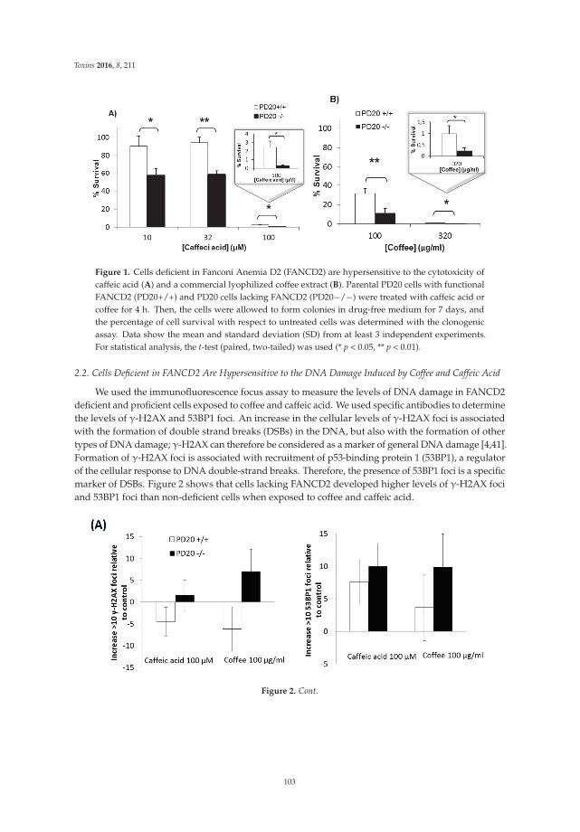
Toxins 2016, 8, 211
Figure 1. Cells deficient in Fanconi Anemia D2 (FANCD2) are hypersensitive to the cytotoxicity ofcaffeic acid (A) and a commercial lyophilized coffee extract (B). Parental PD20 cells with functionalFANCD2 (PD20+/+) and PD20 cells lacking FANCD2 (PD20´/´) were treated with caffeic acid orcoffee for 4 h. Then, the cells were allowed to form colonies in drug-free medium for 7 days, andthe percentage of cell survival with respect to untreated cells was determined with the clonogenicassay. Data show the mean and standard deviation (SD) from at least 3 independent experiments.For statistical analysis, the t-test (paired, two-tailed) was used (* p < 0.05, ** p < 0.01).
2.2. Cells Deficient in FANCD2 Are Hypersensitive to the DNA Damage Induced by Coffee and Caffeic Acid
We used the immunofluorescence focus assay to measure the levels of DNA damage in FANCD2deficient and proficient cells exposed to coffee and caffeic acid. We used specific antibodies to determinethe levels of γ-H2AX and 53BP1 foci. An increase in the cellular levels of γ-H2AX foci is associatedwith the formation of double strand breaks (DSBs) in the DNA, but also with the formation of othertypes of DNA damage; γ-H2AX can therefore be considered as a marker of general DNA damage [4,41].Formation of γ-H2AX foci is associated with recruitment of p53-binding protein 1 (53BP1), a regulatorof the cellular response to DNA double-strand breaks. Therefore, the presence of 53BP1 foci is a specificmarker of DSBs. Figure 2 shows that cells lacking FANCD2 developed higher levels of γ-H2AX fociand 53BP1 foci than non-deficient cells when exposed to coffee and caffeic acid.
Figure 2. Cont.
103
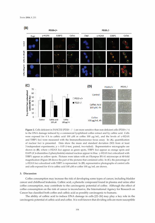
Toxins 2016, 8, 211
Figure 2. Cells deficient in FANCD2 (PD20´/´) are more sensitive than non-deficient cells (PD20+/+)to the DNA damage induced by a commercial lyophilized coffee extract and by caffeic acid. Cellswere exposed for 4 h to caffeic acid 100 μM or coffee 100 μg/mL, and the levels of γ-H2AXand 53BP1 foci were measured with the Immunofluorescence focus assay. In (A), quantificationof nuclear foci is presented. Data show the mean and standard deviation (SD) from at least3 independent experiments; p > 0.05 (t-test, paired, two-tailed). Representative micrographs areshown in (B), where γ-H2AX foci appear as green spots, 53BP1 foci appear as orange spots andDAPI (41,6-diamidino-2-phenylindole)-stained nucleus appear in blue. γ-H2AX foci colocalized with53BP1 appear as yellow spots. Pictures were taken with an Olympus BX 61 microscope at 40-foldmagnification (Figure 2B shows the part of the pictures that contained cells). In (C), the percentage ofγ-H2AX foci colocalized with 53BP1 is represented. In (D), representative photographs of control cellsand cells exposed for 4 h to caffeic acid 100 μM or coffee 100 μg/mL are shown.
3. Discussion
Coffee consumption may increase the risk of developing some types of cancer, including bladdercancer and childhood leukemia. Caffeic acid, a phenolic compound found in plasma and urine aftercoffee consumption, may contribute to the carcinogenic potential of coffee. Although the effect ofcoffee consumption on the risk of cancer is inconclusive, the International Agency for Research onCancer has classified both coffee and caffeic acid as possibly carcinogenic to humans.
The ability of caffeic acid to induce DNA damage in cells [22–26] may play a key role in thecarcinogenic potential of caffeic acid and coffee. It is well-known that dividing cells are more susceptible
104
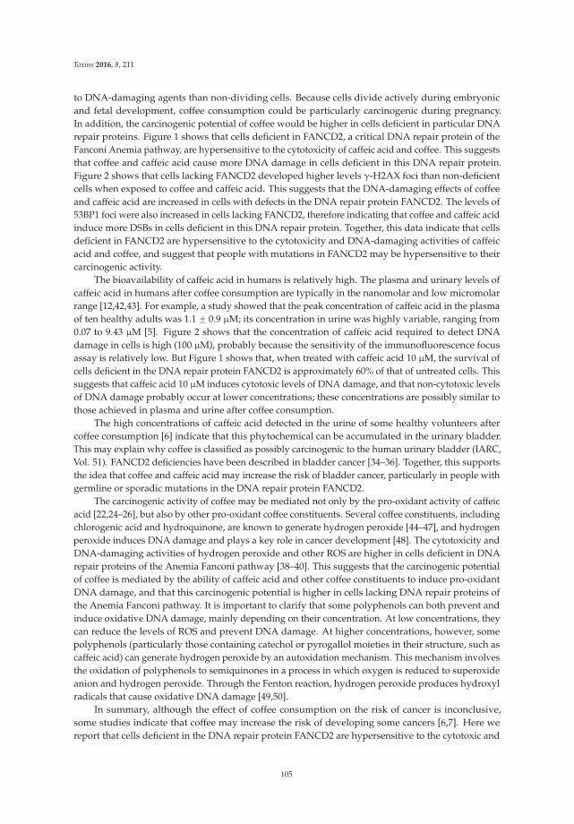
Toxins 2016, 8, 211
to DNA-damaging agents than non-dividing cells. Because cells divide actively during embryonicand fetal development, coffee consumption could be particularly carcinogenic during pregnancy.In addition, the carcinogenic potential of coffee would be higher in cells deficient in particular DNArepair proteins. Figure 1 shows that cells deficient in FANCD2, a critical DNA repair protein of theFanconi Anemia pathway, are hypersensitive to the cytotoxicity of caffeic acid and coffee. This suggeststhat coffee and caffeic acid cause more DNA damage in cells deficient in this DNA repair protein.Figure 2 shows that cells lacking FANCD2 developed higher levels γ-H2AX foci than non-deficientcells when exposed to coffee and caffeic acid. This suggests that the DNA-damaging effects of coffeeand caffeic acid are increased in cells with defects in the DNA repair protein FANCD2. The levels of53BP1 foci were also increased in cells lacking FANCD2, therefore indicating that coffee and caffeic acidinduce more DSBs in cells deficient in this DNA repair protein. Together, this data indicate that cellsdeficient in FANCD2 are hypersensitive to the cytotoxicity and DNA-damaging activities of caffeicacid and coffee, and suggest that people with mutations in FANCD2 may be hypersensitive to theircarcinogenic activity.
The bioavailability of caffeic acid in humans is relatively high. The plasma and urinary levels ofcaffeic acid in humans after coffee consumption are typically in the nanomolar and low micromolarrange [12,42,43]. For example, a study showed that the peak concentration of caffeic acid in the plasmaof ten healthy adults was 1.1 ˘ 0.9 μM; its concentration in urine was highly variable, ranging from0.07 to 9.43 μM [5]. Figure 2 shows that the concentration of caffeic acid required to detect DNAdamage in cells is high (100 μM), probably because the sensitivity of the immunofluorescence focusassay is relatively low. But Figure 1 shows that, when treated with caffeic acid 10 μM, the survival ofcells deficient in the DNA repair protein FANCD2 is approximately 60% of that of untreated cells. Thissuggests that caffeic acid 10 μM induces cytotoxic levels of DNA damage, and that non-cytotoxic levelsof DNA damage probably occur at lower concentrations; these concentrations are possibly similar tothose achieved in plasma and urine after coffee consumption.
The high concentrations of caffeic acid detected in the urine of some healthy volunteers aftercoffee consumption [6] indicate that this phytochemical can be accumulated in the urinary bladder.This may explain why coffee is classified as possibly carcinogenic to the human urinary bladder (IARC,Vol. 51). FANCD2 deficiencies have been described in bladder cancer [34–36]. Together, this supportsthe idea that coffee and caffeic acid may increase the risk of bladder cancer, particularly in people withgermline or sporadic mutations in the DNA repair protein FANCD2.
The carcinogenic activity of coffee may be mediated not only by the pro-oxidant activity of caffeicacid [22,24–26], but also by other pro-oxidant coffee constituents. Several coffee constituents, includingchlorogenic acid and hydroquinone, are known to generate hydrogen peroxide [44–47], and hydrogenperoxide induces DNA damage and plays a key role in cancer development [48]. The cytotoxicity andDNA-damaging activities of hydrogen peroxide and other ROS are higher in cells deficient in DNArepair proteins of the Anemia Fanconi pathway [38–40]. This suggests that the carcinogenic potentialof coffee is mediated by the ability of caffeic acid and other coffee constituents to induce pro-oxidantDNA damage, and that this carcinogenic potential is higher in cells lacking DNA repair proteins ofthe Anemia Fanconi pathway. It is important to clarify that some polyphenols can both prevent andinduce oxidative DNA damage, mainly depending on their concentration. At low concentrations, theycan reduce the levels of ROS and prevent DNA damage. At higher concentrations, however, somepolyphenols (particularly those containing catechol or pyrogallol moieties in their structure, such ascaffeic acid) can generate hydrogen peroxide by an autoxidation mechanism. This mechanism involvesthe oxidation of polyphenols to semiquinones in a process in which oxygen is reduced to superoxideanion and hydrogen peroxide. Through the Fenton reaction, hydrogen peroxide produces hydroxylradicals that cause oxidative DNA damage [49,50].
In summary, although the effect of coffee consumption on the risk of cancer is inconclusive,some studies indicate that coffee may increase the risk of developing some cancers [6,7]. Here wereport that cells deficient in the DNA repair protein FANCD2 are hypersensitive to the cytotoxic and
105

Toxins 2016, 8, 211
DNA-damaging activities of a commercial lyophilized coffee extract and caffeic acid (an importantmetabolite found in plasma and urine after coffee consumption). These data suggest that peoplewith Fanconi Anemia, or healthy people who develop sporadic mutations in FANCD2, may behypersensitive to the carcinogenic activity of coffee.
4. Materials and Methods
4.1. Chemicals and Cell Lines
Caffeic acid was purchased from Sigma (ě98.0%, Sigma-Aldrich, St. Louis, MO, USA).A commercial lyophilized coffee extract (NESCAFÉ® Classic, Barcelona, Spain) was used.In all experiments, caffeic acid and the coffee extract were tested individually, not mixed together.The human Fanconi deficient (PD20 FANCD2´/´) and proficient (PD20 FANCD2´/´ complementedwith FANCD2) cells were kindly provided by Dr. Thomas Helleday and by Dr. Jordi Surrallés Calonge.Cells were maintained in DMEM supplemented with 2 mM glutamine, 50 μg/mL penicillin, 50 μg/mLstreptomycin and 20% fetal bovine serum. Cells were cultured at 37 ˝C in a humidified atmospherecontaining 5% CO2.
4.2. Clonogenic Assay
Cell survival was measured with the clonogenic assay. Cells were plated at low density onto 6 cmPetri dishes. Cells were treated with caffeic acid or coffee for 4 h; then drugs were removed and freshmedia was added to allow the cells to grow for 7 days. Colonies were stained with methylene blueprepared in methanol (4 g/L). Surviving colonies made up 50 cells per colony were counted and thedata were corrected according to cloning efficiencies of control cells.
4.3. Immunofluorescence γ-H2AX and 53BP1 Focus Assay
To evaluate DNA damage, detection of γ-H2AX and 53BP1 foci were quantified byimmunofluorescence using the focus assay. The γ-H2AX focus assay is based on the ability ofdouble-strand breaks (DSBs) to trigger phosphorylation of histone H2AX on Ser-139, which leads to theformation of nuclear foci that can be visualized with anti-γ-H2AX antibodies. Following the inductionof double strand breaks, formation of γ-H2AX foci is associated with recruitment of p53-bindingprotein 1 (53BP1). Accumulation of 53BP1 can also be visualized as foci with anti-53BP1 antibodies.After treatments, cells were washed three times with PBS, and incubated for 30 s with cold 0.1%Triton-X in PBS to pre-extract soluble proteins. Afterward, cells were fixed with 4% paraformaldehydein PBS for 10 min at room temperature and washed three times with PBS. After fixation, cells werepermeabilized with 0.5% Triton X-100 in PBS for 5 min and then blocked three times with 0.1% Tween20, 1% BSA in PBS for 5 min each. Cells were then incubated for 1 h with a mouse anti-γ-H2AXmonoclonal antibody (Upstate; 1:800 dilution). Cells were washed three times with PBS and blockedthree times prior to the incubation with a secondary anti-mouse antibody linked to Alexa Fluor 488(Invitrogen; 1:500 dilution) for 1 h. Cells were washed with PBS, blocked, and incubated overnightwith the rabbit anti-53BP1 primary antibody (Bethyl, 1:1000 dilution). After incubation, cells werewashed with PBS, blocked and washed again with PBS as indicated before. DNA was stained withDAPI (41,6-diamidino-2-phenylindole) and immunofluorescence was observed at 40-fold magnificationwith an Olympus BX 61 microscope. A total of at least 100 cells/dose were scored, and cells with 10 ormore foci were scored as positive [44,51].
Author Contributions: S.M. and M.L-L. conceived and designed the experiments; E.B-M., J.M.C.-M., M.L.O. andE.G.-M. performed the experiments and analyzed the data; E.B-M. and M.L.-L. wrote the paper.
Conflicts of Interest: The authors declare no conflict of interest.
106
Toxins 2016, 8, 211
Abbreviations
ALL acute lymphoblastic leukemiaAML acute myeloid leukemiaDSBs double strand breaksFA Fanconi anemiaIARC International Agency for Research on CancerROS reactive oxygen species
References
1. Ludwig, I.A.; Clifford, M.N.; Lean, M.E.; Ashihara, H.; Crozier, A. Coffee: Biochemistry and potential impacton health. Food Funct. 2014, 5, 1695–1717. [CrossRef] [PubMed]
2. Higdon, J.V.; Frei, B. Coffee and health: A review of recent human research. Crit. Rev. Food Sci. Nutr. 2006,46, 101–123. [CrossRef] [PubMed]
3. Ding, M.; Satija, A.; Bhupathiraju, S.N.; Hu, Y.; Sun, Q.; Han, J.; Lopez-Garcia, E.; Willett, W.; van Dam, R.M.;Hu, F.B. Association of Coffee Consumption with Total and Cause-Specific Mortality in 3 Large ProspectiveCohorts. Circulation 2015, 132, 2305–2315. [CrossRef] [PubMed]
4. Bohn, S.K.; Blomhoff, R.; Paur, I. Coffee and cancer risk, epidemiological evidence, and molecularmechanisms. Mol. Nutr. Food Res. 2014, 58, 915–930. [CrossRef] [PubMed]
5. Sugiyama, K.; Sugawara, Y.; Tomata, Y.; Nishino, Y.; Fukao, A.; Tsuji, I. The association between coffeeconsumption and bladder cancer incidence in a pooled analysis of the Miyagi Cohort Study and OhsakiCohort Study. Eur. J. Cancer Prev. 2016. [CrossRef] [PubMed]
6. Arab, L. Epidemiologic evidence on coffee and cancer. Nutr. Cancer 2010, 62, 271–283. [CrossRef] [PubMed]7. Wu, W.; Tong, Y.; Zhao, Q.; Yu, G.; Wei, X.; Lu, Q. Coffee consumption and bladder cancer: A meta-analysis
of observational studies. Sci. Rep. 2015, 5, 9051. [CrossRef] [PubMed]8. Bonaventure, A.; Rudant, J.; Goujon-Bellec, S.; Orsi, L.; Leverger, G.; Baruchel, A.; Bertrand, Y.; Nelken, B.;
Pasquet, M.; Michel, G.; et al. Childhood acute leukemia, maternal beverage intake during pregnancy, andmetabolic polymorphisms. Cancer Causes Control 2013, 24, 783–793. [CrossRef] [PubMed]
9. Thomopoulos, T.P.; Ntouvelis, E.; Diamantaras, A.A.; Tzanoudaki, M.; Baka, M.; Hatzipantelis, E.; Kourti, M.;Polychronopoulou, S.; Sidi, V.; Stiakaki, E.; et al. Maternal and childhood consumption of coffee, tea and colabeverages in association with childhood leukemia: A meta-analysis. Cancer Epidemiol. 2015, 39, 1047–1059.[CrossRef] [PubMed]
10. Cheng, J.; Su, H.; Zhu, R.; Wang, X.; Peng, M.; Song, J.; Fan, D. Maternal coffee consumption duringpregnancy and risk of childhood acute leukemia: A metaanalysis. Am. J. Obstet. Gynecol. 2014, 210, 151.[CrossRef] [PubMed]
11. Orsi, L.; Rudant, J.; Ajrouche, R.; Leverger, G.; Baruchel, A.; Nelken, B.; Pasquet, M.; Michel, G.; Bertrand, Y.;Ducassou, S.; et al. Parental smoking, maternal alcohol, coffee and tea consumption during pregnancy,and childhood acute leukemia: The ESTELLE study. Cancer Causes Control 2015, 26, 1003–1017. [CrossRef][PubMed]
12. Farah, A.; Monteiro, M.; Donangelo, C.M.; Lafay, S. Chlorogenic acids from green coffee extract are highlybioavailable in humans. J. Nutr. 2008, 138, 2309–2015. [CrossRef] [PubMed]
13. Clifford, M.N. Chlorogenic acids and other cinnamates – nature, occurrence and dietary burden.J. Sci.Food Agric. 1999, 79, 362–372. [CrossRef]
14. Sato, Y.; Itagaki, S.; Kurokawa, T.; Ogura, J.; Kobayashi, M.; Hirano, T.; Sugawara, M.; Iseki, K. In vitroand in vivo antioxidant properties of chlorogenic acid and caffeic acid. Int. J. Pharm. 2011, 403, 136–138.[CrossRef] [PubMed]
15. Kono, Y.; Kobayashi, K.; Tagawa, S.; Adachi, K.; Ueda, A.; Sawa, Y.; Shibata, H. Antioxidant activity ofpolyphenolics in diets. Rate constants of reactions of chlorogenic acid and caffeic acid with reactive speciesof oxygen and nitrogen. Biochim. Biophys. Acta 1997, 1335, 335–342. [CrossRef]
16. Liang, G.; Shi, B.; Luo, W.; Yang, J. The protective effect of caffeic acid on global cerebral ischemia-reperfusioninjury in rats. Behav. Brain Funct. 2015, 11, 18. [CrossRef] [PubMed]
107
Toxins 2016, 8, 211
17. Rosendahl, A.H.; Perks, C.M.; Zeng, L.; Markkula, A.; Simonsson, M.; Rose, C.; Ingvar, C.; Holly, J.M.;Jernstrom, H. Caffeine and Caffeic Acid Inhibit Growth and Modify Estrogen Receptor and Insulin-likeGrowth Factor I Receptor Levels in Human Breast Cancer. Clin. Cancer Res. 2015, 21, 1877–1887. [CrossRef][PubMed]
18. Bakuradze, T.; Lang, R.; Hofmann, T.; Schipp, D.; Galan, J.; Eisenbrand, G.; Richling, E. Coffee consumptionrapidly reduces background DNA strand breaks in healthy humans: Results of a short term repeated uptakeintervention study. Mol. Nutr. Food Res. 2016, 60, 682–686. [CrossRef] [PubMed]
19. Rehman, M.U.; Sultana, S. Attenuation of oxidative stress, inflammation and early markers of tumorpromotion by caffeic acid in Fe-NTA exposed kidneys of Wistar rats. Mol. Cell. Biochem. 2011, 357, 115–124.[CrossRef] [PubMed]
20. Hagiwara, A.; Hirose, M.; Takahashi, S.; Ogawa, K.; Shirai, T.; Ito, N. Forestomach and kidney carcinogenicityof caffeic acid in F344 rats and C57BL/6Nx C3H/HeN F1 mice. Cancer Res. 1991, 51, 5655–5660. [PubMed]
21. Szeto, Y.T.; Collins, A.R.; Benzie, I.F. Effects of dietary antioxidants on DNA damage in lysed cells using amodified comet assay procedure. Mutat. Res. 2002, 500, 31–38. [CrossRef]
22. Bhat, S.H.; Azmi, A.S.; Hadi, S.M. Prooxidant DNA breakage induced by caffeic acid in human peripherallymphocytes: Involvement of endogenous copper and a putative mechanism for anticancer properties.Toxicol. Appl. Pharmacol. 2007, 218, 249–255. [CrossRef] [PubMed]
23. Maistro, E.L.; Angeli, J.P.; Andrade, S.F.; Mantovani, M.S. In vitro genotoxicity assessment of caffeic, cinnamicand ferulic acids. Genet. Mol. Res. 2011, 10, 1130–1140. [CrossRef] [PubMed]
24. Inoue, S.; Ito, K.; Yamamoto, K.; Kawanishi, S. Caffeic acid causes metal-dependent damage to cellular andisolated DNA through H2O2 formation. Carcinogenesis 1992, 13, 1497–1502. [CrossRef] [PubMed]
25. Li, Y.; Trush, M.A. Reactive oxygen-dependent DNA damage resulting from the oxidation of phenoliccompounds by a copper-redox cycle mechanism. Cancer Res. 1994, 54, 1895s–1898s. [PubMed]
26. Babich, H.; Schuck, A.G.; Weisburg, J.H.; Zuckerbraun, H.L. Research strategies in the study of thepro-oxidant nature of polyphenol nutraceuticals. J. Toxicol. 2011, 2011, 467305. [CrossRef] [PubMed]
27. Knudson, A.G. Cancer genetics. Am. J. Med. Genet. 2002, 111, 96–102. [CrossRef] [PubMed]28. Greenman, C.; Stephens, P.; Smith, R.; Dalgliesh, G.L.; Hunter, C.; Bignell, G.; Davies, H.; Teague, J.;
Butler, A.; Stevens, C.; et al. Patterns of somatic mutation in human cancer genomes. Nature 2007, 446,153–158. [CrossRef] [PubMed]
29. Burrell, R.A.; McGranahan, N.; Bartek, J.; Swanton, C. The causes and consequences of genetic heterogeneityin cancer evolution. Nature 2013, 501, 338–345. [CrossRef] [PubMed]
30. Stratton, M.R.; Campbell, P.J.; Futreal, P.A. The cancer genome. Nature 2009, 458, 719–724. [CrossRef][PubMed]
31. Errol, C.F.; Walker, G.C.; Siede, W. DNA Repair and Mutagenesis; ASM Press: Washington, DC, USA, 1995.32. D’Andrea, A.D.; Grompe, M. Molecular biology of Fanconi anemia: Implications for diagnosis and therapy.
Blood 1997, 90, 1725–1736. [PubMed]33. Willers, H.; Kachnic, L.A.; Luo, C.M.; Li, L.; Purschke, M.; Borgmann, K.; Held, K.D.; Powell, S.N. Biomarkers
and mechanisms of FANCD2 function. J. Biomed. Biotechnol. 2008, 2008, 821529. [CrossRef] [PubMed]34. Panneerselvam, J.; Pickering, A.; Zhang, J.; Wang, H.; Tian, H.; Zheng, J.; Fei, P. A hidden role of the
inactivated FANCD2: Upregulating DeltaNp63. Oncotarget 2013, 4, 1416–1426. [CrossRef] [PubMed]35. Zhang, J.; Zhao, D.; Park, H.K.; Wang, H.; Dyer, R.B.; Liu, W.; Klee, G.G.; McNiven, M.A.; Tindall, D.J.;
Molina, J.R.; et al. FAVL elevation in human tumors disrupts Fanconi anemia pathway signaling andpromotes genomic instability and tumor growth. J. Clin. Investig. 2010, 120, 1524–1534. [CrossRef] [PubMed]
36. Panneerselvam, J.; Park, H.K.; Zhang, J.; Dudimah, F.D.; Zhang, P.; Wang, H.; Fei, P. FAVL impairment of theFanconi anemia pathway promotes the development of human bladder cancer. Cell Cycle 2012, 11, 2947–2455.[CrossRef] [PubMed]
37. Tischkowitz, M.D.; Morgan, N.V.; Grimwade, D.; Eddy, C.; Ball, S.; Vorechovsky, I.; Langabeer, S.; Stoger, R.;Hodgson, S.V.; Mathew, C.G. Deletion and reduced expression of the Fanconi anemia FANCA gene insporadic acute myeloid leukemia. Leukemia 2004, 18, 420–425. [CrossRef] [PubMed]
38. Joenje, H.; Arwert, F.; Eriksson, A.W.; de Koning, H.; Oostra, A.B. Oxygen-dependence of chromosomalaberrations in Fanconi’s anaemia. Nature 1981, 290, 142–143. [CrossRef] [PubMed]
39. Saito, H.; Hammond, A.T.; Moses, R.E. Hypersensitivity to oxygen is a uniform and secondary defect inFanconi anemia cells. Mutat. Res. 1993, 294, 255–262. [CrossRef]
108
Toxins 2016, 8, 211
40. Takeuchi, T.; Morimoto, K. Increased formation of 8-hydroxydeoxyguanosine, an oxidative DNA damage, inlymphoblasts from Fanconi’s anemia patients due to possible catalase deficiency. Carcinogenesis 1993, 14,1115–1120. [CrossRef] [PubMed]
41. Monteiro, F.L.; Baptista, T.; Amado, F.; Vitorino, R.; Jeronimo, C.; Helguero, L.A. Expression and functionalityof histone H2A variants in cancer. Oncotarget 2014, 5, 3428–4343. [CrossRef] [PubMed]
42. Wittemer, S.M.; Ploch, M.; Windeck, T.; Muller, S.C.; Drewelow, B.; Derendorf, H.; Veit, M. Bioavailabilityand pharmacokinetics of caffeoylquinic acids and flavonoids after oral administration of Artichoke leafextracts in humans. Phytomedicine 2005, 12, 28–38. [CrossRef] [PubMed]
43. Renouf, M.; Guy, P.A.; Marmet, C.; Fraering, A.L.; Longet, K.; Moulin, J.; Enslen, M.; Barron, D.; Dionisi, F.;Cavin, C.; et al. Measurement of caffeic and ferulic acid equivalents in plasma after coffee consumption:Small intestine and colon are key sites for coffee metabolism. Mol. Nutr. Food Res. 2010, 54, 760–766.[CrossRef] [PubMed]
44. Burgos-Moron, E.; Calderon-Montano, J.M.; Orta, M.L.; Pastor, N.; Perez-Guerrero, C.; Austin, C.; Mateos, S.;Lopez-Lazaro, M. The Coffee Constituent Chlorogenic Acid Induces Cellular DNA Damage and Formationof Topoisomerase I- and II-DNA Complexes in Cells. J. Agric. Food Chem. 2012, 60, 7384–7391. [CrossRef][PubMed]
45. Long, L.H.; Halliwell, B. Coffee drinking increases levels of urinary hydrogen peroxide detected in healthyhuman volunteers. Free Radic. Res. 2000, 32, 463–467. [CrossRef] [PubMed]
46. Hiramoto, K.; Kida, T.; Kikugawa, K. Increased urinary hydrogen peroxide levels caused by coffee drinking.Biol. Pharm. Bull. 2002, 25, 1467–1471. [CrossRef] [PubMed]
47. Halliwell, B.; Long, L.H.; Yee, T.P.; Lim, S.; Kelly, R. Establishing biomarkers of oxidative stress:The measurement of hydrogen peroxide in human urine. Curr. Med. Chem. 2004, 11, 1085–1092. [CrossRef][PubMed]
48. Lopez-Lazaro, M. Dual role of hydrogen peroxide in cancer: Possible relevance to cancer chemopreventionand therapy. Cancer Lett. 2007, 252, 1–8. [CrossRef] [PubMed]
49. Akagawa, M.; Shigemitsu, T.; Suyama, K. Production of hydrogen peroxide by polyphenols andpolyphenol-rich beverages under quasi-physiological conditions. Biosci. Biotechnol. Biochem. 2003, 67,2632–2640. [CrossRef] [PubMed]
50. Mochizuki, M.; Yamazaki, S.; Kano, K.; Ikeda, T. Kinetic analysis and mechanistic aspects of autoxidation ofcatechins. Biochim. Biophys. Acta 2002, 1569, 35–44. [CrossRef]
51. Orta, M.L.; Calderon-Montano, J.; Dominguez, I.; Pastor, N.; Burgos-Moron, E.; Lopez-Lazaro, M.;Cortes, F.; Mateos, S.; Helleday, T. 5-Aza-21-deoxycytidine causes replication lesions that require Fanconianemia-dependent homologous recombination for repair. Nucleic Acids Res. 2013, 41, 5827–5836. [CrossRef][PubMed]
© 2016 by the authors; licensee MDPI, Basel, Switzerland. This article is an open accessarticle distributed under the terms and conditions of the Creative Commons Attribution(CC-BY) license (http://creativecommons.org/licenses/by/4.0/).
109
toxins
Review
Roles of Dietary Phytoestrogens on the Regulation ofEpithelial-Mesenchymal Transition in DiverseCancer MetastasisGeum-A. Lee, Kyung-A. Hwang * and Kyung-Chul Choi *
Laboratory of Biochemistry and Immunology, College of Veterinary Medicine, Chungbuk National University,Cheongju, Chungbuk 361-763, Korea; [email protected]* Correspondence: [email protected] (K.-A.H.); [email protected] (K.-C.C.);
Tel.: +82-43-249-1745 (K.-A.H.); +82-43-261-3664 (K.-C.C.); Fax: +82-43-267-3150 (K.-A.H. & K.-C.C.)
Academic Editor: Carmela FimognariReceived: 6 March 2016; Accepted: 19 May 2016; Published: 24 May 2016
Abstract: Epithelial-mesenchymal transition (EMT) plays a key role in tumor progression. The cellsundergoing EMT upregulate the expression of cell motility-related proteins and show enhancedmigration and invasion. The hallmarks of EMT in cancer cells include changed cell morphologyand increased metastatic capabilities in cell migration and invasion. Therefore, prevention ofEMT is an important tool for the inhibition of tumor metastasis. A novel preventive therapy isneeded, such as treatment of natural dietary substances that are nontoxic to normal human cells, buteffective in inhibiting cancer cells. Phytoestrogens, such as genistein, resveratrol, kaempferol and3,31-diindolylmethane (DIM), can be raised as possible candidates. They are plant-derived dietaryestrogens, which are found in tea, vegetables and fruits, and are known to have various biologicalefficacies, including chemopreventive activity against cancers. Specifically, these phytoestrogensmay induce not only anti-proliferation, apoptosis and cell cycle arrest, but also anti-metastasis byinhibiting the EMT process in various cancer cells. There have been several signaling pathwaysfound to be associated with the induction of the EMT process in cancer cells. Phytoestrogens weredemonstrated to have chemopreventive effects on cancer metastasis by inhibiting EMT-associatedpathways, such as Notch-1 and TGF-beta signaling. As a result, phytoestrogens can inhibit or reversethe EMT process by upregulating the expression of epithelial phenotypes, including E-cadherin,and downregulating the expression of mesenchymal phenotypes, including N-cadherin, Snail, Slug,and vimentin. In this review, we focused on the important roles of phytoestrogens in inhibitingEMT in many types of cancer and suggested phytoestrogens as prominent alternative compoundsto chemotherapy.
Keywords: dietary phytoestrogens; DIM; kaempferol; resveratrol; genistein; epithelial-mesenchymaltransition; cancer metastasis
1. Introduction
Phytochemicals are chemical compounds that occur naturally in plants, amounting to as manyas 4000 different chemicals. Some phytochemicals have the biological significance of inhibiting theinvasion of different species of plants by acting as toxic compounds [1,2]. This characteristic ofphytochemicals has been utilized in curing diverse human diseases. A vast array of plant-derivednatural compounds has been reported to have substantial chemopreventive effects against cancer,in opposition to the health risk of environmental carcinogens [3]. At present, inhibiting humancarcinogenesis using plant-derived compounds is considered as a vital and urgent challenge, despitesome phytochemicals having been used for targeting many forms of cancer as major sources of highlyeffective conventional drugs [4–6].
Toxins 2016, 8, 162 110 www.mdpi.com/journal/toxins
Toxins 2016, 8, 162
Among diverse groups of phytochemicals, phytoestrogens, which are plant-derived xenoestrogensand mostly found in soy, vegetables and fruits, are considered as strong sources of cancer-preventivephytonutrition to inhibit the development and progression of many types of cancer [4,7].
As a kind of xenoestrogen, phytoestrogens specifically have distinctive cancer-preventive effects onestrogen-related cancers. In general, sex hormones, including estrogens, have been known to be closelylinked to the pathogenesis of several types of cancer in the reproductive organs [8–10]. Many diseases,like breast, ovarian, endometrial and cervical cancers, have been called estrogen-receptor (ER)-positivecancers, because the actions of estrogen related to cancer biology are mediated via ERs, which compriseERα and ERβ, mostly present in the nucleus [11,12]. Independently of endogenous estrogens,selective estrogen receptor modulators (SERMs), which are a class of drugs that act on ER, actwith agonistic or antagonistic actions in several target tissues [13]. Phytoestrogens, which arealso a kind of SERM, are known to bind ER with affinities at least 10,000-times lower than thatof 17β-estradiol (E2) and also act as ER agonists or antagonists [14]. The chemopreventive effects ofphytoestrogens are associated with their antagonistic effects on ER [15]. In addition to the actionsof phytoestrogens via ERs, they could have protective effects on the initiation and progression ofestrogen-related cancers by specifically inhibiting the circulating precursors of estrogens [16–20].In addition, they can inhibit crucial steroidogenic enzyme activity, including the conversion of E2from circulating hormones, such as androgens and estrogen sulfate [20]. In estrogen biosynthesis andmetabolism, phytoestrogens have been shown to inhibit several crucial enzymes in aromatase pathway,such as 17β- and 3β-hydroxysteroid dehydrogenase (HSD), which catalyze the dehydrogenationof 17-hydroxysteroids in steroidogenesis and control the interconversion of androstenedione andtestosterone, and E2 and estrone, respectively [21,22].
Phytoestrogens are generally classified into four main classes: isoflavones (genistein, daidzein,kaempferol), lignans (secoisolariciresinol, matairesinol, pinoresinol, lariciresinol), coumestan (coumestrol)and stilbenes (resveratrol) [23,24]. Western populations have been known to intake more foodscontaining lignans, while Asian populations eat more soy foods containing isoflavones [25]. Lignans areincluded in diverse groups of non-flavonoid compounds widely distributed in whole grain cereals,beans, berries, nuts and various seeds [26,27]. A wealth of lignans exists as secoisolariciresinol,matairesinol, lariciresinol and pinoresinol, and they are converted into enterolignans by the intestinalmicrobiota to be absorbed into the human body [27,28]. Tea, fruits, vegetables and grains account forover 85% of the daily intake of lignans, such as matairesinol and secoisolariciresinol [29]. Isoflavonesare naturally-occurring phenolic flavonoid compounds, known to act as phytoestrogens in mammals.Soybeans are the most common source of isoflavones among vegetables; the major isoflavones insoybean are genistein and daidzein, which are well-known phytoestrogens [30]. Coumestans occurmainly in bean sprouts during germination, and the main compound in this subgroup is coumestrol,mostly found in peas and beans [23]. Stilbenoids are hydroxylated derivatives of stilbene and belongto the family of phenylpropanoids [31]. Resveratrol is a typical stilbenoid, which is mainly found ingrapes and wines [32].
In the present article, we will review the effect of four kinds of phytoestrogens, genistein, resveratrol,kaempferol and 3,31-diindolylmethane (DIM), on cancer progression via epithelial-mesenchymaltransition (EMT). As shown in Figure 1, genistein, kaempferol and resveratrol are phenolic compounds:genistein and kaempferol are isoflavones, having a common flavone structure; resveratrol is aderivative of diphenylethane; and DIM is an active indole compound originated from indole-3-carbinol(I3C), an inactive form of indole. They are actively-studied phytoestrogens that have great potential todisplay anti-cancer effects.
111
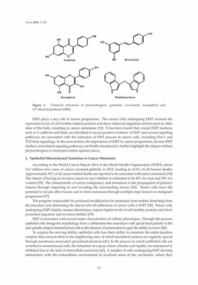
Toxins 2016, 8, 162
Figure 1. Chemical structures of phytoestrogens, genistein, resveratrol, kaempferol and3,31-diindolylmethane (DIM).
EMT plays a key role in tumor progression. The cancer cells undergoing EMT increase theexpression levels of cell motility-related proteins and show enhanced migration and invasion to othersites of the body, resulting in cancer metastasis [33]. It has been found that crucial EMT markers,such as E-cadherin and Snail, are identified to secure positive evidence of EMT, and several signalingpathways are associated with the induction of EMT process in cancer cells, including Not-1 andTGF-beta signalings. In the next section, the importance of EMT in cancer progression, diverse EMTmarkers and related signaling pathways are briefly introduced to further highlight the impact of thesephytoestrogens in chemoprevention against cancer.
2. Epithelial-Mesenchymal Transition in Cancer Metastasis
According to the World Cancer Report 2014 of the World Health Organization (WHO), about14.1 million new cases of cancer occurred globally in 2012, leading to 14.6% of all human deaths.Approximately 90% of all cancer-related deaths are reported to be associated with tumor metastasis [34].The chance of having an invasive cancer in one’s lifetime is estimated to be 42% for men and 38% forwomen [35]. The characteristic of cancer malignancy and metastasis is the propagation of primarytumors through migrating to and invading the surrounding tissues [36]. Tumor cells have thepotential to invade other tissues and to form metastasis through multiple steps known as malignantprogression [37].
The program responsible for profound modification for metastasis that enables detaching fromthe junctions and dismissing the lateral cell-cell adhesions of cancer cells is EMT [38]. Tumor cellsundergoing EMT display unique phenotypes, express higher levels of cell motility proteins and showpromoted migration and invasion abilities [39].
EMT is associated with several major characteristics of cellular phenotypes. Through this process,epithelial cells change the morphology from a cobblestone-like monolayer with apical basal polarity to flatand spindle-shaped mesenchymal cells in the absence of polarization to gain the ability to move [40].
To acquire the moving ability, epithelial cells lose their ability to maintain the entire junctioncomplex that connects them to the neighboring ones, of which basolateral surfaces are regularly spacedthrough membrane-associated specialized junctions [41]. In the process in which epithelial cells areswitched to mesenchymal cells, the formation of a space where a barrier and rigidity are maintained isinhibited due to the lack of intercellular junctions [42]. A number of cells undergoing EMT developinteractions with the extracellular environment in localized areas of the carcinoma, where they
112
Toxins 2016, 8, 162
involve the loss of intercellular cohesion, the disruption of extracellular matrix (ECM), modificationsof the cytoskeleton, increased motility and invasion into the extracellular space [43]. Particularly,EMT is related to the expression of extracellular matrix proteases, such as matrix metalloproteases(MMPs) and urokinase-type plasminogen activator (uPA), which can degrade the ECM linked to theplasma membrane and localized to invadopodia during metastasis [44–46]. Meanwhile, EMT is areversible process that can convert to its inverse process, called the mesenchymal-epithelial transition(MET). The cells undergoing MET increase cell-cell adhesion and return to epithelial phenotypes,which also play a role during embryonic development and pathological processes [47,48]. As a result,primary cancer cells lose cell-cell adhesion via EMT by E-cadherin repression, break through thebasement membrane and enter the bloodstream through intravasation. Later, the circulating tumorcells exit the bloodstream to migrate to the specific metastatic sites where they undergo MET for clonaloutgrowth [49].
A diversity of molecules associated with the process of EMT has been established, and some crucialmolecules have been employed as biological markers to determine the process. As a typical moleculein the adherent junctions, E-cadherin, which is a transmembrane glycoprotein of the type I cadherinfamily and a crucial epithelial marker, has been found to inactivate and repress tumor progressionby maintaining intact cell-cell interactions and inhibiting cell mobility, invasion and metastasis inhuman cancer [50,51]. The loss of E-cadherin expression is allowed for a critical step in the progressionof invasive carcinoma by causing the EMT event. The loss of many epithelial markers (includingE-cadherin, occludin, claudins and beta-catenin) induces the expression of mesenchymal markers(including N-cadherin, Snail, vimentin, R-cadherin and cadherin-11) and acquisition of mesenchymalcharacteristics, such as cell motility and invasion [52,53]. Additionally, the zinc-finger transcriptionfactors, including Snail, Slug, and ZEB 1 and 2, have been shown to induce the EMT process by directlybinding to the E-box of the E-cadherin promoter and suppressing the activity of E-cadherin [54], whileTwist, another type of E-cadherin repressor, indirectly downregulates E-cadherin transcription [55].
It has been found that crucial EMT markers are associated with several signaling pathways inthe induction of the EMT process in cancer cells. Snail has been found to be a critical factor in TGF-βsignaling to resist cell death and to inhibit apoptosis [56]. As the most important factor that triggersEMT, TGF-β1 mediates the EMT process via numerous intracellular signaling pathways, including theSmad pathway, mitogen-activated protein kinases (MAPK), PI3K/Akt and small GTPases in HL-60leukemia, Panc-1 human pancreatic and MDA-MB-231 breast cancer cells [57–59]. The overactivationof the TGF-β pathway in hepatocellular carcinoma cells confers a mesenchymal-like and an increasedmigratory capacity to the cells and finally contributes to tumor progression thorough the crosstalkwith the chemokine CXCL12 pathway in liver tumor cells [60]. The Wnt signaling pathway activatesβ-catenin and several EMT-inducing transcription factors, such as Slug and Twist [34]. This signalingpathway induces cancer cell proliferation, motility and intravasation. Furthermore, in in vivo studies,the Wnt pathway displayed an important role in regulating EMT progression of colorectal cancer [61],and the Wnt-β-catenin was activated in the mesenchyme of the cardiac cushion during EMT inzebrafish and mouse embryo [62,63]. The cancer development of organs has been regulated by Notch-1signaling, which directly promotes Snail, Slug and NF-κB in BxPC-3 human pancreatic cancer cell [64].Notch-1 signaling also induced cell proliferation, survival and EMT by increasing NF-κB transcriptionalactivity in many human malignancies, including pancreatic and breast cancer cells [65,66]. In addition,the Hedgehog (Hh) signaling pathway is currently considered as a therapeutic target for anti-cancertreatment, because this pathway is abnormally activated in various types of cancer and contributesto tumor metastasis by inducing EMT. The misregulation of Hh signaling has been implicated as animportant mediator in human pancreatic carcinoma, and specifically the sonic hedgehog pathwaypromotes metastasis and lymphangiogenesis via activation of Akt, EMT and the MMP-9 pathway ingastric cancers [67,68].
Recently, microRNAs (miRNAs) are being considered as an important regulator of EMT in variouscancer cells. They incompletely bind to the 31untranslated region (31UTR) of mRNA to inhibit the
113
Toxins 2016, 8, 162
translations [69]. The incomplete accordance between miRNAs and their targets allows the chancesfor miRNAs to control multiple genes. Moreover, miRNAs have been shown to play a crucial roleduring caner development and progression via the modulation of the expression of their target mRNAtranscripts [70]. High miR-34a levels stimulate MET by reversing Snail and TCF-β-induced EMT [71].As a negative regulator in the EMT process, miR-125a induced MET by the epidermal growth factorreceptor (EGFR) signaling pathway [72]. miR-506 suppresses EMT, cell proliferation, migration andinvasion by upregulating E-cadherin [73]. miR-138 also has a role in the inhibition of EMT andinvasion in SKOV-3 ovarian cancer cells [74]. Another miRNA, miR-30a, was reported to suppresscell motility and EMT via targeting the expression of mesenchymal markers, thereby increasing theepithelial marker in A549 lung and BGC-823 gastric cancer cells [75,76]. On the other hand, miR-106ais associated with cell proliferation and tumor differentiation, and miR-7 is linked with metastasisand EMT [77]. In the case of miR-10b, its high expression upregulated the EMT and the expressionof EMT-related proteins in metastatic tumors and induced the changed spindle-like morphology,cell migration and overexpression of N-cadherin, Snail, Slug and Twist [78,79]. Specifically, the miR-200family is significant for reducing the ZEB levels, cell migration and TGF-β-induced EMT [80,81].Expression of the miR-200 family was increased or decreased in the process of metastasis: miR-200was downregulated in the EMT process, while it was upregulated during the re-epithelialization ofdistal metastasis [82].
3. Phytoestrogens and Their Actions on Cancer Cells Undergoing EMT
Since the dysregulation of proteins in signaling pathways involved in EMT is associated withcancer progression, they could be potentially targeted as prognostic markers or therapeutic targets ofcancer metastasis [83]. Phytoestrogens having a chemopreventive effect on cancer progression seem toinhibit the EMT process through various channels.
3.1. Genistein
Genistein (40,5,7-trihydroxyisoflavone), having a heterocyclic diphenolic structure similar toestrogen, is a typical isoflavonoid found in a number of plants, including soybeans, peas, lentils andother beans [84,85]. As a phytoestrogen, it has an ability to bind and activate ERs, preferentiallyERβ rather than ERα [86,87]. The higher binding affinity for ERβ of genistein has been associatedwith its action as an estrogen antagonist and having chemopreventive activity in estrogen-responsivecancers [88].
Anti-proliferative and chemopreventive effects of genistein have been extensively investigatedin hormone-related, as well as non-hormone-related cancers in which genistein affects manycrucial cellular functions related to carcinogenesis, including cell proliferation, apoptosis, cell cycleprogression, migration, metastasis and invasion [89]. Recent studies have elucidated that genisteinmay have the potential to inhibit cancer metastasis by specifically regulating the EMT process viadiverse signaling pathways.
Notch-1 signaling is an important pathway to upregulate the expression of EMT markers, ZEB1and 2, Slug and vimentin, leading to the EMT, migration and drug resistance of pancreatic cancercells [90,91]. In AsPC-1 pancreatic cancer cells, Notch-1 overexpression affected the expression ofmiRNAs: overexpression of Notch-1 led to increased expression of miR-21 and decreased expressionof miR-200b. Re-expression of miR-200b led to inhibition of the EMT process by inducing decreasedexpression of ZEB1 and vimentin and increased expression of E-cadherin. Genistein treatment wasfound to attenuate the acquisition of EMT by AsPC-1 cells by promoting re-expression of miR-200b,which was repressed by Notch-1 signaling [90].
In a recent study, genistein suppressed the EMT of BG-1 ovarian cancer cells, which wasactivated by E2 and endocrine-disrupting chemicals, such as bisphenol A (BPA) and nonylphenol (NP),by downregulating the TGF-β pathway [92]. As a result, genistein not only suppressed the migrationof BG-1 cells, but also diminished the expression of mesenchymal markers (vimentin) and metastasis
114

Toxins 2016, 8, 162
markers (MMP-2 and cathepsin D) [92]. Genistein also effectively inhibited TGF-β-induced invasionand metastasis in the Panc-1 pancreatic cancer cell line through Smad4-dependent and independentpathways through p38 MAPK [58]. The EMT process was also reversed by genistein in the HepG2hepatocellular carcinoma cell line by downregulating the nuclear factor of activated T cells 1(NFAT1) [93]. NFAT1 was known to function in cell-autonomous actions, like invasion, migration,differentiation and proliferation in tumors [94]. Although the underlying mechanism has not beenfound yet, genistein suppressed invasive growth of LNCaP prostate cancer cells through the reversalof EMT, even at low concentrations (less than 15 micromoles/L genistein), which did not affect cellproliferation [95].
3.2. Resveratrol
Resveratrol (trans-3,4,5-trihydroxystilbene) is one of the stilbene phytoalexins, first found inthe roots of the oriental medicinal plant Polygonum cuspidatum (Kojo-kon in Japanese) [96], and alsoexists in diverse vegetables, including berries, peanuts and red grape [97]. Resveratrol is knownto be produced naturally when the plant is injured under attack by pathogens, such as bacteria orfungi [98]. Therefore, the proper infection of Botrytis cinerea (the fungus responsible for grey mold)is needed to obtain maximal concentrations of resveratrol within wine [99]. The characteristic ofresveratrol as a phytoestrogen has been verified by its capability to mainly bind to ER and to regulatethe transcription of estrogen-responsive target genes [100]. Many studies showed that resveratrolbinds to ERβ with a higher affinity than to ERα, though it binds with 7000-fold lower affinity than E2,and that it acts as an agonist or an antagonist in the cells expressing ER [101,102]. Resveratrol can alsoregulate androgen receptor (AR)-mediated actions as a chemopreventive agent against prostate cancer:it inhibited androgen-stimulated cell growth and gene expression by repressing the expression andfunction of the AR in LNCaP prostate cancer cells [103].
In addition to its sex hormone-related actions, resveratrol has been found to be very helpful ininhibiting diabetes, heart disease and diverse cancers, because it possesses various bioactive properties,such as anti-oxidation, anti-proliferation, anti-inflammation and induction of apoptosis [104].Specifically, the anti-oxidative efficacy of resveratrol to prevent the ROS generation and oxidative stressthat may drive epithelial cells into an EMT program can be an effective characteristic of resveratrol toprevent the EMT of cancer cells. Actually, modulation of oxidative stress may be an efficient therapeutictool for the inhibition of cancer progression [105]. Resveratrol inhibited the hypoxia-enhancedproliferation, invasion and EMT process in Saos-2 osteosarcoma cells via downregulation of theHIF-1α protein [106]. A previous study also revealed that resveratrol effectively suppressed thehypoxia-driven ROS-induced invasive and migratory ability of pancreatic cancer cells by inhibitingthe Hh signaling pathway, which is able to regulate the EMT [107].
In addition, a recent finding indicated that resveratrol suppressed invasion and metastasisin gastric cancer by inhibiting the Hh signaling pathway and EMT [108]. In PC-3 and LNCaPprostate cancer cell lines, lipopolysaccharide (LPS) was used to trigger EMT, but resveratrol inhibitedLPS-induced morphological changes, cell motility and invasiveness, the expression of EMT markersand inhibited the expression of glioma-associated oncogene homolog 1 (Gli1), suggesting thatresveratrol has in part the ability to inhibit the EMT process through the Hh signaling pathway [109].
Similar to genistein, resveratrol abrogates the TGF-β1-induced EMT process for cancer progression.In LoVo colorectal cancer cells, resveratrol inhibited the invasive and migratory ability of LoVo cells,increased the expression of E-cadherin and repressed the expression of vimentin, via the inhibitionof the TGF-β1/Smads signaling pathway [110]. Other studies also support the role of resveratrolin EMT inhibition. Xu et al. reported that resveratrol reversed EMT by inhibiting AKT signaling inpancreatic cancer [111]. Previously, AKT1 was also shown to promote EMT, as well as to increasemetastasis in squamous cancer and sarcoma [112]. On the contrary, AKT1, but not AKT2 and AKT3,inhibited EMT in breast cancer, depressing Twist1 activation [112]. In the MCF-7 breast cancer cell line,resveratrol was found to inhibit EGF-induced EMT via inhibition of the EGF-mediated Erk pathway
115
Toxins 2016, 8, 162
activation [113]. The role of resveratrol in inhibiting EMT induction was demonstrated in A549 lungcancer cells [114].
3.3. Kaempferol
Kaempferol (3,5,7-trihydroxy-2-(4-hydroxyphenyl)-4H-1-benzopyran-4-one) is one of theflavonoids found in many edible plants, like tea, broccoli, cabbage, beans and tomato, and itsname was derived due to its specific source of the rhizome of Kaempferi galangal L., known as apopular traditional aromatic plant [115,116]. As one of the phytoestrogens due to its polyphenolicstructure, kaempferol also exerts anti-proliferative and anti-carcinogenic actions though ER, AR,the aryl hydrocarbon receptor (AhR) and the progesterone receptor (PR) signaling pathways in manytypes of cancer [117–119].
Apoptosis is one of the main pathways for kaempferol to induce the anti-carcinogenic effect.In some cells, kaempferol induced apoptosis by stimulating the enzyme activity of caspases, which area group of the cysteine proteases that are important initiators or effectors of the apoptosis process [65].For caspase-3, it activates apoptosis by inducing DNA fragmentation and chromatin condensation innucleus [120]. Kaempferol decreased the mitochondria potential by the stimulation of caspase-3 activity,resulting in the apoptosis of human lung non-small carcinoma cells [121,122]. In caspase independentpathways, kaempferol also promoted apoptosis by translocating apoptosis-inducing factor (AIF) intonucleus. AIF, which exists mainly in the space between the inner and outer mitochondrial membrane,was translocated into nuclei by kaempferol to induce nuclear condensation and large-scale DNAfragmentation [123,124].
Kaempferol also seems to inhibit cancer invasion and metastasis via the inhibition of EMT.Specifically for lung cancer, kaempferol was well known to suppress cancer migration, invasivenessand metastasis by modulating the expression of EMT proteins. Kaempferol significantly reducedthe expression of MMP and mesenchymal markers and repressed metastasis and EMT by theTGF-β-dependent signaling pathway in non-small cell lung cancer [125]. In A549 lung cancer cells,kaempferol exerted the suppression of TGF-β1-induced EMT, migration and metastasis by blockingSmad3 as an important mediator of TGF-β signaling. In this study, PI3K/Akt signaling stimulatedEMT and cell migration by directly phosphorylating Smas3, but kaempferol repressed EMT and cellmigration by inhibiting Akt1-mediated phosphorylation of Smad3 [126]. The effect of kaempferol onEMT in relation to cancer progression has not been fully demonstrated yet, except for lung cancer.Although not in cancer, kaempferol was found to alleviate fibrotic airway remodeling via bronchialEMT by modulating protease-activated receptor-1 (PAR1) activation, which was entailed by TGF-β,suggesting that it may be a potential therapeutic agent targeting asthmatic airway constriction [127].Since kaempferol is considered to have obvious anti-EMT efficacies, it will be applicable to othercancers for the prevention of cancer metastasis induced by EMT. According to a recent review onribosomal S6 kinase (RSK) isoforms, the synthetic version of kaempferol, kaempferol-glycoside, caneffectively target the invasion and metastasis of cancer by inhibiting RKS isoforms that promoteinvasion and tumor metastasis [128].
3.4. DIM
Brassica vegetables, such as cabbage, Brussels sprouts and broccoli, are rich in indole-3-carbinol(I3C), which is one of the active phytonutrients and a precursor of many different compounds,especially DIM. I3C undergoes acid-catalyzed dehydration and polymerization to be DIM in anacidic environment [129].
DIM has been shown to abrogate the proliferation of human cancer cells of prostate, breast,colon, ovary and pancreas [130]. Especially, it has the potential role of AhR signaling by actingas one of the selective AhR modulators (SAhRMs) in mammary carcinogenesis prevention [131].With respect to anti-cancer mechanisms, DIM encourages apoptotic cell death by downregulationof NF-κB, survivin and Bcl-2 as anti-apoptotic factors and upregulation of Bax, a pro-apoptotic
116
Toxins 2016, 8, 162
factor [132,133]. One surprising ability of DIM as a potential anti-cancer compound is its selectiveinduction of apoptosis in cancerous cells, but not in normal cells [134]. Furthermore, DIM activates cellcycle modulators, p21 and p27, leading to G1 cell cycle arrest in breast, ovarian, prostate and coloncancer cell lines [132,135].
In addition to the protective effects of DIM against tumorigenesis, DIM has more effects ininhibiting chemotaxis and metastasis by inactivating of CXCR4 and CXCL12 at low concentrationsby affecting AhR and ER in carcinogenesis [136]. The urokinase plasminogen activator (uPA) systemis confirmed to have potential effects in cell migration, angiogenesis, cancer invasion and metastasis.Some studies have shown that DIM can downregulate uPA in the inhibition of tumor progressionin prostate and breast cancer [137,138]. Furthermore, DIM inhibited cell proliferation, migrationand metastasis by directly inactivating vascular endothelial growth factor (VEGF) and MMP and byinvolving the degradation of the basement membrane in original vessels, endothelial cell activationand migration [139].
DIM has been reported to repress tumor malignancy via inhibiting the EMT process. DIM deterredEMT in prostate cancer cells by blocking AR signaling and the expression of prostate-specific antigen(PSA), an AR-target gene, resulting in the reduction of the expression of EMT markers, ZEB1, N-cadherinand fibronectin [140]. DIM upregulated the protein expression of E-cadherin and downregulatedthe protein expression of vimentin by attenuating miR-92a and the NF-κB receptor activator.The PI3K/Akt/mTOR/NF-κB signaling pathway has been known to induce migration, invasionand EMT. However, DIM reversed EMT by modulating the PI3K/Akt/mTOR/NF-κB signaling.DIM also decreased the expression of mesenchymal markers ZEB1, vimentin and Slug and changedthe morphology of pancreatic cancer cells into the epithelial form [141,142].
Recently, miRNA has been shown to have a crucial role in the initiation, progression and metastasisof cancer, and DIM has been shown to function as miRNA regulators affecting cancer metastasis andgrowth by modulating EMT [143]. DIM can repair the miRNA deformity and inhibit the mutationof miRNA in vinyl carbamate-induced mouse lung cancer [144]. According to a recent study byLi et al., DIM treatment decreased the expression of vimentin, ZEB1 and Slug as EMT markers withreversal of EMT in pancreatic cancer cells via the increased expression of miR-200 [145]. DIM promotedthe expression of the let-7 family, while it reversed the tumor progression and EMT by preventingZEB1 [145], and it conducted the reduction of enhancer of zeste homolog 2 (EZH2) intervening in EMTphenotypic cells [146]. In a recent study, DIM also showed depression of miR-34a and inhibition ofcancer growth via AR signaling in vivo [147]. In addition, DIM upregulated the protein expression ofE-cadherin and downregulated the protein expression of vimentin by attenuating miR-92a and theNF-κB receptor activator [148]. In this way, DIM as an miRNA controller can lead to the repression ofEMT and to a new therapy in prostate cancer.
Li et al. also demonstrated that DIM could be effective against pancreatic cancer by reversingthe EMT phenomenon by upregulating the expression of miR-200 and the let-7 family, which aretypically lost in many other cancers [145]. Wu et al. showed that DIM inhibited the metastasis ofnasopharyngeal carcinoma (NPC) and EMT by promoting the expression of mesenchymal markers,Snail and Slug [149]. Moreover, DIM reversed E-cadherin expression in NPC, which experienced theEMT process, and DIM-induced E-cadherin expression has confirmed as having a significant positivecorrelation with the long-time outcome of NPC patients [150].
4. Conclusions
The phytoestrogens have generated considerable interests as alternatives for hormone replacementtherapy (HRT) and chemopreventive compounds for recent years, because they have diversebioactivities, low toxicity for the human body and easy acceptance as dietary supplements.
In the present study, we suggested significant roles of phytoestrogens in the inhibition of cancerprogression via EMT, which is a crucial process inducing cancer migration, invasion and metastasis.Genistein, resveratrol, kaempferol and DIM have been evaluated to effectively repress the EMT process
117
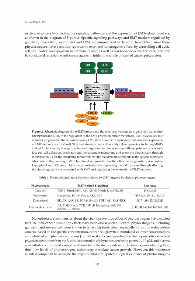
Toxins 2016, 8, 162
in diverse cancers by affecting the signaling pathways and the expression of EMT-related markers,as shown in the diagram of Figure 2. Specific signaling pathways and EMT markers regulated bygenistein, resveratrol, kaempferol and DIM, are summarized in Table 1. In addition, since thesephytoestrogens have been also reported to exert anti-carcinogenic effects by controlling cell cycle,cell proliferation and apoptosis in hormone-related, as well as non-hormone-related cancers, they maybe considered as effective anti-cancer agents to inhibit the whole process of cancer progression.
Figure 2. Schematic diagram of the EMT process and the roles of phytoestrogens, genistein, resveratrol,kaempferol and DIM, in the regulation of the EMT process in cancer metastasis. EMT plays a key rolein tumor progression. The cells undergoing EMT show E-cadherin repression, but increased expressionof EMT markers, such as Snail, Slug and vimentin, and cell motility-related proteins, including MMPsand uPA. As a result, they gain enhanced migration and invasion capabilities: primary cancer cellslose cell-cell adhesion, break through the basement membrane and enter the bloodstream throughintravasation. Later, the circulating tumor cells exit the bloodstream to migrate to the specific metastaticsites, where they undergo MET for clonal outgrowth. On the other hand, genistein, resveratrol,kaempferol and DIM may inhibit cancer metastasis by repressing the EMT process through affectingthe signaling pathways associated with EMT and regulating the expression of EMT markers.
Table 1. Potential signal transductions related to EMT targeted by dietary phytoestrogens.
Phytoestrogen EMT-Related Signalings Reference
Genistein TGF-β, Smad, PI3K, Akt, NF-kB, Notch-1, MAPK, ER [58,90,92]
Resveratrol Hedgehog, TGF-β, Smad, AKT, EGF [107,108,110,111,113,114]
Kaempferol ER, AR, AhR, PR, TGF-β, Smad3, PI3K/Akt, RAF/ERK [117–119,125,126,128]
Diindolylmethane AR, PI3K/Akt/mTOR/NF-κB, Hedgehog, miR-200,RANKL, β-catenin [140,141,143,145,147,148,150]
Nevertheless, controversies about the chemopreventive effect of phytoestrogens have existedbecause their cancer promoting effects have been also reported. Several phytoestrogens, includinggenistein and resveratrol, were known to have a biphasic effect, especially in hormone-dependentcancers: based on the specific concentration, cancer cell growth is stimulated at lower concentrationsand inhibited at higher concentrations [19]. More skepticism regarding the chemopreventive effects ofphytoestrogens came from the in vitro concentration of phytoestrogen being generally 10 μM, and plasmaconcentrations of >10 μM cannot be attainable by the dietary intake of phytoestrogen-containing food;thus, low levels of phytoestrogen, rather, may stimulate cancer growth. However, this insistenceis still incompetent to abrogate the experimental and epidemiological evidence of phytoestrogens
118
Toxins 2016, 8, 162
supporting anti-proliferative and anti-carcinogenic effects. Accordingly, more investigations of therelationship between the hazardous and chemopreventive effects of phytoestrogens in the bodyare needed at this stage. Besides, the poor bioavailability of phytoestrogens, such as resveratrol,in the body is another pitfall in the application of phytoestrogens, which has to be improved [97].The toxicological issues posed by phytoestrogens also should be considered in their applications. Forinstance, the possibility that serum genistein concentrations found in soy-fed infants may inducethymic and immune abnormalities was raised [151]. High doses of genistein (>5 μM) may act as atopoisomerase II inhibitor and a DNA damaging genotoxin [152]. Genistein was reported to be capableof altering the toxicological behaviors of the endocrine-active pesticide methoxychlor and likely otherendocrine-active compounds, as well [153]. In this way, the adverse effects of phytoestrogens onnormal cellular function have been found. As with many other compounds, there are many pros andcons associated with phytoestrogens, and thus, it is urgently needed to shape the development ofguidelines for the use of phytoestrogens for maximizing health benefits, as well as minimizing theadverse effects [154].
From this review, we may suggest that phytoestrogens are potent compounds that abrogatethe cell migration, invasion and metastasis of cancer by effectively suppressing the EMT process.Phytoestrogen treatments are a promising way to prevent cancer development as safer alternatives forthe natural strategies against cancers, though we need more research and interest for going forward toclinical trials.
Acknowledgments: This work was supported by a National Research Foundation of Korea (NRF) grant fundedby the Ministry of Education, Science and Technology (MEST) of the Republic of Korea (2014R1A1A2055295).This work was supported by a grant from the Next-Generation BioGreen 21 Program (No. PJ011355-2015),Rural Development Administration, Republic of Korea.
Conflicts of Interest: None of the authors have any conflicts of interest to declare.
References
1. Cragg, G.M.; Grothaus, P.G.; Newman, D.J. Impact of natural products on developing new anti-cancer agents.Chem. Rev. 2009, 109, 3012–3043. [CrossRef] [PubMed]
2. Fitter, A. Ecology. Making allelopathy respectable. Science 2003, 301, 1337–1338. [CrossRef] [PubMed]3. Acharya, A.; Das, I.; Singh, S.; Saha, T. Chemopreventive properties of indole-3-carbinol, diindolylmethane
and other constituents of cardamom against carcinogenesis. Recent Pat. Food Nutr. Agric. 2010, 2, 166–177.[CrossRef] [PubMed]
4. Sporn, M.B.; Suh, N. Chemoprevention of cancer. Carcinogenesis 2000, 21, 525–530. [CrossRef] [PubMed]5. Cragg, G.M.; Newman, D.J. Plants as a source of anti-cancer agents. J. Ethnopharmacol. 2005, 100, 72–79.
[CrossRef] [PubMed]6. Thakur, V.S.; Deb, G.; Babcook, M.A.; Gupta, S. Plant phytochemicals as epigenetic modulators: Role in
cancer chemoprevention. AAPS J. 2014, 16, 151–163. [CrossRef] [PubMed]7. Allred, K.F.; Yackley, K.M.; Vanamala, J.; Allred, C.D. Trigonelline is a novel phytoestrogen in coffee beans.
J. Nutr. 2009, 139, 1833–1838. [CrossRef] [PubMed]8. Santen, R.J.; Boyd, N.F.; Chlebowski, R.T.; Cummings, S.; Cuzick, J.; Dowsett, M.; Easton, D.; Forbes, J.F.;
Key, T.; Hankinson, S.E.; et al. Critical assessment of new risk factors for breast cancer: Considerations fordevelopment of an improved risk prediction model. Endocr. Relat. Cancer 2007, 14, 169–187. [CrossRef] [PubMed]
9. Giacalone, P.L.; Daures, J.P.; Ouafik, L.; Martin, P.M.; Laffargue, F.; Maudelonde, T. Steroids and adrenomedullingrowth patterns in human ovarian cancer cells: Estrogenic-regulation assay. Gynecol. Oncol. 2003, 91, 651–656.[CrossRef]
10. Chung, S.H.; Franceschi, S.; Lambert, P.F. Estrogen and ERalpha: Culprits in cervical cancer?Trends Endocrinol. Metab. 2010, 21, 504–511. [CrossRef] [PubMed]
11. Hwang, K.A.; Kang, N.H.; Yi, B.R.; Lee, H.R.; Park, M.A.; Choi, K.C. Genistein, a soy phytoestrogen, preventsthe growth of BG-1 ovarian cancer cells induced by 17beta-estradiol or bisphenol A via the inhibition of cellcycle progression. Int. J. Oncol. 2013, 42, 733–740. [PubMed]
119
Toxins 2016, 8, 162
12. Ahn, H.N.; Jeong, S.Y.; Bae, G.U.; Chang, M.; Zhang, D.; Liu, X.; Pei, Y.; Chin, Y.W.; Lee, J.; Oh, S.R.; et al.Selective estrogen receptor modulation by larrea nitida on MCF-7 Cell proliferation and immature rat uterus.Biomol. Ther. 2014, 22, 347–354. [CrossRef] [PubMed]
13. Shang, Y.; Brown, M. Molecular determinants for the tissue specificity of SERMs. Science 2002, 295, 2465–2468.[CrossRef] [PubMed]
14. Kuiper, G.G.; Lemmen, J.G.; Carlsson, B.; Corton, J.C.; Safe, S.H.; van der Saag, P.T.; van der Burg, B.;Gustafsson, J.A. Interaction of estrogenic chemicals and phytoestrogens with estrogen receptor beta.Endocrinology 1998, 139, 4252–4263. [PubMed]
15. Zhao, E.; Mu, Q. Phytoestrogen biological actions on Mammalian reproductive system and cancer growth.Sci. Pharm. 2011, 79, 1–20. [CrossRef] [PubMed]
16. Harris, D.M.; Besselink, E.; Henning, S.M.; Go, V.L.; Heber, D. Phytoestrogens induce differential estrogenreceptor alpha- or Beta-mediated responses in transfected breast cancer cells. Exp. Biol. Med. 2005, 230, 558–568.
17. Hsieh, C.Y.; Santell, R.C.; Haslam, S.Z.; Helferich, W.G. Estrogenic effects of genistein on the growth ofestrogen receptor-positive human breast cancer (MCF-7) cells in vitro and in vivo. Cancer Res. 1998, 58, 3833–3838.[PubMed]
18. Ju, Y.H.; Fultz, J.; Allred, K.F.; Doerge, D.R.; Helferich, W.G. Effects of dietary daidzein and its metabolite,equol, at physiological concentrations on the growth of estrogen-dependent human breast cancer (MCF-7)tumors implanted in ovariectomized athymic mice. Carcinogenesis 2006, 27, 856–863. [CrossRef] [PubMed]
19. Magee, P.J.; Rowland, I.R. Phyto-oestrogens, their mechanism of action: Current evidence for a role in breastand prostate cancer. Br. J. Nutr. 2004, 91, 513–531. [CrossRef] [PubMed]
20. Rice, S.; Whitehead, S.A. Phytoestrogens and breast cancer—Promoters or protectors? Endocr. Relat. Cancer2006, 13, 995–1015. [CrossRef] [PubMed]
21. Kirk, C.J.; Harris, R.M.; Wood, D.M.; Waring, R.H.; Hughes, P.J. Do dietary phytoestrogens influencesusceptibility to hormone-dependent cancer by disrupting the metabolism of endogenous oestrogens?Biochem. Soc. Trans. 2001, 29, 209–216. [CrossRef] [PubMed]
22. Labrie, F.; Luu-The, V.; Lin, S.X.; Labrie, C.; Simard, J.; Breton, R.; Belanger, A. The key role of 17 β-hydroxysteroiddehydrogenases in sex steroid biology. Steroids 1997, 62, 148–158. [CrossRef]
23. Murkies, A.L.; Wilcox, G.; Davis, S.R. Clinical review 92, Phytoestrogens. J. Clin. Endocrinol. Metab. 1998,83, 297–303. [PubMed]
24. Pilsakova, L.; Riecansky, I.; Jagla, F. The physiological actions of isoflavone phytoestrogens. Physiol. Res.2010, 59, 651–664. [PubMed]
25. Hutchins, A.M.; Lampe, J.W.; Martini, M.C.; Campbell, D.R.; Slavin, J.L. Vegetables, fruits, and legumes: Effecton urinary isoflavonoid phytoestrogen and lignan excretion. J. Am. Diet. Assoc. 1995, 95, 769–774. [CrossRef]
26. Milder, I.E.; Arts, I.C.; van de Putte, B.; Venema, D.P.; Hollman, P.C. Lignan contents of Dutch plant foods:A database including lariciresinol, pinoresinol, secoisolariciresinol and matairesinol. Br. J. Nutr. 2005,93, 393–402. [CrossRef] [PubMed]
27. Zamora-Ros, R.; Knaze, V.; Lujan-Barroso, L.; Kuhnle, G.G.; Mulligan, A.A.; Touillaud, M.; Slimani, N.;Romieu, I.; Powell, N.; Tumino, R.; et al. Dietary intakes and food sources of phytoestrogens in the EuropeanProspective Investigation into Cancer and Nutrition (EPIC) 24-h dietary recall cohort. Eur. J. Clin. Nutr. 2012,66, 932–941. [CrossRef] [PubMed]
28. Heinonen, S.; Nurmi, T.; Liukkonen, K.; Poutanen, K.; Wahala, K.; Deyama, T.; Nishibe, S.; Adlercreutz, H.In vitro metabolism of plant lignans: New precursors of mammalian lignans enterolactone and enterodiol.J. Agric. Food Chem. 2001, 49, 3178–3186. [CrossRef] [PubMed]
29. Boker, L.K.; Van der Schouw, Y.T.; De Kleijn, M.J.; Jacques, P.F.; Grobbee, D.E.; Peeters, P.H. Intake of dietaryphytoestrogens by Dutch women. J. Nutr. 2002, 132, 1319–1328. [PubMed]
30. Manjanatha, M.; Shelton, S.; Bishop, M.; Lyn-Cook, L.; Aidoo, A. Dietary effects of soy isoflavones daidzeinand genistein on 7,12-dimethylbenz[a]anthracene-induced mammary mutagenesis and carcinogenesis inovariectomized Big Blue transgenic rats. Carcinogenesis 2006, 27, 1970–1979. [CrossRef] [PubMed]
31. Sobolev, V.S.; Horn, B.W.; Potter, T.L.; Deyrup, S.T.; Gloer, J.B. Production of stilbenoids and phenolic acidsby the peanut plant at early stages of growth. J. Agric. Food Chem. 2006, 54, 3505–3511. [CrossRef] [PubMed]
32. Jang, M.; Cai, L.; Udeani, G.O.; Slowing, K.V.; Thomas, C.F.; Beecher, C.W.; Fong, H.H.; Farnsworth, N.R.;Kinghorn, A.D.; Mehta, R.G.; et al. Cancer chemopreventive activity of resveratrol, a natural product derivedfrom grapes. Science 1997, 275, 218–220. [CrossRef] [PubMed]
120
Toxins 2016, 8, 162
33. Heerboth, S.; Housman, G.; Leary, M.; Longacre, M.; Byler, S.; Lapinska, K.; Willbanks, A.; Sarkar, S. EMT andtumor metastasis. Clin. Transl. Med. 2015, 4. [CrossRef] [PubMed]
34. Spano, D.; Heck, C.; De Antonellis, P.; Christofori, G.; Zollo, M. Molecular networks that regulate cancermetastasis. Semin. Cancer Biol. 2012, 22, 234–249. [CrossRef] [PubMed]
35. Siegel, R.L.; Miller, K.D.; Jemal, A. Cancer statistics, 2016. CA Cancer J. Clin. 2016, 66, 7–30. [CrossRef][PubMed]
36. Voulgari, A.; Pintzas, A. Epithelial-mesenchymal transition in cancer metastasis: Mechanisms, markers andstrategies to overcome drug resistance in the clinic. Biochim. Biophys. Acta 2009, 1796, 75–90. [CrossRef][PubMed]
37. Hanahan, D.; Weinberg, R.A. The hallmarks of cancer. Cell 2000, 100, 57–70. [CrossRef]38. Yap, A.S.; Brieher, W.M.; Gumbiner, B.M. Molecular and functional analysis of cadherin-based adherens
junctions. Annu. Rev. Cell Dev. Biol. 1997, 13, 119–146. [CrossRef] [PubMed]39. Onder, T.T.; Gupta, P.B.; Mani, S.A.; Yang, J.; Lander, E.S.; Weinberg, R.A. Loss of E-cadherin promotes
metastasis via multiple downstream transcriptional pathways. Cancer Res. 2008, 68, 3645–3654. [CrossRef][PubMed]
40. Boyer, B.; Thiery, J.P. Epithelium-mesenchyme interconversion as example of epithelial plasticity. APMIS1993, 101, 257–268. [CrossRef] [PubMed]
41. Thiery, J.P. Cell adhesion in development: A complex signaling network. Curr. Opin. Genet. Dev. 2003, 13, 365–371.[CrossRef]
42. Miyoshi, J.; Takai, Y. Structural and functional associations of apical junctions with cytoskeleton.Biochim. Biophys. Acta 2008, 1778, 670–691. [CrossRef] [PubMed]
43. Yang, J.; Weinberg, R.A. Epithelial-mesenchymal transition: At the crossroads of development and tumormetastasis. Dev. Cell 2008, 14, 818–829. [CrossRef] [PubMed]
44. Friedl, P.; Wolf, K. Tumour-cell invasion and migration: Diversity and escape mechanisms. Nat. Rev. Cancer2003, 3, 362–374. [CrossRef] [PubMed]
45. Polette, M.; Nawrocki-Raby, B.; Gilles, C.; Clavel, C.; Birembaut, P. Tumour invasion and matrixmetalloproteinases. Crit. Rev. Oncol. Hematol. 2004, 49, 179–186. [CrossRef] [PubMed]
46. Son, H.; Moon, A. Epithelial-mesenchymal Transition and Cell Invasion. Toxicol. Res. 2010, 26, 245–252.[CrossRef] [PubMed]
47. Davies, J.A. Mesenchyme to epithelium transition during development of the mammalian kidney tubule.Acta Anat. 1996, 156, 187–201. [CrossRef] [PubMed]
48. Foroni, C.; Broggini, M.; Generali, D.; Damia, G. Epithelial-mesenchymal transition and breast cancer: Role,molecular mechanisms and clinical impact. Cancer Treat. Rev. 2012, 38, 689–697. [CrossRef] [PubMed]
49. Chaffer, C.L.; Weinberg, R.A. A perspective on cancer cell metastasis. Science 2011, 331, 1559–1564. [CrossRef][PubMed]
50. Perl, A.K.; Wilgenbus, P.; Dahl, U.; Semb, H.; Christofori, G. A causal role for E-cadherin in the transitionfrom adenoma to carcinoma. Nature 1998, 392, 190–193. [CrossRef] [PubMed]
51. Thiery, J.P.; Sleeman, J.P. Complex networks orchestrate epithelial-mesenchymal transitions. Nat. Rev. Mol.Cell Biol. 2006, 7, 131–142. [CrossRef] [PubMed]
52. Bailey, J.M.; Singh, P.K.; Hollingsworth, M.A. Cancer metastasis facilitated by developmental pathways: Sonichedgehog, Notch, and bone morphogenic proteins. J. Cell. Biochem. 2007, 102, 829–839. [CrossRef] [PubMed]
53. Zeisberg, M.; Neilson, E.G. Biomarkers for epithelial-mesenchymal transitions. J. Clin. Investig. 2009,119, 1429–1437. [CrossRef] [PubMed]
54. Batlle, E.; Sancho, E.; Franci, C.; Dominguez, D.; Monfar, M.; Baulida, J.; Garcia De Herreros, A.The transcription factor snail is a repressor of E-cadherin gene expression in epithelial tumour cells.Nat. Cell Biol. 2000, 2, 84–89. [CrossRef] [PubMed]
55. Yang, M.H.; Wu, M.Z.; Chiou, S.H.; Chen, P.M.; Chang, S.Y.; Liu, C.J.; Teng, S.C.; Wu, K.J. Direct regulation ofTWIST by HIF-1alpha promotes metastasis. Nat. Cell Biol. 2008, 10, 295–305. [CrossRef] [PubMed]
56. Thiery, J.P. Epithelial-mesenchymal transitions in development and pathologies. Curr. Opin. Cell Biol. 2003,15, 740–746. [CrossRef] [PubMed]
57. Bani-Hani, A.H.; Campbell, M.T.; Meldrum, D.R.; Meldrum, K.K. Cytokines in epithelial-mesenchymaltransition: A new insight into obstructive nephropathy. J. Urol. 2008, 180, 461–468. [CrossRef] [PubMed]
121
Toxins 2016, 8, 162
58. Han, L.; Zhang, H.W.; Zhou, W.P.; Chen, G.M.; Guo, K.J. The effects of genistein on transforming growthfactor-beta1-induced invasion and metastasis in human pancreatic cancer cell line Panc-1 in vitro. Chin. Med. J.2012, 125, 2032–2040. [PubMed]
59. Zhang, Y.E. Non-Smad pathways in TGF-β signaling. Cell Res. 2009, 19, 128–139. [CrossRef] [PubMed]60. Bertran, E.; Crosas-Molist, E.; Sancho, P.; Caja, L.; Lopez-Luque, J.; Navarro, E.; Egea, G.; Lastra, R.;
Serrano, T.; Ramos, E.; et al. Overactivation of the TGF-β pathway confers a mesenchymal-like phenotypeand CXCR4-dependent migratory properties to liver tumor cells. Hepatology 2013, 58, 2032–2044. [CrossRef][PubMed]
61. Hu, T.H.; Yao, Y.; Yu, S.; Han, L.L.; Wang, W.J.; Guo, H.; Tian, T.; Ruan, Z.P.; Kang, X.M.; Wang, J.; et al.SDF-1/CXCR4 promotes epithelial-mesenchymal transition and progression of colorectal cancer by activationof the Wnt/beta-catenin signaling pathway. Cancer Lett. 2014, 354, 417–426. [CrossRef] [PubMed]
62. Gitler, A.D.; Lu, M.M.; Jiang, Y.Q.; Epstein, J.A.; Gruber, P.J. Molecular markers of cardiac endocardialcushion development. Dev. Dyn. 2003, 228, 643–650. [CrossRef] [PubMed]
63. Liebner, S.; Cattelino, A.; Gallini, R.; Rudini, N.; Iurlaro, M.; Piccolo, S.; Dejana, E. β-catenin is required forendothelial-mesenchymal transformation during heart cushion development in the mouse. J. Cell Biol. 2004,166, 359–367. [CrossRef] [PubMed]
64. Wang, Z.; Banerjee, S.; Li, Y.; Rahman, K.M.; Zhang, Y.; Sarkar, F.H. Down-regulation of notch-1 inhibits invasionby inactivation of nuclear factor-kappaB, vascular endothelial growth factor, and matrix metalloproteinase-9in pancreatic cancer cells. Cancer Res. 2006, 66, 2778–2784. [CrossRef] [PubMed]
65. Miyamoto, Y.; Maitra, A.; Ghosh, B.; Zechner, U.; Argani, P.; Iacobuzio-Donahue, C.A.; Sriuranpong, V.; Iso, T.;Meszoely, I.M.; Wolfe, M.S.; et al. Notch mediates TGF alpha-induced changes in epithelial differentiationduring pancreatic tumorigenesis. Cancer Cell 2003, 3, 565–576. [CrossRef]
66. Shao, S.; Zhao, X.; Zhang, X.; Luo, M.; Zuo, X.; Huang, S.; Wang, Y.; Gu, S.; Zhao, X. Notch1 signalingregulates the epithelial-mesenchymal transition and invasion of breast cancer in a Slug-dependent manner.Mol. Cancer 2015, 14, 28. [CrossRef] [PubMed]
67. Thayer, S.P.; di Magliano, M.P.; Heiser, P.W.; Nielsen, C.M.; Roberts, D.J.; Lauwers, G.Y.; Qi, Y.P.; Gysin, S.;Fernandez-del Castillo, C.; Yajnik, V.; et al. Hedgehog is an early and late mediator of pancreatic cancertumorigenesis. Nature 2003, 425, 851–856. [CrossRef] [PubMed]
68. Yoo, Y.A.; Kang, M.H.; Lee, H.J.; Kim, B.H.; Park, J.K.; Kim, H.K.; Kim, J.S.; Oh, S.C. Sonic hedgehog pathwaypromotes metastasis and lymphangiogenesis via activation of Akt, EMT, and MMP-9 pathway in gastriccancer. Cancer Res. 2011, 71, 7061–7070. [CrossRef] [PubMed]
69. Gregory, R.I.; Chendrimada, T.P.; Cooch, N.; Shiekhattar, R. Human RISC couples microRNA biogenesis andposttranscriptional gene silencing. Cell 2005, 123, 631–640. [CrossRef] [PubMed]
70. Filipowicz, W.; Jaskiewicz, L.; Kolb, F.A.; Pillai, R.S. Post-transcriptional gene silencing by siRNAs andmiRNAs. Curr. Opin. Struct. Biol. 2005, 15, 331–341. [CrossRef] [PubMed]
71. Siemens, H.; Jackstadt, R.; Hunten, S.; Kaller, M.; Menssen, A.; Gotz, U.; Hermeking, H. miR-34 and SNAILform a double-negative feedback loop to regulate epithelial-mesenchymal transitions. Cell Cycle 2011,10, 4256–4271. [CrossRef] [PubMed]
72. Dahl, K.D.C.; Dahl, R.; Kruichak, J.N.; Hudson, L.G. The epidermal growth factor receptor responsivemiR-125a represses mesenchymal morphology in ovarian cancer cells. Neoplasia 2009, 11, 1208–1215. [CrossRef]
73. Yang, D.; Sun, Y.; Hu, L.; Zheng, H.; Ji, P.; Pecot, C.V.; Zhao, Y.; Reynolds, S.; Cheng, H.; Rupaimoole, R.; et al.Integrated analyses identify a master microRNA regulatory network for the mesenchymal subtype in serousovarian cancer. Cancer Cell 2013, 23, 186–199. [CrossRef] [PubMed]
74. Yeh, Y.M.; Chuang, C.M.; Chao, K.C.; Wang, L.H. MicroRNA-138 suppresses ovarian cancer cell invasionand metastasis by targeting SOX4 and HIF-1alpha. Int. J. Cancer 2013, 133, 867–878. [CrossRef] [PubMed]
75. Cheng, C.W.; Wang, H.W.; Chang, C.W.; Chu, H.W.; Chen, C.Y.; Yu, J.C.; Chao, J.I.; Liu, H.F.; Ding, S.L.;Shen, C.Y. MicroRNA-30a inhibits cell migration and invasion by downregulating vimentin expression andis a potential prognostic marker in breast cancer. Breast Cancer Res. Treat. 2012, 134, 1081–1093. [CrossRef][PubMed]
76. Kumarswamy, R.; Mudduluru, G.; Ceppi, P.; Muppala, S.; Kozlowski, M.; Niklinski, J.; Papotti, M.;Allgayer, H. MicroRNA-30a inhibits epithelial-to-mesenchymal transition by targeting Snai1 and isdownregulated in non-small cell lung cancer. Int. J. Cancer 2012, 130, 2044–2053. [CrossRef] [PubMed]
122

Toxins 2016, 8, 162
77. Liu, Z.; Gersbach, E.; Zhang, X.; Xu, X.; Dong, R.; Lee, P.; Liu, J.; Kong, B.; Shao, C.; Wei, J.J. miR-106arepresses the Rb tumor suppressor p130 to regulate cellular proliferation and differentiation in high-gradeserous ovarian carcinoma. Mol. Cancer Res. 2013, 11, 1314–1325. [CrossRef] [PubMed]
78. Ma, L.; Teruya-Feldstein, J.; Weinberg, R.A. Tumour invasion and metastasis initiated by microRNA-10b inbreast cancer. Nature 2007, 449, 682–688. [CrossRef] [PubMed]
79. Zhang, L.; Sun, J.; Wang, B.; Ren, J.C.; Su, W.; Zhang, T. MicroRNA-10b triggers the Epithelial-MesenchymalTransition (EMT) of laryngeal carcinoma Hep-2 cells by directly targeting the E-cadherin. Appl. Biochem. Biotechnol.2015, 176, 33–44. [CrossRef] [PubMed]
80. Gregory, P.A.; Bert, A.G.; Paterson, E.L.; Barry, S.C.; Tsykin, A.; Farshid, G.; Vadas, M.A.; Khew-Goodall, Y.;Goodall, G.J. The miR-200 family and miR-205 regulate epithelial to mesenchymal transition by targetingZEB1 and SIP1. Nat. Cell Biol. 2008, 10, 593–601. [CrossRef] [PubMed]
81. Korpal, M.; Lee, E.S.; Hu, G.; Kang, Y. The miR-200 family inhibits epithelial-mesenchymal transition andcancer cell migration by direct targeting of E-cadherin transcriptional repressors ZEB1 and ZEB2. J. Biol. Chem.2008, 283, 14910–14914. [CrossRef] [PubMed]
82. Dykxhoorn, D.M. MicroRNAs and metastasis: Little RNAs go a long way. Cancer Res. 2010, 70, 6401–6406.[CrossRef] [PubMed]
83. Guarino, M. Epithelial-mesenchymal transition and tumour invasion. Int. J. Biochem. Cell Biol. 2007, 39, 2153–2160.[CrossRef] [PubMed]
84. Kim, S.H.; Kim, C.W.; Jeon, S.Y.; Go, R.E.; Hwang, K.A.; Choi, K.C. Chemopreventive and chemotherapeuticeffects of genistein, a soy isoflavone, upon cancer development and progression in preclinical animal models.Lab. Anim. Res. 2014, 30, 143–150. [CrossRef] [PubMed]
85. Perabo, F.G.; Von Low, E.C.; Ellinger, J.; von Rucker, A.; Muller, S.C.; Bastian, P.J. Soy isoflavone genistein inprevention and treatment of prostate cancer. Prostate Cancer Prostatic Dis. 2008, 11, 6–12. [CrossRef] [PubMed]
86. Huang, J.; Nasr, M.; Kim, Y.; Matthews, H.R. Genistein inhibits protein histidine kinase. J. Biol. Chem. 1992,267, 15511–15515. [PubMed]
87. Ju, Y.H.; Allred, K.F.; Allred, C.D.; Helferich, W.G. Genistein stimulates growth of human breast cancer cellsin a novel, postmenopausal animal model, with low plasma estradiol concentrations. Carcinogenesis 2006,27, 1292–1299. [CrossRef] [PubMed]
88. Pelekanou, V.; Leclercq, G. Recent insights into the effect of natural and environmental estrogens onmammary development and carcinogenesis. Int. J. Dev. Biol. 2011, 55, 869–878. [CrossRef] [PubMed]
89. Pavese, J.M.; Farmer, R.L.; Bergan, R.C. Inhibition of cancer cell invasion and metastasis by genistein.Cancer Metastasis Rev. 2010, 29, 465–482. [CrossRef] [PubMed]
90. Bao, B.; Wang, Z.; Ali, S.; Kong, D.; Li, Y.; Ahmad, A.; Banerjee, S.; Azmi, A.S.; Miele, L.; Sarkar, F.H. Notch-1induces epithelial-mesenchymal transition consistent with cancer stem cell phenotype in pancreatic cancercells. Cancer Lett. 2011, 307, 26–36. [CrossRef] [PubMed]
91. Kim, A.Y.; Kwak, J.H.; Je, N.K.; Lee, Y.H.; Jung, Y.S. Epithelial-mesenchymal transition is associated with acquiredresistance to 5-Fluorocuracil in HT-29 colon cancer cells. Toxicol. Res. 2015, 31, 151–156. [CrossRef] [PubMed]
92. Kim, Y.S.; Choi, K.C.; Hwang, K.A. Genistein suppressed epithelial-mesenchymal transition and migrationefficacies of BG-1 ovarian cancer cells activated by estrogenic chemicals via estrogen receptor pathway anddownregulation of TGF-beta signaling pathway. Phytomedicine 2015, 22, 993–999. [CrossRef] [PubMed]
93. Dai, W.; Wang, F.; He, L.; Lin, C.; Wu, S.; Chen, P.; Zhang, Y.; Shen, M.; Wu, D.; Wang, C.; et al. Genisteininhibits hepatocellular carcinoma cell migration by reversing the epithelial-mesenchymal transition: Partialmediation by the transcription factor NFAT1. Mol. Carcinog. 2015, 54, 301–311. [CrossRef] [PubMed]
94. Liu, J.F.; Zhao, S.H.; Wu, S.S. Depleting NFAT1 expression inhibits the ability of invasion and migration ofhuman lung cancer cells. Cancer Cell Int. 2013, 13, 41. [CrossRef] [PubMed]
95. Zhang, L.L.; Li, L.; Wu, D.P.; Fan, J.H.; Li, X.; Wu, K.J.; Wang, X.Y.; He, D.L. A novel anti-cancer effect ofgenistein: Reversal of epithelial mesenchymal transition in prostate cancer cells. Acta Pharmacol. Sin. 2008,29, 1060–1068. [CrossRef] [PubMed]
96. Nonomura, S.; Kanagawa, H.; Makimoto, A. Chemical Constituents of Polygonaceous Plants. I. Studieson the Components of Ko-J O-Kon. (Polygonum Cuspidatum Sieb. Et Zucc.). J. Pharm. Soc. Jpn. 1963,83, 988–990.
97. Carter, L.G.; D’Orazio, J.A.; Pearson, K.J. Resveratrol and cancer: Focus on in vivo evidence.Endocr. Relat. Cancer 2014, 21, R209–R225. [CrossRef] [PubMed]
123
Toxins 2016, 8, 162
98. Fremont, L. Biological effects of resveratrol. Life Sci. 2000, 66, 663–673. [CrossRef]99. Adrian, M.; Rajaei, H.; Jeandet, P.; Veneau, J.; Bessis, R. Resveratrol oxidation in botrytis cinerea conidia.
Phytopathology 1998, 88, 472–476. [CrossRef] [PubMed]100. Ashby, J.; Tinwell, H.; Pennie, W.; Brooks, A.N.; Lefevre, P.A.; Beresford, N.; Sumpter, J.P. Partial and weak
oestrogenicity of the red wine constituent resveratrol: Consideration of its superagonist activity in MCF-7cells and its suggested cardiovascular protective effects. J. Appl. Toxicol. 1999, 19, 39–45. [CrossRef]
101. Bowers, J.L.; Tyulmenkov, V.V.; Jernigan, S.C.; Klinge, C.M. Resveratrol acts as a mixed agonist/antagonistfor estrogen receptors alpha and beta. Endocrinology 2000, 141, 3657–3667. [PubMed]
102. Le Corre, L.; Chalabi, N.; Delort, L.; Bignon, Y.J.; Bernard-Gallon, D.J. Resveratrol and breast cancerchemoprevention: Molecular mechanisms. Mol. Nutr. Food Res. 2005, 49, 462–471. [CrossRef] [PubMed]
103. Mitchell, S.H.; Zhu, W.; Young, C.Y. Resveratrol inhibits the expression and function of the androgen receptorin LNCaP prostate cancer cells. Cancer Res. 1999, 59, 5892–5895. [PubMed]
104. Pervaiz, S. Resveratrol: From grapevines to mammalian biology. FASEB J. 2003, 17, 1975–1985. [CrossRef][PubMed]
105. Leone, L.; Mazzetta, F.; Martinelli, D.; Valente, S.; Alimandi, M.; Raffa, S.; Santino, I. Klebsiella pneumoniaeIs Able to Trigger Epithelial-Mesenchymal Transition Process in Cultured Airway Epithelial Cells. PLoS ONE2016, 11, e0146365. [CrossRef] [PubMed]
106. Sun, Y.; Wang, H.; Liu, M.; Lin, F.; Hua, J. Resveratrol abrogates the effects of hypoxia on cell proliferation,invasion and EMT in osteosarcoma cells through downregulation of the HIF-1α protein. Mol. Med. Rep.2015, 11, 1975–1981. [CrossRef] [PubMed]
107. Li, W.; Cao, L.; Chen, X.; Lei, J.; Ma, Q. Resveratrol inhibits hypoxia-driven ROS-induced invasive andmigratory ability of pancreatic cancer cells via suppression of the Hedgehog signaling pathway. Oncol. Rep.2016, 35, 1718–1726. [CrossRef] [PubMed]
108. Gao, Q.; Yuan, Y.; Gan, H.Z.; Peng, Q. Resveratrol inhibits the hedgehog signaling pathway andepithelial-mesenchymal transition and suppresses gastric cancer invasion and metastasis. Oncol. Lett.2015, 9, 2381–2387. [CrossRef] [PubMed]
109. Li, J.; Chong, T.; Wang, Z.; Chen, H.; Li, H.; Cao, J.; Zhang, P.; Li, H. A novel anticancer effect of resveratrol:Reversal of epithelialmesenchymal transition in prostate cancer cells. Mol. Med. Rep. 2014, 10, 1717–1724.[PubMed]
110. Ji, Q.; Liu, X.; Han, Z.; Zhou, L.; Sui, H.; Yan, L.; Jiang, H.; Ren, J.; Cai, J.; Li, Q. Resveratrol suppressesepithelial-to-mesenchymal transition in colorectal cancer through TGF-β1/Smads signaling pathwaymediated Snail/E-cadherin expression. BMC Cancer 2015, 15, 97. [CrossRef] [PubMed]
111. Xu, Q.; Zong, L.; Chen, X.; Jiang, Z.; Nan, L.; Li, J.; Duan, W.; Lei, J.; Zhang, L.; Ma, J.; et al. Resveratrol in thetreatment of pancreatic cancer. Ann. N. Y. Acad. Sci. 2015, 1348, 10–19. [CrossRef] [PubMed]
112. Li, C.W.; Xia, W.; Lim, S.O.; Hsu, J.L.; Huo, L.; Wu, Y.; Li, L.Y.; Lai, C.C.; Chang, S.S.; Hsu, Y.H.; et al. AKT1inhibits epithelial-to-mesenchymal transition in breast cancer through phosphorylation-dependent Twist1degradation. Cancer Res. 2016. [CrossRef] [PubMed]
113. Vergara, D.; Valente, C.M.; Tinelli, A.; Siciliano, C.; Lorusso, V.; Acierno, R.; Giovinazzo, G.; Santino, A.;Storelli, C.; Maffia, M. Resveratrol inhibits the epidermal growth factor-induced epithelial mesenchymaltransition in MCF-7 cells. Cancer Lett. 2011, 310, 1–8. [CrossRef] [PubMed]
114. Wang, H.; Zhang, H.; Tang, L.; Chen, H.; Wu, C.; Zhao, M.; Yang, Y.; Chen, X.; Liu, G. Resveratrol inhibitsTGF-β1-induced epithelial-to-mesenchymal transition and suppresses lung cancer invasion and metastasis.Toxicology 2013, 303, 139–146. [CrossRef] [PubMed]
115. Calderon-Montano, J.M.; Burgos-Moron, E.; Perez-Guerrero, C.; Lopez-Lazaro, M. A review on the dietaryflavonoid kaempferol. Mini Rev. Med. Chem. 2011, 11, 298–344. [CrossRef] [PubMed]
116. Huang, L.; Yagura, T.; Chen, S. Sedative activity of hexane extract of Keampferia galanga L. and its activecompounds. J. Ethnopharmacol. 2008, 120, 123–125. [CrossRef] [PubMed]
117. Boam, T. Anti-androgenic effects of flavonols in prostate cancer. Ecancermedicalscience 2015, 9, 585. [CrossRef][PubMed]
118. Puppala, D.; Gairola, C.G.; Swanson, H.I. Identification of kaempferol as an inhibitor of cigarettesmoke-induced activation of the aryl hydrocarbon receptor and cell transformation. Carcinogenesis 2007, 28,639–647. [CrossRef] [PubMed]
124
Toxins 2016, 8, 162
119. Toh, M.F.; Mendonca, E.; Eddie, S.L.; Endsley, M.P.; Lantvit, D.D.; Petukhov, P.A.; Burdette, J.E. Kaempferolexhibits progestogenic effects in ovariectomized rats. J. Steroids Horm. Sci. 2014, 5, 136. [PubMed]
120. Kang, J.W.; Kim, J.H.; Song, K.; Kim, S.H.; Yoon, J.H.; Kim, K.S. Kaempferol and quercetin, componentsof Ginkgo biloba extract (EGb 761), induce caspase-3-dependent apoptosis in oral cavity cancer cells.Phytother. Res. 2010, 24 (Suppl. S1), S77–S82. [CrossRef] [PubMed]
121. Niering, P.; Michels, G.; Watjen, W.; Ohler, S.; Steffan, B.; Chovolou, Y.; Kampkotter, A.; Proksch, P.; Kahl, R.Protective and detrimental effects of kaempferol in rat H4IIE cells: Implication of oxidative stress andapoptosis. Toxicol. Appl. Pharmacol. 2005, 209, 114–122. [CrossRef] [PubMed]
122. Samhan-Arias, A.K.; Martin-Romero, F.J.; Gutierrez-Merino, C. Kaempferol blocks oxidative stress incerebellar granule cells and reveals a key role for reactive oxygen species production at the plasma membranein the commitment to apoptosis. Free Radic. Biol. Med. 2004, 37, 48–61. [CrossRef] [PubMed]
123. Leung, H.W.; Lin, C.J.; Hour, M.J.; Yang, W.H.; Wang, M.Y.; Lee, H.Z. Kaempferol induces apoptosis in humanlung non-small carcinoma cells accompanied by an induction of antioxidant enzymes. Food Chem. Toxicol.2007, 45, 2005–2013. [CrossRef] [PubMed]
124. Liou, A.K.; Clark, R.S.; Henshall, D.C.; Yin, X.M.; Chen, J. To die or not to die for neurons in ischemia,traumatic brain injury and epilepsy: A review on the stress-activated signaling pathways and apoptoticpathways. Prog. Neurobiol. 2003, 69, 103–142. [CrossRef]
125. Liang, S.Q.; Marti, T.M.; Dorn, P.; Froment, L.; Hall, S.R.; Berezowska, S.; Kocher, G.; Schmid, R.A.; Peng, R.W.Blocking the epithelial-to-mesenchymal transition pathway abrogates resistance to anti-folate chemotherapyin lung cancer. Cell Death Dis. 2015, 6, e1824. [CrossRef] [PubMed]
126. Jo, E.; Park, S.J.; Choi, Y.S.; Jeon, W.K.; Kim, B.C. Kaempferol suppresses transforming growth factor-β1-Inducedepithelial-to-mesenchymal transition and migration of A549 lung cancer cells by inhibiting Akt1-mediatedphosphorylation of Smad3 at Threonine-179. Neoplasia 2015, 17, 525–537. [CrossRef] [PubMed]
127. Gong, J.H.; Cho, I.H.; Shin, D.; Han, S.Y.; Park, S.H.; Kang, Y.H. Inhibition of airway epithelial-to-mesenchymaltransition and fibrosis by kaempferol in endotoxin-induced epithelial cells and ovalbumin-sensitized mice.Lab. Investig. 2014, 94, 297–308. [CrossRef] [PubMed]
128. Sulzmaier, F.J.; Ramos, J.W. RSK isoforms in cancer cell invasion and metastasis. Cancer Res. 2013,73, 6099–6105. [CrossRef] [PubMed]
129. Anderton, M.J.; Manson, M.M.; Verschoyle, R.D.; Gescher, A.; Lamb, J.H.; Farmer, P.B.; Steward, W.P.;Williams, M.L. Pharmacokinetics and tissue disposition of indole-3-carbinol and its acid condensationproducts after oral administration to mice. Clin. Cancer Res. 2004, 10, 5233–5241. [CrossRef] [PubMed]
130. Banerjee, S.; Kong, D.; Wang, Z.; Bao, B.; Hillman, G.G.; Sarkar, F.H. Attenuation of multi-targetedproliferation-linked signaling by 3,3'-diindolylmethane (DIM): From bench to clinic. Mutat. Res. 2011,728, 47–66. [CrossRef] [PubMed]
131. Marques, M.; Laflamme, L.; Benassou, I.; Cissokho, C.; Guillemette, B.; Gaudreau, L. Low levels of3,31-diindolylmethane activate estrogen receptor alpha and induce proliferation of breast cancer cells in theabsence of estradiol. BMC Cancer 2014, 14, 524. [CrossRef] [PubMed]
132. Gong, Y.; Sohn, H.; Xue, L.; Firestone, G.L.; Bjeldanes, L.F. 3,31-Diindolylmethane is a novel mitochondrialH(+)-ATP synthase inhibitor that can induce p21(Cip1/Waf1) expression by induction of oxidative stress inhuman breast cancer cells. Cancer Res. 2006, 66, 4880–4887. [CrossRef] [PubMed]
133. Hong, C.; Firestone, G.L.; Bjeldanes, L.F. Bcl-2 family-mediated apoptotic effects of 3,31-diindolylmethane(DIM) in human breast cancer cells. Biochem. Pharmacol. 2002, 63, 1085–1097. [CrossRef]
134. Li, Y.; Chinni, S.R.; Sarkar, F.H. Selective growth regulatory and pro-apoptotic effects of DIM is mediated byAKT and NF-kappaB pathways in prostate cancer cells. Front. Biosci. 2005, 10, 236–243. [CrossRef] [PubMed]
135. Choi, H.J.; Lim do, Y.; Park, J.H. Induction of G1 and G2/M cell cycle arrests by the dietary compound3,31-diindolylmethane in HT-29 human colon cancer cells. BMC Gastroenterol. 2009, 9, 39. [CrossRef] [PubMed]
136. Hsu, E.L.; Chen, N.; Westbrook, A.; Wang, F.; Zhang, R.; Taylor, R.T.; Hankinson, O. CXCR4 and CXCL12down-regulation: A novel mechanism for the chemoprotection of 3,31-diindolylmethane for breast andovarian cancers. Cancer Lett. 2008, 265, 113–123. [CrossRef] [PubMed]
137. Ahmad, A.; Kong, D.; Sarkar, S.H.; Wang, Z.; Banerjee, S.; Sarkar, F.H. Inactivation of uPA and its receptoruPAR by 3,31-diindolylmethane (DIM) leads to the inhibition of prostate cancer cell growth and migration.J. Cell. Biochem. 2009, 107, 516–527. [CrossRef] [PubMed]
125
Toxins 2016, 8, 162
138. Ahmad, A.; Kong, D.; Wang, Z.; Sarkar, S.H.; Banerjee, S.; Sarkar, F.H. Down-regulation of uPA and uPARby 3,31-diindolylmethane contributes to the inhibition of cell growth and migration of breast cancer cells.J. Cell. Biochem. 2009, 108, 916–925. [CrossRef] [PubMed]
139. Kong, D.; Li, Y.; Wang, Z.; Banerjee, S.; Sarkar, F.H. Inhibition of angiogenesis and invasion by3,31-diindolylmethane is mediated by the nuclear factor-kappaB downstream target genes MMP-9 anduPA that regulated bioavailability of vascular endothelial growth factor in prostate cancer. Cancer Res. 2007,67, 3310–3319. [CrossRef] [PubMed]
140. Kong, D.; Sethi, S.; Li, Y.; Chen, W.; Sakr, W.A.; Heath, E.; Sarkar, F.H. Androgen receptor splice variantscontribute to prostate cancer aggressiveness through induction of EMT and expression of stem cell markergenes. Prostate 2015, 75, 161–174. [CrossRef] [PubMed]
141. Ahmad, A.; Biersack, B.; Li, Y.; Kong, D.; Bao, B.; Schobert, R.; Padhye, S.B.; Sarkar, F.H. Targeted regulationof PI3K/Akt/mTOR/NF-kappaB signaling by indole compounds and their derivatives: Mechanistic detailsand biological implications for cancer therapy. Anticancer Agents Med. Chem. 2013, 13, 1002–1013. [CrossRef][PubMed]
142. Lamouille, S.; Derynck, R. Emergence of the phosphoinositide 3-kinase-Akt-mammalian target of rapamycinaxis in transforming growth factor-beta-induced epithelial-mesenchymal transition. Cells Tissues Organs2011, 193, 8–22. [CrossRef] [PubMed]
143. Sethi, S.; Li, Y.; Sarkar, F.H. Regulating miRNA by natural agents as a new strategy for cancer treatment.Curr. Drug Targets 2013, 14, 1167–1174. [CrossRef] [PubMed]
144. Melkamu, T.; Zhang, X.; Tan, J.; Zeng, Y.; Kassie, F. Alteration of microRNA expression in vinyl carbamate-inducedmouse lung tumors and modulation by the chemopreventive agent indole-3-carbinol. Carcinogenesis 2010,31, 252–258. [CrossRef] [PubMed]
145. Li, Y.; VandenBoom, T.G., 2nd; Kong, D.; Wang, Z.; Ali, S.; Philip, P.A.; Sarkar, F.H. Up-regulation of miR-200 andlet-7 by natural agents leads to the reversal of epithelial-to-mesenchymal transition in gemcitabine-resistantpancreatic cancer cells. Cancer Res. 2009, 69, 6704–6712. [CrossRef] [PubMed]
146. Kong, D.; Heath, E.; Chen, W.; Cher, M.L.; Powell, I.; Heilbrun, L.; Li, Y.; Ali, S.; Sethi, S.; Hassan, O.;et al. Loss of let-7 up-regulates EZH2 in prostate cancer consistent with the acquisition of cancer stem cellsignatures that are attenuated by BR-DIM. PLoS ONE 2012, 7, e33729. [CrossRef] [PubMed]
147. Watson, G.W.; Beaver, L.M.; Williams, D.E.; Dashwood, R.H.; Ho, E. Phytochemicals from cruciferousvegetables, epigenetics, and prostate cancer prevention. AAPS J. 2013, 15, 951–961. [CrossRef] [PubMed]
148. Li, Y.; Kong, D.; Ahmad, A.; Bao, B.; Sarkar, F.H. Targeting bone remodeling by isoflavone and3,31-diindolylmethane in the context of prostate cancer bone metastasis. PLoS ONE 2012, 7, e33011. [CrossRef][PubMed]
149. Wu, T.; Chen, C.; Li, F.; Chen, Z.; Xu, Y.; Xiao, B.; Tao, Z. 3,31-Diindolylmethane inhibits the invasionand metastasis of nasopharyngeal carcinoma cells and by regulation of epithelial mesenchymal transition.Exp. Ther. Med. 2014, 7, 1635–1638. [PubMed]
150. Xu, L.; Jiang, Y.; Zheng, J.; Xie, G.; Li, J.; Shi, L.; Fan, S. Aberrant expression of β-catenin and E-cadherin iscorrelated with poor prognosis of nasopharyngeal cancer. Hum. Pathol. 2013, 44, 1357–1364. [CrossRef] [PubMed]
151. Yellayi, S.; Naaz, A.; Szewczykowski, M.A.; Sato, T.; Woods, J.A.; Chang, J.; Segre, M.; Allred, C.D.;Helferich, W.G.; Cooke, P.S. The phytoestrogen genistein induces thymic and immune changes: A humanhealth concern? Proc. Natl. Acad. Sci. USA 2002, 99, 7616–7621. [CrossRef] [PubMed]
152. Klein, C.B.; King, A.A. Genistein genotoxicity: Critical considerations of in vitro exposure dose.Toxicol. Appl. Pharmacol. 2007, 224, 1–11. [CrossRef] [PubMed]
153. You, L. Phytoestrogen genistein and its pharmacological interactions with synthetic endocrine-activecompounds. Curr. Pharm. Des. 2004, 10, 2749–2757. [CrossRef] [PubMed]
154. Patisaul, H.B.; Jefferson, W. The pros and cons of phytoestrogens. Front. Neuroendocrinol. 2010, 31, 400–419.[CrossRef] [PubMed]
© 2016 by the authors; licensee MDPI, Basel, Switzerland. This article is an open accessarticle distributed under the terms and conditions of the Creative Commons Attribution(CC-BY) license (http://creativecommons.org/licenses/by/4.0/).
126
toxins
Article
Pueraria mirifica Exerts Estrogenic Effects in theMammary Gland and Uterus and Promotes MammaryCarcinogenesis in Donryu Rats
Anna Kakehashi 1,*, Midori Yoshida 2,†, Yoshiyuki Tago 1, Naomi Ishii 1, Takahiro Okuno 1,Min Gi 1 and Hideki Wanibuchi 1
1 Department of Molecular Pathology, Osaka City University Graduate School of Medicine,1-4-3 Asahi-machi, Abeno-ku, Osaka 545-8585, Japan; [email protected] (Y.T.);[email protected] (N.I.); [email protected] (T.O.); [email protected] (M.G.);[email protected] (H.W.)
2 Division of Pathology, Biological Safety Research Center, National Institute of Health Sciences,Ministry of Health, Labour and Welfare, 1-18-1 Kamiyoga, Setagaya-ku, Tokyo 158-8501, Japan
* Correspondence: [email protected]; Tel.: +81-6-6645-3737† Present address: Food Safety Commission, Cabinet Office, Government of Japan, 5-2-20 Akasaka, Minato-ku,
Tokyo 107-6122, Japan; [email protected].
Academic Editor: Carmela FimognariReceived: 3 June 2016; Accepted: 13 September 2016; Published: 4 November 2016
Abstract: Pueraria mirifica (PM), a plant whose dried and powdered tuberous roots are nowwidely used in rejuvenating preparations to promote youthfulness in both men and women,may have major estrogenic influence. In this study, we investigated modifying effects of PM atvarious doses on mammary and endometrial carcinogenesis in female Donryu rats. Firstly, PMadministered to ovariectomized animals at doses of 0.03%, 0.3%, and 3% in a phytoestrogen-lowdiet for 2 weeks caused significant increase in uterus weight. Secondly, a 4 week PM application tonon-operated rats at a dose of 3% after 7,12-dimethylbenz[a]anthracene (DMBA) initiation resulted insignificant elevation of cell proliferation in the mammary glands. In a third experiment, postpubertaladministration of 0.3% (200 mg/kg body weight (b.w.)/day) PM to 5-week-old non-operatedanimals for 36 weeks following initiation of mammary and endometrial carcinogenesis with DMBAand N-ethyl-N′-nitro-N-nitrosoguanidine (ENNG), respectively, resulted in significant increase ofmammary adenocarcinoma incidence. A significant increase of endometrial atypical hyperplasiamultiplicity was also observed. Furthermore, PM at doses of 0.3%, and more pronouncedly, at 1%induced dilatation, hemorrhage and inflammation of the uterine wall. In conclusion, postpubertallong-term PM administration to Donryu rats exerts estrogenic effects in the mammary gland anduterus, and at a dose of 200 mg/kg b.w./day was found to promote mammary carcinogenesis initiatedby DMBA.
Keywords: Pueraria mirifica; mammary gland; uterus; carcinogenesis; estrogenic activity; Donryu rat
1. Introduction
Pueraria mirifica (PM), also known as white Kwao Krua, is a plant found in northern andnortheastern Thailand which belongs to the family of Leguminosae, and the soy, bean, and peasubfamily Papilionoideae. Previously, PM application for treatment of a range of conditions, includingthose related to the aging process, has been reported [1]. Dried and powdered tuberous roots ofPM contain at least 17 chemical compounds with estrogenic biological activities, usually dividedinto three groups: the first group includes teniso flavonoids such as genistin, genistein, daidzein,daidzin, kwakhurin, kwakhurin hydrate, tuberosin, puerarin, mirificin, and puemiricarpene [2,3];
Toxins 2016, 8, 275 127 www.mdpi.com/journal/toxins
Toxins 2016, 8, 275
the second group of coumestans comprises coumestrol, mirificoumestan, mirificoumestan glycol,and mirificoumestan hydrate; and the third features chromenes, such as miroestrol, deoxymiroestrol,and isomiroestrol [2]. All these substances are phytoestrogens with structures similar to that of17 β-estradiol. Miroestrol, the phytoestrogen with the highest estrogenic activity among all thoseisolated from PM, is considered similar to estriol, which is considered the safest estrogen forhumans [4–7]. Furthermore, PM has been reported to contain phytoestrogens like β-sitosterol,stigmasterol, campesterol, as well as the cytotoxic non-phytoestrogen spinasterol [3,8]. Puerarinmight account for about half of the total isoflavone content of PM, with lower amounts of genistin anddaidzin present, all these being glycoside forms which can be partially hydrolyzed in the intestine byC-glycosyl bond cleavage to give the respective aglycoside forms: genistein, daidzein, and daidzein [2].
Nowadays, PM is available in tablets, extracts, creams, sprays, and powdered forms, so that itcan be added to other medicinal preparations or herbs, and individual conditions require differentapplications and dosages [1]. It can be readily obtained from internet resources in many countries,including the USA and Japan, and is primarily used for supporting memory, smoothing the skin,increasing hair growth, improving appetite, and providing relief for ailments like osteoporosis and evencancer [9–12]. Continuous administration of PM at 20–100 mg/day for 6 months, or at 100–200 mg/dayfor 12 months, was found to help women having menopause symptoms, while no significant changeswere detected in their hepatic, hematologic, and renal functions [3]. Pretreatment with PM at a highdose (1000 mg/kg body weight (b.w.)/day) for 4 weeks was shown to suppress the development ofmammary tumors induced by 7,12-dimethylbenz[a]anthracene (DMBA) in Sprague–Dawley rats [13].However, the effect of long-term administration at different doses has not yet been clarified in detail.Based on available studies, a safe dosage of PM as a dietary supplement for humans was suggested at1–2 mg/kg b.w./day or about 50–100 mg/day [3]. Nowadays, doses of 20–100 mg/day are commonlyused, but in some cases 200–900 mg/day or even higher (up to 3000 mg/day) are applied. Until now,no serious side effects have been recorded with the prescribed safe dosage, although at high doses PMmay cause epilepsy, diabetes, asthma, and migraine [3].
Despite the data on the benefits of PM, there are reasons for concern that a herb whichexhibits strong estrogen-like properties may stimulate the growth of existing estrogen-sensitivebreast or endometrial tumors, pointing out questions such as: what is a safe dose? Previously,nanomolar concentrations of genistein, present in PM, was shown to induce acid ceramidase(ASAH1) transcription through a GPR30-dependent, pertussis toxin-sensitive pathway that requiresthe activation of c-Src and extracellular signal regulated kinase 1/2 (ERK1/2), thus stimulating breastcancer cell growth [14]. Recently, we further demonstrated that postpubertal administration of soyisoflavones at estrogenic doses promotes mammary and endometrial carcinogenesis in Donryu rats [15].These data call into question the safety of long-term exposure to phytoestrogens with regard to effectson the mammary gland and endometrium. It is of particular importance that concentrations of PMwhich might exert promoting effects on mammary gland and uterine carcinogenesis be determined.Therefore, the present study was carried out to investigate the modifying effects of various doses ofPM on mammary and uterine endometrial carcinogenesis using the Donryu rat model.
2. Results
2.1. Estrogenic Effect of Test Compounds (Short-Term Experiment 1)
After ovariectomy, PM treatment at doses of 0.3% and 3% and isoflavone aglycon (IA) treatmentat a dose of 0.2% resulted in cornification, evident on examination of vaginal smears (Figure 1).
The 0.3% PM treatment was found to exert even stronger estrogenic activity than 0.2% IA.Mean relative uterus weights were significantly elevated in 0.03% (0.13% ± 0.01%, p < 0.05),0.3% (0.31% ± 0.03%, p < 0.05), 3% PM (0.35% ± 0.08%, p < 0.05) and 0.2% IA (0.19% ± 0.04%,p < 0.05) administered ovariectomized rats as compared to the control (0.08% ± 0.01%). Thus,weak, medium, and strong estrogenic activities of PM at doses of 0.03% (20 mg/kg b.w./day),
128
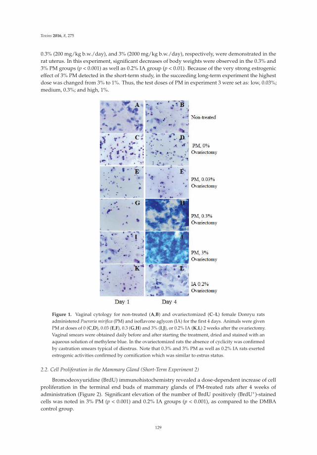
Toxins 2016, 8, 275
0.3% (200 mg/kg b.w./day), and 3% (2000 mg/kg b.w./day), respectively, were demonstrated in therat uterus. In this experiment, significant decreases of body weights were observed in the 0.3% and3% PM groups (p < 0.001) as well as 0.2% IA group (p < 0.01). Because of the very strong estrogeniceffect of 3% PM detected in the short-term study, in the succeeding long-term experiment the highestdose was changed from 3% to 1%. Thus, the test doses of PM in experiment 3 were set as: low, 0.03%;medium, 0.3%; and high, 1%.
Figure 1. Vaginal cytology for non-treated (A,B) and ovariectomized (C–L) female Donryu ratsadministered Pueraria mirifica (PM) and isoflavone aglycon (IA) for the first 4 days. Animals were givenPM at doses of 0 (C,D), 0.03 (E,F), 0.3 (G,H) and 3% (I,J), or 0.2% IA (K,L) 2 weeks after the ovariectomy.Vaginal smears were obtained daily before and after starting the treatment, dried and stained with anaqueous solution of methylene blue. In the ovariectomized rats the absence of cyclicity was confirmedby castration smears typical of diestrus. Note that 0.3% and 3% PM as well as 0.2% IA rats exertedestrogenic activities confirmed by cornification which was similar to estrus status.
2.2. Cell Proliferation in the Mammary Gland (Short-Term Experiment 2)
Bromodeoxyuridine (BrdU) immunohistochemistry revealed a dose-dependent increase of cellproliferation in the terminal end buds of mammary glands of PM-treated rats after 4 weeks ofadministration (Figure 2). Significant elevation of the number of BrdU positively (BrdU+)-stainedcells was noted in 3% PM (p < 0.001) and 0.2% IA groups (p < 0.001), as compared to the DMBAcontrol group.
129
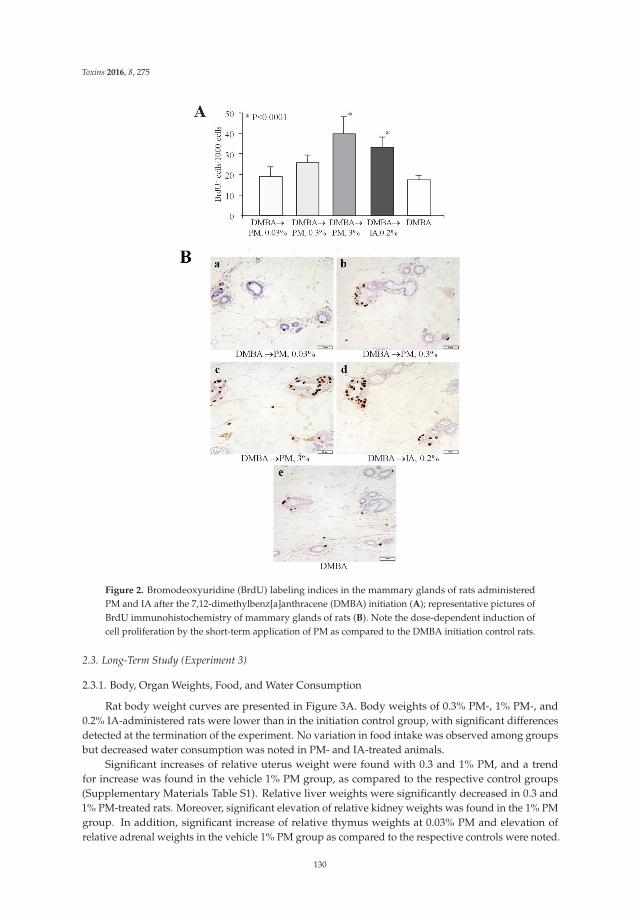
Toxins 2016, 8, 275
Figure 2. Bromodeoxyuridine (BrdU) labeling indices in the mammary glands of rats administeredPM and IA after the 7,12-dimethylbenz[a]anthracene (DMBA) initiation (A); representative pictures ofBrdU immunohistochemistry of mammary glands of rats (B). Note the dose-dependent induction ofcell proliferation by the short-term application of PM as compared to the DMBA initiation control rats.
2.3. Long-Term Study (Experiment 3)
2.3.1. Body, Organ Weights, Food, and Water Consumption
Rat body weight curves are presented in Figure 3A. Body weights of 0.3% PM-, 1% PM-, and0.2% IA-administered rats were lower than in the initiation control group, with significant differencesdetected at the termination of the experiment. No variation in food intake was observed among groupsbut decreased water consumption was noted in PM- and IA-treated animals.
Significant increases of relative uterus weight were found with 0.3 and 1% PM, and a trendfor increase was found in the vehicle 1% PM group, as compared to the respective control groups(Supplementary Materials Table S1). Relative liver weights were significantly decreased in 0.3 and1% PM-treated rats. Moreover, significant elevation of relative kidney weights was found in the 1% PMgroup. In addition, significant increase of relative thymus weights at 0.03% PM and elevation ofrelative adrenal weights in the vehicle 1% PM group as compared to the respective controls were noted.
130
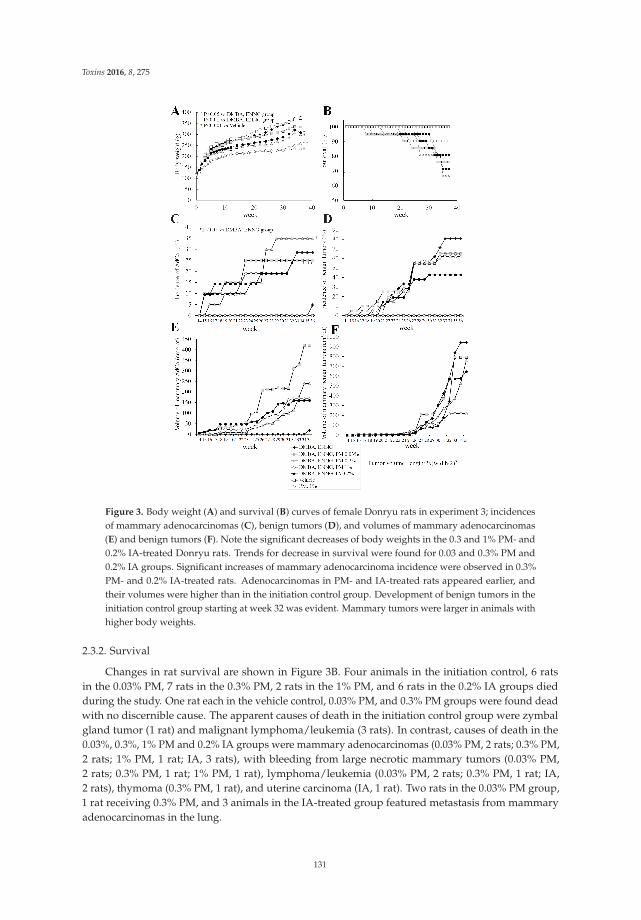
Toxins 2016, 8, 275
Figure 3. Body weight (A) and survival (B) curves of female Donryu rats in experiment 3; incidencesof mammary adenocarcinomas (C), benign tumors (D), and volumes of mammary adenocarcinomas(E) and benign tumors (F). Note the significant decreases of body weights in the 0.3 and 1% PM- and0.2% IA-treated Donryu rats. Trends for decrease in survival were found for 0.03 and 0.3% PM and0.2% IA groups. Significant increases of mammary adenocarcinoma incidence were observed in 0.3%PM- and 0.2% IA-treated rats. Adenocarcinomas in PM- and IA-treated rats appeared earlier, andtheir volumes were higher than in the initiation control group. Development of benign tumors in theinitiation control group starting at week 32 was evident. Mammary tumors were larger in animals withhigher body weights.
2.3.2. Survival
Changes in rat survival are shown in Figure 3B. Four animals in the initiation control, 6 ratsin the 0.03% PM, 7 rats in the 0.3% PM, 2 rats in the 1% PM, and 6 rats in the 0.2% IA groups diedduring the study. One rat each in the vehicle control, 0.03% PM, and 0.3% PM groups were found deadwith no discernible cause. The apparent causes of death in the initiation control group were zymbalgland tumor (1 rat) and malignant lymphoma/leukemia (3 rats). In contrast, causes of death in the0.03%, 0.3%, 1% PM and 0.2% IA groups were mammary adenocarcinomas (0.03% PM, 2 rats; 0.3% PM,2 rats; 1% PM, 1 rat; IA, 3 rats), with bleeding from large necrotic mammary tumors (0.03% PM,2 rats; 0.3% PM, 1 rat; 1% PM, 1 rat), lymphoma/leukemia (0.03% PM, 2 rats; 0.3% PM, 1 rat; IA,2 rats), thymoma (0.3% PM, 1 rat), and uterine carcinoma (IA, 1 rat). Two rats in the 0.03% PM group,1 rat receiving 0.3% PM, and 3 animals in the IA-treated group featured metastasis from mammaryadenocarcinomas in the lung.
131
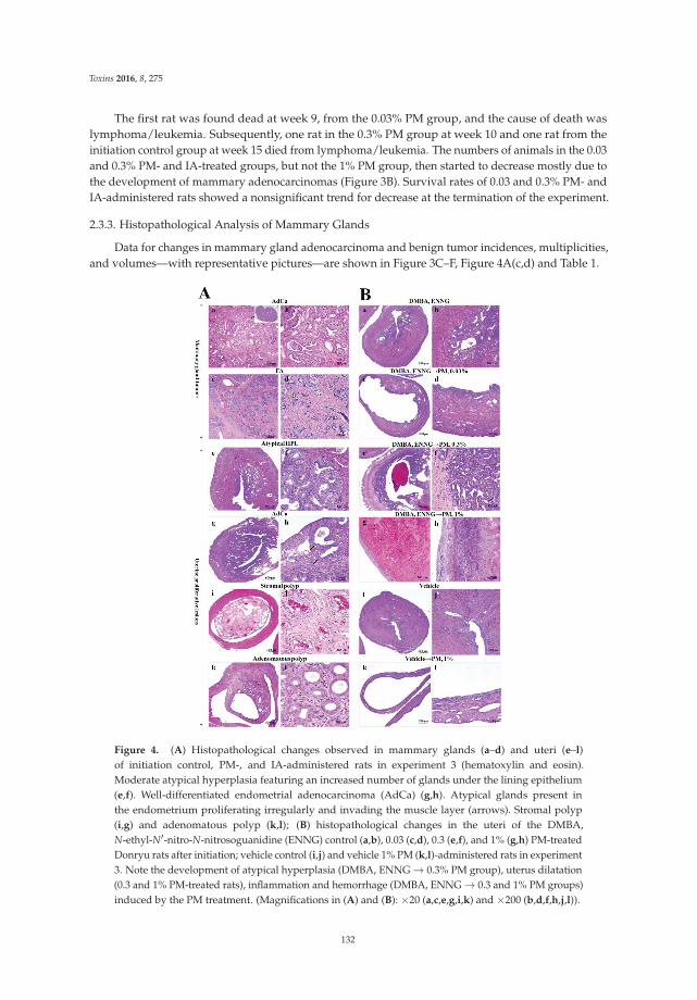
Toxins 2016, 8, 275
The first rat was found dead at week 9, from the 0.03% PM group, and the cause of death waslymphoma/leukemia. Subsequently, one rat in the 0.3% PM group at week 10 and one rat from theinitiation control group at week 15 died from lymphoma/leukemia. The numbers of animals in the 0.03and 0.3% PM- and IA-treated groups, but not the 1% PM group, then started to decrease mostly due tothe development of mammary adenocarcinomas (Figure 3B). Survival rates of 0.03 and 0.3% PM- andIA-administered rats showed a nonsignificant trend for decrease at the termination of the experiment.
2.3.3. Histopathological Analysis of Mammary Glands
Data for changes in mammary gland adenocarcinoma and benign tumor incidences, multiplicities,and volumes—with representative pictures—are shown in Figure 3C–F, Figure 4A(c,d) and Table 1.
Figure 4. (A) Histopathological changes observed in mammary glands (a–d) and uteri (e–l)of initiation control, PM-, and IA-administered rats in experiment 3 (hematoxylin and eosin).Moderate atypical hyperplasia featuring an increased number of glands under the lining epithelium(e,f). Well-differentiated endometrial adenocarcinoma (AdCa) (g,h). Atypical glands present inthe endometrium proliferating irregularly and invading the muscle layer (arrows). Stromal polyp(i,g) and adenomatous polyp (k,l); (B) histopathological changes in the uteri of the DMBA,N-ethyl-N′-nitro-N-nitrosoguanidine (ENNG) control (a,b), 0.03 (c,d), 0.3 (e,f), and 1% (g,h) PM-treatedDonryu rats after initiation; vehicle control (i,j) and vehicle 1% PM (k,l)-administered rats in experiment3. Note the development of atypical hyperplasia (DMBA, ENNG → 0.3% PM group), uterus dilatation(0.3 and 1% PM-treated rats), inflammation and hemorrhage (DMBA, ENNG → 0.3 and 1% PM groups)induced by the PM treatment. (Magnifications in (A) and (B): ×20 (a,c,e,g,i,k) and ×200 (b,d,f,h,j,l)).
132
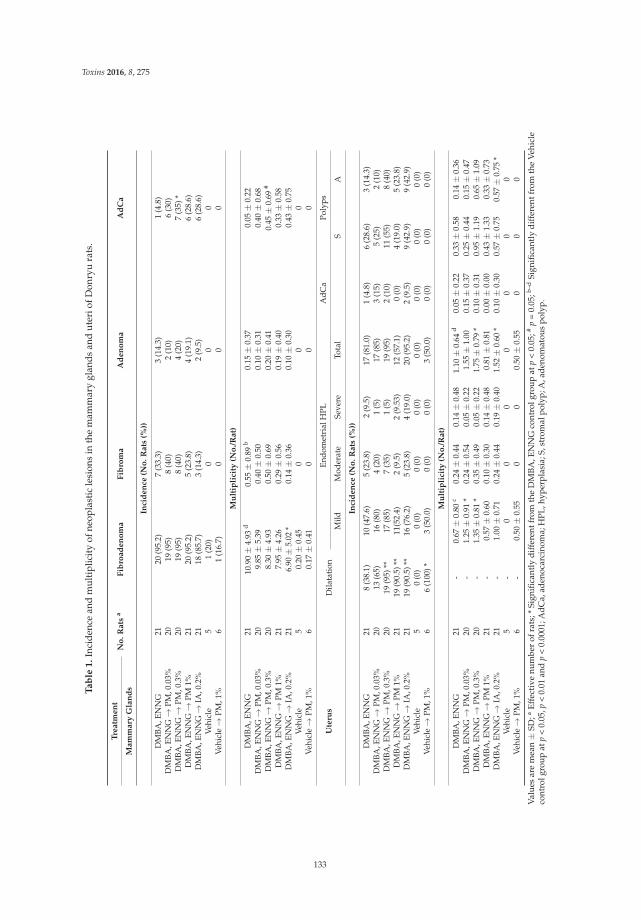
Toxins 2016, 8, 275
Tabl
e1.
Inci
denc
ean
dm
ulti
plic
ity
ofne
opla
stic
lesi
ons
inth
em
amm
ary
glan
dsan
dut
erio
fDon
ryu
rats
.
Trea
tmen
tN
o.R
ats
aFi
broa
deno
ma
Fibr
oma
Ade
nom
aA
dCa
Mam
mar
yG
land
s
Inci
denc
e(N
o.R
ats
(%))
DM
BA,E
NN
G21
20(9
5.2)
7(3
3.3)
3(1
4.3)
1(4
.8)
DM
BA,E
NN
G→
PM,0
.03%
2019
(95)
8(4
0)2
(10)
6(3
0)D
MBA
,EN
NG→
PM,0
.3%
2019
(95)
8(4
0)4
(20)
7(3
5)*
DM
BA,E
NN
G→
PM1%
2120
(95.
2)5
(23.
8)4
(19.
1)6
(28.
6)D
MBA
,EN
NG→
IA,0
.2%
2118
(85.
7)3
(14.
3)2
(9.5
)6
(28.
6)Ve
hicl
e5
1(2
0)0
00
Vehi
cle→
PM,1
%6
1(1
6.7)
00
0
Mul
tipl
icit
y(N
o./R
at)
DM
BA,E
NN
G21
10.9
0±
4.93
d0.
55±
0.89
b0.
15±
0.37
0.05
±0.
22D
MBA
,EN
NG→
PM,0
.03%
209.
85±
5.39
0.40
±0.
500.
10±
0.31
0.40
±0.
68D
MBA
,EN
NG→
PM,0
.3%
208.
30±
4.93
0.50
±0.
690.
20±
0.41
0.45
±0.
69#
DM
BA,E
NN
G→
PM1%
217.
95±
4.26
0.29
±0.
560.
19±
0.40
0.33
±0.
58D
MBA
,EN
NG→
IA,0
.2%
216.
90±
5.02
*0.
14±
0.36
0.10
±0.
300.
43±
0.75
Vehi
cle
50.
20±
0.45
00
0Ve
hicl
e→
PM,1
%6
0.17
±0.
410
00
Ute
rus
Dila
tati
onEn
dom
etri
alH
PLA
dCa
Poly
ps
Mild
Mod
erat
eSe
vere
Tota
lS
A
Inci
denc
e(N
o.R
ats
(%))
DM
BA,E
NN
G21
8(3
8.1)
10(4
7.6)
5(2
3.8)
2(9
.5)
17(8
1.0)
1(4
.8)
6(2
8.6)
3(1
4.3)
DM
BA,E
NN
G→
PM,0
.03%
2013
(65)
16(8
0)4
(20)
1(5
)17
(85)
3(1
5)5
(25)
2(1
0)D
MBA
,EN
NG→
PM,0
.3%
2019
(95)
**17
(85)
7(3
5)1
(5)
19(9
5)2
(10)
11(5
5)8
(40)
DM
BA,E
NN
G→
PM1%
2119
(90.
5)**
11(5
2.4)
2(9
.5)
2(9
.53)
12(5
7.1)
0(0
)4
(19.
0)5
(23.
8)D
MBA
,EN
NG→
IA,0
.2%
2119
(90.
5)**
16(7
6.2)
5(2
3.8)
4(1
9.0)
20(9
5.2)
2(9
.5)
9(4
2.9)
9(4
2.9)
Vehi
cle
50
(0)
0(0
)0
(0)
0(0
)0
(0)
0(0
)0
(0)
0(0
)Ve
hicl
e→
PM,1
%6
6(1
00)*
3(5
0.0)
0(0
)0
(0)
3(5
0.0)
0(0
)0
(0)
0(0
)
Mul
tipl
icit
y(N
o./R
at)
DM
BA,E
NN
G21
-0.
67±
0.80
c0.
24±
0.44
0.14
±0.
481.
10±
0.64
d0.
05±
0.22
0.33
±0.
580.
14±
0.36
DM
BA,E
NN
G→
PM,0
.03%
20-
1.25
±0.
91*
0.24
±0.
540.
05±
0.22
1.55
±1.
000.
15±
0.37
0.25
±0.
440.
15±
0.47
DM
BA,E
NN
G→
PM,0
.3%
20-
1.35
±0.
81*
0.35
±0.
490.
05±
0.22
1.75
±0.
79*
0.10
±0.
310.
95±
1.19
0.65
±1.
09D
MBA
,EN
NG→
PM1%
21-
0.57
±0.
600.
10±
0.30
0.14
±0.
480.
81±
0.81
0.00
±0.
000.
43±
1.33
0.33
±0.
73D
MBA
,EN
NG→
IA,0
.2%
21-
1.00
±0.
710.
24±
0.44
0.19
±0.
401.
52±
0.60
*0.
10±
0.30
0.57
±0.
750.
57±
0.75
*Ve
hicl
e5
-0
00
00
00
Vehi
cle→
PM,1
%6
-0.
50±
0.55
00
0.50
±0.
550
00
Val
ues
are
mea
n±
SD;a
Eff
ecti
venu
mbe
rof
rats
;*Si
gnifi
cant
lyd
iffer
entf
rom
the
DM
BA
,EN
NG
cont
rolg
roup
atp
<0.
05;#
p=
0.05
;b–d
Sign
ifica
ntly
diff
eren
tfro
mth
eV
ehic
leco
ntro
lgro
upat
p<
0.05
,p<
0.01
and
p<
0.00
01;A
dCa,
aden
ocar
cino
ma;
HPL
,hyp
erpl
asia
;S,s
trom
alpo
lyp;
A,a
deno
mat
ous
poly
p.
133
Toxins 2016, 8, 275
Results of histopathological analysis demonstrated significant increase of incidence (p < 0.05)and the strong trend for increase of multiplicity of mammary adenocarcinomas in 0.3% PM- (p = 0.05)and IA-administered rats (Figure 3C and Table 1). There was a significant negative linear trend formultiplicity of fibroadenoma in PM-treated rats, and significant inhibition in IA-treated rats (p < 0.05)(Figure 3D and Table 1).
Macroscopically measured mammary adenocarcinoma volumes were elevated in the 0.03, 0.3,and 1% PM and 0.2% IA groups starting from week 16 as compared to the initiation control rats, withthe highest value observed in the 0.03% PM dose group and the lowest increase induced by PM at adose of 1% (Figure 3E). Unexpectedly, sudden development of benign tumors in the mammary glandsof the initiation control rats was observed at weeks 32–36. Animals with higher body weights hadlarger mammary tumors (Figure 3F).
2.3.4. Histopathological Analysis of Uteri
Data from histopathological examination of rat uteri are shown in Table 1 and Figure 4A(e–l),B(a–l).At termination, the uteri of 0.3 and 1% PM- as well as IA-treated rats after initiation of endometrialcarcinogenesis demonstrated dilatation, increased hemorrhage, and higher numbers of nodulesmacroscopically. In the uteri of N-ethyl-N′-nitro-N-nitrosoguanidine (ENNG)-initiated rats, weobserved various proliferative lesions, with a sequence of changes from atypical hyperplasias toadenocarcinomas (Figure 4A(e–h). In addition, stromal and adenomatous polyps were apparent(Figure 4A(i–l). A significant increase in the multiplicity of total atypical hyperplasias (HPLs)(mild, medium, and severe) was found in 0.3% PM- and IA-administered rats (Table 1). Furthermore,significant elevation of multiplicity of mild atypical HPLs was detected in the uteri of 0.03 and 0.3%PM groups. In addition, multiplicities of stromal and adenomatous polyps tended to increase with the0.3% and 1% PM treatment, while significant elevation was detected in the uteri of the IA-administeredrats (Table 1).
2.3.5. Blood Hematology and Biochemistry
The results of hematological and biochemical examinations of the blood are presented inSupplementary Materials Table S2.
Significantly decreased red blood cell count, Hb, and Ht, but increased platelet count, wereobserved in DMBA- and ENNG-initiated rats as compared to the vehicle control rats. When PM wasadministered at a dose of 1% without initiation, Ht and lymphocyte count were significantly suppressedbut neutrophils elevated, indicating higher levels of inflammation. Furthermore, PM administrationafter the initiation of mammary and uterine carcinogenesis significantly and dose-dependentlysuppressed the platelet counts.
In blood biochemistry, significant inhibition and a trend for decrease in total cholesterol andtriglyceride levels were found in PM- and IA-administered rats after initiation and in the vehicle1% PM group. Moreover, in 1% PM- and IA-treated rats, aspartate aminotransferase (AST) andalanine aminotransferase (ALT) were suppressed, whereas alkaline phosphatase (ALP) and γ-glutamyltranspeptidase (γ-GTP) were elevated. Furthermore, in the initiation control and 1% PM vehiclegroups, the blood urea nitrogen (BUN) level was increased as compared to the vehicle control group.In addition, significant reduction and trends for decrease of blood calcium levels were found in 1% PMand other PM groups, respectively, as compared to the controls.
3. Discussion
The present results demonstrated that long-term postpubertal exposure to PM at doses higher than200 mg/kg b.w./day exerts estrogenic activity and induces cell proliferation in the mammary glands ofDonryu rats. Furthermore, long-term treatment with PM at 200 mg/kg b.w./day promoted mammaryand endometrial carcinogenesis after DMBA and ENNG initiation. Mammary adenocarcinomasmetastasizing to the lungs were found in 0.03 and 0.3% PM-treated rats, as well as 0.2% IA treated
134
Toxins 2016, 8, 275
rats. In this study, the modifying effects of 0.3% PM on mammary gland and uterus were comparablewith those of 0.2% IA. In the medium and especially in the high dose PM groups, decreases of rat bodyweights, adipose deposition, and total cholesterol and triglyceride levels in the blood were obvious,likely due to antilipogenic effects of estrogenic compounds described previously, or perhaps to thedecrease of water intake of rats given PM [16]. In the long-term experiment, we observed significantincrease of mammary adenocarcinoma development induced by PM at a dose of 0.3%. The absence ofdose-dependence in the effects of PM on mammary adenocarcinomas may be related to side effectsexerted at high doses, with best incorporation of its ingredients reaching working “physiological”intracellular concentrations at a dose of 0.3%.
Interestingly, short- and long-term administration of PM applied at medium and high dosesresulted in increase of uterus weight, and dilatation, hemorrhage, and inflammation of the uterinewall. However, the influence of PM on lesion development in the uterus was much less pronounced ascompared to that in the mammary gland—and only atypical hyperplasia was elevated—at 0.3% PM.
Trophic effects of estrogenic compounds on the mammary gland and uterus were previouslysuggested to be due to activation of signaling through estrogen receptors (ERs) ERα and ERβ [17].It was reported that PM phytoestrogens at high doses could effectively outcompete 17 β-estradiolbinding to ERα in MCF-7 cells [3]. We have previously shown that IA at an estrogenic dose of150 mg/kg b.w./day activated ERα or ERβ and downstream AP1 and NF-κB transcriptional factors,also potentiating F-actin signaling in mammary and uterine adenocarcinomas [15]. However, theeffects of biological substances possessing estrogenic activity generally appear to be dependent on thedose. In case of IA intake, “physiological” concentrations are known as those achieved in the serum ofpersons consuming commonly recommended daily doses of isoflavones of 50–100 mg [18]. However,in the case of PM, there are almost no data concerning the concentration of ingredients in the bloodand tissues. The safe dose for humans is considered as 1–2 mg/kg b.w./day. From our results, thedose of PM exerting promoting effects on mammary and uterine carcinogenesis in rats was close to200 mg/kg b.w./day (0.3%).
In female monkeys, daily treatment with 100 mg and 1000 mg/day (about 20 and200 mg/kg b.w./day) of PM for 90 days produced a dose-dependent reduction in the urinary follicularstimulating hormone (FSH), luteinizing hormone (LH) and estradiol levels in the blood, and thesingle dose of 1000 mg disturbed ovarian function and menstrual cycling [19–22]. Furthermore, recentexperiments in mice demonstrated that oral exposure to a nontoxic PM dose of 100 mg/kg b.w./dayfor 8 weeks resulted in prolonged estrous cycles, while 10 mg/kg b.w./day did not induce anychanges in the hypothalamic–pituitary–ovarian–uterine axis, and did not exert estrogenic activityor adverse effects on mating efficiency or reproduction [23]. In addition, development of uterineendometrial hyperplasia and a decrease in the number of growing ovarian follicles, possibly relatedto reduction in the luteinizing hormone (LH) and follicle stimulating hormone (FSH) levels, weredetected after PM application to mice at a dose of 100 mg/kg b.w./day but not at 10 mg/kg b.w./day.Moreover, in studies with gonadectomized female rats, oral treatment with water-suspended PM atdoses of 100 and 1000 mg/kg b.w./day for 2 weeks resulted in a significant increase of uterine weight,remarkable vaginal and uterine proliferation, vaginal cornification, and suppression of reproductivefunctions [24–28]. Recovery after cessation of treatment was dependent on the dosage of PM.
In line with the previous results in rats and mice, in our short-term study we observed estrogeniceffects of PM applied to ovariectomized rats at doses of 200 and 2000 mg/kg b.w./day for 2 weeks.Long-term PM administration at a dose of 0.3% (200 mg/kg b.w./day) was found not only to exertestrogenic activity in the mammary gland and uterus, but also to promote mammary carcinogenesisand induce atypical hyperplasia in the uteri of Donryu rats.
It is important to further mention that the timing of exposure to substances with estrogenicbioactivities is thought to be critical for effects on breast cancer risk. Thus, prepubertal and postpubertalexposure to estrogenic compounds such as genistein could have different effects on cell proliferationin the terminal ductules of mammary glands [29]. In the present case, postpubertal exposure to
135
Toxins 2016, 8, 275
PM, isoflavones or other test compounds with estrogenic activity induced cell proliferation andpromoted mammary and uterine carcinogenesis in our two-step carcinogenesis model with DMBAand ENNG initiation.
We also observed that 1% PM suppressed lymphocyte and platelet counts but elevated neutrophillevels, presumably reflecting effects on the immune system, promoting inflammation, and bone marrowsuppression. Moreover, 1% PM elevated the ALP and γ-GTP as well as the BUN in the blood, whichcould occur if kidneys or liver were damaged. In addition, the present results demonstrated that 1% PMinduced a decrease in blood calcium levels. These data are in line with recent results demonstratingthat long-term treatment of aged menopausal monkeys with 1000 mg/day of PM decreased serumparathyroid hormone (PTH) and calcium levels in the blood likely due to the amelioration of the boneloss caused by estrogen deficiency [11,12].
In conclusion, in the present study long-term postpubertal treatment of Donryu rats with PM at adose of 200 mg/kg b.w./day exerted promoting effects on mammary carcinogenesis after the initiationwith DMBA. Furthermore, PM elevated cell proliferation in the mammary glands of DMBA-initiatedrats, which might lead to the promotion and progression of mammary tumors to greater malignancy.In addition, it inhibited the levels of calcium in the blood, and induced inflammation, hemorrhage,and dilatation of the uterine wall in rats.
4. Experimental Section
4.1. Chemicals
N-ethyl-N′-nitro-N-nitrosoguanidine (ENNG) was obtained from Nakalai Tesque (Kyoto, Japan),7,12-dimethylbenz[a]anthracene (DMBA) from Tokyo Chemical Industry Co. Ltd. (Tokyo, Japan) andpolyethylene glycol (PEG) from Wako Pharmaceutical, Osaka, Japan. Other chemicals were fromSigma Chemical Co. (St Louis, MO, USA) or Wako Pharmaceutical (Osaka, Japan).
4.2. Test Compounds
The Pueraria mirifica powder (Lot No.: PM490621) was produced by Seiko Yakuhin KogyoK.K. (Narashino, Chiba, Japan) and consigned by Shiratori Pharmaceutical Co., Ltd., (Narashino,Chiba, Japan) and Pias Corporation (Osaka, Japan). The taxonomic and content identificationwas performed by the Seiko Yakuhin Kogyo K.K. The sample contained miroestrol (5.3 mg/kg;0.00053%), deoxymiroestrol (6.3 mg/kg; 0.00063%), puerarin (6′-O-beta-apiofuranoside: 21.7 mg/kg;0.00217%), daidzin (daidzein-7-O-glucoside: 12.9 mg/kg; 0.00129%), genistin (genistein-7-O-glucoside:8.7 mg/kg; 0.00087%), daidzein (7,4′-dihydroxyisoflavone: 48.2 mg/kg; 0.00482%), genistein(25.5 mg/kg; 0.00255%) and kwakhurin (3-[2-(3,3-dimethylallyl)-4,6-dihydroxy-3-methoxyphenyll-7-hydroxyisoflavone: 3.5 mg/kg; 0.00035%).
Isoflavone aglycon (IA) extract (SoyAct) was from Kikkoman Corporation (Noda City, Chiba,Japan). In the present experiment, test powder diets were prepared as follows: 0.03%, 0.3%, 1, and3% PM diets contained 0.03%, 0.3%, 1, and 3% Pueraria mirifica powder, respectively, in NIH-07PLDpowder diet (Phytoestrogen Low Diet, Oriental Yeast, Tokyo, Japan). The accuracy of dose formulationand uniformity of blending of the diets was confirmed by the analytical chemistry laboratories atOriental Yeast Co., Tokyo, Japan. For the production of IA extract fermentation of soy was performedfollowed by ethanol/water extraction and purification [15].
4.3. Animals
One hundred and seventeen female 4-week-old Crlj:DON (Donryu) rats (Japan SLC, Shizuoka,Japan) were obtained at 5 weeks of age and quarantined for 1 week before the experiment started.The rats were kept in an animal house with a 12 h (8:00–20:00) light/dark cycle, humidity of 50% ± 2%and temperature of 23 ± 1 ◦C. Tap water and NIH-07PLD diet (Phytoestrogen Low Diet, Oriental Yeast,Tokyo, Japan) was given ad libitum. NIH-07PLD diet constituents were carbohydrate, crude protein,
136
Toxins 2016, 8, 275
crude fiber, fat, neutral detergent fiber, ash, fatty acids, amino acids, vitamins, and trace elements withno phytoestrogens. All animals were checked once daily for general behavior and signs of toxicityor a moribund state. Body weights, water and food intakes were measured every week for the first12 weeks, and thereafter every 4 weeks. During the experiment, the specific signs used to determinewhen the animals should be euthanized included no response to stimuli or a comatose condition,changes in heart rate and physical appearance, dyspnea or severe breathing problems, hypothermia,prostration, body weight loss, and changes in food and water intakes. If significant body weightloss or the water and food consumption changes were detected, the relevant animals were checkedmore precisely for other signs of sickness. At euthanization, a systemic macroscopic pathologicalexamination of liver, kidneys, spleen, adrenals, thymus, mammary glands, and uterus was performed.All experimental procedures were conducted with approval and according to the Guidelines of AnimalCare and Use Committee of the Osaka City University Medical School.
4.4. Short-Term Experiment 1
We performed ovariectomies on 25 female Donryu rats (5 weeks of age) with normal estrous cycles.All animals were checked by vaginal cytology to confirm the absence of estrous cycles. Two weeksafter the ovariectomy, for 2 weeks the rats were given (1) PM at doses of 0.03%, 0.3%, and 3%; (2) 0.2%isoflavone aglycone (IA) extract in basal NIH-07PLD diet; or (3) only control diet. Vaginal smearsstained with aqueous solution of methylene blue were used to check the estrogenic activity of the testcompounds. Animal body weights were measured once a week, and general condition was examinedonce a day. The weights of uteri were measured at final necropsy to determine estrogenic effects of testcompounds. Mammary gland, uterus, liver, kidneys, spleen, adrenals, and thymus were subjected tohistopathological analysis.
Donryu rats with a mean body weight of 200 g consumed PM in 15 g diet at doses of 20 (0.03%),200 (0.3%), 667 (1%), and 2000 (3%) mg/kg b.w./day, considered equal to about 0.2, 2.0, and20.0 mg/kg b.w./day (10, 50, and 1000 mg/day) intake by women with mean body weight of 50 kg(the acceptable daily intake (ADI) for rats is considered to be 100 times that of humans as the safetyfactor is 100 (World Health Organization)). As the ADI of PM for humans is 1–2 mg/kg b.w./day,with this extrapolation, the 0.3% PM dose used in our experiment would be accepted as safe forhumans (2 mg/kg b.w./day). In addition, the dose for rats could be also extrapolated to a humanequivalent dose by the body surface area (BSA) normalization method (mg/m2 conversion) [30,31], inwhich multiplication of the human dose by 6.16 is applied (Km human/Km animal = 37/6). In case ofBSA normalization, PM doses of 20, 200, and 2000 mg/kg b.w./day would be equal to 3.2, 32.5, and325 mg/kg b.w./day intake in humans, and the accepted safe dose of PM is close to 0.03%.
Because of the very strong estrogenic effect of 3% PM observed in this experiment, thelargest dose applied in the long-term experiment was changed to 1% (667 mg/kg b.w./day),equal to about 7 mg/kg b.w./day (350 mg/day) for humans, or in the case of BSA normalization,108 mg/kg b.w./day intake.
4.5. Short-Term Experiment 2
DMBA in sesame oil at a dose of 50 mg/kg b.w. was administered by gavage (i.g.) to20 female Donryu rats (5 weeks of age). Another 5 rats were given an equivalent volume of sesameoil alone (~0.5 mL/rat). PM at doses of 0.03%, 0.3%, and 3% and 0.2% IA were administeredin NIH-07PLD diet for 4 weeks starting from the day of DMBA injection. At euthanasia, liver,kidneys, spleen, adrenals, thymus, and uterus were weighed, and samples of mammary glandswere fixed in 10% phosphate-buffered formalin for the histopathological and bromodeoxyuridine(BrdU) immunohistochemical examination.
137
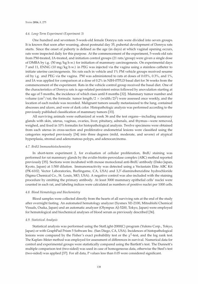
Toxins 2016, 8, 275
4.6. Long-Term Experiment (Experiment 3)
One hundred and seventeen 5-week-old female Donryu rats were divided into seven groups.It is known that soon after weaning, about postnatal day 35, pubertal development of Donryu ratsstarts. Since the onset of puberty is defined as the age (in days) at which vaginal opening occurs,rats were inspected daily for this purpose. At the commencement of the experiment, 5-week-old ratsfrom PM-treated, IA-treated, and initiation control groups (21 rats/group) were given a single doseof DMBA by i.g. (50 mg/kg b.w.) for initiation of mammary carcinogenesis. On experimental days7 and 11, ENNG (10 mg/kg b.w.) in PEG was injected via the vagina using a stainless catheter toinitiate uterine carcinogenesis. Six rats each in vehicle and 1% PM vehicle groups received sesameoil by i.g. and PEG via the vagina. PM was administered to rats at doses of 0.03%, 0.3%, and 1%,and IA was applied for comparison at a dose of 0.2% in NIH-07PLD basal diet for 36 weeks from thecommencement of the experiment. Rats in the vehicle control group received the basal diet. One ofthe characteristics of Donryu rats is age-related persistent estrus followed by anovulation starting atthe age of 5 months, the incidence of which rises until 8 months [32]. Mammary tumor number andvolume (cm3/rat; the formula: tumor length/2 × (width/2)2) were assessed once weekly, and thelocation of each nodule was recorded. Malignant tumors usually metastasized to the lung, containedabscesses and ulcers, and were of dark color. Histopathologic analysis was performed according to thepreviously published classification of mammary tumors [33].
All surviving animals were euthanized at week 36 and the test organs—including mammaryglands with skin, uterus, vaginas, ovaries, liver, pituitary, adrenals, and thymus—were removed,weighed, and fixed in 10% formalin for histopathological analysis. Twelve specimens were obtainedfrom each uterus in cross-section and proliferative endometrial lesions were classified using thecategories reported previously [34] into three degrees (mild, moderate, and severe) of atypicalhyperplasia, stromal and adenomatous polyps, and adenocarcinoma.
4.7. BrdU Immunohistochemistry
In short-term experiment 2, for evaluation of cellular proliferation, BrdU staining wasperformed for rat mammary glands by the avidin-biotin-peroxidase complex (ABC) method reportedpreviously [35]. Sections were incubated with mouse monoclonal anti-BrdU antibody (Dako Japan,Kyoto, Japan) at 1:500 dilution. Immunoreactivity was detected using a Vectastain Elite ABC Kit(PK-6102; Vector Laboratories, Burlingame, CA, USA) and 3,3′-diaminobenzidine hydrochloride(Sigma Chemical Co., St. Louis, MO, USA). A negative control was also included with the stainingprocedure by omitting the primary antibody. At least 3000 mammary epithelial cells’ nuclei werecounted in each rat, and labeling indices were calculated as numbers of positive nuclei per 1000 cells.
4.8. Blood Hematology and Biochemistry
Blood samples were collected directly from the hearts of all surviving rats at the end of the studyafter overnight fasting. An automated hematology analyzer (Sysmex XE-2100, Mitsubishi ChemicalVisuals, Osaka, Japan) and an automatic analyzer (Olympus AJ-5200, Tokyo, Japan) were employedfor hematological and biochemical analyses of blood serum as previously described [36].
4.9. Statistical Analysis
Statistical analysis was performed using the StatLight-2000(C) program (Yukms Corp., Tokyo,Japan) or with GraphPad Prism 5 Software Inc. (San Diego, CA, USA). Incidences of histopathologicallesions were compared by the Fisher’s exact probability test or the χ2-test, and the log rank test.The Kaplan–Meier method was employed for assessment of differences in survival. Numerical data forcontrol and experimental groups were statistically compared using the Bartlett’s test. The Dunnett’smultiple comparison test (two-sided) was used in case of homogeneous data, otherwise the Steel’s test(two-sided) was applied [37]. For all data, P values less than 0.05 were considered significant.
138
Toxins 2016, 8, 275
Supplementary Materials: The following are available online at www.mdpi.com/2072-6651/8/11/275/s1,Table S1: Final body, relative organ weights and chemicals intakes of Donryu rats in Exp. 3, Table S2: Results ofthe hematological and blood biochemical analyses.
Acknowledgments: This work was supported by a Grant-in-Aid for Scientific Research from the Ministry ofHealth, Labor and Welfare of Japan.
Author Contributions: A.K. and H.W. conceived and designed the experiments; A.K., M.Y. and Y.T. performedthe experiments and analyzed the data; M.Y. contributed reagents/materials/analysis tools; A.K., N.I., T.O. andM.G. wrote the paper.
Conflicts of Interest: The authors declare no conflict of interest.
References
1. Subcharoen, P. Thai traditional medicine in the new millennium. J. Med. Assoc. Thail. 2004, 87 (Suppl. S4),S52–S57.
2. Ingham, J.L.; Tabara, S.; Pope, G.S. Chemical Components and Pharmacology of the Rejuvenating Plant Puerariamirifica; Taylor and Fransis: London, UK, 2002.
3. Malaivijitnond, S. Medical applications of phytoestrogens from the Thai herb Pueraria mirifica. Front. Med.2012, 6, 8–21. [CrossRef] [PubMed]
4. Cain, J.C. Miroestrol: An oestrogen from the plant Pueraria mirifica. Nature 1960, 188, 774–777. [CrossRef][PubMed]
5. Shimokawa, S.; Kumamoto, T.; Ishikawa, T.; Takashi, M.; Higuchi, Y.; Chaichantipyuth, C.; Chansakaow, S.Quantitative analysis of miroestrol and kwakhurin for standardisation of Thai miracle herb ‘Kwao Keur’(Pueraria mirifica) and establishment of simple isolation procedure for highly estrogenic miroestrol anddeoxymiroestrol. Nat. Prod. Res. 2013, 27, 371–378. [CrossRef] [PubMed]
6. Chansakaow, S.; Ishikawa, T.; Seki, H.; Sekine, K.; Okada, M.; Chaichantipyuth, C. Identification ofdeoxymiroestrol as the actual rejuvenating principle of “Kwao Keur”, Pueraria mirifica. The known miroestrolmay be an artifact. J. Nat. Prod. 2000, 63, 173–175. [CrossRef] [PubMed]
7. Pope, G.S.; Grundy, H.M.; Jones, H.E.H.; Tait, S.A.S. The oestrogenic substance (miroestrol) from the tuberousroots of Pueraria mirifica. J. Endocrinol. 1958, 17, XV–XVI.
8. Chansakaow, S.; Ishikawa, T.; Sekine, K.; Okada, M.; Higuchi, Y.; Kudo, M.; Chaichantipyuth, C. Isoflavonoidsfrom Pueraria mirifica and their estrogenic activity. Planta Med. 2000, 66, 572–575. [CrossRef] [PubMed]
9. Tiyasatkulkovit, W.; Charoenphandhu, N.; Wongdee, K.; Thongbunchoo, J.; Krishnamra, N.;Malaivijitnond, S. Upregulation of osteoblastic differentiation marker mRNA expression in osteoblast-likeUMR106 cells by puerarin and phytoestrogens from Pueraria mirifica. Phytomedicine 2012, 19, 1147–1155.[CrossRef] [PubMed]
10. Tiyasatkulkovit, W.; Malaivijitnond, S.; Charoenphandhu, N.; Havill, L.M.; Ford, A.L.; VandeBerg, J.L.Pueraria mirifica extract and puerarin enhance proliferation and expression of alkaline phosphatase and type Icollagen in primary baboon osteoblasts. Phytomedicine 2014, 21, 1498–1503. [CrossRef] [PubMed]
11. Suthon, S.; Jaroenporn, S.; Charoenphandhu, N.; Suntornsaratoon, P.; Malaivijitnond, S. Anti-osteoporoticeffects of Pueraria candollei var. mirifica on bone mineral density and histomorphometry in estrogen-deficientrats. J. Nat. Med. 2016, 70, 225–233. [CrossRef] [PubMed]
12. Trisomboon, H.; Malaivijitnond, S.; Suzuki, J.; Hamada, Y.; Watanabe, G.; Taya, K. Long-term treatmenteffects of Pueraria mirifica phytoestrogens on parathyroid hormone and calcium levels in aged menopausalcynomolgus monkeys. J. Reprod. Dev. 2004, 50, 639–645. [CrossRef] [PubMed]
13. Cherdshewasart, W.; Panriansaen, R.; Picha, P. Pretreatment with phytoestrogen-rich plant decreases breasttumor incidence and exhibits lower profile of mammary ERalpha and ERbeta. Maturitas 2007, 58, 174–181.[CrossRef] [PubMed]
14. Lucki, N.C.; Sewer, M.B. Genistein stimulates MCF-7 breast cancer cell growth by inducing acid ceramidase(ASAH1) gene expression. J. Biol. Chem. 2011, 286, 19399–19409. [CrossRef] [PubMed]
15. Kakehashi, A.; Tago, Y.; Yoshida, M.; Sokuza, Y.; Wei, M.; Fukushima, S.; Wanibuchi, H. Hormonally activedoses of isoflavone aglycones promote mammary and endometrial carcinogenesis and alter the moleculartumor environment in Donryu rats. Toxicol. Sci. 2012, 126, 39–51. [CrossRef] [PubMed]
139
Toxins 2016, 8, 275
16. Santana, A.C.; Soares da Costa, C.A.; Armada, L.; de Paula Lopes Gonzalez, G.; dos Santos Ribeiro, M.;de Sousa dos Santos, A.; de Carvalho, J.J.; do Nascimento Saba, C.C. Fat tissue morphology of long-termsex steroid deficiency and estrogen treatment in female rats. Fertil. Steril. 2011, 95, 1478–1481. [CrossRef][PubMed]
17. Messina, M.J.; Wood, C.E. Soy isoflavones, estrogen therapy, and breast cancer risk: Analysis and commentary.Nutr. J. 2008, 7, 17. [CrossRef] [PubMed]
18. Wuttke, W.; Jarry, H.; Seidlova-Wuttke, D. Isoflavones-safe food additives or dangerous drugs?Ageing Res. Rev. 2007, 6, 150–188. [CrossRef] [PubMed]
19. Trisomboon, H.; Malaivijitnond, S.; Watanabe, G.; Taya, K. Estrogenic effects of Pueraria mirifica onthe menstrual cycle and hormone-related ovarian functions in cyclic female cynomolgus monkeys.J. Pharmacol. Sci. 2004, 94, 51–59. [CrossRef] [PubMed]
20. Trisomboon, H.; Malaivijitnond, S.; Watanabe, G.; Taya, K. Ovulation block by Pueraria mirifica: A study ofits endocrinological effect in female monkeys. Endocrine 2005, 26, 33–39. [CrossRef]
21. Trisomboon, H.; Malaivijitnond, S.; Watanabe, G.; Cherdshewasart, W.; Taya, K. The estrogenic effect ofPueraria mirifica on gonadotrophin levels in aged monkeys. Endocrine 2006, 29, 129–134. [CrossRef]
22. Trisomboon, H.; Malaivijitnond, S.; Cherdshewasart, W.; Watanabe, G.; Taya, K. Assessment of urinarygonadotropin and steroid hormone profiles of female cynomolgus monkeys after treatment withPueraria mirifica. J. Reprod. Dev. 2007, 53, 395–403. [CrossRef] [PubMed]
23. Jaroenporn, S.; Malaivijitnond, S.; Wattanasirmkit, K.; Watanabe, G.; Taya, K.; Cherdshewasart, W.Assessment of fertility and reproductive toxicity in adult female mice after long-term exposure toPueraria mirifica herb. J. Reprod. Dev. 2007, 53, 995–1005. [CrossRef] [PubMed]
24. Malaivijitnond, S.; Kiatthaipipat, P.; Cherdshewasart, W.; Watanabe, G.; Taya, K. Different effects ofPueraria mirifica, a herb containing phytoestrogens, on LH and FSH secretion in gonadectomized female andmale rats. J. Pharmacol. Sci. 2004, 96, 428–435. [CrossRef] [PubMed]
25. Malaivijitnond, S.; Chansri, K.; Kijkuokul, P.; Urasopon, N.; Cherdshewasart, W. Using vaginal cytology toassess the estrogenic activity of phytoestrogen-rich herb. J. Ethnopharmacol. 2006, 107, 354–360. [CrossRef][PubMed]
26. Cherdshewasart, W.; Kitsamai, Y.; Malaivijitnond, S. Evaluation of the estrogenic activity of the wildPueraria mirifica by vaginal cornification assay. J. Reprod. Dev. 2007, 53, 385–393. [CrossRef] [PubMed]
27. Cherdshewasart, W.; Sriwatcharakul, S.; Malaivijitnond, S. Variance of estrogenic activity of thephytoestrogen-rich plant. Maturitas 2008, 61, 350–357. [CrossRef] [PubMed]
28. Gomuttapong, S.; Pewphong, R.; Choeisiri, S.; Jaroenporn, S.; Malaivijitnond, S. Testing of the estrogenicactivity and toxicity of Stephania venosa herb in ovariectomized rats. Toxicol. Mech. Methods 2012, 22, 445–457.[CrossRef] [PubMed]
29. Lamartiniere, C.A.; Moore, J.B.; Brown, N.M.; Thompson, R.; Hardin, M.J.; Barnes, S. Genistein suppressesmammary cancer in rats. Carcinogenesis 1995, 16, 2833–2840. [CrossRef] [PubMed]
30. Freireich, E.J.; Gehan, E.A.; Rall, D.P.; Schmidt, L.H.; Skipper, H.E. Quantitative comparison of toxicity ofanticancer agents in mouse, rat, hamster, dog, monkey, and man. Cancer Chemother. Rep. 1966, 50, 219–244.[PubMed]
31. Reagan-Shaw, S.; Nihal, M.; Ahmad, N. Dose translation from animal to human studies revisited. FASEB J.2008, 22, 659–661. [CrossRef] [PubMed]
32. Nagaoka, T.; Takegawa, K.; Takeuchi, M.; Maekawa, A. Effects of reproduction on spontaneous developmentof endometrial adenocarcinomas and mammary tumors in Donryu rats. Jpn. J. Cancer Res. 2000, 91, 375–382.[CrossRef] [PubMed]
33. Costa, I.; Solanas, M.; Escrich, E. Histopathologic characterization of mammary neoplastic lesions inducedwith 7,12 dimethylbenz(alpha)anthracene in the rat: A comparative analysis with human breast tumors.Arch. Pathol. Lab. Med. 2002, 126, 915–927. [PubMed]
34. Nagaoka, T.; Takeuchi, M.; Onodera, H.; Matsushima, Y.; Ando-Lu, J.; Maekawa, A. Sequential observationof spontaneous endometrial adenocarcinoma development in Donryu rats. Toxicol. Pathol. 1994, 22, 261–269.[CrossRef] [PubMed]
35. Kakehashi, A.; Hagiwara, A.; Imai, N.; Wei, M.; Fukushima, S.; Wanibuchi, H. Induction of cell proliferationin the rat liver by the short-term administration of ethyl tertiary-butyl ether. J. Toxicol. Pathol. 2015, 28, 27–32.[CrossRef] [PubMed]
140

Toxins 2016, 8, 275
36. Kakehashi, A.; Ishii, N.; Fujioka, M.; Doi, K.; Gi, M.; Wanibuchi, H. Ethanol-extracted Brazilian propolisexerts protective effects on tumorigenesis in Wistar Hannover rats. PLoS ONE 2016, 11, e0158654. [CrossRef][PubMed]
37. Kobayashi, K.; Kanamori, M.; Ohori, K.; Takeuchi, H. A new decision tree method for statistical analysis ofquantitative data obtained in toxicity studies on rodent. Sangyo Eiseigaku Zasshi 2000, 42, 125–129. [PubMed]
© 2016 by the authors; licensee MDPI, Basel, Switzerland. This article is an open accessarticle distributed under the terms and conditions of the Creative Commons Attribution(CC-BY) license (http://creativecommons.org/licenses/by/4.0/).
141


MDPI AG
St. Alban-Anlage 66
4052 Basel, Switzerland
Tel. +41 61 683 77 34
Fax +41 61 302 89 18
http://www.mdpi.com
Toxins Editorial Office
E-mail: [email protected]
http://www.mdpi.com/journal/toxins


MDPI AG St. Alban-Anlage 66 4052 Basel Switzerland
Tel: +41 61 683 77 34 Fax: +41 61 302 89 18
www.mdpi.com ISBN 978-3-03842-379-9