Automated Identification of Subcellular Organelles by Coherent Anti-Stokes Raman Scattering
Upload
independentCategory
view
3download
0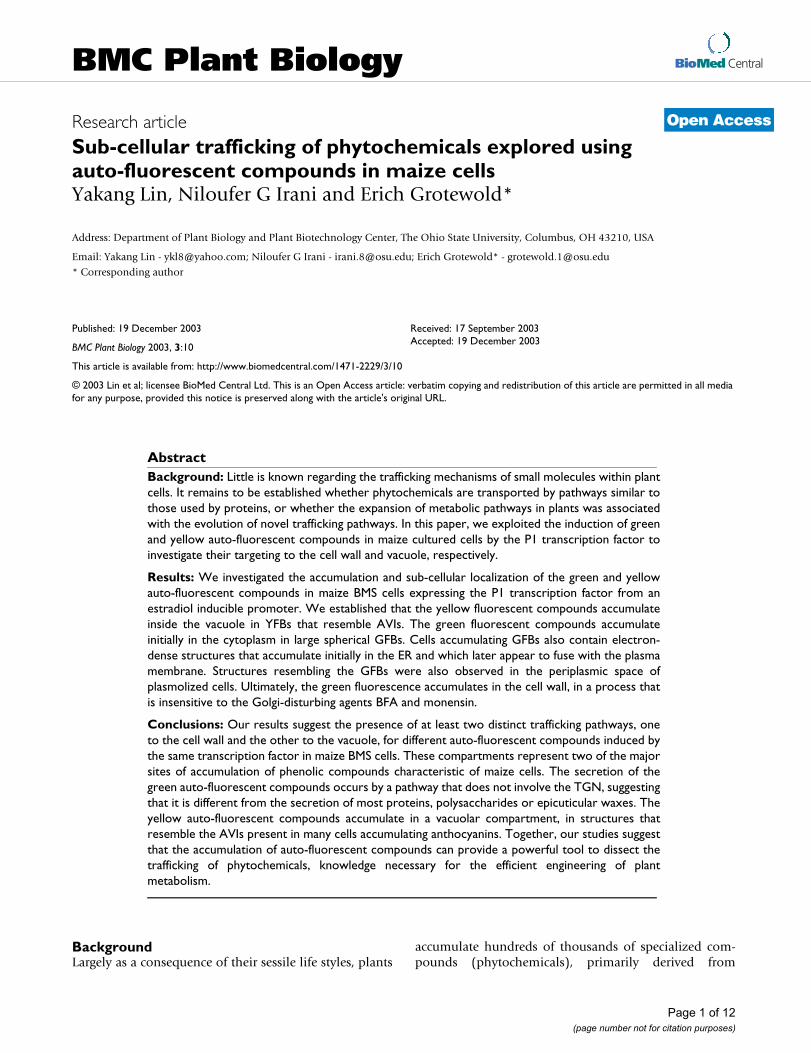
BioMed CentralBMC Plant Biology
ss
Open AcceResearch articleSub-cellular trafficking of phytochemicals explored using auto-fluorescent compounds in maize cellsYakang Lin, Niloufer G Irani and Erich Grotewold*Address: Department of Plant Biology and Plant Biotechnology Center, The Ohio State University, Columbus, OH 43210, USA
Email: Yakang Lin - [email protected]; Niloufer G Irani - [email protected]; Erich Grotewold* - [email protected]
* Corresponding author
AbstractBackground: Little is known regarding the trafficking mechanisms of small molecules within plantcells. It remains to be established whether phytochemicals are transported by pathways similar tothose used by proteins, or whether the expansion of metabolic pathways in plants was associatedwith the evolution of novel trafficking pathways. In this paper, we exploited the induction of greenand yellow auto-fluorescent compounds in maize cultured cells by the P1 transcription factor toinvestigate their targeting to the cell wall and vacuole, respectively.
Results: We investigated the accumulation and sub-cellular localization of the green and yellowauto-fluorescent compounds in maize BMS cells expressing the P1 transcription factor from anestradiol inducible promoter. We established that the yellow fluorescent compounds accumulateinside the vacuole in YFBs that resemble AVIs. The green fluorescent compounds accumulateinitially in the cytoplasm in large spherical GFBs. Cells accumulating GFBs also contain electron-dense structures that accumulate initially in the ER and which later appear to fuse with the plasmamembrane. Structures resembling the GFBs were also observed in the periplasmic space ofplasmolized cells. Ultimately, the green fluorescence accumulates in the cell wall, in a process thatis insensitive to the Golgi-disturbing agents BFA and monensin.
Conclusions: Our results suggest the presence of at least two distinct trafficking pathways, oneto the cell wall and the other to the vacuole, for different auto-fluorescent compounds induced bythe same transcription factor in maize BMS cells. These compartments represent two of the majorsites of accumulation of phenolic compounds characteristic of maize cells. The secretion of thegreen auto-fluorescent compounds occurs by a pathway that does not involve the TGN, suggestingthat it is different from the secretion of most proteins, polysaccharides or epicuticular waxes. Theyellow auto-fluorescent compounds accumulate in a vacuolar compartment, in structures thatresemble the AVIs present in many cells accumulating anthocyanins. Together, our studies suggestthat the accumulation of auto-fluorescent compounds can provide a powerful tool to dissect thetrafficking of phytochemicals, knowledge necessary for the efficient engineering of plantmetabolism.
BackgroundLargely as a consequence of their sessile life styles, plants
accumulate hundreds of thousands of specialized com-pounds (phytochemicals), primarily derived from
Published: 19 December 2003
BMC Plant Biology 2003, 3:10
Received: 17 September 2003Accepted: 19 December 2003
This article is available from: http://www.biomedcentral.com/1471-2229/3/10
© 2003 Lin et al; licensee BioMed Central Ltd. This is an Open Access article: verbatim copying and redistribution of this article are permitted in all media for any purpose, provided this notice is preserved along with the article's original URL.
Page 1 of 12(page number not for citation purposes)

BMC Plant Biology 2003, 3 http://www.biomedcentral.com/1471-2229/3/10
secondary metabolism [1]. These phytochemicals areoften synthesized in cellular compartments different fromwhere they accumulate [2]. Many phytochemicals aresecreted, either in a constitutive fashion, or in response tospecific biotic or abiotic stress conditions. The secretion ofphytochemicals has been best described for gland cells [3]and for roots [4]. In some cases, the secretion involvesspecific proteins, such as ABC transporters [5], responsi-ble for example in the secretion of antifungal terpenoidsin Nicotiana plumbaginifolia [6]. In other instances, vesiclesparticipate in the transport of phytochemicals from thesite of synthesis to the cell surface, as shown for the secre-tion of capsaicinoid vanillyl amides in placentae from redpepper [7], or for the secretion of epicuticular waxes bycork cells in sorghum [8]. In most cases, however, nomechanisms have been proposed to explain the transportof small molecules, often highly reactive or toxic, withincells, or even between cells, as observed in alkaloid bio-synthesis in C. roseus [9].
In contrast to the ease by which the transit of fluorescence-tagged proteins is followed, the study of the pathways bywhich small molecules move from one sub-cellular com-partment to another has been complicated by the difficul-ties associated with tracking these movements.Phytochemicals are difficult to observe within the cellusing immunological techniques, and pigmented com-pounds such as the flavonoid-derived anthocyaninsbecome colored only after they reach their final destina-tion, in this case the vacuole or vacuole-like compartment[10]. The anthocyanin pigments accumulate in the centralvacuole. As is the case for other phenylpropanoids and fla-vonoids, anthocyanins are likely to be synthesized on thecytoplasmic face of the endoplasmic reticulum (ER)[11,12]. The transport of anthocyanins to the vacuolarcompartment may involve specialized glutathione S-transferases that serve as flavonoid carrier proteins[13,14]. More recently, the tt12 gene from Arabidopsiswas shown to encode a member of the MATE (multidrugand toxic compound extrusion) family of transportersinvolved in the sequestration of flavonoids to the vacuoleof seed coat endothelium [15].
We have exploited the accumulation of two auto-fluores-cent compounds induced by the P1 transcription factor inBlack Mexican Sweet (BMS) maize cells in culture [16] tostart to dissect possible mechanisms by which small mol-ecules traffic within plant cells. P1 encodes a R2R3 MYBtranscription factor that regulates the accumulation of 3-deoxy flavonoids in maize floral organs [17]. The expres-sion of P1 in BMS cells resulted in the accumulation ofseveral flavonoids and phenylpropanoids [16]. P1-expressing BMS cells display a number of morphologicalchanges, including a dilatation of the ER and the accumu-lation of electron-dense structures that appear to accumu-
late originally in the ER to then migrate to the cell surfaceto fuse with the plasma membrane [16]. P1-expressingBMS cells also accumulated an increased quantity of yel-low- and green auto-fluorescent compounds targeted tothe vacuole and cell wall, respectively [16].
Here, we provide a detailed characterization of the BMScells accumulating green and yellow auto-fluorescentcompounds after the expression of P1. These compoundsaccumulate in green and yellow fluorescent bodies (GFBand YFB respectively), which are targeted to the cell walland vacuole, respectively. The YFBs accumulate in thelarge central vacuole in structures that resemble anthocy-anic vacuolar inclusions (AVIs) [18]. We show that thesecretion of the green auto-fluorescent compounds to thecell wall is not inhibited by agents that disrupt the trans-Golgi network (TGN). Together, our studies suggest thatthese auto-fluorescent compounds represent a powerfultool in dissecting the trafficking of phytochemicals, andindicate a possible novel secretion mechanism that doesnot involve the TGN network.
ResultsAccumulation of green and yellow auto-fluorescent bodies in maize BMS cellsThe large size of Black Mexican Sweet (BMS) maize cellsprovides a good opportunity to investigate the sub-cellu-lar trafficking of phytochemicals. BMS cells expressing theP1 R2R3 MYB transcription factor from the CaMV 35Spromoter (35S::P1) accumulate yellow- and green auto-fluorescent compounds targeted to the vacuole and cellwalls, respectively [16]. Because 35S::P1 cells in culturetend to suppress the expression of the P1 transgene afterextended propagation, we investigated the trafficking andsub-cellular localization of the yellow- and green auto-flu-orescent compounds in BMS cells expressing P1 from anestradiol inducible promoter (ERE::P1, [19]). The addi-tion of 10 µM estradiol to ERE::P1 BMS cells induces theaccumulation of green- and yellow auto-fluorescent com-pounds, often co-existing in the same cell (Fig. 1).
The accumulation of the green auto-fluorescent com-pounds is first observed in the cytoplasm of ERE::P1 cells2–3 days after the addition of estradiol (Fig. 2A). Six toseven days after induction, a fraction of the cells startsshowing green fluorescence on the cell surface. Interest-ingly, even after 9–10 days following the induction withestradiol, only 2% of the total cells in a culture containgreen fluorescent compounds in either the cytoplasm orthe cell surface (Fig. 2A). In contrast to the green auto-flu-orescence, the yellow auto-fluorescent compounds are fre-quently found in cells not treated with estradiol (Fig. 2B),but the induction of P1 expression increases the numberof cells accumulating these compounds (Fig. 2B), as wellas the amount of the yellow fluorescence in the cells. The
Page 2 of 12(page number not for citation purposes)

BMC Plant Biology 2003, 3 http://www.biomedcentral.com/1471-2229/3/10
fluorescent spectral properties of methanol extractsobtained from ERE::P1 BMS cells induced for 7 days werecompared to those of control BMS cells not expressing P1.Maximum fluorescence was observed at 568 nm (in arather broad peak, excitation 440 – 460 nm), consistentwith the microscopic visualization of the green and yellowfluorescence with an FITC filter [16].
Accumulation of the yellow fluorescent compounds to the central vacuoleThe yellow auto-fluorescent compounds accumulate inthe central vacuole in the form of discrete structures(YFBs, yellow fluorescent bodies) (Fig. 3A,3B). Thenumber of YFBs per vacuole is variable, with some vacu-oles having just a few (Fig. 3A), while others can have 50or more YFBs (Fig. 1). The yellow fluorescence can also befound in the cytoplasm in the form of small bodies that
Accumulation of green and yellow auto-fluorescent compounds in maize cells expressing the P1 geneFigure 1Accumulation of green and yellow auto-fluorescent compounds in maize cells expressing the P1 gene. Confocal microscopy image of a maize BMS cell expressing the P1 gene from the estradiol-inducible promoter (false colors in image). The green and yellow fluorescent bodies are indicated as GFB and YFB respectively.
Page 3 of 12(page number not for citation purposes)

BMC Plant Biology 2003, 3 http://www.biomedcentral.com/1471-2229/3/10
resemble vesicles (Fig. 1). Thus, it is tempting to speculatethat the cytoplasm is the initial site of synthesis of the yel-low auto-fluorescent compounds, which traffic to the vac-uole through vesicle-like structures.
The accumulation of the YFBs in the vacuole is intriguingand resembles the anthocyanic vacuolar inclusions (AVIs)often found in the petals of many red and blue flowers[18]. Under the epifluorescence microscope, the YFBshave a regular boundary (Fig. 3A,3B). However, transmis-sion electron microscope (TEM) micrographs show vacu-olar electron-dense bodies of a size similar to the YFBs, yetwith an irregular boundary (Fig. 3C,3D). While we sus-
pect that the fixing conditions might be responsible forthe irregular boundary in the TEM micrographs, we can-not conclusively conclude from these findings whether amembrane encircles the vacuolar YFBs or not.
Transport of the green fluorescent compounds to the cell wallSimilar to the yellow fluorescent compounds, the greenauto-fluorescent compound initially accumulates in thecytoplasm in the form of small bodies that resemble vesi-cles, which coalesce into very large GFBs (Fig. 4A,4B). Atlonger times, after the induction with estradiol, anincreased number of cells accumulate GFBs close to the
Accumulation of green and yellow auto-fluorescence in ERE::P1 cellsFigure 2Accumulation of green and yellow auto-fluorescence in ERE::P1 cells. (A) Accumulation of green fluorescence in ERE:: P1 cells after induction with estradiol. The y-axis indicates the percentage of cells with cytoplasmic GFBs (green line) or cell wall green fluorescence (orange line). (B) Accumulation of yellow auto-fluorescence in ERE::P1 cells treated (blue line) or not treated (black line) with estradiol. The y-axis indicates the percentage of cells displaying yellow fluorescence.
0
0.2
0.4
0.6
0.8
1
1.2
1.4
1 2 3 4 5 6 7 8 9 1 0
A
0
Days after induction with estradiol
% o
f ce
lls w
ith
gree
n fl
uore
scen
ce
Cytoplasmic GFBFluorescence in cell wall
B
1 2 3 4 5 6 70
1 0
2 0
3 0
4 0
5 0
6 0
7 0
% o
f ce
lls w
ith
yello
w f
luor
esce
nce
Days after induction with estradiol
+ 10 µM estradiol- estradiol
Page 4 of 12(page number not for citation purposes)

BMC Plant Biology 2003, 3 http://www.biomedcentral.com/1471-2229/3/10
cell wall (Fig. 4C), suggesting a migration of these struc-tures towards the periphery of the cells. The green fluores-cence observed in the cell walls (Fig. 4D) indicates thatthis is the ultimate destiny of the green auto-fluorescentcompounds. Consistent with these observations, proto-plasts of ERE::P1 cells induced for 7–10 days with estra-diol accumulate GFBs in the cytoplasm, but lack greenfluorescence in the cell surface (not shown). To furtherexamine the accumulation of the green auto-fluorescentcompounds in the cell surface, we took advantage of therare examples of plasmolized cells normally present inBMS cultures. When we examined a large number of estra-diol-induced ERE::P1 BMS cells, we frequently found
structures resembling the GFBs in the periplasmic space ofplasmolized cells (Fig. 5A,5B).
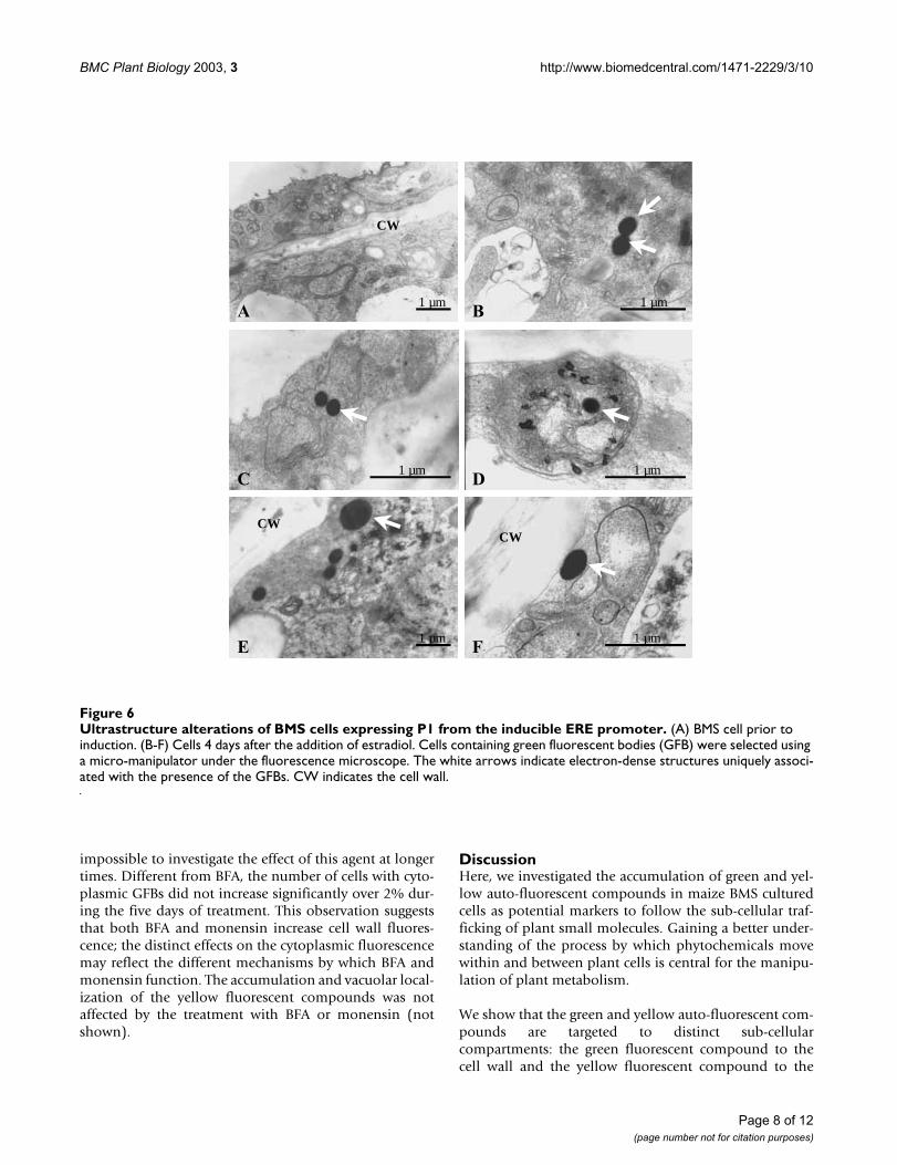
The presence of GFBs coincides with the presence of ER-derived electron-dense bodiesTo better determine the origin and the anatomy of theGFBs, cells accumulating these bodies were separated witha micromanipulator from a ERE::P1 BMS suspension cul-ture induced for 4 days with estradiol. Cells were fixed andexamined by transmission electron microscopy (TEM).Green fluorescing cells contained electron-dense sphericalbodies (indicated with arrows in Fig. 6). These electron-dense bodies, absent in cells not accumulating GFBs, arefound closely associated with structures that resemble the
Localization of the YFB to the vacuoleFigure 3Localization of the YFB to the vacuole. (A) Epifluorescence microscopy and (B) bright field image of a cell accumulating YFBs in the large central vacuole. (C) TEM micrograph showing YFBs in the central vacuole, and (D) corresponds to a higher magnification of the region in a box in (C).
A B
C D
1 µm5 µm
Page 5 of 12(page number not for citation purposes)

BMC Plant Biology 2003, 3 http://www.biomedcentral.com/1471-2229/3/10
expanded ER (Fig. 6C) characteristic of BMS over-express-ing P1, as well as close to the plasma membrane (Fig.6E,6F). The size of these electron dense bodies rarelyexceeds 300 nm (Fig. 6). Similar electron-dense structureswere previously observed in 35S::P1 cells [16]. Contrary tothe GFBs, which were sometimes observed in the periplas-mic space, the electron-dense structures were found tofuse with the plasmalemma [16].
Effect of Golgi-disturbing agents on the secretion of the green fluorescent compoundsTo determine whether the secretion of the green auto-flu-orescent compounds involves the Golgi apparatus and thetrans Golgi network (TGN), we investigated the effect of
Golgi-disturbing agents with respect to the accumulationand distribution of the green auto-fluorescent com-pounds present in induced ERE::P1 cells. Brefeldin A(BFA) blocks protein secretion by promoting the disas-sembly of the Golgi apparatus [20,21]. The treatment ofplant cells with BFA results in the complete loss of vesicleformation from the TGN and the generation of ER-Golgihybrid compartments [22]. We rationalized that, if theTGN were involved in the secretion of the green auto-flu-orescent compound to the cell wall, as is the case for thesecretion of cell wall polysaccharides [23], epicuticularwaxes [8] and proteins, then the treatment with BFAshould abolish the cell wall fluorescence.
Localization of GFBs in ERE::P1 induced BMS cellsFigure 4Localization of GFBs in ERE::P1 induced BMS cells. (A-D) Epifluorescence microscopy of cells present in ERE::P1 cells induced for 7–10 days with 10 µM estradiol representing the accumulation of GFBs in the cytoplasm (A, B), their migration to the cell surface (C) and release of green fluorescence to the cell wall (D).
A B
C D
20 µm
20 µm20 µm
20 µm
Page 6 of 12(page number not for citation purposes)

BMC Plant Biology 2003, 3 http://www.biomedcentral.com/1471-2229/3/10
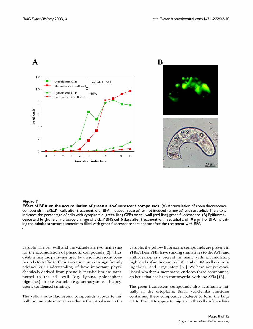
When ERE::P1 cells were treated simultaneously with 10µM estradiol and 10 µg/ml of BFA, we observed adramatic increase in the number of cells accumulatingcytoplasmic GFBs (Fig. 7A). At eight days, for example,more than 16% of the estradiol- and BFA-treated cellsaccumulated green fluorescence (Fig. 7A), compared withless than 1% of the cells treated only with estradiol (Fig.2A). The increase in green fluorescence-accumulating cellswas already evident at 2–4 days after induction and con-tinued until day ten, at which time cells started accumu-lating brown pigmentation, suggestive of cell death (notshown). Strikingly, however, the number of cells withgreen fluorescence in the cell wall increased proportion-ally, yet in a delayed fashion, to the number of cells withGFBs (Fig. 7A). The treatment of ERE::P cells with just 10µg/ml of BFA (no estradiol) resulted in a very low induc-tion of green fluorescence-accumulating cells (Fig. 7A),presumably due to the low level of P1 expression in theabsence of the hormone. In addition to the increased pres-ence of cytoplasmic and cell wall fluorescence, estradiol-induced cells treated with BFA often accumulated abnor-mal membranous structures filled with green fluorescence(Fig. 7B). These structures were only present in cells
treated with BFA and were never observed in ERE::P1 cellstreated only with estradiol. These structures may corre-spond to ER compartments filled with green fluorescentcompounds, which would further suggest the ER as thepossible site of initial accumulation for this compounds.Alternatively, the tubular organizations (Fig. 7B) may cor-respond to the sponge-like structures also observed intobacco BY-2 cells after prolonged exposure (5 hours) toBFA [22].
To conclusively establish that the integrity of the TGN isnot necessary for the secretion of the green auto-fluores-cent compound, we treated ERE::P1 cells with estradioland monensin, another TGN disturbing agent that has adifferent mechanism of action than BFA [21]. Treatmentof ERE::P1 cells with 10 µM estradiol and 10 µM mon-ensin resulted in more than 17% of the ERE::P cells accu-mulating green fluorescence in the cell wall after just 2days, up to 30% after 3 days, while in the control with just10 µM monensin (no estradiol) only 0.5% ERE::P1 cellscontained green fluorescence compound(s) in the cellwalls (not shown). However, the treatment with mon-ensin resulted in accelerated cell death, making it
Accumulation of green fluorescence in the periplasmic space in plasmolized ERE::P1 BMS cellsFigure 5Accumulation of green fluorescence in the periplasmic space in plasmolized ERE::P1 BMS cells. (A) Epifluores-cence microscopy and (B) bright field image of a plasmolyzed BMS cell accumulating two green fluorescent bodies in the peri-plasmic space.
Page 7 of 12(page number not for citation purposes)
BMC Plant Biology 2003, 3 http://www.biomedcentral.com/1471-2229/3/10
impossible to investigate the effect of this agent at longertimes. Different from BFA, the number of cells with cyto-plasmic GFBs did not increase significantly over 2% dur-ing the five days of treatment. This observation suggeststhat both BFA and monensin increase cell wall fluores-cence; the distinct effects on the cytoplasmic fluorescencemay reflect the different mechanisms by which BFA andmonensin function. The accumulation and vacuolar local-ization of the yellow fluorescent compounds was notaffected by the treatment with BFA or monensin (notshown).
DiscussionHere, we investigated the accumulation of green and yel-low auto-fluorescent compounds in maize BMS culturedcells as potential markers to follow the sub-cellular traf-ficking of plant small molecules. Gaining a better under-standing of the process by which phytochemicals movewithin and between plant cells is central for the manipu-lation of plant metabolism.
We show that the green and yellow auto-fluorescent com-pounds are targeted to distinct sub-cellularcompartments: the green fluorescent compound to thecell wall and the yellow fluorescent compound to the
Ultrastructure alterations of BMS cells expressing P1 from the inducible ERE promoterFigure 6Ultrastructure alterations of BMS cells expressing P1 from the inducible ERE promoter. (A) BMS cell prior to induction. (B-F) Cells 4 days after the addition of estradiol. Cells containing green fluorescent bodies (GFB) were selected using a micro-manipulator under the fluorescence microscope. The white arrows indicate electron-dense structures uniquely associ-ated with the presence of the GFBs. CW indicates the cell wall.
D
F
C
BA
E
CW
CWCW
1 µm 1 µm
1 µm 1 µm
1 µm 1 µm
Page 8 of 12(page number not for citation purposes)
BMC Plant Biology 2003, 3 http://www.biomedcentral.com/1471-2229/3/10
vacuole. The cell wall and the vacuole are two main sitesfor the accumulation of phenolic compounds [2]. Thus,establishing the pathways used by these fluorescent com-pounds to traffic to these two structures can significantlyadvance our understanding of how important phyto-chemicals derived from phenolic metabolism are trans-ported to the cell wall (e.g. lignins, phlobaphenepigments) or the vacuole (e.g. anthocyanins, sinapoylesters, condensed tannins).
The yellow auto-fluorescent compounds appear to ini-tially accumulate in small vesicles in the cytoplasm. In the
vacuole, the yellow fluorescent compounds are present inYFBs. These YFBs have striking similarities to the AVIs andanthocyanoplasts present in many cells accumulatinghigh levels of anthocyanins [18], and in BMS cells express-ing the C1 and R regulators [16]. We have not yet estab-lished whether a membrane encloses these compounds,an issue that has been controversial with the AVIs [18].
The green fluorescent compounds also accumulate ini-tially in the cytoplasm. Small vesicle-like structurescontaining these compounds coalesce to form the largeGFBs. The GFBs appear to migrate to the cell surface where
Effect of BFA on the accumulation of green auto-fluorescent compoundsFigure 7Effect of BFA on the accumulation of green auto-fluorescent compounds. (A) Accumulation of green fluorescence compounds in ERE::P1 cells after treatment with BFA, induced (squares) or not induced (triangles) with estradiol. The y-axis indicates the percentage of cells with cytoplasmic (green line) GFBs or cell wall (red line) green fluorescence. (B) Epifluores-cence and bright field microscopic image of ERE::P BMS cell 6 days after treatment with estradiol and 10 µg/ml of BFA indicat-ing the tubular structures sometimes filled with green fluorescence that appear after the treatment with BFA.
B
0
2
4
6
8
1 0
1 2
0 1 2 3 4 5 6 7 8 9 1 0
Days after induction
% o
f ce
lls
Cytoplasmic GFB
Fluorescence in cell wall
Cytoplasmic GFBFluorescence in cell wall
+estradiol +BFA
+BFA
A
20 µm
Page 9 of 12(page number not for citation purposes)
BMC Plant Biology 2003, 3 http://www.biomedcentral.com/1471-2229/3/10
the green fluorescent compounds are released to the cellwall. The presence of the GFBs strongly correlates with theappearance of electron-dense spherical bodies evidentunder the TEM. Similar to the GFBs, these spherical struc-tures can be found at the cell surface, even fusing to theplasma membrane. They are also present inside theexpanded ER, suggesting that the ER is probably their ini-tial accumulation site [16]. Interestingly, a recent study onArabidopsis roots determined that at least two flavonoidbiosynthetic enzymes, chalcone synthase and chalconeflavanone isomerase, are localized in electron-dense bod-ies that resemble in size and localization the electron-dense structures observed here [24]. Similar vesicle-likestructures have been also previously observed for thelocalization of a flavonol O-glucosyltransferase in Chrysos-plenium americanum cells [25], and for Primula kewensis'chalcone synthase [26]. These findings open the possibil-ity that cytoplasmic structures resembling the electron-dense bodies described here function not only as the sitesof storage or transport of specific phytochemicals, but alsoparticipate in specific steps in their synthesis, similar towhat has been found for alkaloid vesicles [27].
While it is tempting to speculate that electron-dense struc-tures as observed in the TEMs and the GFBs, so farobserved only in P1-expressing cells, are one and thesame, their sizes are strikingly different. The GFBs areoften up to 3 µm in diameter, while the spherical electron-dense structures are rarely larger than 0.3 µm in size. Wecannot rule out the possibility that the size difference is aconsequence of sample fixation for the TEM studies. Alter-natively, the GFBs and the electron-dense bodies may cor-respond to different structures induced by the expressionof P1 or by the accumulation of P1-regulated compounds.
The treatment of ERE::P1 cells with estradiol and the TGNdisturbing agents BFA or monensin does not result in aninhibition of the accumulation of the green fluorescencein the cell wall, as it would be expected if the secretion ofthese compounds follows a pathway similar to proteins,i.e. through the TGN. Rather, the accumulation of thegreen fluorescence increases dramatically in the cell wall.An enzymatic polymerization or degradation of the greenfluorescent compounds in the cell wall could explain theincrease in cell wall fluorescence in the presence of BFA ormonensin. The secretion of the enzyme responsible forthis would be blocked by BFA and monensin, resulting inthe observed increase in cell wall green fluorescence. Incontrast, the accumulation of the yellow fluorescent com-pounds in the vacuole is not increased by BFA or mon-ensin, suggesting that the transport of the green andyellow fluorescent compounds to the cell wall and thevacuole respectively, occurs by different mechanisms.
The results presented here suggest the existence of a secre-tion pathway in BMS cells for the green fluorescent com-pounds that does not require the TGN. Structural analyseshave previously suggested the existence of secretion path-ways that involve the direct fusion of the ER to the plasmamembrane, or of vesicles derived from the ER that do notappear to involve the TGN [3]. What is the nature of theGFBs present in the BMS cells expressing P1? Based on ourcurrent knowledge, we cannot rule out the formal possi-bility that the GFBs are proteinaceous or crystalline innature. Indeed, we have been so far unable to isolate thesebodies by differential centrifugation from gently lysedprotoplasts, in conditions under which the YFBs retaintheir identity and fluorescent properties (not shown).However, the observation of GFBs in the periplasmicspace of induced ERE::P1 BMS cells resembles the secre-tion of exosomes by many animal cells in culture [28].Exosomes are believed to derive from late multi-vesicularcompartments such as endosomes or the secretory lyso-somes characteristic of dendritic and some hemapoieticcells [29]. A possible origin of the GFBs from an endo-somal or vacuolar compartment would be consistent withthe observed insensitivity to BFA and monensin, as asecretion pathway originating from these compartmentswould be after the Golgi block caused by the drugs. Alter-natively, the GFBs could correspond to some specializeddomain of the ER that has the ability to fuse with theplasma membrane. Determining the identity of the GFBsand the presence of similar structures in cells secretingother metabolites should provide fundamental informa-tion regarding the mechanisms used by plants for the traf-ficking of phytochemicals to the cell surface.
ConclusionsThe green and yellow auto-fluorescent compounds arepotentially powerful tools to investigate the mechanismsby which specialized compounds (phytochemicals) trafficwithin plant cells, from their site of synthesis to the com-partment where they ultimately accumulate. While thechemical identity and the site of synthesis of these com-pounds remain to be established, it is evident that the pos-sibility to follow these compounds within the cell by theirfluorescing properties will allow the isolation and bio-chemical characterization of the structures in which theyaccumulate. The secretion of the green fluorescent com-pounds evidently occurs by a mechanism distinct fromthe secretion of proteins. Whether this involves a special-ized vesicular system is currently under investigation.Establishing the pathways involved in the sub-cellulartrafficking of phytochemicals is essential for the successfulengineering of plant metabolism.
Page 10 of 12(page number not for citation purposes)

BMC Plant Biology 2003, 3 http://www.biomedcentral.com/1471-2229/3/10
MethodsTreatment of BMS cells with estradiol and Golgi disturbing agentsBMS cells were grown and maintained in the conditionspreviously described [30]. Newly sub-cultured cells wereused for treatments of estradiol and Golgi disturbingagents. For the induction with estradiol, 25 µl of a stocksolution of 20 mM β-estradiol 17-propionate (Sigma) inethanol was added to 50 ml of BMS cell culture suspen-sion. For the treatments with the Golgi disturbing agents,stock solutions of 20 mg/ml of BFA (Sigma) and 20 mMof monensin (Sigma) were prepared in ethanol. The treat-ment with BFA was done at a final concentration of 10 µg/ml and monensin at 10 µM. For microscopyexaminations, 0.5–1 ml of cell culture was taken out every24 hours after treatment.
Microscopy methodsThe BMS cells were diluted to 100,000 cells/ml with cellculture medium. For epifluorescence experiments, 50 µlof diluted cells were loaded on slides with a 20 × 40 mmcover slip. The cells containing GFBs or YFBs were countedand photographed under a Zeiss Axiovert-100 fluorescentmicroscope coupled to a Bio-Rad MRC-1024 imaging sys-tem, using a blue filter adequate for FITC detection. Pic-tures images were taken using Kodak 35 mm slide film(160 T). At least 10,000 living cells were observed in eachsample.
Electron MicroscopyThe cells containing YFBs and GFBs were picked up usinga micro-manipulator (MMO-203, Narishige) under thefluorescence microscope. After double fixations in 2.5%glutaraldehyde and 1% OsO4, the cells were embedded inSpurr's Low Viscosity resin. Ultrathin sections were pre-pared with an ultramicrotome (Reichert Ultracut E). Thesections were mounted on 100 mesh nickel grids coatedwith Formvar film and stained with 1% uranyl acetate for10 min and 0.3% lead citrate for 5 min. Observation andphotography were conducted using a Philips 301 trans-mission electron microscope.
List of abbreviationsAVI, anthocyanin vacuolar inclusions; BFA, Brefeldin A;BMS, Black Mexican Sweet; ER, endoplasmic reticulum;GFB, green fluorescent body; TEM, transmission electronmicroscope; TGN, trans Golgi Network; YFB, yellow fluo-rescent body.
Authors' contributionsY.L. carried out all the microscopy experiments of ERE::P1cells. N.I. investigated the effect of BFA on the TGN net-work in BMS cells. E.G. was involved in the design andsupervision of this project and drafted the manuscript.
AcknowledgmentsWe thank John Rogers for suggestions at the beginning of these studies, Biao Ding and George Heine for comments on this manuscript. We acknowledge the help of Troy Paddock with the chemical analysis of the flu-orescent compounds. This work was supported by a grant from the National Science Foundation to E.G. (MCB-0130062). Correspondence should be addressed to E.G.
References1. Verpoorte R: Plant secondary metabolism. In Metabolic engineer-
ing of plant secondary metabolism Edited by: Verpoorte R, Alfermann AW.Kluwer Academic Publishers, Dordrecht; 2000:1-29.
2. Grotewold E: Subcellular trafficking of phytochemicals. Rec ResDevel Plant Physiol 2001, 2:31-48.
3. Kronestedt-Robards E, Kronestedt AW: Exocytosis in gland cells Cam-bridge Univ. Press, U.K; 1991.
4. Walker TS, Bais HP, Grotewold E, Vivanco JM: Root exudation andrhizosphere biology. Plant Physiol 2003, 132:44-51.
5. Theodoulou FL: Plant ABC transporters. Biochim Biophys Acta2000, 1465:79-103.
6. Jasinski M, Stukkens Y, Degand H, Purnelle B, Marchand-Brynaert J,Boutry M: A plant plasma membrane ATP binding cassette-type transporter is involved in antifungal terpenoidsecretion. Plant Cell 2001, 13:1095-1107.
7. Zamski E, Shoham O, Palevitch D, Levy A: Ultrastructure of capa-icinoid-secreting cells in pungent and nonpungent red pep-per (Capsicum anuum L.) cultivars. Bot Gaz 1987, 148:1-6.
8. Jenks MA, Rich PJ, Ashworth EN: Involvement of cork cells in thesecretion of epicuticular wax filaments on Sorghum bicolor(L.) moench. Int J Plant Sci 1994, 155:506-518.
9. St.-Pierre B, Vazquez-Flota F, De Luca V: Multicellular compart-mentation of Catharanthus roseus alkaloid biosynthesis pre-dicts intercellular translocation of a pathway intermediate.Plant Cell 1999, 11:887-900.
10. Stafford HA: Flavonoid metabolism CRC Press, Inc, Boca Raton, USA;1990.
11. Hrazdina G, Wagner GJ: Metabolic pathways as enzyme com-plexes: evidence for the synthesis of phenylpropanoids andflavonoids on membrane associated enzyme complexes. ArchBiochem Biophys 1985, 237:88-100.
12. Winkel-Shirley B: Evidence of enzyme complexes in the phe-nylpropanoid and flavonoid pathways. Physiol Plant 1999,107:142-149.
13. Marrs KA, Alfenito MR, Lloyd AM, Walbot V: A glutathione S-transferase involved in vacuolar transfer encoded by themaize gene bronze 2. Nature 1995, 375:397-400.
14. Mueller LA, Goodman CD, Silady RA, Walbot W: AN9, a petuniaglutathione S-transferase required for anthocyanin seques-tration, is a flavonoid-binding protein. Plant Physiol 2000,123:1561-1570.
15. Debeaujon I, Peeters AJM, Leon-Kloosterziel KM, Koornneef M: TheTRANSPARENT TESTA12 gene of Arabidopsis encodes a multi-drug secondary transporter-like protein required for flavo-noid sequestration in vacuoles of the seed coat endothelium.Plant Cell 2001, 13:853-871.
16. Grotewold E, Chamberlain M, St Claire G, Swenson J, Siame BA, But-ler LG, Snook M, Bowen B: Engineering secondary metabolismin maize cells by ectopic expression of transcription factors.Plant Cell 1998, 10:721-740.
17. Grotewold E, Drummond B, Bowen B, Peterson T: The Myb-homologous P gene controls phlobaphene pigmentation inmaize floral organs by directly activating a flavonoid biosyn-thetic gene subset. Cell 1994, 76:543-553.
18. Markham KR, Gould KS, Winefield CS, Mitchell KA, Bloor SJ, BoaseMR: Anthocyanic vacuolar inclusions – their nature and signif-icance in flower colouration. Phytochemistry 2000, 55:327-336.
19. Bruce W, Folkerts O, Garnaat C, Crasta O, Roth B, Bowen B:Expression profiling of the maize flavonoid pathway genescontrolled by estradiol-inducible transcription factors CRCand P. Plant Cell 2000, 12:65-80.
20. Klausner RD, Donaldson JG, Lippincott-Schwartz J: Brefeldin A:Insights into the control of membrane traffic and organellestructure. J Cell Biol 1992, 116:1071-1080.
Page 11 of 12(page number not for citation purposes)
BMC Plant Biology 2003, 3 http://www.biomedcentral.com/1471-2229/3/10
Publish with BioMed Central and every scientist can read your work free of charge
"BioMed Central will be the most significant development for disseminating the results of biomedical research in our lifetime."
Sir Paul Nurse, Cancer Research UK
Your research papers will be:
available free of charge to the entire biomedical community
peer reviewed and published immediately upon acceptance
cited in PubMed and archived on PubMed Central
yours — you keep the copyright
Submit your manuscript here:http://www.biomedcentral.com/info/publishing_adv.asp
BioMedcentral
21. Dinter A, Berger EG: Golgi-disturbing agents. Histochem Cell Biol1998, 109(5-6):571-590.
22. Ritzenthaler C, Nebenfuhr A, Movafeghi A, Stussi-Garaud C, BehniaL, Pimpl P, Staehelin LA, Robinson DG: Re-evaluation of theeffects of brefeldin A on plant cells using tobacco Bright Yel-low 2 cells expressing Golgi-targeted green fluorescent pro-tein and COPI antisera. Plant Cell 2002, 14:237-261.
23. Driouich A, Faye L, Staehelin LA: The plant Golgi apparatus: afactory for complex polysaccharides and glycoproteins.Trends Biochem Sci 1993, 18:210-214.
24. Saslowsky D, Winkel-Shirley B: Localization of flavonoidenzymes in Arabidopsis roots. Plant J 2001, 27:37-48.
25. Latchinian-Sadek L, Ibrahim RK: Flavonol ring B-specific O-gluco-syltransferases: purification, production of polyclonal anti-bodies, and immunolocalization. Arch Biochem Biophys 1991,289:230-236.
26. Schopker H, Kneisel M, Beerhues L, Robenek H, Wiermann R: Phe-nylalanine ammonia-lyase and chalcone synthase in glands ofPrimula kewensis (Wats, W)-Immunofluorescence andimmunogold localization. Planta 1995, 196:712-719.
27. Facchini PJ: Alkaloid biosynthesis in plants: biochemistry, cellbiology, molecular regulation, and metabolic engineeringapplications. Annu Rev Plant Physiol Plant Mol Biol 2001, 52:29-66.
28. Thery C, Zitvogel L, Amigorena S: Exosomes: composition, bio-genesis and function. Nat Rev Immunol 2002, 2(8):569-579.
29. Andrews NW: Regulated secretion of conventionallysosomes. Trends Cell Biol 2000, 10:316-321.
30. Dias A, Brown J, Bonello P, Grotewold E: Metabolite profiling asa functional genomics tool. In Plant Functional Genomics: Methodsand Protocols Volume 236. Edited by: Grotewold E. Humana Press, NJ;2003:415-426.
Page 12 of 12(page number not for citation purposes)
Copyright © 2022 FDOKUMEN