Biological Properties of Dietary Micronutrients: Antioxidant ...
129
Biological Properties of Dietary Micronutrients: Antioxidant Capacity and Structure-Activity Relationships INAUGURAL–DISSERTATION zur Erlangung des Doktorgrades der Mathematisch-Naturwissenschaftlichen Fakultät der Heinrich-Heine-Universität Düsseldorf vorgelegt von Alejandro José Betancor Fernández aus Las Palmas de Gran Canaria (Spanien) Düsseldorf, November 2003
-
Upload
khangminh22 -
Category
Documents
-
view
1 -
download
0
Transcript of Biological Properties of Dietary Micronutrients: Antioxidant ...

Biological Properties of Dietary Micronutrients:
Antioxidant Capacity and Structure-Activity Relationships
INAUGURAL–DISSERTATION
zur Erlangung des Doktorgrades der Mathematisch-Naturwissenschaftlichen Fakultät
der Heinrich-Heine-Universität Düsseldorf
vorgelegt von
Alejandro José Betancor Fernández
aus Las Palmas de Gran Canaria (Spanien)
Düsseldorf, November 2003

Gedruckt mit der Genehmigung der Mathematisch-Naturwissenschaftlichen
Fakultät der Heinrich-Heine-Universität Düsseldorf.
Referent: Prof. Dr. Dr. h. c. Helmut Sies
Korreferent: Prof. Dr. Peter Proksch
Tage der mündlichen Prüfungen: 4.12.03, 8.12.03 und 10.12.03

To my parents / A mis padres

INDEX OF CONTENTS
1. INTRODUCTION
1.1 Free Radicals, Reactive Oxygen Species and Disease 1
1.2 The Oxidative Modification Hypothesis of Atherosclerosis 2
1.2.1 Oxidation of LDL 3
1.2.2 Degree of LDL Oxidation and Biological Activity 6
1.3 Evidence for an Increased Oxidative Status in
Atherosclerotic or CVD Patients 7
1.4 The Role of Antioxidants in the Prevention of
Atherosclerosis; Human Studies 8
1.5 Dietary Micronutrients with Antioxidant and Further Anti-
atherogenic Properties 11
1.5.1 Vitamin E 11
1.5.2 Carotenoids 17
1.5.3 Polyphenols 21
1.6 Aim of the Study 26
2. MATERIALS AND METHODS
2. 1 Chemicals 28
2. 2 Instruments 30
2. 3 Preparation of Stock Solutions 31
2. 4 Antioxidant Activity of Dietary Micronutrients in Model
Systems 32
2. 4. 1 Trolox Equivalent Antioxidant Capacity (TEAC ) 32
2. 4. 2 Oxygen Radical Absorption Capacity (ORAC) 35
2. 4. 3 Inhibition of Nitration of Tyrosine by Peroxynitrite 37
2. 5 Cell Culture Study on Anti-atherogenic Effects of Dietary
Micronutrients and α-CEHC 37
2. 5. 1 LDL Preparation and Oxidation 38
2. 5. 2 Cell Culture 38
2. 5. 3 Cytotoxicity 39
2. 5. 4 Uptake of oxLDL 40

2. 5. 5 Short-term Variations of the Redox State 41
2. 5. 6 Long-term Variations of the Redox State 41
2. 5. 7 Apoptotic Cell Death 43
2. 5. 8 Protein Measurement 44
2. 6 Human Intervention Study on the Effects of Smoking
on Vitamin E Metabolism after a Single Dose of Vitamin E 44
2. 6. 1 Study Design 44
2. 6. 2 Determination of α- and γ-CEHC in Serum 44
2. 6. 3 Determination of α- and γ-Tocopherol in Serum 46
2. 7 Statistics 46
3. RESULTS
3. 1 Antioxidant Activity of Dietary Micronutrients in Model
Systems 48
3. 1. 1 Trolox Equivalent Antioxidant Capacity (TEAC ) 48
3. 1. 2 Oxygen Radical Absorbance Capacity (ORAC) 57
3. 1. 3 Inhibition of Tyrosine Nitration by Peroxynitrite 59
3. 2 Cell Culture Study on Anti-atherogenic Effects of Dietary
Micronutrients and α-CEHC 61
3. 2. 1 Cytotoxicity 61
3. 2. 2 Uptake of oxLDL 65
3. 2. 3 Short-term Variations of the Redox State 67
3. 2. 4 Long-term Variations of the Redox State 68
3. 2. 5 Apoptotic Cell Death 72
3. 3 Human Intervention Study on the Effects of Smoking
on Vitamin E Metabolism after a Single Dose of Vitamin E 74
3. 3. 1 HPLC Analysis of α- and γ-CEHC in Serum 74
3. 3. 2 HPLC Analysis of α- and γ-Tocopherol 75
3. 3. 3 General Remarks 76
3. 3. 4 Differences between Smokers and Nonsmokers
in Vitamin E Metabolism 79
3. 3. 5 Further Data of Relevance 80

4. DISCUSSION
4. 1 Antioxidant Activity of Dietary Micronutrients in Model Systems 84
4. 1. 1 TEAC and ORAC Assays 84
4. 1. 2 Methodological Aspects 88
4. 1. 3 Inhibition of Peroxynitrite-mediated Tyrosine Nitration 90
4. 2 Cell Culture Study on the Anti-atherogenic Effects of Dietary
Micronutrients and α-CEHC 91
4. 3 Human Intervention Study on the Effects of Smoking on
Vitamin E Metabolism after a Single Dose of Vitamin E 93
5. SUMMARY 97
6. BIBLIOGRAPHY 99

Acronyms and conventional names
4-HNE 4-hydroxynonenal
5-HpETE 5-hydroperoxy-eicosatetraenoic acid
AAPH 2,2´-azobis(2-amidinopropane) hydrochloride
ABTS 2,2´-azino-bis(3-ethylbenzthiazoline-6-sulfonic acid
Ac-DEVD-pNA acetyl-Asp-Glu-Val-Asp-para-nitroanilide
ATP adenosine triphosphate
AUC area under the curve
AVED ataxia with vitamin E deficiency (familial isolated vitamin E
deficiency)
BHT 2,6-di-tert-butyl-4-methylphenol
CE cholesterol esters
CHD coronary heart disease
CVD cardiovascular disease
DCF 2´,7´-dichlorofluorescein
DCFH-DA 2´,7´-dichlorodihydrofluorescein diacetate
DEVD-CHO Asp-Glu-Val-Asp-aldehyde
DiI 1,1´-dioctadecyl-3,3,3´,3´-tetramethyl-indocarbocyanine
perchlorate
DMSO dimethyl sulfoxide
DTNB 5,5´-dithio-bis(2-nitrobenzoic acid)
DTT 1,4-dithio-threitol
EC (-)-epicatechin
EDTA ethylene-diamine-tetraacetic acid
EGCG epigallocatechin gallate
FCS fetal calf serum
FRAP ferric reducing ability of plasma
GAE gallic acid equivalents
GJC gap junction (intercellular) communication
GSH reduced glutathione
GSSG glutathione disulfide
HDL high density lipoproteins
HEPES 4-(2-hydroxyethyl)-1-piperazineethanesulfonic acid
HNBA 4-hydroxy-3-nitrobenzoic acid

IC50 half-maximal inhibitory concentration
IDL intermediate density lipoproteins
IHD ischemic heart disease
Kpi potassium phosphate buffer
LDL low density lipoprotein
MAEC (primary) mouse aorta endothelial cells
MDA malondialdehyde
MI myocardial infarction
MM-LDL minimally-modified LDL
MTT 3-(4,5-dimethylthiazole-2-yl)-2,5-diphenyl tetrazolium
NADPH nicotinamide-adenine dinucleotide phosphate, reduced
nLDL native LDL
ORAC oxygen radical absorption capacity
oxLDL oxidized LDL
PBS phosphate buffered saline
PMSF phenylmethylsulfonylfluorid
PNA para-nitroanilide
PUFA polyunsaturated fatty acids
RNS reactive nitrogen species
ROS reactive oxygen species
SDS sodium dodecyl sulfate
SMC smooth muscle cells
SSA 5-sulfosalicylic acid
TBARS thiobarbituric acid-reactive substances
TE Trolox equivalents
TEAC Trolox equivalent antioxidant capacity
TEAH tetraethylammonium hydroxide
TG triglycerides
TNB 5-thio-2-nitrobenzoic acid
TP total phenols
TPA 12-O-tetradecanoylphorbol-13-acetate
TRAP total peroxyl radical trapping parameter
Trolox 6-hydroxy-2,5,7,8-tetramethylchroman-2-carboxylic acid
VLDL very-low density lipoprotein

VP 2-vinyl-pyridine
α-CEHC 2,5,7,8-tetramethyl-2-(2´-carboxyethyl)-6-hydroxychroman
α-TTP α-tocopherol transfer protein
γ-CEHC 2,7,8-trimethyl-2-(2´-carboxyethyl)-6-hydroxychroman
Reactive Species
H2O2 hydrogen peroxide .OH hydroxyl radical
NO nitric oxide
ONOO- peroxynitrite
O2.- superoxide radical anion
Aminoacid Codes
Asp aspartate
Glu glutamate
Val valine

1. Introduction
1
1. INTRODUCTION
1. 1 Free Radicals, Reactive Oxygen Species and Disease
Reactive oxygen species (ROS) and reactive nitrogen species (RNS) are
generated in cells in the normal course of metabolism. Some, but not all, of these
ROS/RNS are free radicals, i. e., entities which contain one or more unpaired
electrons but are capable of independent existence. The main site for ROS
production is the mitochondrial electron transport system, where the superoxide
anion radical (O2.-) is generated and, following its dismutation, hydrogen peroxide
(H2O2) [Chance et al., 1979]. In the presence of transition elements like iron, hydroxyl
radical (.OH) generation may follow as a result of the Fenton chemistry. Other
sources of O2.- are the peroxisomal fatty acid metabolism and the detoxication from
xenobiotics via microsomal P450 enzymes [Sies, 1986]. The latter are a minor source
of ROS in physiological conditions, but may be an important generator of oxidants in
some specific subjects (e.g. smokers).
Nitric oxide (NO) is the most important RNS in vivo as it is an endogenous
regulator of several cellular functions (e. g. vasodilation, neurotransmission and roles
in the immune system) [Ignarro, 1990]. Under physiological conditions, the
generation of nitrosating species such as S-nitroso-glutathione or other S-
nitrosothiols (RSNO) is likely and such compounds may participate in trans-
nitrosation reactions [Al-Mustafa et al., 2001]. Key chemical reactions can, on the
other hand, lead to more reactive species, potentially more toxic than NO. Reaction
of NO with O2.- yields the highly toxic peroxynitrite (ONOO-). Steady-state
concentrations of ONOO- may be significant in the vicinity of activated macrophages,
which generate NO and O2.- concomitantly [Fukuto, 1995].
Oxidative stress has been defined as an imbalance between oxidants
(ROS/RNS) and antioxidants in favour of the former, leading to damage [Sies, 1985].
Experimental evidence largely supports a role for oxidative stress in the
pathogenesis and/or progression of a number of diseases [Sies, 1991]. It is involved
in tumor initiation, promotion, and progression, plays a role in reperfusion injury after
ischemia, and, in inflammatory disorders such as rheumatoid arthritis, phagocytic
cells (mainly neutrophils) are steadily activated and release O2.-, H2O2 and the highly
oxidising hypohalous acids.

1. Introduction
2
Various cellular signaling pathways and the expression of certain genes are also
oxidation-sensitive. ROS may thereby elicit changes in functional processes of the
cell that ultimately lead to apoptosis, to increased proliferation, or to immune or other
responses guiding to cellular malfunction. Redox-sensitive transcription factors in
mammalian cells include activating protein (AP)-1 and nuclear factor (NF)-κB [Abate
et al., 1990; Schreck et al., 1991]. Signaling pathways that are activated by stress
stimuli including oxidative stress include the mitogen-activated protein kinases
(MAPK) -comprising the extracellular signal-regulated kinases (ERK) 1 and 2, the
p38 subgroup and the c-Jun-N-terminal kinases (JNK)- and the phosphoinositide-3
kinase (PI3K)/Akt pathway [Klotz, 2002]. In rat liver epithelial cells ONOO- induced an
immediate activation of p38 and a progressive activation of ERK1/2 and JNK;
activation was attenuated by selenite supplementation, suggesting a protective role
of glutathione peroxidase in vivo [Schieke et al., 1999]. Besides, ONOO- elicited a
decrease in gap junction intercellular communication (GJC) likely via ERK activation
[Sharov et al., 1999]. Singlet oxygen (1O2), which may be generated photochemically
by visible or UV light irradiation on photosensitizers such as porphyrins, was found to
activate p38 and JNK in similar manner to UVA in skin fibroblasts [Klotz et al., 1999].
1. 2 The Oxidative Modification Hypothesis of Atherosclerosis
Atherosclerosis is a progressive occlusion of large and medium-sized arteries,
basically consisting in the thickening of the intima (most inner layer of arteries) due to
a combination of fibrosis, smooth muscle cell proliferation, and the development of an
extracellular lipid core. In its more advanced stages all three coats of the arterial wall
(tunica intima, tunica media, and tunica adventitia) become affected. The luminal side
of the intima, a monolayer of endothelial cells (endothelium), loses its antithrombotic
properties under this pathological state and the affected zone is prone to thrombus
formation [Navab et al., 1995].
At present, atherosclerosis is accepted as the primary mechanism leading to
cardiovascular diseases (CVD), the main cause of death in most developed
countries. Various risk factors for atherosclerosis have been identified, such as fat-
rich diets, sedentary lifestyles, hypercholesterolemia, hypertension, smoking,
diabetes mellitus, and family history of premature coronary heart disease (CHD)
[Vogel, 1997].

1. Introduction
3
Figure 1. 1 Atherosclerotic artery showing intima thickening (due to foam cell and lipid accumulation)
and thrombus formation.
The key determinants of the initiation of early lesions are [Schwartz et al., 1993]:
i) a local hemodynamic environment with domains of low shear and reverse flow; this
enhances localised intimal influx and accumulation of plasma lipoproteins, mainly
low-density lipoprotein (LDL) and lipoprotein a -Lp(a)-;
ii) increased net intimal oxidative stress;
iii) focal monocyte recruitment to the arterial intima;
iv) monocyte/macrophage activation in the intima leading to foam cell formation.
1. 2. 1 Oxidation of LDL
LDL is the fraction of human lipoproteins with a buoyant density between 1.019
and 1.063 kg/L. A particle of LDL has a mean molecular mass of 2.5.106 D and a
mean diameter of 20 nm. Approximately 50% of the mass of LDL is cholesterol,
making the particle the major sterol transporter in the circulation [Esterbauer at al.,
1992]. 80% of the cholesterol is esterified, primarily with linoleic acid. The more

1. Introduction
4
lipophilic core of the particle is formed by cholesterol esters (CE) and tryglicerides
(TG), corresponding to 41 and 3% of the total LDL mass, respectively, and free
cholesterol, phospholipids and proteins constitute the particle surface in direct
contact with plasma (Figure 1.2). About 66% of the cholesterol carried by LDL is
returned to the liver, the rest is transported to extrahepatic tissues. Uptake of LDL in
all tissues is regulated by the LDL or apo B/E receptor, a 96 kD protein which
appears in the cytoplasmic membranes of cells in response to their need for
cholesterol. The major apolipoprotein in LDL, apo B100, is located at the surface and
recognized by the LDL receptor. LDL is then endocyted via so-called coated pits into
vesicles formed by the protein clathrin, with subsequent fusion of endosomes and
lysosomes, apolipoprotein digestion and cholesterol de-esterification [Löffler, 2002].
Figure 1. 2 Structure of an LDL particle showing the core and surface components
LDL passes readily across the endothelium. Within the subendothelial space,
LDL gets trapped into extracellular matrix proteins and is exposed to radical-triggered
oxidative alterations by surrounding cells, including endothelial cells, smooth muscle
cells (SMC), and infiltrated macrophages. Candidate effectors of LDL oxidation in the
artery wall are 15-lipoxygenase and enzymes present in neutrophils and endothelial
cells, such as myeloperoxidase, NAD(P)H oxidase, and NO synthase, together with
traces of metal ions [Esterbauer et al., 1992].
A large number of procedures have been applied to obtain LDL with the same
oxidative modifications in vitro. LDL incubated in plain cell medium with 5 µmol/L
copper (II) was found to have the same chemical and biological properties as the
phospholipid
apo B100
cholesteryl ester
triglyceride
free cholesterol
phospholipid
apo B100
cholesteryl ester
triglyceride
free cholesterol

1. Introduction
5
LDL modified by incubation with rabbit aortic endothelial cells in the same medium
(containing traces of iron and copper) for the same time period [Steinbrecher et al.,
1984]. The process was inhibited by chelating agents, confirming the requirement of
trace elements. Thus, LDL oxidated with copper (II) has been widely employed since
then to assess biological activities of oxidatively-modified LDL in cellular and animal
experiments.
In isolated LDL, peroxidation of lipids by copper (II) further required the presence
of pre-formed lipid hydroperoxides [Thomas and Jackson, 1991]. Thus, the
mechanism underlying this type of LDL oxidation appears to be the oxidative and
reductive decomposition of peroxides by transition elements yielding peroxyl (LOO.)
and alcoxyl (LO.) radical species [Rice-Evans et al., 1996]:
LOOH + Cu2+ LOO. + Cu+ + H+
LOOH + Cu+ LO. + Cu2+ + OH-
This leads to the propagation of radical attack on the polyunsaturated fatty acids
(PUFA) yielding new peroxides, until all PUFA are consumed.
In vivo, cells may produce a range of redox reagents which can react with LDL
directly or reduce any transition metal present, facilitating lipid peroxide
decomposition and subsequent uncontrolled PUFA peroxidation. Injury to cells and
tissues may enhance the toxicity of ROS by releasing metal ions from storage sites,
decompartmentalized hem proteins or metalloproteins by interaction with accessible
proteases or oxidants [Rive-Evans et al., 1996].
The first barrier preventing lipid peroxidation is that formed by the antioxidants
within the LDL particle, including vitamin E and carotenoids. During a first lag phase,
in which conjugated dienes are not detected and only minimal lipid peroxidation
occurs, LDL becomes depleted of antioxidants, with α-tocopherol as the first and β-
carotene as the last one. Conjugated dienes then result from oxidation of PUFA with
isolated double bonds to PUFA hydroperoxides with conjugated double bonds, which
may be detected by UV-spectrophotometry at 234 nm. Finally, lipid hydroperoxides
decompose to aldehydes and other products [Esterbauer et al., 1992].

1. Introduction
6
1. 2. 2 Degree of LDL Oxidation and Biological Activity
Oxidatively-modified LDLs have been categorized in minimally-modified LDL
(MM-LDL) and extensively oxidized LDL (oxLDL) in view of a number of different
biological activities attributed to each. However, it should be noted that up to date no
consensus concerning the differentiation between both types of oxidized LDL has
been reached, and even nomenclature differs in the literature.
From a functional aspect, properties such as moderate cytotoxicity, induction of
smooth muscle cell (SMC) proliferation, increase of monocyte adhesion to
endothelium, or stimulation of monocyte-chemotactic protein-1 (MCP-1) release from
endothelial cells have been ascribed to MM-LDL. OxLDL has been shown to be
chemotactic for circulating monocytes and SMC, but it diminishes the motility of
macrophages, thus inhibiting egression of foam cell from the arterial lession. It is
immunogenic, inducing antibody formation, highly cytotoxic, and induces the release
of a number of pro-inflamatory cytokines and chemokines from endothelium
[Esterbauer et al., 1997].
Chemically, this change of biological function has been attributed to oxidative
modifications in apo B100. Aldehydes generated as secondary products of lipid
peroxidation react with aminoacid residues of apo B, mainly lysine, giving Schiff´s
bases and Michael adducts. This usually occurs when thiobarbituric acid-reactive
substances (TBARS) reach a threshold value of 25 mol/mol LDL [Esterbauer et al.,
1990]. The ε- amino group of lysine residues is positively charged at physiological
pH. Hence, the protein becomes more negatively charged after reaction with
aldehydes. Such a degree of oxidation is reached after about 12 to 24 h of incubation
with copper (II) depending on the conditions, and represents the conversion of
minimally-modified LDL into oxLDL [Esterbauer et al., 1990]. The degree of
modification may be followed by protein electrophoresis.
Modifications on apo B100 lead to the formation of new epitopes which are not
recognized by the ubiquitous LDL receptor, but by other more tissue-specific so-
called scavenger receptors [Jürgens, 1990]. Expression of some of the scavenger
receptors has been found to be increased in the atherosclerotic lesion [Ozer at al.,
2003]. SR-A, the major scavenger receptor known for macrophages, is responsible
for massive oxLDL uptake leading to foam cell formation. CD-36 is expressed in
monocytes/macrophages, capillary endothelial cells, SMC, adipocytes, and platelets.
The scavenger receptor LOX-1 is most abundant in endothelial cells.

1. Introduction
7
1. 3. Evidence for an Increased Oxidative Status in Atherosclerotic or CVD
Patients
The understanding of the impact of oxLDL on the mechanism of atherogenesis
and the underlying biochemical pathways is still limited. Nevertheless, if the
oxidative-modification hypothesis is valid, one should be able to encounter proofs of
oxidative damage in vivo. A broad range of approaches has been developed with this
aim. However, appropriate biomarkers of in vivo oxidation in terms of specificity,
stability and reproducibility are scarce. At present the concentration of F2-
isoprostanes, prostaglandin-like compounds formed from non-enzymatic free radical-
dpendent peroxidation of long-chain PUFA, is thought to be the most reliable
biomarker indicating lipid peroxidation.
Table 1. 3 Biomarkers of oxidative damage
Biomarker Methodological characteristics
Lipid peroxidation
TBARS and MDA Not specific. Formation of artifacts possible. Not stable.
Conjugated dienes Not specific. Interference with other components.
Lipid hydroperoxides, oxysterols (HPLC)
Specific. Lack of stability.
Pentane/ethane exhalation
Difficult sampling. Interference with isoprene.
Urine isoprostanes (HPLC - MS)
Specific, reproducible. Lack of stability (short half-life)
Antioxidant status
Total antioxidant capacity as measured with LDL lag phase
Not specific for lipid peroxidation. Ex vivo experiments.
Antioxidant pattern Specific and reproducible.
Adapted from Griffiths et al., 2002
Malondialdehyde-modified and oxidized LDL are formed in atherosclerotic lesions
[Hammer et al., 1995]. Increased levels of MDA-modified LDL have been detected in
the plasma of patients with ischemic heart disease (IHD) [Holvoet and Collen, 1998]
and hypertension [Marchesi et al., 1996]. In addition, decreased lag phases of LDL

1. Introduction
8
oxidation as compared with controls have been found in coronary artery disease
[Cominacini, 1993] or hypertensive patients [Maggi et al., 1993], and evidence for an
inverse relationship between oxidation resistance of LDL and both severity of
myocardial infarction (MI) [Fainaru et al., 2002] and coronary estenosis [van de Vijver
et al., 1998] has been provided. On the other hand, a number of human studies did
not show any correlation between markers of lipid peroxidation or LDL modification
and the risk of CVD.
1. 4 The Role of Antioxidants in the Prevention of Atherosclerosis; Human
Studies
Many observational studies support a relationship between the intake of
antioxidant-rich diets and a lower risk for CVD, and it may be affirmed that at least at
this stage positive findings exceed indefinite or negative results. Results of the
largest, most reliable prospective studies focussed on cardiovascular end-points
state a consistent decrease in risk of CVD with increasing fruit and vegetable
consumption [Lindsay and Astley, 2002]. Consumption of antioxidant-rich food
(vegetables and fruits) with adequate frequency [Verlangieri et al., 1985] or diets low
in fat and high in fruit and vegetables [Diaz et al., 1997; Gey et al., 1993] are
inversely correlated with mortality due to CVD. Most promising results are related to
the risk of IHD, which was 15% lower comparing the 90th and 10th centile of fruit and
vegetable intake [Law and Morris, 1998]. A relative risk of 0.69 was found in the
highest quintile of intake (5–6 servings per day), compared with the lowest quintile
[Joshipura et al., 1999].
A number of observational studies examined the relationships between dietary
intake of vitamin C, vitamin E, carotenoids, flavonoids and the incidence of CVD, on
the assumption that these micronutrients exert in vivo the same protective action
towards LDL oxidation as established in vitro.
a) Vitamin C: Three studies [Vollset and Bjelke, 1983; Enstrom et al., 1992; Gey
et al., 1987 and 1993] found an inverse relationship between vitamin C intake and
risk of CVD. In the EPIC-Norfolk study, plasma ascorbate levels were inversely
correlated to mortality from all causes, including CVD and IHD in men and women
[Khaw et al., 2001]. On the other hand, further prospective studies have found either
no association between vitamin C intake and risk [Rim et al., 1993] or only a weak
and non-significant trend [Stampfer et al., 1993; Knekt et al., 1994].

1. Introduction
9
b) Vitamin E: The two largest prospective dietary intake studies so far [Rimm et
al., 1993; Stampfer et al., 1993] found a significant inverse correlation between
vitamin E intake and the incidence of CVD. Others such as the Basel Prospective
Study [Gey et al., 1993; Gey and Puska, 1989], that followed 2,000 Swiss males with
a mean age of 62 years for 12 years, showed that there was no relationship between
the incidence of CVD or stroke and plasma vitamin E levels (probably because the
Basel population already had high plasma α-tocopherol levels [median 34.6 µmol/L]).
c) β-Carotene: Subjects with low plasma β-carotene and vitamin C had the
highest risk for IHD and stroke in the Basel study. In a case-control study [Kardinaal
et al., 1993] which measured vitamin E and β-carotene in adipose tissue of patients
who had an acute MI and in controls, the former had significantly lower plasma levels
of β-carotene. A 75% lower risk for fatal MI was found in the highest quartile of
carotene intake [Gaziano et al., 1995; van Poppel, 1996]. Some reviews of
observational studies [Gaziano and Hennekens, 1993; Tavani and La Vecchia, 1999]
also suggest that carotenoid or β-carotene-rich diets prevent CVD.
d) Polyphenols: One of the largest studies undertaken to examine the possible
link between flavonoid intake and CVD has shown a significant lower incidence of
coronary heart disease among 805 elderly men [Hertog et al., 1993]. A significant
decrease in risk was found in men consuming more than 18.3 mg flavonoids/day
(552 men over 15 years, with no previous stroke) [Keli et al., 1996]. Tea, onion and
apple consumption showed similar effects [Knekt et al., 1996; Peters et al., 2001;
Rimm et al., 1996; Yochum et al., 1999].
Some large-scale, controlled antioxidant supplementation trials specifically
designed to evaluate the effect of vitamin E, vitamin C, or β-carotene on the
incidence of CVD have been conducted as well and are listed in Table 1.4. The
outcomes are less promising than those of observational studies, and to a certain
extent contradictory. The interpretation of results is complex due to several reasons.
Often surveys are not long enough and lack health-relevant end points, or the effects
on chronic diseases that develop over the years are neglected. The ability of
controlling compliance in long-term prospective studies is however quite limited.
High-risk populations are often monitored, and results are thus not predictive for
healthy subjects. Precise monitoring of food intake (e. g. through food frequency
questionnaires) and regarding individual factors like age, gender, smoking, or
physical activity in the evaluation of outcomes is indispensable.

1. Introduction
10
Several theories have been proposed to explain the discrepancies between
observational and intervention studies. It has been speculated that the dose of the
vitamin supplements was too low for risk populations. It has also been suggested that
the biological activity shown in observational studies was possibly assigned to wrong
dietary constituents. Pro-oxidant effects of antioxidants, which are observed under
certain in vitro conditions, cannot be excluded as the cause underlying failure of
intervention [Mensink et al., 2003]. It should be noted that double-blind, placebo
controlled studies with nutritionally relevant doses have not been undertaken yet.
Table 1. 4 Intervention studies examining cardiovascular disease end-points
Study Antioxidant or Effect Reference
Mixture Heart Protection Study
20,536 with coronary/arte-rial disease or diabetes. 5 years
600 mg vitamin E; 250 mg vitamin C; 20 mg β-carotene
None Heart Protection Study collaborative group, 2002
Cancer prevention study
29,584 men and women. 5.25 years
30 mg vitamin E; 15 mg β-carotene; 50 mg selenium
None Blot et al., 1993
Vitamin E Cambridge Heart Oxidation Study (CHAOS)
Studies on patients with pre-existing disease or who were smokers). 2,002 participants. 1.4 years
536 or 567 mg α−TE/day
Prevention. Improvement in risk of a second cardiovascular event but no improvement in overall mortality. 77% reduction in fatal MI 47% reduction in fatal and nonfatal MI
Stephens et al., 1996
α-Tocopherol,
β-carotene Cancer Prevention Study (ATBC)
29,133 male smokers. 6 years
50 mg α−tocopherol Small decrease of fatal CHD; increased risk of haemorrhagic stroke
Virtamo et al., 1998
Gruppo Italia-no per lo Studio della Sopravivenza nell'Infarto Miocardico (GISSI)
11,324 patients with recent MI. 3.5 years
300 mg α−tocopherol and/or 1 g ω-3-PUFA
None Gruppo Italiano per lo Studio della Sopravivenza nell'Infarto Miocardico (1999)
Heart Outcomes Prevention Evaluation (HOPE)
9,541 high-risk men and women. 4–6 years
400 IU vitamin E/day and/or 10 mg ramipril
None Lonn et al., 2001
Primary Prevention Project (PPP)
4,495 participants
300 mg dl-α-tocophe-rol
53% reduction in peripheral artery disease only
Kochsiek et al., 2001

1. Introduction
11
Table 1. 4 (continued) ββ-Carotene β-carotene and retinal efficacy trial (CARET)
14,254 individuals. Over 4 years
30 mg β-carotene; 25,000 IU retinyl palmitate
Increased risk of cardiac death
Goodman et al., 1996
Physicians Health Study
22,701 males, smokers and non-smokers. 12 years
β-carotene 50 mg alternate days
None Hennekens et al., 1996
ATBC 29,584 males, 6 years
(a) 20 mg β-carotene; or (b) 20 mg β-carote-ne and 50 mg vit. A
No effect. Increased risk for those with previous myocardial infarction
Virtamo et al., 1998
Adapted from Lindsay and Astley, 2002
1. 5 Dietary Micronutrients with Antioxidant and Further Anti-atherogenic
Properties
1. 5. 1 Vitamin E
Vitamin E is a generic term comprising a series of naturally occurring tocopherols
and tocotrienols (α-, β-, γ-, δ-isomers) with three chiralic centres all having R-
configuration. Intake of α- and γ-tocopherol greatly supersedes that of β- and δ-
tocopherol. Main dietary sources of α-tocopherol are vegetable oils (olive, sunflower,
rapeseed), foods containing or made of such oils (margarine, bakery products) and
grains, legumes and dairy foods. γ-Tocopherol predominates in soy-, corn- and
rapeseed oils and in cacao butter. Animal-derived products contain mainly α-
tocopherol. The recommended dietary allowance (RDA) proposed in the newest
report on dietary reference intakes for antioxidants in the USA is 15 mg/day of 2R-
stereoisomeric forms of α-tocopherol [Standing Committee on the Scientific
Evaluation of Dietary Reference, 2000], whereas estimated recommended intakes
set by the German, Austrian and Swiss Nutrition Societies are 14 and 12 mg α-
tocopherol equivalents for males and females, respectively [German, Austrian and
Swiss Nutrition Societies, 2000]. α-Tocopherol equivalents involve the contribution of
all eight naturaly occurring forms of vitamin E and of all seven stereoisomers of RRR-
α-tocopherol, after adjustment for bioavailability using previously determined
equivalence (e.g., γ-tocopherol is assumed to have 10% the availability of α-

1. Introduction
12
tocopherol). α-Tocopherol plasma concentrations below 12 µmol/l are considered as
indicative of vitamin E deficiency [Standing Committee on the Scientific Evaluation of
Dietary Reference, 2000].
Absorption of tocopherols and tocotrienols takes place mainly in the proximal
intestine [Muller et al., 1974], comprising the incorporation into mixed micelles and
subsequent uptake by enterocytes via passive diffusion. Total absorption rates
between 25 and 75% have been reported [Cohn, 1997], being dependent on a
number of factors. For instance, in patients with cholestatic liver disease or
pancreatitis where secretion of bile acids, pancreatic juice, or both, is severely
diminished, a concurrent vitamin E malabsorption has been shown [Traber and Sies,
1996]. In addition, a pre-requisite for micelle formation is the simultaneous intake of
fat, which is every time ingested in different amounts and thus stimulates bile flow
and secretion of pancreatic enzymes distinctly. Micelle formation might be affected
by dietary fibre or substances that help decrease fat absorption (orlistat,
cholestyramines, stanol and sterol esters in margarines), which have various
concomitant effects on vitamin E absorption [Melia et al., 1996; Gylling et al., 1999;
Kersting et al., 2000]. The type of fat vitamin E is associated with is also important, e.
g., absorption is much more efficient in the presence of medium-chain triglycerides
rather than long-chain triglycerides [Gallo-Torres, 1980].
Enterocytes incorporate tocopherols into chylomicrons. These are transferred
into the circulation via the lymphatic system [Traber and Sies, 1996]. At this stage,
discrimination between different forms of vitamin E has been excluded [Traber and
Kayden, 1989]. Before chylomicron remnants enter the liver, some vitamin E has
already been transferred to other lipoproteins [Massey, 1984] and to tissues [Traber
et al., 1985]; this is likely the major route by which tissues take up vitamin E forms
other than RRR-α-tocopherol.
In hepatocytes, the α-Tocopherol Transfer Protein (α-TTP) selectively enriches
very low-density lipoproteins (VLDL) with the 2R-stereoisomers of α-tocopherol.
Relative to RRR-α-tocopherol, the affinity of α-TTP is 38% towards β-tocopherol, 9%
to γ-tocopherol, 2% to δ-tocopherol, and 12% to α-tocotrienol [Hosomi et al., 1997].
The absolute configuration at the C2-position is determinant for binding to the α-TTP:
there is a marked preference for 2R isomers; after a single dose or long-term
supplementation with RRR-α-tocopherol, twofold higher plasma concentrations are
reached as compared to equal doses of all-rac-α-tocopherol [Burton et al., 1998].

1. Introduction
13
Differences between plasma concentrations of α-tocopherol and those of other forms
of vitamin E, between the stereoisomers of α-tocopherol, and to some extent
between tissue concentrations of all vitamers reflect the differences in affinity of α-
TTP. Thus, α-tocopherol accounts for 90% of the human body's tocopherol content,
even though dietary intake of γ-tocopherol equals or supersedes that of α-tocopherol
in USA [Cohn et al., 1992]. Plasma or serum concentrations of α-tocopherol are
typically 20–35 µmol/L (or 4.5–6.0 µmol��α-tocopherol/mmol cholesterol); γ-tocopherol
concentrations are 5–15% those of α-tocopherol. Approximately half of the total α-
tocopherol in blood is found in low-density lipoproteins (LDL), the major carriers for
cholesterol and α-tocopherol, providing tissues with both via the LDL receptor; the
other half is evenly distributed between VLDL and high-density lipoproteins (HDL)
[Ribaya-Mercado et al., 1995]. �� Nevertheless, the large inter-individual differences in plasma vitamin E
concentrations in response to vitamin E supplementation may only be explained as
result of an interaction of diverse factors. Mutations in the α-TPP gene, genetic
polymorphisms in apolipoproteins, lipid processing enzymes, and lipoprotein
receptors seem to be involved. AVED (Ataxia with Vitamin E Deficiency) patients
have extremely low plasma α-tocopherol levels due to a very low α-TPP activity
[Schuelke et al., 2000]. Different phenotypes of apo E have an influence on vitamin E
levels [Peroutka and Dreon, 2000]. α-Tocopherol plasma concentrations only
increase to a plateau level of about 70–80 µmol/l after long-term supplementation
with high doses, and α-tocopherol plasma concentrations linearly correlate with total
plasma lipids [Schultz et al., 1995]. Thus, it has been speculated that plasma lipids
determine the individual transport capacity for α-tocopherol in blood.
Recently, a 46-kDa �� α-tocopherol-associated protein (TAP) with a widespread
distribution in human tissues has been identified [Stocker et al., 1999]. TAP has been
suggested to play a central role in tocopherol transport, signaling, secretion and/or
adjustment of the tocopherol composition of membranes [Zimmer et al., 2000] and
has structural motifs that place it in a family of hydrophobic ligand-binding proteins.
The current knowledge on vitamin E metabolism presumes a hydroxylation (ω-
oxidation) step and subsequent shortening of the phytyl side chain of tocopherols
and tocotrienols, leaving the chroman ring intact, and resulting in the formation of
water-soluble carboxy-ethyl-hydroxy-chromans (CEHC) (Figure 1.5), which have

1. Introduction
14
Figure 1. 5 α-Tocopherol degradation in vivo following two different metabolic pathways. Taken from
Stahl et al., 2002.
been detected in blood and urine [Schultz et al., 1995; Stahl et al., 1999; Swanson et
al., 1999]. Precursors of α- and γ-CEHC have been detected in urine and accordingly
named α- and γ-CMBHC (Carboxy-Methyl-Butyl-Hydroxy-Chroman) or CPHC
(Carboxy-Pentyl-Hydroxy-Chroman) [Schuelke et al., 2000; Parker et al., 2000].
1. Introduction
15
Urinary α- and γ-CEHC appear as glucuronic acid and sulfate conjugates [Schultz et
al., 1997]; in serum, about 30% of α-CEHC is present as glucuronide conjugate,
while γ-CEHC does not appear to be conjugated [Stahl et al., 1999]. γ-CEHC has
been shown to exhibit antioxidant activity [Appenroth et al., 2001] and other biological
effects, such as inhibition of inflammatory reactions both at cellular level [Jiang et al.,
2000] and in rats [Jiang et al., 2003], as well as acting as a natriuretic factor in rats
[Wechter et al., 1996].
For α-CEHC, functions in vivo are unknown. It has been proposed as a marker
for super-optimal vitamin E status, since its very low urine levels only increase
significantly when α-tocopherol plasma concentrations reach a threshold of 30–50
µmol/L, corresponding to 7–9 µmol/g total lipids [Schultz et al., 1995].
Oxidation of α- and γ-tocopherol to α- and γ-CEHC has been shown to be
mediated by either CYP3A, the major class of cytochrome P450 enzymes in
mammalian liver [Birringer at al., 2001], and/or CYP4F2 [Sontag and Parker, 2002],
which demonstrates the involvement of the xenobiotic-detoxicating system in vitamin
E metabolism. Thus, more information in the area of genetic polymorphisms or
inductive/inhibitory mechanisms of these isoenzymes might help to explain the
constant individual but highly variable inter-individual response to repeated doses of
vitamin E [Roxborough et al., 2000].
• Biological functions and anti-atherogenic properties.
Even though the first physiological function attributed to vitamin E was its role in
fertility and foetal development, underlying mechanisms remain unclear [Hoppe and
Krennrich, 2000]. Vitamin E acts as an antioxidant by scavenging free radicals, which
can directly or indirectly initiate or propagate lipid peroxidation. Tocopherols also
quench singlet oxygen and might protect membranes against this ROS. α-
Tocopherol reacts slowly with superoxide and at an almost diffusion-controlled rate
with hydroxyl radicals.
Tocopherols and tocotrienols inhibit lipid peroxidation, e. g. in LDL, largely
because they scavenge lipid peroxyl radicals much faster than these radicals can
react with adjacent fatty acid side-chains or with membrane proteins (the rate
constant for the reaction of α-tocopherol with peroxyl radicals is some four orders of
magnitude higher than that for reaction of peroxyl radicals with lipids). In membranes,
1. Introduction
16
vitamin E is anchored in the hydrocarbon part of the bilayer by the phytyl tail,
positioning the chroman ring, which is responsible for the antioxidant activity, towards
the membrane interface, probably rendering it capable of interacting both with free
radicals in the aqueous phase and the membrane.
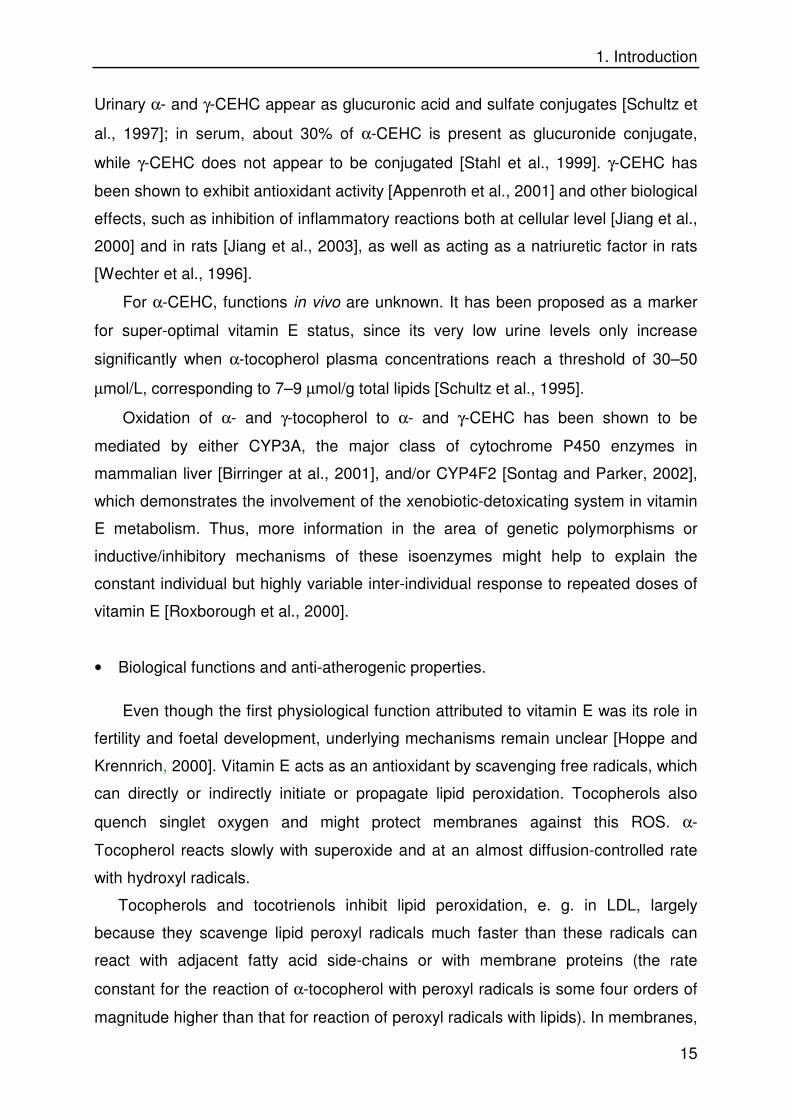
The formed tocopheroxyl radicals might react with a further peroxyl radical to give
non-radical products (tocopherylquinones, see Figure 1.5). One tocopherol molecule
is thus capable of terminating two peroxidation chains (stoichiometric factor of 2).
Chemical reactivity towards peroxyl radicals differs considerably among the
tocopherols, in the sequence α->γ->δ->β-. Some reducing agents such as ascorbate,
cysteine and glutathione can regenerate vitamin E from the tocopheroxyl radical,
which could explain the synergistic effects and inhibition of lipid peroxidation by
vitamin E and ascorbate. However, there is still some doubt as to whether this
reaction occurs in vivo.
The tocopherol quinone is subsequently transformed to both tocopheronic acid
and the tocopheronolactone derived therefrom, the so-called Simon metabolites
(Figure 1.5). Although these have been detected as glucuronides or sulfates in urine,
some controversy exists concerning their authenticity, and it has been suggested that
they are artefacts generated during sample preparation [Schultz et al., 1997]. Data
from other groups propose that they are to some extent authentic [Pope et al., 2000].
Tocopherols can reduce Fe(III) to Fe(II) and Cu(II) to Cu(I) and thus may exert some
pro-oxidant effects in vitro. The α-tocopheroxyl radical can abstract hydrogen from
PUFA to yield alkenyl radicals [Carr et al., 2000], although the rate constant is about
five orders of magnitude lower than the rate constant for the reaction of peroxyl
radicals with PUFA. It is not clear if this pro-oxidant action has a functional meaning
in vivo.
Apart from its antioxidant activity, further anti-atherogenic properties of α-
tocopherol have been demonstrated in vitro. In several experiments on cell culture
the four tocopherol isoforms were shown to exert different biological effects. Non-
antioxidant functions unique to α-tocopherol have thus been postulated and later
demonstrated experimentally. α-Tocopherol was found to induce vascular smooth
muscle cell growth arrest [Boscoboinik et al., 1991]. Inhibtion of protein kinase -C
(PKC) activity was found to be the basis of this effect and of further protective
activities of α-tocopherol, e. g., the inhibition of thrombin-induced endothelin
secretion from endothelial cells [Martin-Nizard et al., 1998] or the impaired assembly

1. Introduction
17
of NAD(P)H oxidase in monocytes [Cachia et al., 1998]. α-Tocopherol inhibited the
aggregation of human platelets by a PKC-dependent mechanism both in vitro and in
vivo. α-Tocopherol was found to activate the phosphatase PP2A, which
dephosphorylates PKC [Ricciarelli et al., 1998]. PKC is currently accepted as a
common denominator in a number of cell events regulated by α-tocopherol, such as
cell proliferation, cell adhesion, or enhancement of immune response [Ricciarelli et
al., 2002].
There is also evidence of transcriptional regulation by α-tocopherol, e. g., it
inhibits liver collagen α1(I) gene expression [Chojkier et al., 1998], and upregulates
α-tropomyosin expression in rat vascular smooth muscle cells [Aratri et al., 1999]. In
SMC and monocytes/macrophages, the oxLDL scavenger receptors SR-A and CD36
are down-regulated at transcriptional level by α-tocopherol [Teupser at al., 1999;
Ricciarelli et al., 2000]. In rats, liver α-TPP and its mRNA are modulated by vitamin E
deficiency [Shaw and Huang, 1998]. The involvement of PKC in these events has not
always been examined.
1. 5. 2 Carotenoids
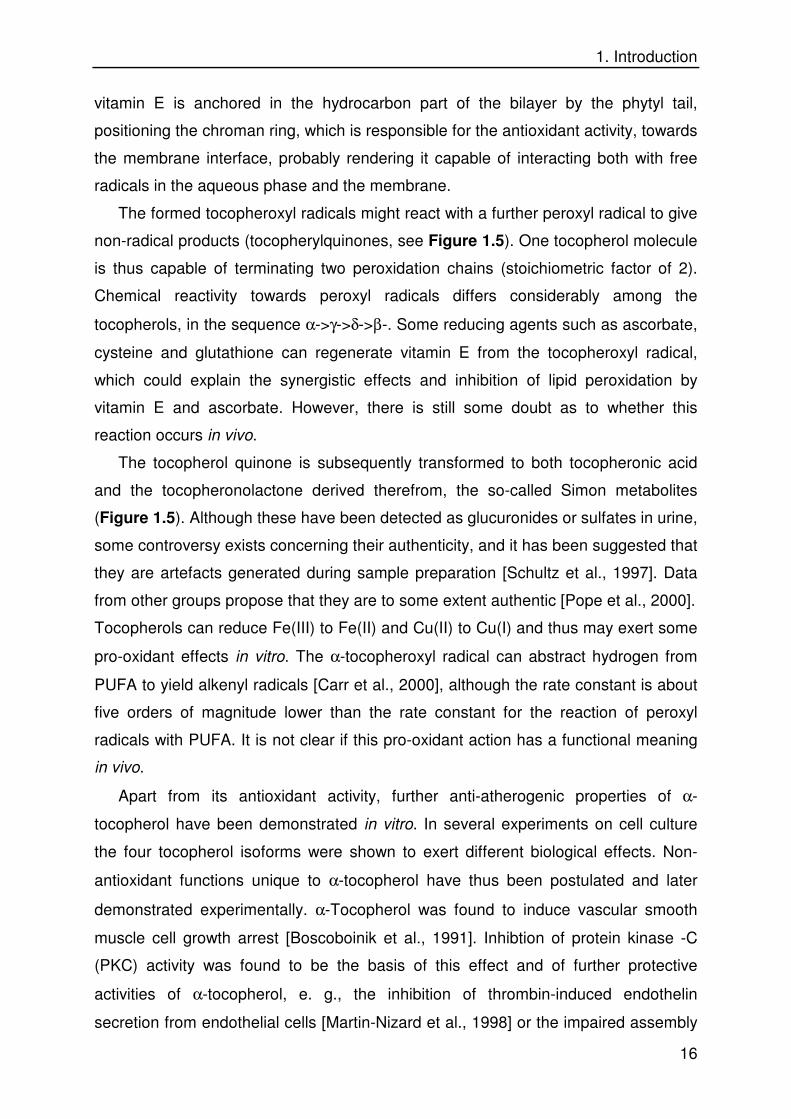
Carotenoids are a family of tetraterpenes (C40) with a central polyene chain
differently substituted at the ends. At least 60 carotenoids occur in fruit and
vegetables consumed by humans. Besides the provitamin A carotenoids (α- and β-
carotene and β-cryptoxanthin), lycopene and the xanthophylls lutein and zeaxanthin
are the major dietary carotenoids (Figure 1. 6). Their major roles in plants are light
harvesting and quenching excited states of oxygen that might be formed during
photosynthesis. There are no recommended daily intakes of the carotenoids,
although a number of conversion factors have been suggested for the conversion of
retinol precursors to vitamin A [Southgate, 2002]. The carotenoid profile in human
plasma is determined by the variety of fruits and vegetables ingested with the diet.
1. Introduction
18
Figure 1. 6 Molecular structure of some dietary carotenoids.
Location and physical form of carotenoids in foods are important determinants of
their bioavailability [Castenmiller and West, 1998; de Pee et al., 1998]. Disruption of
cellular structure (e.g. during mastication) or the transfer of carotenoids to the lipid
phase during processing (e.g. cooking with oil) are excellent facilitators of carotenoid
absorption. Fatty acid esters of carotenoids are hydrolysed in the intestinal lumen
before mucosal uptake, most likely by the carboxylic ester hydrolase secreted by the
pancreas [Wingerath et al., 1995]. Co-ingestion of fat with the meal, and the type of
dietary fat ingested are other factors with impact on bioavailability. Alcohol intake and
smoking have been identified as having an adverse effect on serum carotenoid levels
in several population-based studies [Albanes et al., 1997].
Incorporated in mixed micelles, carotenoids are absorbed by the mucosa of the
small intestine, mainly in the duodenum. No active carrier or transporter proteins for
carotenoids are known. In enterocytes, some cleavage to retinal/retinol occurs (see
below) and both the remaining carotenoid fraction and the cleavage products are
β-carotene
α-carotene
+2
2+zeaxanthin
+2
2+
lutein
+2
2+astaxanthin
2
2
capsanthin
2+2
2+
canthaxanthin
2
2
1. Introduction
19
incorporated into chylomicrons, secreted into the lymph and subsequently into the
blood. In the liver, hepatocytes integrate most of the carotenoids into lipoproteins,
which are then released into the systemic circulation [Stahl et al., 2002].
For humans, quantitative data on carotenoid absorption are scant. It is accepted
that absorption efficiency decreases as the dose of the carotenoid increases, and the
carotenoid plasma profile is dependent on the actual dietary carotenoid intake. Under
normal dietary intake the hydrocarbon carotenoids (mainly β-carotene and lycopene)
and the oxo-carotenoids (mainly lutein) are present at concentrations of 0.1 to 0.6
µmol/l in blood [Polidori et al. 2001]. Carotenes are mainly transported in LDL and
oxo-carotenoids in HDL and LDL. It has been suggested that carotenoids are
exchanged between lipoproteins, and apparently there is a greater exchange of
xanthophylls than of carotenes [Stahl et al, 2002].
Not all factors controlling carotenoid uptake into tissues are yet identified [Stahl
et al., 1992]. The enrichment of the human macula lutea specifically with lutein and
zeaxanthin and the pronounced presence of β-carotene in the pineal gland and
corpus luteum of cattle suggests a selective uptake. Lycopene concentrations are
highest in testis and adrenal tissue [Clinton, 1998]. Hydrocarbon carotenoids
accumulate in the adipose tissue [Su et al., 1998] and skin [Ribaya-Mercado et al.,
1995]. Erythrocytes, leucocytes, and cell membranes also contain carotenoids
[Fotouhi et al., 1996].
Pro-vitamin A carotenoids are partly cleaved to retinoid compounds in the
enterocyte and, to a lesser extent, in the liver and other tissues. Central cleavage of
the polyene chain is catalysed by β-carotene-15,15'-oxygenase, yielding two
molecules of retinal. Isolation and characterization of this enzyme has confirmed a
cytosolic location, not bound to membranes, and the requirement of ferrous iron
[Redmond et al., 2001; von Lintig and Vogt, 2000; Wyss et al., 2000]. Excentric
cleavage has been found to occur in vitro leading to apo-carotenals [Wang et al.,
1991]. Non-enzymatic excentric oxidation might also occur under conditions of
oxidative stress. The metabolism of non-pro-vitamin A carotenoids is less known.
Two lycopene metabolites in cycled form [Khachik et al., 1997] and dehydration
products of lutein have been found. Both central [Redmond et al., 2001] and
excentric [Kiefer et al., 2001] cleavage of lycopene have been reported and found to
be very limited.

1. Introduction
20
In both the intestine and the liver, retinal is reduced to retinol and stored as retinyl-
palmitate. Plasma retinoid-binding protein and lipoproteins are responsible for
transport in serum and for the regulation of blood levels of retinoids.
• Antioxidant an other biological properties.
Carotenoids have been reported to react with ROS and virtually all radical
species likely to be encountered in a biological system and thus protect cells from
oxidative stress [Sies at al., 1992]. Mechanisms include:
- Quenching triplet-state sensitizers, such as flavins and porphyrins, which may either
abstract a hydrogen atom or an electron from various molecules leading to further
radical-catalyzed damage or react with ground-state oxygen to form singlet oxygen
[Young et al., 1996; Sundquist et al., 1993].
- Quenching singlet oxygen directly, which results almost entirely from energy
transfer and yields ground-state oxygen and a triplet excited carotenoid. The energy
is then dissipated in the solvent as heat [Stahl and Sies, 1993]. The contribution of
chemical quenching is thereby minor (<0.5% for β-carotene).
- Radical addition with adduct formation: a lipid peroxyl radical may add at any place
along the carotenoid polyene chain, resulting in a carbon-centered radical, which is
resonance-stabilized and interferes with the propagation step in lipid peroxidation
[Sies at al., 1992]. It has been postulated that at high oxygen tensions this radical
could react reversibly with molecular oxygen to form a new peroxyl radical. By
cleavage of the resulting peroxyl bond this carotenoid peroxyl radical might generate
additional radicals, resulting in a pro-oxidative effect [Truscott, 1996]. Recent
investigation reported, however, that addition radicals would not react with molecular
oxygen even at high pressures either in polar nor in non-polar solvents [El-Agamaey
and Mc Garvey, 2003]. At low partial oxygen pressures β-carotene is an efficient
antioxidant [Burton and Ingold, 1984], interrupting substrate oxidation by peroxyl
radicals. At physiological levels of oxygen carotenoids have antioxidant activity.
- Electron transfer: these reactions result either in the formation of the cation radical
CAR+., the anion radical CAR-. or in the generation of an alkyl radical CAR. (Liebler,
1993)
- Hydrogen abstraction at the allylic position: Woodal et al. (1997) observed the
formation of the 4-methoxy and 4, 4´-dimethoxy derivatives (non-radical products) of
1. Introduction
21
β-carotene when reacted with peroxyl radical initiators in the presence of methanol,
which is explained through this mechanism.
Some carotenoids have been shown to exert, beyond the role of provitamin A
carotenoids in the vision process, non-antioxidant biological properties which may
underlie beneficial effects observed in epidemiological studies in the field of
prevention of neoplastic disorders. Lycopene inhibits growth of mammary,
endometrial and lung cancer cells [Levy et al., 1995]. Lycopene, β-carotene [Prakash
et al., 2001], and retinoic acid [Zhu et al., 1997] suppress growth of different lines of
human breast cancer cells. Carotenes, lycopene, some xantophylls and their
metabolites show a stimulatory effect on gap junctional intercellular communication
[see Stahl et al., 2002 for review]. Besides, the role of retinoids in a large number of
processes related to embryogenesis, morphogenesis, growth, differentiation and
fertility is widely accepted [Hansen et al., 2000].
Anti-atherogenic properties of carotenoids or vitamin A have been explained
almost exclusively through antioxidant activities. However, it has been suggested that
retinoid signaling is involved in atherosclerosis since in vivo studies of retinoid
administration after vascular injury have documented positive changes in vessel
geometry, such as attenuation of neointimal mass or accelerated reendothelia-
lization. A panel of retinoids with different selectivity for retinoid receptors was shown
to inhibit dose-dependently platelet-derived growth factor (PDGF)/insulin-stimulated
growth of human coronary artery SMC [Wakino et al., 2001]. Retinoic acid and
synthetic retinoids were shown to regulate proliferation, migration and differentiation
of SMC by a retinoic acid receptor (RAR)-α dependent signaling pathway [Neuville et
al., 1999]. Furthermore, all-trans-retinoic acid was found to stimulate CD36
expression through RAR binding and thus increase uptake of oxLDL in macrophages
[Wuttge et al., 2001].
1. 5. 3 Polyphenols
Polyphenols are a large variety of phenylpropanoid- or polyacetate-based
compounds which are almost exclusive of higher vascular plants, including cinnamic
acid derivatives (e. g. chlorogenic acid), diarylheptanoids (curcuminoids), flavonoids,
and proanthocyanidins [Stahl et al., 2002]. Over 4000 different flavonoids have been
isolated from plants (Figure 1.7). In the human diet, chlorogenic acid (coffee), ferulic

1. Introduction
22
Figure 1. 7 Subclasses of the flavonoid family
acid (cereals), flavones and flavonols (onions, tea, apples), catechins and other
flavan-3-ols (tea, grapes, cocoa), and isoflavones (soy and black beans) constitute
the major classes. There is no recommended daily intake for these compounds.
However, as a rough guide, the total polyphenol intake probably lies between 100
and 1000 mg / day.
In saliva, some degalloylation of the flavan-3-ol gallate esters, such as
epigallocatechin gallate, already occurs [Yang et al., 1999]. In the stomach, flavonoid
glycosides [Hollman and Katan, 1999] and hydroxycinnamate esters [Rechner et al.,
2001] are not modified, but procyanidin oligomers have been shown to decompose
on interaction with acidic gastric juice ex vivo essentially to epicatechin monomeric
and dimeric units [Spencer et al., 2000]. The large amounts of epicatechin released
can be absorbed by enterocytes.
Major factors influencing extent and rate of absorption of phenols are: (i) uptake
and interactions in the small intestine, including conjugation; (ii) metabolism by the
O A
B C 1
2 3
4 5 6 7 8 1'
2' 3' 4' 5'
6'
O
O H A
B C 1
2 5
6 7 8 1'
2' 3' 4' 5'
6'
O
O
A B
C 1
2 3
5 6 7 8 1'
2' 3' 4' 5'
6'
O
O
A B
C 1
2 5
6 7 8 1'
2' 3' 4' 5'
6'
O
O O H
A B
C 1
2 5 6
7 8 1' 2' 3'
4' 5'
6' 4
3
Ground Structure
Flavan-3-ol
Flavone
Flavanone
Flavon-3-ol
A B
C 1
2 5
6 7 8
1' 2'
3' 4' 5'
6'
O
O
Isoflavone
OH
3 4

1. Introduction
23
Figure 1. 8 Molecular structures of gallate derivatives of epicatechin
bacterial microflora of the colon and subsequent absorption of the resulting
metabolites, and (iii) biliary excretion after hepatic biotransformation and/or
conjugation [Lin et al., 1999].
The sugar moiety plays an important role in the absorption of flavonol glycosides.
Pharmacokinetic data suggest that quercetin glucoside is absorbed from the small
intestine, whereas quercetin rutinoside was absorbed from the colon after
deglycosylation [Hollman et al., 1999]. Glucosylated flavonoids might be carried into
the small intestine enterocyte via active transport (intestinal Na+-glucose
cotransporter SGLT1), which has been shown in vitro for quercetin glucosides [Gee
et al., 1998]. Alternatively, they might be hydrolysed by lactase phloridzin hydrolase
(LPH), a β-glucosidase on the outside of the brush border membrane [Day et al.,
2000], and the aglycone absorbed by passive diffusion in the small intestine. On the
other hand, in ileostomy patients, who lack a colon and thus bacterial flora,
absorption of the quercetin glucosides from onions (52%) was better than that of the
pure aglycone (24%) [Hollman et al., 1995], which confirms that some glycosides are
absorbed without prior hydrolysis by microorganisms. Also in this model, it was found
that 33% of chlorogenic acid, the major dietary hydroxycinnamic acid compound, was
absorbed in the small intestine.

1. Introduction
24
Conjugation of hydroxyl groups with glucuronic acid, sulfate or glycine or O-
methylation of the catechols is reported is a crucial step in the metabolism of plant
phenols. Flavonoids are substrates for small intestinal UDP-glucuronyl transferases
and catechol-O-methyltransferases, whose presence in enterocytes has also been
suggested [Okushio et al., 1999]. Conjugation reactions such as glucuronidation and
methylation occur in the jejunal and ileal sections of the small intestine [Manach et
al., 1998; Spencer et al., 1999]. Deglycosylation of glucosides can occur previously in
the small intestine or afterwards in the liver, depending on the nature and position of
the sugar residue. Further modificaons in the liver include methylation, sulfation and
glucuronidation.
Catabolism and scission of the flavonoid rings and demethylation and
dehydroxylation of the resulting phenolic acids, are, to a great extent, catalysed by
enzymes present in intestinal microorganisms [Hackett, 1986]. Bacterial enzymes
may catalyse hydrolysis of glucuronides, sulfates and glycosides, dehydroxylation,
demethylation, reduction of double bonds, ring cleavage, and decarboxylation of
some phenolic acids [Hollman and Katan 1998].
Liver
Oral IngestionFlavanols, e.g epicatechin
Gu
t L
um
en
Gut Wall
Hepatic Portalvein
Faeces
Degradationby microflora
Blood
Bile
Peripheral Tissues
Kidneys
Urine
Phase I +IIMetabolism(glucuronidation)(O-methylation)(sulphation)
Colon
Blood-BrainBarrierPhase I +II
Metabolism(deglycosylation)(glucuronidation)(O-methylation)
BRAIN
?
Flavonols, e.g kaempferolO
OH
OH
OH
OH
OO
OH
OH
OH
COO-
O
O
OH
OH
OH
HO
CH3
O
OH
OH
OH
COO-
O
O
OH
OH
O
HO
CH3
O-methylated epicatechin
O-methylated epicatechin glucuronide
Epicatechin glucuronide
Liver
Oral IngestionFlavanols, e.g epicatechin
Oral IngestionFlavanols, e.g epicatechin
Gu
t L
um
en
Gut Wall
Hepatic Portalvein
Faeces
Degradationby microfloraDegradationby microflora
Blood
Bile
Peripheral Tissues
Kidneys
Urine
Phase I +IIMetabolism(glucuronidation)(O-methylation)(sulphation)
Phase I +IIMetabolism(glucuronidation)(O-methylation)(sulphation)
Colon
Blood-BrainBarrierPhase I +II
Metabolism(deglycosylation)(glucuronidation)(O-methylation)
Phase I +IIMetabolism(deglycosylation)(glucuronidation)(O-methylation)
BRAIN
?
Flavonols, e.g kaempferolO
OH
OH
OH
OH
OO
OH
OH
OH
COO-
O
O
OH
OH
OH
HO
CH3
O
OH
OH
OH
COO-
O
O
OH
OH
O
HO
CH3
O-methylated epicatechin
O-methylated epicatechin glucuronide
Epicatechin glucuronide
Figure 1. 9 Phenol absorption and metabolism. Changes in epicatechin structure are illustrated as an
example. Taken from Stahl et al., 2002

1. Introduction
25
As far as flavonols are concerned (Figure 1.7), evidence for the presence of
quercetin conjugates in humans has been obtained. Quercetin aglycone could not be
detected in plasma [Manach et al., 1998]. Major urinary and biliary conjugates are the
glucuronides and sulfates of quercetin, 3'-O-methyl-quercetin and 4'-O-methyl-
quercetin [Gross et al., 1996]. After consumption of onions by humans, the major
plasma metabolites were quercetin 3'-sulfate and quercetin 3-glucuronide [Day et al.,
2001]. Flavonols are degraded to a great extent by colonic microflora to phenylacetic
and phenylpropionic acids. For quercetin, reported metabolites are 3-
hydroxycinnamic acid, 3-hydroxyphenylacetic acid, 3,4-dihydroxyphenylacetic acid
(homoprotocatechuic acid) and 3-methoxy, 4-hydroxyphenylacetic acid (homovanillic
acid) [Stahl et al., 2002].
Oral administration of flavones or flavonones leads to the formation of 3-
hydroxyphenylpropionic acid, 3-hydroxycinnamic acid, and 4-hydroxy-3-methoxy-
phenylpropionic acid [Booth et al., 1958; Scheline, 1991].
Regarding flavan-3-ols, especially epicatechin metabolites, the most predominant
conjugates in human plasma and urine are glucuronides and sulfates of epicatechin
and 3'-O-methyl epicatechin [Donovan et al., 1999]. Methylated and glucuronidated
metabolites have not been detected in rat bile. The mechanism of flavan-3-ol
metabolism is supposed to involve glucuronidation in the small intestine followed by
O-methylation in the liver and kidney [Piskula and Terao, 1998]. Specific markers of
catechin metabolism are the valerolactones, which can be detected in human plasma
and urine after a single ingestion of green tea [Li et al., 2000].
Ferulic acid has long been determined as a biomarker for the absorption and
metabolism of both dietary caffeic and ferulic acid derivatives [Booth et al., 1957].
The large intestine is the site of absorption and metabolism of quinic acid esters such
as chlorogenic acid [Rechner et al., 2001]. Colonic microflora provides esterase
activity [Plumb et al., 1999] and additionally could be, with the liver, responsible for
O-methylation of caffeic acid to yield ferulic and isoferulic acids [Chesson et al.,
1999].
• Antioxidant effects and further biological activities.
Plant phenols are antioxidants that can prevent lipid peroxidation and thus are
potent inhibitors of the modifications of LDL. Mechanisms include [Briviba and Sies,
1994):

1. Introduction
26
- scavenging initiating radicals such as .OH, and O2.-
- binding metal ions
- scavenging lipid peroxyl radicals
- inhibiting enzymatic systems responsible for free radical production, such as
cyclooxygenases, lipoxygenases, myeloperoxidase and xanthine oxidase.
The formation of 5-hydroperoxy-eicosatetraenoic acid (5-HpETE) by reaction of
arachidonic acid with recombinant human 5-lipoxygenase was significantly inhibited
by (-)-epicatechin in a dose-dependent manner with a 50% inhibitory concentration
(IC50) of 22 µmol/L. Among the procyanidin fractions isolated from the seeds of
Theobroma cacao, only the dimer fraction and, to a lesser extent, the trimer through
pentamer fractions exhibited comparable effects [Schewe et al., 2002]. Quercetin and
other flavonoids were found to modulate the time course of the reaction of rabbit
reticulocyte and soybean 15-lipoxygenases. The flavone luteolin turned out to be the
most potent inhibitor of the mammalian enzyme with an IC50 of 0.6 µmol/L followed
by baicalein (1 µmol/L) and fisetin (1.5 µmol/L) [Sadik et al., 2003].
Furthermore, experimental work in the last years has provided evidence of non-
antioxidant functions of flavonoids which may be related to cancer preventive effects.
Epicatechin exhibited stimulatory effects on GJC in WB-F344 rat liver epithelial cells
after 24-72 h of incubation; inhibitory effects of the tumor promoter 12-O-
tetradecanoylphorbol-13-acetate (TPA) on GJC were largely suppressed when
epicatechin or genistein (40 µmol/L) were present during the incubation [Ale-Agha et
al., 2002]. Other effects include: suppression of vascular smooth muscle cell
proliferation by epigallocatechin, which partially inhibited the JNK/stress-activated
protein kinase (SAPK) signal transduction pathway [Lu et al., 1998]; baicalein, a
flavonoid from a Chinese herb (Scuttelaria baicalensis) was shown to exert an
inhibitory effect on the proliferative response of PDGF on rabbit vascular smooth
muscle cells [Huang et al., 1994]. In addition, several flavonoids, including quercetin,
were able to improve the cell glutathione status in part through regulation of γ-
glutamylcysteine synthetase gene expression [Myhrstad et al., 2002].
1. 6 Aim of the Study
(1) To assess the antioxidant activity of various types of dietary micronutrients and
polyphenol-containing pharmaceutical preparations by challenging with different
oxidative insults in model systems. The antioxidant activity of the α-tocopherol

1. Introduction
27
metabolite α-CEHC and of compounds present in the preparations should be
studied as well. The effect of solvent polarity, reaction conditions and nature of
pro-oxidants applied will be considered.
(2) To investigate the effects of pre-treating vascular endothelial cells (primary mouse
aorta endothelial cells) with selected antioxidants (α-tocopherol, α-CEHC,
epicatechin and epigallocatechin gallate) on the subsequent incubation with
oxidatively-modified LDL. Along with direct or indirect effects of antioxidant
nature, intervention of these compounds at functional processes (oxLDL uptake,
cell viability, apoptotic death) are to be determined.
(3) To monitor both the serum levels and the degree of biotransformation of α- and γ-
tocopherol (by measuring the corresponding CEHC metabolites) in smoking and
nonsmoking volunteers who are given a single oral dose of vitamin E, in order to
compare outcomes between both groups. In smokers, the oxidative status is
increased, and xenobiotic-detoxicating enzymes (such as cytochrome P450-
dependent monoxygenases) may be induced. Those two factors may thus modify
the physiological levels of certain dietary antioxidants in blood.

2. Materials and Methods
28
2. MATERIALS AND METHODS
2. 1 Chemicals
Following chemicals were used:
L-ascorbic acid Merck, Darmstadt (Germany)
astaxanthin BASF, Ludwigshafen (Germany)
2,2´-azino-bis(3-ethylbenzthiazoline-6-sulfonic acid (ABTS) Sigma-Aldrich, Steinheim (Germany)
2,2´-azobis(2-amidinopropane) hydrochloride (AAPH) Polysciences, Warrington (USA)
canthaxanthin BASF
α-carotene BASF
caspase-3 assay kit Sigma-Aldrich
2-(rac) �-α-CEHC BASF
2-(rac)-γ-CEHC BASF
2´,7´-dichlorodihydrofluorescein diacetate (DCFH-DA) Sigma-Aldrich
chlorogenic acid Sigma-Aldrich
copper (II) chloride (CuCl2) Sigma-Aldrich
curcumin Extrasynthese, Lyon (France)
1,1´-dioctadecyl-3,3,3´,3´-tetramethyl-indocarbocyanine perchlorate (DiI)
Sigma-Aldrich
epicatechin Sigma-Aldrich
ethylenediaminetetraacetic acid (EDTA) Merck
Folin-Ciocalteau´s reagent 2N Sigma-Aldrich
gallic acid Sigma-Aldrich
glutathione disulfide (GSSG) Sigma-Aldrich
harpagoside Phytochem, Ichenhausen (Germany) hydrochloric acid (HCl) 25% Merck
4-(2-hydroxyethyl)-1-piperazineethanesulfonic acid (HEPES) Boehringer-Mannheim, Mannheim (Germany)
4-hydroxy-3-nitrobenzoic acid (HNBA) Sigma-Aldrich
kaempferol Extrasynthese
lutein BASF
luteolin Sigma-Aldrich
dimethyl sulfoxide (DMSO) Sigma-Aldrich
3-(4,5-dimethylthiazole-2-yl)-2,5-diphenyl tetrazolium (MTT) Sigma-Aldrich
nicotinamide-adenine dinucleotide phosphate, reduced (NADPH)
Roche, Mannheim (Germany)
3-nitrotyrosine Sigma-Aldrich
R-phycoerythrin from Porphyra tenera Sigma-Aldrich
potassium bromide (KBr) Merck
potassium di-hydrogen phosphate (KH2PO4) Merck
di-potassium hydrogen phosphate (K2HPO4) Merck
potassium peroxodisulfate (persulfate) (K2S2O8) Sigma-Aldrich
sodium bicarbonate (NaHCO3) Merck
sodium carbonate (Na2CO3) Merck
sodium chloride (NaCl) Merck
sodium dodecyl-sulfate (SDS) Merck

2. Materials and Methods
29
sodium sulfate (Na2SO4) Merck
5-sulfosalicylic acid (SSA) Sigma-Aldrich
tetraethylammonium hydroxide (TEAH) Sigma
5,5´-dithio-bis(2-nitrobenzoic acid) (DTNB) Sigma-Aldrich
1,4-dithio-threitol (DTT) Boehringer-Mannheim
RRR-α-tocopherol Cognis Deutschland, Düsseldorf (Germany)
RRR-γ-tocopherol Cognis Deutschland
R-Trolox Sigma-Aldrich
L-tyrosine Sigma-Aldrich
2-vinyl-pyridine (VP) Sigma-Aldrich
All solvents used were purchased from Merck (Darmstadt, Germany) or Sigma-
Aldrich (Steinheim, Germany). Minimum quality grade was p. A. Gaseous nitrogen
purity grade 5.0 was provided by Linde (Höllriegelskreuth, Germany).
The following pharmaceutical trade products containing standardized natural
extracts were provided by Truw Arzneimittel (Gütersloh, Germany):
• Curcu-Truw capsules, containing ethanolic extract of turmeric rhizomes (dry
drug-to-solvent ratio 13-25:1, 27% curcuminoids); Truw Arzneimittel
(Gütersloh, Germany)
• Rheuma-Sern capsules, containing aqueous extract of devil´s claw roots (dry
drug-to-solvent ratio 2:1, 2.60% harpagoside); Truw Arzneimittel (Gütersloh,
Germany)
• Hepar-SL forte capsules, containing aqueous extract of artichoke leaves (dry
drug-to-solvent ratio 4-6:1); Sertürner Arzneimittel, Berlin (Germany)
• KwaiN coated tablets, containing fresh garlic extract (standardized to 1.0-
1.4% alliin); Lichtwer Pharma, Berlin (Germany)
• Ameu soft capsules, containing salmon oil concentrate (min. 35% n-3 fatty
acids); Omega Pharma, Berlin (Germany)
The vitamin E capsules used for the intervention study were from Hermes
(Munich, Germany).

2. Materials and Methods
30
2. 2 Instruments
a) UV/VIS and fluorescence spectroscopy
DU 530 Life Science UV/VIS Spectrometer (Beckman, Munich, Germany)
LS-5 Fluorescence/Luminiscence Spectrometer (Perkin-Elmer, Überlingen,
Germany)
Victor 1420 Multilabel Counter (Wallac, Turku, Finland)
b) Microscopy
Axiovert 100TV (Zeiss, Oberkochen, Germany) coupled with a Hamamatsu Digital
Camera (Hamamatsu Photonics, Hamamatsu, Japan)
c) Centrifuges and Rotors
Hettich Universal 30 RF (Hettich, Tuttlingen, Germany)
L7-55M Ultracentrifuge / Rotor SW 41 (Beckman)
d) High Performance Liquid Chromatography
Pump: La Chrom L-7100 (Merck-Hitachi, Darmstadt, Germany)
Integrator: D-7500 Integrator 7480 (Merck-Hitachi)
Analysis of 3-nitrotyrosine:
Autosampler: 655 A-40 Autosampler 7480 (Merck-Hitachi)
Column: 4.6 x 150 mm Lichrospher 100 RP 18 (Merck)
Detector: La Chrom UV/VIS Detector L-7420 (Merck-Hitachi)
Analysis of carotenoids:
Column: 4.1 x 250 mm Spherisorb ODS2 C-18 (Bischoff Chromatography,
Leonberg, Germany)
Detector: La Chrom UV/VIS Detector L-7420 (Merck-Hitachi)
Analysis of serum α- and γ- CEHC:
Column: 4 x 250 mm Lichrospher 100 RP 18, endcapped (Merck-Hitachi)
Detector: Coulochem 5100A; Analytical Cell 5011; Conditioning Cell 5021
(ESA, Inc., Chelmsford, USA)
Analysis of α- and γ-tocopherol:
Column: 4.6 x 250 mm Suplex pKb-100 (Supelco, Bellefonte, USA)
Detector: La Chrom UV/VIS Detector L-7420 (Merck-Hitachi)

2. Materials and Methods
31
2. 3 Preparation of Stock Solutions
a) Pure Compounds
Stock solutions of micronutrients were prepared at concentrations ranging from 1
to 3 mmol/L. Flavonoids and hydroxycinnamates were dissolved in ethanol,
methanol/water 50:50 or acetone; α-tocopherol in ethanol; carotenoids in ethanol and
dichloromethane; harpagoside in water; α- and γ-CEHC in ethanol; Trolox and
ascorbic acid in water, 75 mM phosphate buffer (pH 7.0) and ethanol.
b) Pharmaceutical Preparations
Content weight of each capsule of turmeric, devil´s claw, artichoke and salmon
oil was calculated as the mean difference of weight between the whole capsule and
the capsule cover (n=7).
Fractions A, B and C: Five entire garlic coated tablets and the contents of four to
six capsules of turmeric, devil´s claw or artichoke extract were crushed in a mortar
until homogeneity of particle size. The products were treated with solvents of different
lipophilicity to obtain fractions for testing. To 10 to 100 mg of powder either 1 mL of
water, methanol/water 70:30, or ethanol was added. After 24 h of incubation under
agitation at 4°C in the dark, all suspensions were centrifuged at 5,000 rpm for 10 min
and the supernatants collected. The pellets were washed with 0.5 mL solvent, left for
two hours at 4°C in the dark and centrifuged. Supernatants from the same solvent
were combined. The extracts were designated as fraction A (water), fraction B
(methanol/water 70:30), and fraction C (ethanol). The clear supernatants and the
solutions of the pure compounds were stored at –80°C until use. Storage time was
less than 7 days.
Fraction D: Extraction of highly lipophilic compounds. A highly lipophilic fraction
(Fraction D) was prepared as follows. 1 g of powder was suspended in 50 mL
acetone/water (75:25 v/v) for two hours in the dark and vacuum-filtered through a
Büchner funnel. The residue was extracted again until the filtrate was colourless. The
filtrates were transferred to a decanting funnel; 150 mL of diethyl ether was added,
and the funnel was shaken. Bidistilled water was added to separate the phases. The
upper phase containing the lipophilic compounds was washed several times with
water. The ether solution was filtered through a solid bed of Na2SO4 and dried in a

2. Materials and Methods
32
rotavapor. The residue was dissolved in acetone and named fraction D. Fraction D of
salmon oil was a 200 mg/mL solution of the capsule contents in hexane.
2. 4 Antioxidant Activity of Dietary Micronutrients in Model Systems
2. 4. 1 Trolox Equivalent Antioxidant Capacity (TEAC )
a) Background
The TEAC assay is based on the ability of different compounds to scavenge a
long-lived free radical generated by oxidation of 2,2´-azino-bis(3-ethylbenzthiazoline-
6-sulfonic acid (ABTS) in aqueous solution with potassium persulfate. ABTS and
potassium persulfate react following a stoichiometry of 2:1. The obtained stable
radical has four absorption maxima in the VIS spectrum at 415, 645, 734 and 815 nm
and is blue- green colored. The half-life of the radical in solution has been estimated
to be 24 h.
Figure 2. 1 Absorption spectrum of the ABTS radical
ABTS is a bivalent anion in solution. Thus, the net charge after one-electron
donation is –1. However, the radical so obtained is commonly defined in the literature
as radical cation or ABTS•+ and the use of the more accurate form ABTS•- is rare. To
avoid confusion, also in this work the radical is referred to as ABTS•+.
0
1
2
380 480 580 680 780 880
wavelength (nm)
abso
rban
ce (
AU
)

2. Materials and Methods
33
As a species capable of trapping one-electron, ABTS•+ was used to test the
ability of compounds to one-electron donation, which results in partial bleaching of
the blue-green color of the reaction mixture. Trolox, a water-soluble analogue of α-
tocopherol, was used as a reference compound.
b) Experimental Procedure [Re et al., 1999]
Following substances were tested:
Pure compounds:
• carotenoids: β-carotene, α-carotene, zeaxanthin, lutein, capsorubin,
capsanthin, canthaxanthin and astaxanthin.
• flavonoids: epicatechin, luteolin and kaempferol.
• others: α-CEHC, RRR-α-tocopherol, ascorbic acid, curcumin, chlorogenic
acid and harpagoside.
Pharmaceutical preparations:
• turmeric, artichoke, devil´s claw and garlic: Fractions A to D.
• salmon oil: Fraction D.
A fresh stock solution of ABTS radical was prepared every day by adding 14
mmol/L ABTS to 4.9 mmol/L potassium persulfate; oxidation of ABTS was thus
incomplete. The maximal intensity due to oxidation was measured after 6 hours.
At the beginning of the experiment the ABTS•+ solution was diluted to yield an
absorbance of 0.7 at 734 nm, either in ultra-pure water or in ethanol depending on
the polarity of the solvent used for the stock solutions of the samples. The extinction
coefficients of ABTS•+ at 734 nm in water and ethanol are very similar: 1.5 x10⁴ M-
1cm-1 and 1.6 x 10⁴ M-1cm-1, respectively.
.+

2. Materials and Methods
34
10 µL of sample was added to 1 mL of the ABTS•+ solution. The mixtures were
vortexed briefly and absorbance at 734 nm was measured 1, 3, 6 and 9 min after
addition. For the pure compounds, the final concentrations tested were 2.5, 5, 10 and
15 µmol/L; each concentration was tested in triplicate. Of every extract fraction five
replicates were prepared and at least four dilutions of each were tested. A solvent
control was run in each series.
The percentage loss of absorbance determined after 6 or 9 min was plotted as a
function of the concentration of the samples. Absorbance values generally remained
constant after the first minute.
The antioxidant activity of a compound or mixture using the TEAC assay was
quantified by dividing the slope or gradient of the plots of percentage loss of
absorbance vs. concentration for each sample by the slope of the curve for Trolox.
Thus, the TEAC assay assumes a value of 1 for Trolox per definition and the results
corresponding to the samples are expressed as Trolox Equivalents (TE).
c) Determination of Total Phenols (TP)
The amount of TP in the fractions of pharmaceutical preparations was
determined following the Folin-Ciocalteau method [Singleton and Rossi, 1965], based
on a colorimetric oxidation/reduction reaction between the phenols and the Folin-
Ciocalteau´s reagent. The Folin-Ciocalteau´s reagent is a complex oxidizing agent
comprised of heteropolyphosphotungstate-molybdate; upon reaction with phenols, a
blue-coloured product is obtained consisting of a mixture of the 1-, 2-, 4-, and 6-
electron reduction products of the tungstate reagents, and the 2-, 4-, and 6-electron
reduction products of the molybdate reagents. It should be noted that all phenolic
molecules with no differentiation between phenol monomers, dimers and larger
phenolic compounds are determined in this assay.
0.1 mL sample was diluted with 5.4 mL bi-distilled water and mixed with 4 mL of
7.5% sodium carbonate and 0.5 mL of 2 N Folin-Ciocalteau´s reagent. After 2 h
incubation in the dark, absorbance was measured at 765 nm. A gallic acid standard
curve was plotted and results were expressed as gallic acid equivalents (GAE).

2. Materials and Methods
35
d) Carotenoid Analysis by HPLC
Aliquotes of the Fraction D of the natural extracts were taken (volumes ranging
from 10 to 100 µL, depending on the dilution of the dry material), then dried under
nitrogen in the absence of light and injected into the HPLC system described above.
UV/VIS detection was set at 450 nm. The mobile phase comprised a binary gradient
at a constant flow of 1.5 mL/min. The initial composition of the eluent (acetone/water
75:25) was held for 5 min. Then, a linear gradient was applied for 5 min to yield a
final composition of acetone/water of 95:5, which was held for 7 min. Finally, the
column was washed for 3 min with acetone [Mínguez-Mosquera and Hornero-
Méndez, 1993]. For quantification, the external standard mode was applied.
Carotenoids not listed in 2. 1 were isolated from natural sources such as paprika
oleoresine by HPLC.
2. 4. 2 Oxygen Radical Absorption Capacity (ORAC)
a) Background
Phycobiliproteins are accessory photosynthetic proteins occurring in
cyanobacteria (blue-green algae) and in two groups of eukaryotic algae, the red
algae and the cryptomonads. R-phycoerythrin is a phycobiliprotein occurring in
various red algae. R- and B-phycoerythrin are the most remarkable phycobiliproteins
with respect to quaternary structure stability and to spectroscopic features. Despite
their complex multisubunit structure (αβ)6γ, no dissociation is observed at
concentrations as low as 10-15 mol/L. Besides, R-phycoerythrin carries 34 covalently
attached tetrapyrrole prosthetic groups which are resposible for the fluorescence
properties of the macromolecule. The intensity of fluorescence is highly sensitive to
conformation and chemical integrity of the protein and of the prostetic groups.
Phycoerythrins rapidly lose fluorescence when subjected to peroxyl or hydroxyl
radical attack. This may be in turn retarded in the presence of radical scavengers.
In the present study, AAPH was used as a radical generator to initiate R-
phycoerythrin loss of integrity and loss of fluorescence. AAPH is a diazo-compound
which decomposes in aqueous oxygenated solutions in a temperature-dependent
manner to yield peroxyl radicals:

2. Materials and Methods
36
A – N = N – A A. + N2 + A.
A + O2 AOO.
where A = HCl . HN = C (NH2) C (CH3)2 − .
b) Experimental Procedure
The Oxygen Radical Absorption Capacity (ORAC), e. g. the ability to retard R-
phycoerythrin complete loss of fluorescence due to peroxyl radical attack, was
determined as described by [Cao and Prior, 1999] for α-CEHC, RRR-α-tocopherol,
ascorbate and epicatechin
0.1 mL of test substance was mixed in a fluorescence cuvette with 0.1 mL of 3.4
mg/L R-phycoerythrin (final concentration) and 1.75 mL 75 mmol/L phosphate buffer
(pH 7.0). The mixture was equilibrated for 10 min at 37°C and then fluorescence was
measured at 540 nm (excitation) and 565 nm (emission) wavelenghts. This value
was used as baseline and referred to as 100% fluorescence. After addition of 50 µL
of 4 mmol/L AAPH, the cuvette was vortexed briefly. Fluorescence was first recorded
exactly 15 seconds after AAPH addition and then every 6 min until the fluorescence
was less than 5% of the starting value. The rack containing the cuvettes was kept at
37°C in a water bath. Appropriate solvent controls as well as a standard containing
0.1 mL of 1 µM Trolox were run in each assay. Each compound was tested in
triplicate at four concentrations.
Final results were expressed as ORAC values using the formula proposed by
Cao et al. (15):
ORAC value (µmol/L) = 20 x k x (Ssample-Scontrol)/(STrolox-Scontrol)
k is the dilution factor of the sample (usually 1/20), and S the area under the
curve (AUC) of fluorescence intensity vs. time. Thus, ORAC values depend on the
inhibition rate as well as the inhibition time caused by the presence of antioxidants as
both parameters are enclosed in the AUC. According to the formula, the ORAC value
for Trolox is 1.

2. Materials and Methods
37
2. 4. 3 Inhibition of Nitration of Tyrosine by Peroxynitrite
Protection against peroxynitrite-triggered nitration of tyrosine was performed as
described by [Pannala et al., 1997] with minor modifications. A 200 µmol/L tyrosine
stock solution was prepared in 100 mmol/L phosphate buffer/0.1 mmol/L EDTA (pH
7.3) and the concentration was confirmed spectrophotometrically at 274 nm (ε= 1405
M-1 cm-1). 350 µmol/L peroxynitrite was added by bolus addition to 100 µmol/L
tyrosine under vortexing at 25°C. The potential effects of cosolution of α-CEHC,
Trolox or (-)-epicatechin (0-40 µmol/L) with tyrosine were examined.
50 µL of each sample containing 100 µM 4-hydroxy-3-nitrobenzoic acid (HNBA)
as an internal standard was then injected into a C-18 reverse-phase column using an
autosampler. The eluent used for separation was a mixture of 50 mmol/L potassium
phosphate buffer (pH 7):acetonitrile (95:5) subject to the next step gradient (1.0
mL/min flow rate):
5 min 8 min 13 min
buffer:acetonitrile 95:5 50:50 95:5 95:5
3-Nitrotyrosine was detected at 430 nm. Ratios of peak areas of 3-nitrotyrosine
standard vs. internal standard were used for calibration and quantification.
2. 5 Cell Culture Study on Anti-atherogenic Effects of Dietary Micronutrients
and αα-CEHC
RRR-α-tocopherol, α-CEHC, epicatechin and epigallocatechin gallate were the
antioxidants selected to explore potential protective effects on endothelial cells which
were stressed with oxidatively-modified LDL. Stock solutions were prepared in
ethanol (3 to 5 mmol/L).

2. Materials and Methods
38
2. 5. 1 LDL Preparation and Oxidation
Blood from 2 healthy non-smoking volunteers was obtained by venipuncture,
allowed to clot for 10 min and centrifuged for 15 min at 4,500 rpm to obtain the
serum. The LDL fraction of serum was isolated using the short-run ultracentrifugation
method [Kleinveld et al., 1992]. For that purpose, 1.87 mL of the so-called “heavy
solution” (354 g KBr, 153 g NaCl, and 100 µg EDTA in 1 L ultra-pure water) was
added to 0.9 mL serum in a polyallomer tube (Beckman, Munich, Germany) to reach
a final density of 1.33 g/mL. The mixture was then overlayed with 0.15 mol/L NaCl up
to the tube edge and 6 tubes were centrifuged together at 4°C / 120,000 g for 2 h to
allow separation of lipoprotein bands.
The orange-colored LDL bands were collected, combined and stored with 100
µmol/L EDTA under nitrogen at 4°C for up to 3 days. Salts and EDTA were removed
by gel-filtration over a Sephadex G-25 separation column (modell PD-10, Amersham
Pharmacia Biotech, Freiburg, Germany).
LDL concentration was determined indirectly by measuring total cholesterol with
an enzymatic colorimetric test (CHOD-PAP, Boehringer Mannheim, Germany) and
applying a conversion factor of 2200 ± 163 mol cholesterol/mol LDL [Esterbauer, H.
et al., 1990] .
100 nmol/L purified LDL in PBS was incubated with 5 µmol/L CuCl2 at 37°C and
the extent of oxidation was followed by measuring conjugated diene formation at 234
nm every 10 min. Oxidation was stopped at a level of 250 nmol conjugated
dienes/mg protein (approx. 80 min) by placing the reaction vessel on ice. Oxidized
LDL were used immediately in cell culture experiments to avoid further extensive
oxidation before incorporation into the cells. Oxidized LDL will be abbreviated with
the term oxLDL; however, note that under these conditions and time of oxidation a
so-called minimally-modified LDL is obtained (see Introduction).
2. 5. 2 Cell Culture
Primary murine aorta endothelial cells (MAEC) were kindly provided by Dr.
Cristoph V. Suschek, Heinrich-Heine-University Düsseldorf. Cells were grown in 550
mL culture flasks (Greiner, Frickenhausen) using RPMI-1640 culture medium (Sigma-

2. Materials and Methods
39
Aldrich, Steinheim) to which 10% fetal calf serum (Greiner), 2 mmol/L L-glutamine
(Sigma) and 20 mg/mL penicilline/streptomycine (Sigma) were added. Cells were
incubated in a Binder CO2-Incubator Line CB (Tuttlingen, Germany) incubator (set at
37°C/ 5% carbon dioxide/ water-vapour saturation) until confluence, washed with
PBS and subsequently detached from the flask ground using 3 mL of 0.5 mg/L
trypsin/PBS (2:1 v/v). The isolated cells were resuspended in growth medium and
divided into new culture flasks at dilutions ranging from 1:5 to 1:10. For experiments,
cells were allowed to grow on 6 cm dishes or 24-well plates (Greiner) until 90%
confluence and then cultured for 24 hours in serum-deprived RPMI-1640 medium.
2. 5. 3 Cytotoxicity
Cytotoxic effects of samples on MAEC were studied using a spectrophotometric
test for cell viability, the MTT test. This assay is based on the physiological ability of
mitochondria to oxidize 3-(4,5-dimethylthiazole-2-yl)-2,5-diphenyl tetrazolium to
formazan, which takes place in intact and early apoptotic cells but not in dead cells.
MAEC were grown on 24-well culture plates until 90% confluence, washed once
in PBS and subsequently treated according to one of these three series of
experiments:
a) Cytotoxicity of Test Compounds: RRR-α-tocopherol, α-CEHC, epicatechin and
epigallocatechin gallate stocks were diluted to 30 and 15 µmol/L in RPMI-1640
medium containing 0.5% FCS. Incubation with MAEC for 24 h followed. A control
containing 1% ethanol was run in each series.
b) Cytotoxicity of Native and Oxidatively-modified LDL: nLDL (200 nmol/L) or
oxLDL (12.5, 25, 50, 75, 100 and 200 nmol/L) in RPMI-1640 medium containing
0.5% FCS were pipetted onto the cell monolayers and effects were determined after
24 h. A control comprising only 0.5% FCS was run in each assay.
c) Cytotoxicity of Native and Oxidatively-modified LDL on Antioxidant-preloaded
Cells: After 20 h cell preincubation with either 15 µmol/L epigallocatechin gallate or
30 µmol/L α-tocopherol, α-CEHC or epicatechin, cells were washed twice with PBS
and incubated further 32 h with 30 nmol/L moLDL in RPMI-1640 medium containing
0.5% FCS. The concentrations of antioxidants and oxLDL were selected in

2. Materials and Methods
40
accordance to the results of the experiments detailed under a) and b). Appropriate
controls without antioxidants and/or oxLDL were applied.
MTT stock was prepared by dissolving 5 mg in 1 mL PBS. Working solutions
consisted of a 1:10 dilution in FCS-free culture medium without phenol red. After
treatment, 200 µL MTT working solution was added to each well and cells were
incubated under usual culture conditions further 3 h. The reaction was stopped by
adding 200 µL SDS/0.1 mol/L HCl (10% w/v). After overnight incubation at 37°C,
formazan release was measured at 570 nm.
2. 5. 4 Uptake of oxLDL
The time course of uptake of nLDL and oxLDL by MAEC was followed
fluorometrically.
2 mL of 1.5 µM nLDL was incubated overnight with the lipophilic fluorescent
probe 1,1´-dioctadecyl-3,3,3´,3´-tetramethyl-indocarbocyanine perchlorate (DiI, 50 µL
of a 3 mg/mL solution in DMSO) under agitation, then isolated using the short-run
ultracentrifugation mode, filtered over a PD-10 column and finally diluted to 0.1
µmol/L in phenol-red free RPMI-1640 medium 0% FCS. A part was stored as labelled
nLDL fraction, the rest was oxidized with 1 µmol/L CuCl2 up to 250 nmol conjugated
dienes/mg protein. MAEC were incubated with either labelled nLDL or labelled oxLDL
(100 nmol/L) and uptake was followed using one of these approaches:
a) Semiquantitative Method, by observing the cells under a confocal fluorescent
microscope (excitation filter 450-490 nm, emmision filter 570-610 nm).
b) Quantitative Method. Confluent cells grown on 6 cm dishes were incubated
with the DiI-labelled LDL, washed in PBS at different time points and collected in 500
µL Tris-HCl (10 mmol/L, pH 7) using a plastic cell scraper (Greiner). 3 mL of ice-cold
chloroform/methanol 1:2 (v/v) was then added to the cell suspensions, the mixture
was vortexed for 30 s and sonicated for 10 s using a Branson Sonifier 250 (Branson
Ultrasonics, Danburg, USA). Phase separation was achieved by addition of 2 mL
chloroform and 1 mL KCl 1 mol/L and centrifugation at 2000 rpm for 5 min.
Fluorescence of the lower chloroform phase was measured in a fluorescence

2. Materials and Methods
41
spectrometer at 549 and 565 nm absorption and emission wavelenghts, respectively;
if needed, samples were reconcentrated under a mild stream of nitrogen.
2. 5. 5 Short-term Variations of the Redox State
The approach to assess the cell redox state within 10 h after addition of oxLDL
was based on the fluorogenic properties of 2´,7´-dichlorodihydrofluorescein diacetate
(DCDHF-DA), a lipophilic probe which is readily taken up by cells, stored mainly at
the internal cell membrane compartment and subsequently deesterified by cellular
esterases. Only the deacetylated form (DCF) is fluorescent, in contrast to the parent
compound.
Cells were treated with test compounds and/or LDL as described in 2. 5. 3. Cells
were washed once with PBS 2, 4, 6, 8, and 10 h after starting incubation with either
nLDL or oxLDL, and 100 µmol/L DCDHF-DA in 0% FCS culture medium was added.
After 30 min incubation at usual culture conditions, cells were washed once again
with PBS and subsequently 0.5 mL phenol red-free medium was added. Finally,
fluorescence was measured in a plate reader (excitation 485 nm, emission 535 nm).
2. 5. 6 Long-term Variations of the Redox State
Overall redox state during the 24 h following oxLDL addition was monitored as
changes in levels of total free thiols and glutathione. Free thiols including reduced
glutathione (GSH) are oxidized by 5,5´-dithio-bis(2-nitrobenzoic acid) (DTNB) to
produce disulfides under stoichiometric formation of 5-thio-2-nitrobenzoic acid (TNB).
The amount of TNB formed can be followed at 405 or 412 nm.
a) Measurement of Total Free Thiols: After being stressed for 0, 2, 6, 10 and 24 h
with oxLDL, cells were collected in 500 µL PBS containing 2 mmol/L PMSF and 1
µg/mL leupeptin, shortly sonicated and centrifuged to obtain the supernatants.
Supernatants were derivatized with DTNB (50 nmol dissolved in NaHCO3 10 mg/mL
per mg protein), and TNB release followed at 412 nm. Supernatants of cells treated
with nLDL were run as controls.
b) Measurement of Glutathione: Glutathione was measured using the DTNB-
glutathione disulfide (GSSG) reductase recycling procedure [Anderson, 1985] in its

2. Materials and Methods
42
two main applications, for non-bound total glutathione and for GSSG alone.
Confluent cells on 24-well plates were collected in 250 µL ice-cold HCl 0.01 N and
immediately frozen at –80°C. Measurements were performed within the 2 following
days. After thawing out on ice, cells were sonicated for 5 s and lysates centrifuged at
15,000 rpm for 3 min. The supernatants were collected and deproteinized by adding
5-sulfosalycilic acid (SSA, 5% w/v final concentration) followed by vortexing and
recentrifuging at 15,000 rpm for 3 min. The composition of the solutions used is
detailed below:
Stock solutions (prepared daily)
Derivatization reagent NADPH
39.6 mg DTNB 6.25 NADPH
100 mg NaHCO3 1 mL bi-distilled water
10 mL PBS / 6 mmol/L EDTA
Working solution (for 10 samples)
480 µL derivatization reagent
100 µL NADPH stock solution
9.42 mL PBS / 6 mmol/L EDTA
For single measurements, 900 µL working solution was mixed with 75 µL sample
in a cuvette and absorbance at 412 nm was set at 0. 25 µL Glutathione reductase
(final activity: 2.66 U/mL) was added and reaction kinetics was followed for 3 min.
Reaction rates (dA/min) in the linear range were used for calculations.
i) Total Non-bound Glutathione: Working samples were obtained as detailed above,
without ulterior modifications. Thus, all GSSG is converted into GSH and both GSH
and GSSG are measured as GSH equivalents, according to the following reactions:
2 GSH + DTNB GSSG + TNB
Glutathione Reductase GSSG + NADPH + H+ 2 GSH + NADP+

2. Materials and Methods
43
A sample blank lacking GSSG was used to determine the background rate.
Appropiate GSSG standards also containing SSA were run and a standard curve
was plotted.
ii) GSSG: To 100 µL of the protein-free supernatant, 3 µL 2-vinyl-pyridine (VP) and
58 µL triethanolamine/water 1:10 (v/v) were added. VP at the concentrations used
reacts with GSH without inhibiting the glutathione reductase. After 1 h derivatisation,
75 µL samples were used as detailed above. GSSG standards and a sample blank
containing VP were run.
2. 5. 7 Apoptotic Cell Death
a) Involvement of Apoptosis in oxLDL-induced Cytotoxicity was assessed by:
• evaluating morphological changes using confocal microscopy during 48 h
treatment with oxLDL and
• incubating cells with the cell-permeable caspase-3 inhibitor Asp-Glu-Val-Asp-
aldehyde (DEVD-CHO, 100 µmol/L) for 30 min, then stressing with oxLDL for
24 h and finally performing the MTT assay for cell viability as detailed above. A
control without inhibitor was run simultaneously.
b) Caspase-3 Activity
Caspase-3 activity was determined using a colorimetric caspase-3 assay kit
(Sigma) on a 96-well plate. The reaction mixture (100 µL) contained 30 µL cell lysate
and 10 µL of the caspase-3 substrate acetyl-Asp-Glu-Val-Asp-p-nitroanilide (Ac-
DEVD-pNA, 200 µmol/L final concentration) in assay buffer (20 mmol/L HEPES, pH
7.4, 0.1% CHAPS, 5 mmol/L DTT, 2 mmol/L EDTA). When the substrate is cleaved,
the yellow compound p-nitroanilide (pNA) is released and can be measured at 405
nm. A control containing also 10 µL of DEVD-CHO (20 µmol/L final concentration)
was carried out to account for non-specific hydrolysis of the substrate. Besides, a
caspase-3 positive control comprising both substrate and caspase-3 (0.1 µg/mL final
concentration) was performed.
2. Materials and Methods
44
2. 5. 8 Protein Measurement
Protein concentration in cell lysates was determined using the Lowry-based RC-
DC protein assay (Bio-Rad, Hercules, USA).
2. 6 Human Intervention Study on the Effects of Smoking on Vitamin E
Metabolism after a Single Dose of Vitamin E
2. 6. 1 Study Design
The intervention study presented here is an extension of the work initiated by
Dragan Radosavac. For a better interpretation of the outcomes, data corresponding
to former volunteers were added to novel data produced for the present work.
Twenty-nine healthy, free-living adults (males and females) were recruited for the
study. 17 volunteers were smokers, 12 non-smokers. None of the participants
ingested vitamin supplements during the study. Women were not pregnant or in the
lactation phase. None of the participants suffered from lipid malabsorption. Written,
informed consent was obtained from each volunteer.
All subjects received a single oral dose of vitamin E (Hermes, Munich) containing
306 mg of RRR-α-tocopherol and 1.77 mg of γ-tocopherol. Capsules were taken
together with a meal.
Blood was obtained by venipuncture before ingestion of the capsule (baseline
level) and 2, 6, 12, 24, 35, 50 and 74 h after intake of vitamin E. Blood was allowed
to clot for 10 min and centrifuged for 15 min at 4,500 rpm to obtain the serum.
Samples were stored at –40°C until analysis.
2. 6. 2 Determination of α- and γ-CEHC in Serum [Stahl et al., 1999]
a) Sample Preparation
Extraction Solvent A: To a stock solution of 100 mL hexane/dichloromethane 5:2, 350
µL of 3 mg/mL α-tocopherol in ethanol was added freshly every day.
2. Materials and Methods
45
Extraction Solvent B: 350 µL of 3 mg/mL α-tocopherol in ethanol was added freshly
to 100 mL hexane/dichloromethane 1:1 every day.
500 µL serum was pipetted into a glass tube and 25 µL of 10 mg/mL ascorbate in
water was added. Lipids were removed from serum by addition of 5 mL of extraction
solvent A, 1 min vortexing and centrifugation for phase separation at 15°C for 5 min
at 5000 rpm. The upper organic phase was discarded and the remaining aqueous
phase acidified with 20 µL of acetic acid (100%) and extracted twice by addition of 5
mL of extraction solvent B, 1 min vortexing and subsequent centrifugation at 15°C for
5 min at 5000 rpm. The two organic layers obtained were combined and the solvent
was evaporated under a gentle stream of nitrogen at room temperature. For HPLC
analysis the dry residue was dissolved in 80 µL of acetonitrile containing 12.5 µg α-
tocopherol/mL to avoid prompt oxidation. The solution was diluted to 200 µL with bi-
distilled water and filtered through a 0.45 µm polyvinylidene fluoride filter (Lida
Manufacturing Co., Kenosha, USA). 100 µL sample was injected.
b) HPLC Analysis
For the mobile phase, 25 mL of tetraethylammonium hydroxide (TEAH, 20% in
water) and 186 mg EDTA were added to 620 mL water and the pH adjusted to 4.5
with approx. 5 mL acetic acid (100%). After filtration through a 0.22 µm hydrophilic
Millipore Type GV filter, 350 mL acetonitrile was added and the mixture degassed
with helium for about 4 min. The eluent was left to recirculate in the HPLC system for
about 12 h with the detector switched on to stabilize the baseline response.
For analysis of the samples the system was also used in recirculating mode and
the flow was set at 1 mL/min. For coulometric detection, the analytical cell was set at
+0.35 V and the conditioning cell at –0.1 V.
External standard mode was used for calculation. Stock solutions of α- and γ-
CEHC were prepared in ethanol and their concentrations calculated using the
extinction coefficients of ε289 nm= 3230 M-1.cm-1 and ε297 nm= 3620 M-1.cm-1
respectively. Stock solutions were diluted in acetonitrile/water 40:60 and 100 µL
sample was injected.

2. Materials and Methods
46
2. 6. 3 Determination of α- and γ-Tocopherol in Serum [Aust et al., 2001]
a) Sample Preparation
200 µl Serum was added to 1 mL KPi-Puffer (2 mmol/L KPi, pH 7,2 , 250 mg/L
EDTA, 250 mg/L ascorbate) in a polypropylene tube, and 20 µL of an ethanolic
solution of δ-tocopherol was added. Proteins were precipitated by addition of 1 mL
ethanol and vortexing. 6 mL of an extraction solution (n-hexane/dichloromethane, 5:1
v/v, 0,01 % BHT) were added. After 1 min agitation and 10 min sonication, samples
were centrifuged for phase separation for 3 min at 3,000 rpm.
5 mL of the organic phase was then given to one new polypropylene tube and
dried under nitrogen stream. Residues were subsequently resolved in di-ethylether
and dried again under nitrogen. After reconstitution with 20 µL dichloromethane, 180
µL of mobile phase A was added and 50 µL was injected into the HPLC system.
b) HPLC Analysis
The mobile phase was a mixture of following solutions:
A methanol/acetonitril/2-propanol, 54:44:2, v/v/v,
B methanol/acetonitril/water/2-propanol, 46:37:15:2, v/v/v/v
Following gradient was followed at 1 mL/min:
5 min 10 min 12 min 5 min
A:B 64:36 64:36 100:0 100:0 64:36
Tocopherols were detected at 292 nm.
2. 7 Statistics
Statistical analysis was performed using the STATISTICA 5.5 software package
for Windows and Microsoft Excel 2000.
In the study of in vitro antioxidant activities of single compounds and in cell
culture experiments, parametric tests were performed (1 or 2-way variance analysis

2. Materials and Methods
47
followed by Tuckey´s HSD test; t-Student test). Levels of significance chosen were:
(*) or (§) p ≤ 0.05; (**) or (§§) p ≤ 0.001.
For parameters corresponding to the different fractions of the pharmaceutical
preparations (TEAC and content of total phenols) data were analysed using non-
parametric tests (Friedman´s test; individual values were compared using the Mann-
Whitney U test). The effect of sample concentration on ABTS•+ reduction was
assessed using the Kruskal-Wallis test. Significant differences were considered at p ≤
0.01 and expressed by different small letters or (*).
For the intervention study, non-parametric tests were performed, including the
Wilcoxon rank-sum test for comparison between time points and the Friedman´s test.
All data are presented as mean ± standard deviation (SD).

3. Results
48
3 RESULTS
3. 1 Antioxidant Activity of Dietary Micronutrients in Model Systems
The first aim of this work was to characterise antioxidant properties of different
classes of micronutrients which are consumed with the diet at a regular basis. For
this section, the antioxidant activity was regarded as the ability of a substance to act
as a radical scavenger or reducing species in vitro in the absence of cells. Test
substances included vitamin E, carotenoids, flavonoids and hydroxycinnamic acid
derivatives. α-CEHC, a metabolite of α-tocopherol and α-tocotrienol in human blood
and urine, was assessed for the first time for antioxidant capacity. Not only single
compounds but complex mixtures in form of plant extracts were also investigated.
3. 1. 1 Trolox Equivalent Antioxidant Capacity (TEAC )
The TEAC assay gives a first approximate measure of the antioxidant activity of
compounds (see Discussion), determined by the decolorization of the ABTS•+. The
reduction of the radical is related to the decrease of absorbance at 734 nm. Precision
of the methodology was calculated in terms of within-day and day-to-day coefficients
of variation (c.v.) for all substances, related to the TEAC values obtained. Within-day
coefficients of variation coefficients ranged from 5.2% to 11.8%. Inter-day c.v. varied
between 0.4% and 5.7%.
a) Single Compounds
The reaction of the stable radical with most of the compounds tested was
completed after 1 min, the first time point studied. Main exceptions were carotenoids
and curcumin, which showed a further small inhibitory effect up to 6 min of reaction
time. Also, a good linear correlation between the concentrations used and the
percentage ABTS•+ reduction was found for all single compounds studied, with the
exception of curcumin. The linear response range of curcumin ranged only from 0 to
approximately 4 µmol/L.

3. Results
49
Table 3. 1 Trolox Equivalent Antioxidant Capacity (TEAC) of single compounds.
test compound TEAC (µmol Trolox equivalents)*
flavonoids- epicatechin 1.45 ± 0.03
hydroxycinnamates epigallocatechin gallate 2.45 ± 0.13
luteolin 2.18 ± 0.10
kaempferol 1.39 ± 0.09
chlorogenic acid 1.20 ± 0.08
curcumin 1.92 ± 0.09
monophenols α-tocopherol 0.97 ± 0.04
α-CEHC 0.96 ± 0.02
carotenes β-carotene 3.56 ± 0.42
α-carotene 2.32 ± 0.67
xanthophylls- zeaxanthin 3.28 ± 0.37
ketocarotenoids lutein 1.58 ± 0.38
capsorubin 5.05 ± 0.30
capsanthin 1.61 ± 0.20
canthaxanthin 0.59 ± 0.16
astaxanthin 2.61 ± 0.71
others harpagoside 0.04 ± 0.02
ascorbic acid 1.26 ± 0.07
Trolox 1
*µmolar equivalents of Trolox with the same antioxidant activity as 1 µmol of test compound.
Compounds were tested in triplicate at four different concentrations.
The TEAC values were calculated by dividing the slope of the concentration-
response curves for each test compound by the slope of the concentration-response
curve of Trolox (Figure 3. 2). Curcumin, luteolin, kaempferol, chlorogenic acid,
epicatechin and epigallocatechin gallate exhibited an antioxidant activity superior to
Trolox (1 per definition). Among them, epigallocatechin gallate showed the most
pronounced ability to reduce ABTS•+. α-Tocopherol and α-CEHC showed about the
same activity as Trolox. Carotenoids covered a broad range of antioxidant activity
levels. Harpagoside showed only 4% of the Trolox activity. Ascorbic acid was a
slightly better reducing agent compared to Trolox.

3. Results
50
0
200
400
600
800
0 2 4 6 8 10
time (min)
abso
rban
ce (7
34 n
m) x
100
0 (A
. U.)
epicatechin
ascorbate
Trolox
.�WRFRSKHURO
.�&(+&
A
Figure 3. 2 (A) Reaction of the ABTS.+ solution with test compounds (10 µmol/L); (B) Dose –
response curves for test compounds at ABTS.+ reduction. n = 3
b) Pharmaceutical Preparations
It has been suggested that dietary supplements or other pharmaceutical
preparations which are rich in specific antioxidants may be useful to improve the
antioxidant status of the organism and contribute to the preventive effects associated
0
20
40
60
80
100
0 2 4 6 8 10 12 14 16
concentration (µmol/L)
AB
TS
.+ re
du
ctio
n (
%)
epicatechin
ascorbate
Trolox
.�WRFRSKHURO
.�&(+&
B

3. Results
51
with an increased antioxidant intake. Reactive oxygen species generated
endogenously in excess play a role in inflammatory processes and may trigger the
pathogenesis of several degenerative diseases such as atherosclerosis, cancer, or
cataract [Halliwell and Gutteridge, 1999]; antioxidants supplied with the normal diet or
with special pharmaceutical formulations scavenge these intermediates protecting
the organism at the cellular and molecular level and may thus contribute to the
prevention of such diseases [Sies et al., 1992]. Therefore, the antioxidant capacity of
five common pharmaceutical preparations derived from turmeric, artichoke, devil´s
claw, garlic and salmon was studied using the TEAC assay. In order to estimate the
chemical properties of the dietary constituents responsible for the major contribution
to the antioxidant activity, four different fractions with different lipophilicity were
obtained by solid/liquid extraction (one unique liquid/liquid extraction in the case of
salmon oil) according to this scheme (details in 2. 3):
Turmeric, artichoke, devil´s claw and garlic preparations
fraction A: water
fraction B: methanol/water 70:30
fraction C: ethanol
fraction D: acetone/water 75:25 followed by diethyl ether
Samon oil preparation
fraction D: hexane
The outcomes were expressed as µmol Trolox equivalents (TE) per gram of dry
matter (referred to the contents of the capsule or the entire garlic coated tablet):
Table 3. 3 Trolox Equivalent Antioxidant Capacity of fractions of pharmaceutical preparations
TEAC (µmol Trolox equivalents / g)
preparation n fraction A* fraction B fraction C fraction D
turmeric 5 375 ± 55a 506 ± 63b 1257 ± 82c 599 ± 59b
artichoke 5 193 ± 39a 302 ± 46b 3.7 ± 0.7c 0.1 ± 0.1d
devil´s claw 5 394 ± 24a 302 ± 57b 4.6 ± 1.1c 2.2 ± 0.6d
garlic 5 14.4 ± 2.7a 5.6 ± 1.0b 0.1 ± 0.0c 0.0 ± 0.0c
salmon oil 5 --- --- --- 3.9 ± 0.6 * each fraction was tested at least at four concentrations. a, b, c, d different small letters indicate significant differences between two fractions within each preparation; p ≤ 0.01.

3. Results
52
Figure 3. 4 Trolox Equivalent Antioxidant Capacity of fractions of pharmaceutical preparations
represented in a logarithmic scale. n = 5 (each fraction was tested at least at four concentrations).
A linear relationship for concentration vs. response was found for all fractions of
each preparation except in fractions B, C and D of the turmeric extract preparation,
where a bi-phasic response was observed (Figure 3.5). Thus, the TEAC values
corresponding to curcumin and the turmeric extract fractions were calculated using
the slope in the linear range.
Turmeric (Curcuma longa L., Zingiberaceae) rhizome extracts are rich in the
yellow-reddish pigment curcumin and other curcuminoids, derivatives of
hydroxycinnamic acid. The main medical indication for turmeric extract is the
treatment of disorders of liver and gallbladder. Curcumin has been shown to exhibit
anti-carcinogenic activity and possess anti-inflammatory properties (Hänsel et al.,
1997).
Fractions C and D of the turmeric preparation exhibited 100% reduction at 0.15
and 0.5 mg dry matter/mL, respectively, whereas fractions A and B were less
efficient. The concentration-dependent effect of all fractions was found to be
significant (p ≤ 0.05) using the Kruskall-Wallis test.
1
10
100
1000
10000
turmeric artichoke devil´s claw garlic salmon oil
TE
AC
(µ
mo
l TE
/g)
fraction A
fraction B
fraction C
fraction D

3. Results
53
0
25
50
75
100
125
0 1 2 3 4 5
mg turmeric rhizome extract / 10 mL
AB
TS
.+ R
edu
ctio
n (
%)
fraction A
fraction B
fraction C
fraction D
Figure 3. 5 Reduction of the ABTS radical cation in the presence of fractions A to D of the turmeric
preparation. Concentration is expressed as mg dry matter equivalents per 10 mL solvent; n = 3 - 5.
Curcumin is the major, almost exclusive, polyphenol in the ethanolic extract of
turmeric. The VIS spectrum obtained for fraction C of the turmeric preparation was
similar to that of curcumin in ethanol, showing a maximum at 424 nm. On the basis of
the content of curcuminoids provided by the manufacturer, and the TEAC value
determined for curcumin, the antioxidant activity of fraction C (1257 ± 82 µmol TE/g)
could be largely ascribed to the presence of curcumin (to 90%). In fraction D, where
carotenoids should be most soluble, �� RQO\� β-carotene was detected by HPLC at a
level corresponding to 0.69 µg / g dry matter, implying that the contribution of
carotenoids to the antioxidant activity of the product is of minor importance.
Pharmacological properties of artichoke (Cynara scolymus L., Compositae)
leaf extracts are attributed to caffeoylquinic acids, such as chlorogenic acid, and their
derivatives, mainly cynarin, but also to the variety of glycosides of the flavone
luteolin. Artichoke extracts are used to treat disorders of liver and gallbladder, as well
as dyspepsia, atherosclerosis or diabetes [Wegener and Fintelmann, 1999].

3. Results
54
Figure 3. 6 Chemical structure of expected constituents in the pharmaceutical preparations: (A)
curcumin; (B) chlorogenic acid; (C) cynarin; (D) luteolin; (E) kaempferol; (F) harpagoside
For the artichoke leaf-based product, the highest TEAC was found in fraction B,
corresponding to 302 ± 46 µmol TE/g. Fraction A exhibited somewhat less activity
and very little antioxidant activity was found in fractions C and D.
The antioxidant activities measured as TEAC for two abundant compounds in
artichoke leaves, luteolin and chlorogenic acid, were 2.18 ± 0.10 and 1.20 ± 0.08
µmol TE/µmol respectively (Table 3.1). This is in accordance with the fact that these
compounds are very good soluble in methanol/water 70:30 and somewhat less
OOH
OH OH
OCH3
OCH3
OHOH
O
OH
O
OH
OOH
OH
OHO
O
OH
O
OH
O OH
OH
O
OH
OH
O
OH
O
OH
OH
OH
OOH
OH O
OH
OH
O
OH
OH
OH
O
O
O
OH OH
H
O
CH3 OH
A
B C
D E
F

3. Results
55
soluble in water alone. No carotenoids nor significant antioxidant activity were found
in fraction D.
Extracts of devil´s claw (Harpagophytum procumbens [BURCH.] D.C. ex
MEISSN., Pedaliaceae) roots are used in the treatment of arthritis and other
inflammatory processes [Chantre et al 2000]. Iridoid-glycosides, among them
harpagoside, harpagide and procumbide, are the most abundant compounds in the
roots; pharmacological activity is tentatively ascribed to them. Luteolin and
kaempferol are also present in small amounts.
The devil´s claw extract tested here is particularly rich in water-soluble
antioxidants, since the major TEAC was found in fractions A and B. Pure
harpagoside was very good soluble in water but showed very poor antioxidant activity
in the TEAC assay (Table 3.1). In Fractions C and D, very low activity was detected.
Traces of β-carotene (2.34 µg/g) and β-cryptoxanthin (less than 0.1 µg/g) were found
in fraction D. The antioxidant activity of kaempferol, a frequent flavonol in devil´s claw
roots, was 1.39 ± 0,09 µmol TE/µmol.
Garlic (Allium sativum L., Amaryllidaceae) exhibits cardioprotective, lipid-
lowering, and anti-thrombotic effects [Borek 2001]. The protective effect appears to
be related to the presence of organosulfur compounds [Bianchini and Vainio, 2001].
Fractions A and B of the garlic coated tablets exerted little antioxidant activity in the
TEAC assay, whereas almost no effect was detected for fractions C and D. No
carotenoids were found in fraction D.
Salmon oil concentrate is a suitable source of polyunsaturated n-3 fatty acids,
such as eicosapentaenoic or docosahexaenoic acids. Dietary intervention trials
provide evidence that an increased intake of n-3 fatty acids is correlated with a lower
incidence of coronary heart disease [Harris and Isley 2001]. The fatty acid-free (non-
saponifiable) fraction of the salmon oil containing vitamin E (added for stabilization by
the manufacturer) was obtained. Fraction D, corresponding to the oil dissolved in
hexane, showed an antioxidant activity very similar to that of the non-saponifiable
fraction (3.9 µmol TE/g); α-tocopherol concentration was 4.3 µmol/g oil as
determined photometrically. Thus, the antioxidant activity of the salmon oil product
could be wholly ascribed to α-tocopherol (TEAC for α-tocopherol was 0.97 ± 0.04
TE). No carotenoids were found in fraction D.

3. Results
56
b) Determination of Total Phenols (TP)
The concentration of Folin-active compounds was particularly high in the turmeric
fractions, followed by devil´s claw and artichoke. Good coefficients of linear
regression between TEAC values and contents of TP were found for all test
preparations.
The results confirm the observations that moderately water-soluble polyphenols
are responsible for the antioxidant activity of artichoke leaf extract and that
polyphenols of high hydrophilicity account for the antioxidant properties of devil´s
claw root extract. Three different curcuminoids (curcumin, monodemethoxy-curcumin
and bisdemethoxycurcumin), covering the large solubility spectrum from methanol to
ether screened here, are present in Curcuma longa rhizomes and may well account
for the high activities found in fractions B to D. It should be noted that organosulfur
compounds may interfere in the results for garlic extract as some are Folin-active.
Table 3. 7 Total phenols in fractions of pharmaceutical preparations and correlation
with the antioxidant activity.
TP (mg GAE / g dry matter) r TEAC/TP
preparation n fraction A fraction B fraction C fraction D
turmeric 5 30.1 ± 2.8a 46.0 ± 4.1b 124.7 ± 6.0c 46.2 ± 4.6d 0.983*
artichoke 5 14.8 ± 3.1a 25.5 ± 6.9b 1.0 ± 0.5c 0.0 ± 0.0d 0.973*
devil´s claw 5 37.4 ± 4.5a 29.9 ± 7.1a 0.7 ± 0.2b 0.3 ± 0.1c 0.986*
garlic 5 0.8 ± 0.4a 0.9 ± 0.3a 0.0 ± 0.0b 0.0 ± 0.0b 0.688*
salmon 5 --- --- --- 0.5 ± 0.1 ---
TP = Total Phenols
GAE = Gallic Acid Equivalents a, b, c, d different small letters indicate significant differences between two fractions within one
preparation; p ≤ 0.05.
r is the coefficient of linear regression between TEAC and TP within each preparation; (*) p ≤ 0.01.

3. Results
57
3. 1. 2 Oxygen Radical Absorption Capacity (ORAC)
The ORAC assay was performed as a method complementary to the TEAC to
gain further information on the radical scavenging activities of compounds of interest
which were used in subsequent experiments in cell culture, e. g., α-CEHC, α-
tocopherol and epicatechin.
In Figure 3.8, the decay in fluorescence of R-phycoerythrin when exposed to
peroxyl radicals generated by AAPH in the absence or presence of radical
scavengers is shown. Each antioxidant caused a delay in the loss of fluorescence (a
so-called lag phase), and an increase in the area under the curve “AUC” [%
fluorescence intensity of the initial value . minute] which was proportional to the
concentration of the radical scavenger in the reaction mixture at time zero. A linear
correlation between ORAC values and concentrations (0-4 µmol/L) for all the
substances tested was found (r2 > 0,9972). Within-day coefficients of variation ranged
from 2.0 to 9.6%. In order to calculate day-to-day coefficient of variations, data were
collected over three days. The corresponding results ranged between 4.3 and 16.7%.
α-CEHC exerted a slightly higher peroxyl quenching activity than Trolox but without
significant difference. Epicatechin showed the best protective effects of the
compounds tested. Ascorbate was included as an alternative positive control in all
series of experiments; even though it was active, the ORAC value for ascorbate was
much lower than the corresponding TEAC value, and was about one half the score
for Trolox, indicating a different reactivity of ascorbate towards peroxyl radicals in
comparison to ABTS•+. In the case of α-tocopherol, the necessary presence of high
amounts of ethanol as organic co-solvent modified the reaction kinetics, which was
interpreted as an impairment of protein integrity. Indeed, a major restriction inherent
to this assay are the problems associated with the testing of highly lipophilic
compounds or those of intermediate polarity.
Oxygen Radical Absorbance Capacity of single compounds
Trolox epicatechin α-CEHC α-tocopherol ascorbate
ORAC (µmol TE) 1.00 ± 0.34 2.64 ± 0.25** 1.42 ± 0.35 n. p. 0.58 ± 0.39*
t-Student test was applied to determine significant differences with Trolox; (*) p ≤ 0.05; (**) p ≤ 0.001. Each compound was tested in triplicate at four concentrations. n. p. = not practicable

3. Results
58
0
2
4
6
8
10
12
0 1 2 3 4
concentration (µmol/L)
OR
AC
(µ
mo
l Tro
lox
equ
ival
ents
) epicatechin
ascorbate
Trolox
.�&(+&
B
Figure 3. 8 (A) Fluorescence of R-phycoerythrin after addition of AAPH and extension of the lag
phase with antioxidants at different concentrations. Fluorescence was recorded at 37°C every 6
min until intensity was less than 5% of the first reading. Each compound was tested in triplicate at
four different concentrations. (B) Dose-response curve by test compounds in the ORAC assay.
0
20
40
60
80
100
0 10 20 30 40 50 60 70 80
time (min)
R-p
hyco
ery
thri
n f
luo
resc
ence
(%
sta
rt v
alu
e)
control
Trolox 1 µM
Trolox 2 µM
.�&(+&����0ascorbate 2 µM
epicatechin 2 µM
A

3. Results
59
3. 1. 3 Inhibition of Tyrosine Nitration by Peroxynitrite
a) Analysis and Chromatography
The amount of initial tyrosine was checked spectrophotometrically before the
experiments; the amount of 3-nitrotyrosine formed was determined from calibration
curves plotted using authentic 3-nitrotyrosine. 4-Hydroxy-3-nitrobenzoic acid (HNBA)
was used as an internal standard. Retention time for 3-nitrotyrosine was 4.3 min and
for HNBA 5.7 min. Mobile phase was allowed to flow up to 26 min according to the
gradient specified in 2. 4. 3 to elute the test compounds and their reaction products.
For calibration plots, peak area ratios of 3-nitrotyrosine:internal standard were plotted
against the injected concentration of both compounds. Linear relation was given with
Abs
orba
nce
(430
nm
)
(min)
a b c
Fig 3. 9 HPLC chromatograms showing (a) maximal amount of 3-nitrotyrosine formed when 350
µmol/L peroxynitrite were added to 100 µmol/L tyrosine, corresponding to 22.3 µmol/L 3-
nitrotyrosine; (b) effect of adding 10 µmol/L epicatechin to the reaction mixture; (c) effect of adding
20 µmol/L epicatechin. Peak at 4.3 min corresponds to 3-nitrotyrosine and peak at 5.7 min to 4-
hydroxy-3-nitrobenzoic acid (internal standard).

3. Results
60
r2 > 0.995. Limit of quantification for 3-nitrotyrosine was 1.5 µmol/L. Within-day and
day-to-day coefficients of variation were less than 5%.
b) Results
As shown in Figure 3.10, α-CEHC and Trolox showed a comparable inhibitory
capacity against the nitrating activity of peroxynitrite. Epicatechin was the most
efficient inhibitor of tyrosine nitration. Reaction with α-tocopherol was not viable.
In the absence of test compound, but in the presence of vehicle (0.4% methanol),
the amount of 3-nitrotyrosine generated was 21.6 ± 1.9 µmol/L, which was
considered as 100% yield of 3-nitrotyrosine or 0% inhibition of tyrosine nitration. Half-
maximal inhibitory concentrations (IC50) of epicatechin, α-CEHC and Trolox were 12,
46 and 52 µmol/L respectively.
Figure 3. 10 Inhibition of peroxynitrite-mediated formation of 3-nitrotyrosine. n = 3.
-20
0
20
40
60
80
100
120
0 10 20 30 40 50 60 70 80
antioxidant concentration (µmol/L)
inh
ibit
ion
(%
)
epicatechin
Trolox
.�&(+&

3. Results
61
3. 2 Cell Culture Study on the Anti-atherogenic Effects of Dietary Micronu-
trients and αα-CEHC
In the former section, a structurally broad range of dietary constituents and
several natural extracts were characterised for the antioxidant activity in vitro. In the
case of the pharmaceutical preparations, the contribution of specific groups of natural
occurring compounds to the antioxidant capacity was elucidated. Of the oxidative
challenges applied in the former experiments, ABTS•+ unquestionably helps
identifying the electron-donating ability of substances due to its spectroscopic
features and enormous stability, but is an artificial radical not occurring in living
organisms. On the other hand, peroxyl radicals (ORAC) and peroxynitrite are reactive
species being constantly formed in vivo and are produced in excess in pathological
conditions where oxidative imbalance plays a crucial role.
For the present section, the antioxidant activity of α-tocopherol and α-CEHC as
well as of epicatechin and epigallocatechin gallate, two hydrophilic compounds with
high TEAC scores, was assessed in mouse aorta endothelial cell (MAEC) culture
exposed to oxidatively-modified human LDL. A novel perspective was approached: 1.
Test compounds were given to cells before incubation with the oxidized LDL to get
information of intracellular effects alone. 2. The relation to indicators of cytotoxicity,
redox state, and cell death by apoptosis was studied.
3. 2. 1 Cytotoxicity
It is known that LDL oxidized in vitro by means of various prooxidants causes
damage to endothelial cells which can lead to death depending on the concentration
and time of incubation applied. Thus, the first issues to be explored were whether the
antioxidants alone have some effect on endothelial cell survival, or if loading the cells
with those antioxidants before exposure to oxLDL enhances cell survival.
Using the MTT test, treatment with antioxidant micronutrients (15 or 30 µmol/L)
for 24 h was found to have no significant effect on cell viability. Cell morphology was
not changed as observed by optical microscopy. The only exception was
epigallocatechin gallate at 30 µmol/L. Treatment led to a 40% decay of in
mitochondrial function (MTT assay) as compared to solvent control. Consequently,

3. Results
62
concentrations selected for later preincubation experiments were: 30 µmol/L for α-
tocopherol, α-CEHC and epicatechin, and 15 µmol/L for epigallocatechin gallate.
Figure 3. 11 Cell viability after 24 h treatment with the different antioxidants. Viability was measured
as MTT oxidation to the blue-coloured compound formazan and referred to the solvent control; n = 4.
Significant differences to the control were calculated: (**) p ≤ 0.001. EC = epicatechin; EGCG =
epigallocatechin gallate
Incubation of mouse aorta endothelial cells (MAEC) with 200 nmol/L nLDL over
24 h did not induce mitochondrial disfunction (MTT Test) nor abnormal cell structure
patterns were detected. However, stressing with oxLDL for the same time elicited a
concentration-dependent decrease of cell viability, as shown in Figure 3.12.
40
60
80
100
120
1% ethanol .�WRFRSKHURO .�&(+& EC EGCG
MT
T t
urn
ove
r (%
)
15 µmol/L 30 µmol/L
**

3. Results
63
Figure 3. 12 Cell viability after 24 h exposure to oxLDL. Viability was measured as MTT
transformation to the blue-coloured formazan and referred to the cells treated with 200 nmol/L nLDL
(100%); n = 5. Significant differences to the control were calculated. (**) p ≤ 0.001.
30 nmol/L (causing approx. 50% cell death) was the amount of oxLDL selected
for subsequent experiments. At this level, first detectable microscopic changes in cell
morphology occurred after 20 h incubation. Cell bodies were shrunken, cell nuclei
and chromatin appeared condensed. Later, blebbing of plasma membrane became
more evident and cell detachment from the flask surface followed. Figure 3.13 shows
a typical picture of changes in cell morphology after 24 h incubation with nLDL (30
nmol/L) in comparison to oxLDL. Furthermore, cell blebbing and suspension in the
medium after stressing for 48 h with oxLDL is shown.
The most interesting result of the viability tests is that the gradual decay due to
incubation with oxLDL (30 nmol/L) could be prevented in part when cells were pre-
treated with epigallocatechin gallate (15 µmol/L), epicatechin (30 µmol/L) or α-
tocopherol (30 µmol/L). Differences to cells treated with oxLDL alone were
statistically significant at 6 h and afterwards. However, pre-incubating cells with α-
CEHC had no impact on cell survival (Figure 3.14). Therefore, successive expe-
0
20
40
60
80
100
120
200
nmol/L
nLDL
12.5 25 50 75 100 200
MT
T t
urn
ove
r (%
)
**********
**
nmol/L oxLDL

3. Results
64
Figure 3. 13 Morphology of MAEC under following conditions: (A) 30 nmol/L nLDL, 24 h; (B) 30
nmol/L oxLDL, 24h; (C) 30 nmol/L oxLDL, 48 h. Yellow arrows show: (1) cell body shrinkage; (2)
nucleus and chromatin condensation; (3) cell membrane blebbing, and (4) cell detachment from the
culture surface.
A B
C
1
2
3
4

3. Results
65
Figure 3. 14 Cell viability after stressing with 30 nmol/L oxLDL. Cells were pre-treated for 20 h with
the antioxidants specified in the legend. Concentrations were: 30 µmol/L α-CEHC, α-tocopherol and
EC or 15 µmol/L EGCG. n = 3. EC = epicatechin; EGCG = epigallocatechin gallate.
riments were performed in order to elucidate the mechanisms underlying prolonged
survival of cells exposed to oxLDL after pre-treatment with the test antioxidant
species.
3. 2. 2 Uptake of oxLDL
The first question to be addressed was whether pre-loading the cells with
antioxidants would retard or diminish cellular uptake of oxLDL and thereby prevent
loss of viability. For that purpose, nLDL and oxLDL were labelled with the fluorescent
probe DiI and uptake was followed as described in 2. 5. 4. DiI-labelled nLDL and
oxLDL were taken up by MAEC at a very similar rate as followed by confocal
fluorescence microscopy. For both types of LDL, subcellular distribution of
fluorescence was identical. Fluorescent images were superimposed to those taken
with visible light microscopy. Results are shown in Figure 3.15; images of cells
treated with nLDL are shown on the left side and those treated with oxLDL are shown
on the right. Pictures A and B were taken after 2 h, C and D after 5 h and E and F af-
20
40
60
80
100
0 10 20 30
time (h)
MT
T t
urn
ove
r (%
)
oxLDL
.�WRFRSKHURO���R[/'/
.�&(+&���R[/'/EC + oxLDL
EGCG + oxLDL

3. Results
66
Figure 3. 15 Over-time cellular uptake of DiI-labelled nLDL (left) and oxLDL (right). (A) and (B) were
taken after 2 h, (C) and (D) after 5 h and (E) and (F) after 8 h incubation. Blue arrows show: (1) cell
nuclei; (2) cytoplasmic localisation of fluorescence, and (3) increase of intracellular fluorescence and
of the total number of fluorescent cells over time.
A B
C D
E F
2 1
3

3. Results
67
ter 8 h incubation. Over-time increase of intracellular fluorescence and exclusive
cytoplasmic distribution of fluorescence with no accumulation in the cell nuclei are
evident. Besides, the number of fluorescence emitting cells increased over-time.
Changes in cell morphology were not detected even after 8 h of incubation.
In Table 3.16, uptake of oxLDL is presented as a function of fluorescence
intensity associated with the probe DiI in the chloroform phase obtained by extraction
of the cell lysates. Even when cells were pre-incubated with antioxidants, oxLDL-
treated cells showed a similar amount of uptake as compared to those treated with
nLDL, which served as a control.
Table 3. 16 Uptake of DiI-labelled oxLDL
Relative uptake of DiI-labelled oxLDL (%)*
control 1% ethanol 30 µmol/L
α-tocopherol
30 µmol/L
α-CEHC
30 µmol/L EC
15 µmol/L EGCG
6 h 100 ± 9.3 94.3 ± 10.3 103.5 ± 12.6 94.6 ± 11.3 97.6 ± 17.1 104.6 ± 11.7
10 h 100 ± 11.4 99.4 ± 9.6 93.2 ± 15.6 105.3 ± 14.1 105.3 ± 6.8 106.2 ± 7.8
* percentage amount of intracellular DiI after incubation with 100 nmol/L DiI-labelled oxLDL for 6 or 10
h, relative to the control ( = 100%; cells treated with the same concentration of DiI-labelled nLDL). n =
5. No significant differences with the control were found.
3. 2. 3 Short-term Variations of the Redox State
In the first experiments performed to assess if the test compounds were acting as
antioxidants inside the cells, short-term alterations of the cellular redox state were
followed. Intracellular fluorescence due to DCDHF-DA oxidation to DCF is correlated
with the amount of reactive oxygen species (ROS) present. Thus, when cells were
exposed to oxLDL a rise of ROS was observed; however, it was not perceptible after
treatment with nLDL (both 30 nmol/L). On the other hand, pre-treatment with α-
tocopherol (30 µmol/L) counteracted this effect, with an inhibition of 30% at the point
of greatest fluorescence intensity (6 h). Pre-incubation with the other antioxidants
had no effect. It should be pointed out that inhibition effects observed using this

3. Results
68
assay cannot be ascribed categorically to ROS scavenging, since modulation of ROS
production by several antioxidants cannot be excluded.
3. 2. 4 Long-term Variations of the Redox State
a) Measurement of Total Free Thiols
Total free thiols in the cell include not only GSH but also sulfhydryl moieties in
other molecules such as proteins, where cysteinyl residues involved and not involved
in disulfide bonds coexist. The balance of the thiol pool of a cell is a highly regulated
process where glutathione reductase, glutathione peroxidase and NADPH produced
in the pentose-phosphate route play an important role; ROS including lipid peroxides
from oxLDL readily oxidize sulfhydryl groups and in excess they may impair the thiol
protective system.
0
20
40
60
80
100
120
nLDL 1% ethanol .�WRFRSKHURO .�&(+& EC EGCG
intr
acel
lula
r fl
uo
resc
ence
(%
of
the
max
.)
0 h 2 h 4 h 6 h 10 h
**
*
**
§§
§§
§§
§§
Figure 3. 17 Intracellular amount of ROS calculated as DCF-associated fluorescence (% of the
maximal value measured). Cells were pre-treated with either 30 µmol/L α-CEHC, α-tocopherol or
epicatechin (EC); 15 µmol/L epigallocatechin gallate (EGCG); or 1% ethanol and subsequently
incubated with 30 nmol/L oxLDL for up to 10 h. Cells incubated with 30 nmol/L nLDL were run in
each series. n = 3. (§§) indicates significant differences between oxLDL-treated and nLDL-treated
cells; p ≤ 0.001. (*) indicates significant differences between antioxidant-pre-treated cells and cells
pre-treated with 1% ethanol and then exposed to 30 nmol/L oxLDL; p ≤ 0.05. (**) p ≤ 0.001.
oxLDL

3. Results
69
Exposure to 30 nmol/L nLDL for up to 24 h did not affect the free thiol pool of
MAEC (data not shown). In combination with the results exposed for cell viability,
morphology and ROS production, it is clear that natural oxidation of lipoprotein
constituents inside the cell did not cause any detectable changes within the first 24 h
of incubation.
However, stressing with oxLDL lowered the level of thiols by 50% after 10 h and
by 65% after 24 h indicating a striking deterioration of the cell redox sate. Pre-
treatment with α-tocopherol, epicatechin (both 30 µmol/L) or epigallocatechin gallate
(15 µmol/L) was able to diminish this effect in a significant manner after 6 h
incubation with oxLDL, as shown in Figure 3.18; α-CEHC showed no significant
protective activity. Cells pre-treated with catechins were best protected with thiol
amounts at 24 h twofold higher than those of oxLDL-stressed cells at the same time
point.
0
20
40
60
80
100
120
1% ethanol .�WRFRSKHURO .�&(+& EC EGCG
free
th
iols
(%
of
the
max
.)
0 h 2 h 6 h 10 h 24 h
*
*
*
**
**
**
****
**
Figure 3. 18 Amount of free thiols measured over time using derivatization with DTNB. Cells were
pre-treated with either 30 µmol/L α-CEHC, α-tocopherol or epicatechin (EC); 15 µmol/L
epigallocatechin gallate (EGCG); or 1% ethanol, and subsequently exposed to 30 nmol/L oxLDL up
to 24 h. Cells incubated with 30 nmol/L nLDL were run in each series. n = 3. (*) indicates significant
differences between antioxidant-pre-treated cells and cells pre-treated with 1% ethanol and then
challenged with 30 nmol/L oxLDL; p ≤ 0.05. (**) p ≤ 0.001.
oxLDL

3. Results
70
b) Measurement of Glutathione
In the DTNB-GSSG reductase recycling procedure, total glutathione is measured
as the sum of GSH plus twice the amount of GSSG. However, it should be noted that
other forms of glutathione exist in the cell, e. g., mixed disulfides or thioethers. A
decrease in the levels of total glutathione may result from processes other than GSH
oxidation to GSSG, such as from conjugation to other molecules, inhibition of its
synthesis de novo, or leakage of GSSG into the medium, a well known phenomenon.
Cells which were treated with antioxidants alone for 24 h did not undergo any
significant changes in GSSG or total glutathione levels when compared to the solvent
control (data not shown).
Table 3. 19 Intracellular level of glutathione upon incubation with nLDL or oxLDL.
total glutathione
(nmol / mg protein)
glutathione disulfide
(nmol / mg protein)
n 3 h 24 h 3 h 24 h
control* 3 34.7 ± 4.5 39.8 ± 4.5 1.3 ± 1.0 1.2 ± 1.0
nLDL 3 35.8 ± 4.8 34.7 ± 4.8 1.4 ± 1.2 2.0 ± 1.2
oxLDL 3 31.6 ± 7.2 14.2 ± 7.2 9.7 ± 0.7 12.2 ± 0.7
α-tocopherol§ 3 37.3 ± 6.3 28.7 ± 6.3 2.0 ± 0.9 22.6 ± 6.3
α-CEHC§ 3 32.5 ± 5.0 13.5 ± 5.0 7.7 ± 2.3 11.3 ± 2.3
EC§ 3 36.9 ± .8 17.6 ± 1.8 8.6 ± 0.4 5.3 ± 0.4
EGCG§§ 3 29.6 ± 5.0 14.3 ± 5.0 13.3 ± 1.9 4.0 ± 1.9
* cells incubated with 1% ethanol (instead of antioxidants) for 24 h and kept subsequently in serum-
free medium (instead of LDL) for the time frame specified. § cells pre-incubated with 30 µmol/L of the corresponding antioxidant for 24 h and challenged
subsequently with 30 nmol/L oxLDL for the time frame specified. §§ cells pre-incubated with 15 µmol/L of EGCG for 24 h and challenged subsequently with 30 nmol/L
oxLDL for the time frame specified.
EC = epicatechin; EGCG = epigallocatechin gallate
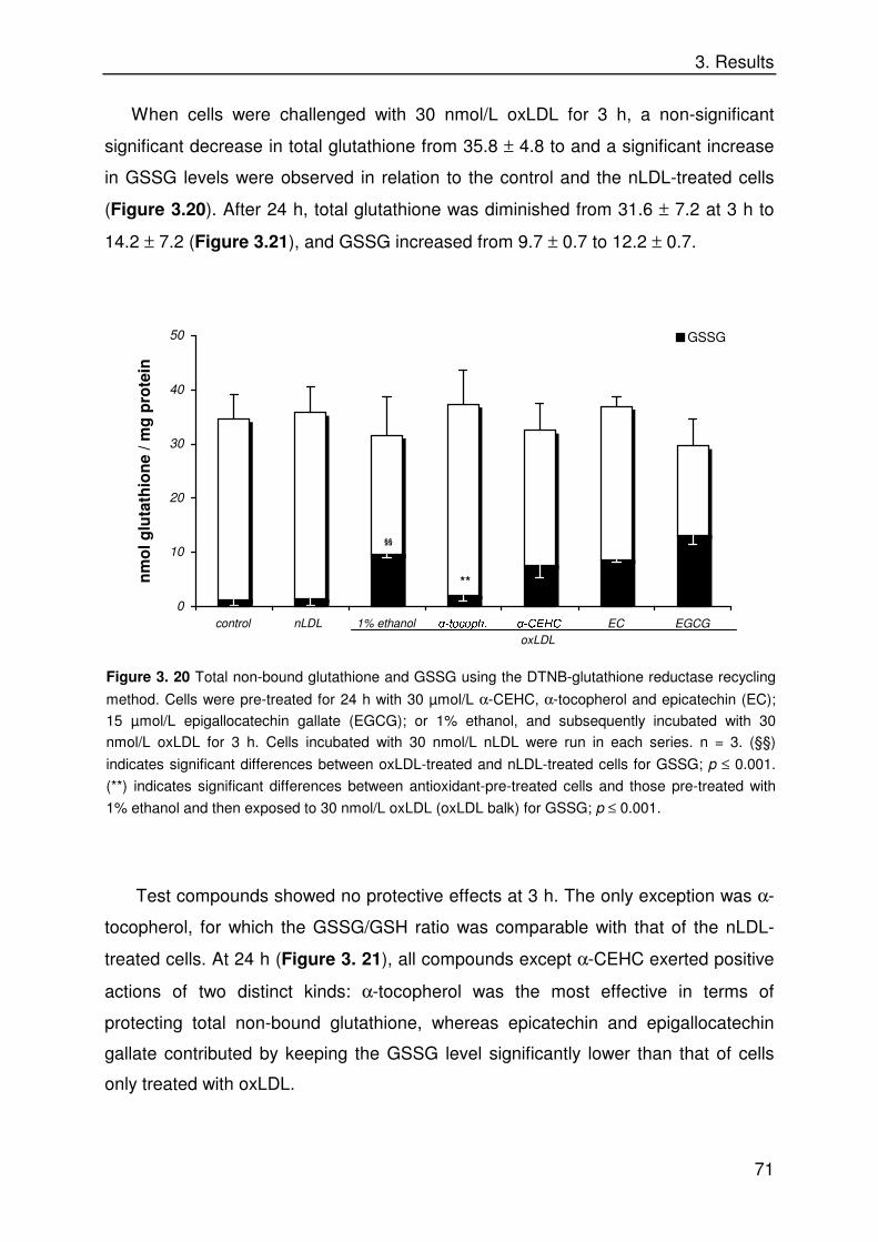
3. Results
71
When cells were challenged with 30 nmol/L oxLDL for 3 h, a non-significant
significant decrease in total glutathione from 35.8 ± 4.8 to and a significant increase
in GSSG levels were observed in relation to the control and the nLDL-treated cells
(Figure 3.20). After 24 h, total glutathione was diminished from 31.6 ± 7.2 at 3 h to
14.2 ± 7.2 (Figure 3.21), and GSSG increased from 9.7 ± 0.7 to 12.2 ± 0.7.
Test compounds showed no protective effects at 3 h. The only exception was α-
tocopherol, for which the GSSG/GSH ratio was comparable with that of the nLDL-
treated cells. At 24 h (Figure 3. 21), all compounds except α-CEHC exerted positive
actions of two distinct kinds: α-tocopherol was the most effective in terms of
protecting total non-bound glutathione, whereas epicatechin and epigallocatechin
gallate contributed by keeping the GSSG level significantly lower than that of cells
only treated with oxLDL.
Figure 3. 20 Total non-bound glutathione and GSSG using the DTNB-glutathione reductase recycling
method. Cells were pre-treated for 24 h with 30 µmol/L α-CEHC, α-tocopherol and epicatechin (EC);
15 µmol/L epigallocatechin gallate (EGCG); or 1% ethanol, and subsequently incubated with 30
nmol/L oxLDL for 3 h. Cells incubated with 30 nmol/L nLDL were run in each series. n = 3. (§§)
indicates significant differences between oxLDL-treated and nLDL-treated cells for GSSG; p ≤ 0.001.
(**) indicates significant differences between antioxidant-pre-treated cells and those pre-treated with
1% ethanol and then exposed to 30 nmol/L oxLDL (oxLDL balk) for GSSG; p ≤ 0.001.
0
10
20
30
40
50
control nLDL 1% ethanol .�WRFRSK� .�&(+& EC EGCG
nm
ol g
luta
thio
ne
/ mg
pro
tein
GSSG
**
§§
oxLDL
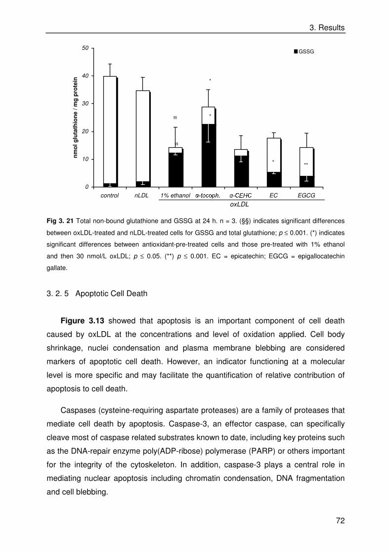
3. Results
72
Fig 3. 21 Total non-bound glutathione and GSSG at 24 h. n = 3. (§§) indicates significant differences
between oxLDL-treated and nLDL-treated cells for GSSG and total glutathione; p ≤ 0.001. (*) indicates
significant differences between antioxidant-pre-treated cells and those pre-treated with 1% ethanol
and then 30 nmol/L oxLDL; p ≤ 0.05. (**) p ≤ 0.001. EC = epicatechin; EGCG = epigallocatechin
gallate.
3. 2. 5 Apoptotic Cell Death
Figure 3.13 showed that apoptosis is an important component of cell death
caused by oxLDL at the concentrations and level of oxidation applied. Cell body
shrinkage, nuclei condensation and plasma membrane blebbing are considered
markers of apoptotic cell death. However, an indicator functioning at a molecular
level is more specific and may facilitate the quantification of relative contribution of
apoptosis to cell death.
Caspases (cysteine-requiring aspartate proteases) are a family of proteases that
mediate cell death by apoptosis. Caspase-3, an effector caspase, can specifically
cleave most of caspase related substrates known to date, including key proteins such
as the DNA-repair enzyme poly(ADP-ribose) polymerase (PARP) or others important
for the integrity of the cytoskeleton. In addition, caspase-3 plays a central role in
mediating nuclear apoptosis including chromatin condensation, DNA fragmentation
and cell blebbing.
0
10
20
30
40
50
control nLDL 1% ethanol .�WRFRSK� .�&(+& EC EGCG
nm
ol g
luta
thio
ne
/ mg
pro
tein
GSSG
*
§§
*
§§
*
**
oxLDL

3. Results
73
Fig. 3. 22 Caspase 3 activity 0, 2 and 4 h after addition of 30 nmol/L nLDL or oxLDL and effect of
pre-incubation with antioxidants. n = 5. (§§) indicates significant differences between oxLDL-treated
and nLDL-treated cells; p ≤ 0.001. (*) indicates significant differences between antioxidant-pre-treated
cells and those pre-treated with 1% ethanol and then 30 nmol/L oxLDL; p ≤ 0.05. (**) p ≤ 0.001. Upper
graph: caspase 3 contribution to cell death triggered by incubating with 30 nmol/L oxLDL for 24 h.
Cells were pre-incubated with (left column) or without (middle column) a caspase-3 specific inhibitor
for 30 min prior to oxidative challenge. EC = epicatechin; EGCG = epigallocatechin gallate.
Involvement of caspase-3 in the cytotoxic effects of oxLDL was demonstrated.
Addition of the caspase-3 inhibitor DEVD-CHO before incubation with oxLDL
increased cell viability by 50% after 24 h when compared with cells only treated with
oxLDL. About 50% cell death after 24 h could be thus directly attributed to caspase-3
activation (small panel in Figure 3.22).
Caspase 3 activity rapidly increased within the first 4 h in cells which were pre-
incubated with 1% ethanol or α-CEHC; with the flavonols, caspase 3 increase was
moderate, and with α-tocopherol it was extremely low.
0
1
2
3
4
nLDL oxLDL .-tocopherol .-CEHC epicatechin epig. gallate
Absorbance
405 nm
(AU x 10
0 h
2 h
4 h
**
**
**
**
*
** **
§§
§§
0
25
50
75
100
125
nLDL oxLDL 100 µM DEVD-
CHO/ oxLDL
MTT Turnover (%)
*
0
1
2
3
4
nLDL oxLDL .-tocopherol .-CEHC EC EGCG
Ab
sorb
ance
405
nm
(
AU
x 1
0)
0 h
2 h
4 h
**
**
**
**
*
** **
§§
§§
0
25
50
75
100
125
nLDL oxLDL 100 µM DEVD-
CHO/ oxLDL
MT
T T
urn
ove
r (%
)
*
1% ethanol
oxLDL
3. Results
74
3. 3 Human Intervention Study on the Effects of Smoking on Vitamin E Meta-
bolism after a Single Dose of Vitamin E
α- and γ-CEHC are metabolites of α- and γ-tocopherol and -tocotrienol in human
blood; γ-CEHC (also called LLU-α) has been shown by others to possess antioxidant
activity and effects at cellular level such as prevention of inflammatory responses on
in macrophages and epithelial cells or growth arrest of a prostata carcinoma cell line;
for α-CEHC, functions in vivo are unknown.
In this work, α-CEHC has been shown to act as an antioxidant in aqueous
solution, being able to scavenge ABTS•+, peroxyl radicals and peroxynitrite with an
efficacy comparable to Trolox. Thus, so far it may be affirmed that α- and γ-CEHC
contribute as antioxidants to the antioxidant defense system; in contrast to their
parent compounds, which are active in lipoproteins and cell membranes, they likely
act in the aqueous phase. Therefore, the question was addressed whether conditions
of oxidative stress may influence the levels of α- and γ-CEHC in vivo. Smoking is a
situation where oxidative stress is increased [Polidori et al., 2001; Polidori et al.,
2003] and was investigated in this study.
3. 3. 1 HPLC Analysis of α- and γ-CEHC in Serum
Under the conditions of analysis (for details see 2. 6. 2), recovery of α-CEHC
was 99.6 ± 1.5% (n = 4) and that of γ-CEHC was 87.9 ± 2.8% (n = 3). Addition of
antioxidants (vitamin E, vitamin C) during extraction was necessary to avoid
decomposition of the analytes.
A chromatogram of a human serum sample is shown in Figure 3.23. The
analytes are clearly separated and eluted with retention times of 12.3 min for α-
CEHC and 8.6 min for γ-CEHC. The limit of quantification in serum was 1.5 nmol/L
for both compounds; the detection limit was 0.75 nmol/L at a signal to noise ratio of
3/1. For both compounds the signal was linear in the range of 0.75-750 nmol/L,
related to the final concentration in the sample. Linear regression coefficients were
0.9997 for α-CEHC and 0.9999 for γ-CEHC. Coefficients of variation for within-day
precision in three independent experiments ranged from 2.6 to 5.1% for α-CEHC and

3. Results
75
from 2.8 to 3.9% for γ-CEHC. Inter-day precision was calculated with coefficients of
variation of 2.7% for α-CEHC and 1.8% for γ-CEHC.
3. 3. 2 HPLC Analysis of α- and γ-Tocopherol
Reference compounds were used for tocopherol identification. Pool serum was
regularly analysed and served as quality controls for tocopherol analysis. δ-
tocopherol (2 µg/sample) was used as an internal standard. Recovery of the internal
standard was reproducible during the study. Coefficient of variation corresponding to
the height of the internal standard signal was 15%. For quantitation, the ratio
between peak height of the analyte and that of δ-tocopherol was used; based on a
regression curve calculated from calibration samples (r > 0.998), the concentration of
the analytes in the samples was calculated.
Fig 3. 23 HPLC chromatogram of standards (left);
γ-CEHC standard (201.5 nmol/L) eluted at 8.6
min, α-CEHC standard (177.3 nmol/L) eluted at
12.3 min. The chromatogram on the right shows a
human serum sample.
0 5 10 15 20 0 5 10 15 20 25 30 35
2 m
V
time (min)
γ-CEHC
γ-CEHC
α-CEHC
α-CEHC

3. Results
76
3. 3. 3 General Remarks
There was a pronounced inter-individual variability in the uptake and
biotransformation of the orally applied α-tocopherol (306 mg), which was
independent of factors like smoking or gender. This phenomenon is already known
not only for α-tocopherol but also for other isomers of vitamin E. Using the non-
parametric Wilcoxon rank-sum test, over-time changes of all parameters were
significant (p < 0.001). Besides, certain trends of interest deserve special attention.
In Figure 3.25, individual responses for α-tocopherol serum levels in smoking
and non-smoking subjects are shown. The maximal concentrations of α-tocopherol
occur between 6 and 12 h after supplementation. From Figure 3.26, where mean
values and standard deviations are presented, a net average increase of 8.87 µmol/L
for smokers and 12.90 µmol/L for nonsmokers was calculated at the tmax (12
Abs
orba
nce
292
Fig 3. 24 Typical HPLC chromatogram from
tocopherol analysis in human serum under the
conditions specified in 2. 6. 3.
inte
rnal
sta
ndar
d
γ-to
coph
erol
α
-toc
ophe
rol
time (min)
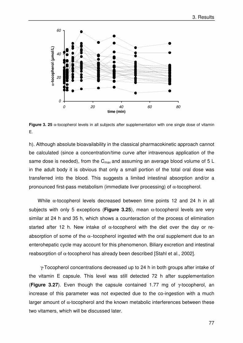
3. Results
77
Figure 3. 25 α-tocopherol levels in all subjects after supplementation with one single dose of vitamin
E.
h). Although absolute bioavailability in the classical pharmacokinetic approach cannot
be calculated (since a concentration/time curve after intravenous application of the
same dose is needed), from the Cmax and assuming an average blood volume of 5 L
in the adult body it is obvious that only a small portion of the total oral dose was
transferred into the blood. This suggests a limited intestinal absorption and/or a
pronounced first-pass metabolism (immediate liver processing) of α-tocopherol.
While α-tocopherol levels decreased between time points 12 and 24 h in all
subjects with only 5 exceptions (Figure 3.25), mean α-tocopherol levels are very
similar at 24 h and 35 h, which shows a counteraction of the process of elimination
started after 12 h. New intake of α-tocopherol with the diet over the day or re-
absorption of some of the α−tocopherol ingested with the oral supplement due to an
enterohepatic cycle may account for this phenomenon. Biliary excretion and intestinal
reabsorption of α-tocopherol has already been described [Stahl et al., 2002].
γ-Tocopherol concentrations decreased up to 24 h in both groups after intake of
the vitamin E capsule. This level was still detected 72 h after supplementation
(Figure 3.27). Even though the capsule contained 1.77 mg of γ-tocopherol, an
increase of this parameter was not expected due to the co-ingestion with a much
larger amount of α-tocopherol and the known metabolic interferences between these
two vitamers, which will be discussed later.
0
20
40
60
0 20 40 60 80
time (min)
αα
-to
cop
her
ol (
µm
ol/L
)

3. Results
78
Figure 3. 26 α-Tocopherol serum concentrations in smokers and nonsmokers after supplementation
with vitamin E. Concentrations in the right graph are corrected for cholesterol.
It should be noted that baseline α-tocopherol levels corresponding to both groups
together but not those of γ-tocopherol were significantly correlated with baseline
cholesterol levels (r = 0.680; p ≤ 0.001). Hence, α-tocopherol was also expressed per
g serum cholesterol, the corrected values were plotted to search for differences with
the former values and are shown in Figure 3.26. It is obvious that kinetic patterns are
the same for cholesterol-normalized or uncorrected α-tocopherol, however,
differences between smokers and nonsmokers are more obvious. Also noteworthy is
the fact that baseline levels of α-tocopherol were about 10 fold those of γ-tocopherol,
irrespective of smoking and gender, whereas no linear correlations between baseline
α-tocopherol and γ-tocopherol levels were found.
0
10
20
30
40
50
0 20 40 60 80
time (h)
αα
-to
cop
her
ol (
µm
ol/L
)
smokers
nonsmokers
0
10
20
30
0 20 40 60 80
time (h)
µm
ol α α
-to
cop
her
ol/
g C
ho
lest
ero
l
smokers
nonsmokers

3. Results
79
α-Tocopherol concentrations were 2000-fold higher than those of its metabolite
α-CEHC; γ-tocopherol levels were only 17 to 19 times higher than those
corresponding to γ-CEHC. This is a striking indicator of the different metabolism of
both vitamers in humans. On the other hand, no significant correlations between
baseline α-tocopherol and α-CEHC levels nor between baseline γ-tocopherol and γ-
CEHC levels were found. Thus, under unsupplemented conditions, the fates of α-
and γ-tocopherol rather depend on inter-individual variability in the metabolic
machinery than on blood tocopherol concentrations.
Figure 3. 27 γ-tocopherol serum concentrations after a single oral dose of vitamin E.
3. 3. 4 Differences between Smokers and Nonsmokers in Vitamin E Metabolism
Baseline levels of α-tocopherol were similar for smokers and nonsmokers (21.74
± 6.51 and 22.40 ± 7.84 µmol/L, respectively). However, the increment was
proportionally higher in nonsmokers (59% at the tmax) when compared to smokers
(only 38%). Despite sharing a comparable kinetics, mean values for nonsmokers
were higher than those for smokers at all time points. The direct consequence was
that smokers reached baseline levels of α-tocopherol faster (at 50 h, vs. 72 h or later
for nonsmokers).
0
1
2
3
4
0 20 40 60 80
time (h)
-to
cop
her
ol (
µm
ol/L
)
smokers
nonsmokers

3. Results
80
Differences in serum levels of α-CEHC between both groups were almost
undetectable. However, the trend was inverse to that detailed above for α-tocopherol,
showing smokers slightly higher values (Figure 3. 28).
Figure 3. 28 α-CEHC (left) and γ-CEHC (right) levels in smokers and nonsmokers after a single dose
of vitamin E.
The most evident differences between both groups were found in γ-CEHC levels.
The mean baseline level of γ-CEHC in nonsmokers is 1.5-fold higher than that of
smokers. This difference is maintained or even amplified over time (after a short
convergence of the values) upon supplementation. In view of the data shown here, it
is clear that a single dose of α-tocopherol accelerates the degradation of
endogenous γ-tocopherol for a short period of time in smokers, but it is uncertain in
which way it affects γ-tocopherol metabolism in smokers. However, it may be
affirmed that a larger amount of γ-tocopherol is continually transformed to γ-CEHC in
smokers, irrespective of the supplementation with α-tocopherol.
3. 3. 5 Further data of relevance
Very few intervention studies on vitamin E metabolism in humans involving blood
levels of CEHC´s have been performed up to date, and the number of subjects
0
15
30
45
60
0 20 40 60 80
time (h)
CE
HC
(n
mo
l/L)
smokers
nonsmokers
0
50
100
150
200
250
300
0 20 40 60 80
time (h)
-CE
HC
nm
ol/L
smokers
nonsmokers

3. Results
81
investigated is still very low. Hence, normal or reference values of those metabolites
are missing. So far, it seems clear that the enormous range of responses in blood
tocopherol levels among individuals after supplementation is also reflected in CEHC
levels.
In our study, two volunteers were found for whom values or orders of magnitude
of some parameters clearly differ from median and mean value of their corresponding
group.
Subject 1 was a healthy 26-year-old male smoker with a body mass index (BMI)
of 22.89; subject 2 was also a healthy male smoker, aged 33, with a BMI of 19.79.
Subject 1 was slightly hypercholesterolemic. Baseline data of these volunteers as
compared to the mean ± standard deviation corresponding to all smokers studied are
presented in Table 3.29:
Table 3. 29 Baseline serum concentrations of subjects 1 and 2 in comparison to
baseline levels of smokers.
cholesterol
(g/L)
αα-tocopherol
(µmol/L.g chol)
γγ-tocopherol
(µmol/L)
αα-CEHC
(nmol/L)
γγ-CEHC
(nmol/L)
n 13 13 17 14 14
subject 1 2.13 (2) 7.9 (13) 1.7 (10) 36.7 (1) 1096 (2)
subject 2 1.44 (9) 16.6 (2) 4.9 (1) 13.7 (7) 1099 (1)
smokers:
median 1.58 12.7 2.1 14.5 130
mean ± SD 1.64 ± 0.36 13.0 ± 2.7 2.3 ± 1.2 16.5 ± 8.3 153 ± 79
Ranks of the subjects for each parameter are presented in brackets. (1) = the highest.
In both subjects, overall γ-CEHC levels were 6 to 7 times higher than the mean
for smokers. In subject 1, overall α-CEHC levels were the highest (2 to 5 times higher
than the mean for smokers) and overall α-tocopherol (including non-normalized
values) was the lowest among smokers. Subject 2 was normocholesterolemic and
showed very high overall levels of α- and γ-tocopherol. His α-CEHC levels after the
baseline were about one half the values corresponding to the mean for smokers.

3. Results
82
An apparent metabolic ratio was calculated for every time point by dividing the
concentration of each metabolite (α- or γ-CEHC, nmol/L) by the concentration of the
corresponding tocopherol (µmol/L).The word apparent is added to clarify that:
a) This parameter does not correspond to the metabolic ratio used in formal
pharmacokinetics, for which urine amounts of the metabolite are needed. Thus, the
major factor neglected is renal excretion of the metabolite; it should be pointed out,
though, that renal clearance in healthy subjects mainly depends on the solubility and
molecular weight of the substance excreted itself.
b) It is assumed that α- and γ-CEHC are the major final metabolites of α- and γ-
tocopherol, respectively.
From Figure 3.30, it is evident that the increase in α-CEHC levels following
vitamin E intake greatly exceeded the increase in α-tocopherol levels, since the
apparent metabolic ratio increased to two-fold of the baseline at 12/24 h. This reflects
that acute loads of α-tocopherol are subject to a rapid and massive degradation so
that the pool of α-tocopherol already present is preserved.
Putting the data from Table 3.29 and Figure 3.30 together, it may be affirmed
that subject 1 is an extensive metabolizer of both vitameres; subject 2 holds a large
endogenous pool of vitamin E, metabolizes γ-tocopherol extraordinarily fast and is a
slow metabolizer of newly-ingested α-tocopherol.

3. Results
83
Figure 3. 30 Over-time α-tocopherol and γ-tocopherol apparent metabolic ratios. These were
calculated by dividing concentrations of each metabolite (CEHC, in nmol/L) by the levels of the
corresponding parent compound (tocopherol, in µmol/L) at all time points. Plotted values represent
mean ± SD for all smokers or all nonsmokers; single ratios for two volunteers are also presented (see
3. 3. 5).
0
1
2
3
4
5
6
0 20 40 60 80
time (h)
αα
-to
cop
her
ol m
etab
olic
rat
io (
nm
ol/µ
mo
l)smokers
nonsmokers
subject 1
subject 2
0
500
1000
1500
2000
2500
0 20 40 60 80
time (h)
γγ-to
cop
her
ol m
etab
olic
rat
io (
nm
ol/µ
mo
l)
smokers
nonsmokers
subject 1
subject 2

4. Discussion
84
4. DISCUSSION
4. 1 Antioxidant Activity of Dietary Micronutrients in Model Systems
Nearly all single dietary micronutrients were active in the systems investigated. A
potential antioxidant activity of harpagoside, due to the presence of hydroxyl groups
in the bicyclic ring (see Figure 3.6), was not confirmed, however. In general, the
ranking of antioxidant activities of the single compounds obtained by applying the
TEAC assay (see Table 3.1) could be explained by specific structural features (see
below), and the activities of dietary extract fractions were strictly related to the
contents of polyphenols (Table 3.7).
4. 1. 1 TEAC and ORAC Assays
• Polyphenols – phenylpropanoids
Among flavonoids, the flavonol kaempferol, containing a single 4´-hydroxyl group
on the B ring (Figure 3.6), exerted the lowest activity measured in the TEAC assay
(Table 3.1). Both the flavan-3-ol epicatechin and the flavone luteolin, which share a
catechol group as B ring showed intermediate activities. Epigallocatechin gallate,
with three neighbouring hydroxyl groups on the B ring (Figure 1.8), exhibited a much
better antioxidant activity than epicatechin. It is thus evident that the electron-
donating capacity of the molecule becomes enhanced by increasing the number of
adjacent hydroxyl groups on the B ring. It should be pointed out that among
flavonoids with a 4´-monohydroxyl group (e. g. apigenin, pelargonidin), kaempferol
has been identified as the best antioxidant due to the following reasons: 1) the B ring
is conjugated with the C ring (2, 3 positions and the 4-carbonyl group) and 2) the 3-
hydroxyl group enhances the potential for conjugation [Pannala et al., 2001]. This
should be the rationale for the TEAC value of kaempferol being so close to that of
epicatechin.
In cholorogenic acid, the electron delocalization over the catechol ring is
expanded over the propenoate chain (Figure 3.6). In view of the TEAC values,
however, the antioxidant activity is higher when a chromanol ring is bound to the
catechol as in flavonoids.
Both catechol rings of curcumin are partially hindered by methylation of one of
the hydroxyl groups (Figure 3.6). Nonetheless, the introduction of a methoxy group

4. Discussion
85
ortho to phenol increases the antioxidant activity of unsubstituted monophenols by
probable two mechanisms: 1) it enhances the stability of the phenoxyl radical by an
inductive effect, and 2) steric hindrance disturbs the formation of hydrogen bonds to
the medium, thus enhancing the transfer of the hydrogen atom (in a manner
comparable to the methyl groups in α-tocopherol). It seems that curcuminoids act as
classical chain-breaking antioxidants like α-tocopherol; their antioxidant activity
decreases in hydrogen bond forming media and synthetic, non-phenolic
curcuminoids are mostly inactive [Barclay et al., 2000]. It is noteworthy that curcumin
is stabilized by a keto-enol tautomeric form, which confers the molecule its
characteristic color, under formation of an intramolecular hydrogen bond so that
hydrogen abstraction from the central carbon is unlikely and should not contribute to
the antioxidant properties.
Two distinct patterns have been described concerning the general mechanisms
underlying the antioxidant activity of phenylpropanoids. It appears that the oxidation
of catechol-containing flavonoids consists of a two-electron donation process yielding
a semiquinone and successively an ortho-quinone [Jovanovic et al., 1998, Pannala et
al., 2001]. On the other hand, hindered and single phenols react via formation of
phenoxyl radicals, of which a part dimerise and another fraction may continue to act
as antioxidants yielding phenolates. The biphasic responses obtained for curcumin
and the turmeric fractions B, C and D (Figure 3.5) are hence explained as a result of
curcumin radicals being formed and coupled in the reaction mixture to yield insoluble
dimers and eventually larger polymers that precipitate [Masuda et al., 1999]. Such
reactions are favoured in the present system at curcumin concentrations above 4
µmol/L.
• Monophenolic compounds
α-Tocopherol and α-CEHC exhibited similar antioxidant capacity as Trolox in the
TEAC assay due to their similar molecular structure. α-Tocopherol could not be
tested in the ORAC assay (see below).
• Carotenoids
All the carotenoids induced a decrease in the radical cation absorption. However,
results demonstrate substantial differences between the various types of carotenoids.
4. Discussion
86
The higher scores of β-carotene and zeaxanthin (β-rings only) with respect to α-
carotene and lutein (one β-ring plus one ε-ring) are explained by the fact that the
double bond in ε-rings is not conjugated with the polyene chain, and electron
delocalisation is thus more restricted (Figure 1.6). Besides, the negative charge
density on C-4 of the ε-ring is clearly higher than that on C-4 of the β-ring
[Hernández-Blanco et al., in preparation]. In xanthophylls, the charge distribution is
further affected by the presence of hydroxyl groups. In the 3´-hydroxyl-ε-ring (lutein),
the charge skip between carbon 3´ –with a net positive charge in all compounds- and
4´ is maximised. These differences in charge distribution likely affect reactions
involving terminal rings, such as electron abstraction. The stability of the carotenoid
radical generated by such mechanism when an ε-ring is present is lower and may
account for the lower TEAC value of ε-ring-containing compounds.
It is remarkable that the presence of a hydroxyl group decreases the antioxidant
activity in the case of lutein, when compared with α-carotene, but not that of
zeaxanthin in comparison with β-carotene (non-significant difference between TEAC
values). It has been speculated that the 3´-position on ε-rings is a reactive one (e. g.,
for hydrogen abstraction and/or hydroxylation), and might be hindered by the
hydroxyl groups as in lutein or isozeaxanthin [Palozza et al., 1995; El-Tinay et al.,
1970]. This effect has also been observed in the reaction of different carotenoids with
AMVN and AIBN [Woodall et al., 1997].
Capsorubin exerted the highest radical quenching activity. This ketocarotenoid
has two 3-hydroxyl-6-oxo-κ end groups. The carbonyl groups cause a pronounced
electronic charge skip between C-6 of the κ-groups and the polyene chain
[Hernández-Blanco et al., in preparation], and strongly stabilise the polyene chain by
an electron withdrawal effect, which is most appropriate for the delocalisation of the
unpaired electron of the carotenoid radical intermediates generated by addition. Even
though charge distribution over the central part of the molecule is the same in
capsorubin and capsanthin, a very different distribution is observed at the end groups
due to the asymmetry of the latter, which accounts for its lower antioxidant ability with
respect to capsorubin. This effect has been reported previously by other authors
[Terao, 1989; Martin et al., 1999; Pérez-Gálvez and Mínguez-Mosquera, 2002].
Astaxanthin has been reported to be a good antioxidant in comparison with other
carotenes and xanthophylls [Terao, 1989; Miki, 1991]. A symmetrical charge

4. Discussion
87
distribution along the molecule is observed in canthaxanthin and astaxanthin, with
high charge skips close to the hydroxyl and keto groups, but not immediate to the
polyene chain as in the case of capsorubin [Hernández-Blanco et al., in preparation].
As addressed in the introduction, antioxidants supplied with the diet or with
dietary supplements undergo a complex process until they reach the target tissue,
involving liberation from the food matrix, absorption through the gastro-intestinal tract
and distribution into different organs via blood circulation. Partial degradation of
micronutrients to inactive compounds may occur even before target tissues are
reached, e. g. due to colonic microorganisms or immediate biotrans-formation in the
liver after gut absorption. Thus, it should be pointed out that the antioxidant activity of
micronutrients in vivo is influenced by a number of factors not contemplated in
conditions of experimentation in vitro.
• Pharmaceutical Preparations
In the turmeric preparation, curcuminoids were the most abundant constituents.
The contribution of β-carotene, as single carotenoid detected, to the antioxidant
capacity was less than 0.1% of the absolute amount. This indicates that further
antioxidant compounds must be present, since the water-soluble fraction A shows a
high activity. Since this fraction is rich in phenols (Table 3.7) but curcumin is
insoluble in water, phenolic decomposition products of curcumin, such as ferulic acid,
vanillin or vanillic acid (Masuda et al 1999) may account for this effect.
The major antioxidant capacity of the artichoke leaf preparation was found in
fractions A and B coinciding with the presence of phenols. Most flavonoid glucosides
as well as hydroxycinnamates are soluble in these solvents [Macheix and Fleuriet
1998]. Artichoke leaves are particularly rich in caffeic acid and caffeic acid esters with
quinic acid (e. g. chlorogenic acid and the diester cynarin, which is almost exclusively
found in artichokes). Since water is the extraction solvent used in the manufacture of
the commercial product, the low antioxidant properties found in the more lipophilic
fractions C and D are realistic.
Results for devil´s claw preparation are consistent with the fact that water-
extractable substances account for about 70% the dry weight of the roots. In view of
the weak antioxidant activity of harpagoside, contribution by other iridoids, such as
harpagide or procumbide, to the antioxidant activity, is excluded because of their high

4. Discussion
88
structural similarity. According to linear regression data, phenols significantly
contribute to the antioxidant activity. Major phenols in devil´s claw roots are glycated
forms of flavonoids like those of kaempferol or luteolin.
The garlic tablets rendered low antioxidant activities for all fractions. There are
more water-soluble than lipid-soluble antioxidants present in garlic extract. TEAC and
total phenols correlated among treatments but other compounds present in garlic
extract are also Folin-active, particularly those containing sulfhydryl and disulfide
groups.
Results for the salmon oil reveal that all the antioxidant activity found in the oil in
solution is related to α-tocopherol, added to the product for stabilization.
It should be noted that the results obtained for each fraction of each preparation
are not additive, as some of the antioxidant constituents dissolve in more than one
medium.
Today, 59% of the population in USA report taking dietary supplements on a
regular basis. Of these regular supplement users, almost half (46%) take a multiple
vitamin/mineral product on a daily basis and more than a third (35%) take single
vitamins, such as vitamin C, vitamin E, or vitamin B complex. Almost 23% regularly
consume herb/plant and specialty supplements (such as glucosamine/chondroitin).
15% adults currently consume such plant supplements as garlic, ginseng and St.
John's Wort [Dietary Supplement Education Alliance, 2001].
Even though the manufacturer´s claims on health benefits of dietary supplements
become increasingly regulated, such statistics constitute a rationale for the chemical
evaluation of those products and the closer examination of potential biological
activities derived therefrom. The total antioxidant capacity of widespread dietary
supplements and the relationship to their antioxidant contents was assessed in this
work as contribution to that concern.
4. 1. 2 Methodological Aspects
The ranks of antioxidant capacity were dependent on the assay applied (e. g. for
ascorbic acid), yet for some of the compounds consistent with already published
values [Pannala et al., 1991; Cao and Pryor, 1999; Re at al., 1999]. The diversity of
nature of the radicals (or radical generators) and discrepancy in the redox potential of

4. Discussion
89
the oxidant/reductant pair account for this effect. Testing antioxidant activities or
capacities in more than one system is thus indispensable.
There has been an intense debate on the question which measure for Total
Antioxidant Capacity is most appropriate from the increasing number of methods
being developed, especially in order to prospect biological fluids such as blood
plasma [Duthie, 1999; Prior and Cao, 1999; Rice-Evans, 2000]. Prior and Cao
repeatedly claimed the superiority of their method, the ORAC assay, over others like
the TRAP or the FRAP assays, for being able to determine both inhibition time and
inhibition percentage and unique in taking the reaction to completion, which enables
the use of an “area under the curve (AUC)” technique for quantification [Cao and
Prior, 1999]. However, later improvements made on the original TEAC assay (the
ferryl myoglobin/ABTS procedure) [Miller et al., 1993], by substituting
H2O2/metmyoglobin by potassium persulfate [Re et al., 1999] –the procedure applied
in this work-, provided the TEAC assay in practice with the same advantages which
the ORAC has.
To be tested in the ORAC assay, α-tocopherol needed a high amount of organic
co-solvent, thereby changing the habitual reaction kinetics, which led to assume that
the presence of ethanol had affected the stability of the protein. The ORAC value for
α-tocopherol has been reported to be 1.0 by making the score for the more
hydrosoluble α-tocopheryl succinate applicable for α-tocopherol (Cao et al., 1993) –
however, the peroxyl radical scavenging capacity of α-tocopherol resides in the free
6-hydroxyl moiety of the chroman ring, which is protected and slowly released in α-
tocopherol succinate. Likewise, an ORAC value of 1 was assigned to α-tocopherol
under the consideration of Trolox being the water- soluble analogue of α-tocopherol
[Pieri at al., 1994], even though such a statement is tentative.
Intra- and inter-day variability concedes the TEAC assay a relative superiority. It
is noteworthy that coefficients of variability obtained in the ORAC assay for this work
are much higher than those corresponding to experiments which were performed
with an automated analyser [Cao et al., 1995]. On the other hand, neither the TEAC
nor the ORAC assay at their actual state of development are reliable methods to
prospect the total antioxidant capacity of biological fluids [Rice-Evans, 2000].
In favour of the ORAC assay count both a more physiological reaction with
peroxyl radicals and the use of a biological target (protein) as the marker of oxidative
damage; the novel TEAC assay is nevertheless much less time-consuming and

4. Discussion
90
applicable to lipophilic compounds such as α-tocopherol. Both may be performed as
complementary methods to screen the antioxidant activity of single compounds or
mixtures except for biological fluids as plasma.
4. 1. 3 Inhibition of Peroxynitrite-mediated Tyrosine Nitration
Epicatechin was the most efficient inhibitor of tyrosine nitration. α-CEHC and
Trolox showed a comparable inhibitory capacity.
In Trolox and α-CEHC, the benzene moiety of the chromanol ring is completely
substituted like in α-tocopherol so that nitration is not possible, and pure electron
donation must be responsible for the protective effects shown. Recent data for α-
tocopherol suggest that the reaction occurs via two-electron transfer yielding a stable
α-tocopherol-quinone which would not be reduced back to α-tocopherol e. g. by
ascorbic acid [Hogg et al., 1994]. Vitamin E has been subject of profound research to
elucidate its role as RNS scavenger [Christen at al., 1997; Goss at al., 1999].
In epicatechin, the possibility of nitration on the cromanol ring is also low since
the two hydroxyl groups are located meta to each other and π-electron delocalisation
is weakened. However, positions ortho to the hydroxyl groups on the catechol ring
(2´and 5´) are strongly activated by an electron resonance effect (+M). This should
enable the formation of stable nitro- adducts in those positions upon reaction with
peroxynitrite. It is likely that epicatechin thereby competes with an intermediate
tyrosine radical, since epicatechin´s IC50 against peroxynitrite-mediated dityrosine
formation has been shown to be comparable to its IC50 of 3-nitrotyrosine generation
[Schröder et al., 2001]
The antioxidant properties of α-CEHC were examined in the three model
systems with positive results. A number of synthetic water-soluble vitamin E
analogues are being prospected for advantages over the lipophilic tocopherols in the
via of administration (i.v. vs. p.o.) and/or in the ability to offset sudden rises of
reactive species as occurring in reperfusion after ischemia [Abadie at al., 1993],
radical-mediated hepatotoxicity [Campo et al., 2001], myocardial infarction [Campo et
al., 1994] and possibly other types of CVD [Campo et al., 1997]. These compounds
share an intact or acetylated chroman ring but vary in the type of functional groups or
length of the side chain. Some have succeeded at animal experimentation and in
initial intervention trials [Chowienczyk et al., 2000]. Furthermore Trolox, a further

4. Discussion
91
water-soluble α-tocopherol analogue, is widely employed in in vitro, cell or animal
model studies. Trolox significantly enhanced cardiac recovery after ischaemia
/reperfusion, both when it was perfused in isolated hearts and after its oral
administration to rats, and its activity was better than that of vitamin E [Sagach et al.,
2002]. Since the solubility and structural features of such analogues are shared by α-
CEHC, with the plus of being a natural-occurring compound, α-CEHC should be
tested for similar applications.
4. 2 Cell Culture Study on the Anti-atherogenic Effects of Dietary Micro-
nutrients and αα-CEHC
Findings from the cellular pro-atherogenic system suggest that:
• pre-treatment with α-tocopherol, epicatechin and epigallocatechin gallate at
physiological concentrations is able to transiently prevent cell damage and
significantly prolong cell survival, and
• pre-incubation with α-CEHC has neither impact on cell damage nor can enlarge
cell viability even at super-physiological levels.
Only α-tocopherol was able to scavenge efficiently intracellular ROS that were
generated as an early event in response to oxLDL (Figure 3.17). Membrane-bound
NAD(P)H oxidase has been found to be responsible for the bulk of ROS (mainly O2.-)
produced in oxLDL-stressed endothelial cells [Heinloth et al., 2000] which is in turn
activated through the LOX-1 receptor [Cominacini et al., 2000] NOS impairment
occurs concomitantly and NO production is drastically diminished [Galle et al., 2000].
α-Tocopherol is mainly positioned within membranes and thus closer to the site of
O2.- generation, which may account for its superiority in ROS scavenging.
Since the amounts of DiI-labelled oxLDL and DiI-labelled nLDL taken up by
MAEC were similar at each point studied (Figures 3.15 and 3.16), it appears that the
uptake rates for both lipoprotein types were similar. Scavenger receptors (mainly
LOX-1 in endothelial cells) take up highly oxidized LDL at a faster rate than the nLDL
receptor is able of taking up mildly oxidized LDL. Thus, one could assume that the
labelled oxLDL used here (obtained at conditions of low oxidation) was recognized
and taken up mainly by the LDL receptor.
OxLDL at the mild level of oxidation applied in our experiments caused a
significant decrease of total levels of glutathione and a deterioration of the

4. Discussion
92
glutathione oxidative status by increasing GSSG levels. The three dietary
antioxidants prevented glutathione depletion by two apparently different mechanisms.
α-Tocopherol showed a protective effect on total glutathione but could not restrain
GSSG levels over a long time. Both catechins were able to maintain levels of GSSG
significantly lower to those of cells without pre-treatment with antioxidants, but had no
positive effect on total glutathione, at least in view of the data corresponding to 24 h
(Figures 3.20 and 3. 21).
Comparable effects were observed in another study where bovine endothelial
cells were challenged with mildly oxidized LDL. α-Tocopherol was shown to prevent
glutathione and ATP depletion as well as to delay the cytotoxic effects. Rutin exerted
a similar protection of intracellular glutathione and ATP, however, it did not prevent
membrane lipid peroxidation efficiently, whereas α-tocopherol did (for 48 h) [Schmitt
et al., 1995]. Thus, it can be postulated that the preserving effect on total glutathione
by α-tocopherol (Figure 3.21) may be related to its membrane protective action
which hampers leakage of glutathione to the medium. Catechins likely act dissolved
in the cytosol thus preventing glutathione oxidation more efficiently than α-tocopherol
(Figure 3.21) but might not effectively prevent glutathione leakage caused by
membrane damage.
The results for total free thiols reflect the effects of the test compounds on
glutathione preservation. Only the active compounds α-tocopherol, epicatechin and
epigalocatechin gallate were also active in protecting the thiol pool over time,
whereas α-CEHC was inactive in both sets of experiments (Figures 3.18 to 3.21).
Reduced glutathione is responsible for sustaining the thiol pool in the cell, including
free sulfhydryl groups of enzymes which may be crucial for their activity. Ranks of
test compounds in mantaining levels of reduced glutathione after 24 h
(epigallocatechin gallate = epicatechin > α-tocopherol > α-CEHC, Figure 3.21) were
the same as ranks corresponding to the ability of keeping levels of total free thiols at
the same time point (Figure 3.18).
oxLDLs have been reported to have opposite effects on glutathione levels of
cultured cells. In macrophages, they were found to induce a rapid depletion followed
by an adaptive increase triggered by a rise of γ−glutamylcysteine synthetase (γ−GCS)
heavy subunit levels [Shen and Sevanian, 2001]. In human vascular endothelial cells,
such compensating response was observed after 24 h incubation with oxLDL [Cho et
al., 1999]. In another study, different lipid fractions from oxLDL caused partial

4. Discussion
93
glutathione depletion without a later balancing rise [Therond et al., 2000], as also
shown here.
There is substantial evidence that oxLDL activates multiple apoptotic signaling
pathways in different cell types. Mildly oxidized LDL induced apoptosis in primary
cultures of human endothelial cells and SMC. Signals were mediated by
intramembrane domains of both Fas and tumor necrosis factor (TNF) receptors I and
II, accompanied by an increase of proapototic and a decrease in antiapoptotic
proteins of the Bcl-2 family, and resulted in marked activation of class I and II
caspases [Napoli et al., 2000]. In the human endothelial cell line ECV-304, LDLs
oxidized under mild conditions induced apoptosis by a mechanism involving both
derivatization of cell proteins with 4-hydroxynonenal (4-HNE) and ubiquitination, and
late inhibition of endogenous proteolysis by the proteasome pathway [Vieira et al.,
2000].
A role for apoptosis in cell death triggered by oxLDL was also observed in our
experiments, in view of the changes in cell morphology and caspase-3 activation.
The three dietary micronutrients were able to diminish this effect, in the order α-
tocopherol > epigallocatechin gallate > epicatechin (Figure 3.22). Even though this
was a relative early event, obviously it must play a role in the decrease of late-onset
oxLDL-mediated cytotoxicity, which is observable in Figure 3.14.
4. 3 Human Intervention Study on the Effects of Smoking on Vitamin E Meta-
bolism after a Single Dose of Vitamin E
Results of the intervention study indicate differences in tocopherol metabolism
between smokers and nonsmokers. Moreover, even though virtually all vitamin E in
the oral dose was RRR-α-tocopherol, divergences were more obvious in γ-tocopherol
metabolism. The decrease in γ-tocopherol levels after intake of a high dose of α-
tocopherol (Figure 3.27) is accounted for by the fact that newly ingested α-
tocopherol is readily sorted out by the liver α-TPP and released to the circulation
within VLDLs, whereby larger amounts of endogenous γ-tocopherol are metabolized.
Nevertheless, this effect appears to be irrespective of smoking and cannot explain
the overall significantly higher levels of γ-CEHC for smokers on its own (Figure 3.28).
In preceding studies, responses (AUC) to the administration of a single dose of
deuterated α-tocopherol were consistently and significantly lower in smokers than in

4. Discussion
94
non-smokers [Munro et al., 1997], and smoking was found to enhance plasma α-
tocopherol disappearance [Traber et al., 2001]. There is substantial evidence that the
oxidative status in smokers is increased. Arterial tissue vitamin E and C are
significantly lower in smokers [Mezzetti et al. 1995]; significantly lower plasma
carotenoid and vitamin C levels were found in male smokers as compared to
nonsmokers [Ross et al., 1995]; and quitting cigarette smoking increases plasma
levels of a number of antioxidant micronutrients and improves plasma resistance
towards oxidative challenge [Polidori et al., 2003]. Thus, an increased oxidative
stress has been made in part responsible for the lower response and higher
clearance of vitamin E in smokers [Traber at al., 2001].
Recent contributions in the field of vitamin E metabolism have assigned a role for
two different cytochrome P450-dependent isoenzymes, CYP3A and CYP4F, at the
initial stage of tocopherol degradation (ω-hydroxylation of the phytyl chain) [Birringer
at al., 2001; Sontag and Parker, 2002]. A role for Phase I enzymes suggests that
vitamin E metabolism may be a process of relatively high complexity, due to the large
number of isoenzymes, their different degrees of activation among tissues such as
liver, lung or kidney, and the well-known variety of inter-individual responses
associated with inherited and environmental factors [Guengerich, 2001].
The effects of smoking on gene regulation of such enzymes have not been
completely elucidated yet. Smoking is thought to induce mainly CYP1A1 and
CYP2E1. This effect has been observed in liver and lung of “smoking” mice [Villard et
al., 1994; Villard at al., 1998]. The role of CYP3A is still unclear, however. CYP3A
has been shown to be induced in liver microsomes but not in lung of “smoking” mice
[Villard et al., 1994], and also results for human smokers confer a relative importance
to this isoenzyme [Wanwimolruk et al., 1993 and 1995]. Therefore, it may be
speculated that induction either of CYP3A or of other cytochrome P450-dependent
isoenzymes latently involved in tocopherol metabolism accounts for the higher rate of
γ-tocopherol metabolism in smokers.
In addition, it has been postulated that smoking may have an induction effect on
postpandrial lipolysis of trigliceride-rich lipoproteins (mainly chylomicrons) [Zaratin et
al., 2001]. The transfer of vitamin E between circulating lipoproteins depends not only
upon α-TPP selectivity/activity, but also upon lipolysis of VLDL (mainly by lipoprotein
lipase) in the bloodstream [Parks et al., 2000], so it is reasonable that this effect
might increase α-tocopherol distribution and thus accelerate clearance from plasma.

4. Discussion
95
α- and γ-CEHC serum levels in humans have been measured in three studies up
to date and the number of subjects examined is still very limited. The levels found in
the present work for most of all volunteers are very similar to those already published
[Stahl et al., 1999, Galli et al., 2002, Radosavac et al., 2002]. Nonetheless, two of the
smokers rendered unexpectedly high levels (Table 3. 29).
In subject 2, a normocholesterolemic man with very high baseline levels of both
tocopherols, biotransformation of γ-tocopherol to γ-CEHC was greatly enhanced, but
that of α-tocopherol to α-CEHC was considerably diminished (Figure 3.30). It may be
postulated that, in the case of simultaneous super-optimal levels of α- and γ-
tocopherol, α-tocopherol is especially preserved, since the more α-tocopherol is
bound to α-TPP -which is by far more selective for α-tocopherol −, the larger amount
of γ-tocopherol is available for metabolism. α-CEHC has been related to a super-
optimal α-tocopherol status [Schultz et al., 1995]. α-CEHC was only detected in urine
when a threshold level of α-tocopherol in blood was reached, which was interpreted
as a result of α-TPP becoming saturated and remaining α-tocopherol being
metabolized. It seems feasible that γ-CEHC is the metabolite which is over-produced
when both the α-TPP capacity is saturated and endogenous levels of γ-tocopherol
are high, since the metabolic enzymes CYP3A and CYP4F2 exert a notably higher
catalytic activity on γ-tocopherol in comparison with α-tocopherol [Sontag and Parker,
2002; Birringer et al., 2002].
Tocopherol levels in subject 1 were the lowest among all smokers. Also overall α-
tocopherol corrected by cholesterol was very low even after α-tocopherol intake,
showing that the binding capacity for α-tocopherol of cholesterol-rich lipoproteins
(mainly LDL) was under-utilized. Thus, the rationale for those data must be different
from that exposed for subject 2, even though CEHC levels in subject 1 were also
extremely high. A similar situation is found in the literature concerning AVED patients
[Schuelke et al., 2000]. A broad range of mutations in the α-TPP gene of those
patients lead to an extreme decrease in α-TPP´s binding capacity for α-tocopherol,
which results in blood α-tocopherol levels below 10 µmol/L and very high amounts of
α-CEHC in urine. Even though subject 1 is completely healthy, his serum levels of α-
tocopherol were not far above the plasma level which is considered as indicative of
vitamin E deficiency (12 µmol/L) [Standing Committee on the Scientific Evaluation of
Dietary Reference, 2000]. It is thus suggested that there may be (still unknown)

4. Discussion
96
mutations of the α-TPP gene which compromise α-TPP´s activity in a less dramatic
manner than in AVED patients and thus do not result in any clinical outcome.
In summary, vitamin E metabolism appears to be as variable among individuals
as its absorption is. The limited number of subjects studied up to date is not enough
to establish normal values of serum CEHC. The existence of healthy subjects with
very outlying concentrations as shown here should be further investigated as this
could provide for closer understanding of activation/induction patterns.

5. Summary
97
5. SUMMARY
The antioxidant properties of selected micronutrients (vitamin E, vitamin E
derivatives, carotenoids, and polyphenolic compounds) were assessed. Studies were
performed at three different experimental levels:
(1) in cell-free model systems. The Trolox Equivalent Antioxidant Capacity
(TEAC) was measured for all compounds and for pharmaceutical preparations with a
broad range of therapeutic indications (derived from turmeric rhizome, devil´s claw
root, artichoke leaf, garlic, and salmon oil); for selected compounds both the Oxygen
Radical Absorbance Capacity (ORAC) and the half-maximal inhibitory concentration
(IC50) against peroxynitrite-mediated nitration of tyrosine were determined.
(2) at the cellular level, under pro-atherogenic conditions. Primary mouse aorta
endothelial cells were incubated with oxidatively-modified low density lipoproteins
(oxLDL) in different time frames. The effects of pre-treating cells with selected dietary
micronutrients or 2,5,7,8-tetramethyl-2(2´-carboxyethyl)-6-hydroxychroman (α-
CEHC) on indicators of cell viability, redox state, amount of oxLDL uptake, and
apoptosis were investigated.
(3) in a human intervention trial. In smokers, oxidative stress is increased and
xenobiotic-detoxicating enzymes, e. g. cytochrome P450-dependent monoxy-
genases, are induced. Both factors may modify the levels of certain antioxidants in
blood and affect their metabolism. 29 smoking and nonsmoking volunteers received
a single oral dose of vitamin E. Both the serum levels of α- and γ-tocopherol and their
major metabolites (α- and γ-CEHC) were monitored for 72 h and both groups were
compared.
• to (1): Most of the test micronutrients and α-CEHC exhibited antioxidant
activities in the TEAC and ORAC assays. The differences in activity observed in the
TEAC assay were assigned to particular structural features of each compound: in
flavonoids, the number of neighbouring hydroxyl groups on ring B was most
important; in carotenoids, molecular symmetry, the position of double bonds and
absence/presence of hydroxyl groups in the terminal rings were the most decisive
factors; the length of the side chain had no influence on the antioxidant activity of α-
tocopherol, its structural analogue Trolox, and α-CEHC. The turmeric extract was rich
in lipophilic and hydrophilic antioxidants. The antioxidant capacity of artichoke and
devil´s claw extracts was considerably lower and was significantly correlated with the

5. Summary
98
polyphenol content. Garlic and salmon oil preparations were almost inactive in the
TEAC assay.
• to (2): α-Tocopherol and catechin compounds counteracted oxLDL-induced
cell damage to some extent over a period of 24 h. Involved mechanisms comprise
ROS scavenging, maintenance of glutathione and the total thiol redox status, and
inhibition/repression of caspase-3.
• to (3): In smokers, overall levels of the γ-tocopherol metabolite γ-CEHC were
significantly higher and those of α-CEHC non-significantly higher than in
nonsmokers, even though this effect appeared to be independent of α-tocopherol
supplementation. Since γ-tocopherol is preferentially metabolized compared to α-
tocopherol, it may be speculated that biotransformation is enhanced in smokers due
to induction of vitamin E degrading enzymes, e. g. cytochrome P450-dependent
monoxygenases.
99
6. BIBLIOGRAPHY
�� Abadie, C., Baouali, A. B., Maupoil, V., Rochette, L. (1993) An α-tocopherol analogue with
antioxidant activity improves myocardial function during ischemia-reperfusion in isolated working
rat hearts. Free Rad. Biol. Med. 15: 209-215
�� Abate, C., Patel, L., Rauscher, F. J., III, Curran, T. (1990) Redox regulation of fos and jun DNA-
binding activity in vitro. Science 249: 1157-1161
�� Albanes, D., Virtamo, J., Taylor, P. R., Rautalahti, M., Pietinen, P., Heinonen, O. P. (1997) Effects
of supplemental beta-carotene, cigarette smoking, and alcohol consumption on serum carotenoids
in the alpha-tocopherol, beta-carotene cancer prevention study. Am. J. Clin. Nutr. 66: 366-372
�� Ale-Agha, N., Stahl, W., Sies, H. (2002) (-)-Epicatechin effects in rat liver epithelial cells:
stimulation of gap junctional communication and counteraction of its loss due to the tumor
promoter 12-O-tetradecanoylphorbol-13-acetate. Biochem. Pharmacol. 63: 2145-2149
�� Al-Mustafa, A. H., Sies, H., Stahl, W. (2001) Sulfur-to-nitrogen transnitrosation: transfer of nitric
oxide from S-nitroso compounds to diethanolamine and the role of intermediate sulfur-to-sulfur
transnitrosation.Toxicology 163: 127-136
�� Anderson, M. E. (1985) Determination of glutathione and glutathione disulfide in biological
samples. Methods in Enzymology 113: 548-555
�� Appenroth, D., Karge, E., Kiessling, G., Wechter, W. J., Winnefeld, K., Fleck, C. (2001) LLU-α, an
endogenous metabolite of γ-tocopherol, is more effective against metal nephrotoxicity in rats then
γ-tocopherol. Toxicol. Lett. 122: 255-265
�� Aratri, E., Spycher, S. E., Breyer, I., Azzi, A. (1999) Modulation of alpha-tropomyosin expression
by alpha-tocopherol in rat vascular smooth muscle cells. FEBS Lett. 447: 91-94
�� Assefa, Z., Vantieghem, A., Declercq, W., Vandenabeele, P., Vandenheede, J. R., Merlevede, W.,
de Witte, P., Agostinis, P. (1999) The activation of the c-Jun N-terminal kinase and p38 mitogen-
activated protein kinase signaling pathways protects HeLa cells from apoptosis following
photodynamic therapy with hypericin. J. Biol. Chem. 274: 8788-8796
�� Aust, O., Sies, H., Stahl, W., Polidori, M. C. (2001) Analysis of lipophilic antioxidants in human
serum and tissues: tocopherols and carotenoids. J. Chromatog. A. 936: 83-93
�� Barclay, L. R., Vinqvist, M. R., Mukai, K., Goto, H., Hashimoto, Y., Tokunaga, A., Uno, H (2000)
On the antioxidant mechanism of curcumin: classical methods are needed to determine
antioxidant mechanism and activity. Org. Lett. 2: 2841-2843
�� Bianchini, F., Vainio, H. (2001) Allium vegetables and organosulfur compounds: do they help
prevent cancer? Environ. Health Perspect. 109: 893-902
�� Birringer, M., Drogan, D., Brigelius-Flohé, R. (2001) Tocopherols are metabolized in HepG2 cells
by side chain ω-oxidation and consecutive β-oxidation. Free Rad. Biol. Med. 31: 226-232.

100
�� Birringer, M., Pfluger, P., Kluth, D., Landes, N., Brigelius-Flohe, R. (2002) Identities and
differences in the metabolism of tocotrienols and tocopherols in HepG2 cells. J. Nutr. 132: 3113-
3318
�� Blot, W. J., Li, J. Y., Taylor, P. R., Guo, W., Dawsey, S., Wang, G. Q., Yang, C. S., Zheng, S. F.,
Gail, M., Li, G. Y. (1993) Nutrition intervention trials in Linxian, China: supplementation with
specific vitamin/mineral combinations, cancer incidence, and disease-specific mortality in the
general population. J. Natl. Cancer Inst. 85: 1483-1492
�� Booth, A. N., Emerson, O. H., Jones, F. T., DeEds, F. (1957) Urinary metabolites of caffeic and
chlorogenic acids. J. Biol. Chem. 229: 51-59
�� Booth, A. N., Jones, F. T., DeEds, F. (1958) Metabolic fate of hesperidin, eriodictyol,
homoeriodictyol, and diosmin. J. Biol. Chem. 230: 661-668
�� Borek, C. (2001) Antioxidant health effects of aged garlic extract. J. Nutr. 131S: 1010S-1015S
�� Boscoboinik, D., Szewczyck, A., Hensey, C., Azzi, A. (1991) Inhibition of cell proliferation by α-
tocopherol. Role of protein kinase C. J. Biol. Chem. 266: 6188-6194.
�� Briviba, K., Sies, H. (1994) Nonenzymatic antioxidant defense systems. In: Balz, Frei (ed.) Natural
antioxidants in human health and disease. Academic Press, Inc., London, pp. 107-122
�� Burton, G. W., Traber, M. G., Acuff, R. V., Walters, D. N., Kayden, H., Hughes, L., Ingold, K. U.
(1998) Human plasma and tissue alpha-tocopherol concentrations in response to supplementation
with deuterated natural and synthetic vitamin E. Am. J. Clin. Nutr. 67: 669-684
�� Cachia, O., Benna, J. E., Pedruzzi, E., Descomps, B., Gougerot-Pocidalo, M. A., Leger, C. L.
(1998) alpha-tocopherol inhibits the respiratory burst in human monocytes. Attenuation of
p47(phox) membrane translocation and phosphorylation. J. Biol. Chem. 273: 32801-32805
�� Campo, G. M., Ceccarelli, S., Squadrito, F., Altavilla, D., Dorigotti, L., Caputi, A. P. (1997)
Raxofelast, (IRFI 016) A new hydrophilic vitamin E-like antioxidant agent. Cardiovasc. Drugs
Rev.15: 157-173.
�� Campo, G. M., Squadrito, F., Ceccarelli, S., Calo, M., Avenoso, A., Campo, S., Squadrito,G.,
Altavilla, D. (2001) Reduction of carbon tetrachloride-induced rat liver injury by IRFI 042, a novel
dual vitamin E-like antioxidant. Free Rad. Res. 34: 379-393
�� Campo, G. M., Squadrito, F., Ioculono, M., Altavilla, D., Calapai, G., Zingarelli, B., Scuri, R.,
Caputi, A. P.(1994) Reduction of myocardial infarct size in rat by IRLF-048, a selective analogue
of vitamin E. Free Rad. Biol. Med. 16: 427-435
�� Cao, G., Alessio, H. M., Cutler, R. G. (1993) Oxygen-radical absorbance capacity assay for
antioxidants. Free Rad. Biol. Med. 14: 303-311
�� Cao, G., Prior, R. L. (1999) Measurement of oxygen radical absorbance capacity in biological
samples. Methods in Enzymology 299: 50-62

101
�� Cao, G., Verdon, C. P., Wu, A. H. P., Wang, H. Prior, R. (1995) Automated assay of oxygen
radical absorbance capacity with the COBAS FARA II. Clin. Chem. 41: 1738-1744
�� Carr, A. C., Frei, B. (1999) Toward a new recommended dietary allowance for vitamin C based on
antioxidant and health effects in humans. Am. J. Clin. Nutr. 69: 1086-1107
�� Castenmiller, J. J., West, C. E. (1998) Bioavailability and bioconversion of carotenoids. Annu. Rev.
Nutr. 18: 19-38
�� Chance, B., Sies, H., Boveris, A. (1979) Hydroperoxide metabolism in mammalian organs.
Physiol. Rev. 59: 527-605
�� Chantre, P., Cappelaere, A., Leblan, D., Guedon, D., Vandermander, J., Fournie, B. (2000)
Efficacy and tolerance of Harpagophytum procumbens versus diacerhein in treatment of
osteoarthritis. Phytomedicine 7: 177-183
�� Chesson, A., Provan, G. J., Russell, W. R., Scobbie, L., Richardson, A. J., Stewart, C. (1999)
Hydroxycinnamic acids in the digestive tract of livestock and humans. J. Sci. Food Agric. 79: 373-
378
�� Cho, S., Hazama, M., Urata, Y., Goto, S., Horiuchi, S., Sumikawa, K., Kondo, T. (1999) Protective
role of glutathione synthesis in response to oxidized low density lipoprotein in human vascular
endothelial cells. Free Rad. Biol Med. 26: 589-602
�� Chojkier, M., Houglum, K., Lee, K. S., Buck, M. (1998) Long- and short-term D-alpha-tocopherol
supplementation inhibits liver collagen alpha1(I) gene expression. Am. J. Physiol. 275 (6 Pt 1):
G1480-1485
�� Chowienczyk, P. J., Brett, S. E., Gopaul, N. K., Meeking, D., Marchetti, M., Russell-Jones, D. L.,
Anggard, E. E., Ritter, J. M. (2000) Oral treatment with an antioxidant (raxofelast) reduces
oxidative stress and improves endothelial function in men with type II diabetes. Diabetologia 43:
974-977
�� Christen, S., Woodall, A. A., Shigenaga, M. K., Southwell-Keely, P. T., Duncan, M. W., Ames, B.
N. (1997) Gamma-tocopherol traps mutagenic electrophiles such as NO(X) and complements
alpha-tocopherol: physiological implications. Proc. Natl. Acad. Sci. USA. 94: 3217-3222
�� Clinton, S. K. (1998) Lycopene: chemistry, biology, and implications for human health and
disease. Nutr. Rev. 56: 35-51
�� Cohn, W. (1997) Bioavailability of vitamin E. Eur. J. Clin. Nutr. 51S1: 80S–85S
�� Cohn, W., Gross, P., Grun, H., Loechleiter, F., Muller, D. P., Zulauf, M. (1992) Tocopherol
transport and absorption. Proc. Nutr. Soc. 51: 179-188
�� Cominacini, L., Garbin, U., Pastorino, A. M., Davoli, A., Campagnola, M., De Santis A., Pasini, C.,
Faccini, G. B., Trevisan, M. T., Bertozzo, L., et al. (1993) Predisposition to LDL oxidation in
patients with and without angiographically established coronary artery disease. Atherosclerosis.
99: 63-70
102
�� Cominacini, L., Pasini, A. F., Garbin, U., Davoli, A., Tosetti, M. L., Campagnola, M., Rigoni, A.,
Pastorino, A. M., Lo Cascio, V., Sawamura, T. (2000) Oxidized low density lipoprotein (ox-LDL)
binding to ox-LDL receptor-1 in endothelial cells induces the activation of NF-kappaB through an
increased production of intracellular reactive oxygen species. J. Biol. Chem. 275: 12633-12638
�� Davies M. J. (1989) Direct detection of radical production in the ischaemic and reperfused
myocardium: current status. Free Rad. Res. Commun. 7: 275-284
�� Day, A. J., Canada, F. J., Diaz, J. C., Kroon, P. A., Mclauchlan, R., Faulds, C. B., Plumb, G. W.,
Morgan, M. R., Williamson, G. (2000) Dietary flavonoid and isoflavone glycosides are hydrolysed
by the lactase site of lactase phlorizin hydrolase. FEBS Lett. 468: 166-170
�� Day, A. J., Mellon, F., Barron, D., Sarrazin, G., Morgan, M. R., Williamson, G. (2001) Human
metabolism of dietary flavonoids: identification of plasma metabolites of quercetin. Free Radic.
Res. 35: 941-952
�� de Pee, S., West, C. E., Permaesih, D., Martuti, S., Muhilal, Hautvast, J.G. (1998) Orange fruit is
more effective than are dark-green, leafy vegetables in increasing serum concentrations of retinol
and beta-carotene in schoolchildren in Indonesia. Am. J. Clin. Nutr. 68: 1058-1067
�� Deutsche Gesellschaft für Ernährung e.V. (D.G.E.), Österreichische Gesellschaft für Ernährung
(Ö.G.E.), and Schweizerische Vereinigung für Ernährung (S.V.E.) (2000) Referenzwerte für die
Nährstoffzufuhr, 1st ed. Umschau Verlag, Frankfurt am Main
�� Diaz, M. N., Frei, B., Vita, J. A., Keaney, J. F. Jr. (1997) Antioxidants and atherosclerotic heart
disease. N. Engl. J. Med. 337, 408-416
�� Dietary Supplement Education Alliance (2001) Dietary supplement barometer survey – an
executive summary. Available at the Dietary Supplemet Information Bureau website:
www.supplementinfo.org
�� Donovan, J. L., Bell, J. R., Kasim-Karakas, S., German, J. B., Walzem, R. L., Hansen, R. J.,
Waterhouse, A. L. (1999) Catechin is present as metabolites in human plasma after consumption
of red wine. J. Nutr. 129: 1662-1668
�� Duthie, G. G. (1999) Determination of activity of antioxidants in human subjects. Proc. Nutr. Soc.
58: 1015-1024.
�� El-Agamey, A., McGarvey, D. J. (2003) Evidence for a lack of reactivity of carotenoid addition
radicals towards oxygen: a laser flash photolysis study of the reactions of carotenoids with
acylperoxyl radicals in polar and non-polar solvents. J. Am. Chem. Soc. 125: 3330-40
�� El-Tinay, A. H., Chichester C. O. (1970) Oxidation of beta-carotene. Site of initial attack. J. Org.
Chem. 35: 2290-2293
�� Enstrom, J. E., Kanim, L. E., Klein, M. A. (1992) Vitamin C intake and mortality among a sample of
the United States population. Epidemiology 3: 194-202

103
�� Esterbauer, H., Dieber-Rotheneder, M., Waeg, G., Striegl, G., Jürgens, G. (1990) Biochemical,
structural and functional properties of oxidized low-density lipoprotein. Chemical Res. Toxicol. 3:
77-92
�� Esterbauer, H., Gebicki, J., Puhl, H., Jürgens, G. (1992) The role of lipid peroxidation and
antioxidants in oxidative modification of LDL. Free Rad. Biol. Med. 13: 341-390
�� Esterbauer, H., Schmidt, R. Hayn, M. (1997) Relationships among oxidation of low-density
lipoprotein, antioxidant protection, and atherosclerosis. In: Sies, H. (ed.) Advances in
Pharmacology vol. 38: Antioxidants in disease mechanisms and therapy. Academic Press Inc.,
London, pp. 425-455
�� Fainaru, O., Fainaru, M., Assali, A. R., Pinchuk, I., Lichtenberg, D. (2002) Acute myocardial
infarction is associated with increased susceptibility of serum lipids to copper-induced peroxidation
in vitro. Clin. Cardiol. 25: 63-68
�� Fotouhi, N., Meydani, M., Santos, M. S., Meydani, S. N., Hennekens, C. H., Gaziano, J. M. (1996)
Carotenoid and tocopherol concentrations in plasma, peripheral blood mononuclear cells, and red
blood cells after long-term beta-carotene supplementation in men. Am. J. Clin. Nutr. 63: 553-558
�� Frenkel, K. (1992) Carcinogen-mediated oxidant formation and oxidative DNA damage.
Pharmacol. Ther. 53: 127-166
�� Fukuto, J. M. (1995) Chemistry of nitric oxide: biologically relevant aspects. Adv. Pharmacol. 34:
1-15
�� Galle, J., Lehmann-Bodem, C., Hübner, U., Heinloth, A., Wanner, C. (2000) CyA and OxLDL
cause endothelial dysfunction in isolated arteries through endothelin-mediated stimulation of O2-.
formation. Nephrol. Dial. Transplant. 15: 339-346
�� Galli, F., Lee, R., Dunster, C., Kelly, F. J. (2002) Gas chromatography mass spectrometry analysis
of carboxyethyl-hydroxychroman metabolites of α- and γ-tocopherol in human plasma. Free Rad.
Biol. Med. 22: 333-340
�� Gallo-Torres, H. E. (1980) Absorption, transport and metabolism. In: Machlin, L. J. (ed.) Vitamin E:
a comprehensive treatise. Marcel Dekker, New York, pp 170-192
�� Gaziano, J. M., Hennekens, C. H. (1993) The role of beta-carotene in the prevention of
cardiovascular disease. Ann. N.Y. Acad. Sci. 691: 148-155
�� Gaziano, J. M., Johnson, E. J., Russell, R. M., Manson, J. E., Stampfer, M. J., Ridker, P. M., Frei,
B., Hennekens, C. H., Krinsky, N. I. (1995) Discrimination in absorption or transport of beta-
carotene isomers after oral supplementation with either all-trans- or 9-cis-beta-carotene. Am. J.
Clin. Nutr. 61: 1248-1252
�� Gee, J. M., DuPont, M. S., Rhodes, M. J., Johnson, I. T. (1998) Quercetin glucosides interact with
the intestinal glucose transport pathway. Free Rad. Biol. Med. 25: 19-25

104
�� Gey, K. F. (1998) Vitamins E plus C and interacting conutrients required for optimal health. A
critical and constructive review of epidemiology and supplementation data regarding
cardiovascular disease and cancer. Biofactors 7: 113-174
�� Gey, K. F., Brubacher, G. B., Stahelin, H. B. (1987) Plasma levels of antioxidant vitamins in
relation to ischemic heart disease and cancer. Am. J. Clin. Nutr. 45S5: 1368S-1377S
�� Gey, K. F., Moser, U. K., Jordan, P., Stahelin, H. B., Eichholzer, M., Ludin, E. (1993) Increased
risk of cardiovascular disease at suboptimal plasma concentrations of essential antioxidants: an
epidemiological update with special attention to carotene and vitamin C. Am. J. Clin. Nutr. 57S5:
787S-797S
�� Gey, K. F., Puska, P. (1989) Plasma vitamins E and A inversely correlated to mortality from
ischemic heart disease in cross-cultural epidemiology. Ann. N. Y. Acad. Sci. 570: 268-282
�� Gey, K. F., Puska, P., Jordan, P., Moser, U. K. (1991) Inverse correlation between plasma vitamin
E and mortality from ischemic heart disease in cross-cultural epidemiology. Am. J. Clin. Nutr.
53S1: 326S-334S
�� Ghiselli, A., Serafini, M., Natella, F., Scaccini, C. (2000) Total antioxidant capacity as a tool to
assess redox status: critical view and experimental data. Free Rad. Biol. Med. 29: 1106-114
�� Goss, S. P., Hogg, N., Kalyanaraman, B. (1999) The effect of alpha-tocopherol on the nitration of
gamma-tocopherol by peroxynitrite. Arch. Biochem. Biophys. 363: 33-40
�� Griffiths, H. R., Moller, L., Bartosz, G., Bast,A., Bertoni-Freddari, C., Collins, A., Cooke, M.,
Coolen, S., Haenen, G., Hoberg, A. M., Loft, S., Lunec, J., Olinski, R., Parry, J., Pompella, A.,
Poulsen, H., Verhagen, H., Astley, S. B. (2002) Biomarkers. Mol Aspects Med. 23: 101-208
�� Gross, M., Pfeiffer, M., Martini, M., Campbell, D., Slavin, J., Potter, J. (1996) The quantitation of
metabolites of quercetin flavonols in human urine. Cancer Epidemiol. Biomarkers Prev. 5: 711-720
�� Gruppo Italiano per lo Studio della Sopravivenza nell'Infarto miocardico (1999) Dietary
supplementation with n-3 polyunsaturated fatty acids and vitamin E after myocardial infarction:
results of the GISSI-Prevenzione trial. Lancet 354(9177): 447-455
�� Guengerich, F. P. (2001) Cytochrome P450. In: Ioannides, C. (ed.) Enzyme systems that
metabolise drugs and other xenobiotics, John Wiley Sons Ltd, London, pp 33-55
�� Gylling, H., Puska, P., Vartiainen, E., Miettinen, T. A. (1999) Retinol, vitamin D, carotenes and
alpha-tocopherol in serum of a moderately hypercholesterolemic population consuming sitostanol
ester margarine. Atherosclerosis 145: 279-285
�� Hackett, A. M. (1986) The metabolism of flavonoid compounds in mammals. In: Cody, V.,
Middleton, E., Harborne, J. (eds.) Plant flavonoids in biology and medicine. Biochemical,
pharmaceutical, structure–activity relationships. Alan R. Liss Inc., New York, pp: 177-194

105
�� Hammer, A., Kager, G., Dohr, G., Rabl, H., Ghassempur, I., Jurgens, G. (1995) Generation,
characterization, and histochemical application of monoclonal antibodies selectively recognizing
oxidatively modified apoB-containing serum lipoproteins. Arterioscler. Thromb. Vasc. Biol. 15: 704-
13
�� Hansen, L. A., Sigman, C. C., Andreola, F., Ross, S. A., Kelloff, G. J., De Luca, L. M. (2000)
Retinoids in chemoprevention and differentiation therapy. Carcinogenesis 21: 1271-1279
�� Harris, W. S., Isley, W. L. (2001) Clinical trial evidence for the cardioprotective effects of omega-3
fatty acids. Curr. Atheroscler. Rep. 3: 174-179
�� Heart Protection Study Collaborative Group. (2002) MRC/BHF Heart Protection Study of
antioxidant vitamin supplementation in 20,536 high-risk individuals: a randomised placebo-
controlled trial. Lancet 360(9326): 23-33
�� Heinloth, A., Heermeier, K., Raff, U., Wanner, C., Galle, J. (2000) Stimulation of NADPH oxidase
by oxidized low-density lipoprotein induces proliferation of human vascular endothelial cells. J.
Am. Soc. Nephrol. 11: 1819-1825
�� Hennekens, C. H., Buring, J. E., Manson, J. E., Stampfer, M., Rosner, B., Cook, N. R., Belanger,
C., LaMotte, F., Gaziano, J. M., Ridker, P. M., Willett, W., Peto, R. (1996) Lack of effect of long-
term supplementation with beta carotene on the incidence of malignant neoplasms and
cardiovascular disease. N. Engl. J. Med. 334: 1145-1149
�� Hernández-Blanco, I., Pérez-Gálvez, A., Martin, H. D., Betancor-Fernández, A., Sies, H., Stahl, W.
(in preparation) Scavenging of free radicals with different oxidation potential by dietary
carotenoids. Influence of structural features and carotenoid concentration on the anti- prooxidant
reaction balance
�� Hertog, M. G., Feskens, E. J., Hollman, P. C., Katan, M. B., Kromhout, D. (1993) Dietary
antioxidant flavonoids and risk of coronary heart disease: the Zutphen Elderly Study. Lancet 342
(8878): 1007-1011
�� Hogg, N., Joseph, J., Kalyanaraman, B. (1994) The oxidation of α-tocopherol and Trolox by
peroxynitrite. Arch. Biochem. Biophys. 314: 153-158
�� Hollman, P. C., Bijsman, M. N., van Gameren, Y., Cnossen, E. P., Vries, J. H., Katan, M. B. (1999)
The sugar moiety is a major determinant of the absorption of dietary flavonoid glycosides in man.
Free Rad. Res. 31: 569-573
�� Hollman, P. C., de Vries, J. H., Leeuwen, S. D., Mengelers, M. J., Katan, M. B. (1995) Absorption
of dietary quercetin glycosides and quercetin in healthy ileostomy volunteers. Am. J. Clin. Nutr. 62:
1276-1282
�� Hollman, P. C., van der Gaag, M. S., Mengelers, M. J., van Trijp, J. M., de Vries, J. H., Katan, M.
B. (1996) Absorption and disposition kinetics of the dietary antioxidant quercetin in man. Free
Rad. Biol. Med. 21: 703-707

106
�� Holvoet, P., Collen, D. (1998) Oxidation of low density lipoproteins in the pathogenesis of
atherosclerosis. Atherosclerosis 137S: 33S-38S.
�� Hoppe, P. P., Krennrich, G. (2000) Bioavailability and potency of natural-source and all-racemic
alpha-tocopherol in the human: a dispute. Eur. J. Nutr. 39:183-193
�� Hosomi, A., Arita, M., Sato, Y., Kiyose, C., Ueda, T., Igarashi, O., Arai, H., Inoue, K. (1997) Affinity
for alpha-tocopherol transfer protein as a determinant of the biological activities of vitamin E
analogs. FEBS Lett. 409: 105-108
�� Ignarro, L. J. (1990) Nitric oxide. A novel signal transduction mechanism for transcellular
communication. Hypertension 16: 477-483
�� Jiang, Q., Ames, B. N. (2003) Gamma-tocopherol, but not alpha-tocopherol, decreases
proinflammatory eicosanoids and inflammation damage in rats. FASEB J. 17: 816-822
�� Jiang, Q., Elson-Schwab, I., Courtemanche, C., Ames, B. (2000) γ-Tocopherol and its major
metabolite, in contrast to α-tocopherol, inhibit cyclooxygenase activity in macrophages and
epithelial cells. Proc. Natl. Acad. Sci. USA. 97: 11494-11499
�� Joshipura, K. J., Ascherio, A., Manson, J. E., Stampfer, M. J., Rimm, E. B., Speizer, F. E.,
Hennekens, C. H., Spiegelman, D., Willett, W. C. (1999) Fruit and vegetable intake in relation to
risk of ischemic stroke. J. Am. Med. Assoc. 282: 1233-1239
�� Jovanovic, S. V., Steenken, S., Simic, M. G., Hara, Y. (1998) Antioxidant properties of flavonoids:
reduction potentials and electron transfer reactions of flavonoid radicals. In: Rice-Evans, C. A.,
Packer, L. (eds) Flavonoids in health and disease, 1st edn. Marcel Dekker, Inc.,New York, pp.
137-161
�� Jürgens, G. (1990) Modified serum lipoproteins and atherosclerosis. Ann. Rep. Med. Chem.
25:169-176
�� Kardinaal, A. F., Kok, F. J., Ringstad, J., Gomez-Aracena, J., Mazaev, V. P., Kohlmeier, L., Martin,
B. C., Aro, A., Kark, J. D., Delgado-Rodriguez, M., et al. (1993) Antioxidants in adipose tissue and
risk of myocardial infarction: the EURAMIC Study. Lancet 342(8884): 1379-1384
�� Keli, S. O., Hertog, M. G., Feskens, E. J., Kromhout, D. (1996) Dietary flavonoids, antioxidant
vitamins, and incidence of stroke: the Zutphen Study. Arch. Intern. Med. 156: 637-642
�� Kersting, F., Selenka, A., Walch, S. (2000) Effects of cholestyramine on vitamin E levels in
patients treated with statins, J. Clin. Pharmacol. 40: 1476-1479
�� Khachik, F., Spangler, C. J., Smith, J. C. Jr., Canfield, L. M., Steck, A., Pfander, H. (1997)
Identification, quantification, and relative concentrations of carotenoids and their metabolites in
human milk and serum. Anal. Chem. 69: 1873-1881
�� Khaw, K. T., Bingham, S., Welch, A., Luben, R., Wareham, N., Oakes, S., Day, N. (2001) Relation
between plasma ascorbic acid and mortality in men and women in EPIC-Norfolk prospective
study: a prospective population study, Lancet 357(9257): 657-663

107
�� Kiefer, C., Hessel, S., Lampert, J. M., Vogt, K., Lederer, M. O., Breithaupt, D. E., von Lintig, J.
(2001) Identification and characterization of a mammalian enzyme catalyzing the asymmetric
oxidative cleavage of provitamin A. J. Biol. Chem. 276: 14110-14116
�� Kleinveld, H. A., Hak-Lemmers, H. L., Stalenhoef, A. F., Demacker, P. N. (1992) Improved
measurement of low-density-lipoprotein susceptibility to copper-induced oxidation: application of a
short procedure for isolating low-density lipoprotein. Clin. Chem. 38: 2066-2072
�� Klotz, L. O., Pellieux, C., Briviba, K., Pierlot, C., Aubry, J. M., Sies, H. (1999) Mitogen-activated
protein kinase (p38-, JNK-, ERK-) activation pattern induced by extracellular and intracellular
singlet oxygen and UVA. Eur. J. Biochem. 260: 917-922
�� Klotz, L.-O. (2002) Oxidant-induced signaling: effects of peroxynitrite and singlet oxygen. Biol.
Chem. 383: 443-456
�� Knekt, P., Aromaa, A., Alfthan, G., Maatela, J., Hakama, M., Hakulinen, T., Teppo, L. (1996) Re:
prospective study of serum micronutrients and ovarian cancer. J. Natl. Cancer Inst. 88: 1408
�� Knekt, P., Reunanen, A., Jarvinen, R., Seppanen, R., Heliovaara, M., Aromaa, A. (1994)
Antioxidant vitamin intake and coronary mortality in a longitudinal population study. Am. J.
Epidemiol. 139: 1180-1189
�� Kochsiek, K., Lehmacher, W., Stiefelhagen, P., Wehling, M., Weirauch, T. R. (2001) Primary
prevention of cardiovascular events with ASS and vitamin E- The primary prevention project.
Internist 42: 1705-1707
�� Law, M. R., Morris, J. K. (1998) By how much does fruit and vegetable consumption reduce the
risk of ischaemic heart disease? Eur. J. Clin. Nutr. 52: 549-556
�� Levy, J., Bosin, E., Feldman, B., Giat, Y., Miinster, A., Danilenko, M., Sharoni, Y. (1995) Lycopene
is a more potent inhibitor of human cancer cell proliferation than either alpha-carotene or beta-
carotene. Nutr. Cancer. 24: 257-266
�� Li, C., Lee, M. J., Sheng, S., Meng, X., Prabhu, S., Winnik, B., Huang, B., Chung, J. Y., Yan, S.,
Ho, C. T., Yang, C. S. (2000) Structural identification of two metabolites of catechins and their
kinetics in human urine and blood after tea ingestion. Chem. Res. Toxicol. 13; 177-184
�� Liebler, D. C. (1993) Antioxidant reactions of carotenoids. Ann. N. Y. Acad. Sci. 669: 20-31
�� Lin, J. H., Chiba, M., Baillie, T. A. (1999) Is the role of the small intestine in first-pass metabolism
overemphasized? Pharmacol. Rev. 51: 135-158
�� Lindsay, D. G., Astley, S. B. (2002) European research on the functional effects of dietary
antioxidants - EUROFEDA. Mol. Aspects Med. 23: 1-38
�� Löffler, G. Stoffwechsel von Phosphoglyceriden, Sphingolipiden und Cholesterin. (2003) In:
Löffler, G., Petrides, P. E. (eds.) Biochemie und Pathobiochemie, 7th ed. Springer Verlag Berlin
Heidelberg, pp. 599-628

108
�� Lonn, E. M., Yusuf, S., Dzavik, V., Doris, C. I., Yi, Q. L., Smith, S., Moore-Cox, A., Bosch, J.,
Riley, W. A., Teo, K.-K. (2001) Effects of rampil and vitamin E in atherosclerosis- The study to
evaluate carotid ultrasound changes in patients with rampil and vitamin E (SECURE). Circulation
63: 919-925
�� Lu, L. H., Lee, S. S., Huang, H. C. (1998) Epigallocatechin suppression of proliferation of vascular
smooth muscle cells: correlation with c-jun and JNK. Br. J. Pharmacol. 124: 1227-1237
�� Maggi, E., Marchesi, E., Ravetta, V., Falaschi, F., Finardi, G., Bellomo, G. (1993) Low-density
lipoprotein oxidation in essential hypertension. J. Hypertens. 11: 1103-1111
�� Manach, C., Morand, C., Crespy, V., Demigne, C., Texier, O., Regerat, F., Remesy, C. (1998)
uercetin is recovered in human plasma as conjugated derivatives which retain antioxidant
properties. FEBS Lett. 426: 331-336
�� Marchesi, E., Martignoni, A., Salvini, M., Catalano, O., Maggi, E., Negro, C., Traversa, B.,
Bellomo, G. (1996) Carotid intima-media thickening and in vivo LDL oxidation in patients with
essential hypertension. J. Hum. Hypertens. 10: 577-82
�� Martin, H. D., Jäger, C., Ruck, C., Schmidt, M., Walsh, R. Paust, J. (1999) Anti- and prooxidant
properties of carotenoids. J. Prakt. Chem. 341: 302-308
�� Martin-Nizard, F., Bouliier, A., Fruchart, J. C, Duriez, P. (1998) Alpha-tocopherol but not beta-
tocopherol inhibits thrombin-induced PKC activation and endothelin secretion in endothelial cells.
J. Cardiovasc. Risk. 5: 339-345
�� Massey, J. B. (1984) Kinetics of transfer of alpha-tocopherol between model and native plasma
lipoproteins. Biochim. Biophys. Acta 793: 387-392
�� Masuda, T., Hidaka, K., Shinohara, A., Maekawa, T., Takeda, Y., Yamaguchi, H. (1999) Chemical
studies on antioxidant mechanism of curcuminoid: analysis of radical reaction products from
curcumin. J. Agric. Food Chem. 47: 71-77
�� Melia, A. T., Koss-Twardy, S. G., Zhi, J. (1996) The effect of orlistat, an inhibitor of dietary fat
absorption, on the absorption of vitamins A and E in healthy volunteers. J. Clin. Pharmacol. 36:
647-653
�� Mensink, R. P., Aro, A., Den Hond, E., German, J. B., Griffin, B. A., ter Meer, H.-U., Mutanen, M.,
Pannemans, D., Stahl, W. (2003) PASSCLAIM – Diet-related cardiovascular disease. Eur. J. Nutr.
42S1: 6S-27S
�� Mezzetti, A., Lapenna, D., Pierdomenico, S. D., Calafiore, A. M., Costantini, F., Riario-Sforza, G.,
Imbastaro, T., Neri M., Cuccurullo, F. (1995) Vitamins E, C and lipid peroxidation in plasma and
arterial tissue of smokers and non-smokers. Atherosclerosis. 112: 91-99
�� Miki, W. (1991) Biological functions and activities of animal carotenoids. Pure Appl. Chem. 63:
141-146

109
�� Miller, N. J., Rice-Evans, C., Davies, M. J., Gopinathan, V., Milner, A.. (1993) A novel method for
measuring antioxidant capacity and its application to monitoring the antioxidant status in
premature neonates. Clin. Sci. (Lond.) 84: 407-412
�� Mínguez-Mosquera, M. I., Hornero-Méndez, D. (1993) Separation and quantification of the
carotenoid pigments in red peppers (Capsicum annuum L.), paprika, and oleoresin by reversed-
phase HPLC. J. Agric. Food Chem. 41: 1616-1620
�� Muller, D. P., Harries, J. T., Lloyd, J. K. (1974) The relative importance of the factors involved in
the absorption of vitamin E in children. Gut 15: 966-971
�� Munro, L. H., Burton, G., Kelly, F. J. (1997) Plasma RRR-alpha-tocopherol concentrations are
lower in smokers than in non-smokers after ingestion of a similar oral load of this antioxidant
vitamin. Clin. Sci. (Lond) 92: 87-93
�� Myhrstad, M.C., Carlsen, H., Nordstrom, O., Blomhoff, R., Moskaug, J. O. (2002) Flavonoids
increase the intracellular glutathione level by transactivation of the gamma-glutamylcysteine
synthetase catalytical subunit promoter. Free Rad. Biol. Med. 32: 386-393
�� Napoli, C., Quehenberger, O., De Nigris, F., Abete, P., Glass, C. K., Palinski, W. (2000) Mildly
oxidized low density lipoprotein activates multiple apoptotic signaling pathways in human coronary
cells. FASEB J. 14: 1996-2007
�� Navab, M., Fogelman, A. M., Berliner, J. A., Territo, M. C., Demer, L. L., Frank, J. S., Watson, A.
D., Edwards, P. A., Lusis, A. J. (1995) Pathogenesis of atherosclerosis. Am. J. Cardiol. 76S, 18S-
32S
�� Neuville, P., Yan, Z., Gidlof, A., Pepper, M. S., Hansson, G. K., Gabbiani, G., Sirsjo, A. (1999)
Retinoic acid regulates arterial smooth muscle cell proliferation and phenotypic features in vivo
and in vitro through an RARalpha-dependent signaling pathway. Arterioscler. Thromb. Vasc. Biol.
19: 1430-1436
�� Okushio, K., Suzuki, M., Matsumoto, N., Nanjo, F., Hara, Y. (1999) Identification of (−)-epicatechin
metabolites and their metabolic fate in the rat. Drug Metab. Dispos. 27: 309-316
�� Ozer, N. K., Aytan, N., Yat, D., Negris, Y. (2003) Multifaces of vitamin E against atherosclerosis.
Free Rad. Res. 37S1: 38S
�� Palozza, P., Luberto, C., Bartoli,G.M. (1995) The effect of fatty acid unsaturation on the
antioxidant activity of beta-carotene and alpha-tocopherol in hexane solutions.
Free Rad. Biol. Med. 18: 943-948
�� Pannala, A. S., Chan, T. S., O´Brien, J. O., Rice-Evans, C. (2001) Flavonoid B-ring chemistry and
antioxidant activity: fast reaction kinetics. Biochem. Biophys. Res. Com. 282: 1161-1168
�� Pannala, A., Rice-Evans. C., Halliwell, B., and Singh, S. (1997) Inhibition of peroxynitrite-mediated
tyrosine nitration by catechin polyphenols. Bioch. Biophys. Res. Com. 232: 164-168
�� Parker, R. S., Sontag, T. J., Swanson, J. E. (2000) Cytochrome P4503A-dependent metabolism of
tocopherols and inhibition by sesamin. Biochem. Biophys. Res. Commun. 277: 531-534

110
�� Parks, E. J., Dare, D., Frazier, K. B., Hellerstein, M. K., Neese, R. A., Hughes, E., Traber, M. G.
(2000) Dependence of plasma alpha-tocopherol flux on very low-density triglyceride clearance in
humans. Free Rad. Biol. Med. 29: 1151-1159
�� Pérez-Gálvez, A., Mínguez-Mosquera, M. I. (2002) Degradation of non-esterified and esterified
xanthophylls by free radicals. Biochim. Biophys. Acta 1569: 31-34
�� Peroutka, S. J., Dreon, D. M. (2000) The value of genotyping for apolipoprotein E alleles in
relation to vitamin E supplementation. Eur. J. Pharmacol. 410: 161-163
�� Peters, U., Poole, C., Arab, L. (2001) Does tea affect cardiovascular disease? A meta-analysis.
Am. J. Epidemiol. 154: 495-503
�� Pieri, C., Marra, M., Moroni, F., Recchioni, R., Marcheselli, F. (1994) Melatonin: a peroxyl radical
scavenger more effective than vitamin E. Life Sci. 55: 271-276
�� Piskula, M. K., Terao, J. (1998) Accumulation of (−)-epicatechin metabolites in rat plasma after
oral administration and distribution of conjugation enzymes in rat tissues. J. Nutr. 128: 1172-1178
�� Plumb, G. W., Garcia-Conesa, M. T., Kroon, P. A., Rhodes, M., Ridley, S., Williamson, G. (1999)
Metabolism of chlorogenic acid by human plasma, liver, intestine and gut microflora. J. Sci. Food
Agric. 79: 390-392
�� Polidori, M. C., Mecocci, P., Stahl, W., Sies, H. (2003) Cigarette smoking cessation increases
plasma levels of several antioxidant micronutrients and improves resistance towards oxidative
challenge. Br. J. Nutr. 90: 147-150
�� Polidori, M. C., Stahl, W., Eichler, O., Niestroj, I., Sies, H. (2001) Profiles of antioxidants in human
plasma. Free Radic. Biol. Med. 30: 456-462
�� Pope, S. A., Clayton, P. T., Muller, D. P. (2000) A new method for the analysis of urinary vitamin E
metabolites and the tentative identification of a novel group of compounds. Arch. Biochem.
Biophys. 381: 8-15
�� Prakash, P., Russell, R. M., Krinsky, N. I. (2001) In vitro inhibition of proliferation of estrogen-
dependent and estrogen-independent human breast cancer cells treated with carotenoids or
retinoids. J. Nutr. 131: 1574-1580
�� Prior, R. L. Cao, G. (1999) In vivo total antioxidant capacity: comparison of different analytical
methods. Free Rad. Biol. Med. 27: 1173-1191
�� Radosavac, D., Graf, P., Polidori, M. C., Sies, H., Stahl, W. (2002) Tocopherol metabolites
2,5,7,8-tetramethyl-2-(2´-carboxyethyl)-6-hydroxychroman (α-CEHC) and 2,7,8-trimethyl-2-(2´-
carboxyethyl)-6-hydroxychroman (γ-CEHC) in human serum after a single dose of vitamin E. Eur.
J. Nutr. 41: 119-124
�� Re, R., Pellegrini, N., Proteggente, A., Pannala, A., Yang, M., Rice-Evans, C. (1999) Antioxidant
activity applying an improved ABTS radical cation decolorization assay. Free Rad. Biol. Med. 26:
1231-1237
111
�� Rechner, A. R., Spencer, J. P., Kuhnle, G., Hahn, U., Rice-Evans, C. A. (2001) Novel biomarkers
of the metabolism of caffeic acid derivatives in vivo. Free Rad. Biol. Med. 30: 1213-1222
�� Redmond, T. M., Gentleman, S., Duncan, T., Yu, S., Wiggert, B., Gantt, E., Cunningham, F. X. Jr.
(2001) Identification, expression, and substrate specificity of a mammalian beta-carotene 15,15'-
dioxygenase. J. Biol. Chem. 276: 6560-6565
�� Ribaya-Mercado, J. D., Ordovas, J. M., Russell, R. M. (1995) Effect of beta-carotene
supplementation on the concentrations and distribution of carotenoids, vitamin E, vitamin A, and
cholesterol in plasma lipoprotein and non-lipoprotein fractions in healthy older women. J. Am. Coll.
Nutr. 14: 614-620
�� Ricciarelli, R., Tasinato, A., Clement, S., Ozer, N. K., Boscoboinik, D., Azzi, A. (1998) alpha-
Tocopherol specifically inactivates cellular protein kinase C alpha by changing its phosphorylation
state. Biochem J. 334: 243-249
�� Ricciarelli, R., Zingg, J. M., Azzi, A. (2000) Vitamin E reduces the uptake of oxidized LDL by
inhibiting CD36 scavenger receptor expression in cultured aortic smooth muscle cells. Circulation
102: 82-87
�� Ricciarelli, R., Zingg, J.-M., Azzi, A. (2002) The 80th anniversary of vitamin E: beyond its
antioxidant properties. Biol. Chem. 383: 457-465
�� Rice-Evans, C. (2000) Measurement of total antioxidant activity as a marker of antioxidant status
in vivo: procedures and limitations. Free Rad. Res. 33S :59S-66S
�� Rice-Evans, C., Leake, D., Bruckdorfer, K. R., Diplock, A. T. (1996) Practical approaches to low
density lipoprotein oxidation: whys, wherefores and pitfalls. Free Rad. Res. 25: 285-311
�� Rimm, E. B., Katan, M. B., Acherio, A., Stampfer, M. J., Willer, W. C. (1996) Relation between
intake of flavonoids and risk for coronary heart disease in male health professionals. Arch. Intern.
Med. 125: 384-389
�� Rimm, E. B., Stampfer, M. J., Ascherio, A., Giovannucci, E., Colditz, G. A., Willett, W. C. (1993)
Vitamin E consumption and the risk of coronary heart disease in men. N. Engl. J. Med. 328: 1450-
1456
�� Ross, M. A., Crosley, L. K., Brown, K. M., Duthie, S. J., Collins, A. C., Arthur, J. R., Duthie, G. G.
(1995) Plasma concentrations of carotenoids and antioxidant vitamins in Scottish males:
influences of smoking. Eur. J. Clin. Nutr. 49: 861-865
�� Roxborough, H. E., Burton, G. W., Kelly, F. J. (2000) Inter- and intra-individual variation in plasma
and red blood cell vitamin E after supplementation. Free Rad. Res. 33: 437-445
�� Sadik C. D, Sies, H, Schewe, T. (2003) Inhibition of 15-lipoxygenases by flavonoids: structure-
activity relations and mode of action. Biochem. Pharmacol. 65: 773-781
�� Sagach, V. F., Scrosati, M., Fielding, J., Rossoni, G., Galli, C., Visioli, F. (2003) The water-soluble
vitamin E analogue Trolox protects against ischaemia/reperfusion damage in vitro and ex vivo. A
comparison with vitamin E. Pharmacol. Res. 45 :435-439

112
�� Scheline, R. R. (1991) Metabolism of oxygen heterocyclic compounds. In: CRC handbook of
mammalian metabolism of plant compounds, CRC Press, Boca Ranton, pp 279-284
�� Schewe T, Kuhn H, Sies H. (2002) Flavonoids of cocoa inhibit recombinant human 5-
lipoxygenase. J. Nutr. 132: 1825-1829
�� Schieke, S. M., Briviba, K., Klotz, L. O., Sies, H. (1999) Activation pattern of mitogen-activated
protein kinases elicited by peroxynitrite: attenuation by selenite supplementation. FEBS Lett. 448:
301-303
�� Schmitt, A., Salvayre, R., Delchambre, J., Negre-Salvayre, A. (1995) Prevention by alpha-
tocopherol and rutin of glutathione and ATP depletion induced by oxidized LDL in cultured
endothelial cells. Br. J. Pharmacol. 116: 1985-1990
�� Schreck, R., Rieber, P., Baeuerle, P. A. (1991) Reactive oxygen intermediates as apparently
widely used messengers in the activation of the NF-κB transcription factor and HIV-1. EMBO J.
10: 2247-2258
�� Schroeder, P., Zhang, H., Klotz, L. O., Kalyanaraman, B., Sies, H. (2001) (-)-Epicatechin inhibits
nitration and dimerization of tyrosine in hydrophilic as well as hydrophobic environments.
Biochem. Biophys. Res. Commun. 289: 1334-1338
�� Schuelke, M., Elsner, A., Finckh, B., Kohlschütter, A., Hübner, C., Brigelius-Flohé, R. (2000)
Urinary alpha-tocopherol metabolites in alpha-tocopherol transfer protein-deficient patients. J.
Lipid. Res. 41:1543-1551
�� Schultz, M., Leist, M., Elsner, A., Brigelius-Flohe, R. (1997) Alpha-carboxyethyl-6-hydroxychroman
as urinary metabolite of vitamin E. Methods in Enzymology. 282: 297-310
�� Schultz, M., Leist, M., Petrzika, M., Gassmann, B., Brigelius-Flohe, R. (1995) Novel urinary
metabolite of alpha-tocopherol, 2,5,7,8-tetramethyl-2(2'-carboxyethyl)-6-hydroxychroman, as an
indicator of an adequate vitamin E supply? Am. J. Clin. Nutr. 62S6: 1527S–1534S
�� Schwartz, C. J., Valente, A. J., Hildebrandt, E. F. (1993) The pathogenesis of atherosclerosis and
coronary heart disease: an evolving consensus. In: Born, G. V. R., Schwartz, C. J. (eds.) New
horizons in coronary heart disease. Current Science, London, pp. 1-10
�� Sharov, V. S., Briviba, K., Sies, H. Peroxynitrite diminishes gap junctional communication:
protection by selenite supplementation. IUBMB Life 48: 379-384
�� Shaw, H. M., Huang, C. (1998) Liver alpha-tocopherol transfer protein and its mRNA are
differentially altered by dietary vitamin E deficiency and protein insufficiency in rats. J. Nutr. 128:
2348-2354
�� Shen, L., Sevanian, A. (2001) OxLDL induces macrophage gamma-GCS-HS protein expression: a
role for oxLDL-associated lipid hydroperoxide in GSH synthesis. J. Lipid Res. 42: 813-823
�� Sies, H. (1985) Oxidative stress: introductory remarks. In: Sies, H. (ed.) Oxidative Stress.
Academic Press, London, pp 1-8

113
�� Sies, H. (1986) Biochemistry of oxidative stress. Angew. Chem. 25: 1058-1071
�� Sies, H. (1991) Oxidative stress: from basic research to clinical application. Am. J. Med. 91S: 31S-
38S
�� Sies, H., Stahl, W., Sundquist, A. R. (1992) Antioxidant functions of vitamins. Vitamins E and C,
beta-carotene, and other carotenoids. Ann. N. Y. Acad. Sci. 669: 7-20
�� Singleton, V. L., Rossi, J. (1965) Colorimetry of total phenolic with phosphomolybdic-
phosphotungstic acid reagents. Am. J. Enol. Vitic. 16: 144-158
�� Sontag, T. J., Parker, R. S. (2002) Cytochrome P450 omega-hydroxylase pathway of tocopherol
catabolism. Novel mechanism of regulation of vitamin E status. J. Biol. Chem. 277: 25290-25296.
�� Southgate, D. A. T. (2002) McCance and Widdowson's the Composition of Foods, 5th ed. Royal
Society of Chemistry/CRC Press, Cambridge
�� Spencer, J. P., Chaudry, F., Pannala, A. S., Srai, S. K., Debnam, E., Rice-Evans, C. (2000)
Decomposition of cocoa procyanidins in the gastric milieu. Biochem. Biophys. Res. Commun. 272:
236-241
�� Spencer, J. P., Chowrimootoo, G., Choudhury, R., Debnam, E. S., Srai, S. K., Rice-Evans, C.
(1999) The small intestine can both absorb and glucuronidate luminal flavonoids. FEBS Lett. 458:
224-230
�� Stahl, W., Graf, P., Brigelius-Flohe, R., Wechter, W., Sies, H. (1999) Quantification of the alpha-
and gamma-tocopherol metabolites 2,5,7,8-tetramethyl-2-(2'-carboxyethyl)-6-hydroxychroman and
2,7,8-trimethyl-2-(2'-carboxyethyl)-6-hydroxychroman in human serum. Anal. Biochem. 275: 254-
259
�� Stahl, W., Nicolai, S., Briviba, K., Hanusch, M., Broszeit, G., Peters, M., Martin, H. D., Sies, H.
(1997) Biological activities of natural and synthetic carotenoids: induction of gap junctional
communication and singlet oxygen quenching. Carcinogenesis 18: 89-92
�� Stahl, W., Schwarz, W., Sundquist, A. R., Sies, H. (1992) Cis–trans isomers of lycopene and beta-
carotene in human serum and tissues. Arch. Biochem. Biophys. 294: 173-177
�� Stahl, W., Sies, H. (1993) Physical quenching of singlet oxygen and cis-trans isomerization of
carotenoids. Ann. N. Y. Acad. Sci. 691: 10-9
�� Stahl, W., van den Berg, H., Arthur, J., Bast, A., Dainty, J., Faulks, R. M., Gartner, C., Haenen, G.,
Hollman, P., Holst, B., Kelly, F. J., Polidori, M. C., Rice-Evans, C., Southon, S., van Vliet, T., Vina-
Ribes, J., Williamson, G., Astley, S. B. (2002) Bioavailability and metabolism. Mol. Aspects Med.
23: 39-100
�� Stampfer, M. J., Hennekens, C. H., Manson, J. E., Colditz, G. A., Rosner, B., Willett, W. C. (1993)
Vitamin E consumption and the risk of coronary disease in women. N. Engl. J. Med. 328 (20),
1444-1449

114
�� Standing Committee on the Scientific Evaluation of Dietary Reference (2000) Dietary Reference
Intakes for Vitamin C, Vitamin E, Selenium, and Carotenoids. National Academy Press,
Washington
�� Steinbrecher, U. P., Parthasarathy, S., Leake, D. S., Witzum, J. l., Steinberg, D. (1984)
Modification of low density lipoprotein by endothelial cells involves lipid peroxidation and
degradation of low density lipoprotein phosppholipids. Proc. Nat. Acad. Sci. USA. 81: 3883-3887
�� Stephens, N. G., Parsons, A., Schofield, P. M., Kelly, F., Cheeseman, K., Mitchinson, M. J. (1996)
Randomised controlled trial of vitamin E in patients with coronary disease: Cambridge Heart
Antioxidant Study (CHAOS). Lancet 347(9004): 781-786
�� Stocker, A., Zimmer, S., Spycher, S. E., Azzi, A. (1999) Identification of a novel cytosolic
tocopherol-binding protein: structure, specificity, and tissue distribution, IUBMB Life 48: 49-55
�� Su, L. C., Bui, M., Kardinaal, A., Gomez-Aracena, J., Martin-Moreno, J., Martin, B., Thamm, M.,
Simonsen, N., van't Veer, P., Kok, F., Strain, S., Kohlmeier, L. (1998) Differences between plasma
and adipose tissue biomarkers of carotenoids and tocopherols. Cancer Epidemiol. Biomarkers
Prev. 7: 1043-1048
�� Sundquist, A. R., Hanusch, M., Stahl, W., Sies H. (1993) Cis/trans isomerization of carotenoids by
the triplet carbonyl source 3-hydroxymethyl-3,4,4-trimethyl-1,2-dioxetane. Photochem. Photobiol.
57: 785-91.
�� Swanson, J. E., Ben, R. N., Burton, G. W., Parker, R. S. (1999) Urinary excretion of 2,7,8-
trimethyl-2-(beta-carboxyethyl)-6-hydroxychroman is a major route of elimination of gamma-
tocopherol in humans. J. Lipid Res. 40: 665-671
�� Tavani, A., La Vecchia, C. (1999) Beta-carotene and risk of coronary heart disease. A review of
observational and intervention studies. Biomed. Pharmacother. 53: 409-416
�� Terao, J. (1989) Antioxidant activity of β-carotene-related carotenoids in solution. Lipids 24: 659-
661
�� Teupser, D., Thiery, J., Seidel, D. (1999) Alpha-tocopherol down-regulates scavenger receptor
activity in macrophages. Atherosclerosis 144: 109-115
�� Therond, P., Abella, A., Laurent, D., Couturier, M., Chalas, J., Legrand, A., Lindenbaum, A. (2000)
In vitro study of the cytotoxicity of isolated oxidized lipid low-density lipoproteins fractions in
human endothelial cells: relationship with the glutathione status and cell morphology.Free Rad.
Biol. Med. 28: 585-596
�� Thomas, C. E., Jackson, R. L. (1991) Lipid hydroperoxide involvement in copper-dependent and
independent oxidation of low density lipoproteins. J. Pharmacol. Exp. Ther. 256: 1182-1188
�� Traber, M. G., Kayden, H. J. (1989) Preferential incorporation of alpha-tocopherol vs gamma-
tocopherol in human lipoproteins. Am. J. Clin. Nutr. 49: 517-526

115
�� Traber, M. G., Olivecrona, T., Kayden, H. J. (1985) Bovine milk lipoprotein lipase transfers
tocopherol to human fibroblasts during triglyceride hydrolysis in vitro. J. Clin. Invest. 75: 1729-
1734
�� Traber, M. G., Sies, H. (1996) Vitamin E in humans: demand and delivery. Annu. Rev. Nutr. 16:
321-347
�� Traber, M. G., Winklhofer-Roob, B. M., Roob, J. M., Khoschsorur, G., Aigner, R., Cross, C.,
Ramakrishnan, R., Brigelius-Flohe, R. (2001) Vitamin E kinetics in smokers and nonsmokers. Free
Rad. Biol. Med. 31: 1368-1374
�� Truscott, T. G. (1996) Beta-carotene and disease: a suggested pro-oxidant and anti-oxidant
mechanism and speculations concerning its role in cigarette smoking. J. Photochem. Photobiol. B.
35: 233-5
�� van de Vijver, L. P., Kardinaal, A. F., van Duyvenvoorde, W., Kruijssen, D. A., Grobbee, D. E., van
Poppel, G., Princen, H. M. (1998) LDL oxidation and extent of coronary atherosclerosis.
Arterioscler. Thromb. Vasc. Biol. 18: 193-199
�� van Poppel, G. (1996) Epidemiological evidence for beta-carotene in prevention of cancer and
cardiovascular disease. Eur. J. Clin. Nutr. 50S3: 57S-61S
�� Verlangieri, A. J., Kapeghian, J. C., El-Dean, S., Bush, M. (1985) Fruit and vegetable consumption
and cardiovascular mortality. Med. Hypotheses. 16: 7-15
�� Vieira, O., Escargueil-Blanc, I., Jurgens, G., Borner, C., Almeida, L., Salvayre, R., Negre-Salvayre,
A. (2000) Oxidized LDLs alter the activity of the ubiquitin-proteasome pathway: potential role in
oxidized LDL-induced apoptosis. FASEB J. 14: 532-542
�� Villard, P. H., Herber, R., Seree, E. M., Attolini, L., Magdalou, J., Lacarelle, B. (1998) Effect of
cigarette smoke on UDP-glucuronosyltransferase activity and cytochrome P450 content in liver,
lung and kidney microsomes in mice. Pharmacol. Toxicol. 82: 74-79
�� Villard, P. H., Seree, E., Lacarelle, B., Therene-Fenoglio, M. C., Barra, Y., Attolini, L., Bruguerole,
B., Durand, A., Catalin, J. (1994) Effect of cigarette smoke on hepatic and pulmonary cytochromes
P450 in mouse: evidence for CYP2E1 induction in lung. Biochem. Biophys. Res. Commun. 202:
1731-1737
�� Virtamo, J., Rapola, J. M., Ripatti, S., Heinonen, O. P., Taylor, P. R., Albanes, D., Huttunen, J. K.
(1998) Effect of vitamin E and beta carotene on the incidence of primary nonfatal myocardial
infarction and fatal coronary heart disease. Arch. Intern. Med. 158: 668-675
�� Vogel, R. A. (1997) Coronary risk factors, endothelial function, and atherosclerosis: a review. Clin.
Cardiol. 20: 426-432.
�� Vollset S. E., Bjelke, E. (1983) Does consumption of fruit and vegetables protect against stroke?
Lancet 2(8352): 742
�� von Lintig, J., Vogt, K. (2000) Filling the gap in vitamin A research. Molecular identification of an
enzyme cleaving beta-carotene to retinal. J. Biol. Chem. 275: 11915-11920

116
�� Wakino, S., Kintscher, U., Kim, S., Jackson, S., Yin, F., Nagpal, S., Chandraratna, R. A., Hsueh,
W. A., Law, R. E. (2001) Retinoids inhibit proliferation of human coronary smooth muscle cells by
modulating cell cycle regulators. Arterioscler. Thromb. Vasc. Biol. 21: 746-751
�� Wang, X. D., Tang, G. W., Fox, J. G., Krinsky, N. I., Russell, R. M. (1991) Enzymatic conversion of
beta-carotene into beta-apo-carotenals and retinoids by human, monkey, ferret, and rat tissues.
Arch. Biochem. Biophys. 285: 8-16
�� Wanwimolruk, S., Wong, S. M., Coville, P. F., Viriyayudhakorn, S., Thitiarchakul, S. (1993)
Cigarette smoking enhances the elimination of quinine. Br. J. Clin. Pharmacol. 36: 610-614.
�� Wanwimolruk, S., Wong, S. M., Zhang, H., Coville, P. F., Walker R. J. (1995) Metabolism of
quinine in man: identification of a major metabolite, and effects of smoking and rifampicin
pretreatment. J. Pharm. Pharmacol. 47: 957-963
�� Wechter, W. J., Kantoci, D., Murray, E. D. Jr, D'Amico, D. C., Jung, M. E., Wang, W. H. (1996) A
new endogenous natriuretic factor: LLU-alpha. Proc Natl Acad Sci U S A. 93: 6002-6007
�� Wegener, T., Fintelmann, V. (1999) [Pharmacological properties and therapeutic profile of
artichoke (Cynara scolymus L.)]. Wien. Med. Wochenschr. 149: 241-247
�� Wingerath, T., Stahl, W., Sies, H. (1995) beta-Cryptoxanthin selectively increases in human
chylomicrons upon ingestion of tangerine concentrate rich in beta-cryptoxanthin esters. Arch.
Biochem. Biophys. 324: 385-390
�� Woodall, A. A., Lee, S. W., Weesie, R. J., Jackson, M.J., Britton, G. (1997) Oxidation of
carotenoids by free radicals: relationship between structure and reactivity. Biochim. Biophys. Acta
1336: 33-42
�� Woodall, A. A., Wai-Ming Lee, S., Weesie, R. J., Jackson, M. J.. Britton, G. (1997) Oxidation of
carotenoids by free radicals: relationship between structure and reactivity. Biochim. Biophys. Acta.
1336: 33-42
�� Wuttge D. M., Romert, A., Eriksson, U., Torma, H., Hansson, G. K., Sirsjo A. (2001) Induction of
CD36 by all-trans retinoic acid: retinoic acid receptor signaling in the pathogenesis of
atherosclerosis. FASEB J. 15: 1221-1223.
�� Wyss, A., Wirtz, G., Woggon, W., Brugger, R., Wyss, M., Friedlein, A., Bachmann, H., Hunziker,
W. (2000) Cloning and expression of beta,beta-carotene 15,15'-dioxygenase. Biochem. Biophys.
Res. Commun. 271: 334-336
�� Xia, Z., Dickens, M., Raingeaud, J., Davis, R. J., Greenberg, M. E. (1995) Opposing effects of
ERK and JNK-p38 MAP kinases on apoptosis. Science 270: 1326-1331
�� Yang, C. S., Lee, M. J., Chen, L. (1999) Human salivary tea catechin levels and catechin esterase
activities: implication in human cancer prevention studies. Cancer Epidemiol. Biomarkers Prev. 8:
83-89
�� Yochum, L., Kushi, L.H., Meyer, K., Folsom, A. R. (1999) Dietary flavonoid intake and risk of
cardiovascular disease in postmenopausal women. Am. J. Epidemiol. 149: 943-949

117
�� Young, A. J., Frank, H. A. (1996) Abstract energy transfer reactions involving carotenoids:
quenching of chlorophyll fluorescence. J. Photochem. Photobiol. B. 36: 3-15
�� Zaratin, A. C., Bertolami, M. C., Faludi, A. A., Rocha, J. C., Nunes, V. S., Nakandakare, E. R.,
Quintao, E. C., de Faria E. C. (2001) Acute in vivo chylomicron metabolism and postalimentary
lipoprotein alterations in normolipidemic male smokers. Clin. Chim. Acta. 305: 99-105
�� Zhu, W. Y., Jones, C. S., Kiss, A., Matsukuma, K., Amin, S., De Luca, L. M. (1997) Retinoic acid
inhibition of cell cycle progression in MCF-7 human breast cancer cells. Exp Cell Res. 234: 293-
299
�� Zimmer, S., Stocker, A., Sarbolouki, M. N., Spycher, S. E., Sassoon, J., Azzi, A. (2000) A novel
human tocopherol-associated protein: cloning, in vitro expression, and characterization, J. Biol.
Chem. 275: 25672-25680

118
ACKNOWLEDGEMENTS
This research work was carried out at the Institut für Biochemie und
Molekularbiologie I, Heinrich-Heine-Universität Düsseldorf.
I would like to express my sincere gratitude to the following people:
Prof. Dr. Dr. Helmut Sies, for confiding in me to carry out the PhD thesis at his
institute, for constant support through these three years, and for his cordiality and
openness with all members of the institute.
Prof. Dr. Peter Proksch, for taking over the co-supervision of my thesis.
Prof. Dr. Wilhelm Stahl, for giving me space in his group and introducing me in the
fascinating area of dietary antioxidant micronutrients. His constructive criticism,
commitment, and encouragement during both the experimental and the writing phase
were key to conclude this work.
All the superb colleagues I have worked with in the “Group Stahl ”: Dr. Niloofar Ale-
Agha, Dr. Ahmed Al-Mustafa, Timur Anlasik, Dr. Olivier Aust, Juan Carlos Carrillo,
Felicitas Daubrawa, Dr. Olaf Eichler, Dr. Angelika Junghans, Karin Neukam, Dr.
Antonio Pérez-Gálvez, Priv.-Doz. Dr. M. Cristina Polidori, Dragan Radosavac,
Sabrina de Almeida, Dr. Emanuela Camera and both Heide Krahl and Peter Graf for
helpful technician expertise.
Priv.-Doz. Dr. M. Cristina Polidori, for her valuable guidance, always having time to
hear me, and providing for a fruitful exchange of ideas; Dr. Antonio Pérez-Gálvez,
who performed the HPLC-analysis of carotenoids, and was an immense source of
scientific lucidity and motivation. I further thank both of them for their familiar attitude,
for their encouraging words, and particularly for sharing their “joy of parenthood” so
generously with us.
Dr. Niloofar Ale-Agha, for teaching me useful cell culture techniques, her huge
support, confidence, and caring attitude; Juan Carlos Carrillo, for his close
comradeship, encouragement, and fine sense of humor.
All colleagues at the institute, including scientific and non-scientific staff, for a friendly
work environment. My warmest thanks to Elisabeth Sauerbier and Andrea Borchardt
for their expertise technical assistance, to Kotb Abdelmohsen for his patient help and

119
motivating conversations, and to Marlies Scholtes for her constant comprehension
and precious assistance.
My brothers, sister, and friends, who are there in good and bad moments; Thomas
Seiffert, for being the most supportive and motivating companion, and my parents, to
whom all achievements in my life are dedicated, for their enormous love and constant
dedication.
Düsseldorf, November 2003.
Alejandro José Betancor-Fernández

120
ERKLÄRUNG
Hiermit erkläre ich, dass ich die vorliegende Dissertation eigenständig und ohne
unerlaubte Hilfe angefertigt und diese in der vorliegenden oder in ähnlicher Form
noch bei keiner anderen Institution eingereicht habe.
(Alejandro José Betancor Fernández)
Düsseldorf, 27. Oktober 2003