
Molecular Basis and Gene Therapies of Cystic Fibrosis - Unglue.it
210
Molecular Basis and Gene Therapies of Cystic Fibrosis Printed Edition of the Special Issue Published in Genes www.mdpi.com/journal/genes John Engelhardt and Claude Ferec Edited by
-
Upload
khangminh22 -
Category
Documents
-
view
0 -
download
0
Transcript of Molecular Basis and Gene Therapies of Cystic Fibrosis - Unglue.it

Molecular Basis and Gene Therapies of Cystic Fibrosis • John Engelhardt and Claude Ferec
Molecular Basis and Gene Therapies of Cystic Fibrosis
Printed Edition of the Special Issue Published in Genes
www.mdpi.com/journal/genes
John Engelhardt and Claude FerecEdited by

Molecular Basis and Gene Therapies ofCystic Fibrosis


Molecular Basis and Gene Therapies ofCystic Fibrosis
Editors
John Engelhardt
Claude Ferec
MDPI • Basel • Beijing • Wuhan • Barcelona • Belgrade • Manchester • Tokyo • Cluj • Tianjin

Editors
John Engelhardt
University of Iowa
USA
Claude Ferec
University of Western Brittany
France
Editorial Office
MDPI
St. Alban-Anlage 66
4052 Basel, Switzerland
This is a reprint of articles from the Special Issue published online in the open access journal
Genes (ISSN 2073-4425) (available at: https://www.mdpi.com/journal/genes/special issues/Cystic
Fibrosis).
For citation purposes, cite each article independently as indicated on the article page online and as
indicated below:
LastName, A.A.; LastName, B.B.; LastName, C.C. Article Title. Journal Name Year, Volume Number,
Page Range.
ISBN 978-3-03943-683-5 (Hbk)
ISBN 978-3-03943-684-2 (PDF)
c© 2020 by the authors. Articles in this book are Open Access and distributed under the Creative
Commons Attribution (CC BY) license, which allows users to download, copy and build upon
published articles, as long as the author and publisher are properly credited, which ensures maximum
dissemination and a wider impact of our publications.
The book as a whole is distributed by MDPI under the terms and conditions of the Creative Commons
license CC BY-NC-ND.

Contents
About the Editors . . . . . . . . . . . . . . . . . . . . . . . . . . . . . . . . . . . . . . . . . . . . . . vii
Soon H. Choi, Rosie E. Reeves, Guillermo S. Romano Ibarra, Thomas J. Lynch,
Weam S. Shahin, Zehua Feng, Grace N. Gasser, Michael C. Winter, T. Idil Apak Evans,
Xiaoming Liu, Meihui Luo, Yulong Zhang, David A. Stoltz, Eric J. Devor, Ziying Yan and
John F. Engelhardt
Detargeting Lentiviral-Mediated CFTR Expression in Airway Basal Cells Using miR-106bReprinted from: Genes 2020, 11, 1169, doi:10.3390/genes11101169 . . . . . . . . . . . . . . . . . . 1
Zhongyu Liu, Justin D. Anderson, Lily Deng, Stephen Mackay, Johnathan Bailey,
Latona Kersh, Steven M. Rowe and Jennifer S. Guimbellot
Human Nasal Epithelial Organoids for Therapeutic Development in Cystic FibrosisReprinted from: Genes 2020, 11, 603, doi:10.3390/genes11060603 . . . . . . . . . . . . . . . . . . . 21
Huibi Cao, Rongqi Duan and Jim Hu
Overcoming Immunological Challenges to Helper-Dependent Adenoviral Vector-MediatedLong-Term CFTR Expression in Mouse AirwaysReprinted from: Genes 2020, 11, 565, doi:10.3390/genes11050565 . . . . . . . . . . . . . . . . . . . 35
Nika V. Petrova, Nataliya Y. Kashirskaya, Tatyana A. Vasilyeva, Elena I. Kondratyeva,
Elena K. Zhekaite, Anna Y. Voronkova, Victoria D. Sherman, Varvara A. Galkina,
Eugeny K. Ginter, Sergey I. Kutsev, Andrey V. Marakhonov and Rena A. Zinchenko
Analysis of CFTR Mutation Spectrum in Ethnic Russian Cystic Fibrosis PatientsReprinted from: Genes 2020, 11, 554, doi:10.3390/genes11050554 . . . . . . . . . . . . . . . . . . . 47
Xuefeng Deng, Wei Zou, Ziying Yan and Jianming Qiu
Establishment of a Recombinant AAV2/HBoV1 Vector Production System in Insect CellsReprinted from: Genes 2020, 11, 439, doi:10.3390/genes11040439 . . . . . . . . . . . . . . . . . . . 61
Brajesh K. Singh, Ashley L. Cooney, Sateesh Krishnamurthy and Patrick L. Sinn
Extracellular Vesicle-Mediated siRNA Delivery, Protein Delivery, and CFTR Complementationin Well-Differentiated Human Airway Epithelial CellsReprinted from: Genes 2020, 11, 351, doi:10.3390/genes11040351 . . . . . . . . . . . . . . . . . . . 79
Anthony J. Fischer, Samuel H. Kilgore, Sachinkumar B. Singh, Patrick D. Allen,
Alexis R. Hansen, Dominique H. Limoli and Patrick M. Schlievert
High Prevalence of Staphylococcus aureus Enterotoxin Gene Cluster Superantigens in CysticFibrosis Clinical IsolatesReprinted from: Genes 2019, 10, 1036, doi:10.3390/genes10121036 . . . . . . . . . . . . . . . . . . 95
Thierry Bienvenu, Maureen Lopez and Emmanuelle Girodon
Molecular Diagnosis and Genetic Counseling of Cystic Fibrosis and Related Disorders:New ChallengesReprinted from: Genes 2020, 11, 619, doi:10.3390/genes11060619 . . . . . . . . . . . . . . . . . . . 107
Virginie Scotet, Carine L’Hostis and Claude Ferec
The Changing Epidemiology of Cystic Fibrosis: Incidence, Survival and Impact of the CFTRGene DiscoveryReprinted from: Genes 2020, 11, 589, doi:10.3390/genes11060589 . . . . . . . . . . . . . . . . . . . 123
v

Matthew D. Strub and Paul B. McCray, Jr.
Transcriptomic and Proteostasis Networks of CFTR and the Development of Small MoleculeModulators for the Treatment of Cystic Fibrosis Lung DiseaseReprinted from: Genes 2020, 11, 546, doi:10.3390/genes11050546 . . . . . . . . . . . . . . . . . . . 137
Alice Francoise and Genevieve Hery-Arnaud
The Microbiome in Cystic Fibrosis Pulmonary DiseaseReprinted from: Genes 2020, 11, 536, doi:10.3390/genes11050536 . . . . . . . . . . . . . . . . . . . 165
Philip M. Farrell, Michael J. Rock and Mei W. Baker
The Impact of the CFTR Gene Discovery on Cystic Fibrosis Diagnosis, Counseling,and Preventive TherapyReprinted from: Genes 2020, 11, 401, doi:10.3390/genes11040401 . . . . . . . . . . . . . . . . . . . 183
vi
About the Editors
John Engelhardt please add John Engelhardt Biographical Notes.
Claude Ferec MD-PhD, Pharm, Prof of Genetics. I have 35 years of experience in genetics
research, with an emphasis on applying molecular analytical technologies to achieve a better
understanding of complex genetic disorders. My team in Brest for a long time has been involved in
the study of two genetic disorders particularly present in our isolated Celtic population: cystic fibrosis
and haemochromatosis. We also study other disorders, such as hereditary pancreatitis and polycystic
kidney disease. Focusing on cystic fibrosis (CF), we propose to illustrate what has been the road map
of our research projects during the last thirty years and to show how the impact of gene discovery
and genetic and genomic progresses has dramatically modified our view on predictive medicine;
personalized medicine; and, not the least, patient care. 1) Mapping and cloning the gene responsible
for the disorder: After the CFTR gene was cloned in the late 1990s, we immediately embarked on the
CF genetic analysis consortium with the aim of identifying the molecular defects of the gene. We were
the first to identify nearly all the mutants in a large population of 3 million inhabitants (Ferec et al.
Nat Genet 1992) and—to make a long story short—our lab has identified more than 400 mutations
and set up new methods to scan the 27 exons of the gene in only one week (Audrezet et al. Hum Mol
Genet 1993; Le Marechal et al. Hum Genet 2001; Audrezet et al. J Mol Diagn 2008). We were also
the first to perform a systematic screen of genomic rearrangements in the CFTR gene, leading to the
identification of a large number of gross deletions (Audrezet et al. Hum Mutat 2004) and, through
a worldwide collaborative study, to describe the distribution of these rearrangements in different
populations of the world (Ferec et al. Eur J Hum Genet 2006). We finally set up a custom CGH
array assay to precisely narrow down these deletions/duplications (Quemener et al. Hum Mutat
2010).2) Study of genotype/phenotype correlations: The genotype/phenotype correlations among CF
patients sharing the same mutation is complex, suggesting that the phenotype is influenced, beyond
environmental factors, by factors such as modifier genes or the long-distance regulation of the gene
itself (Ferec et al. Hum Mol Genet 1993; Braun et al. J Cyst Fibros 2006). Knowledge of mutations
in the gene has completely modified the spectrum of phenotypes associated with CFTR dysfunction.
As, for example, CFTR-related disorders such as sterility in men with absence of vas deferens are
associated with specific mutated alleles (Chillon et al. N Engl J Med 1995). 3) Development of genetic
epidemiology: The high incidence of CF in our geographic area (Brittany) combined with our long
experience in newborn screening for this disease have led us to develop, in the last twenty years,
a research program devoted to the genetic epidemiology of CF. This program aims to measure the
changes observed in the incidence, survival, and clinical outcomes of CF. The pilot newborn screening
project implemented in our area thirty years ago was an excellent example of a successful program
combining a biochemical marker test with, for the first time, a mutation screening test (Scotet et al.
Lancet 2000). 4) The development of regulation and functional study of the CFTR protein: In this
field, our aim is to identify new proteins interacting with the wild-type CFTR protein. We have
shown for the first time that AnxA5 interacts directly with CFTR and regulates its normal function
(Trouve et al. Biochim Biophys Acta 2007). Indeed, we have shown that AnxA5 is involved in the
cell surface localization of the F508del CFTR and that the Cl channel function of the mutated CFTR
is increased, indicating that the mechanisms regulating AnxA5 are potential therapeutic targets in
CF (Le Drevo et al. Biochim Biophys Acta 2008). We also showed, for the first time, that the altered
vii

apoptosis observed in CF under stress conditions (inflammation, infection) is due to altered Cal-1,
Csp12, and mostly Csp-3 activation (Kerbiriou et al. PLoS One 2009). 5) Impact of gene discovery
on health policies: The discovery of the CFTR gene, the identification of its mutations, and the
development of newborn screening and the prenatal molecular diagnosis test have dramatically
changed the epidemiology of CF. As a model in Brittany, a region of 3 million inhabitants where
all the mutated alleles are identified, we set up a newborn screening pilot program as early as 1989,
proposed a prenatal test to accurately identify at-risk families, and systematically proposed in affected
families a cascade screening for mutation carrier detection. We were able to assess the impact of those
public health policies (Scotet et al. Lancet 2000; Scotet et al. Prenat Diagn 2008, Dugueperoux et al.
J Cyst Fibros 2016). In our area, around 37,000 births occur each year and a mean of 11 newborns
are screened positive for CF. This leads to a CF incidence of 1/3300, which is decreasing (Scotet et al.
Orphanet J Rare Dis. 2012). The results of those different policies have decreased the incidence of CF
by one third (Scotet et al. Hum Genet 2003). I am well prepared to serve as Principal Investigator on
this project, entitled “Origin of F508del-CF and Heterozygote Selective Advantage: Role of Arsenic”.
In fact, our INSERM team is uniquely well prepared for this project because of expertise in genetic
analysis of both the genes responsible for CF (CFTR) and haemochromatosis (HFE) as well as our
large number of stored DNA specimens. During 20 years of collaboration with Prof Farrell, we have
explored explanations for the relatively high frequency of the F508del allele with studies of ancient
DNA and modern DNA from trios to identify when and where F508del arose and its pattern of
dissemination (Farrell et al. Nature Precedings 2007; Farrell et al. Eur J Hum Genet 2018). Now,
we are ready to determine its origin more specifically and why there must have been a selective
advantage for the F508del/wt carrier.
viii

genesG C A T
T A C G
G C A T
Article
Detargeting Lentiviral-Mediated CFTR Expressionin Airway Basal Cells Using miR-106b
Soon H. Choi 1, Rosie E. Reeves 1, Guillermo S. Romano Ibarra 2, Thomas J. Lynch 1,
Weam S. Shahin 1, Zehua Feng 1, Grace N. Gasser 1, Michael C. Winter 1, T. Idil Apak Evans 1,
Xiaoming Liu 1, Meihui Luo 1, Yulong Zhang 1, David A. Stoltz 3, Eric J. Devor 4, Ziying Yan 1 and
John F. Engelhardt 1,*
1 Department of Anatomy and Cell Biology, University of Iowa, Carver College of Medicine,Iowa City, IA 52242, USA; [email protected] (S.H.C.); [email protected] (R.E.R.);[email protected] (T.J.L.); [email protected] (W.S.S.); [email protected] (Z.F.);[email protected] (G.N.G.); [email protected] (M.C.W.); [email protected] (T.I.A.E.);[email protected] (X.L.); [email protected] (M.L.); [email protected] (Y.Z.);[email protected] (Z.Y.)
2 Molecular Medicine Program, University of Iowa, Carver College of Medicine, Iowa City, IA 52246, USA;[email protected]
3 Department of Internal Medicine, University of Iowa, Carver College of Medicine, Iowa City, IA 52246, USA;[email protected]
4 Department of Obstetrics and Gynecology, University of Iowa, Carver College of Medicine,Iowa City, IA 52246, USA; [email protected]
* Correspondence: [email protected]
Received: 12 September 2020; Accepted: 2 October 2020; Published: 6 October 2020
Abstract: Lentiviral-mediated integration of a CFTR transgene cassette into airway basal cells isa strategy being considered for cystic fibrosis (CF) cell-based therapies. However, CFTR expression ishighly regulated in differentiated airway cell types and a subset of intermediate basal cells destined todifferentiate. Since basal stem cells typically do not express CFTR, suppressing the CFTR expressionfrom the lentiviral vector in airway basal cells may be beneficial for maintaining their proliferativecapacity and multipotency. We identified miR-106b as highly expressed in proliferating airway basalcells and extinguished in differentiated columnar cells. Herein, we developed lentiviral vectorswith the miR-106b-target sequence (miRT) to both study miR-106b regulation during basal celldifferentiation and detarget CFTR expression in basal cells. Given that miR-106b is expressed in the293T cells used for viral production, obstacles of viral genome integrity and titers were overcome bycreating a 293T-B2 cell line that inducibly expresses the RNAi suppressor B2 protein from flock housevirus. While miR-106b vectors effectively detargeted reporter gene expression in proliferating basalcells and following differentiation in the air–liquid interface and organoid cultures, the CFTR-miRTvector produced significantly less CFTR-mediated current than the non-miR-targeted CFTR vectorfollowing transduction and differentiation of CF basal cells. These findings suggest that miR-106bis expressed in certain airway cell types that contribute to the majority of CFTR anion transport inairway epithelium.
Keywords: miRNA; airway basal cell; CFTR; gene therapy; lentivirus
1. Introduction
Cystic fibrosis (CF) is an inherited disease caused by mutations in the cystic fibrosis transmembraneconductance regulator (CFTR) gene [1]. CFTR is expressed primarily in epithelial cells of multipleorgans. CFTR plays an important role in transepithelial anion transport important for regulating
Genes 2020, 11, 1169; doi:10.3390/genes11101169 www.mdpi.com/journal/genes1
Genes 2020, 11, 1169
airway surface fluid volume, viscosity, and pH [2]. Lung disease with CF involves thick viscous mucusand chronic bacterial infections and is the primary cause of mortality. Gene and cell-based therapies forCF lung disease are gaining momentum, but knowledge gaps do remain regarding the target airwaycell types that can prevent or reverse lung disease once a functional CFTR gene is expressed [3].
Both the proximal and distal airways express CFTR, but the landscape of cell types and CFTRexpression patterns differ in these two levels of the airway. In the proximal airways, basal cells areconsidered the major stem cell precursor for ciliated cells, goblet cells, ionocytes, and other specializedcell types [3,4]. CFTR is expressed at widely divergent levels in a subset of proximal airway basalcells, secretory (goblet) cells, and ionocytes [5,6]. In the distal airway, basal and club cells are generallyconsidered multipotent or bipotent stem cells, respectively, and can both give rise to ciliated cells. CFTRis most abundantly expressed in club secretory cells of bronchioles and alveolar type II cells [3,7,8].
Delivery of the CFTR gene to the CF airway basal cell is of particular interest in CF cell-basedtherapies, as this stem cell target has the ability to self-renew and differentiate into secretory cells(goblet or club), ciliated cells, and ionocytes. Lentiviral vectors have advantages over other widelyused gene delivery vectors, such as adeno-associated vector (AAV), because lentiviruses integrateinto the host genome and persist following cell division. However, CFTR is not typically expressedin multipotent airway basal cells but is rather expressed in transitional (intermediate) basal cellsfated to become secretory cells [3,6,7]. Given that the functional role of CFTR expression in basal celldifferentiation is unknown, methods to regulate transgene-derived CFTR expression in multipotentand transitional basal cell states and mimic endogenous patterns of expression could provide greaterefficacy in CF cell therapy approaches.
We hypothesized that this pattern of expression could be achieved by suppressing CFTR expressionin multipotent basal cells via miRNA-mediated silencing. This approach of suppressing transgeneexpression in a specific cell type is most often referred to as “detargeting”. To this end, we sought toidentify a miRNA that was selectively expressed in multipotent basal cells and identified miR-106b.The target sequence of miR-106b was then incorporated into the 3′-untranslated region (UTR) ofreporter and CFTR transgene cassettes encoded within bicistronic and bidirectional lentiviral vectors.Here, we describe the challenges and solutions for vector production using this approach, the analysisof dual reporter gene vectors that demonstrate the efficiency of basal cell detargeting of transgeneexpression, and the functional consequences of downregulating CFTR expression in CF humanbasal cells by assessing their capacities for generating CFTR currents following differentiation.We believe these vectors created will provide new opportunities for studying pathways that controllineage-commitment of airway basal cells, understanding cell type-specific functions of CFTR function,and ultimately aid in developing more effective gene therapy approaches for CF.
2. Materials and Methods
2.1. Proviral Vector Plasmid Construction
pLV-dt/EGFP is a proviral lentiviral transfer plasmid. It is derived from pLent6/V5-GW/lacZ(Invitrogen) by inserting a phosphoglycerate kinase 1 promoter (PGK) driven dTomato expressioncassettes (PGK-dTomato or dt) in the same direction as the lentiviral genomic transcript and a humancytomegalovirus enhancer beta-actin promoter (CBA) driven nuclear EGFP expression cassettes(CBA-EGFP) in opposite orientations. The EGFP reporter has the SV40 large T antigen nuclearlocalization signal sequence attached to its C-terminus.
pLV-dt/ΔEGFP is derived from pLV-dt/EGFP following deletion of the CBA promoter. Thisvector was used to confirm that enhanced titers generated in the presence of B2 was due to antisensetranscripts derived from the CBA-EGFP expression cassette.
pLV-dt/EGFP-miRT and pLV-dt/EGFP-RmiRT are derivates of pLV-dt/EGFP. pLV-dt/EGFP-miRThas four tandem 21-nt long sequences complementary to miR-106b (4×miR-target or miRT) withinthe 3’ UTR of the CBA-EGFP cassette. pLV-dt/EGFP-RmiRT is the control vector with the miRT
2

Genes 2020, 11, 1169
sequences placed in the reverse orientation (reverse-4× miRT or RmiRT) within the 3′ UTR of theCBA-EGFP cassette.
pLV-dt/CFTR-miRT was constructed by deleting the CBA-EGFP cassette from pLV-dt/EGFP-miRTbut leaving the miRT within the vector. pLV-dt/CFTR-Ø was constructed by deleting both theCBA-EGFP and cassette from pLV-dt/EGFP-miRT. Subsequently, the PGK-CFTR fragment was clonedin the opposite orientation to the Tomato cassette and the CBA promoter placed in front of the Tomatotransgene cassette.
TripZ-B2 was constructed using a binary plant vector pCassRZ containing FHV RNA1cDNA (a generous gift from Jang-Kyun Seo and ALN Rao) as template for amplifyingB2 by PCR using a 5′ forward primer encoding an AgeI site (underlined) (5′-AAAAAAACCGGTGCCGCCACCATGCCAAGCAAACTCGCGCTAATCC-3′) and a 3′ reverse primerencoding a MluI site (underlined) (5′-AAAAAAACGCGTTTTCGGGCTAGAACGGGTGTGGGTG-3′).The resulting B2 gene PCR product was digested with AgeI and MluI, and subcloned into TripZ vector(Thermo Scientific) under the control of tetracycline response element.
All lentiviral vector plasmids were amplified by transforming Stbl3 competent Escherichia coli(E. coli.) (ThermoFisher Scientific, #C7373, Waltham, MA, USA). DNA purification was carried outusing QIAprep Miniprep kits (QIAGEN, #27104, Hilden, Germany) and Nucleobond Xtra Maxi EF kits(Takara, #740414, Kusatsu, Japan). All vector plasmids were Sanger sequenced to confirm integrity.
2.2. Cell Culture and Human Basal Expansion
The human embryonic kidney cell line HEK293T was used for vector production and culturedin Dulbecco’s modified Eagle’s medium (DMEM) with 10% fetal bovine serum (FBS) and 1%Penicillin/Streptomycin (P/S). Primary human airway epithelial cells were isolated from the dissectedtracheobronchial airway of CF (ΔF508/G551D) and non-CF lungs obtained at the time of lungtransplantation and were obtained from the Cells and Tissue Core at the University of Iowa CarverCollege of Medicine. When lentivirus transduced basal cell cultures were expanded for FACS isolation,they were cultured under dual SMAD signaling inhibition using Small Airway Epithelial GrowthMedium (SAGM; Lonza, #CC-3118, Basel, Switzerland) supplemented with extra additives (SAGM-EA)on tissue culture plates precoated with Collagen IV (Sigma, #C7521, St. Louis, MO, USA), as previouslydescribed [9]. For experiments that used unsorted populations of lentivirus transduced human basalcells passaged only 2–3 times, cells were cultured in Bronchial Epithelial Cell Growth Medium BulletKit(BEGM; Lonza, #CC-3170, Basel, Switzerland) and directly seeded onto a transwell filter culture at anair–liquid interface.
2.3. Generation of Differentiated Air–Liquid Interface Cultures
Polarized human airway epithelial cultures were generated at an air–liquid interface by seeding2 × 105 basal cells onto transwell inserts with polyester membrane (Corning, #3450, Corning, NY,USA) that was precoated with collagen IV (Sigma #C7521, St. Louis, MO, USA). Seeding occurredin SAGM-EA or BEGM, depending on the experimental design, and at 24 h post-seeding the basalcell culture medium was replaced with PneumaCult ALI medium (StemCell Technologies, Vancouver,Canada) in both the apical and basal chambers. The next day, the apical chamber media was aspirated,and the basal chamber media was replaced every other day for a minimum of 21 days before analysis.
2.4. microRNA Inhibitor Transfection
Anti-miR miRNA inhibitor for has-miR-106b (ThermoFisher Scientific, Assay ID AM10067,#AM17000, Waltham, MA, USA) and anti-miR miRNA inhibitor negative Control #1 (ThermoFisherScientific, #AM17010, Waltham, MA, USA) were used to transfect the 293T cells transduced withLV-dt/EGFP-miRT. The transfection procedure followed the RNAi transfection protocol providedwith Lipofectamine RNAiMAX Transfection Reagent (ThermoFisher Scientific, #13778100, Waltham,MA, USA).
3
Genes 2020, 11, 1169
2.5. Lentiviral Vector Production
Lentiviral vector production was performed using a previously published protocol [10] withslight modifications in 293T and 293T-B2 cells. When the 293T-B2 cells were used, doxycyclinewas added at the time of Ca2PO4 transfection (500 ng/mL) with viral production vector: pMD2.G(VSV-G envelope expressing vector), psPAX2 (packaging vector) and the proviral vector plasmid(pLV-dt/EGFP-miRT, pLV-dt/EGFP-RmiRT, pLV-dt/CFTR-miRT, or pLV-dt/CFTR-Ø). At ~12–16 hpost-transfection, the medium was changed to DMEM with 2% FBS. At 24 h and 48 h after thefirst medium change, the medium containing lentivirus is harvested and filtered (0.4 μm pore size).The virus was concentrated ~100-fold using a Lenti-X Concentrator (Takara, #631232, Kusatsu, Japan)and then resuspended in a medium of choice. Lentiviral vector titers were calculated by serial dilutionon 293T cells followed by flow cytometry for Tomato expression at 3 days post-infection, as previouslydescribed [10].
2.6. Creation of the Doxycycline-Inducible 293T-B2 Cell Line
The TripZ-B2 plasmid described above was used to produce a lentiviral vector for transductionof 293T cells. The virally transduced cells were selected with puromycin treatment (3 μg/mL) for5 days. After that, 0.25 μg/mL of puromycin was used for 293T-B2 maintenance and expansion. B2 wasinduced by addition of doxycycline to the culture medium (Sigma, #D9891), as described above.
2.7. qPCR miRNA Arrays
Total RNA was extracted using the miRVana miRNA isolation kit (Ambion, #AM1560, Austin,TX, USA). RNA quality and concentrations were analyzed on a NanoDrop M-1000 spectrophotometerand an Agilent 2100 Bioanalyzer. RNAs with quality scores >7.00 were used for expression assays.RNA concentrations were standardized to 200 ng/μL. TaqMan low-density miRNA arrays (TLDAs)(Applied Biosystems, #4444913, Foster City, CA, USA) were used to assess miRNA expression levels inproliferating basal cells grown in SAGM-EA. Reverse transcription of 600 ng total RNA was carriedout using a TaqMan miRNA reverse transcriptase kit (Applied Biosystems, #4366596) with MegaplexRT primers, Human Pool (Applied Biosystems, #4399966). Samples were loaded onto the TLDA,which utilizes 384 wells preloaded with specific miRNA probes and primers in each well. The TLDAdata were processed on an Applied Biosystems Model 7900 Genetic Analyzer, and the data wereanalyzed using the Applied Biosystems StatMiner software. Each sample was analyzed in triplicate,and each Ct value was normalized to the Ct value of RNU48 endogenous RNA control. Relativequantification of each miRNA was performed using the ΔΔCt method. Statistical significance of the foldchange was assessed using two-tailed t-tests. p-values of <0.05 were taken as statistically significant.
2.8. Quantitative Real-Time PCR of miRNAs
TaqMan miRNA assays for homo sapiens (has) miR-106b, miR-25, miR-93, and RNU-48 arefrom ThermoFisher′s MicroRNA Analysis products (#4427975), and their Assay IDs are 000442,000403, 000432, and 001006, respectively. The qPCR was performed according to their protocol(thermofisher.com/taqmanfiles, Waltham, MA, USA).
2.9. Transduction of Human Primary Airway Basal Cells
Primary human basal cells were plated on 6-well plates at 25–30% confluence for lentivirusinfection in the presence of DEAE-Dextran (6 μg/mL) [11]. On the day following plating, the lentiviralvector solution was mixed with culture medium (2 mL total volume with ~5 × 10⁶ transduction units(TU)) and added to each well and incubated overnight before the medium was changed. Typically,the level of transduction based on Tomato expression was 30–50% of cells.
4

Genes 2020, 11, 1169
2.10. Organoid Culture
The membranes of 24-well transwells (Corning) were coated with 20 μL of a 1:1PneumaCult-ALI:cold Matrigel (Corning, #354277) mixture and then incubated at 37 ◦C for 30 min.The airway basal cells (~11,000 cells/well) in the medium and cold Matrigel are mixed at a 1:1 (v/v)ratio and 50 μL of the Matrigel/cell mixture was applied onto the transwell. After incubation at 37 ◦Cfor 30 min, PneumaCult-ALI (StemCell Technologies, #05001) was added on top of the Matrigel in theapical chamber and basal chamber. The medium was then changed every other day and the organoidswere analyzed after ~3 weeks by staining with Hoescht 33342 (10 μg/mL) for one hour and imaged liveon a confocal microscope (LSM 880, Zeiss, Oberkochen, Germany).
2.11. Immunohistochemistry and Microscopy
ALI membranes were fixed in 4% paraformaldehyde (PFA) overnight prior to washing withphosphate-buffered saline (PBS) and embedding in Tissue-Tek® O.C.T. Compound (OCT) frozen blocks.Frozen sections were cut at 10 μm and post-fixed in 4% PFA for 20 min, rinsed three times with PBS,and then incubated in blocking buffer containing 20% donkey serum, 0.5% triton X-100, 1 mM CaCl2in PBS for 1 h. Samples were then blocked with 1% donkey serum and then incubated with primaryantibody in diluent buffer containing 1% donkey serum, 0.5% triton X-100 and 1 mM CaCl2 in PBSovernight at 4 ◦C. Slides were then washed twice with PBS and then incubated with secondary antibodyin diluent buffer at room temperature for 1 h. Nuclei were stained with Hoescht 33342 (10 μg/mL).The primary antibodies were chicken anti-GFP (1:1000, Aves Lab, #GFP-1020) and rabbit anti-keratin 5(1:500, BioLegend, #PRB160P). The secondary antibodies used were Alexa Fluor 488 labeled donkeyanti-chicken IgG (1:250, Jackson ImmunoResearch, #703-546-155, West Grove, PA, USA) and AlexaFluor 647 labeled donkey anti-rabbit IgG (1:250, Jackson ImmunoResearch, #711-606-152, West Grove,PA, USA). Slides were washed three times with PBS and then mounted with Aquamount (ThermoScientific, VWR #41799-008, Waltham, MA, USA). Images of stained slides were obtained using anLSM 880 confocal microscope (Zeiss, Oberkochen, Germany).
2.12. Flow Cytometry
We used fluorescence-activated cell sorting (FACS) to isolate pure populations of Tomato-positivebasal cells from LV-dt/EGFP-RmiRT and LV-dt/EGFP-miRT transduced cultures. These cells werethen seeded into ALI cultures and differentiated for 21 days. Cells were then dissociated withAccutase (StemCell Technologies, #07920), centrifuged at 200 RCM for 5 min, and resuspended in1mL PBS without calcium or magnesium chloride. To evaluate EGFP expression in various celltypes, cells were fixed and permeabilized using the Foxp3 Fixation/Permeabilization kit following themanufacturer’s protocol (eBiosciences/ThermoFisher #005523-00, Waltham, MA, USA). Cells werestained with the following antibodies: BSND (Abcam clone EPR14270, Cambridge, UK), MUC5AC(Novus clone 45M1, Littleton, CO, USA), acetylated alpha tubulin (Cell Signaling clone D20G3conjugated to Alexa 647), p63 (Abcam clone EPR5701 conjugated to Alexa647). BSND and MUC5ACwere stained with goat anti-rabbit and goat anti-mouse polyclonal antibodies conjugated to Alexa 647(Invitrogen/ThermoFisher; #A-21244 and #A21235, Waltham, MA, USA). Stained cells were then runon an Attune N×T Flow Cytometer (ThermoFisher, Waltham, MA, USA) and analyzed using FlowJoversion 10.7 (Ashland, OR, USA).
2.13. Short-Circuit Current Measurements
Short-circuit currents were measured in CF ALI cultures generated from LV-dt/EGFP-RmiRT andLV-dt/EGFP-miRT transduced basal cells following differentiation for at least 3 weeks. Transwellswere placed under VCC MC8 voltage clamps and P2300 Ussing chambers (Physiologic Instruments,San Diego, CA, USA) with low chloride buffer in the apical chamber and high chloride buffer inthe basal chamber, as previously described [12,13]. The change in current was assessed after the
5
Genes 2020, 11, 1169
sequential addition of the following antagonists and agonists: 100 μM amiloride (ENaC inhibitor),100 μM 4,4′-Diisothiocyano-2,2′-stilbenedisulfonic acid (DIDS) (a general chloride channel blocker thatdoes not affect CFTR), 100 μM 3-Isobutyl-1-methylxanthine (IBMX) and 10 μM forskolin (to increaseintracellular cAMP levels which activate CFTR), and 50 μM GlyH101 (a CFTR channel blocker).
2.14. Statistical Analysis
Statistical analysis and graphical presentation were performed using Microsoft Excel (version 16.41,Redmond, WA, USA), GraphPad Prism (version 8, San Diego, CA, USA), and RStudio (version 1.3.959,Boston, MA, USA). Statistical significance in the TLDA data was analyzed using Student’s t-test withoutassuming a consistent standard deviation between genes and adjusted for multiple comparisons usinga false discovery rate approach using a two-stage linear step-up procedure of Benjamini, Kriegerand Yekutieli, with Q = 5%. Correlation of miRNA expression between passage 3 and 18 was testedusing linear regression analysis in RStudio (version 1.3.959, Boston, MA, USA). One-way ANOVA andBonferroni’s multiple comparisons test were used for lentiviral vector titration. One-way ANOVAand Tukey’s multiple comparisons test were used for Isc analysis and qPCR. One-way ANOVA andDunnett’s multiple comparisons test were used for cell type analysis flow cytometry.
3. Results
3.1. Basal Cells Stably Express miR-106b in Conditional Reprogramming Proliferative Cultures forLong-Term Culture
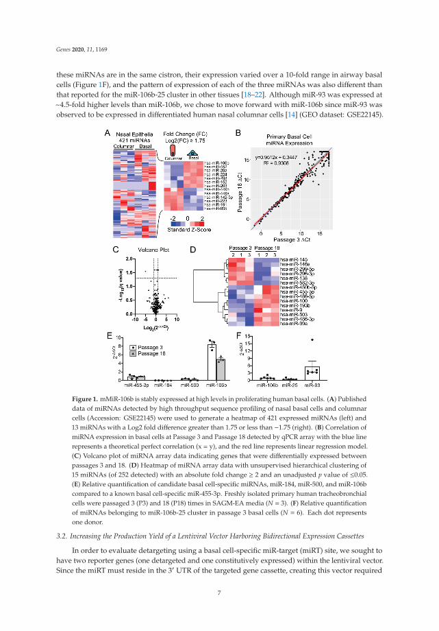
To select a miRNA for detargeting experiments, we accessed publicly available data throughNCBI Gene Expression Omnibus (GEO) under serial number GSE22145 that compared basal cellsvs. columnar cells in nasal airway [14] and found seven miRNAs that were consistently expressed inbasal cells but not columnar cells from the nasal epithelia of three donors (Figure 1A). To evaluatethe expression of miRNA expression in our cultured human tracheobronchial basal cells expanded inSAGM-EA [9], we used a TaqMan low-density array (TLDA; Applied Biosystems) to quantify relativeexpression of 377 miRNAs (Supplemental Table S1). Expression of 252 miRNAs was consistentlydetected in basal cells at passage 3 and at passage 18 (Figure 1B). Of these miRNAs, only nine changedsignificantly between passage 3 and 18 (FDR test with Q = 5%) and 171 miRNAs did not exceeda ±2-fold change in expression in the passage (Figure 1C). Using a less stringent test, expression of15 miRNAs changed significantly between passage 3 and 18 (unadjusted t-test p ≤ 0.05 and absolutefold change ≥ 2) (Figure 1D).
Comparison of our array data with the nasal miRNA sequencing study demonstrated thatmiR-106b was one of the few miRNAs that was not expressed in columnar cells. Other miRNAs thatwere basal cell-specific in the nasal study included miR-184 and miR-500. miR-500 was detected atlower levels than miR-106b in our array study and miR-184 was undetectable. In this regard, miR-106bappeared to be the ideal miRNA to use in basal cell detargeting. We decided that our candidatemiRNA should have a higher expression level than that of miR-455-3p, which has been reportedto effectively inhibit MUC1 in human epithelial basal cells [15]. To more quantitively evaluate theexpression of miR-106 in reference to low (miR-500) and very low (miR-184) basal cell expressingmiRNAs, we performed single-plex qPCR for these miRNAs in comparison to that of miR-455-3p(Figure 1E). As expected, the expression levels of miR-184 expression was very low and miR-500a wasabsent, while miR-106b was more than 11-fold higher than the level of miR-455-3p. Moreover, miR-106bwas stable on passage, decreasing by only 30% during the 15 passages. These findings confirmed thevalidity of the array data and suggested miR-106b was a top candidate for basal cell detargeting.
miR-106b, miR-25 and miR-93 belong to the miR-106b-25 cluster that is located in the 13thintron of mini-chromosome maintenance complex component 7 gene (MCM7) [16,17]. We preparedmiRNA samples from six donors and analyzed the relative expression of these miRNAs (Figure 1F).The expression levels were fairly consistent between the six random donor samples. Notably, although
6
Genes 2020, 11, 1169
these miRNAs are in the same cistron, their expression varied over a 10-fold range in airway basalcells (Figure 1F), and the pattern of expression of each of the three miRNAs was also different thanthat reported for the miR-106b-25 cluster in other tissues [18–22]. Although miR-93 was expressed at~4.5-fold higher levels than miR-106b, we chose to move forward with miR-106b since miR-93 wasobserved to be expressed in differentiated human nasal columnar cells [14] (GEO dataset: GSE22145).
Figure 1. mMiR-106b is stably expressed at high levels in proliferating human basal cells. (A) Publisheddata of miRNAs detected by high throughput sequence profiling of nasal basal cells and columnarcells (Accession: GSE22145) were used to generate a heatmap of 421 expressed miRNAs (left) and13 miRNAs with a Log2 fold difference greater than 1.75 or less than −1.75 (right). (B) Correlation ofmiRNA expression in basal cells at Passage 3 and Passage 18 detected by qPCR array with the blue linerepresents a theoretical perfect correlation (x = y), and the red line represents linear regression model.(C) Volcano plot of miRNA array data indicating genes that were differentially expressed betweenpassages 3 and 18. (D) Heatmap of miRNA array data with unsupervised hierarchical clustering of15 miRNAs (of 252 detected) with an absolute fold change ≥ 2 and an unadjusted p value of ≤0.05.(E) Relative quantification of candidate basal cell-specific miRNAs, miR-184, miR-500, and miR-106bcompared to a known basal cell-specific miR-455-3p. Freshly isolated primary human tracheobronchialcells were passaged 3 (P3) and 18 (P18) times in SAGM-EA media (N = 3). (F) Relative quantificationof miRNAs belonging to miR-106b-25 cluster in passage 3 basal cells (N = 6). Each dot representsone donor.
3.2. Increasing the Production Yield of a Lentiviral Vector Harboring Bidirectional Expression Cassettes
In order to evaluate detargeting using a basal cell-specific miR-target (miRT) site, we sought tohave two reporter genes (one detargeted and one constitutively expressed) within the lentiviral vector.Since the miRT must reside in the 3′ UTR of the targeted gene cassette, creating this vector required
7
Genes 2020, 11, 1169
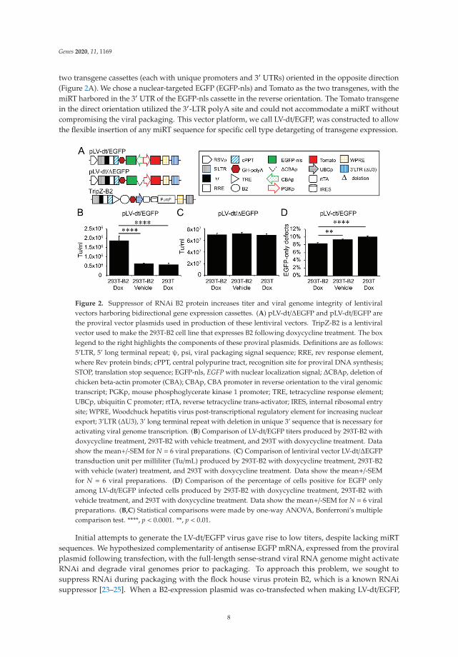
two transgene cassettes (each with unique promoters and 3′ UTRs) oriented in the opposite direction(Figure 2A). We chose a nuclear-targeted EGFP (EGFP-nls) and Tomato as the two transgenes, with themiRT harbored in the 3′ UTR of the EGFP-nls cassette in the reverse orientation. The Tomato transgenein the direct orientation utilized the 3′-LTR polyA site and could not accommodate a miRT withoutcompromising the viral packaging. This vector platform, we call LV-dt/EGFP, was constructed to allowthe flexible insertion of any miRT sequence for specific cell type detargeting of transgene expression.
Figure 2. Suppressor of RNAi B2 protein increases titer and viral genome integrity of lentiviralvectors harboring bidirectional gene expression cassettes. (A) pLV-dt/ΔEGFP and pLV-dt/EGFP arethe proviral vector plasmids used in production of these lentiviral vectors. TripZ-B2 is a lentiviralvector used to make the 293T-B2 cell line that expresses B2 following doxycycline treatment. The boxlegend to the right highlights the components of these proviral plasmids. Definitions are as follows:5’LTR, 5’ long terminal repeat; ψ, psi, viral packaging signal sequence; RRE, rev response element,where Rev protein binds; cPPT, central polypurine tract, recognition site for proviral DNA synthesis;STOP, translation stop sequence; EGFP-nls, EGFP with nuclear localization signal; ΔCBAp, deletion ofchicken beta-actin promoter (CBA); CBAp, CBA promoter in reverse orientation to the viral genomictranscript; PGKp, mouse phosphoglycerate kinase 1 promoter; TRE, tetracycline response element;UBCp, ubiquitin C promoter; rtTA, reverse tetracycline trans-activator; IRES, internal ribosomal entrysite; WPRE, Woodchuck hepatitis virus post-transcriptional regulatory element for increasing nuclearexport; 3’LTR (ΔU3), 3’ long terminal repeat with deletion in unique 3’ sequence that is necessary foractivating viral genome transcription. (B) Comparison of LV-dt/EGFP titers produced by 293T-B2 withdoxycycline treatment, 293T-B2 with vehicle treatment, and 293T with doxycycline treatment. Datashow the mean+/-SEM for N = 6 viral preparations. (C) Comparison of lentiviral vector LV-dt/ΔEGFPtransduction unit per milliliter (Tu/mL) produced by 293T-B2 with doxycycline treatment, 293T-B2with vehicle (water) treatment, and 293T with doxycycline treatment. Data show the mean+/-SEMfor N = 6 viral preparations. (D) Comparison of the percentage of cells positive for EGFP onlyamong LV-dt/EGFP infected cells produced by 293T-B2 with doxycycline treatment, 293T-B2 withvehicle treatment, and 293T with doxycycline treatment. Data show the mean+/-SEM for N = 6 viralpreparations. (B,C) Statistical comparisons were made by one-way ANOVA, Bonferroni’s multiplecomparison test. ****, p < 0.0001. **, p < 0.01.
Initial attempts to generate the LV-dt/EGFP virus gave rise to low titers, despite lacking miRTsequences. We hypothesized complementarity of antisense EGFP mRNA, expressed from the proviralplasmid following transfection, with the full-length sense-strand viral RNA genome might activateRNAi and degrade viral genomes prior to packaging. To approach this problem, we sought tosuppress RNAi during packaging with the flock house virus protein B2, which is a known RNAisuppressor [23–25]. When a B2-expression plasmid was co-transfected when making LV-dt/EGFP,
8
Genes 2020, 11, 1169
the resulting virus titer was ~3 times higher than that without B2 (data not shown). We then useda lentivector to stably integrate a B2 gene expression cassette into 293T cells, however, persistent B2expression in 293T cells was toxic. Thus, we generated a 293T cell line that expresses a doxycyclineinducible (Tet-on) B2 protein using a TRIPZ vector (Figure 2A). We first tested different concentrationsof doxycycline and two time points of doxycycline addition, at the time of proviral vector transfectionor at the first media change after transfection. We observed that addition of 500 ng/mL doxycycline atthe time of transfection produced highest virus titer while maintaining health of the producer cells.B2 mRNA induction by doxycycline was verified by qPCR (data not shown). Indeed, the lentiviralvector LV-dt/EGFP titer was significantly increased by doxycycline induced B2 expression during thevirus production (Figure 2B). To confirm that the mechanism of reduced titers of LV-dt/EGFP wasdue to antisense EGFP transcripts, we created a second control vector (LV-dt/ΔEGFP) which lackedthe CBA-promoter controlling EGFP expression. Titers of LV-dt/ΔEGFP, which lacked expression ofEGFP transcripts with complementary to the viral genome, were not affected by the induction of B2(Figure 2C).
To calculate viral titers in the above experiments, we used titration transduction assays on 293cells followed by flow cytometry. We noticed that there were LV-dt/EGFP transduced cells that werepositive for only EGFP or Tomato. This suggested that mutations or deletions within the proviralgenomes likely occurred prior to packaging. During reverse transcription, a reverse transcriptase maychange its templates 8 to 10 times [26] contributing to diversity of the lentivirus in the wild. Thisis a drawback to lentiviral vectors. We sought to evaluate whether inhibiting RNAi pathways withB2 would improve integrity of the packaged LV-dt/EGFP genomes. To this end, we compared thepercentage of LV-dt/EGFP transduced 293T cells that only expressed EGFP (defective particles) fromthree types of viral preparation conditions: 1) 293T-B2 cells induced with doxycycline, 2) 293T-B2 cellnot induced with doxycycline, and 3) 293T cells induced with doxycycline. Results from these flowcytometry comparisons demonstrated that group-1 and group-2 had ~20% and ~10% fewer defectiveparticles than group-3, respectively (Figure 2D). We hypothesize that low level expression of B2 in theuninduced group-2 viral preparations improved integrity of the viral genomes when compared to293T preparations lacking B2 (group-3).
3.3. Detargeting EGFP Expression in Proliferating Basal Cells.
To test whether the miR-106b target sequence (miRT) could be used to effectively detarget geneexpression in basal cells, we generated a lentivirus vector that contained a nuclear targeted EGFPwith the 3′-UTR miR-106b target sequence (pLV-dt/EGFP-miRT) and a control lentivirus vector withthe miR-106bT sequence in reverse orientation (pLV-dt/EGFP-RmiRT; Figure 3A). We infected humanepithelial basal cells grown in SAGM-EA with LV-dt/EGFP-miRT or LV-dt/EGFP-RmiRT and analyzedEGFP and Tomato expression by flow cytometry and fluorescent imaging. As hypothesized, the nucleiof the LV-dt/EGFP-RmiRT transduced basal cells were EGFP-positive, whereas basal cells transducedwith the LV-dt/EGFP-miRT vector were EGFP-negative (Figure 3B). Thus, the miR-106b target sequencein the 3′-UTR appeared to successfully detarget EGFP expression in basal cells. To verify thatmiR-106b was indeed responsible for EGFP knock-down, we transfected FACS isolated Tomato-positiveLV-dt/EGFP-miRT transduced cells with a miR-106b inhibitor. As expected, the LV-dt/EGFP-miRTtransduced cells transfected with the miR-106b inhibitor recovered nuclear EGFP expression, while themock transfected negative control cells did not (Figure 3C). The quantification of EGFP-positive only(Q4), Tomato-positive only (Q1), EGFP/Tomato-double-positive (Q2) and double-negative (Q3) cellsare shown in the quadrants generated by flow cytometry (Figure 3D).
9

Genes 2020, 11, 1169
Figure 3. Incorporation of miR-106b target sequence (miRT) into the 3′-UTR of EGFP effectivelydetargets lentiviral-mediated expression in proliferating basal cells. (A) Diagram of the bidirectionalpromoter proviral lentiviral plasmids (pLV-dt/EGFP-miRT and pLV-dt/EGFP-RmiRT) used to generatelentivirus and test detargeting in basal cells. The box legend to the right highlights the components ofthese proviral plasmids as described in detail within the Figure 2A legend. LV-dt/EGFP-miRT is theexperimental vector harboring a CBA promoter driven nuclear targeted EGFP (EGFP-nls) with miR-106btarget sequence (4×miR target or mirT) in the reverse orientation. In the forward direction, the PGKpromoter drives expression of the Tomato reporter, which is unaffected by miR-106b. LV-dt/EGFP-RmiRTis a control vector with the miRNA target sequence in the reverse orientation. (B) LV-dt/EGFP-miRTand LV-dt/EGFP-RmiRT viruses were used to transduce primary human airway basal cell in SAGM-EAcultures. The Tomato-positive (red) cells indicate the virally transduced cells. EGFP expression isseen in dt/EGFP-RmiRT control transduced cells but not in cells transduced with the detargetedLV-dt/EGFP-miRT vector. Scale bar, 100 μm. (C) Basal cells transduced with LV-dt/EGFP-miRT vector,and FACS isolated for Tomato-positive cells, were transfected with miRT-106b inhibitor sequences toblock detargeting or mock transfected. Scale bar, 100 μm. (D) The cells in (B and C) were analyzed byflow cytometer and are shown in dot plots to the right of the corresponding images for each condition.The percentage of cells are indicated in each quadrant: Q1 (Tomato-positive only cells), Q2 (Tomatoand EGFP double-positive cells), Q3 (EGFP-positive only cells), and Q4 (non-fluorescent cells).
3.4. Basal Cell miRT-106b Detargeting is Partially Maintained in Differentiated ALI Cultures and Organoids
miR-106 is highly expressed in proliferating basal cells grown in SAGM-EA media and basal celldetargeting with miRT-106b is highly effective (Figure 3). To determine if miR-106 expression in basalcells of differentiated cultures was sufficient for detargeting, we studied the EGFP expression profiles ofALI and organoid cultures generated from LV-dt/EGFP-miRT and LV-dt/EGFP-RmiRT transduced basalcells. Approximately 40% of the basal cell population was transduced and the cells were not subjectedto FACS prior to making ALI cultures or airway organoids. ALI cultures generated from these two
10
Genes 2020, 11, 1169
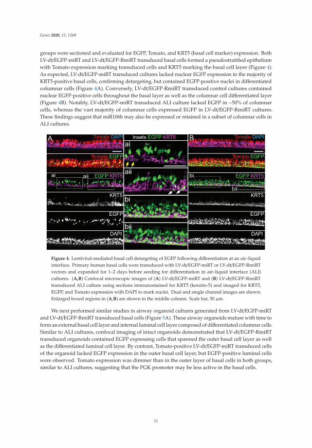
groups were sectioned and evaluated for EGFP, Tomato, and KRT5 (basal cell marker) expression. BothLV-dt/EGFP-miRT and LV-dt/EGFP-RmiRT transduced basal cells formed a pseudostratified epitheliumwith Tomato expression marking transduced cells and KRT5 marking the basal cell layer (Figure 4).As expected, LV-dt/EGFP-miRT transduced cultures lacked nuclear EGFP expression in the majority ofKRT5-positive basal cells, confirming detargeting, but contained EGFP-positive nuclei in differentiatedcolumnar cells (Figure 4A). Conversely, LV-dt/EGFP-RmiRT transduced control cultures containednuclear EGFP-positive cells throughout the basal layer as well as the columnar cell differentiated layer(Figure 4B). Notably, LV-dt/EGFP-miRT transduced ALI culture lacked EGFP in ~50% of columnarcells, whereas the vast majority of columnar cells expressed EGFP in LV-dt/EGFP-RmiRT cultures.These findings suggest that miR106b may also be expressed or retained in a subset of columnar cells inALI cultures.
Figure 4. Lentiviral-mediated basal cell detargeting of EGFP following differentiation at an air–liquidinterface. Primary human basal cells were transduced with LV-dt/EGFP-miRT or LV-dt/EGFP-RmiRTvectors and expanded for 1–2 days before seeding for differentiation in air–liquid interface (ALI)cultures. (A,B) Confocal microscopic images of (A) LV-dt/EGFP-miRT and (B) LV-dt/EGFP-RmiRTtransduced ALI culture using sections immunostained for KRT5 (keratin-5) and imaged for KRT5,EGFP, and Tomato expression with DAPI to mark nuclei. Dual and single channel images are shown.Enlarged boxed regions in (A,B) are shown in the middle column. Scale bar, 50 μm.
We next performed similar studies in airway organoid cultures generated from LV-dt/EGFP-miRTand LV-dt/EGFP-RmiRT transduced basal cells (Figure 5A). These airway organoids mature with time toform an external basal cell layer and internal luminal cell layer composed of differentiated columnar cells.Similar to ALI cultures, confocal imaging of intact organoids demonstrated that LV-dt/EGFP-RmiRTtransduced organoids contained EGFP expressing cells that spanned the outer basal cell layer as wellas the differentiated luminal cell layer. By contrast, Tomato-positive LV-dt/EGFP-miRT transduced cellsof the organoid lacked EGFP expression in the outer basal cell layer, but EGFP-positive luminal cellswere observed. Tomato expression was dimmer than in the outer layer of basal cells in both groups,similar to ALI cultures, suggesting that the PGK promoter may be less active in the basal cells.
11
Genes 2020, 11, 1169
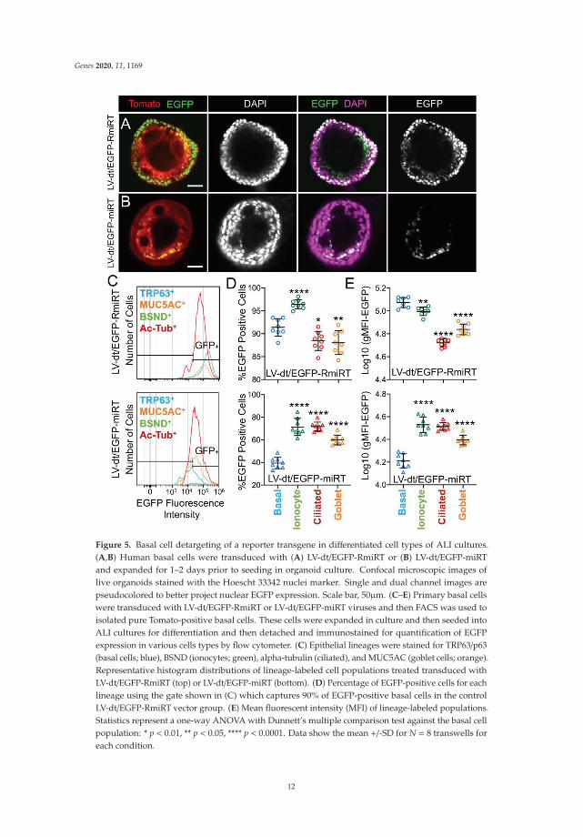
Figure 5. Basal cell detargeting of a reporter transgene in differentiated cell types of ALI cultures.(A,B) Human basal cells were transduced with (A) LV-dt/EGFP-RmiRT or (B) LV-dt/EGFP-miRTand expanded for 1–2 days prior to seeding in organoid culture. Confocal microscopic images oflive organoids stained with the Hoescht 33342 nuclei marker. Single and dual channel images arepseudocolored to better project nuclear EGFP expression. Scale bar, 50μm. (C–E) Primary basal cellswere transduced with LV-dt/EGFP-RmiRT or LV-dt/EGFP-miRT viruses and then FACS was used toisolated pure Tomato-positive basal cells. These cells were expanded in culture and then seeded intoALI cultures for differentiation and then detached and immunostained for quantification of EGFPexpression in various cells types by flow cytometer. (C) Epithelial lineages were stained for TRP63/p63(basal cells; blue), BSND (ionocytes; green), alpha-tubulin (ciliated), and MUC5AC (goblet cells; orange).Representative histogram distributions of lineage-labeled cell populations treated transduced withLV-dt/EGFP-RmiRT (top) or LV-dt/EGFP-miRT (bottom). (D) Percentage of EGFP-positive cells for eachlineage using the gate shown in (C) which captures 90% of EGFP-positive basal cells in the controlLV-dt/EGFP-RmiRT vector group. (E) Mean fluorescent intensity (MFI) of lineage-labeled populations.Statistics represent a one-way ANOVA with Dunnett’s multiple comparison test against the basal cellpopulation: * p < 0.01, ** p < 0.05, **** p < 0.0001. Data show the mean +/-SD for N = 8 transwells foreach condition.
12

Genes 2020, 11, 1169
To quantify the extent of detargeting in basal cells, we transduced primary human basal cellswith LV-dt/EGFP-miRT or LV-dt/EGFP-RmiRT vector systems and FACS isolated Tomato-positive cellsfor expansion in SAGM-EA prior to seeding into ALI cultures. Well-differentiated ALI cultures werethen dissociated, and the single cell suspension of epithelial cells was fixed and stained for markers ofbasal cells (TRP63), ciliated cells (acetylated tubulin), goblet cells (MUC5AC), and ionocytes (BSND).These populations were then subjected to flow cytometer and the percentages of EGFP-positive cellsfor each cell phenotype quantified (Figure 5C–E). As expected from confocal imaging of ALI cultures(Figure 4), the fluorescent intensity of all cell types in the LV-dt/EGFP-miRT group was lower than thatof LV-dt/EGFP-RmiRT, suggesting that inclusion of the miRT-106b target sequences generally reducesexpression of the EGFP transgene. However, quantification of the percentage of EGFP-positive cellsdemonstrated the largest drop for miRT vs. RmiRT expression in TRP63-positive basal cells (2.3-fold)(Figure 5D). Furthermore, in LV-dt/EGFP-RmiRT transduced cells, the percentage of EGFP-positivebasal cells was significantly lower than ciliated and goblet cells, whereas the opposite was observedin LV-dt/EGFP-miRT transduced cells (Figure 5D). Additionally, the mean fluorescent intensity (MFI,calculated as the geometric mean) of EGFP was the highest in basal cells of the LV-dt/EGFP-RmiRTcontrol group, supporting confocal imaging of ALI demonstrating the strongest EGFP expression inKRT5-positive basal cells with this vector (Figure 4B, bi, bii). By contrast, the MFI was the lowest inthe LV-dt/EGFP-miRT transduced basal cells as compared to ionocytes, ciliated cells and goblet cells(Figure 5E), similar to those observed histologic studies (Figure 4A, ai, aii). Overall, these findingssuggest that the miRT-106b sequences effectively reduce expression of EGFP in basal cells.
An unexpected finding from these cellular phenotyping studies of LV-dt/EGFP-miRT- andLV-dt/EGFP-RmiRT-transduced epithelia was a significant shift in the number of goblet cells andionocytes (Table 1). The largest shift occurred in the percentage of MUC5AC-positive goblet cells,rising 2-fold (p < 0.0001) in LV-dt/EGFP-miRT transduced epithelia as compared to the RmiRT controlvector. By contrast, the percentage of ionocytes marginally declined in the LV-dt/EGFP-miRT group(p < 0.0388), while the percentage of basal cells and ciliated cells was not significantly different betweenthe two groups. These findings raise the interesting possibility that high-level expression of mRNAcontaining the miRT sequence could potentially sequester miR-106b and impact processes involved ingoblet cell and ionocyte specification.
Table 1. Distribution of cell types in differentiated ALI cultures.
Vector% Basal Cells
(TRP63+)% Ionocytes
(BSND+)% Ciliated Cells(Ac-Tubulin+)
% Goblet Cells(MUC5AC+)
LV-dt/EGFP-RmiRT 21.8+/−3.8 * 0.82+/−0.02 44.3+/−1.6 11.2+/−0.8
LV-dt/EGFP-miRT 22.4+/−1.5 0.72+/−0.04 41.5+/−0.9 21.9+/−1.2
p-value ** 0.7768 0.0388 0.1567 <0.0001
The percentage of viable cells positive for each of the phenotypic cellular markers is shown (TRP63-basal cells;BSND-ionocytes; acetylated tubulin-ciliated cells; MUC5AC-goblet cells). Cells not positive for any of the fourantibodies were 21.9% and 12.5% for LV-dt/EGFP-RmiRT- and LV-dt/EGFP-miRT-transduced epithelia, respectively.* Mean +/-SEM. ** Statistical comparisons by Welch’s t test.
3.5. Basal Cell-Detargeting of CFTR Expression Alters Functional Complementation in CF Airway Epithelia
To test our primary hypothesis that detargeting of CFTR in basal cells would improvecomplementation in CF airway epithelia, we replaced EGFP in LV-dt/EGFP-miRT with CFTR togenerate the pLV-dt/CFTR-miRT lentiviral vector. Our control vector (pLV-dt/CFTR-Ø) was identical topLV-dt/CFTR-miRT but lacked the miR-106b target sequences (Figure 6A). Freshly isolated CF humantracheobronchial basal cells were transduced with each vector and expanded 4 days before seedinginto transwells for ALI culture. Contrary to our initial hypothesis, ALI cultures transduced withLV-dt/CFTR-Ø gave rise to ~3.5-fold greater CFTR-mediated CI– currents than that of LV-dt/CFTR-miRTtransduced ALI cultures (Figure 6B), even though both cultures expressed similar levels of CFTR
13

Genes 2020, 11, 1169
mRNA, which were 3.2-fold (LV-dt/CFTR-Ø) and 2.7-fold (LV-dt/CFTR-miRT) higher levels thanthe mock-infected group. Characteristic of CFTR, these currents were induced by cAMP agonists(IBMX/Forskolin) and inhibited by the CFTR channel blocker GlyH101. The slightly lower expressionof CFTR mRNA in the LV-dt/CFTR-miRT transduced cultures was expected, consistent with detargetedexpression in basal cells.
Figure 6. Detargeting CFTR expression in basal cells impacts the level of complementation in CFairway epithelia. (A) Diagram of lenti-vector containing CFTR expression cassette in reverse orientation.The PGK promoter (PGKp) drives expression of CFTR with the miR-106b target sequence (4× miRtarget or mirT) in the 3’UTR. CBA promoter drives expression of Tomato as a reporter gene for viraltransduction. pLV-dt/CFTR-Ø is a control vector with no miRT sequence. Box (below) is a legend for eachshape in the diagram that highlights the components of these proviral plasmids as described in detailwithin the Figure 2A legend. (B) Short-circuit current (Isc) measurements of differentiated air-liquidinterface cultures seeded with transduced at basal cells. Mock, mock-infected cells. PGK-CFTR-Ø, cellstransduced by LV-dt/CFTR-Ø; PGK-CFTR-miRT, cells transduced by LV-dt/CFTR-miRT. Amiloride wasused to block ENaC-mediated Na+ currents. 4,4′-Diisothiocyanatostilbene-2,2′-disulfonic acid (DIDS)was use to inhibit most non-CFTR chloride channel. 3-isobutyl-2-methylxanthine (IBMX) and Forskolinwas used for activate CFTR channels. N-(2-naphtalenyl)-(3.5-dibromo-2.4-dihydroxyphenyl)methyleneglycine hydrazide (GlyH101) was used to block CFTR. Data show the mean +/-SEM for N = 6 transwellsfor each condition. (C) Relative quantification of CFTR mRNA normalized to GAPDH mRNA from eachsample used in B. For B and C, the statistics used is one-way ANOVA, Tukey’s multiple comparisonstest. ****, p < 0.0001. **, p = 0.0025. Data show the mean +/-SEM for N = 3 independent samples foreach condition.
4. Discussion
Multipotent basal cells are generally considered the primary stem cell of the large conductingairways [27] and thus are a primary target for stem cell-based genetic therapies for CF. CFTR isexpressed in a subpopulation of transitional basal cells (i.e., intermediate basal cells) that are fated to
14

Genes 2020, 11, 1169
become secretory cells (i.e., goblet cells and club cells) [6,28]. CFTR is also expressed at low levels ina subpopulation of secretory cells and at high levels in pulmonary ionocytes [5,6]. The function of CFTRexpression in transitional basal cells remains unclear, but it stands to reason that this expression couldbe a precursor state to CFTR-expressing daughter cells [3]. The contribution of CFTR expression insecretory cells and ionocytes to the overall level of transepithelial ion transport in airway epithelium isalso a source of controversy [3]. Given that CFTR is not expressed in proliferating airway basal cells andthe potential that basal cell CFTR expression may impact fate decisions, we reasoned that developinga lentiviral vector that could more closely reproduce endogenous multipotent and transitional basalcell CFTR expression patterns would have utility for CF cell-based therapies. Thus, we sought todevelop a lentiviral vector that would repress CFTR expression in proliferating multipotent basal cellsand activate CFTR expression in transitional basal cells as they commit to differentiate.
Gene replacement cell-based therapies for CF will require the expansion of basal cells inconditionally reprogrammed culture and the introduction of a corrected CFTR gene using an integratingapproach (e.g., lentivirus) [29,30]. It was on this rationale that we designed a lentiviral vector withbi-directional promoters capable of carrying miRT sequences within a heterologous 3’-UTR of thereverse oriented target gene. Through bioinformatics and experimentation, we identified miR-106b asbeing highly expressed in both proliferating human tracheobronchial and nasal basal cells (Figure 1),but absent in differentiated columnar cells. Thus, the miRT for miR-106b appeared to be suitable forapproaching our studies. Notably, the regulated miRT gene must be placed in the reverse orientationsince a heterologous UTR in the direct orientation would prematurely terminate the viral genomicRNA during virus production [31]. Lentiviruses with reverse-oriented expression cassettes producelow viral titers due to a double-stranded RNA response and cleavage of the vector RNA genome bycellular Dicer [31,32]. However, previous attempts have successfully produced high titer virus whenan inducible promoter is used to drive the reverse-oriented gene of interest [31].
Our studies required the use of a strong promoter for both expression cassettes and, like others,we found titers to be low within our bidirectional pLV-dt/EGFP vector. We improved the inherentlylow titer using a suppressor of RNA silencing (SRS), B2 protein from flock house virus. Althoughmost viral infections activate RNAi responses in the cell against the virus [33,34], only a few havelooked into using RNAi suppressors in animal viral vector production [35,36]. In lentiviruses, potentialSRSs include Nef and Tat [37,38]; Tat is included with psPAX2 packaging plasmid and may helpprotect the lentiviral genome during virus production. By utilizing the B2 protein, pLV-dt/EGFP vectorproduction was further increased 3-fold (Figure 2). In this study, we only used B2, but other SRSs ofsome well-known plant and animal virus SRSs [33,39,40] are worth investigating and may furtherimprove virus production with a bi-directional vector. One downside of using the 293T-B2 cell line isthat the cells seem to be even less tightly attached to the surface of the culture dish than the wild-type293T, so when adding transfection reagents or media it should be carried out very carefully. To mitigatethis problem, poly-lysine coating the culture dish may help the cells to attach more tightly and helpimprove the virus titer.
Our studies evaluating miR-106b-mediating detargeting using the pLV-dt/EGFP-miRT vectorsystem demonstrated robust shut-off of the EGFP-miRT reporter in proliferating basal cells (Figure 3).Adjusting for differences in functional titer, the miRT-106b reduced EGFP expression 54-fold inproliferating human basal cells as compared to the RmiRT-106b control vector. However, when thepLV-dt/EGFP-miRT basal cells were differentiated at an ALI, there was a subset of basal cells thatwere not detargeted and a subset of columnar cells that were detargeted (Figure 4). We do not knowwhether quiescent G0 and intermediate basal cells express miR-106b and this could impact detargetingof pLV-dt/EGFP-miRT in KRT5-positive basal cells. The finding of miRT-106b detargeted columnarcells also suggests that at least in differentiated ALI culture systems, miR-106b expression is expressedor retained in a larger subset of columnar cells than previously observed in human nasal epithelia [14].Culture conditions likely impacted miR-106b expression and the level of detargeting since organoidcultures demonstrated robust basal cell detargeting with the pLV-dt/EGFP-miRT as compared to the
15
Genes 2020, 11, 1169
RmiRT-106b control vector. It is also worth noting that in ALI cultures the CBA promoter used to driveEGFP expression in the pLV-dt/EGFP is robustly expressed in basal cells, while the PGK promoter usedto drive Tomato expression is more active in columnar cells and less active in basal cells (Figure 4).These differences were even more greatly accentuated in organoid cultures (Figure 5A,B) where veryweak EGFP expression was observed in the luminal cell layer for both vector systems.
Due to the relative activity of the CBA and PGK promoters in basal vs. columnar cells of ALIcultures and organoids, we altered the sequence of the promoters in our pLV-dt/CFTR-miRT andpLV-dt/CFTR-Ø vector systems. Since the PGK promoter was weaker in basal cells and stronger incolumnar cells, it was used to drive CFTR expression, thus accentuating the detargeting by miRT-106bin basal cells and facilitating CFTR expression in differentiated cells that participate in ion transport(Figure 6A). Similarly, the CBA promoter had greater expression in basal cells and thus was usedto drive Tomato expression. Contrary to our original hypothesis, CF ALI cultures transduced withLV-dt/CFTR-miRT had significantly less functional correction of CFTR currents as compared toLV-dt/CFTR-Ø (Figure 6B).
While the reason for the observed difference in CFTR complementation is currently unknown, thereare three potential explanations that warrant further investigation. First, LV-dt/EGFP-miRT transducedALI cultures had a subpopulation of columnar cells that were Tomato-positive and EGFP-negative.This phenotype was rarely observed in control ALI transduced with the pLV-dt/EGFP-RmiRT vector.Thus, these results would be consistent with expression of miR-106b in a subset of columnar cells thatcontribute to CFTR-mediated current.
Second, expression of the EGFP-miRT-106b transcript in basal cells led to significant changes intwo cell populations when differentiated at ALI (i.e., ionocytes and goblet cells) (Table 1). The decreasein the percentage in ionocytes was relatively small (12%), while the increase in goblet cells was large(200%). Both of these cellular compartments express CFTR in a subpopulation of each cell type.One possibility for this vector-related shift in differentiated cell types is that high-level expression ofEGFP-miRT-106b transcripts may sequester miR-106b and act like a miR-inhibitor. Thus, it is possiblethat inhibiting miR-106b activates differentiation toward columnar cells that cannot participate inCFTR-mediated ion transport. This possibility can be supported by two interpretations of CFTR mRNAlevels shown in Figure 6C, which are not mutually exclusive. The mild reduction in total CFTR mRNAlevels in LV-dt/CFTR-miRT transduced epithelia is consistent with successful CFTR detargeting inbasal cells, where the decrease may represent expression of CFTR in basal cells. However, we cannotrule out that inhibiting miR-106b may have led to an expansion of cell types that cannot facilitateCFTR-mediated anion transport, and that we cannot currently account for with the flow cytometrypanel described. Future studies using single-cell RNAseq could help to understand the shift in cellularcompartments and their CFTR expression patterns.
The last formal possibility for explaining these results, however unlikely, is the contribution ofbasal cell CFTR expression to transepithelial anion transport. Current wisdom suggests that onlychannels that reside in the apical and basolateral membranes of polarized epithelia contribute totransepithelial ion movement. However, very little is known about why CFTR is expressed in basalcells, so we cannot rule this out as a formal possibility.
miR-106b is one of the three miRNAs in the polycistronic miR-106b ~25 cluster within an intron ofthe MCM7 gene. MCM7 is part of the DNA replication initiation complex, but its expression is notnecessarily coupled to that of miR-106b [19,20]. miR-106b can also play roles in cell-cycle regulationof both stem cells and cancer cells [16,41]. For example, expression of this miRNA enhances cellgrowth [42], promotes migration of certain cancer cells [43], and promotes cell cycle progression [44].In SAGM-EA conditionally reprogramming media, basal cells are locked into a self-renewing state.However, it remains unclear if miR-106b plays a role in cell cycle progression of basal cells culturedunder these conditions. We did not observe a major difference in morphology or growth of cellsexpressing the miRT-106b target in either EGFP or CFTR transcripts, and this might suggest that
16

Genes 2020, 11, 1169
sequestration of miR-106b from its native targets does not occur if these biologic functions of miR-106bare relevant to airway basal cells.
5. Conclusions
This study has strengths and limitations. One limitation includes a clearer understanding ofcellular expression patterns of miR-106b in ALI cultures. While we had initial chosen miR-106b asa candidate based on its lack of expression in nasal columnar cells, our reporter gene expression studiessuggest that it may be expressed in a subset of columnar cells. Future studies evaluating miR-106bexpression in FACS-isolated cell types would allow for a clearer interpretation of how the miRT-106bsequence alters CFTR complementation. A second limitation is the fact the promoters used in thebicistronic vectors studied have slightly different activities in basal vs. columnar cells. The use ofa bidirectional promoter might be a better approach for future studies, however, the most commonlyused major immediate-early cytomegaloviruses enhancer/promoter is typically inactivated in lentiviralvectors by methylation. A strength of these studies includes the development of miRT-106b vectors thatcan clearly detarget expression in proliferating basal cells. Such a vector system can be used to studybasal cell differentiation through the regulated expression of transcription factors that would otherwiseterminally differentiate proliferating basal cells. A second strength includes the novel findings thatmiR-106b appears to be expressed in specific populations of cells that contribute to CFTR-mediatedtransepithelial ion transport and/or that certain cell types can express CFTR mRNA but not participatein CFTR-dependent transepithelial anion transport. Although more research is needed to understandthe mechanism, the finding itself has implications for CF gene therapy as it implies unique cellulartargets for CFTR complementation.
Supplementary Materials: The following are available online at http://www.mdpi.com/2073-4425/11/10/1169/s1,Table S1: PCR array table.
Author Contributions: Conceptualization, S.H.C., Z.Y., and J.F.E.; methodology, S.H.C., R.E.R., E.J.D., G.S.R.I.,D.A.S., T.J.L., G.N.G., Z.F., M.C.W., T.I.A.E., W.S.S., Y.Z., Z.Y., and J.F.E.; formal analysis, S.H.C., R.E.R., G.S.R.I.,D.A.S., E.J.D., T.J.L., W.S.S., Z.Y., and J.F.E.; investigation, S.H.C., R.E.R., E.J.D., G.S.R.I., X.L., M.L. Z.Y., and J.F.E.;resources, E.J.D., D.A.S., Z.Y., and J.F.E.; writing—original draft preparation, S.H.C., and R.E.R.; writing—reviewand editing, T.J.L., G.S.R.I., D.A.S., Z.Y., and J.F.E.; supervision, Z.Y., and J.F.E.; project administration, J.F.E..;funding acquisition, J.F.E. All authors have read and agreed to the published version of the manuscript.
Funding: This work was supported by grants from the National Institutes of Health P30 DK054759, P01 HL152960,R01 DK047967 to J.F.E., and a grant from the Cystic Fibrosis Foundation to J.F.E. G.S.R.I. was supported by twoT32s HL007638 and GM007337.
Acknowledgments: University of Iowa, Carver College of Medicine, Flow Cytometry core, Genomics Core,Microscopy Core, Tissue Culture Core. ALN Rao (University of California at Riverside) and Jang-kyun Seo(Seoul National University, South Korea) for the plasmid containing FHV RNA1 gene.
Conflicts of Interest: The authors declare no conflict of interest.
Abbreviations
CF cystic fibrosisCFTR cystic fibrosis transmembrane conductance regulatormiR microRNA3’-UTR 3’-untranslated regionPGK mouse phosphoglycerate kinase 1 promoterCBA CMV early enhancer-chicken beta-actin promoterTomato dTomato, dtSAGM-EA Small Airway Epithelial Growth Medium with extra additives
17
Genes 2020, 11, 1169
References
1. Riordan, J.R.; Rommens, J.M.; Kerem, B.; Alon, N.; Rozmahel, R.; Grzelczak, Z.; Zielenski, J.; Lok, S.;Plavsic, N.; Chou, J.L.; et al. Identification of the cystic fibrosis gene: Cloning and characterization ofcomplementary DNA. Science 1989, 245, 1066–1073. [CrossRef] [PubMed]
2. Xie, Y.; Ostedgaard, L.; Abou Alaiwa, M.H.; Lu, L.; Fischer, A.J.; Stoltz, D.A. Mucociliary Transport in Healthyand Cystic Fibrosis Pig Airways. Ann. Am. Thorac. Soc. 2018, 15, S171–S176. [CrossRef] [PubMed]
3. Tang, Y.; Yan, Z.; Engelhardt, J.F. Viral Vectors, Animal Models, and Cellular Targets for Gene Therapy ofCystic Fibrosis Lung Disease. Hum. Gene Ther. 2020, 31, 524–537. [CrossRef] [PubMed]
4. Rock, J.R.; Randell, S.H.; Hogan, B.L. Airway basal stem cells: A perspective on their roles in epithelialhomeostasis and remodeling. Dis. Model. Mech. 2010, 3, 545–556. [CrossRef] [PubMed]
5. Montoro, D.T.; Haber, A.L.; Biton, M.; Vinarsky, V.; Lin, B.; Birket, S.E.; Yuan, F.; Chen, S.; Leung, H.M.;Villoria, J.; et al. A revised airway epithelial hierarchy includes CFTR-expressing ionocytes. Nature 2018, 560,319–324. [CrossRef] [PubMed]
6. Plasschaert, L.W.; Zilionis, R.; Choo-Wing, R.; Savova, V.; Knehr, J.; Roma, G.; Klein, A.M.; Jaffe, A.B.A single-cell atlas of the airway epithelium reveals the CFTR-rich pulmonary ionocyte. Nature 2018, 560,377–381. [CrossRef] [PubMed]
7. Carraro, G.; Mulay, A.; Yao, C.; Mizuno, T.; Konda, B.; Petrov, M.; Lafkas, D.; Arron, J.R.; Hogaboam, C.M.;Chen, P.; et al. Single Cell Reconstruction of Human Basal Cell Diversity in Normal and IPF Lung. Am. J.Respir. Crit. Care Med. 2020. [CrossRef]
8. Xu, Y.; Mizuno, T.; Sridharan, A.; Du, Y.; Guo, M.; Tang, J.; Wikenheiser-Brokamp, K.A.; Perl, A.T.; Funari, V.A.;Gokey, J.J.; et al. Single-cell RNA sequencing identifies diverse roles of epithelial cells in idiopathic pulmonaryfibrosis. JCI Insight 2016, 1, e90558. [CrossRef]
9. Mou, H.; Vinarsky, V.; Tata, P.R.; Brazauskas, K.; Choi, S.H.; Crooke, A.K.; Zhang, B.; Solomon, G.M.;Turner, B.; Bihler, H.; et al. Dual SMAD Signaling Inhibition Enables Long-Term Expansion of DiverseEpithelial Basal Cells. Cell Stem Cell 2016. [CrossRef]
10. Barde, I.; Salmon, P.; Trono, D. Production and titration of lentiviral vectors. Curr. Protoc. Neurosci. 2010, 53,4–21. [CrossRef]
11. Denning, W.; Das, S.; Guo, S.; Xu, J.; Kappes, J.C.; Hel, Z. Optimization of the transductional efficiency oflentiviral vectors: Effect of sera and polycations. Mol. Biotechnol. 2013, 53, 308–314. [CrossRef]
12. Sun, X.; Olivier, A.K.; Liang, B.; Yi, Y.; Sui, H.; Evans, T.I.; Zhang, Y.; Zhou, W.; Tyler, S.R.; Fisher, J.T.; et al.Lung phenotype of juvenile and adult cystic fibrosis transmembrane conductance regulator-knockout ferrets.Am. J. Respir. Cell Mol. Biol. 2014, 50, 502–512. [CrossRef] [PubMed]
13. Yan, Z.; Sun, X.; Feng, Z.; Li, G.; Fisher, J.T.; Stewart, Z.A.; Engelhardt, J.F. Optimization of RecombinantAdeno-Associated Virus-Mediated Expression for Large Transgenes, Using a Synthetic Promoter and TandemArray Enhancers. Hum. Gene Ther. 2015, 26, 334–346. [CrossRef] [PubMed]
14. Marcet, B.; Chevalier, B.; Luxardi, G.; Coraux, C.; Zaragosi, L.E.; Cibois, M.; Robbe-Sermesant, K.; Jolly, T.;Cardinaud, B.; Moreilhon, C.; et al. Control of vertebrate multiciliogenesis by miR-449 through directrepression of the Delta/Notch pathway. Nat. Cell Biol. 2011, 13, 693–699. [CrossRef] [PubMed]
15. Martinez-Anton, A.; Sokolowska, M.; Kern, S.; Davis, A.S.; Alsaaty, S.; Taubenberger, J.K.; Sun, J.; Cai, R.;Danner, R.L.; Eberlein, M.; et al. Changes in microRNA and mRNA expression with differentiation of humanbronchial epithelial cells. Am. J. Respir. Cell Mol. Biol. 2013, 49, 384–395. [CrossRef]
16. Mehlich, D.; Garbicz, F.; Wlodarski, P.K. The emerging roles of the polycistronic miR-106b approximately25 cluster in cancer-A comprehensive review. Biomed Pharmacother. 2018, 107, 1183–1195. [CrossRef]
17. Kim, Y.K.; Kim, V.N. Processing of intronic microRNAs. EMBO J. 2007, 26, 775–783. [CrossRef]18. Zhou, Y.; Hu, Y.; Yang, M.; Jat, P.; Li, K.; Lombardo, Y.; Xiong, D.; Coombes, R.C.; Raguz, S.; Yague, E.
The miR-106b~25 cluster promotes bypass of doxorubicin-induced senescence and increase in motility andinvasion by targeting the E-cadherin transcriptional activator EP300. Cell Death Differ. 2014, 21, 462–474.[CrossRef]
19. Chuang, T.D.; Luo, X.; Panda, H.; Chegini, N. miR-93/106b and their host gene, MCM7, are differentiallyexpressed in leiomyomas and functionally target F3 and IL-8. Mol. Endocrinol. 2012, 26, 1028–1042. [CrossRef]
20. Haldar, S.; Roy, A.; Banerjee, S. Differential regulation of MCM7 and its intronic miRNA cluster miR-106b-25during megakaryopoiesis induced polyploidy. RNA Biol. 2014, 11, 1137–1147. [CrossRef]
18

Genes 2020, 11, 1169
21. Kan, T.; Sato, F.; Ito, T.; Matsumura, N.; David, S.; Cheng, Y.; Agarwal, R.; Paun, B.C.; Jin, Z.; Olaru, A.V.;et al. The miR-106b-25 Polycistron, Activated by Genomic Amplification, Functions as an Oncogene bySuppressing p21 and Bim. Gastroenterology 2009, 136, 1689–1700. [CrossRef]
22. Smith, A.L.; Iwanaga, R.; Drasin, D.J.; Micalizzi, D.S.; Vartuli, R.L.; Tan, A.C.; Ford, H.L. The miR-106b-25cluster targets Smad7, activates TGF-beta signaling, and induces EMT and tumor initiating cell characteristicsdownstream of Six1 in human breast cancer. Oncogene 2012, 31, 5162–5171. [CrossRef] [PubMed]
23. Chao, J.A.; Lee, J.H.; Chapados, B.R.; Debler, E.W.; Schneemann, A.; Williamson, J.R. Dual modes ofRNA-silencing suppression by Flock House virus protein B2. Nat. Struct. Mol. Biol. 2005, 12, 952–957.[CrossRef] [PubMed]
24. Li, H.; Li, W.X.; Ding, S.W. Induction and suppression of RNA silencing by an animal virus. Science 2002, 296,1319–1321. [CrossRef]
25. Lingel, A.; Simon, B.; Izaurralde, E.; Sattler, M. The structure of the flock house virus B2 protein, a viralsuppressor of RNA interference, shows a novel mode of double-stranded RNA recognition. EMBO Rep.2005, 6, 1149–1155. [CrossRef] [PubMed]
26. Rawson, J.M.O.; Nikolaitchik, O.A.; Keele, B.F.; Pathak, V.K.; Hu, W.S. Recombination is required for efficientHIV-1 replication and the maintenance of viral genome integrity. Nucleic. Acids Res. 2018, 46, 10535–10545.[CrossRef] [PubMed]
27. Rock, J.R.; Onaitis, M.W.; Rawlins, E.L.; Lu, Y.; Clark, C.P.; Xue, Y.; Randell, S.H.; Hogan, B.L. Basal cellsas stem cells of the mouse trachea and human airway epithelium. Proc. Natl. Acad. Sci. USA 2009, 106,12771–12775. [CrossRef] [PubMed]
28. Engelhardt, J.F.; Yankaskas, J.R.; Ernst, S.A.; Yang, Y.; Marino, C.R.; Boucher, R.C.; Cohn, J.A.; Wilson, J.M.Submucosal glands are the predominant site of CFTR expression in the human bronchus. Nat. Genet. 1992, 2,240–248. [CrossRef]
29. Flotte, T.R.; Ng, P.; Dylla, D.E.; McCray, P.B., Jr.; Wang, G.; Kolls, J.K.; Hu, J. Viral vector-mediated andcell-based therapies for treatment of cystic fibrosis. Mol. Ther. 2007, 15, 229–241. [CrossRef]
30. Yan, Z.; McCray, P.B., Jr.; Engelhardt, J.F. Advances in gene therapy for cystic fibrosis lung disease.Hum. Mol. Genet. 2019, 28, R88–R94. [CrossRef]
31. Poling, B.C.; Tsai, K.; Kang, D.; Ren, L.; Kennedy, E.M.; Cullen, B.R. A lentiviral vector bearing a reverseintron demonstrates superior expression of both proteins and microRNAs. RNA Biol. 2017, 14, 1570–1579.[CrossRef]
32. Liu, Y.P.; Vink, M.A.; Westerink, J.T.; Ramirez de Arellano, E.; Konstantinova, P.; Ter Brake, O.; Berkhout, B.Titers of lentiviral vectors encoding shRNAs and miRNAs are reduced by different mechanisms that requiredistinct repair strategies. RNA 2010, 16, 1328–1339. [CrossRef] [PubMed]
33. Ding, S.W.; Han, Q.; Wang, J.; Li, W.X. Antiviral RNA interference in mammals. Curr. Opin. Immunol. 2018,54, 109–114. [CrossRef] [PubMed]
34. Son, K.N.; Liang, Z.; Lipton, H.L. Double-Stranded RNA Is Detected by Immunofluorescence Analysis inRNA and DNA Virus Infections, Including Those by Negative-Stranded RNA Viruses. J. Virol. 2015, 89,9383–9392. [CrossRef] [PubMed]
35. Rauschhuber, C.; Mueck-Haeusl, M.; Zhang, W.; Nettelbeck, D.M.; Ehrhardt, A. RNAi suppressor P19 can bebroadly exploited for enhanced adenovirus replication and microRNA knockdown experiments. Sci. Rep.2013, 3, 1363. [CrossRef]
36. Liu, Y.; Zhang, L.; Zhang, Y.; Liu, D.; Du, E.; Yang, Z. Functional analysis of RNAi suppressor P19 onimproving baculovirus yield and transgene expression in Sf9 cells. Biotechnol. Lett. 2015, 37, 2159–2166.[CrossRef]
37. Aqil, M.; Naqvi, A.R.; Bano, A.S.; Jameel, S. The HIV-1 Nef protein binds argonaute-2 and functions as a viralsuppressor of RNA interference. PLoS ONE 2013, 8, e74472. [CrossRef]
38. Bennasser, Y.; Le, S.Y.; Benkirane, M.; Jeang, K.T. Evidence that HIV-1 encodes an siRNA and a suppressor ofRNA silencing. Immunity 2005, 22, 607–619. [CrossRef]
39. de Vries, W.; Berkhout, B. RNAi suppressors encoded by pathogenic human viruses. Int. J. Biochem. Cell Biol.2008, 40, 2007–2012. [CrossRef]
40. Maillard, P.V.; van der Veen, A.G.; Poirier, E.Z.; Reis e Sousa, C. Slicing and dicing viruses: Antiviral RNAinterference in mammals. EMBO J. 2019, 38. [CrossRef]
19

Genes 2020, 11, 1169
41. Mens, M.M.J.; Ghanbari, M. Cell Cycle Regulation of Stem Cells by MicroRNAs. Stem Cell Rev. Rep. 2018, 14,309–322. [CrossRef]
42. Li, Y.; Wang, Y.; Zhou, H.; Yong, Y.; Cao, Y. miR-106b promoted growth and inhibited apoptosis ofnasopharyngeal carcinoma cells by suppressing the tumor suppressor PTEN. Int. J. Clin. Exp. Pathol. 2016, 9,7078–7086.
43. Cheng, Y.; Guo, Y.; Zhang, Y.; You, K.; Li, Z.; Geng, L. MicroRNA-106b is involved in transforming growthfactor beta1-induced cell migration by targeting disabled homolog 2 in cervical carcinoma. J. Exp. Clin.Cancer Res. 2016, 35, 11. [CrossRef] [PubMed]
44. Ivanovska, I.; Ball, A.S.; Diaz, R.L.; Magnus, J.F.; Kibukawa, M.; Schelter, J.M.; Kobayashi, S.V.; Lim, L.;Burchard, J.; Jackson, A.L.; et al. MicroRNAs in the miR-106b family regulate p21/CDKN1A and promotecell cycle progression. Mol. Cell Biol. 2008, 28, 2167–2174. [CrossRef] [PubMed]
© 2020 by the authors. Licensee MDPI, Basel, Switzerland. This article is an open accessarticle distributed under the terms and conditions of the Creative Commons Attribution(CC BY) license (http://creativecommons.org/licenses/by/4.0/).
20

genesG C A T
T A C G
G C A T
Article
Human Nasal Epithelial Organoids for TherapeuticDevelopment in Cystic Fibrosis
Zhongyu Liu 1,2,†, Justin D. Anderson 1,2,†, Lily Deng 2, Stephen Mackay 1,2, Johnathan Bailey 2,
Latona Kersh 1,3, Steven M. Rowe 1,2,3,4 and Jennifer S. Guimbellot 1,2,*
1 Gregory Fleming James Cystic Fibrosis Research Center, University of Alabama at Birmingham (UAB),Birmingham, AL 35294, USA; [email protected] (Z.L.); [email protected] (J.D.A.);[email protected] (S.M.); [email protected] (L.K.); [email protected] (S.M.R.)
2 Department of Pediatrics, Division of Pulmonary and Sleep Medicine, UAB, Birmingham, AL 35233, USA;[email protected] (L.D.); [email protected] (J.B.)
3 Department of Medicine, Division of Pulmonary, Allergy, and Critical Care Medicine, UAB,Birmingham, AL 35294, USA
4 Department of Cell, Developmental and Integrative Biology, UAB, Birmingham, AL 35294, USA* Correspondence: [email protected]; Tel.: +1-205-234-0250; Fax: +1-205-975-5983† These authors contributed equally to this work.
Received: 6 April 2020; Accepted: 27 May 2020; Published: 29 May 2020
Abstract: We describe a human nasal epithelial (HNE) organoid model derived directly from patientsamples that is well-differentiated and recapitulates the airway epithelium, including the expressionof cilia, mucins, tight junctions, the cystic fibrosis transmembrane conductance regulator (CFTR),and ionocytes. This model requires few cells compared to airway epithelial monolayer cultures,with multiple outcome measurements depending on the application. A novel feature of the modelis the predictive capacity of lumen formation, a marker of baseline CFTR function that correlateswith short-circuit current activation of CFTR in monolayers and discriminates the cystic fibrosis (CF)phenotype from non-CF. Our HNE organoid model is amenable to automated measurements offorskolin-induced swelling (FIS), which distinguishes levels of CFTR activity. While the apical side isnot easily accessible, RNA- and DNA-based therapies intended for systemic administration could beevaluated in vitro, or it could be used as an ex vivo biomarker of successful repair of a mutant gene.In conclusion, this highly differentiated airway epithelial model could serve as a surrogate biomarkerto assess correction of the mutant gene in CF or other diseases, recapitulating the phenotypic andgenotypic diversity of the population.
Keywords: cystic fibrosis; CFTR; human nasal epithelial cells; organoids; biomarker; functional assay;CFTR modulators; pre-clinical in vitro models
1. Introduction
Cystic fibrosis (CF) is an autosomal recessive genetic disorder caused by disease-causing variantsin the CF transmembrane conductance regulator (CFTR) gene. CFTR is expressed on the epithelialcells of several organs, including the lung, intestine, pancreas, and liver. In the lung, functionalCFTR regulates ion transfer in the airway lumen, balancing the salt, pH, and fluid content as wellas influencing the viscosity and organization of mucus. In CF, this balance is impaired, resulting inthick secretions poorly transported out of the lung by the mucociliary apparatus, leading to a cycleof inflammation and infection that ultimately causes significant morbidity and mortality among theCF population.
Identifying an effective cure for CF is critical. While new drugs called CFTR modulators have beenidentified that are highly effective for the treatment of CF [1–3], they target the protein defect in CFTR
Genes 2020, 11, 603; doi:10.3390/genes11060603 www.mdpi.com/journal/genes21

Genes 2020, 11, 603
but do not permanently correct the genetic mutation. Furthermore, although a recent three-drug CFTRmodulator therapy was approved for ~90% of patients as defined by the genotype [4,5], no treatmentfor the underlying defect exists for the remaining 10%. This includes patients with rare mutationsunsuccessfully rescued by modulators. Ideally, correction of the mutation using gene therapy or othernucleotide-based approaches would be permanently curative.
The availability of in vitro models to test correction of the genetic defect in CF has been somewhatlimited. In this study, we describe the development of an in vitro model derived from patientsamples (nasal brush biopsies), which recapitulates the highly differentiated airway epithelium.While we and others reported similar models derived from nasal tissue, some [6–9] are grown fromdifferentiated cells already in an intact monolayer, either from excised nasal polyps or brush biopsies.In these cases, the apical membrane is to the exterior of the structure, and the basolateral side is notaccessible. These spheroids are free-floating, which makes imaging difficult, are of limited quantity,and are derived from cells that are not passaged or expanded, making higher throughput applicationsimpossible. One similar model [10] employs a labor-intensive technical approach that is not amenableto high-throughput applications, which the authors cite as a specific limitation. In contrast, our organoidassay utilizes small amounts of biopsy material that are expanded to millions of cells, which can providesufficient replicates for moderate-to-high-throughput applications; was optimized for uniformity inorganoid distribution and morphology to aid in automated imaging and analysis; and has a greatercapacity to detect subtle differences. The model has several potential outcome measures, includingquantitating the kinetics of CFTR rescue on fluid transport; fluorescent microscopy to qualitativelydetermine gene expression; and evaluating the functional activities through the use of novel imagingtechniques (micro-optical coherence tomography, μOCT). These are useful to assess the impact of anycurative therapy targeted to CFTR mutations [11–16]. Because this model is derived directly frompatient samples, we postulate that it will reproduce the phenotypic and genotypic diversity that existsamong the CF population and will be more likely to predict the effectiveness of the therapy acrossall patients.
2. Materials and Methods
2.1. Patient Samples
Non-CF healthy control subjects (n = 12, age 16–40 years) and subjects with CF (n = 36,age 1–51 years) were recruited to this study after written informed consent was obtained. For asubset of subjects with CF who contributed cells for functional assessment, basic descriptive dataregarding their age, genotype, pancreatic sufficiency status, and baseline sweat chloride (whenavailable) is described in the Supplementary Material S1. The study was approved by the Universityof Alabama Institutional Review Board under protocol number IRB-151030001. One brush biopsyfrom each nare was obtained using cytology brushes (Medical Packaging CYB1, Cat# CYB-1, length:8 inches, width approximately 7 mm, Camarillo, CA, USA) under direct visualization of the inferiorturbinate via an otoscope and sterile 9-mm speculum. The cell number obtained from brushing wasvaried between subjects, ranging from 2.5 × 10⁵ to 2 × 10⁶.
2.2. Cell Culture and Expansion
Expansion of primary human nasal epithelial cells (HNEs) was adapted from conditionalreprogramming culture (CRC) methods that have been previously published [17,18]. In brief,after collection, cells were immediately placed in Roswell Park Memorial Institute (RPMI) 1640 media(Thermo Scientific, Waltham, MA, USA) and processed within two hours. Depending on the numberof cells collected, cells were either cryopreserved and/or expanded in co-culture with irradiated3T3 fibroblasts for up to 14 days in a T75 flask containing F-media with 10 μM Y27632 (StemgentStemolecule, Beltsville, MD, USA) [17]. At a seeding density of at least 2.5 × 10³/cm2, one T75 willyield 2 × 10⁶ to 4 × 10⁶ cells. For the initial three days of expansion culture only, Amphotericin B
22
Genes 2020, 11, 603
2.5 μg/mL (Sigma-Aldrich Corp., St. Louis, MO, USA), Tobramycin 100 μg/mL (Alfa Aesar, Ward Hill,MA, USA), Ceftazidime 100 μg/mL (Sigma-Aldrich Corp., St. Louis, MO, USA), and Vancomycin100 μg/mL (Alfa Aesar, Ward Hill, MA, USA) were added to the media of the CF-derived cells [10].Cells were discarded if less than 80% confluent by day 14. HNEs were recovered after trypsinizationin the expansion flask (0.05% trypsin-EDTA (ethylenediaminetetraacetic acid)). No cells greater thanpassage 3 were used to avoid a known reduction in CFTR expression at high passage numbers [19].CF and non-CF cells behaved similarly during expansion, consistent with prior reports [20–22].
2.3. Differentiated HNE Culture
Organoids were cultured either in a Transwell system (polyester membrane, area 0.33 cm2,pore size: 0.4 μm; Corning Inc., Corning, NY, USA) or 15-well angiogenesis slides (ibidi USA, Inc.,Fitchburg, WI, USA). Slides were selected to enhance the optical imaging of organoids for morphologyand functional assessment. For angiogenesis slides, Matrigel (Corning Inc., Corning, NY, USA) with atotal protein concentration of at least 9 mg/mL was used for organoid culture. Each well of the slideswas coated with 5 μL of cold 100% Matrigel on ice and slides were placed into a cell culture incubator at37 ◦C for at least 30 min. HNEs were resuspended into a 20% matrigel and Ultroser-G (USG) media [23]suspension at 500 cells/μL. For each replicate well, 5 μL of the suspension (2500 cells/replicate) wereseeded into imaging slide wells coated with matrigel, incubated at 37 ◦C for one hour, and coveredwith 50 μL of USG media. The media was exchanged every other day until the cells were used forexperiments. For organoids grown in a Transwell system, a cold Transwell insert was coated with100 μL of 100% Matrigel on ice, then put into a cell culture incubator at 37 ◦C for at least 30 min.Sixty microliters of the HNE cell suspension described above were seeded into Transwells coatedwith matrigel, incubated at 37 ◦C for one hour, then USG media was added to the lower chamber.For monolayer cultures, Transwell inserts were coated with 0.06 mg/mL in 0.2% acetic acid of humanplacenta collagen type IV (Sigma-Aldrich Corp., St. Louis, MO, USA). After expansion, HNEs wereseeded into the Transwells with the density of 5 × 10⁵ cells/insert in F-media for two days. USG mediawas exchanged every other day until the cells were well differentiated and used for the short-circuitcurrent (Isc) experiments, around 28 days in culture [24].
2.4. Organoid Fixation and Immunoflurescence
Organoids derived from cells donated by 11 unique individals (6 with CF) were harvested after28 days of culture by replacing media with 50 μL of cold 1X phosphate buffered saline (PBS) in each wellon ice and pipetting 3–5 times. For histology and immunofluorescence of cross-sectioned organoids,all dissociated organoids from one ibidi slide (15 wells) were combined into a conical tube on ice.Total volume was adjusted to 10 mL with cold 1XPBS and centrifuged at 4 ◦C, at 300×g for 5 min,and supernatant was removed. Then, 60 μL of warm Histogel (Thermo Scientific, Waltham, MA, USA)was mixed with the organoid pellet, and immediately transferred to a histology mold. Once solid,the mold block was fixed with 4% paraformaldehyde overnight at 4 ◦C. After embedding in paraffin,the block was then cut into 5-μm cross-sections, fixed onto glass slides, and stained using hematoxylinand eosin (H&E). Some cross-sections were used for immunofluorescence with details describedbelow. Histology was imaged by a Nikon Ts2 microscope. For whole mount immunofluorescence,organoids from one to two wells were pipetted into an eight-well glass bottom chamber slide (ibidiUSA, Inc., Fitchburg, WI, USA), which was pre-treated with Cell-Tak (Corning Inc., Corning, NY, USA),removing excess liquid by pipette. The chamber slide was placed into a 37 ◦C incubator for 40 min toenhance organoid adherence to the glass bottom. After gently washing with 1X PBS 3 times, organoidswere fixed with 4% paraformaldehyde (Electron Microscopy Sciences, Hatfield, PA, USA) for 30 minat room temperature (RT), and washed and stored in PBS until immunostaining. Immunofluorescentstaining used modifications of previous methods [25–28]. Briefly, to reduce auto-fluorescence, 250 μLof 50 mM NH4Cl in 1X PBS were added into each well of the slides at RT for 30 min while gentlyshaking. After washing with 1X PBS twice, cultures were permeabilized by 0.1% Triton X-100
23
Genes 2020, 11, 603
(Alfa Aesar, Ward Hill, MA, USA) for 30 min at RT and then blocked with 2% BSA (Thermo Scientific,Waltham, MA, USA) plus 0.1% Triton X-100 in PBS for one hour at RT. All antibody solutions wereprepared with 2% BSA plus 0.1% Triton X-100 in PBS. Cultures were incubated with primary antibodiesat 4 ◦C for 2 days as follows: Anti-human CFTR (R&D Systems, Inc., Minneapolis, MN, R domain,MAB1660; 1:100), anti-human ZO-1 (Zona occludens 1; Thermo Scientific, Waltham, MA, USA,MA3-39100-A647; 1:1000), anti-human MUC5B (Mucin 5b; Sigma-Aldrich Corp., St. Louis, MO, USA,HPA008246; 1:100), anti-β IV tubulin (Tubulin β type IV; Abcam, Cambridge, MA, USA, ab11315;1:100) for cilia, and anti-FOXI1 for Ionocytes (Forkhead box I1; Sigma-Aldrich Corp., St. Louis, MO,USA, HPA071469; 1:100). Cross-sections were incubated with primary antibodies at 4 ◦C overnight asfollows: Anti-human MUC5AC (Mucin 5AC; Thermo Fisher Scientific, Waltham, MA, USA, MA512178;1:100) for mucin and anti-acetylated tubulin (Tubulin α-4A; Sigma-Aldrich Corp., St. Louis, MO, USA,T7451; 1:100) for cilia. After thoroughly washing with PBS plus 0.3% Triton X-100 three times, 5 minfor each time while shaking, all secondary antibodies from Invitrogen were diluted at 1:2000 andincubated at 4 ◦C for 2 days, except for cross-sections, which were incubated at RT in the dark forone hour. After incubation, the slides were washed thoroughly with PBS with 0.3% Triton X-100 andNucBlue (2 drops/mL for 30 min; 4′, 6-diamidino-2-phenylindole (DAPI); Thermo Scientific, Waltham,MA, USA) in 2% BSA plus 0.3% Triton X-100 was utilized for nuclear staining. Organoids were imagedwith either a Nikon Ts2 or confocal microscope (Nikon A1R-HD25).
2.5. Imaging and Analysis of Organoids
Organoids were also imaged by either the automated image system in Biotek Lionheart FXor micro-optical coherence tomography (μOCT) [15] in an environmentally controlled chamber at37 ◦C and 5% CO2. Gen5 ImagePrime software (BioTek, Winooski, VT, USA) in the Lionheart wasused for image processing and automated quantitation of the organoid size and count in each well.The forskolin-induced swelling (FIS) assay was adapted from assays described previously [9,29].FIS assays were performed by 21 days of culture. The organoids for the FIS assay were pre-incubatedwith NucBlu (Thermo Scientific, Waltham, MA, USA) for 1 h prior to stimulation and imaging.All treatment conditions were diluted in Dulbecco’s PBS and added to media at a 1:1 ratio. The organoidswere stimulated with a cocktail of forskolin 10 μM and IBMX 100 μM (FI). Brightfield and fluorescent(DAPI) images of the organoids were then taken in each well of a slide (every 20 min, for 8 h intotal) using Lionheart FX. The total surface area (TSA) from the sum of all organoids in the well wasautomatically determined by the software for each condition at each time point. As an additionalquality control, DAPI images were compared to the brightfield to ensure accurate masking of TSA.The change in TSA relative to the baseline (time 0), or fractional change, was calculated for eachwell (3–5 individual wells per condition) and averaged for each time point. For calculation of thebaseline luminal ratio (BLR), brightfield images of 3–5 wells per subject were masked using the polygonfunction of the NIS-Elements Basic Research software (Nikon Instruments Inc., Melville, NY, USA) byoutlining the whole organoid and another mask outlining the lumen of the same organoid. Typically,40 organoids were analyzed per subject between 3–5 wells, with a minimum of 10 organoids per well.Organoids with lumens that were not able to be visualized and masked accurately were excluded fromthe analysis. This method was chosen to avoid overestimating the severity of CFTR dysfunction foreach subject by assigning these an arbitrarily chosen small area. For μOCT imaging, organoid cultureswere placed in an environmentally controlled chamber, images were constructed, and the ciliary beatfrequency (CBF) was determined as described previously [15,30,31].
2.6. Short-Circuit Current Measurements of Monolayers
Electrophysiological experiments to measure the transport of ions across epithelial monolayerswere accomplished using established methods [32]. Reagents were introduced into bath solution in theUssing chamber (Physiologic Instruments, San Diego, CA, USA) in the following order: Amiloride,100 μM (apical) to inhibit the ENaC current; forskolin, 10 μM (apical and basal) to stimulate CFTR;
24
Genes 2020, 11, 603
VX-770 10 μM (apical and basal) to further potentiate the current; and CFTRinh-172 10 μM (apical).Electrophysiological data were collected and analyzed using Acquire and Analyze 2.3 software(Physiologic Instruments, San Diego, CA, USA) [32].
2.7. Statistical Analysis
One-way ANOVA was used for baseline lumen ratio comparisons. Two-way ANOVA withTukey’s multiple comparisons test was used to evaluate differences between the average fractionalchange among people with a different CFTR function at 1 and at 8 h. Pearson’s correlation coefficientwas calculated for the correlation analysis. Fisher’s exact test was used for culture success comparisonbetween CF and non-CF. All statistical analysis was performed in GraphPad Prism 8 (GraphPad,La Jolla, CA, USA).
3. Results
3.1. HNE Organoid Morphology Reflects That of Airway Epithelial Layers In Vivo
Initial optimization of the assay included iterative testing of the Matrigel concentration, culturemedium, cell seeding density, and type of culture vessel. After initial expansion of the nasal epithelialcells, we obtained organoids suitable for further testing from 88% of the successfully expanded samples.There were no significant differences in the culture success between CF and non-CF samples, similar toprior reports [20–22]. The average number of organoids formed in each well with the seeding densityof 500 cells/μL was 31 (non-CF) and 37 (CF). Lumens were visible as early as day 3 of culture, and inthe majority of samples by day 7 (Figure 1A–C). By 21 days of culture, the total surface area (TSA) andluminal area (LA) were significantly different between non-CF and CF patients, with two minimalfunction (MF) mutations or with a residual function (RF) mutation, but not between those with RF andMF (Figure 1D). Fixed cross-sections of the organoids show a robust circular culture with the apicallumen on the interior, and thick and thin epithelial walls, with evidence of cilia forming cells (Figure 2).Other than the lumen size, there were no qualitative differences observed between non-CF and CFcultures up to 42 days of weekly imaging.
Mucus expression was also assessed after 28 days of culture (Figure 3). Immunofluorescentstaining of both MUC5AC and MUC5B was performed to assess the mucins produced, with similarqualitative expression regardless of the genotype. Mucus-producing cells with evidence of mucingranules were detected (Figure 3A). MUC5B shows bundles of mucins in both non-CF and CF organoids,whereas MUC5AC organizes as a diffuse web (Figure 3B,C). Although similar patterns have beenseen in other published works, the differences in mucins seen may be due to the differences in theimmunofluorescent techniques or CF phenotype [10,33]; while we did not observe the finding ina CF organoid, the approach did not allow for quantitative comparisons. Three-dimensional (3D)reconstruction of the confocal image demonstrates the secretion of MUC5B from mucosal cells into thelumen of the organoid (Figure 3D). Light microscopy and μOCT imaging were completed to assessluminal mucus movement (Figure 3E,F). Corresponding videos show a circular pattern of movementof the fluid and mucus within the lumen (Videos S1 and S2).
For both non-CF and CF organoids, cilia expression was assessed in histology sections (Figure 2A,C),whole-mount immunofluorescence with 3-D reconstruction (Figure 4A), immunofluorescent fixedcross-sections (Figure 4B), light microscopy (Figure 4C), and μOCT imaging (Figure 4D). Videos showcilia beating on the luminal (apical) surface of the organoid (Videos S3 and S4), and the ciliarybeat frequency was readily measured (Figure S1). Cilia formation was similar across genotypes.Other markers of differentiation were also assessed with immunofluorescent staining of whole-mountfixed organoids. Apically localized tight junctions (ZO-1), CFTR, and FOXI1 (marker for ionocytes)were identified (Figure 4E–H). ZO-1 staining was similar across genotypes. The detection of CFTR wasseen in both non-CF and CF cultures (MF/MF and MF/RF), similar to prior reports [34]. Apical plasma
25
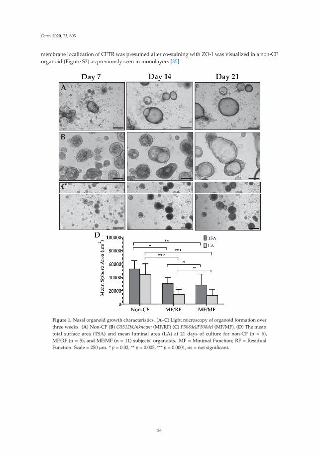
Genes 2020, 11, 603
membrane localization of CFTR was presumed after co-staining with ZO-1 was visualized in a non-CForganoid (Figure S2) as previously seen in monolayers [35].
Figure 1. Nasal organoid growth characteristics. (A–C) Light microscopy of organoid formation overthree weeks. (A) Non-CF (B) G551D/Unknown (MF/RF) (C) F508del/F508del (MF/MF). (D) The meantotal surface area (TSA) and mean luminal area (LA) at 21 days of culture for non-CF (n = 6),MF/RF (n = 5), and MF/MF (n = 11) subjects’ organoids. MF = Minimal Function; RF = ResidualFunction. Scale = 250 μm. * p = 0.02, ** p = 0.005, *** p = 0.0001, ns = not significant.
26

Genes 2020, 11, 603
Figure 2. Representative samples of typical hematoxylin and eosin staining of HNE organoids showstructural markers of differentiation. (A) Fully differentiated cross-section of a non-CF organoid shows adifferentiated monolayer with an open lumen. Scale= 50μm. (B) An example of the lower magnificationof non-CF organoids demonstrates variations in the overall organoid morphology. Scale = 250 μm.(C) Higher magnification of the black frame in panel (A) showing a ciliated apical surface (arrow).Scale = 50 μm. (D) An example of higher magnification of G551D/unknown CF organoids showingthick and thin (arrow) epithelial walls. Scale = 50 μm.
Figure 3. Mucus characteristics in HNE organoids, representative findings from CF and non-CForganoids are shown. (A) MUC5B granules in G551D/N1303K organoid. Scale = 50 μm. (B) Muc5Bwhole-mount IF showing a bundled mucin pattern in F508del/R117H-5T organoid. Scale = 50 μm.(C) MUC5AC fixed cross-section showing a diffuse mucin pattern in a non-CF organoid. Scale = 50 μm.(D) Top panel is the maximum projection of the 3-D hemisphere in the bottom panel. MUC5B isseen entering the lumen (arrow) through the apical surface in a non-CF organoid. Scale = 500 μm.(E) Brightfield microscopy showing a clump of cellular debris and/or mucus in the lumen in a non-CForganoid. Scale = 100 μm. (F) μOCT still of probable mucus in a non-CF organoid. (E,F) correspond toVideos S1 and S2. E = epithelium L = lumen M =mucus.
27
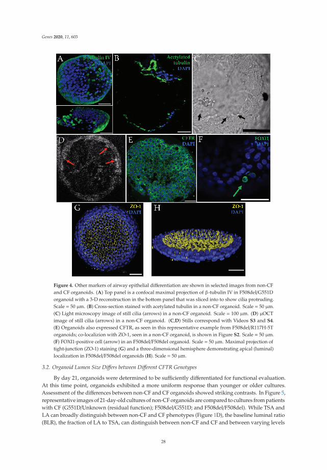
Genes 2020, 11, 603
Figure 4. Other markers of airway epithelial differentiation are shown in selected images from non-CFand CF organoids. (A) Top panel is a confocal maximal projection of β-tubulin IV in F508del/G551Dorganoid with a 3-D reconstruction in the bottom panel that was sliced into to show cilia protruding.Scale = 50 μm. (B) Cross-section stained with acetylated tubulin in a non-CF organoid. Scale = 50 μm.(C) Light microscopy image of still cilia (arrows) in a non-CF organoid. Scale = 100 μm. (D) μOCTimage of still cilia (arrows) in a non-CF organoid. (C,D) Stills correspond with Videos S3 and S4.(E) Organoids also expressed CFTR, as seen in this representative example from F508del/R117H-5Torganoids; co-localizion with ZO-1, seen in a non-CF organoid, is shown in Figure S2. Scale = 50 μm.(F) FOXI1-positive cell (arrow) in an F508del/F508del organoid. Scale = 50 μm. Maximal projection oftight-junction (ZO-1) staining (G) and a three-dimensional hemisphere demonstrating apical (luminal)localization in F508del/F508del organoids (H). Scale = 50 μm.
3.2. Organoid Lumen Size Differs between Different CFTR Genotypes
By day 21, organoids were determined to be sufficiently differentiated for functional evaluation.At this time point, organoids exhibited a more uniform response than younger or older cultures.Assessment of the differences between non-CF and CF organoids showed striking contrasts. In Figure 5,representative images of 21-day-old cultures of non-CF organoids are compared to cultures from patientswith CF (G551D/Unknown (residual function); F508del/G551D; and F508del/F508del). While TSA andLA can broadly distinguish between non-CF and CF phenotypes (Figure 1D), the baseline luminal ratio(BLR), the fraction of LA to TSA, can distinguish between non-CF and CF and between varying levels
28

Genes 2020, 11, 603
of CFTR dysfunction (Figure 6A–C). Furthermore, BLR shows a high correlation (r = 0.94, p = 0.0005)with the baseline forskolin-stimulated Isc in HNE monolayers from the same patients (Figure 6D).
Figure 5. Light microscopy of cultures at 21 days of growth demonstrates variations in the amount ofluminal fluid present within organoids of different CFTR genotypes. Scale = 1000 μm. The genotype(G551D/unknown (MF/RF based on phenotype) is defined in Supplementary Material Table S1.
Figure 6. Baseline lumen ratio. Zoomed-in section of a well to show F508del/G551D organoids(A) masking of the organoid’s total surface area and (B) masking of the same organoids’ luminalareas. (C) Baseline lumen to total surface area ratio comparing non-CF (n = 6) to CF patients withresidual function (n = 5) and minimal function mutations (n = 11). (D) Pearson correlation comparingthe average organoid BLR for each person and the corresponding change in short-circuit current(ΔIsc) after forskolin stimulation in the ussing chamber. Not all subjects from (C) had correspondingshort-circuit current measurements. Filled circle=Non-CF; Open square=MF/RF; Open circle=MF/MF.Scale = 200 μm. * p = 0.02, *** p = 0.0001, and **** p < 0.0001.
29
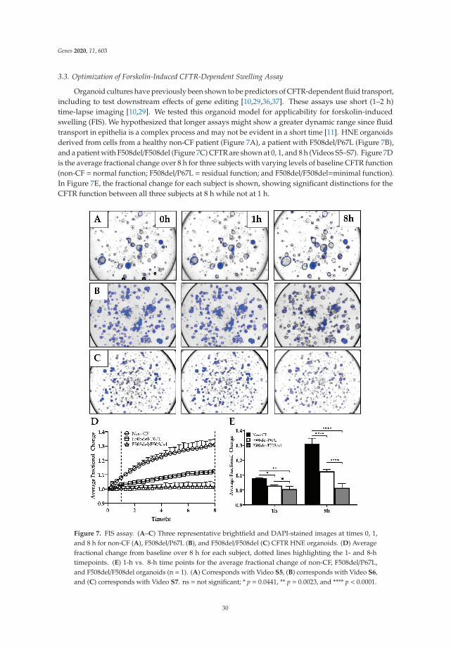
Genes 2020, 11, 603
3.3. Optimization of Forskolin-Induced CFTR-Dependent Swelling Assay
Organoid cultures have previously been shown to be predictors of CFTR-dependent fluid transport,including to test downstream effects of gene editing [10,29,36,37]. These assays use short (1–2 h)time-lapse imaging [10,29]. We tested this organoid model for applicability for forskolin-inducedswelling (FIS). We hypothesized that longer assays might show a greater dynamic range since fluidtransport in epithelia is a complex process and may not be evident in a short time [11]. HNE organoidsderived from cells from a healthy non-CF patient (Figure 7A), a patient with F508del/P67L (Figure 7B),and a patient with F508del/F508del (Figure 7C) CFTR are shown at 0, 1, and 8 h (Videos S5–S7). Figure 7Dis the average fractional change over 8 h for three subjects with varying levels of baseline CFTR function(non-CF = normal function; F508del/P67L = residual function; and F508del/F508del=minimal function).In Figure 7E, the fractional change for each subject is shown, showing significant distinctions for theCFTR function between all three subjects at 8 h while not at 1 h.
Figure 7. FIS assay. (A–C) Three representative brightfield and DAPI-stained images at times 0, 1,and 8 h for non-CF (A), F508del/P67L (B), and F508del/F508del (C) CFTR HNE organoids. (D) Averagefractional change from baseline over 8 h for each subject, dotted lines highlighting the 1- and 8-htimepoints. (E) 1-h vs. 8-h time points for the average fractional change of non-CF, F508del/P67L,and F508del/F508del organoids (n = 1). (A) Corresponds with Video S5, (B) corresponds with Video S6,and (C) corresponds with Video S7. ns = not significant; * p = 0.0441, ** p = 0.0023, and **** p < 0.0001.
30

Genes 2020, 11, 603
4. Discussion and Conclusions
In this manuscript, we provide evidence for a highly differentiated organoid culture that expressesmarkers consistent with human airway epithelial monolayers with reproducible cultures regardless ofthe genotype. The organoid culture shows distinct differences in lumen formation between non-CF andCF cultures with and without residual CFTR function, suggesting a CFTR dependence of the lumensize and fluid transport during organoid development. Along with the significant differences betweenthe lumen formation of CF and non-CF organoids, this difference highly correlates with paralleledshort-circuit measurements of baseline CFTR activity. This has not previously been evaluated in humannasal epithelial organoids, but a similar finding was seen in intestinal organoids [38]. Our swellingdata suggest that this model has the potential for functional assessment of the patient phenotype,identifying both large and small differences (such as residual function, or a modest improvement of onetreatment over another). Furthermore, the model has multiple applications, including FIS, assessmentof mucus expression, transport, cilia activity, and fluorescence microscopy.
We present data that the full effects of CFTR restoration may not be visible over short time frames innasal organoid studies. By lengthening the assay to eight hours, we were able to increase the distinctionbetween differing CFTR activity levels. This result requires further testing with more subjects, but it isan interesting finding that takes into account that fluid transport may not immediately follow CFTRrescue, and the complex systems that result in fluid secretion into the lumen may take some time toshow full effects. The functional assay could be useful in pre-clinical testing, where interventionsdesigned to be delivered via systemic absorption (i.e., from the basolateral side after distributionthrough the blood) can be applied to an in vitro surrogate. Because it requires so little material togenerate many replicates, it is amenable to higher throughput applications using novel compounds oriterative testing of different delivery mechanisms for RNA- and DNA-based therapies. Additionally,it provides a useful ex vivo marker for in vivo gene therapy trials. A nasal brush biopsy is safe, easilyobtained in an outpatient clinical research setting, and can be repeatedly collected from the patient [39].This could be tested to monitor the stability of a genetic repair, which would be helpful to confirma corrected phenotype even if clinical measures (like lung function testing) may not be altered (forexample, if a patient has a normal lung function result at the start of the study).
The differentiation of this model provides multiple readouts for CFTR functional rescue. It ishighly amenable to fluorescent imaging. We used fluorescence for automated imaging of living cellsas well as fixed whole organoids for immunofluorescent staining of specific markers. We observedlimited evidence that the organoid can recapitulate mucin organization also seen in other models [33].Currently, a variety of approaches to deliver gene therapy packages exist, and delivery mechanismstagged with a fluorescent marker for measuring uptake could easily be tested across a variety of patientsamples with this approach. Further, the readout for functional assessment of CFTR rescue is notlimited to the measurement of the total cross-sectional area or lumen over time. Our results show thatμOCT imaging can yield multiple readouts of the lumen area, mucus morphology, ciliary function,and possibly with further testing, mucus viscosity and mucociliary transport speeds.
Our study has some limitations. We present functional data (lumen size and swelling) from alimited number of subjects, while additional subject samples were used for iterative testing to optimizethe culturing and assay methodology to achieve a reproducible assay. Despite the small number ofsubjects, the lumen measurement shows excellent distinction for differences between patient groups.The current analysis limits our baseline luminal ratio assay to manual measurements until furtherrevisions to the automated software can be made. However, because the measurements and output forthe FIS assay are automated, we will now be able to scale up the testing of many patients and correlateclinical outcome measures with the organoid data for future studies.
Our results show that a solid assay with similarities to the robust intestinal organoidforskolin-induced swelling assay can be developed with HNEs. This provides a significant advantagebecause not every CF center has the capacity to collect rectal biopsies on all patients, nor are all patientsreceptive to biopsy collection. However, many CF centers in the United States and elsewhere have
31

Genes 2020, 11, 603
experience in collecting nasal brush biopsies from infants through to adults, with risk comparable tothat of a nasopharyngeal swab for virus detection. The model can assess CFTR dysfunction and rescue,both as a pre-clinical tool for systemically delivered DNA-based therapies and an ex vivo surrogatebiomarker to assess the stability of DNA-based therapy in clinical trials.
Supplementary Materials: The following are available online at http://www.mdpi.com/2073-4425/11/6/603/s1,Table S1: Individual subject demographics and available clinical data for selected participants contributingfunctional data. Figure S1: Example measurement of μOCT ciliary beat frequencies, Figure S2: Organoid CFTR(green) co-localization with apical ZO-1 (red). (A) To illustrate the location of the ZO-1 staining within theorganoid, an α-blended 3-D volume reconstruction was created with the fluorescence intensity increased to revealthe organoid’s shape and orient the reader to the location of the 2D co-localization image below. (B) Magnifiedview of the yellow frame in (A) to better visualize the CFTR and ZO-1 overlay, Video S1: Brightfield video ofluminal movement of mucus, Video S2: μOCT video of luminal movement of mucus, Video S3: Brightfield videoof luminal cilia beating, Video S4: μOCT video of cilia beating, Video S5: FIS assay of non-CF organoids, Video S6:FIS assay of F508del/P67L organoids. Video S7: FIS assay of F508del/F508del organoids.
Author Contributions: Conceptualization, Z.L., J.D.A. and J.S.G.; methodology, Z.L., J.D.A., L.D., S.M. andJ.S.G.; validation, Z.L., J.D.A. and J.S.G.; formal analysis, Z.L. and J.D.A.; investigation, Z.L., J.D.A., L.D. and J.B.;resources, L.K.; writing—original draft preparation, J.S.G.; writing—review and editing, Z.L., J.D.A., S.M.R. andJ.S.G.; visualization, J.D.A.; supervision, J.S.G.; project administration, J.S.G.; funding acquisition, S.M.R. and J.S.G.All authors have read and agreed to the published version of the manuscript.
Funding: This work was supported by the Kaul Pediatric Research Institute Pilot Award and the CysticFibrosis Foundation (GUIMBE1710). J.S.G. is supported by the NIH (K23 HL143167-01A1), and the CysticFibrosis Foundation (GUIMBE18A0-Q). Z.L., J.D.A., L.K., and J.S.G. and S.M.R. are supported by the GregoryFleming James Cystic Fibrosis Center (R35HL135816, NIH DK072482, CFF UAB Research and DevelopmentProgram—Rowe19RO, all to S.M.R.) and the UAB Center for Clinical and Translational Science (UL1TR001417).
Acknowledgments: We gratefully acknowledge the contributions of Guillermo Tearney and Hui Min Leung,Massachusetts General Hospital and Harvard Medical School, for the invention and maintenance of the μOCTimager. We thank Susan Birket and Steven Roberts for technical assistance. We thank the UAB High ResolutionImaging Facility and the UAB Histology Core for assistance. We would also like to thank Jennifer Natt for herreview and copy editing of the manuscript.
Conflicts of Interest: J.S.G. is listed as an inventor on a patent application 20170242033 from the University ofNorth Carolina that describes a similar model. When licensed technology from UNC produces royalties, theinventors receive a share of the revenue. Otherwise, the authors declare no conflict of interest. The funders had norole in the design of the study; in the collection, analyses, or interpretation of data; in the writing of the manuscript,or in the decision to publish the results.
Abbreviations
CF cystic fibrosisCFTR Cystic Fibrosis Transmembrane conductance regulator
References
1. Bell, S.C.; De Boeck, K.; Amaral, M.D. New pharmacological approaches for cystic fibrosis: Promises,progress, pitfalls. Pharmacol. Ther. 2015, 145, 19–34. [CrossRef] [PubMed]
2. Bertoncini, E.; Colomb-Lippa, D. Pulmonology: CFTR modulators for cystic fibrosis. J. Am. Acad.Physician Assist. 2013, 26, 59–60. [CrossRef] [PubMed]
3. Derichs, N. Targeting a genetic defect: Cystic fibrosis transmembrane conductance regulator modulators incystic fibrosis. Eur. Respir. Rev. 2013, 22, 58–65. [CrossRef]
4. Middleton, P.G.; Mall, M.A.; Drevinek, P.; Lands, L.C.; McKone, E.F.; Polineni, D.; Ramsey, B.W.;Taylor-Cousar, J.L.; Tullis, E.; Vermeulen, F.; et al. Elexacaftor-Tezacaftor-Ivacaftor for Cystic Fibrosiswith a Single Phe508del Allele. N. Engl. J. Med. 2019, 381, 1809–1819. [CrossRef] [PubMed]
5. Heijerman, H.G.M.; McKone, E.F.; Downey, D.G.; Van Braeckel, E.; Rowe, S.M.; Tullis, E.; Mall, M.A.;Welter, J.J.; Ramsey, B.W.; McKee, C.M.; et al. Efficacy and safety of the elexacaftor plus tezacaftorplus ivacaftor combination regimen in people with cystic fibrosis homozygous for the F508del mutation:A double-blind, randomised, phase 3 trial. Lancet 2019. [CrossRef]
32
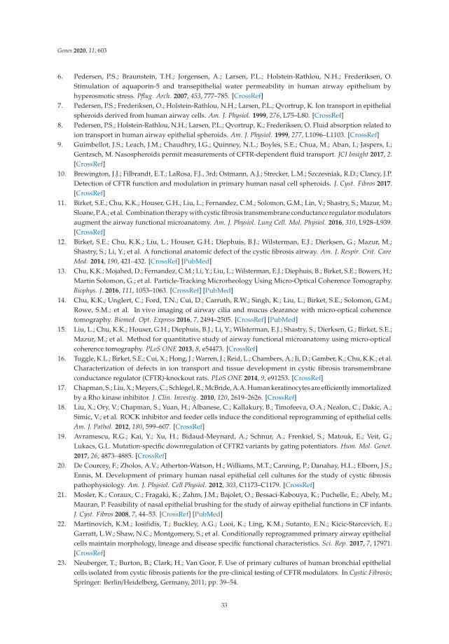
Genes 2020, 11, 603
6. Pedersen, P.S.; Braunstein, T.H.; Jorgensen, A.; Larsen, P.L.; Holstein-Rathlou, N.H.; Frederiksen, O.Stimulation of aquaporin-5 and transepithelial water permeability in human airway epithelium byhyperosmotic stress. Pflug. Arch. 2007, 453, 777–785. [CrossRef]
7. Pedersen, P.S.; Frederiksen, O.; Holstein-Rathlou, N.H.; Larsen, P.L.; Qvortrup, K. Ion transport in epithelialspheroids derived from human airway cells. Am. J. Physiol. 1999, 276, L75–L80. [CrossRef]
8. Pedersen, P.S.; Holstein-Rathlou, N.H.; Larsen, P.L.; Qvortrup, K.; Frederiksen, O. Fluid absorption related toion transport in human airway epithelial spheroids. Am. J. Physiol. 1999, 277, L1096–L1103. [CrossRef]
9. Guimbellot, J.S.; Leach, J.M.; Chaudhry, I.G.; Quinney, N.L.; Boyles, S.E.; Chua, M.; Aban, I.; Jaspers, I.;Gentzsch, M. Nasospheroids permit measurements of CFTR-dependent fluid transport. JCI Insight 2017, 2.[CrossRef]
10. Brewington, J.J.; Filbrandt, E.T.; LaRosa, F.J., 3rd; Ostmann, A.J.; Strecker, L.M.; Szczesniak, R.D.; Clancy, J.P.Detection of CFTR function and modulation in primary human nasal cell spheroids. J. Cyst. Fibros 2017.[CrossRef]
11. Birket, S.E.; Chu, K.K.; Houser, G.H.; Liu, L.; Fernandez, C.M.; Solomon, G.M.; Lin, V.; Shastry, S.; Mazur, M.;Sloane, P.A.; et al. Combination therapy with cystic fibrosis transmembrane conductance regulator modulatorsaugment the airway functional microanatomy. Am. J. Physiol. Lung Cell. Mol. Physiol. 2016, 310, L928–L939.[CrossRef]
12. Birket, S.E.; Chu, K.K.; Liu, L.; Houser, G.H.; Diephuis, B.J.; Wilsterman, E.J.; Dierksen, G.; Mazur, M.;Shastry, S.; Li, Y.; et al. A functional anatomic defect of the cystic fibrosis airway. Am. J. Respir. Crit. CareMed. 2014, 190, 421–432. [CrossRef] [PubMed]
13. Chu, K.K.; Mojahed, D.; Fernandez, C.M.; Li, Y.; Liu, L.; Wilsterman, E.J.; Diephuis, B.; Birket, S.E.; Bowers, H.;Martin Solomon, G.; et al. Particle-Tracking Microrheology Using Micro-Optical Coherence Tomography.Biophys. J. 2016, 111, 1053–1063. [CrossRef] [PubMed]
14. Chu, K.K.; Unglert, C.; Ford, T.N.; Cui, D.; Carruth, R.W.; Singh, K.; Liu, L.; Birket, S.E.; Solomon, G.M.;Rowe, S.M.; et al. In vivo imaging of airway cilia and mucus clearance with micro-optical coherencetomography. Biomed. Opt. Express 2016, 7, 2494–2505. [CrossRef] [PubMed]
15. Liu, L.; Chu, K.K.; Houser, G.H.; Diephuis, B.J.; Li, Y.; Wilsterman, E.J.; Shastry, S.; Dierksen, G.; Birket, S.E.;Mazur, M.; et al. Method for quantitative study of airway functional microanatomy using micro-opticalcoherence tomography. PLoS ONE 2013, 8, e54473. [CrossRef]
16. Tuggle, K.L.; Birket, S.E.; Cui, X.; Hong, J.; Warren, J.; Reid, L.; Chambers, A.; Ji, D.; Gamber, K.; Chu, K.K.; et al.Characterization of defects in ion transport and tissue development in cystic fibrosis transmembraneconductance regulator (CFTR)-knockout rats. PLoS ONE 2014, 9, e91253. [CrossRef]
17. Chapman, S.; Liu, X.; Meyers, C.; Schlegel, R.; McBride, A.A. Human keratinocytes are efficiently immortalizedby a Rho kinase inhibitor. J. Clin. Investig. 2010, 120, 2619–2626. [CrossRef]
18. Liu, X.; Ory, V.; Chapman, S.; Yuan, H.; Albanese, C.; Kallakury, B.; Timofeeva, O.A.; Nealon, C.; Dakic, A.;Simic, V.; et al. ROCK inhibitor and feeder cells induce the conditional reprogramming of epithelial cells.Am. J. Pathol. 2012, 180, 599–607. [CrossRef]
19. Avramescu, R.G.; Kai, Y.; Xu, H.; Bidaud-Meynard, A.; Schnur, A.; Frenkiel, S.; Matouk, E.; Veit, G.;Lukacs, G.L. Mutation-specific downregulation of CFTR2 variants by gating potentiators. Hum. Mol. Genet.2017, 26, 4873–4885. [CrossRef]
20. De Courcey, F.; Zholos, A.V.; Atherton-Watson, H.; Williams, M.T.; Canning, P.; Danahay, H.L.; Elborn, J.S.;Ennis, M. Development of primary human nasal epithelial cell cultures for the study of cystic fibrosispathophysiology. Am. J. Physiol. Cell Physiol. 2012, 303, C1173–C1179. [CrossRef]
21. Mosler, K.; Coraux, C.; Fragaki, K.; Zahm, J.M.; Bajolet, O.; Bessaci-Kabouya, K.; Puchelle, E.; Abely, M.;Mauran, P. Feasibility of nasal epithelial brushing for the study of airway epithelial functions in CF infants.J. Cyst. Fibros 2008, 7, 44–53. [CrossRef] [PubMed]
22. Martinovich, K.M.; Iosifidis, T.; Buckley, A.G.; Looi, K.; Ling, K.M.; Sutanto, E.N.; Kicic-Starcevich, E.;Garratt, L.W.; Shaw, N.C.; Montgomery, S.; et al. Conditionally reprogrammed primary airway epithelialcells maintain morphology, lineage and disease specific functional characteristics. Sci. Rep. 2017, 7, 17971.[CrossRef]
23. Neuberger, T.; Burton, B.; Clark, H.; Van Goor, F. Use of primary cultures of human bronchial epithelialcells isolated from cystic fibrosis patients for the pre-clinical testing of CFTR modulators. In Cystic Fibrosis;Springer: Berlin/Heidelberg, Germany, 2011; pp. 39–54.
33
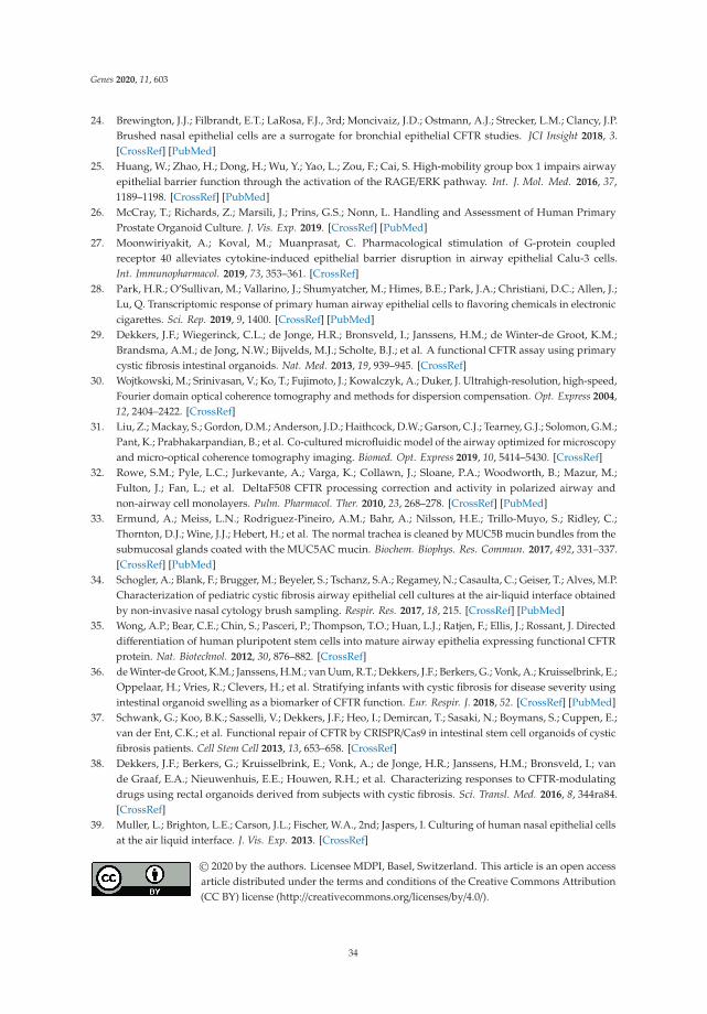
Genes 2020, 11, 603
24. Brewington, J.J.; Filbrandt, E.T.; LaRosa, F.J., 3rd; Moncivaiz, J.D.; Ostmann, A.J.; Strecker, L.M.; Clancy, J.P.Brushed nasal epithelial cells are a surrogate for bronchial epithelial CFTR studies. JCI Insight 2018, 3.[CrossRef] [PubMed]
25. Huang, W.; Zhao, H.; Dong, H.; Wu, Y.; Yao, L.; Zou, F.; Cai, S. High-mobility group box 1 impairs airwayepithelial barrier function through the activation of the RAGE/ERK pathway. Int. J. Mol. Med. 2016, 37,1189–1198. [CrossRef] [PubMed]
26. McCray, T.; Richards, Z.; Marsili, J.; Prins, G.S.; Nonn, L. Handling and Assessment of Human PrimaryProstate Organoid Culture. J. Vis. Exp. 2019. [CrossRef] [PubMed]
27. Moonwiriyakit, A.; Koval, M.; Muanprasat, C. Pharmacological stimulation of G-protein coupledreceptor 40 alleviates cytokine-induced epithelial barrier disruption in airway epithelial Calu-3 cells.Int. Immunopharmacol. 2019, 73, 353–361. [CrossRef]
28. Park, H.R.; O’Sullivan, M.; Vallarino, J.; Shumyatcher, M.; Himes, B.E.; Park, J.A.; Christiani, D.C.; Allen, J.;Lu, Q. Transcriptomic response of primary human airway epithelial cells to flavoring chemicals in electroniccigarettes. Sci. Rep. 2019, 9, 1400. [CrossRef] [PubMed]
29. Dekkers, J.F.; Wiegerinck, C.L.; de Jonge, H.R.; Bronsveld, I.; Janssens, H.M.; de Winter-de Groot, K.M.;Brandsma, A.M.; de Jong, N.W.; Bijvelds, M.J.; Scholte, B.J.; et al. A functional CFTR assay using primarycystic fibrosis intestinal organoids. Nat. Med. 2013, 19, 939–945. [CrossRef]
30. Wojtkowski, M.; Srinivasan, V.; Ko, T.; Fujimoto, J.; Kowalczyk, A.; Duker, J. Ultrahigh-resolution, high-speed,Fourier domain optical coherence tomography and methods for dispersion compensation. Opt. Express 2004,12, 2404–2422. [CrossRef]
31. Liu, Z.; Mackay, S.; Gordon, D.M.; Anderson, J.D.; Haithcock, D.W.; Garson, C.J.; Tearney, G.J.; Solomon, G.M.;Pant, K.; Prabhakarpandian, B.; et al. Co-cultured microfluidic model of the airway optimized for microscopyand micro-optical coherence tomography imaging. Biomed. Opt. Express 2019, 10, 5414–5430. [CrossRef]
32. Rowe, S.M.; Pyle, L.C.; Jurkevante, A.; Varga, K.; Collawn, J.; Sloane, P.A.; Woodworth, B.; Mazur, M.;Fulton, J.; Fan, L.; et al. DeltaF508 CFTR processing correction and activity in polarized airway andnon-airway cell monolayers. Pulm. Pharmacol. Ther. 2010, 23, 268–278. [CrossRef] [PubMed]
33. Ermund, A.; Meiss, L.N.; Rodriguez-Pineiro, A.M.; Bahr, A.; Nilsson, H.E.; Trillo-Muyo, S.; Ridley, C.;Thornton, D.J.; Wine, J.J.; Hebert, H.; et al. The normal trachea is cleaned by MUC5B mucin bundles from thesubmucosal glands coated with the MUC5AC mucin. Biochem. Biophys. Res. Commun. 2017, 492, 331–337.[CrossRef] [PubMed]
34. Schogler, A.; Blank, F.; Brugger, M.; Beyeler, S.; Tschanz, S.A.; Regamey, N.; Casaulta, C.; Geiser, T.; Alves, M.P.Characterization of pediatric cystic fibrosis airway epithelial cell cultures at the air-liquid interface obtainedby non-invasive nasal cytology brush sampling. Respir. Res. 2017, 18, 215. [CrossRef] [PubMed]
35. Wong, A.P.; Bear, C.E.; Chin, S.; Pasceri, P.; Thompson, T.O.; Huan, L.J.; Ratjen, F.; Ellis, J.; Rossant, J. Directeddifferentiation of human pluripotent stem cells into mature airway epithelia expressing functional CFTRprotein. Nat. Biotechnol. 2012, 30, 876–882. [CrossRef]
36. de Winter-de Groot, K.M.; Janssens, H.M.; van Uum, R.T.; Dekkers, J.F.; Berkers, G.; Vonk, A.; Kruisselbrink, E.;Oppelaar, H.; Vries, R.; Clevers, H.; et al. Stratifying infants with cystic fibrosis for disease severity usingintestinal organoid swelling as a biomarker of CFTR function. Eur. Respir. J. 2018, 52. [CrossRef] [PubMed]
37. Schwank, G.; Koo, B.K.; Sasselli, V.; Dekkers, J.F.; Heo, I.; Demircan, T.; Sasaki, N.; Boymans, S.; Cuppen, E.;van der Ent, C.K.; et al. Functional repair of CFTR by CRISPR/Cas9 in intestinal stem cell organoids of cysticfibrosis patients. Cell Stem Cell 2013, 13, 653–658. [CrossRef]
38. Dekkers, J.F.; Berkers, G.; Kruisselbrink, E.; Vonk, A.; de Jonge, H.R.; Janssens, H.M.; Bronsveld, I.; vande Graaf, E.A.; Nieuwenhuis, E.E.; Houwen, R.H.; et al. Characterizing responses to CFTR-modulatingdrugs using rectal organoids derived from subjects with cystic fibrosis. Sci. Transl. Med. 2016, 8, 344ra84.[CrossRef]
39. Muller, L.; Brighton, L.E.; Carson, J.L.; Fischer, W.A., 2nd; Jaspers, I. Culturing of human nasal epithelial cellsat the air liquid interface. J. Vis. Exp. 2013. [CrossRef]
© 2020 by the authors. Licensee MDPI, Basel, Switzerland. This article is an open accessarticle distributed under the terms and conditions of the Creative Commons Attribution(CC BY) license (http://creativecommons.org/licenses/by/4.0/).
34

genesG C A T
T A C G
G C A T
Article
Overcoming Immunological Challenges toHelper-Dependent Adenoviral Vector-MediatedLong-Term CFTR Expression in Mouse Airways
Huibi Cao 1, Rongqi Duan 1 and Jim Hu 1,2,*1 Program of Translational Medicine, The Hospital for Sick Children, Research Institute, 686 Bay Street,
Toronto, ON M5G 0A4, Canada; [email protected] (H.C.); [email protected] (R.D.)2 Departments of Paediatrics and Laboratory Medicine and Pathobiology, University of Toronto, 1 King’s
College Circle, Toronto, ON M5S 1A8, Canada* Correspondence: [email protected]; Tel.: +1-416-813-6412
Received: 10 April 2020; Accepted: 13 May 2020; Published: 18 May 2020
Abstract: Cystic Fibrosis (CF) is caused by mutations in the cystic fibrosis transmembrane conductanceregulator (CFTR) gene, and CF patients require life-long treatment. Although CFTR modulatorsshow a great potential for treating most CF patients, some individuals may not tolerate the treatment.In addition, there is no effective therapy for patients with some rare CFTR mutations, such as class I CFmutations, which lead to a lack of CFTR protein production. Therefore, other therapeutic strategies,such as gene therapy, have to be investigated. Currently, immune responses to gene therapy vectorsand transgene products are a major obstacle to applying CF gene therapy to clinical applications.In this study, we examined the effects of cyclophosphamide on the modulation of host immuneresponses and for the improvement of the CFTR transgene expression in the repeated delivery ofhelper-dependent adenoviral (HD-Ad) vectors to mouse lungs. We have found that cyclophosphamidesignificantly decreased the expression of T cell genes, such as CD3 (cluster of differentiation 3) andCD4, and reduced their infiltration into mouse lung tissues. We have also found that the levels ofthe anti-adenoviral antibody and neutralizing activity as well as B-cell infiltration into the mouselung tissues were significantly reduced with this treatment. Correspondingly, the expression of thehuman CFTR transgene has been significantly improved with cyclophosphamide administrationcompared to the group with no treatment. These data suggest that the sustained expression ofthe human CFTR transgene in mouse lungs through repeated vector delivery can be achieved bytransient immunosuppression.
Keywords: cystic fibrosis; gene therapy; cyclophosphamide; transient immunosuppression
1. Introduction
Cystic fibrosis (CF) is a life-long inherited disease that affects >70,000 patients globally; it is causedby underlying genetic mutations in the cystic fibrosis transmembrane conductance regulator (CFTR)gene [1]. New CFTR-modulator drugs show promise for up to 90% of patients, including patients withCFTR mutations for which early modulators are ineffective [2–5]. Although CFTR modulators showenormous potential for treating most CF patients, they are expensive and patients require lifetimetreatments [6]. In addition, some patients may not tolerate the treatments. It is understandable thatpharmaceutical approaches may not always effectively fix every malfunctioning human body caused bythe same genetic defect [7]. Currently, there is no remedy for some patients with rare CFTR mutations,such as class I mutations, which lead to a lack of production of the CFTR protein [8]. Therefore,alternative therapeutic strategies, such as gene therapy, should be explored.
To date, more than 2600 gene therapy clinical trials have either been completed, are going to be,or have been approved worldwide [9]. Gene therapies for inherited immune disorders, hemophilia,
Genes 2020, 11, 565; doi:10.3390/genes11050565 www.mdpi.com/journal/genes35
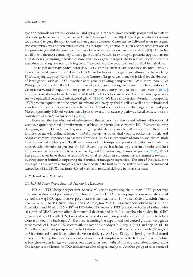
Genes 2020, 11, 565
eye and neurodegenerative disorders, and lymphoid cancers, have recently progressed to a stagewhere drugs have been approved in the United States and Europe [10]. Efficient gene delivery systemsare essential to gene therapy to treat human genetic diseases. Genes can be delivered to target organsand cells with viral and non-viral vectors. As therapeutics, adenoviral (Ad) vectors represent one ofthe promising candidates among current available advance-therapy medical products [11]. Ad vectoris still one of the most commonly utilized gene transfer vectors in a variety of potential applications forlung diseases (including inherited disease and cancer gene therapy). Ad-based vector can efficientlytransduce dividing and non-dividing cells. They can be easily produced and purified in high titers.
The helper-dependent adenoviral (HD-Ad) vector has been developed based on adenovirus bydeleting all viral genes. This makes the HD-Ad vector less immunogenic and allows it to have a largeDNA-carrying capacity [12–14]. This unique feature of large capacity makes it ideal for the deliveryof large genes, such as CFTR, together with gene regulating components. With more than 36 kbDNA payload capacity, HD-Ad vectors can easily carry gene editing components, such as guide RNA,CRISPR-Cas9, and therapeutic donor genes with gene-regulatory elements in the same vector [15–17].Our previous studies have demonstrated that HD-Ad vectors are efficient for transducing airwaysurface epithelial cells and submucosal glands [18,19]. We have shown that abundant therapeuticCFTR protein expression at the apical membrane of airway epithelial cells as well as the submucosalglands of the conduct airways can be achieved by HD-Ad vector delivery to the lungs of mice and pigs.More importantly, HD-Ad vectors have been shown to transduce pig airway basal cells, which areconsidered as stem/progenitor cells [20,21].
However, the transduction of self-renewal tissues, such as airway epithelium with episomalvectors, requires repeated administration to achieve long-term gene correction [22]. Even consideringstem/progenitor cell targeting with gene editing, repeated delivery may be still needed due to the currentlow in vivo gene-targeting efficiency. HD-Ad vectors, as other viral vectors, evoke host innate andadaptive immune responses against capsid proteins. Studies in experimental animals and clinical trialshave shown that antibody and T cell responses can limit transgene expression duration and hinder therepeated administration of gene transfer [23]. Several approaches, including vector modification and hostimmune system modulations, have been investigated for minimizing immune responses [24–26]. All ofthese approaches have shown effects on the partial reduction in inflammation and immune reactions,but they are not fruitful in improving the duration of transgene expression. The aim of this study is toinvestigate how pharmacological agents can modulate the host immune system to allow the sustainedexpression of the CFTR gene from HD-Ad vectors in repeated delivery to mouse airways.
2. Materials and Methods
2.1. HD-Ad Vector Preparation and Delivery to Mice Lungs
HD-Ad-CFTR (helper-dependent adenoviral vector expressing the human CFTR gene) wasprepared as described previously [27]. The purity of the HD-Ad vector preparations was determinedby real-time q-PCR (quantitative polymerase chain reaction). For vector delivery, adult femaleC57Bl/6 mice (Charles River Laboratories, Wilmington, MA, USA) were anesthetized by isofluraneinhalation, and 20 μL of 1.5 × 10¹⁰ of HD-Ad-CFTR vector in PBS (phosphate buffered saline) with40 μg/mL of DEAE-dextran (diethylaminoethyl-dextran) and 0.1% L-α-lysophosphatidylcholine (LPC)(Sigma-Aldrich, Oakville, ON, Canada) were placed in small drops onto one nostril from which theywere aspirated into the lungs. All the mice, including the experiment and control groups, were giventhree rounds of HD-Ad-CFTR vector with the same dose at day 0 (d0), day 60 (d60), and day 120 (d120).Only the experiment group was injected intraperitoneally (ip) with cyclophosphamide (50 mg/kg)at 6 h before and 4 and 8 days after the vector delivery. At 3 and 33 days following the final roundof vector delivery, the mice were sacrificed and blood samples were collected by cardiac puncture.A bronchoalveolar lavage was performed three times, each with 0.9 mL of phosphate-buffered saline.The lungs were collected for RNA isolation and histological analyses. Another group of mice received
36
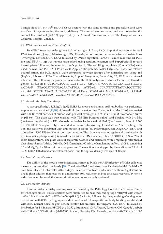
Genes 2020, 11, 565
a single dose of 1.5 × 10¹⁰ HD-Ad-CFTR vectors with the same formula and procedure, and weresacrificed 3 days following the vector delivery. The animal studies were conducted following theAnimal Use Protocol (#48813) approved by the Animal Care Committee of The Hospital for SickChildren, Toronto, Canada.
2.2. RNA Isolation and Real-Time RT-qPCR
Total RNA from mouse lungs was isolated using an RNeasy kit (a simplified technology for totalRNA isolation) (Qiagen, Mississauga, ON, Canada) according to the manufacturer’s instructions(Invitrogen, Carlsbad, CA, USA), followed by DNase digestion. For SYBR Green real-time RT-PCR,the total RNA (1 μg) was reverse-transcribed using random hexamers and SuperScript II reversetranscriptase following the manufacturer’s protocol. The resulting templates (10 ng cDNA) wereused for real-time PCR (ABI Prism 7500, Applied Biosystems, Foster City, CA, USA). For relativequantification, the PCR signals were compared between groups after normalization using 18S(TaqMan, Ribosomal RNA Control Reagents, Applied Biosystems, Foster City, CA, USA) as an internalreference. The following are primer sequences for the PCR analysis of vector CFTR and T cell markergenes: K18CFTR-F: CCTGAGTCCTGTCCTTTCTC, K18CFTR-RCGCTGTCTGTATCCTTTCCTC;mCD3e-F: GGACGATGCCGAGAACATTGA, mCD3e-R: CCAGGTGCTTATCATGCTTCTG;mCD4-F: GCCCTCATATACACACACCTGT, mCD4-R: GCAGCAGCAGCAGCAGCAA; mCD8a-F:GCTCAGTCATCAGCAACTCG, mCD8a-R: GTGAGGGAGTTCGCAGCACT.
2.3. Anti-Ad Antibody Titer Assay
A pan-specific (IgA, IgE, IgGs, IgM) ELISA for mouse anti-human Ad5 antibodies was performed,as previously described [19,28]. A 96-well ELISA plate (Corning Costar, Acton, MA, USA) was coatedwith 5 × 10⁹ viral particles of human Ad5 per well overnight at 4 ◦C in a 100 mM bicarbonate bufferat pH 9.6. The plate was then washed with TBS (Tris-buffered saline) and blocked with 3% BSA(bovine serum albumin) in TBS. Mouse bronchoalveolar lavage fluid (BALF) and serum diluted in 1:200or 1:200,000 TBS, respectively, were added to the wells for overnight incubation. After washing withTBS, the plate was incubated with anti-mouse-Ig-biotin (BD Pharmingen, San Diego, CA, USA) anddiluted in 1:5000 TBS for 3 hr at room temperature. The plate was washed again and incubated withavidin-alkaline phosphatase (Sigma-Aldrich, Oakville, ON, Canada), diluted 1:50,000 in TBS for 2 hr atroom temperature. The plate was subsequently washed and incubated with 1 mg/mL p-nitrophenylphosphate (Sigma-Aldrich, Oakville, ON, Canada) in 100 mM diethanolamine buffer at pH 9.8, containing0.5 mM MgCl2, for 10 min at room temperature. The reaction was stopped by the addition of 25 μL of0.2 M EDTA (ethylenediaminetetraacetic acid) and the optical density was read at 405 nm.
2.4. Neutralizing Abs Assay
The ability of the mouse heat-inactivated serum to block the Ad5 infection of HeLa cells wasmeasured, as described previously [28]. The diluted BALF and serum was incubated with HD-Ad-LacZand then infected HeLa cells. After 3 days, the cells were fixed and stained with an X-gal solution.The highest dilution that resulted in a minimum 50% reduction in blue cells was recorded. When noreduction was observed, the lowest dilution was conservatively assigned.
2.5. CDs Marker Staining
Immunohistochemistry staining was performed by the Pathology Core at The Toronto Centrefor Phenogenomics. Tissue sections were submitted to heat-induced epitope retrieval with citratebuffer (pH 6.0) or with Tris-EDTA buffer (pH 9.0) for 7 min, followed by the quenching of endogenousperoxidase with 0.3% hydrogen peroxide in methanol. Non-specific antibody binding was blockedwith 2.5% normal horse or goat serum (Vector, Laboratories, Burlingame, CA, USA), followed byincubation for 1 h in rat anti-CD3 at a 1:150 dilution (ab11089, Abcam, Toronto, ON, Canada), rabbitanti-CD4 at a 1:500 dilution (ab183685, Abcam, Toronto, ON, Canada), rabbit anti-CD8 at a 1:1000
37
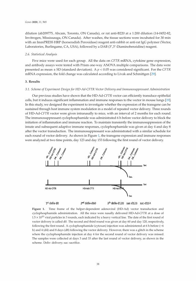
Genes 2020, 11, 565
dilution (ab209775, Abcam, Toronto, ON Canada), or rat anti-B220 at a 1:200 dilution (14-0452-82,Invitrogen, Mississauga, ON Canada). After washes, the tissue sections were incubated for 30 minwith an ImmPRESS HRP (horseradish Peroxidase) reagent anti-rabbit or anti-rat IgG polymer (Vector,Laboratories, Burlingame, CA, USA), followed by a DAB (3′,3′-Diaminobenzidine) reagent.
2.6. Statistical Analysis
Five mice were used for each group. All the data on CFTR mRNA, cytokine gene expression,and antibody assays were tested with Prism one-way ANOVA multiple comparisons. The data werepresented as mean ± SD (standard deviation). A p < 0.05 was considered significant. For the CFTRmRNA expression, the fold change was calculated according to Livak and Schmittgen [29].
3. Results
3.1. Scheme of Experiment Design for HD-Ad-CFTR Vector Delivery and Immunosuppressant Administration
Our previous studies have shown that the HD-Ad-CFTR vector can efficiently transduce epithelialcells, but it induces significant inflammation and immune responses to the vector in mouse lungs [19].In this study, we designed the experiment to investigate whether the expression of the transgene can besustained through host immune system modulation in a model of repeated vector delivery. Three roundsof HD-Ad-CFTR vector were given intranasally to mice, with an interval of 2 months for each round.The immunosuppressant cyclophosphamide was administrated 6 h before vector delivery to block theinitiation of inflammation and immune reaction. To maintain transiently the immunosuppression of theinnate and subsequent adaptive immune responses, cyclophosphamide was given at day 4 and day 8after the vector transduction. The immunosuppressant was administrated with a similar schedule foreach round of vector delivery. As shown in Figure 1, the transgene expression and immune responseswere analyzed at two time points, day 123 and day 153 following the first round of vector delivery.
Figure 1. Time frame of the helper-dependent adenoviral (HD-Ad) vector transduction andcyclophosphamide administration. All the mice were nasally delivered HD-Ad-CFTR at a dose of1.5 × 1010 viral particles in 3 rounds, each indicated by a heavy vertical line. The date of the first round ofvector delivery is called d0. The second and third round was given at day 60 and day 120, respectively,following the first round. A cyclophosphamide (cytoxan) injection was administered at 6 h before (−6h) and 4 (d4) and 8 days (d8) following the vector delivery. However, there was a glitch in the schemewhere the cyclophosphamide injection at day 4 for the second round of vector delivery was missed.The samples were collected at days 3 and 33 after the last round of vector delivery, as shown in thescheme. Deliv: delivery; sac: sacrifice.
38
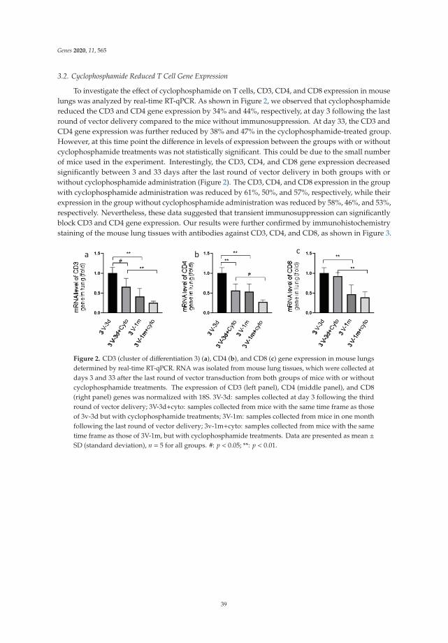
Genes 2020, 11, 565
3.2. Cyclophosphamide Reduced T Cell Gene Expression
To investigate the effect of cyclophosphamide on T cells, CD3, CD4, and CD8 expression in mouselungs was analyzed by real-time RT-qPCR. As shown in Figure 2, we observed that cyclophosphamidereduced the CD3 and CD4 gene expression by 34% and 44%, respectively, at day 3 following the lastround of vector delivery compared to the mice without immunosuppression. At day 33, the CD3 andCD4 gene expression was further reduced by 38% and 47% in the cyclophosphamide-treated group.However, at this time point the difference in levels of expression between the groups with or withoutcyclophosphamide treatments was not statistically significant. This could be due to the small numberof mice used in the experiment. Interestingly, the CD3, CD4, and CD8 gene expression decreasedsignificantly between 3 and 33 days after the last round of vector delivery in both groups with orwithout cyclophosphamide administration (Figure 2). The CD3, CD4, and CD8 expression in the groupwith cyclophosphamide administration was reduced by 61%, 50%, and 57%, respectively, while theirexpression in the group without cyclophosphamide administration was reduced by 58%, 46%, and 53%,respectively. Nevertheless, these data suggested that transient immunosuppression can significantlyblock CD3 and CD4 gene expression. Our results were further confirmed by immunohistochemistrystaining of the mouse lung tissues with antibodies against CD3, CD4, and CD8, as shown in Figure 3.
Figure 2. CD3 (cluster of differentiation 3) (a), CD4 (b), and CD8 (c) gene expression in mouse lungsdetermined by real-time RT-qPCR. RNA was isolated from mouse lung tissues, which were collected atdays 3 and 33 after the last round of vector transduction from both groups of mice with or withoutcyclophosphamide treatments. The expression of CD3 (left panel), CD4 (middle panel), and CD8(right panel) genes was normalized with 18S. 3V-3d: samples collected at day 3 following the thirdround of vector delivery; 3V-3d+cyto: samples collected from mice with the same time frame as thoseof 3v-3d but with cyclophosphamide treatments; 3V-1m: samples collected from mice in one monthfollowing the last round of vector delivery; 3v-1m+cyto: samples collected from mice with the sametime frame as those of 3V-1m, but with cyclophosphamide treatments. Data are presented as mean ±SD (standard deviation), n = 5 for all groups. #: p < 0.05; **: p < 0.01.
39
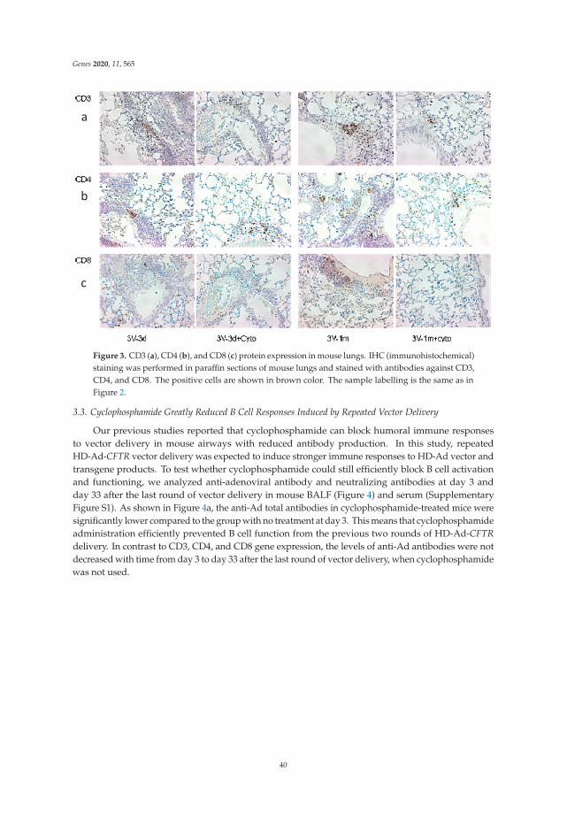
Genes 2020, 11, 565
Figure 3. CD3 (a), CD4 (b), and CD8 (c) protein expression in mouse lungs. IHC (immunohistochemical)staining was performed in paraffin sections of mouse lungs and stained with antibodies against CD3,CD4, and CD8. The positive cells are shown in brown color. The sample labelling is the same as inFigure 2.
3.3. Cyclophosphamide Greatly Reduced B Cell Responses Induced by Repeated Vector Delivery
Our previous studies reported that cyclophosphamide can block humoral immune responsesto vector delivery in mouse airways with reduced antibody production. In this study, repeatedHD-Ad-CFTR vector delivery was expected to induce stronger immune responses to HD-Ad vector andtransgene products. To test whether cyclophosphamide could still efficiently block B cell activationand functioning, we analyzed anti-adenoviral antibody and neutralizing antibodies at day 3 andday 33 after the last round of vector delivery in mouse BALF (Figure 4) and serum (SupplementaryFigure S1). As shown in Figure 4a, the anti-Ad total antibodies in cyclophosphamide-treated mice weresignificantly lower compared to the group with no treatment at day 3. This means that cyclophosphamideadministration efficiently prevented B cell function from the previous two rounds of HD-Ad-CFTRdelivery. In contrast to CD3, CD4, and CD8 gene expression, the levels of anti-Ad antibodies were notdecreased with time from day 3 to day 33 after the last round of vector delivery, when cyclophosphamidewas not used.
40
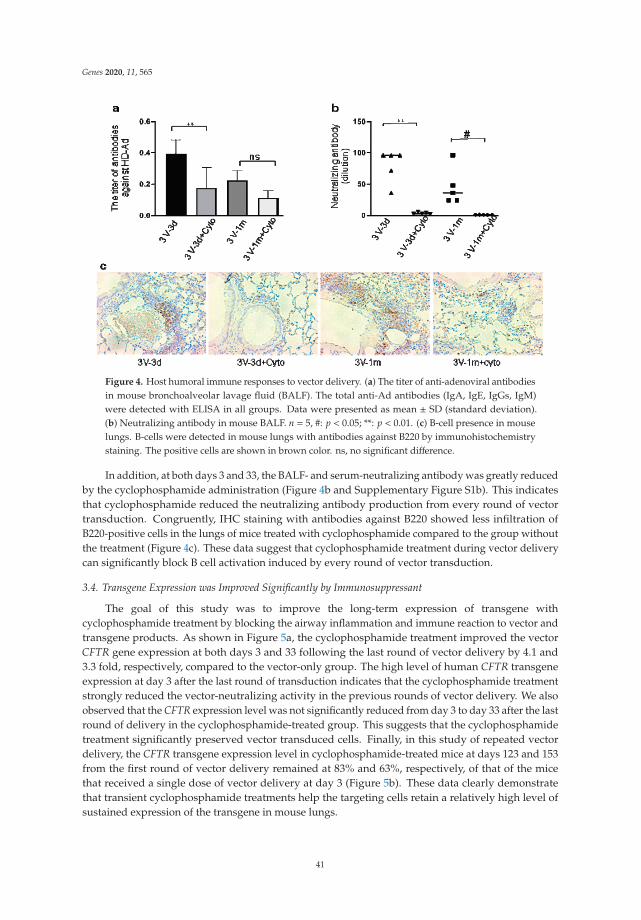
Genes 2020, 11, 565
Figure 4. Host humoral immune responses to vector delivery. (a) The titer of anti-adenoviral antibodiesin mouse bronchoalveolar lavage fluid (BALF). The total anti-Ad antibodies (IgA, IgE, IgGs, IgM)were detected with ELISA in all groups. Data were presented as mean ± SD (standard deviation).(b) Neutralizing antibody in mouse BALF. n = 5, #: p < 0.05; **: p < 0.01. (c) B-cell presence in mouselungs. B-cells were detected in mouse lungs with antibodies against B220 by immunohistochemistrystaining. The positive cells are shown in brown color. ns, no significant difference.
In addition, at both days 3 and 33, the BALF- and serum-neutralizing antibody was greatly reducedby the cyclophosphamide administration (Figure 4b and Supplementary Figure S1b). This indicatesthat cyclophosphamide reduced the neutralizing antibody production from every round of vectortransduction. Congruently, IHC staining with antibodies against B220 showed less infiltration ofB220-positive cells in the lungs of mice treated with cyclophosphamide compared to the group withoutthe treatment (Figure 4c). These data suggest that cyclophosphamide treatment during vector deliverycan significantly block B cell activation induced by every round of vector transduction.
3.4. Transgene Expression was Improved Significantly by Immunosuppressant
The goal of this study was to improve the long-term expression of transgene withcyclophosphamide treatment by blocking the airway inflammation and immune reaction to vector andtransgene products. As shown in Figure 5a, the cyclophosphamide treatment improved the vectorCFTR gene expression at both days 3 and 33 following the last round of vector delivery by 4.1 and3.3 fold, respectively, compared to the vector-only group. The high level of human CFTR transgeneexpression at day 3 after the last round of transduction indicates that the cyclophosphamide treatmentstrongly reduced the vector-neutralizing activity in the previous rounds of vector delivery. We alsoobserved that the CFTR expression level was not significantly reduced from day 3 to day 33 after the lastround of delivery in the cyclophosphamide-treated group. This suggests that the cyclophosphamidetreatment significantly preserved vector transduced cells. Finally, in this study of repeated vectordelivery, the CFTR transgene expression level in cyclophosphamide-treated mice at days 123 and 153from the first round of vector delivery remained at 83% and 63%, respectively, of that of the micethat received a single dose of vector delivery at day 3 (Figure 5b). These data clearly demonstratethat transient cyclophosphamide treatments help the targeting cells retain a relatively high level ofsustained expression of the transgene in mouse lungs.
41

Genes 2020, 11, 565
Figure 5. Expression of human cystic fibrosis transmembrane conductance regulator (CFTR) mRNAin mouse lungs assessed by real-time RT-qPCR. (a) CFTR mRNA levels in lung tissues of mice atdays 3 and 33 following the last round of HD-Ad-CFTR delivery with or without cyclophosphamidetreatments. The transgene expression was normalized with 18S. Data were presented as mean ± SD.n = 5; #: p < 0.05; **: p < 0.01. (b) CFTR mRNA levels in mouselung tissues from 3 days of single dosedelivery (1V-3d), and at days 3 (3V-3d) and 33 (3V-33d) of 3 dose delivery following the last round oftransduction with cyclophosphamide treatment. Data were presented as mean ± SD. Ns indicates nosignificant difference.
4. Discussion
In this repeated gene delivery study, we found the immunosuppressant cyclophosphamidesignificantly blocked the adaptive immune responses induced by the HD-Ad vector, showing a reducedT cell gene expression and T cell and B cell infiltration in mouse lungs. The transient immunosuppressionresulted in a significant level of transgene expression at day 3 after three rounds of HD-Ad vectortransduction and maintained a relatively high level of CFTR gene expression at 33 days after the lastround of delivery. The slightly lower level of transgene expression in mice at day 33 may partiallyresult from epithelial cells’ turn over. These data demonstrated that the sustained expression of thetransgene in mouse lungs can be achieved by the repeated delivery of HD-Ad-CFTR vectors witha transient cyclophosphamide administration.
The sustained expression of transgene at therapeutic levels (achieving lifelong expression) inthe terminal-differentiated cells of epithelia, which are capable of self-renewal, requires the repeatedadministration of vectors. Immune responses against vectors and transgene products are one of themajor obstacles to sustained transgene expression in repeated vector delivery. Strategies to controlunwanted immune responses to transgene products and vectors are needed to achieve the sustainedexpression of therapeutic genes in the lung. Lungs have their own set of immune cells which functionas the frontline of immunity for the airway. Adaptive immune responses can be triggered by vectorantigens or vector-encoded proteins during gene delivery. We and others have shown that genetransfer with Ad or HD-Ad vectors at single doses locally or systemically resulted in the activation of Tcells and humoral immune responses to transgene products and vector antigens [30]. The repeatedadministration of most viral vectors is hindered by the toxicity of T cells and neutralizing or opsonizingthe Abs induced by the vectors.
Thus, strategies including immunosuppression, induction of immune tolerance, or modificationof viral capsids have been tested in vector delivery [22]. For example, Seregin et al. reported thattransient pre-treatment with glucocorticoid can significantly reduce innate and adaptive immuneresponses to Ad vector with systemic delivery without a transgene expression reduction in mouseliver [31]. Immune suppressant prednisone has also been shown to reduce the extent of intramuscularT-cell infiltrates in AAV-treated muscles, which may aid in achieving long-term transgene expression
42
Genes 2020, 11, 565
through promoting PD1-mediated programmed T-cell death [32]. In addition, viral vector serotypeswitching [33] and capsid engineering [34] have been evaluated for viral vector readministration.The repeated liver gene transfer to adult mice with mutations in the liver-specific UGT1 gene by AAVserotype switching, upon neonatal administration, resulted in the lifelong correction of total bilirubinlevels in the mice [35].
Cyclophosphamide is a potent immunosuppressive agent used clinically in autoimmune disordersand the transplantation of allogeneic bone marrow or solid organs to establish peripheral allografttolerance and suppress autoreactive T cells [36]. Our previous study found that cyclophosphamidetreatment significantly improved transgene expression at day 3 following HD-Ad readministration [19].In the repeated airway transduction mouse model, the transient use of cyclophosphamide duringHD-vector administration significantly reduced the expression of the CD3 and CD4 gene and theinfiltration of these cells in the targeting organ. This indicated that cyclophosphamide may cause T celldeath besides its anti-mitotic and anti-replicative effects [36]. Strauss et al. have reported that the effectof cyclophosphamide on T cells is its ability to induce apoptosis, which is a fundamental differencefrom other immunosuppressive agents.
Cyclophosphamide up-regulated Fas (CD95) expression, triggering activation-induced cell death.Comparing the effects of other drugs such as dexamethasone and rapamycin on human peripheralblood T cells, cytotoxic cell lines, and Jurkat cells, only cyclophosphamide triggered cell death [37,38].The induction of apoptosis is also a key mechanism to eliminate activated T cells during the terminationof an immune response.
In addition to T cell effects, cyclophosphamide also affects B cell proliferation and plasma cellantibody production [39]. In this study, HD-Ad vector transduction resulted in anti-Ad antibodyproduction. Such antibodies can bind to and prevent viral vectors from entering target cells, therebyreducing the transgene expression. Transient cyclophosphamide administration significantly preventedanti-Ad antibody production and reduced the neutralizing activity. This reduction is expected toimprove the vector transduction efficiency from the next round of vector delivery and sustainedtransgene expression. These studies established the important concept that the transient suppressionof host immune responses is efficient for the sustained expression of transgene in mouse lungs inrepeated HD-Ad vector delivery.
However, one limitation of this study is that we are not sure whether our results with mice can betranslated into clinical applications, because there are differences between mice and humans in theimmune system. These differences include: activation of immune responses to challenge, the balanceof leukocyte subsets, Toll-like receptors, and antibody subsets [40,41]. Therefore, the safety, dosage,and scheduling of cyclophosphamide administration should be further evaluated in large animalmodels. Cyclophosphamide has immunosuppressive and immunomodulatory properties. Consideringthe risk of infection for persons with CF, the immunosuppressive function of cyclophosphamideand its effect on vector transduction efficiency should be tested in proper CF animal models. In thisstudy, we did not investigate whether cyclophosphamide affects host innate immune responses.However, our previous work has shown that there was no significant change in cytokine production toHD-Ad vector with cyclophosphamide treatment [19]. Strategies with the combination of differentimmunosuppressive agents to target specifically innate and adaptive immune responses at differentstages may also be important. In addition, the scheduling of administration is of particular importancefor the immunological effect [36]. We observed that the expression of CD3, CD4, and CD 8 genesgoes down significantly with time, one month after the last round of delivery with or withoutcyclophosphamide treatment. This observation suggests a window for scheduling the following roundof vector administration in order to avoid the peak of adaptive immune responses.
43
Genes 2020, 11, 565
5. Conclusions
In summary, the immunosuppressant cyclophosphamide can modulate the host immune systemand allow for a more sustained expression of the CFTR gene in repeated vector delivery to mouseairways. In combination with a gene-editing system to target airway stem cells, sustained expressionwith a therapeutic level of transgene may be achievable in the future.
Supplementary Materials: The following are available online at http://www.mdpi.com/2073-4425/11/5/565/s1,Figure S1: Anti-adenoviral antibodies in mouse serum. (a) The total anti-Ad antibodies (IgA, IgE, IgGs, IgM) weredetected with ELISA in all groups. Data were presented as mean ± SD. (b) Neutralizing antibody in mouse sera.n = 5, #: p < 0.05; **: p < 0.01.
Author Contributions: Conceptualization, H.C. and J.H.; methodology, H.C. and R.D.; validation, H.C. andR.D.; formal analysis, H.C.; investigation, H.C.; resources, J.H.; data curation, H.C. and R.D.; writing—originaldraft preparation, H.C.; writing—review and editing, R.D. and J.H.; visualization, H.C.; supervision, J.H.; projectadministration, J.H.; funding acquisition, J.H. All authors have read and agreed to the published version ofthe manuscript.
Funding: This study was funded by the Canadian Institute for Health Research (CIHR) grants (MOP 125882),the Cystic Fibrosis Foundation Therapeutics, Inc. grant (HU15XX0), and the Cystic Fibrosis Canada grant (#3032)to JH.
Acknowledgments: The authors wish to acknowledge the contribution of Vivian Bradaschia and Milan Gangulyin the Pathology Core at The Centre for Phenogenomics for IHC staining and of Jing Wu at The Hospital for SickChildren, Research Institute for real-time RT-PCR analyses. We thank Sara Chowns for reading and commentingon the manuscript.
Conflicts of Interest: All authors have no conflict of interest to declare.
References
1. Corbyn, Z. Promising new era dawns for cystic fibrosis treatment. Lancet 2012, 379, 1475–1476. [CrossRef]2. Davies, J.C.; Moskowitz, S.M.; Brown, C.; Horsley, A.; Mall, M.A.; McKone, E.F.; Plant, B.J.; Prais, D.;
Ramsey, B.W.; Taylor-Cousar, J.L.; et al. VX-659-Tezacaftor-Ivacaftor in Patients with Cystic Fibrosis and Oneor Two Phe508del Alleles. N. Engl. J. Med. 2018, 379, 1599–1611. [CrossRef]
3. Heijerman, H.G.M.; McKone, E.F.; Downey, D.G.; Van Braeckel, E.; Rowe, S.M.; Tullis, E.; Mall, M.A.;Welter, J.J.; Ramsey, B.W.; McKee, C.M.; et al. Efficacy and safety of the elexacaftor plus tezacaftorplus ivacaftor combination regimen in people with cystic fibrosis homozygous for the F508del mutation:A double-blind, randomised, phase 3 trial. Lancet 2019, 394, 1940–1948. [CrossRef]
4. Keating, D.; Marigowda, G.; Burr, L.; Daines, C.; Mall, M.A.; McKone, E.F.; Ramsey, B.W.; Rowe, S.M.;Sass, L.A.; Tullis, E.; et al. VX-445-Tezacaftor-Ivacaftor in Patients with Cystic Fibrosis and One or TwoPhe508del Alleles. N. Engl. J. Med. 2018, 379, 1612–1620. [CrossRef]
5. Middleton, P.G.; Mall, M.A.; Drevinek, P.; Lands, L.C.; McKone, E.F.; Polineni, D.; Ramsey, B.W.;Taylor-Cousar, J.L.; Tullis, E.; Vermeulen, F.; et al. Elexacaftor-Tezacaftor-Ivacaftor for Cystic Fibrosiswith a Single Phe508del Allele. N. Engl. J. Med. 2019, 381, 1809–1819. [CrossRef]
6. Cooney, A.L.; McCray, P.B., Jr.; Sinn, P.L. Cystic Fibrosis Gene Therapy: Looking Back, Looking Forward.Genes 2018, 9, 538. [CrossRef]
7. Brody, H. Gene therapy. Nature 2018, 564, S5. [CrossRef]8. Jennings, M.T.; Flume, P.A. Cystic Fibrosis: Translating Molecular Mechanisms into Effective Therapies.
Ann. Am. Thorac. Soc. 2018, 15, 897–902. [CrossRef]9. Ginn, S.L.; Amaya, A.K.; Alexander, I.E.; Edelstein, M.; Abedi, M.R. Gene therapy clinical trials worldwide
to 2017: An update. J. Gene Med. 2018, 20, e3015. [CrossRef]10. Dunbar, C.E.; High, K.A.; Joung, J.K.; Kohn, D.B.; Ozawa, K.; Sadelain, M. Gene therapy comes of age. Science
2018, 359. [CrossRef]11. Gao, J.; Mese, K.; Bunz, O.; Ehrhardt, A. State-of-the-art human adenovirus vectorology for therapeutic
approaches. FEBS Lett. 2019, 593, 3609–3622. [CrossRef]12. Cao, H.; Koehler, D.R.; Hu, J. Adenoviral vectors for gene replacement therapy. Viral Immunol. 2004, 17,
327–333. [CrossRef]
44
Genes 2020, 11, 565
13. Flotte, T.R.; Ng, P.; Dylla, D.E.; McCray, P.B., Jr.; Wang, G.; Kolls, J.K.; Hu, J. Viral vector-mediated andcell-based therapies for treatment of cystic fibrosis. Mol. Ther. 2007, 15, 229–241. [CrossRef]
14. Koehler, D.R.; Hitt, M.M.; Hu, J. Challenges and strategies for cystic fibrosis lung gene therapy. Mol. Ther.2001, 4, 84–91. [CrossRef]
15. Zhou, Z.P.; Yang, L.L.; Cao, H.; Chen, Z.R.; Zhang, Y.; Wen, X.Y.; Hu, J. In Vitro Validation ofa CRISPR-Mediated CFTR Correction Strategy for Preclinical Translation in Pigs. Hum. Gene Ther. 2019, 30,1101–1116. [CrossRef]
16. Xia, E.; Zhang, Y.; Cao, H.; Li, J.; Duan, R.; Hu, J. TALEN-Mediated Gene Targeting for Cystic Fibrosis-GeneTherapy. Genes 2019, 10, 39. [CrossRef]
17. Xia, E.; Duan, R.; Shi, F.; Seigel, K.E.; Grasemann, H.; Hu, J. Overcoming the Undesirable CRISPR-Cas9Expression in Gene Correction. Mol. Ther. Nucleic Acids 2018, 13, 699–709. [CrossRef]
18. Cao, H.; Machuca, T.N.; Yeung, J.C.; Wu, J.; Du, K.; Duan, C.; Hashimoto, K.; Linacre, V.; Coates, A.L.;Leung, K.; et al. Efficient gene delivery to pig airway epithelia and submucosal glands using helper-dependentadenoviral vectors. Mol. Ther. Nucleic Acids 2013, 2, e127. [CrossRef]
19. Cao, H.; Yang, T.; Li, X.F.; Wu, J.; Duan, C.; Coates, A.L.; Hu, J. Readministration of helper-dependentadenoviral vectors to mouse airway mediated via transient immunosuppression. Gene Ther. 2011, 18, 173–181.[CrossRef]
20. Cao, H.; Ouyang, H.; Grasemann, H.; Bartlett, C.; Du, K.; Duan, R.; Shi, F.; Estrada, M.; Seigel, K.; Coates, A.;et al. Transducing airway basal cells with a helper-dependent adenoviral vector for lung gene therapy.Hum. Gene Ther. 2018. [CrossRef]
21. Levardon, H.; Yonker, L.M.; Hurley, B.P.; Mou, H. Expansion of Airway Basal Cells and Generation ofPolarized Epithelium. Bio. Protoc. 2018, 8. [CrossRef]
22. Carlon, M.S.; Vidovic, D.; Dooley, J.; da Cunha, M.M.; Maris, M.; Lampi, Y.; Toelen, J.; Van den Haute, C.;Baekelandt, V.; Deprest, J.; et al. Immunological ignorance allows long-term gene expression after perinatalrecombinant adeno-associated virus-mediated gene transfer to murine airways. Hum. Gene Ther. 2014, 25,517–528. [CrossRef]
23. Ferrari, S.; Griesenbach, U.; Geddes, D.M.; Alton, E. Immunological hurdles to lung gene therapy. Clin. Exp.Immunol. 2003, 132, 1–8. [CrossRef]
24. Nathwani, A.C.; Tuddenham, E.G.; Rangarajan, S.; Rosales, C.; McIntosh, J.; Linch, D.C.; Chowdary, P.;Riddell, A.; Pie, A.J.; Harrington, C.; et al. Adenovirus-associated virus vector-mediated gene transfer inhemophilia B. N. Engl. J. Med. 2011, 365, 2357–2365. [CrossRef]
25. Martino, A.T.; Basner-Tschakarjan, E.; Markusic, D.M.; Finn, J.D.; Hinderer, C.; Zhou, S.; Ostrov, D.A.;Srivastava, A.; Ertl, H.C.; Terhorst, C.; et al. Engineered AAV vector minimizes in vivo targeting of transducedhepatocytes by capsid-specific CD8+ T cells. Blood 2013, 121, 2224–2233. [CrossRef]
26. Arruda, V.R.; Favaro, P.; Finn, J.D. Strategies to modulate immune responses: A new frontier for gene therapy.Mol. Ther. 2009, 17, 1492–1503. [CrossRef]
27. Palmer, D.J.; Ng, P. Methods for the production of helper-dependent adenoviral vectors. Methods Mol. Biol.2008, 433, 33–53.
28. Koehler, D.R.; Martin, B.; Corey, M.; Palmer, D.; Ng, P.; Tanswell, A.K.; Hu, J. Readministration ofhelper-dependent adenovirus to mouse lung. Gene Ther. 2006, 13, 773–780. [CrossRef]
29. Livak, K.J.; Schmittgen, T.D. Analysis of relative gene expression data using real-time quantitative PCR andthe 2(-Delta Delta C(T)) Method. Methods 2001, 25, 402–408. [CrossRef]
30. Schirmbeck, R.; Reimann, J.; Kochanek, S.; Kreppel, F. The immunogenicity of adenovirus vectors limitsthe multispecificity of CD8 T-cell responses to vector-encoded transgenic antigens. Mol. Ther. 2008, 16,1609–1616. [CrossRef]
31. Seregin, S.S.; Appledorn, D.M.; McBride, A.J.; Schuldt, N.J.; Aldhamen, Y.A.; Voss, T.; Wei, J.; Bujold, M.;Nance, W.; Godbehere, S.; et al. Transient pretreatment with glucocorticoid ablates innate toxicity ofsystemically delivered adenoviral vectors without reducing efficacy. Mol. Ther. 2009, 17, 685–696. [CrossRef]
32. Cramer, M.L.; Shao, G.; Rodino-Klapac, L.R.; Chicoine, L.G.; Martin, P.T. Induction of T-Cell Infiltration andProgrammed Death Ligand 2 Expression by Adeno-Associated Virus in Rhesus Macaque Skeletal Muscleand Modulation by Prednisone. Hum. Gene Ther. 2017, 28, 493–509. [CrossRef]
45
Genes 2020, 11, 565
33. Majowicz, A.; Salas, D.; Zabaleta, N.; Rodriguez-Garcia, E.; Gonzalez-Aseguinolaza, G.; Petry, H.; Ferreira, V.Successful Repeated Hepatic Gene Delivery in Mice and Non-human Primates Achieved by SequentialAdministration of AAV5(ch) and AAV1. Mol. Ther. 2017, 25, 1831–1842. [CrossRef]
34. Vandenberghe, L.H.; Wilson, J.M.; Gao, G. Tailoring the AAV vector capsid for gene therapy. Gene Ther. 2009,16, 311–319. [CrossRef]
35. Bockor, L.; Bortolussi, G.; Iaconcig, A.; Chiaruttini, G.; Tiribelli, C.; Giacca, M.; Benvenuti, F.; Zentilin, L.;Muro, A.F. Repeated AAV-mediated gene transfer by serotype switching enables long-lasting therapeuticlevels of hUgt1a1 enzyme in a mouse model of Crigler-Najjar Syndrome Type I. Gene Ther. 2017, 24, 649–660.[CrossRef]
36. Ahlmann, M.; Hempel, G. The effect of cyclophosphamide on the immune system: Implications for clinicalcancer therapy. Cancer Chemother. Pharm. 2016, 78, 661–671. [CrossRef]
37. Strauss, G.; Osen, W.; Debatin, K.M. Induction of apoptosis and modulation of activation and effectorfunction in T cells by immunosuppressive drugs. Clin. Exp. Immunol. 2002, 128, 255–266. [CrossRef]
38. Al-Homsi, A.S.; Roy, T.S.; Cole, K.; Feng, Y.; Duffner, U. Post-transplant high-dose cyclophosphamide for theprevention of graft-versus-host disease. Biol. Blood Marrow Transplant. 2015, 21, 604–611. [CrossRef]
39. Wojcik, R. Reactivity of the immunological system of rats stimulated with Biolex-Beta HP aftercyclophosphamide immunosuppression. Cent. Eur. J. Immunol. 2014, 39, 51–60. [CrossRef]
40. Tao, L.; Reese, T.A. Making Mouse Models That Reflect Human Immune Responses. Trends Immunol. 2017,38, 181–193. [CrossRef]
41. Mestas, J.; Hughes, C.C. Of mice and not men: Differences between mouse and human immunology.J. Immunol. 2004, 172, 2731–2738. [CrossRef]
© 2020 by the authors. Licensee MDPI, Basel, Switzerland. This article is an open accessarticle distributed under the terms and conditions of the Creative Commons Attribution(CC BY) license (http://creativecommons.org/licenses/by/4.0/).
46

genesG C A T
T A C G
G C A T
Article
Analysis of CFTR Mutation Spectrum in EthnicRussian Cystic Fibrosis Patients
Nika V. Petrova *, Nataliya Y. Kashirskaya, Tatyana A. Vasilyeva, Elena I. Kondratyeva,
Elena K. Zhekaite , Anna Y. Voronkova, Victoria D. Sherman , Varvara A. Galkina,
Eugeny K. Ginter, Sergey I. Kutsev, Andrey V. Marakhonov and Rena A. Zinchenko
Research Centre for Medical Genetics, Moskvorechje Street, 1, 115478 Moscow, Russia;[email protected] (N.Y.K.); [email protected] (T.A.V.); [email protected] (E.I.K.);[email protected] (E.K.Z.); [email protected] (A.Y.V.); [email protected] (V.D.S.);[email protected] (V.A.G.); [email protected] (E.K.G.); [email protected] (S.I.K.);[email protected] (A.V.M.); [email protected] (R.A.Z.)* Correspondence: [email protected]
Received: 30 March 2020; Accepted: 13 May 2020; Published: 15 May 2020
Abstract: The distribution and frequency of the CFTR gene mutations vary considerably betweencountries and ethnic groups. Russians are an East Slavic ethnic groups are native to EasternEurope. Russians, the most numerous people of the Russian Federation (RF), make about 80%of the population. The aim is to reveal the molecular causes of CF in ethnic Russian patients ascomprehensively as possible. The analysis of most common CFTR mutations utilized for CF diagnosisin multiethnic RF population accounts for about 83% of all CF-causing mutations in 1384 ethnicRussian patients. Variants c.1521_1523delCTT (F508del), c.54-5940_273+10250del21kb (CFTRdele2,3),c.2012delT (2143delT), c.2052_2053insA (2184insA), and c.3691delT (3821delT) are most typical for CFpatients of Russian origin. DNA of 154 CF patients, Russian by origin, in whom at least one mutantallele was not previously identified (164 CF alleles), was analyzed by Sanger sequencing followed bythe multiplex ligase-dependent probe amplification (MLPA) method. In addition to the 29 variantsidentified during the previous test for common mutations, 91 pathogenic CFTR variants were alsorevealed: 29 missense, 19 nonsense, 14 frame shift in/del, 17 splicing, 1 in frame ins, and 11 copynumber variations (CNV). Each of the 61 variants was revealed once, and 17 twice. Each of the variantsc.1209G>C (E403D), c.2128A>T (K710X), c.3883delA (4015delA), and c.3884_3885insT (4016insT)were detected for three, c.1766+1G>A (1898+1G>A) and c.2834C>T (S945L) for four, c.1766+1G>C(1898+1G>C) and c.(743+1_744-1)_(1584+1_1585-1)dup (CFTRdup6b-10) for five, c.2353C>T (R785X)and c.4004T>C (L1335P) for six, c.3929G>A (W1310X) for seven, c.580-1G>T (712-1G>T for eight,and c.1240_1244delCAAAA (1365del5) for 11 unrelated patients. A comprehensive analysis of CFTRmutant alleles with sequencing followed by MLPA, allowed not only the identification of 163 of 164unknown alleles in our patient sample, but also expansion of the mutation spectrum with novel andadditional frequent variants for ethnic Russians.
Keywords: cystic fibrosis; CFTR gene; common and new pathogenic variants; ethnic Russian population
1. Introduction
Cystic fibrosis (CF, OMIM#219700) is an autosomal recessive condition resulting from thepathogenic variants in the CF transmembrane regulator (CFTR) gene. CF is a hereditary disease causedby impaired epithelial chloride channel CFTR function. Variants are classified as disease causing,not disease causing, of variable clinical significance, or of unknown clinical significance. More than2000 different variants of the CFTR gene sequence have been revealed, the pathogenicity of 20% ofwhich is established [1,2]. In many populations the most frequent pathogenic variant of the CFTR gene
Genes 2020, 11, 554; doi:10.3390/genes11050554 www.mdpi.com/journal/genes47
Genes 2020, 11, 554
(ABCC7) is F508del, which accounts for approximately two thirds of all CFTR alleles, with a decreasingprevalence from Northwest to Southeast Europe. The remaining third of alleles are substantiallyheterogeneous, with fewer than 20 mutations occurring at a worldwide frequency of more than 0.1%.Some variants can reach a higher frequency in certain populations, due to a founder effect in religious,ethnic or geographical isolates [3]. The spectrum and frequency of CFTR gene sequence variants varysignificantly in different countries and ethnic groups, which suggests the development of regionalmolecular diagnostics protocols to optimize medical and genetic care for CF patients [4].
The diagnosis of CF was proven by typical pulmonary or gastrointestinal symptoms or positiveneonatal screening, or the diagnosis of CF in a sibling, as well as at least one of the following: twopositive sweat chloride tests, or the identification of two CFTR pathologic variants in trans accordingto the guidelines of the European Cystic Fibrosis Society as well as the Russian National Consensus onCystic Fibrosis [5,6].
Molecular genetic studies on CF have been conducted in the Laboratory of Genetic Epidemiologyof the Research Centre for Medical Genetics for a long period of time starting from the year 1989.To date, the laboratory has analyzed the DNA of more than 3400 CF patients, the clinical diagnosis wasconfirmed in the Scientific-Clinical Department for Cystic Fibrosis of the Research Centre for MedicalGenetics. Thereby, 87.4% of the CF patients we examined live in the European part of Russia. More than85% are Russian or come from marriages between Russians and persons belonging to other ethnicgroups. According to the Russian Registry of cystic fibrosis patients of 2017 (RF CF Registry), among atleast 212 pathogenic variants of the CFTR gene eleven variants are the most frequent ones in the RussianFederation (their relative frequencies exceed 1% in the sample of tested patients) and they are F508delwith a share of 52.81%, CFTRdele2,3—6.21%, E92K—3.00%, 2143delT—2.15%, 3849+10kbC>T—2.02%,W1282X—1.90%, 2184insA—1.85%, 1677delTA—1.81%, N1303K—1.54%, G542X—1.35%, and L138inswith 1.24% [7]. All other CFTR variants identified in Russian patients share 12.35%. The frequenciesand spectrum of variants of the CFTR gene vary in different regions. This is caused by specific ethnicbackground of the population, as well as by different population processes occurring on differentterritories inhabited by the same ethnos. Thus, in the North Caucasus Federal District (NCFD), threevariants are the most frequent ones: F508del (25.0%), 1677delTA (21.5%), and W1282X (17.2%) [7].A study of CFTR gene variants’ spectra in different NCFD ethnic groups revealed a high proportionof variant W1282X (88%) for Karachays [8], and variants 1677delTA (81.5%) and E92K (12.5%) forChechens [9]. The most frequent variants in the Volga Federal District (VFD) are F508del (50.5%), E92K(8.7%) and CFTRdele2,3 (5.0%) [7]. A high share of E92K variant in VFD is due to the prevalence of thisvariant for Chuvash (55%) [10]. The second most frequent variant for Chuvash CF patients is F508del(30%) [9], although this value is lower than in the total sample of CF patients (according to the Registryof CF patients in the Russian Federation 2017, [7]).
Russian East Slavic ethnos is the most numerous people in the Russian Federation (RF) (morethan 111,000,000 people), which makes 77.7% of the population of the country according to census of2010 [11]. In the European part of RF, Russians make 85%–90% of the population.
The aim is to describe the Russian-specific spectrum of pathogenic variants of the CFTR gene,testing of which could increase the informativeness of DNA diagnostics in regions with a predominantlyRussian population, as well to establish a basis for forming a patient base for possible targeted therapy.
48

Genes 2020, 11, 554
2. Materials and Methods
Initially, CFTR genotyping of 1384 CF patients (ethnic Russians) from all-Russian sample (3457CF patients) tested in the Laboratory of Genetic Epidemiology, Research Centre for Medical Geneticswere analyzed. The diagnosis of CF was made in the Scientific-Clinical Department for CysticFibrosis, Research Centre for Medical Genetics or in regional CF centers according to the acceptedstandards [10]. Diagnosis was confirmed by analysis of clinical presentation and Gibson–Cooke sweattest, with chloride ion concentrations of 60 mmol/L or higher defining positive result. The assignmentof patients’ Russian ancestry was based on self- or parents’ reports. The study included 154 CF Russianpatients, 90% of whom came from the European part of the Russian Federation and 10% from Siberianor Far Eastern regions, for all of them at least one mutant allele was not identified.
Patients or their parents signed an informed consent to the study. The research protocol wasapproved by the Ethical Committee of Research Centre for Medical Genetics (Research Centre forMedical Genetics, 115522, Moscow, Moskvorechie St., 1, Russian Federation, Protocol No.17/2006 of02.02.2006).
Molecular diagnostics consists of three consecutive stages.First stage included analysis of 33 frequent CFTR variants (c.54-5940_273+10250del21kb
(p.Ser18Argfs*16, CFTRdele2,3), c.254G>A (p.Gly85Glu, G85E), c.262_263delTT (p.Leu88IlefsX22,394delTT), c.274G>A (p.Glu92Lys, E92K), c.350G>A (p.Arg117His, R117H), c.413_415dupTAC(p.Leu138dup; L138ins), c.472dupA (p.Ser158LysfsX5, 604insA), c.489+1G>T (621+1G>T),c.1000C>T (p.Arg334Trp, R334W), c.1040G>C (p.Arg347Pro, R347P), c.1397C>G (p.Ser466X,Ser466X), c.1519_1521delATC (p.Ile507del, I507del), c.1521_1523delCTT (p.Phe508del, F508del),c.1545_1546delTA (p.Tyr515X, 1677delTA), c.1585-1G>A (1717-1G>A), c.1624G>T (p.Gly542X,G542X), c.1652G>A (p.Gly551Asp, G551D), c.1657C>T (p.Arg553X, R553X), c.2012delT (p.Leu671X,2143delT), c.2051_2052delAAinsG (p.Lys684SerfsX38, 2183AA>G), c.2052_2053insA (p.Gln685ThrfsX4,2184insA), c.2657+5G>A (2789+5A>G), c.3140-16T>A (3272-16T>A), c.3476C>T (p.Ser1159Phe, S1159F),c.3475T>C (p.Ser1159Pro; S1159P), c.3535_3536insTCAA (p.Thr1179IlefsX17, 3667ins4), c.3587C>G(p.Ser1196X, S1196X), c.3691delT (p.Ser1231ProfsX4, 3821delT), c.3718-2477C>T (3849+10kbC-T),c.3816_3817delGT (p.Ser1273LeufsX28, 3944delGT), c.3844T>C (p.Trp1282Arg, W1282R), c.3846G>A(p.Trp1282X, W1282X), c.3909C>G (p.Asn1303Lys, N1303K), representing a routine Russian Federationpanel that identifies up to 85% of mutant CF alleles as described previously [12].
Second stage included analysis of CFTR gene coding sequence, exon-intron junctions and5′-UTR sequence by Sanger sequencing as described previously [12]. Variant pathogenicity status(only pathogenic or likely pathogenic variants were reported) was established using consensusrecommendations of the American College of Medical Genetics and Genomics and the Associationfor Molecular Pathology for interpretation of sequence variants and Russian recommendations.The frequencies of identified alleles in general populations were established based on the GnomADbrowser (https://gnomad.broadinstitute.org/). The predicted functional effect of missense variants wasdetermined through SIFT, FATHMM and Radial SVM prediction algorithms as well as GERP++ andPhyloP conservation scores. Intronic and splicing variants were analyzed using Human Splicing Findertool v. 2.4.1. Novel variants were submitted to the CFTR2 website dataset (https://cftr2.org/), CFTR1(http://www.genet.sickkids.on.ca/cftr). Pathogenic variants of the CFTR gene are denoted accordingto the legacy nomenclature, besides novel variants named according to the HGVS nomenclature forNM_000492.4 (CFTR) transcript variant.
49
Genes 2020, 11, 554
Third stage intended to search for large rearrangements in chromosome region 7q31.2(deletions/duplications–CNV) involved the CFTR gene locus by the multiplex ligation-dependentprobe amplification (MLPA) method in case when no pathogenic allele was detected or an allele withuncertain significance was identified at the previous stages. MLPA analysis was performed withSALSA MLPA probemix P091-D2 CFTR (MRC-Holland, Amsterdam, the Netherlands) accordingto the manufacturer’s recommendation. The MLPA results were analyzed using Coffalyser.Net(MRC-Holland) [12].
Variants phase was checked by segregation analysis in proband and healthy parents.The gIVS6a_415_IVS10_987Dup26817bp (CFTRdup6b-10) duplication and its boundaries were
previously described by F.M. Hantash and co-authors [13]: The fragments duplicated started 415 bpdownstream of exon 6a, in IVS6a, and spanned exons 6b, 7, 8, 9, and 10, breaking at 2987 bp downstreamof exon 10 in IVS10. The duplicated region is 26,817 bp. Two pairs of primers have been developed toclarify the boundaries of CFTRdup6b-10 duplications identified in Russian CF patients. One flanks thejunction area of rupture points of intron 11 (10 as in the legacy nomenclature) and intron 6a (6): IVS10F5′-TCAGGAAATGGCAATGGGGT-3′ and IVS6aR 5′-GGCTCTGGTGTGATGATCCATA-3′. A 359 bpfragment from these primers is amplified only from the allele carrying the duplication. The second pair(INT10F 5′-GGGGTTGGGAAGTGATTCCA-3′ and INT10R 5′-GCCATCAGCTAGGCTTCTGTA-3′)flanks the rupture area of the intron 10, amplification occurs only from the normal sequence of theintron 10 of the CFTR gene leading to a product of 234 bp.
To compare variant frequencies, the Fisher test was used. The significance level was considered tobe p ≤ 0.05.
3. Results
When developing a routinely used mutation panel, the laboratory’s own data [14], the resultsof the first collaborative study [15], and studies of other Russian laboratories (in St. Petersburg [16],Bashkortostan [17], and Tomsk [18]) were considered. The panel includes 33 pathogenic variants ofthe CFTR gene identified in patients from different regions of the Russian Federation, as well as thevariants specific for certain ethnic groups [7–10], and allows identification of up to 85% of mutantalleles in all-Russian population [12].
At the first stage, the results of testing 33 pathogenic variants of the CFTR gene in DNA of1384 ethnic Russians with CF (previously performed in the laboratory of genetic epidemiology) wereanalyzed. Thereby, 29 out of 33 tested variants were revealed (Table 1). In addition to F508del andCFTRdele2,3, eight more variants can be referred to as frequent ones for ethnic Russians (frequency ofvariants 2143delT, 3849+10kbC-T and 2184insA exceed 2%, variants N1303K, G542X, E92K, W1282X,and L138ins exceed 1%). The mutation detection rate of the used panel of tested variants is 83% inthe sample of ethnic Russians (Table 1). In 932 patients, two mutant variants were identified, in 426patients only one pathogenic variant was detected, both alleles were not detected in 26 patients.
50

Genes 2020, 11, 554
Table 1. Frequencies of 33 variants of CFTR gene in a sample of 1384 ethnic Russians and in a nationwidesample of CF patients (RF CF Registry) [7].
No. Variants Number % % in RF CF Registry
1 c.1521_1523delCTT (p.Phe508del, F508del) 1522 54.99 52.812 c.54-5940_273+10250del21kb (p.Ser18Argfs*16, CFTRdele2,3) 210 7.59 6.213 c.2012delT (p.Leu671X, 2143delT) 75 2.71 2.154 c.3718-2477C>T (3849+10kbC-T) 65 2.35 2.025 c.2052_2053insA (p.Gln685ThrfsX4, 2184insA) 62 2.24 1.856 c.3909C>G (p.Asn1303Lys, N1303K) 48 1.73 1.547 c.1624G>T (p.Gly542X, G542X) 44 1.59 1.358 c.274G>A (p.Glu92Lys, E92K) 29 1.05 3.009 c.3846G>A (p.Trp1282X, W1282X) 32 1.16 1.90
10 c.413_415dupTAC (p.Leu138dup; L138ins) 31 1.12 1.2411 c.3844T>C (p.Trp1282Arg, W1282R) 21 0.76 0.5512 c.1397C>G (p.Ser466X, Ser466X) 20 0.72 0.5013 c.3691delT (p.Ser1231ProfsX4, 3821delT) 19 0.69 0.4614 c.1000C>T (p.Arg334Trp, R334W) 19 0.69 0.8015 c.262_263delTT (p.Leu88IlefsX22, 394delTT) 15 0.54 0.9416 c.3587C>G (p.Ser1196X, S1196X) 14 0.51 0.4817 c.3816_3817delGT (p.Ser1273LeufsX28, 3944delGT) 12 0.43 0.2718 c.2657+5G>A (2789+5A>G) 10 0.36 0.4819 c.489+1G>T (621+1G>T) 7 0.25 0.1820 c.3140-16T>A (3272-16T>A) 6 0.22 0.3421 c.1657C>T (p.Arg553X, R553X) 5 0.18 0.1822 c.1545_1546delTA (p.Tyr515X, 1677delTA) 5 0.18 1.8123 c.3535_3536insTCAA (p.Thr1179IlefsX17, 3667ins4) 4 0.14 0.1024 c.254G>A (p.Gly85Glu, G85E) 4 0.14 0.1025 c.472dupA (p.Ser158LysfsX5, 604insA) 3 0.11 0.1026 c.2051_2052delAAinsG (p.Lys684SerfsX38, 2183AA>G) 3 0.11 0.0427 c.3475T>C (p.Ser1159Pro; S1159P) 3 0.11 0.1028 c.1040G>C (p.Arg347Pro, R347P) 2 0.07 0.1029 c.350G>A (p.Arg117His, R117H) 1 0.04 0.0430 c.1519_1521delATC (p.Ile507del, I507del) 0 - 031 c.1585-1G>A (1717-1G>A) 0 - 0.0432 c.1652G>A (p.Gly551Asp, G551D) 0 - 0.0433 c.3476C>T (p.Ser1159Phe, S1159F) 0 - 0.11
Identified 2290 82.78Not identified 478 17.22
Total 2768
On the second stage, 154 ethnic Russians affected by CF, for whom one or both mutant alleleswere not identified when analyzing 33 mutations, were selected from the sample of 1384 CF patientsfor further analysis. Their genotypes were presented in Supplementary Table S2. There was a total of164 unidentified mutant alleles of the CFTR gene.
As a result, in addition to 29 identified frequent mutations, 91 pathogenic (or likely pathogenic)genetic variants in the CFTR gene were detected (Table 2). Of these, 29 are missense mutations, 19nonsense mutations, 14 frame-shift mutations (11 deletions and three insertions)), 17 splice-site, onein-frame insertion, 11 large rearrangements (eight deletions and three duplications).
51

Genes 2020, 11, 554
Ta
ble
2.
The
CFT
Rge
neva
rian
tsad
diti
onal
lyid
enti
fied
in15
4pr
evio
usly
scre
ened
Rus
sian
pati
ents
.
No
.V
ari
an
tA
cco
rdin
gto
cDN
AP
rote
inC
ha
ng
eL
eg
acy
Na
me
Ex
on/I
ntr
on
1N
um
be
rM
uta
tio
nT
yp
e
1c.
43de
lCp.
Leu1
5Phe
fsX
117
5del
C1e
2sd
2c.
53+
1G>
T18
5+1G
->T
1i2
s3
c.79
G>
Tp.
Gly
27X
G27
X2e
1n
4c.
115C>
Tp.
Gln
39X
Q39
X2e
1n
5c.
223C>
Tp.
Arg
75X
R75
X3e
1n
6c.
252T>
Ap.
Tyr8
4X3e
2n
7c.
264_
268d
elA
TATT
p.Le
u88P
hefs
X21
3e1
sd8
c.27
4-6T>
C40
6-6T>
C3i
1s
9c.
274G>
Tp.
Glu
92X
E92X
4e1
n10
c.29
3A>
Gp.
Gln
98A
rgQ
98R
4e1
m11
c.35
8G>
Cp.
Ala
120P
ro4e
1m
12c.
422C>
Ap.
Ala
141A
spA
141D
4e1
m13
c.49
0-1G>
C4i
1s
14c.
580-
1G>
T71
2-1G
->T
5i8
s15
c.61
3C>
Ap.
Pro2
05Th
r6a
e1
m16
c.65
0A>
Gp.
Glu
217G
lyE2
17G
6ae
1m
17c.
831G>
Ap.
Trp2
77X
6be
1n
18c.
940G>
Ap.
Gly
314A
rgG
314R
7e1
m19
c.[1
075C>
A;1
079C>
A]
p.[G
ln35
9Lys
;Thr
360L
ys]
Q35
9K/T
360K
7e1
m20
c.10
83G>
Ap.
Trp3
61X
7e2
n21
c.10
86T>
Ap.
Tyr3
62X
Y36
2X7e
1n
22c.
1204
G>
Tp.
Glu
402X
8e1
n23
c.12
09G>
Cp.
Glu
403A
spE4
03D
8e3
m24
c.[1
210−
12[5
];121
0-34
TG[1
2]]
5T;T
G12
7i1
s25
c.12
19de
lGp.
Glu
407A
snfs
X35
9e1
sd26
c.13
52G>
Tp.
Gly
451V
al9e
1m
27c.
1240
_124
4del
CA
AA
Ap.
Asn
415X
1365
del5
9e11
sd28
c.13
64C>
Ap.
Ala
455G
luA
455E
9e1
m29
c.13
82G>
Ap.
Gly
461G
lu9e
1m
30c.
1438
G>
Tp.
Gly
480C
ysG
480C
10e
1m
31c.
1501
A>
Gp.
Thr5
01A
laT5
01A
10e
1m
32c.
1513
A>
Cp.
Asn
505H
is10
e1
m33
c.15
25G>
Cp.
Gly
509A
rg10
e1
m
52

Genes 2020, 11, 554
Ta
ble
2.
Con
t.
No
.V
ari
an
tA
cco
rdin
gto
cDN
AP
rote
inC
ha
ng
eL
eg
acy
Na
me
Ex
on/I
ntr
on
1N
um
be
rM
uta
tio
nT
yp
e
34c.
1528
delG
p.V
al51
0Phe
fsX
1716
60de
lG10
e1
sd35
c.15
84+
1G>
A17
16+
1G>
A10
i1
s36
c.15
89T>
Cp.
Ile5
30Th
r11
e1
m37
c.16
08de
lAp.
Asp
537T
hrfs
X3
11e
2sd
38c.
1646
G>
Ap.
Ser5
49A
snS5
49N
11e
1m
39c.
1705
T>
Cp.
Tyr5
69H
isY
569H
12e
1m
40c.
1735
G>
Tp.
Asp
579T
yrD
579Y
12e
2m
41c.
1766+
2T>
C12
i2
s42
c.17
66+
1G>
A18
98+
1G>
A12
i4
s43
c.17
66+
1G>
C18
98+
1G>
C12
i5
s44
c.17
92_1
793i
nsA
AA
p.Ly
s598
dup
K59
8ins
13e
1i
45c.
1795
dupA
p.Th
r599
Asn
fsX
213
e1
si46
c.19
11de
lGp.
Gln
637H
isfs
X26
2043
delG
13e
2sd
47c.
2128
A>
Tp.
Lys7
10X
K71
0X13
e3
n48
c.21
95T>
Gp.
Leu7
32X
L732
X13
e1
n49
c.22
90C>
Tp.
Arg
764X
R76
4X13
e1
n50
c.23
12de
lAp.
Asn
771T
hrfs
X2
13e
1sd
51c.
2353
C>
Tp.
Arg
785X
R78
5X13
e6
n52
c.23
74C>
Tp.
Arg
792X
R79
2X13
e1
n53
c.24
17A>
Gp.
Asp
806G
lyD
806G
13e
1m
54c.
2589
_259
9del
AA
TTTG
GTG
CT
p.Il
e864
Serf
sX28
2721
del1
114
ae
2sd
55c.
2617
G>
Tp.
Glu
873X
14a
e1
n56
c.26
58-2
A>
G27
90-2
A->
G14
bi
1s
57c.
2780
T>C
p.Le
u927
Pro
L927
P15
e1
m58
c.28
34C>
Tp.
Ser9
45Le
uS9
45L
15e
4m
59c.
2909
G>
Ap.
Gly
970A
spG
970D
16e
1m
60c.
2936
A>
Tp.
Asp
979V
alD
979V
16e
1m
61c.
2988+
1G>
A31
20+
1G->
A16
i1
s62
c.29
89-2
A>
C16
i1
s63
c.29
89-2
A>
G31
21-2
A->
G16
i1
s64
c.31
07C>
Ap.
Thr1
036A
sn17
ae
1m
65c.
3112
C>
Tp.
Gln
1038
X17
ae
1n
66c.
3189
delG
p.Tr
p106
3X17
be
1n
53

Genes 2020, 11, 554
Ta
ble
2.
Con
t.
No
.V
ari
an
tA
cco
rdin
gto
cDN
AP
rote
inC
ha
ng
eL
eg
acy
Na
me
Ex
on/I
ntr
on
1N
um
be
rM
uta
tio
nT
yp
e
67c.
3472
C>
Tp.
Arg
1158
XR
1158
X19
e2
n68
c.34
84C>
Tp.
Arg
1162
XR
1162
X19
e2
n69
c.35
28de
lCp.
Lys1
177S
erfs
X15
3659
delC
19e
2sd
70c.
3763
T>
Cp.
Ser1
255P
roS1
255P
20e
2m
71c.
3775
A>
Tp.
Arg
1259
X20
e1
n72
c.38
72A>
Gp.
Gln
1291
Arg
Q12
91R
20e
1m
73c.
3874
-2A>
G40
06-2
A->
G20
i1
s74
c.38
83de
lAp.
Ile1
295P
hefs
X33
4015
delA
21e
3sd
75c.
3884
_388
5ins
Tp.
Ser1
297P
hefs
X5
4016
insT
21e
3si
76c.
3929
G>
Ap.
Trp1
310X
W13
10X
21e
7n
77c.
3963+
1G>
T40
95+
1G->
T21
i1
s78
c.40
04T>
Cp.
Leu1
335P
roL1
335P
22e
6m
79c.
4242+
1G>
A43
74+
G->
A23
i1
s80
c.42
96_4
297i
nsG
Ap.
Ser1
435G
lyfs
X14
4428
insG
A24
e2
si81
c.(?
-1)_
(158
4+1_
1585
-1)d
elC
FTR
dele
1-10
1C
NV
82c.
(53+
2-54
-1)_
(273+
1_27
4-1)
del
CFT
Rde
le2,
3(no
n21
kb)1
1C
NV
83c.
(273+
1_27
4-1)
_(74
3+1_
744-
1)de
lC
FTR
dele
4-6a
1C
NV
84c.
(273
-1_2
74+
1)_(
869+
1_87
0-1)
del(
1209
-1_1
210+
1)_(
1392+
1_13
93+
1)de
lC
FTR
del4
-7;d
el9-
102
CN
V
85c.
(489+
1_49
0-1)
_(13
92+
1_13
93-1
)del
CFT
Rde
le5-
101
CN
V86
c.(5
3+1_
54-1
)_(1
64+
1_16
5+1)
del
CFT
Rde
le21
2C
NV
87c.
(53+
1_54
-1)_
(869+
1_87
0+1)
del
CFT
Rde
le2-
71
CN
V88
c.(1
679+
1_16
80-1
)_(2
490+
1_24
91-1
)del
(290
8+1_
2909
-1)d
elC
FTR
dele
12,1
3;de
l161
2C
NV
89c.
(743+
1_74
4-1)
_(15
84+
1_15
85-1
)dup
CFT
Rdu
p6b-
10(g
IVS6
a+41
5_IV
S10
+29
87D
up26
817b
p)5
CN
V
90c.
(743
-1_7
44+
1)_(
869+
1_87
0-1)
dup
CFT
Rdu
p6b,
71
CN
V91
c.(4
136+
1_41
37-1
)_(*
1_?)
dup
CFT
Rdu
p23,
241
CN
V
Not
e:(1 )
–exo
nnu
mbe
ring
acco
rdin
gto
lega
cyno
men
clat
ure
,(m
)-m
isse
nse
mu
tati
on,(
n)–n
onse
nse
mu
tati
on,(
sd)–
fram
e-sh
ift
del
etio
n,(s
i)-f
ram
e-sh
ift
inse
rtio
n,(s
)-sp
lice-
site
,(i
)–in
-fra
me
inse
rtio
n,(C
NV
)–co
pynu
mbe
rva
riat
ion
(lar
gere
arra
ngem
ent)
.
54
Genes 2020, 11, 554
4. Discussion
At present, routine DNA testing of patients includes analysis for 33 pathogenic variants in theCFTR gene (Materials and methods section). The spectrum of variants included in the first stage ofmolecular genetic research was developed gradually. Therefore, 1384 ethnic Russian CF patients areincluded in the present study, for whom all 33 variants have been tested.
The choice of the spectrum of mutations for routine analysis is conditioned by the results obtainedin the course of our own studies [14], studies conducted in different laboratories of the RussianFederation on independent samples of CF patients [6,12,16,17], and data on the prevalence of CFTRgene mutations in a global sample of CF patients published by the World Health Organization [18] andpresented in the CFTR mutation database CFTR1 [19].
Mutations c.1521_1523delCTT (F508del), c.1624G>T (G542X), c.1652G>A (G551D), c.1657C>T(R553X), c.3846G>A (W1282X), c.3909C>G (N1303K), c.489+1G>T (621+1G>T), c.350G>A (R117H),and c.1585-1G>A (1717-1G>A), are among ones the most common in the world [18,20]. Therefore, firstof all these mutations were included in the analysis of Russian patients. Variants c.1519_1521delATC(I507del), c.254G>A (G85E), c.3718-2477C>T (3849+10kbC-T), c.1000C>T (R334W), and c.1040G>C(R347P), although not among the most common in the world, are quite common for many populationswith specific ethnic background. In 1993–1995, in order to detect pathogenic variants specific tothe Russian population, a joint study of the coding sequence of the CFTR gene was carried outwith the Institute of Biogenetics (Brest, France) by denaturing gradient gel electrophoresis withsubsequent sequencing in a sample of 50 patients. It was shown that, in addition to the previouslydetected CFTR gene mutations, the mutations c.2012delT (2143delT), c.2052_2053insA (2184insA),c.262_263delTT (394delTT), and c.3691delT (3821delT) can also be considered frequent for ethnicRussian CF patients. [15]. In the collaborative study of Dörk T. with co-authors [21], in which ourlaboratory also participated, the predominant distribution of mutation c.54-5940_273+10250del21kb(CFTRdele2,3) was shown for the populations of Eastern Europe and the relative frequency of thismutation was determined for the studied Russian patients (7.2%); second in frequency after themutation c.1521_1523delCTT (F508del). T.E. Ivashchenko [16] for the first time describes the variantsc.1545_1546delTA (1677delTA), c.3587C>G (S1196X) and c.3844T>C (W1282R), relatively frequent forCF patients from Russia. The variant c.1545_1546delTA (1677delTA) was shown to be common forGeorgian patients, whereas variants c.3587C>G (S1196X) and c.3844T>C (W1282R) were identified forRussian CF patients. In a study conducted in our laboratory, variants c.3535_3536insTCAA (3667ins4),c.3816_3817delGT (3944delGT), c.472dupA (604insA), and c.413_415dupTAC (L138ins) were identifiedand included in the frequent mutations’ panel [14].
4.1. Similarity and Difference of Frequency Profiles of Common CF Variants in Two Samples of RussianPatients and the Data of CFTR2
A comparison of frequency profiles of 33 variants tested at the first stage shows similarity offrequency distributions for ethnic Russian patients and for patients of All-Russian sample (Table 1):the most frequent is c.1521_1523delCTT (F508del) (54.99% and 52.81%, respectively), the second infrequency is c.54-5940_273+10250del21kb (CFTRdele2,3) (7.59% and 6.21%), and frequencies of eightmore variants exceed 1%. This similarity is not surprising, as ethnic Russians make up the majority(over 85%) of CF patients in the Russian Federation. However, there also are differences. Frequenciesof the variants c.1521_1523delCTT (F508del) (p = 0.059), c.54-5940_273+10250del21kb (CFTRdele2,3)(p = 0.018), c.2012delT (2143delT) (p = 0.109), c.3718-2477C>T (3849+10kbC>T), c.2052_2053insA(2184insA), c.3909C>G (N1303K), c.1624G>T (G542X), c.3844T>C (W1282R), c.1397C>G (pSer466X),c.3691delT (3821delT), c.3816_3817delGT (3944delGT) are higher for ethnic Russian patients’ samplethan for the all-Russian one (Table 1, Figure 1). Perhaps, this is due to the fact that these variantsare typical for ethnic Russians and may reflect this ancestry. While frequencies of other variantsprevail in the all-Russian sample, which reflects the fact that these variants prevail among patientsbelonging to other ethnic groups. Thus, the frequency of variant c.1545_1546delTA (1677delTA)
55
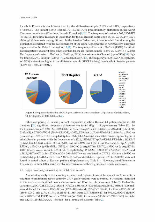
Genes 2020, 11, 554
for ethnic Russians is much lower than for the all-Russian sample (0.18% and 1.81%, respectively,p < 0.0001). The variant c.1545_1546delTA (1677delTA) is predominantly distributed in the NorthCaucasus populations (Chechens, Ingush, Kumyks) [9,12]. The frequency of variant c.262_263delTT(394delTT) for ethnic Russians is lower than for the all-Russian sample (0.54% vs. 0.94%, p = 0.074,although difference is not significant). In the Russian Federation, it is more often found among thepopulation associated with the past settlement of the Finno-Ugric peoples in northwestern Europeanregions and in the Volga-Ural region [12,17]. The frequency of variant c.274G>A (E92K) for ethnicRussian patients is almost three times less than for the all-Russian sample (1.05% vs. 3.00%, p < 0.0001).The frequency of variant c.274G>A (p.Glu92Lys, E92K) is maximum for Chuvash (up to 55%) [10], highfor Tatars (6.67%), Bashkirs (6.25%) [7], Chechens (12.5%) [9]. The frequency of c.3846G>A (p.Trp1282X,W1282X) is significant higher in the all-Russian sample (RF CF Registry) than in ethnic Russian patients(1.16% vs. 1.90%, p = 0.012).
Figure 1. Frequency distribution of CFTR gene variants in three samples of CF patients: ethnic Russians,CF RF Registry, CFTR2 database [22].
When comparing CF-causing variant frequencies in ethnic Russian CF patients to the CFTR2database [22], significant frequency difference was found (Fig. 1, Supplementary Table S1). So,the frequencies of c.54-5940_273+10250del21kb (p.Ser18Argfs*16, CFTRdele2,3), c.2012delT (p.Leu671X,2143delT), c.3718-2477C>T (3849+10kbC-T), c.2052_2053insA (p.Gln685ThrfsX4, 2184insA), c.274G>A(p.Glu92Lys, E92K), c.413_415dupTAC (p.Leu138dup; L138ins) and some other variants appear higher inethnic Russian patients while the frequencies of c.1521_1523delCTT (p.Phe508del, F508del), c.1624G>T(p.Gly542X, G542X), c.2657+5G>A (2789+5A>G), c.489+1G>T (621+1G>T), c.1657C>T (p.Arg553X,R553X), c.254G>A (p.Gly85Glu, G85E), c.1040G>C (p.Arg347Pro, R347P), c.350G>A (p.Arg117His,R117H) were lower. Variants c.3844T>C (p.Trp1282Arg, W1282R), c.3140-16T>A (3272-16T>A), andc.3816_3817delGT (p.Ser1273LeufsX28, 3944delGT) were not listed in CFTR2. Variants c.1652G>A(p.Gly551Asp, G551D), c.1585-1G>A (1717-1G>A), and c.3476C>T (p.Ser1159Phe, S1159F) were notfound in tested cohort of Russian patients (Supplementary Table S1). However, the differences infrequencies in these latter series involve rare variants and their significance remains unknown.
4.2. Sanger Sequencing Detection of the CFTR Gene Variants
As a result of analysis of the coding sequence and regions of exon-intron junctions 80 variants inaddition to preliminary tested common CFTR gene variants were identified. 61 variants identifiedin this work were identified on one chromosome and 17 on two chromosomes (Table 2). Each of thevariants c.1209G>C (E403D), c.2128A>T (K710X), c.3883delA (4015delA) and c.3884_3885insT (4016insT)were detected for three, c.1766+1G>A (1898+1G>A) and c.2834C>T (S945L) for four, c.1766+1G>C(1898+1G>C) and c.(743+1_744-1)_(1584+1_1585-1)dup (CFTRdup6b-10) for five, c.2353C>T (R785X)and c.4004T>C (L1335P) for six, c.3929G>A (W1310X) for seven, c.580-1G>T (712-1G>T) for eight,and c.1240_1244delCAAAA (1365del5) for 11 unrelated patients (Table 2).
56
Genes 2020, 11, 554
Some of genetic variants identified in sequencing were first discovered in this study. Descriptionof 15 is presented in a previously published paper [23]. Nine of these variants are nonsense mutations(c.252T>A (p.Tyr84X), c.831G>A (p.Trp277X), c.1083G>A (p.Trp361X), c.3112C>T (p.Gln1038X))or frame-shift mutations (c.264_268delATATT (p.Leu88PhefsX21), c.1219delG (p.Glu407AsnfsX35),c.1608delA (p.Asp537ThrfsX3), c.1795dupA (p.Thr599AsnfsX2), c.3189delG (p.Trp1063X), resultingin the formation of premature stop codon (Table 1). Variant c.490-1G>C breaks the acceptorsite of 5 exon splicing. These variants belong to the category of PVS1 null variants (pathogenicvariant sequence) according to the criteria of classification of pathogenicity of genetic variants [24].Variant c.1792_1793insAAA (p.Lys598dup) leads to the insertion of lysine into position 598, and clinicalsignificance of the variant is assessed as pathogenic. The clinical significance of the missensemutations (c.358G>C (p.Ala120Pro), c.1382G>A (p.Gly461Glu), c.1513A>C (p.Asn505His), c.1525G>C(p.Gly509Arg)) is assessed as probably pathogenic.
Eight more variants are presented for the first time. Two variants-nonsense mutations (c.1204G>T(p.Glu402X), c.2617G>T (p.Glu873X)) and one deletion with frame shift (c.2312delA (p.Asn771ThrfsX2))are concluded to be PVS1 null variants according to the ACMG classification. Variant c.2989-2A>C is aviolation of the 19 exon splicing site. Three missense mutations (c.613C>A (p.Pro205Thr), c.1352G>T(p.Gly451Val), c.1589T>C (p.Ile530Thr), c.3107C>A (p.Thr1036Asn)), the clinical significance of whichis assessed as probably pathogenic according to the recommendations [24]. The characteristics of thephenotypes of patients who carry rare missense variants are presented in Supplementary Table S3.
4.3. CNV in Russian CF Patients Detected by MLPA
Large rearrangements of the CFTR gene were found for 18 unrelated patients, which is 10.8%(18/166) of the tested mutant alleles and should account for about 1% in the total sample of all mutantalleles in Russians. The MLPA method revealed 11 large rearrangements of the CFTR gene: threeduplications and eight deletions (Table 1). Four of the large rearrangements were detected in severalfamilies. Thus, the duplication of a fragment covering 7–11 (6b–10) exons was detected for fiveunrelated patients. The testing system we developed allowed us to confirm that the duplicationdetected had the same frames as previously described in the literature [13]. In the RF CF Registry 2017,this variant was noted for six more unrelated patients. Thus, CFTRdup6b-10 was detected in elevenunrelated patients. Six of them live in the Volga-Ural region, three in the Central region. It should benoted that two patients from the Volga-Ural region belong to the other ethnic groups: one-Bashkirand one-Udmurt.
Each of the deletions, c.(53+1_54-1)_(164+1_165-1)del (CFTRdele2), c.[(1679-1_1680+1)_(2490+1_2491-1)del[;](2908+1_2989-1)del] (CFTRdele12,13;del16) and c.(273-1_274+1)_(869+1_870-1)del(1209-1_1210+1)_(1392+1_1393+1)del (CFTRdel4-7;del9-10) was detected twice. Complex deletion,CFTRdele12,13;del16, was detected for two patients from unrelated families living in the Moscowregion; deletion CFTRdel4-7;del9-10 for two families from the Kaliningrad region and the Republic ofBuryatia; deletion CFTRdele2 in families from the Transbaikal region and Irkutsk region.
4.4. Detection Rate of Three-Stage Analysis of CFTR Gene in Russian CF Patients
As a result of analysis of the coding sequence and regions of exon-intron junctions and subsequentsearch for large rearrangements, 163 out of 164 alleles were identified that were not detected afterpreliminary testing of frequent variants of the CFTR gene.
In one patient only variant E217G with the F508del in trans was detected after sequencing andMLPA. In NCBI-ClinVar database variant E217G is considered to be variant of conflicting interpretationof pathogenicity (benign; likely benign; uncertain significance) [25]. In the study by Lee J.H. et al. [26]it was shown that non-synonymous E217G mutation in the M470 background caused a 60%–80%reduction in CFTR-dependent Cl− currents and HCO3
− transport activities. So we might suggest thatthe clinical presentation in that patient is due to complex allele E217G-M470 (Supplementary Table S3).
57

Genes 2020, 11, 554
The second mutant allele of the CFTR gene could not be identified in one sample. Failure toidentify the second pathologic mutation in the CFTR gene after sequencing the coding sequence andsearching for large rearrangements may be due to the location of the pathogenic variant either in innerregions of the introns, or in regulatory regions of the CFTR gene, or in regulatory regions outsidethe CFTR gene. Indeed, such variants have been recently identified, for example, c.1680-883A>G,c.2989-313A>T, c.3469-1304C>G, or c.3874-4522A>G, that lead to the creation of a new donor splicesite and the activation of a cryptic acceptor splice site, resulting in the inclusion of an additionalpseudo-exon (PE) and the loss of wild type (WT) CFTR transcripts [27].
5. Conclusions
In a representative sample of CF patients (ethnic Russians), the spectrum of 33 routinelyanalyzed (in Russia) variants of the CFTR gene was studied. It was shown that, out of 29identified variants, frequencies of only 10 exceed 1%, and the mutation detection rate of testingdid not exceed 85%. Consistent use of sequencing and MLPA methods has allowed us toidentify a significant variety of CFTR gene mutations spectrum (91 additional genetic variants),to expand the spectrum of frequent variants (c.1766+1G>C (1898+1G>C), c.2353C>T (R785X),c.(743+1_744-1)_(1584+1_1585-1)dup (CFTRdup6b-10), c.4004T>C (L1335P), c.3929G>A (W1310X),c.580-1G>T (712-1G>T), c.1240_1244delCAAAA (1365del5), detected for five and more unrelatedpatients, to increase the detection rate of identified mutant alleles for Russian CF patients up to 99.4%,consistently using the strategy of Sanger sequencing and MLPA analysis. This information can beuseful for the further optimization of medical genetic counseling in CF high-risk families, for improvingthe neonatal screening program for CF, and for making decision about the possible CFTR modulatorstherapy in the future. The identification of previously unknown CF-pathogenic or likely-pathogenicvariants is a useful piece of information for diagnostic testing not only in Russia, but worldwide,and can be considered as a contribution to the general knowledge about the CFTR variant heterogeneity.
Supplementary Materials: The following are available online at http://www.mdpi.com/2073-4425/11/5/554/s1,Table S1: Frequencies of 33 variants of CFTR gene in the samples of 1384 ethnic Russians and in the CF patients fromCFTR2 database [22], Table S2: Genotypes and CFTR gene variants in 154 Russian patients tested for 33 commonCFTR variants, Table S3: Clinical and demographic characteristics of CF patients with rare missense variants.
Author Contributions: Data curation, N.V.P. and N.Y.K.; Formal analysis, N.V.P. and T.A.V.; Funding acquisition,S.I.K. and R.A.Z.; Investigation, N.V.P.; Project administration, E.K.G., S.I.K. and R.A.Z.; Resources, N.Y.K., E.I.K.,E.K.Z., A.Y.V., V.D.S. and V.A.G.; Supervision, E.K.G. and R.A.Z.; Validation, T.A.V. and A.V.M.; Writing–originaldraft, N.V.P.; Writing–review & editing, A.V.M. All authors have read and agreed to the published version ofthe manuscript.
Funding: The research was partially supported by grant RFBR 20-015-0061A and within the state task of theMinistry of education and science of Russia.
Acknowledgments: The authors are grateful to Stanislav Krasovsky and Elena Amelina for their input to thecollection of the clinical data of adult CF patients. Also the authors would like to thank the National CF PatientRegistry for providing access to patients data and thank the individual regional CF centers representatives forallowing the use of data, https://mukoviscidoz.org/mukovistsidoz-vrossii.html.
Conflicts of Interest: The authors declare no conflict of interest. The funders had no role in the design of thestudy; in the collection, analyses, or interpretation of data; in the writing of the manuscript, or in the decision topublish the results.
References
1. Sosnay, P.R.; Raraigh, K.S.; Gibson, R.L. Molecular Genetics of Cystic Fibrosis Transmembrane ConductanceRegulator: Genotype and Phenotype. Pediatr. Clin. North Am. 2016, 63, 585–598. [CrossRef] [PubMed]
2. Farrell, P.M.; White, T.B.; Howenstine, M.S.; Munck, A.; Parad, R.B.; Rosenfeld, M.; Sommerburg, O.;Accurso, F.J.; Davies, J.C.; Rock, M.J.; et al. Diagnosis of Cystic Fibrosis in Screened Populations. J. Pediatr.2017, 181S, S33–S44. [CrossRef] [PubMed]
58

Genes 2020, 11, 554
3. Castellani, C.; Cuppens, H.; Macek, M., Jr.; Cassiman, J.J.; Kerem, E.; Durie, P.; Tullis, E.; Assael, B.M.;Bombieri, C.; Brown, A.; et al. Consensus on the use and interpretation of cystic fibrosis mutation analysis inclinical practice. J. Cyst. Fibros. 2008, 7, 179–196. [CrossRef] [PubMed]
4. De Boeck, K.; Vermeulen, F.; Dupont, L. The diagnosis of cystic fibrosis. Presse Med. 2017, 46, e97–e108.[CrossRef] [PubMed]
5. Castellani, C.; Duff, A.J.A.; Bell, S.C.; Heijerman, H.G.M.; Munck, A.; Ratjen, F.; Sermet-Gaudelus, I.;Southern, K.W.; Barben, J.; Flume, P.A.; et al. ECFS best practice guidelines: The 2018 revision. J. Cyst. Fibros.2018, 17, 153–178. [CrossRef] [PubMed]
6. National Consensus Project. Cystic Fibrosis: Definition, Diagnostic Criteria, Treatment 2018; Kondratyeva, E.I.,Kashirskaya, N.Y., Kaprqanov, N.I., Eds.; Company BORGES Ldt.: Moscow, Russia, 2018; p. 356.
7. MEDPRAKTIKA-M. Registry of Patients with Cystic Fibrosis in the Russian Federation. 2017; Voronkova, A.Y.,Amelina, E.A., Kashirskaya, N.Y., Kondratyeva, E.I., Krasovsky, S.A., Starinova, M.A., Kapranov, N.I., Eds.;MEDPRAKTIKA-M: Moscow, Russia, 2019; p. 68. (in Russian)
8. Petrova, N.V.; Kashirskaya, N.Y.; Vasilieva, T.A.; Timkovskaya, E.E.; Voronkova, A.Y.; Shabalova, L.A.;Kondrateva, E.I.; Sherman, V.D.; Kapranov, N.I.; Zinchenko, R.A.; et al. High proportion of W1282X mutationin CF patients from Karachai-Cherkessia. J. Cyst. Fibros. 2016, 15, e28–e32. [CrossRef] [PubMed]
9. Petrova, N.V.; Kashirskaya, N.Y.; Saydaeva, D.K.; Polyakov, A.V.; Adyan, T.A.; Simonova, O.I.; Gorinova, Y.V.;Kondratyeva, E.I.; Sherman, V.D.; Novoselova, O.G.; et al. Spectrum of CFTR mutations in Chechen cysticfibrosis patients: High frequency of c.1545_1546delTA (p.Tyr515X; 1677delTA) and c.274G>A (p.Glu92Lys,E92K) mutations in North Caucasus. BMC Med. Genet. 2019, 20, 44. [CrossRef]
10. Stepanova, A.A.; Abrukova, A.V.; Savaskina, E.N.; Poliakov, A.V. Mutation p.E92K is the primary cause ofcystic fibrosis in Chuvashes. Russ. J. Genet. 2012, 48, 731–737. [CrossRef]
11. StatData.ru. Available online: http://www.statdata.ru/nacionalnyj-sostav-rossii (accessed on8 February 2020).
12. Petrova, N.V.; Kondratyeva, E.I.; Krasovsky, S.A.; Polyakov, A.V.; Ivachshenko, T.E.; Pavlov, A.E.;Zinchenko, R.A.; Ginter, E.K.; Kutsev, S.I.; Odinokova, O.N.; et al. National Consensus Project «Cysticfibrosis: Definition, diagnostic criteria, treatment» Section «Genetics of Cystic Fibrosis. Molecular geneticdiagnosis of cystic fibrosis». Meditsinskaya Genet. 2016, 11, 29–45. (in Russian).
13. Hantash, F.M.; Redman, J.B.; Goos, D.; Kammesheidt, A.; McGinniss, M.J.; Sun, W.; Strom, C.M.Characterization of a recurrent novel large duplication in the cystic fibrosis transmembrane conductanceregulator gene. J. Mol. Diagn. 2007, 9, 556–560. [CrossRef]
14. Petrova, N.V.; Timkovskaya, E.E.; Zinchenko, R.A. The analysis of CFTR mutations frequencies in differentpopulations of Russia. Eur. J. Hum. Genet. 2008, 16, 387.
15. Verlingue, C.; Kapranov, N.I.; Mercier, B.; Ginter, E.K.; Petrova, N.V.; Audrezet, M.P.; Ferec, C. Completescreening of mutations in the coding sequence of the CFTR gene in a sample of CF patients from Russia:Identification of three novel alleles. Hum. Mutat. 1995, 5, 205–209. [CrossRef] [PubMed]
16. Ivaschenko, T.E.; Baranov, V.S. Biochemical and Molecular Genetic Basis of Cystic Fibrosis Pathogenesis; Intermedika:Saint-Petersburg, Russia, 2002; p. 256. (in Russian)
17. Korytina, G.F.; Victorova, T.V.; Baykova, G.V.; Khusnutdinova, E.K. Analysis of the Spectra of Mutationsand Polymorphic Loci of Cystic Fibrosis Transmembrane Conductance Regulator in the Population ofBashkortostan. Rus. J. Genet. 2002, 38, 1076–1081. [CrossRef]
18. WHO Human Genetics Programme. The Molecular Genetic Epidemiology of Cystic Fibrosis. Report of a JointMeeting of WHO/ECFTN/ICF(M)/ECFS/World Health Organization (WHO); WHO: Genoa, Italy, 2002; Availableonline: https://www.cfww.org/docs/who/2002/who_hgn_cf_wg_04.02.pdf (accessed on 8 February 2020).
19. Cystic Fibrosis Mutation Database. Available online: http://www.genet.sickkids.on.ca (accessed on5 February 2020).
20. Bobadilla, J.L.; Macek, M., Jr.; Fine, J.P.; Farrell, P.M. Cystic fibrosis: A worldwide analysis of CFTRmutations—Correlation with incidence data and application to screening. Hum. Mutat. 2002, 19, 575–606.[CrossRef] [PubMed]
21. Dörk, T.; Macek, M., Jr.; Mekus, F.; Tümmler, B.; Tzountzouris, J.; Casals, T.; Krebsová, A.; Koudová, M.;Sakmaryová, I.; Macek, M.S.; et al. Characterization of a novel 21-kb deletion, CFTRdele2,3(21kb), in theCFTR gene: A cystic fibrosis mutation of Slavic origin common in Central and East Europe. Hum. Genet.2000, 106, 259–268. [CrossRef] [PubMed]
59

Genes 2020, 11, 554
22. CFTR2. Clinical and Functional Translation of CFTR. Available online: https://www.cftr2.org/ (accessed on5 February 2020).
23. Petrova, N.V.; Marakhonov, A.V.; Vasilyeva, T.A.; Kashirskaya, N.Y.; Ginter, E.K.; Kutsev, S.I.; Zinchenko, R.A.Comprehensive genotyping reveals novel CFTR variants in cystic fibrosis patients from the Russian Federation.Clin. Genet. 2018, 95, 1–4. [CrossRef] [PubMed]
24. Richards, S.; Aziz, N.; Bale, S.; Bick, D.; Das, S.; Gastier-Foster, J.; Grody, W.W.; Hegde, M.; Lyon, E.; Spector, E.;et al. Standards and guidelines for the interpretation of sequence variants: A joint consensus recommendationof the American College of Medical Genetics and Genomics and the Association for Molecular Pathology.Genet. Med. 2015, 17, 405–424. [CrossRef] [PubMed]
25. National Center for Biotechnology Information (NCBI-ClinVar). Available online: https://www.ncbi.nlm.nih.gov/clinvar/ (accessed on 5 February 2020).
26. Lee, J.H.; Choi, J.H.; Namkung, W.; Hanrahan, J.W.; Chang, J.; Song, S.Y.; Park, S.W.; Kim, D.S.; Yoon, J.-H.;Suh, Y.; et al. A haplotype-based molecular analysis of CFTR mutations associated with respiratory andpancreatic diseases. Hum. Mol. Genet. 2003, 12, 2321–2332. [CrossRef] [PubMed]
27. Bergougnoux, A.; Délétang, K.; Pommier, A.; Varilh, J.; Houriez, F.; Altieri, J.P.; Koenig, M.; Férec, C.;Claustres, M.; Lalau, G.; et al. Functional characterization and phenotypic spectrum of three recurrentdisease-causing deep intronic variants of the CFTR gene. J. Cyst. Fibros. 2019, 18, 468–475. [CrossRef][PubMed]
© 2020 by the authors. Licensee MDPI, Basel, Switzerland. This article is an open accessarticle distributed under the terms and conditions of the Creative Commons Attribution(CC BY) license (http://creativecommons.org/licenses/by/4.0/).
60

genesG C A T
T A C G
G C A T
Article
Establishment of a Recombinant AAV2/HBoV1 VectorProduction System in Insect Cells
Xuefeng Deng 1, Wei Zou 1, Ziying Yan 2 and Jianming Qiu 1,*
1 Department of Microbiology, Molecular Genetics and Immunology, University of Kansas Medical Center,Kansas City, KS 66160, USA; [email protected] (X.D.); [email protected] (W.Z.)
2 Department of Anatomy and Cell Biology, University of Iowa, Iowa City, IA 52242, USA;[email protected]
* Correspondence: [email protected]
Received: 27 March 2020; Accepted: 15 April 2020; Published: 17 April 2020
Abstract: We have previously developed an rAAV2/HBoV1 vector in which a recombinantadeno-associated virus 2 (rAAV2) genome is pseudopackaged into a human bocavirus 1 (HBoV1)capsid. Recently, the production of rAAV2/HBoV1 in human embryonic kidney (HEK) 293 cellshas been greatly improved in the absence of any HBoV1 nonstructural proteins (NS). This NS-freeproduction system yields over 16-fold more vectors than the original production system thatnecessitates NS expression. The production of rAAV with infection of baculovirus expression vector(BEV) in the suspension culture of Sf9 insect cells is highly efficient and scalable. Since the replicationof the rAAV2 genome in the BEV system is well established, we aimed to develop a BEV systemto produce the rAAV2/HBoV1 vector in Sf9 cells. We optimized the usage of translation initiationsignals of the HBoV1 capsid proteins (Cap), and constructed a BEV Bac-AAV2Rep-HBoV1Cap,which expresses the AAV2 Rep78 and Rep52 as well as the HBoV1 VP1, VP2, and VP3 at theappropriate ratios. We found that it is sufficient as a trans helper to the production of rAAV2/HBoV1in Sf9 cells that were co-infected with the transfer Bac-AAV2ITR-GFP-luc that carried a 5.4-kboversized rAAV2 genome with dual reporters. Further study found that incorporation of an HBoV1small NS, NP1, in the system maximized the viral DNA replication and thus the rAAV2/HBoV1vector production at a level similar to that of the rAAV2 vector in Sf9 cells. However, the transductionpotency of the rAAV2/HBoV1 vector produced from BEV-infected Sf9 cells was 5–7-fold lowerin polarized human airway epithelia than that packaged in HEK293 cells. Transmission electronmicroscopy analysis found that the vector produced in Sf9 cells had a high percentage of emptycapsids, suggesting the pseudopackage of the rAAV2 genome in HBoV1 capsid is not as efficient as inthe capsids of AAV2. Nevertheless, our study demonstrated that the rAAV2/HBoV1 can be producedin insect cells with BEVs at a comparable yield to rAAV, and that the highly efficient expression of theHBoV1 capsid proteins warrants further optimization.
Keywords: rAAV2/HBoV1; baculovirus; insect cells
1. Introduction
Adeno-associated virus (AAV) and human bocavirus (HBoV) are members in different generaof the parvovirus family [1]. AAV is a nonpathogenic parvovirus and its productive replicationneeds the function of a helper virus [2,3]. In contrast, HBoV1 is a human pathogen that causeslower respiratory tract infections in young children worldwide [4–12]. In vitro, HBoV1 infects onlypolarized human airway epithelium cultured at an air–liquid interface (HAE-ALI), and replicatesautonomously [13–16]. While both are nonenveloped, small, single-stranded (ss) DNA viruses, AAVpackages both the plus- and minus-strand genome with equal efficiency, whereas HBoV1 prefers
Genes 2020, 11, 439; doi:10.3390/genes11040439 www.mdpi.com/journal/genes61
Genes 2020, 11, 439
packaging the minus-strand [14]. HBoV1 has a genome of 5543 nucleotides (nts) in length, which is18.5% (864 nts) larger than the AAV2 genome of 4679 nts [14].
The genome organizations of these two viruses are quite different. HBoV1 uses one promoterto express all viral nonstructural (NS) and structural or capsid (Cap) proteins, but AAV uses threedifferent promoters [4,17]. The coding sequence of AAV consists of two large open reading frames(ORFs) encoding the nonstructural or replication (Rep) proteins and the Cap proteins at the left andright half of the AAV genome, respectively [18]. The large Rep78/68 and the small Rep52/40 proteinsare expressed from the viral mRNAs that are transcribed by an upstream promoter at unit 5 of thegenome (P5) and an internal promoter (P19), respectively. Three AAV Cap proteins, VP1, VP2 andVP3, are expressed from the mRNA transcribed by the P40 promoter [2,19]. In addition, a smallNS protein, assembly-associated protein (AAP), is alternatively translated from the P40-transcribedCap-coding mRNA [20], which plays a role in capsid assembly [21–24]. Recently, another small NSprotein, membrane-associated accessory protein (MAAP), has been identified, which is also expressedfrom the P40-transcribed mRNA through alternative translation [25]. It exists in all AAV serotypesand was believed to play a role in the life cycle of AAV. HBoV1 expresses five NS proteins, NS1,NS2, NS3, NS4, and nuclear protein (NP1), and three Cap proteins, VP1, VP2, and VP3, as well as abocaviral noncoding small RNA (BocaSR) [26–29]. The middle ORF, which is located at the centerof the genome between the left ORF encoding NS1-4 and the right ORF encoding VP1-3, encodesNP1 [4]. NP1 plays an important role in viral DNA replication [30], as well as in regulation of HBoV1cap expression [28,31].
The sequences at both termini of AAV and HBoV1 contain important motifs that are necessary forviral genome replication and virion assembly. In AAV, they are inverted terminal repeats (ITRs) [2,19],but in HBoV1 they are asymmetric with a non-perfectly palindromic hairpin at the left end terminusand a perfectly palindromic hairpin at the right terminus [14]. The transfection of the plasmid cloneof a complete AAV genome in human embryonic kidney (HEK)293 cells leads to the production ofAAV virions, but this only occurs in the presence of infection of a helper virus, such as adenovirusor co-transfection of a plasmid helper harboring all the adenoviral helper genes (E2, E4Orf6, andVA RNA) [32]. While HEK293 cells are not permissive to HBoV1 infection, the transfection of thecloned HBoV1 genome can produce HBoV1 infectious virions. The progeny virions infect HAE-ALIwith a robustly high efficiency, even at a multiplicity of infection (MOI) of at 0.001 viral genome percell [14,15].
Trans-complementation supports the replication and package of the gutless rAAV2 genomecontaining only cis elements of their termini and a gene of interest, which have been effectivelydeveloped as rAAV vectors for gene therapy of genetic diseases [2,19,33]. The safety profiles of therAAV genome have been proven from tremendous preclinical studies and clinical trials of human genetherapy [34–41]. Up to date, two rAAV-based drugs, Luxturna and Zolgensma, have been approvedby the US FDA. Similarly, a recombinant HBoV1 vector (rHBoV1) was produced in HEK293 cells viatrans-complementation [42]. rHBoV1 efficiently transduced HAE-ALI from the apical membrane;however, the safety concern of being derived from a human pathogen limits its application. To overcomethis disadvantage for a safe vector to transduce human airway epithelium from the airway lumenwith the emphasis on gene therapy for cystic fibrosis (CF) lung disease, we successfully developeda cross-genera chimeric parvovirus vector, rAAV2/HBoV1 [42], in which the safety-proven rAAV2genome was packaged into the airway tropic HBoV1 capsid. Importantly, the rAAV2/HBoV1 expandsthe package capacity of the rAAV2 genome by 20%, up to 5.8-kb [42]. Apical application of anrAAV2/HBoV1 carrying a full-length CF transmembrane conductance regulator (CFTR) cDNA of 5.4-kbto CF HAE-ALI cultures, which were made of primary airway epithelial cells of CF patients, efficientlycorrected CFTR-dependent chloride transport [42]. In addition, the rAAV2/HBoV1 vector efficientlytransduced ferret airways in vivo [43]. Therefore, it holds much promise for gene delivery to humanairways, as well as for preclinical trials of CF gene therapy using CF ferret models [44]. Recentlywe have increased the production efficiency of the rAAV2/HBoV1 vector in HEK293 cells through
62
Genes 2020, 11, 439
optimization of cap expression, which approaches a similar level of rAAV2 production in HEK293cells [45]. However, a robust vector production system is in demand for future CF gene therapy inpreclinical and human trials using the rAAV2/HBoV1 vector.
Traditional rAAV vector productions utilize HEK293 cells. During the rAAV2 or the rAAV2/HBoV1production in HEK293 cells, the rescue and replication of the rAAV2 genome require the expression ofAAV rep in addition to the adenoviral helper genes [19,46]. rAAV2 can also be produced in insect cellsby the infection of baculovirus expression vectors (BEVs). The AAV-BEV production system representsa robust and scalable bioprocess [47–52], which only requires one of the large Rep78/68 and one of thesmall Rep52/40 [53]. AAP is required for efficient production of certain serotypes of rAAV vectors inSf9 cells [54,55]. Co-infection of BEVs—one carrying an rAAV2 genome and one expressing AAV2Rep78 and Rep52 along with AAV2 VP1, VP2, and VP3—has been used to produce the rAAV vector ina large quantity at a yield of up to ~105 copies per Sf9 cell, compared to the yield of ~103 copies perHEK293 cell [47,53,54,56].
In this report, we explored the possibility of rAAV2/HBoV1 vector production in the BEV system.Our study demonstrated that the rAAV2/HBoV1 vector can be efficiently produced in a suspensionSf9 culture. In the presence of the expression of HBoV1 NP1, a vector yield similar to that of rAAV2was achieved in Sf9 cells. To our knowledge, this is the first report that the parvoviral cross-generapseudopackage is also effective in insect cells.
2. Materials and Methods
2.1. Cell and Cell CultureHuman embryonic kidney (HEK) 293 cells: HEK293 cells (CRL-1573), obtained from American
Type Culture Collection (ATCC; Manassas, VA, USA), were cultured in Dulbecco’s modified Eagle’smedium (GE Healthcare Life Sciences, Piscataway, NJ, USA) with 10% fetal bovine serum (#F0926,MilliporeSigma, St. Louis, MO, USA)
Insect cells: Sf9 cells (CRL-1711, ATCC) were cultured in suspension in SFX-Insect medium (GEHealthcare, Marlborough, MA, USA) with 2% fetal bovine serum (#F0926, Millipore Sigma; St. Louis,MO, USA) at 27 ◦C.
HAE-ALI cultures: primary human airway cells were isolated from human lung tissues, andthis procedure was carried out at the Tissue and Cell Culture Core of the Center for Gene Therapy,University of Iowa. The primary cells were cultured in the airway basal cell expansion medium(#CC-3118, Lonza, Basel, Switzerland), supplemented with 10 μM of ROCK inhibitor Y-276322, 1 μMof A8301, 1 μM of DMH-1, and 1 μM of CHIR99021 (Tocris Biosciences, Minneapolis, MN, USA) untilconfluency [57]. Then, the cells were collected and seeded onto collagen-coated inserts (Transwells;#3470, Corning, Tewksbury, MA, USA) with a density of 50,000 cells/well. After cell attachment for twodays, the apical and basolateral medium were removed and replaced with a complete Pneumacult-ALImedium (StemCell, Vancouver, Canada) at the basolateral chamber to initiate an airway–liquid interface.The medium was changed every two days, and the ALI-cultured HAE took about four weeks to befully differentiated. We monitored the cultures with a transepithelial electrical resistance using anepithelial Ohmvoltmeter (Millicell-ERS; EMD-Millipore, Burlington, MA, USA), and only HAE-ALIcultures with a resistance value of over 1000 Ω·cm2 were used for subsequent transduction.
2.2. Construction of Baculoviral Expression Shuttle Plasmids and Other HEK293 Cell-Expressing PlasmidspFastBacDual(m): the plasmid pFastBacDual (Invitrogen, Carlsbad, CA, USA) was modified by
inserting a 0.83-kb fragment of λ DNA which contains a SbfI site at each end through SnaBI at nt 3983and MfeI at nt 4815 to obtain the plasmid pFastBacDual (m).
pBac-AAV2ITR-GFP-Luc (5.4-kb): this BAC-AAV transfer plasmid was constructed by replacingthe 0.83-kb λ DNA in pFastBacDual(m) with a 5444-nt ITR-flanked (rAAV) proviral DNA into theat two SbfI sites (Figure 1A). The intermediate rAAV proviral plasmid pAAV-CMV(P10)-GFP-SV40-Luc-bGHpA was derived from pAAV-MCS vector (Cell Biolabs, Inc., San Diego, CA, USA). Foreign
63

Genes 2020, 11, 439
DNA flanking with a pair of SbfI sites was cloned into EcoRI and BamHI digested pAAV-MCS,including the P10 promoter, an open reading frame (ORF) of an enhanced green fluorescent protein(GFP; excised from pEGFP1, Clontech, Palo Alto, CA, USA), SV40 polyadenylation signal (polyA),SV40 early promoter, a FLAG-tagged ORF of firefly luciferase (Luc) and a stuffer from λ DNA (to makethe rAAV2 genome of 5444-nt).
Figure 1. Construction of baculoviral transfer plasmids for vector production in Sf9 cells and theplasmids for rAAV2/HBoV1 vector production in HEK293 cells. (A) BEVs for rAAV2/HBoV1 production.Schematically diagrammed are structures inside the BEVs that were involved in rAAV2/HBoV1production. Bac-AAV2ITR-GFP-Luc carries an rAAV2 genome of 5.4-kb; Bac-AAV2Rep-HBoV1Capexpresses AAV2 Rep proteins and HBoV1 capsid proteins as shown; and Bac-HBoV1NP1 expressesHBoV1 NP1. P10 and Ph are baculoviral promoters, and CMV and SV40 are cytomegaloviral immediateearly and SV40 virus early promoters, respectively. PolyA: polyadenylation signal; Luc: firefly luciferase.(B) Plasmids used for vector production in HEK293 cells. pAAV2ITR-GFP-Luc carries the same rAAV2genome as shown in panel A. pCMVNS*Cap-P5Cap is a two-in-one plasmid. It was derived from theplasmid pHBoV1CMVNS*Cap [28], in which the NS1/2 ORF was early terminated. An AAV2 P5 andP19 driven rep gene was cloned after the CMV promoter-driven HBoV1 cap gene expression cassette.(C,D) Codon optimization. Both wild type (wt) and optimized (opt.) sequences between ATGs ofthe AAV2 Rep78 and Rep52 ORFs (C) and of the HBoV1 VP1 and VP3 ORFs (D) are diagrammed.Nucleotides in red indicate mutations. (E) BEVs for rAAV2 production in Sf9 cells. Bac-AAV2ITR-GFPcarries a GFP expression cassette under both the CMV and P10 promoters. Bac-AAV2Rep-Cap carriesexpression cassettes of AAV2 cap and AAV2 rep under the P10 and Ph promoters, respectively.
64

Genes 2020, 11, 439
pBac-AAV2Rep-HBoV1Cap: to obtain a modified AAV2 rep gene expression cassette of abifunctional Rep78- and Rep52-encoding mRNA, we synthesized a 637-bp DNA fragment containing apartially codon-optimized (opt)Rep78 ORF [56] (Figure 1C), and amplified the full-length optRep78/52ORF using overlapped PCR, which was cloned into pFastBacDual through BglII (BamHI)-XbaI sites andresulted in pFastBacDual-AAV2Rep. We also synthesized a fragment of 390-bp containing an optimizedHBoV1 sequence between VP1 AUG and VP3 AUG [28,58], as shown in Figure 1D, and amplified the fulllength optVP1/2/3 ORF using overlapped PCR, which was then cloned into the pFastBacDual-AAV2Repthrough XhoI-NheI sites to obtain the BEV transfer plasmid pBac-AAV2Rep-HBoV1Cap (Figure 1A).
pBac-HBoV1NP1: HBoV1 NP1 ORF was cloned into pFastBacDual between the XhoI-KpnI sitesto get the transfer construct pBac-HBoV1-NP1 (Figure 1A).
pAAV2ITR-GFP-Luc: to parallel compare the capability of the Sf9 cell and HEK293 cell systems,we made a plasmid, pAAV2ITR-GFP-Luc (Figure 1B), based on the backbone of pAAV-MCSpromoterless (Cell Biolabs). This was achieved by cloning the fragments from the plasmid ofpFastBacDual(m)-AAV2-ITR-GFP-Luc through two NotI sites.
pCMVNS*Cap-P5Rep: this was the HBoV1 cap and AAV2 rep expression two-in-one helperplasmid (Figure 1B), which was constructed by cloning the P5- and P19-driven AAV2 rep expressioncassette from the pAV-Rep2 plasmid [42] into the pCMVNS*Cap [28], which expresses HBoV1 capsidproteins under the cytomegalovirus immediate early promoter (CMV) through a SacII site.
All the plasmids were sequenced to confirm the expressing genes and the critical elements forvirus production at MCLAB (South San Francisco, CA, USA).
2.3. Recombinant Baculovirus Expression vector (BEV) Production
BEVs were generated by transfection of the BEV shuttle plasmid into DH10BacTM E. coli competentcells, following the instructions of the Bac-to-Bac Baculovirus Expression System (Invitrogen, Carlsbad,CA). Bac-AAV2ITR-GFP (Bac-GFP) [47] and Bac-AAV2Rep-Cap (Bac-RepCap2) [56] (Figure 1E) wereobtained from The University of Iowa Viral Vector Core Facility. BEVs were titrated in plaque formingunits (pfu) by a plaque assay as described in the manual of the Bac-to-Bac Baculovirus ExpressionSystem (Invitrogen) or quantified using quantitative real-time PCR (qPCR) with an amplicon targetingto the gentamicin-resistance gene (Probe: 5′ 6FAM-ACA TTC ATC GCG CTT GCT GCC TTC-3′ ZEN/Iowa Black FQ; forward primer: 5′-CGG GAA CTT GCT CCG TAG TAA-3′, and reverse primer:5′-CGC CAA CAA CCG CTT CTT-3′).
2.4. rAAV vector Production
For production of AAV vectors in insect cells, 200 mL of Sf9 cells in suspension culture at a densityof 2 × 10⁶ cells/mL were co-infected with BEVs at an MOI of ~10 (pfu/cell). At 72 hrs post-infection,cells were collected by centrifugation and lysed in phosphate-buffered saline, pH7.4 (PBS). After fourtimes of freezing-thawing, the cells were sonicated at the setting of 70% power for 3 min (1 min sonicatewith 1 min of interval), followed by DNase I treatment at 37 ◦C for 45 min and 10% deoxycholatewith 0.25% Trypsin-EDTA incubation at 37 ◦C for another 30 min. Then, CsCl was added into cellcrude lysate at a final concentration of 0.472 g/mL and incubated at 37 ◦C for 30 min. The mixturewas centrifuged at 3,500 rpm for 30 min, the clarified virus/CsCl solution was transferred into anothertube and adjusted to a final density of about 1.40 g/mL. The final clear mixture was loaded into tubesand centrifuged at 41,000 rpm for 36 hrs at 20 ◦C using an TH641 rotor in a Sorvall™WX (ThermoScientific). After two rounds of CsCl banding, an aliquot (500 μL) of gradient fractions was collectedusing a Gradient Station (BioComp Instruments, Fredericton, N.B., Canada), determined for values ofrefractive index using an Abbe Refractometer, and quantified by qPCR for vector genomes. The peakfractions were dialyzed against PBS buffer.
For the HEK293 cell system, the rAAV2/HBoV1 vector was generated by co-transfection ofpAAV2ITR-GFP-Luc, pCMVNS*Cap-P5Rep, and pHelper that expresses adenoviral E2, E4Orf6 proteinand VA RNA [3] into twenty 150-cm2 plates of HEK293 cells (80% confluency) at a ratio 2:3:1,
65
Genes 2020, 11, 439
as previously described [42]. At 72 hrs post-transfection, cell pellets were collected and treated, andrecombinant vector was purified as described above for infected Sf9 cells.
2.5. Western Blot and Southern Blot Analyses
Western blotting was performed as previously described [59]. For Southern blotting, low molecularweight (Hirt) DNA was extracted from BEV infected Sf9 cells, and the analysis was performed aspreviously described [60], using an AAV2 cap gene probe.
2.6. Quantitative Real Time PCR (qPCR) Analysis of rAAV2/HBoV1
The titers of rAAV2 and rAAV2/HBoV1 in DNase I digestion-resistant particles (DRP) weredetermined by a qPCR method that has been used previously [42]. Briefly, 50 μL aliquots of the sampleswere incubated with 20 units of Benzonase (MilliporeSigma) for 2 hrs at 37 ◦C, followed by 20 μL ofproteinase K (15 mg/mL) at 56 ◦C for 10 min. Viral DNA was extracted using a QIAamp blood minikit (Qiagen, Hilden, Germany), and then eluted in 50 μL of deionized water. A plasmid containing aGFP ORF was used to establish a standard curve for absolute quantification. The amplicon primersand dual-labeled probe were designed using Primer Express (Applied Biosystems, Foster City, CA,USA) and synthesized at IDT Inc. (Coralville, Iowa, USA). The sequences of the primers and probespecific to the GFP coding sequence are as follows: forward primer, 5′-CTG CTG CCC GAC AACCA-3′; reverse primer, 5′-TGT GAT CGC GCT TCT CGT T-3′; and dual-labeled probe, 5′ 6FAM-TACCTG AGC ACC CAG TCC GCC CT-3′ Iowa Black FQ. Probe qPCR MasterMix (Applied BiologicalMaterials Inc., Vancouver, Canada) was used for qPCR following a standard protocol on a 7500 FastReal-Time PCR System (Applied Biosystems, Foster City, CA, USA), and 2 μL of extracted DNA wasused in a reaction volume of 20 μL.
2.7. Transmission Electron MicroscopyFor each recombinant virus, aliquots of 50 μL in the peak fractions were performed for electron
microscopy analysis in the Electron Microscopy Research Laboratory (EMRL) of the University ofKansas Medical Center. Briefly, two to five μL of each sample were spotted onto formvar-coated,carbon-stabilized, 200-mesh copper grids for 1 min and then washed with deionized water. Stainingwas achieved by adding five drops of 2% uranyl acetate. Excess staining was removed immediatelyby adsorption to filter paper, and the samples were then air dried. The grids were examined on aTransmission Electron Microscope (JEM-1400; JEOL, Peabody, MA, USA) at a magnification setting of30,000 × and an accelerating voltage of 100 KV.
2.8. rAAV2/HBoV1 Transduction of HAE-ALI CulturesWe followed a previously described protocol to infect HAE-ALI cultures with rAAV2/HBoV1 [42].
Briefly, proteasome inhibitor doxorubicin and N-acetyl-l-leucyl-l-leucyl-l-norleucine (LLnL) at the finalconcentrations of 2.5 μM and 20 nM, respectively, were added into the culture medium of the basolateralchamber. Then, a total of 109 DRP of rAAV2/HBoV1 in 50 μL of medium were applied directly ontothe apical surface of the airway epithelia at an MOI of ~2000 DRP/cell. At 12 hrs post-infection, themedium and virus were removed from the apical surface, and the basal medium was replaced withfresh medium without addition of proteasome inhibitors.
2.9. Measurement of Luciferase Reporter Expression
Luciferase enzyme activity was examined using a Luciferase Assay System kit (Promega, Madison,WI, USA) following the manufacturer’s instructions. Briefly, HAE cells were collected after EDTA andtrypsin treatments of the HAE-ALI cultures, and equal numbers of the cells from compared HAE-ALIcultures were transferred into wells of a 96-well plate. The wells were then added with 20 μL of 1×Lysis reagent, followed by mixing with 100 μL of Luciferase Assay Reagent and the light produced ona Synergy H1 microplate reader (Synergy H1, BioTek, Winooski, VT, USA) was measured.
66
Genes 2020, 11, 439
2.10. Antibodies UsedA monoclonal antibody (clone 303.9) that reacts with AAV2 Rep78 and Rep52 and a monoclonal
antibody (clone B1) that reacts with AAV2 Cap were purchased from American Research Products, Inc.(Waltham, MA, USA). Rat anti-HBoV1 Cap that reacts with VP1, VP2, and VP3 and rat anti-HBoV1NP1 that recognizes the NP1 protein were made in house and have been described previously [26,31].
3. Results
3.1. Design of the Baculovirus Expression vector SystemWe used two BEVs, Bac-AAV2ITR-GFP and Bac-AAV2Rep-Cap (Figure 1E), to produce rAAV2 in
Sf9 cells, which serves as a comparative control for the test of the production of the rAAV2/HBoV1vector. In Bac-AAV2Rep-Cap, the AAV2 rep and cap genes were modified to allow expression of theRep78 and Rep52 proteins and the VP1, VP2, and VP3 proteins from two species of mRNAs transcribedfrom Ph and P10 baculovirus promoters, respectively [56]. To generate the rAAV2/HBoV1 vector, wesimilarly made two BEVs, Bac-AAV2ITR-GFP-Luc, which harbors a 5.4-kb oversized rAAV2 genome,and Bac-AAV2Rep-HBoV1Cap (Figure 1A), which expresses AAV2 Rep78 and Rep52 under the Phpromoter and HBoV1 VP1, VP2 and VP3 under the P10 promoter. In addition, we made Bac-HBoV1NP1that expresses the HBoV1 NP1 protein to look for a role of the NP1 in vector production.
We also compared the production and biologic properties of the rAAV2/HBoV1 vectors frominsect cell and mammalian cell systems in parallel. To this end, we made the cis and trans plasmidconstructs for rAAV2/HBoV1 vector production in HEK293 cells in the format similar to those used inthe BEVs. The pAAV2ITR-GFP-Luc (5.4-kb) harbors the identical 5.4-kb rAAV2 genome that was inthe transfer BEV Bac-AAV2ITR-GFP-Luc. The pCMVNS*Cap-P5Rep is the HBoV1 cap and AAV2 reptwo-in-one expression plasmid, which expresses HBoV1 NS (NP1, NS3 and NS4) and Cap (VP1, VP2,and VP3) under the cytomegalovirus immediate early promoter (CMV) [27,28] and AAV2 Rep78 andRep 52 under the AAV2 P5 and P19 promoters, respectively (Figure 1B).
3.2. Analyses of Protein Expression and Replication of the rAAV2 Genome in Sf9 CellsTo characterize the expression of AAV Rep and Cap, Sf9 cells grown in suspension culture were
infected with Bac-AAV2Rep-Cap, Bac-AAV2Rep-HBoV1Cap, and Bac-HBoV1NP1, respectively. Theinfected cells were collected at 72 hrs post-infection, and the expression of viral proteins was analyzedby Western blotting. We first examined the expression of AAV2 Rep from the Sf9 cells infected withBac-AAV2Rep-Cap (Figure 2A) and Bac-AAV2Rep-HBoV1Cap (Figure 2B), respectively. The results ofWestern blotting showed that the expressions of the AAV2 Rep by the P10 promoter from these two BEVsdemonstrated a similar pattern, which both expressed AAV2 Rep78 and Rep52 at a ratio close to ~1:2.Of note, when we constructed the Bacmid pBac-AAV2Rep-HBoV1Cap, the codon optimization of AAV2rep (Figure 1C) was adopted from one mRNA transcript, which was used in Bac-AAV2Rep-Cap [56].The HBoV1 Cap expression from the Sf9 cells infected with Bac-AAV2Rep-HBoV1Cap was alsoanalyzed with Western blotting, which confirmed that the optimization of initiation codons (Figure 1D)led to the expression levels of HBoV1 VP1, VP2, and VP3 at a ratio close to ~1:1:10 (Figure 2C), similarto that was observed from the transfection of the pCMVNS*Cap in HEK293 cells [28]. The crypticpolyadenylation signals (pA) resided inside the unique sequence in the VP1 ORF, which serve asthe proximal pA preventing HBoV1 cap transcription in mammalian cells in the absence of NP1expression [28], appeared to not be effective in the insect cells. These results confirmed that theexpression strategy that Bac-AAV2Rep-Cap utilized to express the overlapping genes rep and cap fromthe baculoviral promoters P10 and Ph was applicable for the construction of Bac-AAV2Rep-HBoV1Cap.This AAV2/HBoV1 trans helper possessed the same capability to express AAV2 Rep proteins andalso HBoV1 VP1, VP2 and VP3, and more importantly, they were expressed at the expected ratios.Thus, one BEV was able to express the parvoviral proteins from different genera efficiently withoutmutual disruption.
67

Genes 2020, 11, 439
To determine the function of HBoV1 NP1 during the replication of the rAAV2 genome, we made aBac-HBoV1NP1. It expressed HBoV1 NP1 at ~25 kDa (Figure 2D). Then, Sf9 cells were co-infected withBac-AAV2ITR-GFP-Luc and Bac-AAV2Rep-HBoV1Cap, with or without Bac-HBoV1NP1. The infectedcells were sampled at 72 hrs post-infection and analyzed for the presence of rAAV2 replicative-form(RF) DNA intermediates by Southern blotting (Figure 2E). Although ssDNA was not obviously detectedin both groups, clearly much more double replicative form (dRF) DNA was observed in the presence ofNP1 expression (Figure 2E, compare lanes 2 vs 3). Although NP1 is not required to modulate the HBoV1cap expression in Sf9 cells as it does in mammalian cells, it positively enhances the replication of rAAV2genomes. The mechanism of NP1’s involvement in rAAV2 replication in Sf9 cells remains unclear.
Figure 2. Expression of AAV2 Rep and Cap proteins and HBoV1 Cap and NP1 proteins inBEV-infected Sf9 cells. (A–D) Western blotting. Sf9 insect cells were infected with Bac-AAV2Rep-Cap(A), Bac-AAV2Rep-HBoV1Cap (B,C), or Bac-HBoV1NP1 (D). The infected cells were collected at 72 hrspost-infection and subjected to Western blot analysis. (A) AAV2 Rep proteins were detected with ananti-Rep monoclonal antibody. (B,C) Bac-AAV2Rep-HBoV1Cap generated from transfection of threebacmid (Bacmid-1-3) were infected with Sf9 cells independently. (B) AAV2 Rep proteins were detectedwith an anti-Rep monoclonal antibody, and (C) HBoV1 Cap protein expression was detected withan anti-HBoV1 Cap protein antiserum. (D) HBoV1 NP1 was detected with a rat anti-HBoV1 NP1antiserum. β-actin served as a loading control. Mock, uninfected cells. (E) Southern blotting. Sf9 cellswere infected with Bac-AAV2ITR-GFP-Luc and Bac-AAV2Rep-HBoV1Cap with (+) or without (-)co-infection of Bac-HBoV1NP1. Cells were collected at 72 hrs post-infection and subjected to extractionof lower molecular weight (Hirt) DNAs, which were analyzed by Southern blotting. Mock, uninfectedSf9 cells as a control; M, a marker of a rAAV2ITR-GFP-Luc proviral replicative form (RF) genome of5.4 kb. dRF and mRF, double and monomer RF.
68
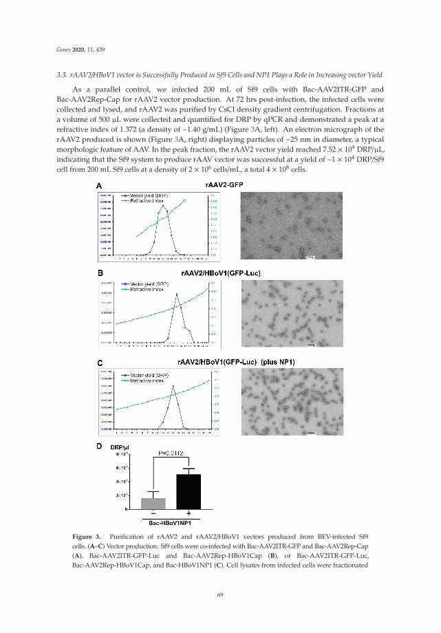
Genes 2020, 11, 439
3.3. rAAV2/HBoV1 vector is Successfully Produced in Sf9 Cells and NP1 Plays a Role in Increasing vector Yield
As a parallel control, we infected 200 mL of Sf9 cells with Bac-AAV2ITR-GFP andBac-AAV2Rep-Cap for rAAV2 vector production. At 72 hrs post-infection, the infected cells werecollected and lysed, and rAAV2 was purified by CsCl density gradient centrifugation. Fractions ata volume of 500 μL were collected and quantified for DRP by qPCR and demonstrated a peak at arefractive index of 1.372 (a density of ~1.40 g/mL) (Figure 3A, left). An electron micrograph of therAAV2 produced is shown (Figure 3A, right) displaying particles of ~25 nm in diameter, a typicalmorphologic feature of AAV. In the peak fraction, the rAAV2 vector yield reached 7.52 × 10⁹ DRP/μL,indicating that the Sf9 system to produce rAAV vector was successful at a yield of ~1 × 10⁴ DRP/Sf9cell from 200 mL Sf9 cells at a density of 2 × 10⁶ cells/mL, a total 4 × 10⁸ cells.
Figure 3. Purification of rAAV2 and rAAV2/HBoV1 vectors produced from BEV-infected Sf9cells. (A–C) Vector production. Sf9 cells were co-infected with Bac-AAV2ITR-GFP and Bac-AAV2Rep-Cap(A), Bac-AAV2ITR-GFP-Luc and Bac-AAV2Rep-HBoV1Cap (B), or Bac-AAV2ITR-GFP-Luc,Bac-AAV2Rep-HBoV1Cap, and Bac-HBoV1NP1 (C). Cell lysates from infected cells were fractionated
69
Genes 2020, 11, 439
by CsCl equilibrium ultracentrifugation. Left panel: qPCR analysis was used to determine the DRP ineach fraction of 0.5 mL (blue line); the density of each fraction was determined as refractive index and isshown by the line in green. Right panel: transmission electron micrographs of rAAV2 or rAAV2/HBoV1vectors, which were negatively stained with a 1% uranyl acetate solution. Bar = 100 nm. (D) Comparisonof rAAV2/HBoV1 production with or without NP1 expression in Sf9 cells. The experiments in panelsB&C were repeated three times in parallel. Purified vectors at the peak fraction were quantified andcompared. Averages and standard deviations are shown. Statistical analysis was performed to get theP value using student “t” test.
The production of the rAAV2/HBoV1 vector was performed with BEV infection to Sf9 cells underthe same conditions for rAAV2. We compared two groups of BEVs with or without expression ofHBoV1 NP1 in parallel by infecting 4 × 10⁸ cells of Sf9 cells: Group I, with Bac-AAV2ITR-GFP-Lucand Bac-AAV2Rep-HBoV1Cap; Group II, with Bac-rAAV2ITR-GFP-Luc, Bac-AAV2Rep-HBoV1Cap,and Bac-HBoV1NP1. At three days post-infection, the infected cells were collected and lysed, andvectors were purified by CsCl density gradient centrifugation. The refractive index and DRP of eachfraction are shown at the left in Figure 3B,C, and the transmission electron microscopy demonstratedthat the rAAV2/HBoV1 vector had a typical parvovirus icosahedral structure that was ~25 nm indiameter as shown at the right in Figure 3B,C. Without expression of HBoV1 NP1, an average vectoryield was 1.6 × 10⁹ DRP/μL in the peak fraction of 500 μL; however, there was a significant increase to5.0 × 109 DRP/μL with the help of Bac-HBoV1NP1, confirming that expression of NP1 significantlyincreased vector yield by three times (Figure 3D). Notably, the expression of NP1 led to an increasein rAAV2 replicative-form (RF) DNA intermediates (Figure 2E), which could be responsible for theenhanced production.
3.4. Comparison of the Transduction Efficiencies Between rAAV2/HBoV1 vectors Produced in Sf9 Cells and inHEK293 Cells
It is encouraging that the yield of rAAV2/HBoV1 produced from the Sf9 cell system was comparableto that of rAAV2 in Sf9 cells in the presence of NP1 expression (5.0 × 10⁹ vs 7.5 × 10⁹ DRP/ul inthe peak fraction of 500 μL). We next characterized its biological function in transducing HAE-ALIcultures. To this end and for fair comparison, we produced rAAV2/HBoV1(293) by transfection ofpAAV2ITR-GFP-Luc, pCMVNS*Cap-P5Rep and pHelper into HEK293 cells of 20 × 145-mm platesand obtained a yield of 2.3 × 10⁹ DRP/μL at the peak fraction (Figure 4A). We apically infected thewell-differentiated HAE-ALI cultures, which were generated from airway epithelial cells from twodifferent donors, with equal amounts of vectors produced from Sf9 cells or HEK293 cells. Proteasomeinhibitors LLnL and doxorubicin were only applied in the basal chamber during the infection periodof 12 hrs [42]. At seven days post-infection, cells were examined for the GFP expression under afluorescence microscope, and images were taken at the same setting (Figure 4B,D). We observed moregreen cells from the infection of rAAV2/HBoV1(293) with relatively stronger intensity of fluorescencethan its counterpart infection transduced of the rAAV2/HBoV1(Sf9). Next, the cells were lysed forquantification of the luciferase activity (Figure 4C,E), which revealed that the rAAV2/HBoV1(293)vector has a transduction efficiency 5–7 times higher than the rAAV2/HBoV1(Sf9) vector.
70

Genes 2020, 11, 439
Figure 4. Comparison of the transduction efficiency between the rAAV2/HBoV1 vectors producedin Sf9 cells and HEK293 cells. (A) rAAV2/HBoV1 vector produced from HEK293 cells. HEK293 cellswere transfected with pAAV2ITR-GFP-Luc, pCMVNS*Cap-P5Rep, and pHelper. Cell lysates frominfected cells were fractionated by CsCl equilibrium ultracentrifugation. Left panel: qPCR analysis wasused to determine the DRP in each fraction (blue line); the density of each fraction was determinedas refractive index using an Abbe refractometer and is shown by the line in green. Right panel: atransmission electron micrograph of rAAV2/HBoV1(293) vectors. (B–E) rAAV2/HBoV1 transduction ofHAE-ALI. HAE-ALI cultures prepared form Donor A (B,C) and Donor B (D,E) were transduced withrAAV2/HBoV1 either produced from Sf9 or HEK293 cells at an MOI of ~2000 DRP/cell from the apicalsurface. The rAAV2/HBoV1 vector was applied directly onto the apical surface of the airway epithelia.HAE cells were examined for GFP expression at 10 days post-transduction. Images were taken withan Eclipse Ti-S microscope (Nikon, Melville, NY, USA) at a magnification of × 20 (B&D). Luciferaseactivity was assayed at 10 days post-transduction (C&E). Averages and standard deviations generatedfrom at least three independent experiments are shown. Statistical analysis was performed to get the Pvalue using student “t” test.
4. Discussion
Cross-genera pseudopackaging between parvoviruses was first established by pseudotyping arAAV genome into a capsid of human parvovirus B19 [61] for a chimeric AAV-B19 vector in HEK293cells, which demonstrated high tropism to human erythroid cells. In 2013, we successfully packagedan rAAV2 genome into the capsid of HBoV1 in HEK293 cells, generating rAAV2/HBoV1 chimeric
71
Genes 2020, 11, 439
vector [42]. The rAAV2/HBoV1 vector has a high tropism for polarized human airway epithelia and isable to encapsidate an oversized rAAV2 genome of 5.8-kb, representing one of the best rAAV vectorsfor gene delivery to human airways and holding much promise for use in preclinical trials of CF genetherapy in ferrets and human trials of CF patients [62].
To meet the high demand of rAAV2/HBoV1 vector production at a large quantity, in this study,we took advantage of the rAAV2 vector production system in insect cells. We modified HBoV1 capgene in Bac-AAV2Rep-HBoV1Cap that expressed VP1, VP2, and VP3 at a ratio of ~1:1:10 in Sf9 cells,and proved that the rAAV2/HBoV1 vector was produced in Sf9 cells. More importantly, with theco-infection of a BEV expressing HBoV1 NP1, the rAAV2/HBoV1 vector was produced at a yield of5.0 × 109 DRP/μL, an equivalent efficiency as that of the rAAV2 vector in Sf9 cells (7.5 × 10⁹ DRP/μL inthe peak fraction) from a small suspension culture (200 mL of Sf9 cells at a density of 2 million/mL)(Table 1).
Table 1. Comparison of vector production in Sf9 vs HEK293 and with or without NP1 expression.
Vector Helper *Vector Yield
in the Peak Fraction (500 μL)
rAAV2/HBoV1(Sf9) None 1.6 × 10⁹ DRP/μL
rAAV2/HBoV1(Sf9) Bac-HBoV1NP1 5.0 × 10⁹ DRP/ul
rAAV2(Sf9) None 7.5 × 10⁹ DRP/μL
rAAV2/HBoV1(293) pHelper 2.3 × 10⁹ DRP/μL
Note: 200 mL of Sf9 cells at a density of 2 × 10⁶ cells/mL (a total of 4 × 10⁸) and 20 145-mm plates of HEK293 cells (atotal of 5 × 10⁸) were infected /transfected for rAAV vector production. Vectors in the peak fraction that has thedensity of 1.40 g/mL in CsCl were quantified after dialyzed. * Helper: other than rep/cap trans complementary.
The yield of rAAV2/HBoV1 in Sf9 cells is ~10–100-fold higher than in HEK293 cells, consideringa yield per cell [47,53,54,56]. The infection of the BEVs to Sf9 cell suspension is simpler than theplasmid transfection to HEK293 cells, and the process is easily scalable for large preparation, e.g., witha Bio-Reactor. It was previously reported that the biological characteristics of Sf9 cell-produced rAAVis equivalent to the HEK293 cell-produced rAAV [47,48,53,63]. However, in contrast, we observed thatthe transduction activity of the rAAV2/HBoV1(Sf9) vector produced from Sf9 cells is 5–7 times lowerthan that of the rAAV2/HBoV1(293) vector packaged in HEK293 cells (Figure 4C,E). We speculatedthat the rAAV2 genomes may not be as well packaged in HBoV1 capsids as that in AAV2 capsids,thus we examined these vector preps under transmission electron microscopy. We noticed that therAAV2/HBoV1(293) vector barely had any empty particles (>95% full particles) (Figure 4A) as did therAAV2 vector produced from Sf9 cells (Figure 3A, right panel), whereas rAAV2/HBoV1(Sf9) vectorshad a high level of empty particles (only 50–60% full particles) (Figure 3B,C, right panels). Infection ofBEV-Rep2Cap2, which was made following the Kotin strategy of Bac-AAV2Rep-Cap [56], expressedAAP in Sf9 cells, and knockout of the AAP decreased rAAV2 yield by 10 times [55]. This suggested thatthe AAP plays an important role in rAAV vector production in Sf9, which is likely through facilitationof the assembly of AAV capsids [22–24]. We currently do not know whether HBoV1 cap also expressesan AAP-like protein that may facilitate the assembly of the HBoV1 capsid, which warrants furtherinvestigation. Recently, glycosylation of rAAV has been reported and likely affects the potency ofvector [64]. The possible variations in glycosylation between the vectors produced in HEK293 and Sf9cells may also impact the transduction.
While the replication of the rAAV2 genome in Sf9 is not the rate limiting step for both rAAV2 andrAAV2/HBoV1 productions, it appears the trans functions for the pseudopackage of the rAAV2 genomein HBoV1 are less efficient than that for packaging it in the capsid of AAV2 or another AAV serotype.We have demonstrated an HBoV1 NS-free production system for rAAV2/HBoV1 in HEK293 cells [45].In such a case, it appears that the expressions of AAV Rep proteins together with the helper componentsof adenovirus are sufficient for the cross-genera pseudopackage. It is clear that the adenovirus helper
72
Genes 2020, 11, 439
functions are not essential to the production of rAAV2 in Sf9 cells; however, it remains unknownwhether they play a role in assisting the package of the rAAV2 genome into the HBoV1 capsid inHEK293 cells. Of note, the helper components from adenovirus are absent in the BEV system. Thus,the AAV2 Rep proteins, especially the AAV2 Rep52, might not be acting as efficiently in Sf9 cells asit does for pseudopackage of the rAAV2 genome in the HBoV1 capsid in HEK293 cells. For futureimprovement, we will tackle whether incorporation of one or more adenovirus helper componentswill solve this problem. Another consideration is the potent involvement of HBoV1 small NS proteins,despite the fact that the NS-free rAAV2/HBoV1 vector production system in HEK293 cells is againstsuch action [45]. However, it is possible that they might confer necessary function in the absence ofadenovirus function. Among them, the NS3 might be the first choice for the test, as it fully contains thehelicase domain of the NS1, which is similar to the AAV2 Rep52 in structure [27] and executes helicaseactivity during viral genome packaging as the AAV2 Rep52 does [65].
In conclusion, we have established a rAAV2/HBoV1 vector production system in suspensionculture of Sf9 cells for pseudopackage of the rAAV2 genome into the HBoV1 capsid. The yieldof the rAAV2/HBoV1 vector is similar to that of rAAV2 produced in suspension Sf9 culture in asmall volume, which is scalable in a large culture of Sf9 cells [49–51,53,66,67]. However, the currentrAAV2/HBoV1-BEV system tends to produce more empty particles than the counterpart rAAV2 vectorsystem. In the future, we will optimize the Sf9 cell production and purification system to reduce emptyparticles and to produce the rAAV2/HBoV1 vector in a large quantity as the suspension Sf9 cell culturecan be easily scaled, which will enable the use of the vector for gene therapy of CF lung disease inlarge animal models.
Author Contributions: Conceptualization, Z.Y. and J.Q.; methodology, X.D.; validation, X.D.; formal analysis,X.D.; investigation, X.D.; resources, X.D. and W.Z.; data curation, X.D.; writing—original draft preparation, X.D.and J.Q.; writing—review and editing, X.D., W.Z., Z.Y., and J.Q.; visualization, X.D.; supervision, J.Q.; projectadministration, J.Q.; funding acquisition, Z.Y. and J.Q. All authors have read and agreed to the published versionof the manuscript.
Funding: This study was supported by PHS grants AI150877 and AI139572 from the National Institute of Allergyand Infectious Diseases. This study was also supported by grant YAN19XX0 from the Cystic Fibrosis Foundation.The Electron Microscope Research Laboratory is supported, in part, by NIH/NIGMS COBRE grant P20GM104936.The JEOL JEM-1400 transmission electron microscope was purchased with funds from NIH grant 1S10RR027564.The funders had no role in study design, data collection and interpretation, or the decision to submit the workfor publication.
Acknowledgments: We thank the members of the Qiu lab for technical support and valuable discussions. Weacknowledge the Electron Microscope Research Laboratory, The University of Kansas Medical Center, for helpwith transmission of electron microscopy.
Conflicts of Interest: The authors declare no conflict of interest.
References
1. Cotmore, S.F.; Agbandje-McKenna, M.; Canuti, M.; Chiorini, J.A.; Eis-Hubinger, A.M.; Hughes, J.; Mietzsch, M.;Modha, S.; Ogliastro, M.; Penzes, J.J.; et al. Ictv Report Consortium ICTV virus taxonomy profile: Parvoviridae.J. Gen. Virol. 2019, 100, 367–368. [CrossRef]
2. Berns, K.I.; Giraud, C. Biology of adeno-associated virus. Curr. Top. Microbiol. Immunol. 1996, 218, 1–23.3. Wang, Z.; Deng, X.; Zou, W.; Engelhardt, J.F.; Yan, Z.; Qiu, J. Human bocavirus 1 is a novel helper for
adeno-associated virus replication. J. Virol. 2017, 91, e00710-17. [CrossRef]4. Qiu, J.; Söderlund-Venermo, M.; Young, N.S. Human parvoviruses. Clin. Microbiol. Rev. 2017, 30, 43–113.
[CrossRef]5. Allander, T.; Jartti, T.; Gupta, S.; Niesters, H.G.; Lehtinen, P.; Osterback, R.; Vuorinen, T.; Waris, M.; Bjerkner, A.;
Tiveljung-Lindell, A.; et al. Human bocavirus and acute wheezing in children. Clin. Infect. Dis. 2007, 44,904–910. [CrossRef]
6. Lin, F.; Zeng, A.; Yang, N.; Lin, H.; Yang, E.; Wang, S.; Pintel, D.; Qiu, J. Quantification of human bocavirus inlower respiratory tract infections in China. Infect. Agents Cancer 2007, 2, 3. [CrossRef] [PubMed]
73

Genes 2020, 11, 439
7. Christensen, A.; Nordbø, S.A.; Krokstad, S.; Rognlien, A.G.; Døllner, H. Human bocavirus in children:Mono-detection, high viral load and viraemia are associated with respiratory tract infection. J. Clin. Virol.2010, 49, 158–162. [CrossRef] [PubMed]
8. Deng, Y.; Gu, X.; Zhao, X.; Luo, J.; Luo, Z.; Wang, L.; Fu, Z.; Yang, X.; Liu, E. High viral load of humanbocavirus correlates with duration of wheezing in children with severe lower respiratory tract infection.PLoS ONE 2012, 7, e34353. [CrossRef] [PubMed]
9. Don, M.; Söderlund-Venermo, M.; Valent, F.; Lahtinen, A.; Hedman, L.; Canciani, M.; Hedman, K.; Korppi, M.Serologically verified human bocavirus pneumonia in children. Pediatr. Pulmonol. 2010, 45, 120–126.[CrossRef]
10. Edner, N.; Castillo-Rodas, P.; Falk, L.; Hedman, K.; Soderlund-Venermo, M.; Allander, T. Life-threateningrespiratory tract disease with human bocavirus-1 infection in a four-year-old child. J. Clin. Microbiol. 2011,50, 531–532. [CrossRef]
11. Kantola, K.; Hedman, L.; Arthur, J.; Alibeto, A.; Delwart, E.; Jartti, T.; Ruuskanen, O.; Hedman, K.;Söderlund-Venermo, M. Seroepidemiology of human bocaviruses 1–4. J. Infect. Dis. 2011, 204, 1403–1412.[CrossRef] [PubMed]
12. Martin, E.T.; Kuypers, J.; McRoberts, J.P.; Englund, J.A.; Zerr, D.M. Human Bocavirus-1 Primary Infectionand Shedding in Infants. J. Infect. Dis. 2015, 212, 516–524. [CrossRef] [PubMed]
13. Dijkman, R.; Koekkoek, S.M.; Molenkamp, R.; Schildgen, O.; van der Hoek, L. Human bocavirus can becultured in differentiated human airway epithelial cells. J. Virol. 2009, 83, 7739–7748. [CrossRef] [PubMed]
14. Huang, Q.; Deng, X.; Yan, Z.; Cheng, F.; Luo, Y.; Shen, W.; Lei-Butters, D.C.; Chen, A.Y.; Li, Y.; Tang, L.; et al.Establishment of a reverse genetics system for studying human bocavirus in human airway epithelia. PLoSPathog. 2012, 8, e1002899. [CrossRef]
15. Deng, X.; Yan, Z.; Luo, Y.; Xu, J.; Cheng, Y.; Li, Y.; Engelhardt, J.; Qiu, J. In vitro modeling of human bocavirus1 infection of polarized primary human airway epithelia. J. Virol. 2013, 87, 4097–4102. [CrossRef]
16. Deng, X.; Li, Y.; Qiu, J. Human bocavirus 1 infects commercially available primary human airway epitheliumcultures productively. J. Virol. Methods 2014, 195, 112–119. [CrossRef]
17. Qiu, J.; Pintel, D.J. The adeno-associated virus type 2 Rep protein regulates RNA processing via interactionwith the transcription template. Mol. Cell. Biol. 2002, 22, 3639–3652. [CrossRef]
18. Qiu, J.; Yoto, Y.; Tullis, G.E.; Pintel, D. Parvovirus RNA processing strategies. In Parvoviruses; Kerr, J.R.,Cotmore, S.F., Bloom, M.E., Linden, M.E., Parish, C.R., Eds.; Hodder Arnold: London, UK, 2006; pp. 253–274.
19. Samulski, R.J.; Muzyczka, N. AAV-mediated gene therapy for research and therapeutic purposes. Annu. Rev.Virol. 2014, 1, 427–451. [CrossRef]
20. Sonntag, F.; Schmidt, K.; Kleinschmidt, J.A. A viral assembly factor promotes AAV2 capsid formation in thenucleolus. Proc. Natl. Acad. Sci. USA 2010, 107, 10220–10225. [CrossRef]
21. Sonntag, F.; Kother, K.; Schmidt, K.; Weghofer, M.; Raupp, C.; Nieto, K.; Kuck, A.; Gerlach, B.; Bottcher, B.;Muller, O.J.; et al. The assembly-activating protein promotes capsid assembly of different adeno-associatedvirus serotypes. J. Virol. 2011, 85, 12686–12697. [CrossRef]
22. Earley, L.F.; Kawano, Y.; Adachi, K.; Sun, X.X.; Dai, M.S.; Nakai, H. Identification and characterization ofnuclear and nucleolar localization signals in the adeno-associated virus serotype 2 assembly-activatingprotein. J. Virol. 2015, 89, 3038–3048. [CrossRef] [PubMed]
23. Tse, L.V.; Moller-Tank, S.; Meganck, R.M.; Asokan, A. Mapping and Engineering Functional Domains of theAssembly-Activating Protein of Adeno-associated Viruses. J. Virol. 2018, 92, e00393-18. [CrossRef] [PubMed]
24. Maurer, A.C.; Cepeda Diaz, A.K.; Vandenberghe, L.H. Residues on adeno-associated virus capsid lumendictate interactions and compatibility with the assembly-activating protein. J. Virol. 2019, 93, e02013-18.[CrossRef] [PubMed]
25. Ogden, P.J.; Kelsic, E.D.; Sinai, S.; Church, G.M. Comprehensive AAV capsid fitness landscape reveals a viralgene and enables machine-guided design. Science 2019, 366, 1139–1143. [CrossRef]
26. Chen, A.Y.; Cheng, F.; Lou, S.; Luo, Y.; Liu, Z.; Delwart, E.; Pintel, D.; Qiu, J. Characterization of the geneexpression profile of human bocavirus. Virology 2010, 403, 145–154. [CrossRef]
27. Shen, W.; Deng, X.; Zou, W.; Cheng, F.; Engelhardt, J.F.; Yan, Z.; Qiu, J. Identification and Functional Analysisof Novel Non-structural Proteins of Human Bocavirus 1. J. Virol. 2015, 89, 10097–10109. [CrossRef]
74

Genes 2020, 11, 439
28. Zou, W.; Cheng, F.; Shen, W.; Engelhardt, J.F.; Yan, Z.; Qiu, J. Nonstructural Protein NP1 of human bocavirus1 plays a critical role in the expression of viral capsid proteins. J. Virol. 2016, 90, 4658–4669. [CrossRef]
29. Wang, Z.; Shen, W.; Cheng, F.; Deng, X.; Engelhardt, J.F.; Yan, Z.; Qiu, J. Parvovirus expresses a smallnoncoding RNA that plays an essential role in virus replication. J. Virol. 2017, 91, e02375-16. [CrossRef]
30. Shen, W.; Deng, X.; Zou, W.; Engelhardt, J.F.; Yan, Z.; Qiu, J. Analysis of the cis and trans requirements forDNA replication at the right end hairpin of the human bocavirus 1 genome. J. Virol. 2016, 90, 7761–7777.[CrossRef]
31. Wang, X.; Xu, P.; Cheng, F.; Li, Y.; Wang, Z.; Hao, S.; Wang, J.; Ning, K.; Ganaie, S.S.; Engelhardt, J.F.;et al. Cellular cleavage and polyadenylation specificity factor 6 (CPSF6) mediates nuclear import of humanbocavirus 1 NP1 protein and modulates viral capsid protein expression. J. Virol. 2020, 94, e01444-19.[CrossRef] [PubMed]
32. Samulski, R.J.; Srivastava, A.; Berns, K.I.; Muzyczka, N. Rescue of adeno-associated virus from recombinantplasmids: Gene correction within the terminal repeats of AAV. Cell 1983, 33, 135–143. [CrossRef]
33. Kearns, W.G.; Afione, S.A.; Fulmer, S.B.; Pang, M.C.; Erikson, D.; Egan, M.; Landrum, M.J.; Flotte, T.R.;Cutting, G.R. Recombinant adeno-associated virus (AAV-CFTR) vectors do not integrate in a site-specificfashion in an immortalized epithelial cell line. Gene Ther. 1996, 3, 748–755.
34. Cideciyan, A.V.; Hauswirth, W.W.; Aleman, T.S.; Kaushal, S.; Schwartz, S.B.; Boye, S.L.; Windsor, E.A.;Conlon, T.J.; Sumaroka, A.; Roman, A.J.; et al. Vision 1 year after gene therapy for Leber’s congenitalamaurosis. N. Engl. J. Med. 2009, 361, 725–727. [CrossRef] [PubMed]
35. Nathwani, A.C.; Tuddenham, E.G.; Rangarajan, S.; Rosales, C.; McIntosh, J.; Linch, D.C.; Chowdary, P.;Riddell, A.; Pie, A.J.; Harrington, C.; et al. Adenovirus-associated virus vector-mediated gene transfer inhemophilia B. N. Engl. J. Med. 2011, 365, 2357–2365. [CrossRef] [PubMed]
36. Gaudet, D.; Methot, J.; Dery, S.; Brisson, D.; Essiembre, C.; Tremblay, G.; Tremblay, K.; de Wal, J.; Twisk, J.; vanden Bulk, N.; et al. Efficacy and long-term safety of alipogene tiparvovec (AAV1-LPLS447X) gene therapyfor lipoprotein lipase deficiency: An open-label trial. Gene Ther. 2013, 20, 361–369. [CrossRef] [PubMed]
37. Flotte, T.R.; Trapnell, B.C.; Humphries, M.; Carey, B.; Calcedo, R.; Rouhani, F.; Campbell-Thompson, M.;Yachnis, A.T.; Sandhaus, R.A.; McElvaney, N.G.; et al. Phase 2 clinical trial of a recombinant adeno-associatedviral vector expressing alpha1-antitrypsin: Interim results. Hum. Gene Ther. 2011, 22, 1239–1247. [CrossRef][PubMed]
38. Gaudet, D.; Methot, J.; Kastelein, J. Gene therapy for lipoprotein lipase deficiency. Curr. Opin. Lipidol. 2012,23, 310–320. [CrossRef]
39. Spencer, H.T.; Riley, B.E.; Doering, C.B. State of the art: Gene therapy of haemophilia. Haemophilia 2016, 22,66–71. [CrossRef]
40. Bennett, J.; Wellman, J.; Marshall, K.A.; McCague, S.; Ashtari, M.; DiStefano-Pappas, J.; Elci, O.U.; Chung, D.C.;Sun, J.; Wright, J.F.; et al. Safety and durability of effect of contralateral-eye administration of AAV2 genetherapy in patients with childhood-onset blindness caused by RPE65 mutations: A follow-on phase 1 trial.Lancet 2016, 388, 661–672. [CrossRef]
41. Feuer, W.J.; Schiffman, J.C.; Davis, J.L.; Porciatti, V.; Gonzalez, P.; Koilkonda, R.D.; Yuan, H.; Lalwani, A.;Lam, B.L.; Guy, J. Gene Therapy for Leber Hereditary Optic Neuropathy: Initial Results. Ophthalmology 2016,123, 558–570. [CrossRef]
42. Yan, Z.; Keiser, N.W.; Song, Y.; Deng, X.; Cheng, F.; Qiu, J.; Engelhardt, J.F. A novel chimeric adeno-associatedvirus 2/human bocavirus 1 parvovirus vector efficiently transduces human airway epithelia. Mol. Ther. 2013,21, 2181–2194. [CrossRef] [PubMed]
43. Yan, Z.; Feng, Z.; Sun, X.; Zhang, Y.; Zou, W.; Wang, Z.; Jensen-Cody, C.; Liang, B.; Park, S.Y.; Qiu, J.; et al.Human bocavirus type-1 capsid facilitates the transduction of ferret airways by adeno-associated virusgenomes. Hum. Gene Ther. 2017, 28, 612–625. [CrossRef] [PubMed]
44. Yan, Z.; Stewart, Z.A.; Sinn, P.L.; Olsen, J.C.; Hu, J.; McCray, P.B., Jr.; Engelhardt, J.F. Ferret and pig modelsof cystic fibrosis: Prospects and promise for gene therapy. Hum. Gene Ther. Clin. Dev. 2015, 26, 38–49.[CrossRef] [PubMed]
45. Yan, Z.; Zou, W.; Feng, Z.; Shen, W.; Park, S.Y.; Deng, X.; Qiu, J.; Engelhardt, J.F. Establishment of a high-yieldrecombinant adeno-associated virus/human bocavirus vector production system independent of bocavirusnonstructural proteins. Hum. Gene Ther. 2019, 30, 556–570. [CrossRef] [PubMed]
75
Genes 2020, 11, 439
46. Samulski, R.J.; Sally, M.; Muzyczka, N. Adeno-associated virus vector. In The Development of Human GeneTherapy; Friedmann, T., Ed.; Cold Spring Harbor: New York, NY, USA, 1999.
47. Urabe, M.; Ding, C.; Kotin, R.M. Insect cells as a factory to produce adeno-associated virus type 2 vectors.Hum. Gene Ther. 2002, 13, 1935–1943. [CrossRef] [PubMed]
48. Virag, T.; Cecchini, S.; Kotin, R.M. Producing recombinant adeno-associated virus in foster cells: Overcomingproduction limitations using a baculovirus-insect cell expression strategy. Hum. Gene Ther. 2009, 20, 807–817.[CrossRef]
49. Meghrous, J.; Aucoin, M.G.; Jacob, D.; Chahal, P.S.; Arcand, N.; Kamen, A.A. Production of recombinantadeno-associated viral vectors using a baculovirus/insect cell suspension culture system: From shake flasksto a 20-L bioreactor. Biotechnol. Prog. 2005, 21, 154–160. [CrossRef]
50. Cecchini, S.; Virag, T.; Kotin, R.M. Reproducible high yields of recombinant adeno-associated virus producedusing invertebrate cells in 0.02- to 200-liter cultures. Hum. Gene Ther. 2011, 22, 1021–1030. [CrossRef]
51. Negrete, A.; Yang, L.C.; Mendez, A.F.; Levy, J.R.; Kotin, R.M. Economized large-scale production of highyield of rAAV for gene therapy applications exploiting baculovirus expression system. J. Gene Med. 2007, 9,938–948. [CrossRef]
52. Negrete, A.; Kotin, R.M. Production of recombinant adeno-associated vectors using two bioreactorconfigurations at different scales. J. Virol. Methods 2007, 145, 155–161. [CrossRef]
53. Kotin, R.M. Large-scale recombinant adeno-associated virus production. Hum. Mol. Genet. 2011, 20, R2–R6.[CrossRef] [PubMed]
54. Kotin, R.M.; Snyder, R.O. Manufacturing clinical grade recombinant adeno-associated virus using invertebratecell lines. Hum. Gene Ther. 2017, 28, 350–360. [CrossRef]
55. Grosse, S.; Penaud-Budloo, M.; Herrmann, A.K.; Borner, K.; Fakhiri, J.; Laketa, V.; Kramer, C.; Wiedtke, E.;Gunkel, M.; Menard, L.; et al. Relevance of assembly-activating protein for adeno-associated virus vectorproduction and capsid protein stability in mammalian and insect cells. J. Virol. 2017, 91, e01198-17. [CrossRef]
56. Smith, R.H.; Levy, J.R.; Kotin, R.M. A simplified baculovirus-AAV expression vector system coupled withone-step affinity purification yields high-titer rAAV stocks from insect cells. Mol. Ther. 2009, 17, 1888–1896.[CrossRef] [PubMed]
57. Mou, H.; Vinarsky, V.; Tata, P.R.; Brazauskas, K.; Choi, S.H.; Crooke, A.K.; Zhang, B.; Solomon, G.M.;Turner, B.; Bihler, H.; et al. Dual SMAD signaling inhibition enables long-term expansion of diverse epithelialbasal cells. Cell Stem Cell 2016, 19, 217–231. [CrossRef] [PubMed]
58. Cecchini, S.; Negrete, A.; Virag, T.; Graham, B.S.; Cohen, J.I.; Kotin, R.M. Evidence of prior exposure tohuman bocavirus as determined by a retrospective serological study of 404 serum samples from adults in theUnited States. Clin. Vaccine Immunol. 2009, 16, 597–604. [CrossRef]
59. Deng, X.; Yan, Z.; Cheng, F.; Engelhardt, J.F.; Qiu, J. Replication of an autonomous human parvovirus innon-dividing human airway epithelium is facilitated through the DNA damage and repair pathways. PLoSPathog. 2016, 12, e1005399. [CrossRef]
60. Guan, W.; Wong, S.; Zhi, N.; Qiu, J. The genome of human parvovirus B19 virus can replicate in non-permissivecells with the help of adenovirus genes and produces infectious virus. J. Virol. 2009, 83, 9541–9553. [CrossRef]
61. Ponnazhagan, S.; Weigel, K.A.; Raikwar, S.P.; Mukherjee, P.; Yoder, M.C.; Srivastava, A. Recombinant humanparvovirus B19 vectors: Erythroid cell-specific delivery and expression of transduced genes. J. Virol. 1998,72, 5224–5230. [CrossRef]
62. Yan, Z.; McCray, P.B., Jr.; Engelhardt, J.F. Advances in gene therapy for cystic fibrosis lung disease. Hum.Mol. Genet. 2019, 28, R88–R94. [CrossRef]
63. Urabe, M.; Nakakura, T.; Xin, K.Q.; Obara, Y.; Mizukami, H.; Kume, A.; Kotin, R.M.; Ozawa, K. Scalablegeneration of high-titer recombinant adeno-associated virus type 5 in insect cells. J. Virol. 2006, 80, 1874–1885.[CrossRef] [PubMed]
64. Aloor, A.; Zhang, J.; Gashash, E.A.; Parameswaran, A.; Chrzanowski, M.; Ma, C.; Diao, Y.; Wang, P.G.; Xiao, W.Site-specific N-glycosylation on the AAV8 capsid protein. Viruses 2018, 10, 644. [CrossRef] [PubMed]
65. King, J.A.; Dubielzig, R.; Grimm, D.; Kleinschmidt, J.A. DNA helicase-mediated packaging ofadeno-associated virus type 2 genomes into preformed capsids. EMBO J. 2001, 20, 3282–3291. [CrossRef]
76

Genes 2020, 11, 439
66. Aslanidi, G.; Lamb, K.; Zolotukhin, S. An inducible system for highly efficient production of recombinantadeno-associated virus (rAAV) vectors in insect Sf9 cells. Proc. Natl. Acad. Sci. USA 2009, 106, 5059–5064.[CrossRef] [PubMed]
67. Mietzsch, M.; Grasse, S.; Zurawski, C.; Weger, S.; Bennett, A.; Agbandje-McKenna, M.; Muzyczka, N.;Zolotukhin, S.; Heilbronn, R. OneBac: Platform for scalable and high-titer production of AAV serotype 1-12vectors for gene therapy. Hum. Gene Ther. 2014, 25, 212–222. [CrossRef] [PubMed]
© 2020 by the authors. Licensee MDPI, Basel, Switzerland. This article is an open accessarticle distributed under the terms and conditions of the Creative Commons Attribution(CC BY) license (http://creativecommons.org/licenses/by/4.0/).
77

genesG C A T
T A C G
G C A T
Article
Extracellular Vesicle-Mediated siRNA Delivery,Protein Delivery, and CFTR Complementation inWell-Differentiated Human Airway Epithelial Cells
Brajesh K. Singh *, Ashley L. Cooney, Sateesh Krishnamurthy and Patrick L. Sinn
Stead Family Department of Pediatrics, Carver College of Medicine, The University of Iowa,Iowa City, IA 52242, USA; [email protected] (A.L.C.); [email protected] (S.K.);[email protected] (P.L.S.)* Correspondence: [email protected]; Tel.: +1-(319)-335-6933
Received: 2 March 2020; Accepted: 24 March 2020; Published: 26 March 2020
Abstract: Extracellular vesicles (EVs) are a class of naturally occurring secreted cellular bodies thatare involved in long distance cell-to-cell communication. Proteins, lipids, mRNA, and miRNA can bepackaged into these vesicles and released from the cell. This information is then delivered to targetcells. Since EVs are naturally adapted molecular messengers, they have emerged as an innovative,inexpensive, and robust method to deliver therapeutic cargo in vitro and in vivo. Well-differentiatedprimary cultures of human airway epithelial cells (HAE) are refractory to standard transfectiontechniques. Indeed, common strategies used to overexpress or knockdown gene expression inimmortalized cell lines simply have no detectable effect in HAE. Here we use EVs to efficiently deliversiRNA or protein to HAE. Furthermore, EVs can deliver CFTR protein to cystic fibrosis donor cellsand functionally correct the Cl− channel defect in vitro. EV-mediated delivery of siRNA or proteins toHAE provides a powerful genetic tool in a model system that closely recapitulates the in vivo airways.
Keywords: exosomes; microvesicles; cystic fibrosis; lung; primary cells
1. Introduction
Primary cultures of well-differentiated human airway epithelial cells (HAE) are a robust model forstudying epithelial cell biology. Cells grown at an air–liquid interface form a polarized, pseudostratifiedcolumnar epithelium that closely resembles the morphology of the in vivo surface epithelium of theconducting airways [1–3]. This model provides an opportunity to study cell biology, disease progression,pathogenesis, and treatments for lung diseases like cystic fibrosis [4]. However, well-differentiatedHAE are refractory to transfection techniques for delivering expression plasmids, small interferingRNA molecules (siRNA) [5–8], and single-stranded oligonucleotides [5,6]. Transfection is possible atthe time of seeding when the cells are still poorly differentiated [9]. Transfecting siRNA into poorlydifferentiated airway cells leads to knockdown of target genes; however, this strategy has its limitationsbecause poorly differentiated HAE are simply less representative of the in vivo airways. As a result,viral-based vectors (such as adenovirus, lentivirus, or adeno-associated virus) are typically employedto deliver genetic material to HAE. Viral vectors can be expensive and time consuming to generate.To date, many effector molecules and approaches have been investigated to increase the transfectionefficiency in HAE [7,9]; yet despite much effort, there remains a need to identify an efficient deliverystrategy in this in vitro model system. The ability to efficiently transfect well-differentiated HAE wouldopen the doors to wide-ranging experimental questions that are currently not feasible, such as probingnovel gene targets for therapeutic rescue of cells lacking cystic fibrosis transmembrane conductanceregulator (CFTR) function [9–12].
Genes 2020, 11, 351; doi:10.3390/genes11040351 www.mdpi.com/journal/genes79
Genes 2020, 11, 351
Extracellular vesicles (EVs) are a family of lipid-bound cellular bodies that are secreted by mostcells in the body, including T cells, B cells, dendritic cells, mast cells, and epithelial cells [13–16]. EVs canbe purified from several types of extracellular body fluids including blood, urine, amniotic fluid, breastmilk, saliva, and cerebrospinal fluid [17]. EVs are also readily collected from the supernatant of variouscultured cell types in vitro [18]. The two main types of EVs are microvesicles (MVs) and exosomes,which are classified based upon their biogenesis, release pathways, size, content, and function [19,20].These vesicles were once thought to be a mechanism for removing unwanted proteins, but manystudies show that EVs are involved in intercellular communications. For example, they package highlyvariable cargo and facilitate transfer of proteins, lipids, microRNAs, and mRNAs from donor cells totarget cells [21–26]. Exosomes bind a wide range of surface receptors (tetraspanins, integrins, CD11band CD18 receptors) and are internalized by phagocytosis or endocytosis [27]. MVs endocytose orpotentially fuse to the cell surface; thereby, integrating lipids and proteins directly into the plasmamembrane of target cells [19]. Both MVs and exosomes are involved in many cellular processes andevolved to deliver multiple types of genetic and other cargo to a wide range of cellular targets [27–31].
Here we demonstrate the utility and functional impact of exosome-mediated delivery of siRNAand MV-mediated delivery of CFTR protein in well-differentiated HAE. Our data provide evidencethat EVs can provide a rapid, inexpensive, and robust tool to deliver small RNAs and proteins into animportant model system.
2. Materials and Methods
2.1. Cell Culture
Primary cultures of human airway epithelia were cultured from trachea and bronchi as describedpreviously [2]. Briefly, epithelial cells suspensions were enzymatically dissociated and seeded at adensity of 5 × 10⁵ cells/cm2 onto collagen-coated, Transwell inserts (Corning Costar polycarbonatefilters) with 0.6 cm2 semipermeable support membranes. The cells were then incubated for 24 h at37 ◦C and 5% CO2. The apical medium was removed and the cells were maintained in Ultroser G(Biosepra SA, Cedex, France) medium for more than 4 weeks at an air-liquid interface. All studiesdescribed in this article received Institutional Review Board approval (IRB ID 9507432).
2.2. Isolation of Extracellular Vesicles (EVs)
To avoid contamination with fetal bovine serum (FBS) derived EVs, FBS was centrifuged at110,000× g for 2 h and the pellet was discarded. The cell culture media of HEK-293T or A549 cellswas replaced with DMEM supplemented with EV cleared FBS one day before supernatant collection.The cell culture supernatants were collected and the EVs were isolated by differential centrifugationas previously described [18,32]. EVs are typically isolated from 60–80 mL of culture supernatant.We collect this supernatant from ~1.5 × 10⁸ cells growing at confluency from 4 × 150 mm culture dishes.Briefly, the cell supernatants were cleared of intact cells and cell debris by centrifugation at 300× g for10 min and 10,000× g for 10 min at 4 ◦C, before the first ultracentrifugation at 30,000× g for 70 min at4 ◦C to pellet MVs (Figure 1A). The supernatant was then filtered through a 0.22 μm Amicon filterbefore the second round of ultracentrifugation at 110,000× g for 70 min at 4 ◦C to pellet exosomes.The MV and exosome pellets were washed in phosphate buffered saline (PBS) once and re-suspendedin 200 μl of PBS or culture media with gentle mixing and stored at −80 ◦C. The recovery of EVs wasestimated by measuring the protein concentration using a Bradford assay. The protein concentrationsvaried from 0.25 to 0.5 μg/μL.
80
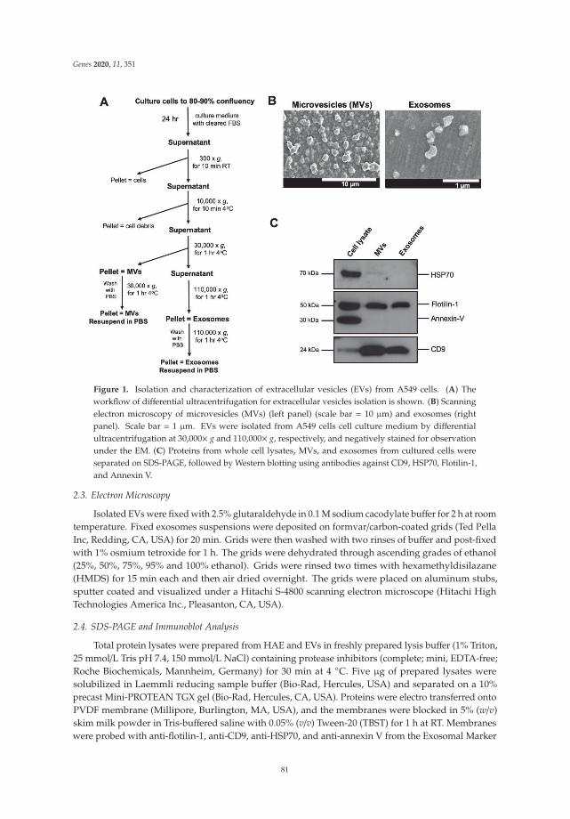
Genes 2020, 11, 351
Figure 1. Isolation and characterization of extracellular vesicles (EVs) from A549 cells. (A) Theworkflow of differential ultracentrifugation for extracellular vesicles isolation is shown. (B) Scanningelectron microscopy of microvesicles (MVs) (left panel) (scale bar = 10 μm) and exosomes (rightpanel). Scale bar = 1 μm. EVs were isolated from A549 cells cell culture medium by differentialultracentrifugation at 30,000× g and 110,000× g, respectively, and negatively stained for observationunder the EM. (C) Proteins from whole cell lysates, MVs, and exosomes from cultured cells wereseparated on SDS-PAGE, followed by Western blotting using antibodies against CD9, HSP70, Flotilin-1,and Annexin V.
2.3. Electron Microscopy
Isolated EVs were fixed with 2.5% glutaraldehyde in 0.1 M sodium cacodylate buffer for 2 h at roomtemperature. Fixed exosomes suspensions were deposited on formvar/carbon-coated grids (Ted PellaInc, Redding, CA, USA) for 20 min. Grids were then washed with two rinses of buffer and post-fixedwith 1% osmium tetroxide for 1 h. The grids were dehydrated through ascending grades of ethanol(25%, 50%, 75%, 95% and 100% ethanol). Grids were rinsed two times with hexamethyldisilazane(HMDS) for 15 min each and then air dried overnight. The grids were placed on aluminum stubs,sputter coated and visualized under a Hitachi S-4800 scanning electron microscope (Hitachi HighTechnologies America Inc., Pleasanton, CA, USA).
2.4. SDS-PAGE and Immunoblot Analysis
Total protein lysates were prepared from HAE and EVs in freshly prepared lysis buffer (1% Triton,25 mmol/L Tris pH 7.4, 150 mmol/L NaCl) containing protease inhibitors (complete; mini, EDTA-free;Roche Biochemicals, Mannheim, Germany) for 30 min at 4 ◦C. Five μg of prepared lysates weresolubilized in Laemmli reducing sample buffer (Bio-Rad, Hercules, USA) and separated on a 10%precast Mini-PROTEAN TGX gel (Bio-Rad, Hercules, CA, USA). Proteins were electro transferred ontoPVDF membrane (Millipore, Burlington, MA, USA), and the membranes were blocked in 5% (w/v)skim milk powder in Tris-buffered saline with 0.05% (v/v) Tween-20 (TBST) for 1 h at RT. Membraneswere probed with anti-flotilin-1, anti-CD9, anti-HSP70, and anti-annexin V from the Exosomal Marker
81
Genes 2020, 11, 351
Antibody Sampler Kit (Cell Signaling Technology, Danvers, MA, USA) diluted in TBST for 1 h.Blots were rinsed and incubated in horseradish peroxidase-conjugated goat anti-sheep IgG (Bio-Rad,Hercules, MA, USA; 1:10,000) or goat anti-mouse IgG (Bio-Rad, Hercules, MA, USA; 1:10,000) for 1 h atRT with shaking. Antibody staining was visualized by chemiluminescence (ECL Plus Western blottingdetection reagents, GE Healthcare, Pittsburgh, PA, USA).
2.5. Dicer-Substrate Short Interfering RNA (DsiRNA) Oligonucleotides
The 27-mer DsiRNAs and digoxigenin (DIG)-labeled DsiRNA were designed and synthesizedby Integrated DNA Technologies (IDT, Coralville, IA, USA) as described earlier [33–35]. The DIGlabel was internally coupled to an amino-dT base in a 2-O′ methyl modified DsiRNA againsthypoxanthine-guanine phosphoribosyltransferase (HPRT). The sequences of DsiRNAs used in thestudy are provided in Table 1.
Table 1. Sequences of DsiRNA used in the study. DNA bases are in bold and 2 O’ methyl bases areunderlined. Amino dT bases coupled to DIG are in italics.
DsiRNA Target mRNA DsiRNA Target Sequence
hHPRT human HPRT 5′ pGCCAGACUUUGUUGGAUUUGAATT
3′ UCGGUCUGAAACAACCUAAACUUUAADIG-HPRT human HPRT 5′ pCCAGUAAAGUUATCACAUGUUCUAG
3′ GUGGUCAUUUCAAUAGUGUACAAGAUCScrambled Not applicable 5′ pCGUUAAUCGCGUAUAAUACGCGUAT
3′ CAGCAAUUAGCGCAUAUUAUGCCGCAUA
2.6. Loading of DsiRNA into Exosomes
Exosomes were loaded with DsiRNA or DIG-labelled DsiRNA by electroporation as previouslydescribed [36]. Briefly, exosomes and DsiRNA were mixed in a 1:1 ratio (wt/wt) in electroporationbuffer to a final concentration of 250 ng/μL. The exosome-DsiRNA mixtures were electroporated in a400 μL volume using 0.4-mm cuvettes at 400 mV and 125 μF capacitance with pulse time of 10–15 ms.To remove non-electroporated DsiRNA, electroporated exosomes first treated with RNase and thenwashed in 1.5 mL of PBS, ultracentrifuged at 110,000× g for 70 min and re-suspended in 50 μL ofculture media.
2.7. Exosome Labeling
Exosomes were stained with 1X CellMask Deep Red Plasma Membrane stain (Thermo FisherScientific, Waltham, MA, USA) for 10 min at 37 ◦C. Unbound CellMask stain were then washed byultracentrifugation at 110,000× g for 70 min in TLA 100.3 rotor (Beckman Coulter, Indianapolis, IN,USA). Finally, exosomes were re-suspended in 100 μL of culture media and stored at −80 ◦C forfurther analysis.
2.8. Confocal Microscopy and Immunostaining
For confocal microscopy, HAE were fixed in 2% paraformaldehyde, permeabilized with 0.2%Triton-X-100 in Superblock (Thermo Fisher Scientific, Waltham, MA, USA), and then blocked in1× Superblock for 1 h at RT. F-actin was stained with either rhodamine-phalloidin (1:100, cat. no.R415, Thermo Fisher Scientific, Waltham, MA, USA) or Alexa Fluor 488-phalloidin (1:100, cat. no.A12379, Thermo Fisher Scientific, Waltham, MA, USA) for 30 min at RT. For immunostaining, HAEwere incubated with primary antibody against mouse anti-CFTR monoclonal antibody (769, CFFT)or anti-Digoxygenin (cat. no. 11333089001, Roche Biochemicals, Mannheim, Germany) overnight at4 ◦C. HAE were then incubated for secondary antibodies was Alexa 488-labeled goat anti-mouse orAlexa 488-labeled goat anti-sheep for 1 h at RT. HAE were mounted on a slide with Vectashield withDAPI (Vector Laboratories Inc., Burlingame, CA, USA). Representative images from three donors are
82
Genes 2020, 11, 351
shown. Z-stacks were acquired on a Leica TCS SP3 confocal microscope (Leica Microsystems Inc.,Buffalo Grove IL, USA) using a 40× or 63× oil-immersion objective.
2.9. Real-Time Quantitative PCR (RT-qPCR)
Total cellular RNA was isolated using Direct-zol™ RNA MiniPrep kit (Zymo Research, Irvine,CA, USA), according to the manufacturer’s protocol. The RNA concentration of samples wasquantified using ND-1000 spectrophotometer (Thermo Fisher Scientific, Waltham, USA). 0.5 μgof total RNA was reverse transcribed by a high-capacity reverse transcription kit (Thermo FisherScientific, Waltham, MA, USA) according to the manufacturer’s instructions. qRT-PCR for HPRTand SFRS9 mRNA were performed in an ABI Prism 7900 HT real-time PCR system (Thermo FisherScientific, Waltham, MA, USA). The PCR conditions were as follows: 95 ◦C for 10 min, 95 ◦C for 15 s,and 60 ◦C for 1 min for 40 cycles. The following primers were used in RT-qPCR analysis: For hHPRTAGGATTTGGAAAGGGTGTTTATTC (forward) and CCCATCTCCTTCATCACATCTC (reverse) andfor hSFRS9: TGCGTAAACTGGATGACACC (forward) and CCTGCTTTGGTATGGAGAGTC (reverse).The mRNA level of HPRT was calculated with normalization to SFRS9 using the 2−ΔΔCT method. Foldchange in expression are means of three technical triplicates from three human donors.
2.10. Isolation of CFTR- or mCherry-Loaded EVs and Delivery to HAE
To isolate EVs loaded with either mCherry or CFTR, A549 cells were transfected with an expressionplasmid or transduced with an adenovirus expressing mCherry (Ad5-mCherry) or CFTR (Ad5-CFTR)under the direction of a short 183-bp synthetic enhancer/promoter F5tg83 as previously described [37,38].Briefly, cells were grown to 70–80% confluency and transduced at MOI = 50 for 4 h. After transduction,cells were washed three times with PBS to wash away unbound viral particles. Fresh culture medium(DMEM supplemented with EV-cleared FBS) was added to the cells. After 48 h EVs were isolated asdescribed above.
To treat the airway epithelia with EVs apically, HAE were first treated apically with 0.1%lysophosphatidylcholine (LPC) in PBS for 2 h. HAE were rinsed three times with PBS before EVswere applied in a volume of 100 μL culture media for 4 h at 37 ◦C. EVs were removed and HAEwere incubated at 37 ◦C under 5% CO2 for the indicated times. In the case of basolateral application,the cultures were inverted, and the EVs were applied to the basolateral surface for 4 h in 80 μl ofculture media at 37 ◦C, 5% CO2. Following the treatment, the EVs were removed, and the cultureswere turned upright and incubated at 37 ◦C under 5% CO2 for the indicated times.
2.11. Ussing Chamber Studies of Well-Differentiated HAE
To measure change in anion channel activity, HAE were mounted into Ussing chambers asdescribed earlier [39]. The apical and basolateral chambers were filled with symmetrical Ringer’ssolution (135 mM NaCl, 5 mM HEPES, 0.6 mM KH2PO4, 2.4 mM K2HPO4, 1.2 mM MgCl2, 1.2 mMCaCl2, 5 mM dextrose). Dextrose was added to this solution immediately before the experiments.The protocol was performed as follows: treatment with amiloride (100 μM), followed by DIDS (100 μM),apical solution was replaced with a low Cl− solution, and CFTR Cl− current was measured as previouslydescribed [40].
3. Results
3.1. Isolation and Characterization of Extracellular Vesicles
EVs were isolated from culture media of HEK-293T or A549 cells. EVs were fractionated into twopopulations by differential ultracentrifugation. The first fraction, containing microvesicles (MVs), wasrecovered from the pellet after centrifugation at 30,000× g, and the second fraction, containing exosomes,was recovered from the pellet after centrifugation at 110,000× g (Figure 1A). We next characterizedthe two fractions for their morphology/size and protein surface markers (Figure 1B,C). As expected,
83

Genes 2020, 11, 351
the MV fraction consisted of large vesicles ranging from 300–1000 nm in diameter (Figure 1B, leftpanel), whereas the second exosome fraction contained smaller size vesicles ranging from 80–150 nm(Figure 1B, right panel). To further confirm the purity of our fractions, proteins from cell lysates,MVs, and exosomes were characterized by immunoblot analysis for EVs specific protein markers [41](Figure 1C). Tetraspanin-like CD9 and flotillin-1 were highly enriched in the EVs, whereas HSP70 andannexin V were not enriched in either the MV or exosome fractions. HSP70 was not detectable in theexosome fraction, but faintly present in the MV fraction, suggesting that large vesicles contain cellularproteins. This suggests the probable differences in biogenesis, where exosomes are secreted throughthe endosome pathway and would present endosome markers, whereas larger vesicles such as MVs areproduced by direct budding from the cell membrane and would contain cellular proteins or proteinsassociated with the plasma membrane. The absence of annexin V from the EV fractions confirms theabsence of apoptotic bodies in our EV preparations. Our results are consistent with previous reportsthat describe the protein composition in exosomes and MVs [19,28].
3.2. Exosomes are Internalized at the Basolateral Surface of HAE
For our initial studies, we asked whether EVs would be taken up by well-differentiated HAE andwhether there is a preference for the apical or basolateral surface. Our standard protocol requires theHAE cultures to be grown on polycarbonate Transwell inserts with a pore size of 400 nm. Becausethis pore size physically limits the access of MVs to the basolateral surface, we focused our efforts onexosomes which are smaller than the pore size of the Transwell insert. To evaluate the efficiency ofexosome uptake by HAE, exosomes derived from HEK-293T cells were labeled with CellMask DeepRed Plasma Membrane stain and applied to the apical (Figure 2A) or basolateral (Figure 2B) surface ofHAE for 4 h. At the indicated time points post-delivery, confocal microscopy was used to evaluateintracellular uptake of CellMask stain. Following apical exosome delivery, few (estimated <1%)CellMask positive cells were observed (Figure 2A). In contrast, following basolateral exosome delivery,we readily observed abundant CellMask positive HAE (Figure 2B). Exosome internalization wasobserved as early as 3 h after basolateral application. Regardless of apical or basolateral exosomedelivery, the CellMask stain was stable for at least 5 days after delivery and remained localized inperinuclear regions below the apical surface. However, the presence of CellMask is not an indication ofthe stability of internalized exosomes, only the internalized labeled lipid bilayers. These data suggestthat exosomes are an efficient delivery vehicle to HAE and that delivery is more efficient from thebasolateral surface than the apical surface.
84
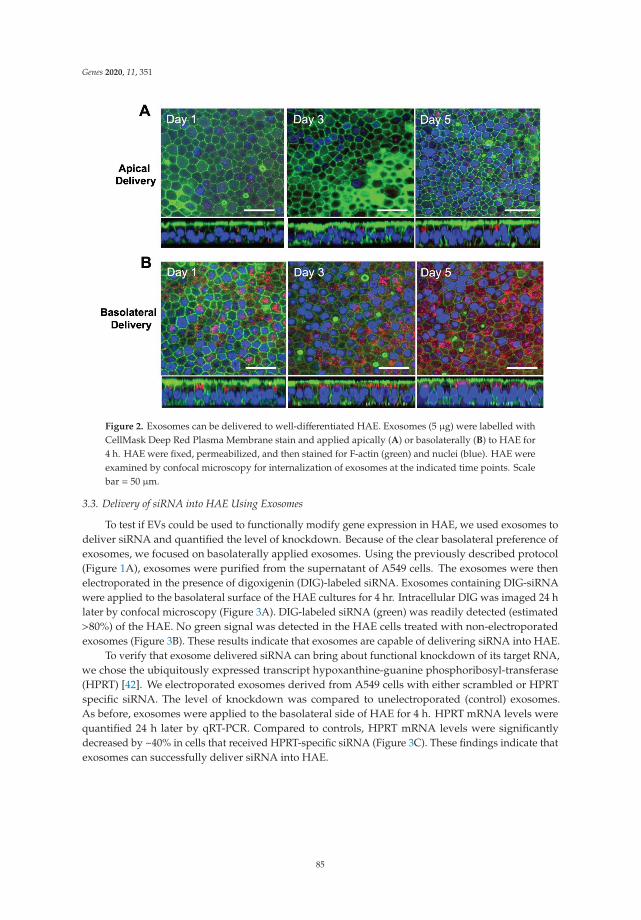
Genes 2020, 11, 351
Figure 2. Exosomes can be delivered to well-differentiated HAE. Exosomes (5 μg) were labelled withCellMask Deep Red Plasma Membrane stain and applied apically (A) or basolaterally (B) to HAE for4 h. HAE were fixed, permeabilized, and then stained for F-actin (green) and nuclei (blue). HAE wereexamined by confocal microscopy for internalization of exosomes at the indicated time points. Scalebar = 50 μm.
3.3. Delivery of siRNA into HAE Using Exosomes
To test if EVs could be used to functionally modify gene expression in HAE, we used exosomes todeliver siRNA and quantified the level of knockdown. Because of the clear basolateral preference ofexosomes, we focused on basolaterally applied exosomes. Using the previously described protocol(Figure 1A), exosomes were purified from the supernatant of A549 cells. The exosomes were thenelectroporated in the presence of digoxigenin (DIG)-labeled siRNA. Exosomes containing DIG-siRNAwere applied to the basolateral surface of the HAE cultures for 4 hr. Intracellular DIG was imaged 24 hlater by confocal microscopy (Figure 3A). DIG-labeled siRNA (green) was readily detected (estimated>80%) of the HAE. No green signal was detected in the HAE cells treated with non-electroporatedexosomes (Figure 3B). These results indicate that exosomes are capable of delivering siRNA into HAE.
To verify that exosome delivered siRNA can bring about functional knockdown of its target RNA,we chose the ubiquitously expressed transcript hypoxanthine-guanine phosphoribosyl-transferase(HPRT) [42]. We electroporated exosomes derived from A549 cells with either scrambled or HPRTspecific siRNA. The level of knockdown was compared to unelectroporated (control) exosomes.As before, exosomes were applied to the basolateral side of HAE for 4 h. HPRT mRNA levels werequantified 24 h later by qRT-PCR. Compared to controls, HPRT mRNA levels were significantlydecreased by ~40% in cells that received HPRT-specific siRNA (Figure 3C). These findings indicate thatexosomes can successfully deliver siRNA into HAE.
85
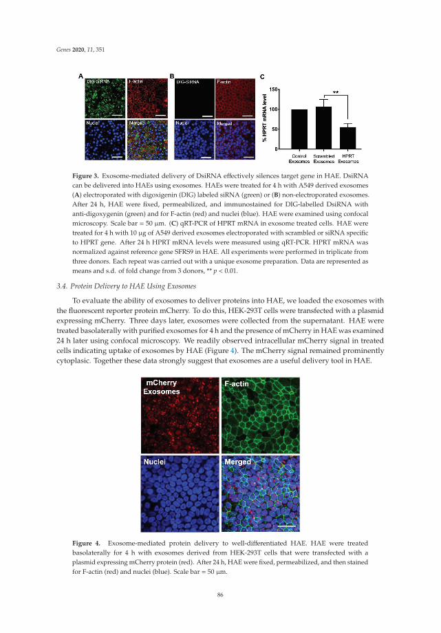
Genes 2020, 11, 351
Figure 3. Exosome-mediated delivery of DsiRNA effectively silences target gene in HAE. DsiRNAcan be delivered into HAEs using exosomes. HAEs were treated for 4 h with A549 derived exosomes(A) electroporated with digoxigenin (DIG) labeled siRNA (green) or (B) non-electroporated exosomes.After 24 h, HAE were fixed, permeabilized, and immunostained for DIG-labelled DsiRNA withanti-digoxygenin (green) and for F-actin (red) and nuclei (blue). HAE were examined using confocalmicroscopy. Scale bar = 50 μm. (C) qRT-PCR of HPRT mRNA in exosome treated cells. HAE weretreated for 4 h with 10 μg of A549 derived exosomes electroporated with scrambled or siRNA specificto HPRT gene. After 24 h HPRT mRNA levels were measured using qRT-PCR. HPRT mRNA wasnormalized against reference gene SFRS9 in HAE. All experiments were performed in triplicate fromthree donors. Each repeat was carried out with a unique exosome preparation. Data are represented asmeans and s.d. of fold change from 3 donors, ** p < 0.01.
3.4. Protein Delivery to HAE Using Exosomes
To evaluate the ability of exosomes to deliver proteins into HAE, we loaded the exosomes withthe fluorescent reporter protein mCherry. To do this, HEK-293T cells were transfected with a plasmidexpressing mCherry. Three days later, exosomes were collected from the supernatant. HAE weretreated basolaterally with purified exosomes for 4 h and the presence of mCherry in HAE was examined24 h later using confocal microscopy. We readily observed intracellular mCherry signal in treatedcells indicating uptake of exosomes by HAE (Figure 4). The mCherry signal remained prominentlycytoplasic. Together these data strongly suggest that exosomes are a useful delivery tool in HAE.
Figure 4. Exosome-mediated protein delivery to well-differentiated HAE. HAE were treatedbasolaterally for 4 h with exosomes derived from HEK-293T cells that were transfected with aplasmid expressing mCherry protein (red). After 24 h, HAE were fixed, permeabilized, and then stainedfor F-actin (red) and nuclei (blue). Scale bar = 50 μm.
86

Genes 2020, 11, 351
3.5. MV-Mediated Delivery of CFTR Protein Corrects Anion Defect in HAE from CF Donors
CFTR complementation restores the anion channel defect in well-differentiated human airwayepithelial cells derived from CF donors (HAECF) [43,44]. Typically, viral vectors are required to deliverthe CFTR cDNA. Here we test whether CFTR protein can be delivered to HAECF using either exosomesor MVs and functionally correct the Cl− channel defect. For these experiments, A549 cells were firsttransduced with Ad-vector (MOI = 50) expressing either mCherry or CFTR. MVs or exosomes werethen purified from the A549 supernatant one day later as described (Figure 1A).
Thus, far we focused our efforts on using exosomes to deliver siRNA or protein to HAE. Exosomesare typically 80–150 nm in diameter and will readily pass through the 400 nm pores of the Transwell insertson which HAE are grown. MVs range in size from 200 to 1000 nm. However, exosomes typically packageproteins <150 kDa; whereas, MVs can package proteins as large as ~300 kDa. The fully glycosylated,mature form of CFTR is ~250 kDa. Thus, for CFTR delivery, we sought to contrast exosomes and MVswhile allowing equivalent access to the basolateral surface. To achieve basolateral access with an apicalapplication, we pretreated HAE with 0.1% lysophosphatidylcholine (LPC). LPC is a natural airwaysurfactant that transiently opens tight junctions and allows access to the basolateral surface. LPC treatmentis a common strategy to improve transduction efficiencies of viral vectors, such as VSV-G pseudotypedlentivirus [45] or adenovirus [44], with preferences for the basolateral surface of polarized epithelia.
EVs (exosomes or MVs) loaded with mCherry were applied to the apical surface of LPC treated HAE.Intracellular localization of mCherry was examined 24 h later by confocal microscopy (Figure 5A,B).We observed equivalent levels of mCherry expression in HAE treated with MVs or exosomes. We nextloaded EVs with CFTR and applied them to the apical surface of LPC treated HAECF. Cells werefixed and immunohistochemisty was used to detect CFTR protein 24 h after EV application. In thiscase, CFTR protein was only detected following application of MVs but not exosomes (Figure 5C,D).In addition, the bioelectric properties of epithelia were analyzed in Ussing chambers. HAECF treatedwith CFTR MVs showed a significant increase in transepithelial Cl− current in response to forskolin and3-isobutyl-1-methylxanthine (F&I) and current was inhibited by the CFTR channel blocker, GlyH-101(Figure 5E) when compared to cells treated with either mCherry MVs, mCherry exosomes, or CFTRexosomes. These data demonstrated that MV-mediated apical delivery of CFTR protein to primaryairway epithelia can correct the anion channel defect in vitro as early as 24 h post treatment.
Figure 5. MV-mediated correction of chloride (Cl−) transport in cystic fibrosis (CF) human primaryepithelia. HAECF were treated apically with A549-derived EVs: (A) mCherry exosomes, (B) mCherryMVs, (C) CFTR exosomes, or (D) CFTR MVs for 4 h. HAECF were pretreated with 0.1% LPC for 2 hbefore EVs treatment. After 24 h, HAECF were fixed, permeabilized, and examined using confocalmicroscopy for mCherry (red) or immunostained for CFTR protein (green). Nuclei were stained withDAPI (blue). Scale bar = 100 μm. (E) Transepithelial Cl− current was measured in HAECF treated witheither mCherry-EVs or CFTR-EVs in Ussing chambers. Change in current was measured in response toforskolin and IBMX (F&I) and GlyH-101. Data are represented as means and s.d. of two HAE culturesfrom three donors, * p < 0.05.
87

Genes 2020, 11, 351
4. Discussion
HAE prepared from donor trachea and bronchi are grown at an air–liquid interface and form apseudostratified columnar epithelium with tight junctions. The cultures include multiple cell typessuch as ciliated cells, non-ciliated cells, goblet cells, and basal cells that recapitulate the surface cells ofthe conducting airways [2,46]. These cells provide an excellent model for studying airway cell biology.Indeed, HAE recapitulate many in vivo barriers to gene delivery. In the present study, we investigatedthe utility of extracellular vesicles (EVs) to deliver different cargos to this model system.
Manipulating genes (either by overexpression or knockdown) is a fundamental strategy forexamining gene function. However, the transfection efficiency in well-differentiated HAE isreproducibly below the limit of detection regardless of the transfection or electroporation reagent [7,9,47].The differentiation process correlates with a highly resistant transfection barrier within 5–6 days afterseeding; this prevents the entry of oligonucleotides no matter the formulation of the transfectionreagent [7,9,47]. Transfection is only possible when conducted at the time of seeding when the cellsare still poorly differentiated [9]. Transfecting siRNA into poorly differentiated airway cells leads toknockdown of target genes; however, this strategy has its limitations because poorly differentiated HAEare simply less representative of the in vivo airways. In the time it takes the epithelia to differentiate(3–4 weeks), the effect of the siRNA gene knockdown will diminish. The established technique forachieving efficient gene transfer in well-differentiated HAE is to use viral vectors such as adenovirus,lentivirus, or AAV. Viral vector can be time consuming and expensive when considering the effort toclone packaging plasmids as well as producing, purifying, and titering the vector.
Due to their small size (~30–100 nm), exosomes are considered nanoparticles. As a class,nanoparticles are used extensively to deliver small molecules, peptides, proteins, DNAs or siRNAs [48];however, many manufactured lipid-based nanoparticles have associated withxicity and trigger adaptiveand innate immune responses [49–52]. Exosomes are naturally adapted to transmit molecular messagesbetween cells without invoking immune responses and are considered a safe means to deliver smalltherapeutic agents such as siRNA and drugs to specific target tissues in a non-cytotoxic manner [53].Several studies demonstrate that an exosome-based delivery system can deliver its cargo to multiplecell types in vitro and in vivo. Exosomes offer significant advantages over synthetic drug deliverysystems including enhanced serum stability, low immunogenicity, and minimal clearance by lung, liver,and spleen [29,53–55] and have reproducibly been used in vivo to deliver siRNAs or miRNAs [21,56–58].In addition, exosomes will deliver other therapeutic agents, such as doxorubicin [59], STAT-3 inhibitorJSI-124 (cucurbitacinI) [60], cytosine deaminase (CD) fused with uracil phosphorybosyltransferase(UPRT) [61], curcumin [62], and catalase (exoCAT) [63]. Thus, exosomes are an effective delivery toolfor multiple materials into multiple cell types.
Our results suggest that exosomes are readily taken up by the cells in well-differentiated HAE andthey can efficiently deliver siRNA. We foresee many applications of this delivery system. For example,siRNAs or proteins that affect the levels of CFTR could be used to screen gene modulators, such asSIN3A, DERL1, ARF4, CDH1, et al. In future studies we aim to push the levels of knockdown furtherby expressing lung epithelial specific peptides (GFE-1 and GFE-2) [64] on the surface of exosomesand/or use a pH-sensitive fusogenic peptide [65] for efficient delivery of the cargo into HAE.
MVs are the class of EVs that are largest in size, and which therefore carry large protein cargo likeCFTR and laminin proteins [66]. In this study, we showed MV-mediated delivery of CFTR protein tocorrect the transepithelial Cl− current in well-differentiated HAE from CF donors. We used both MVsand exosomes to deliver the CFTR protein, but only MVs were capable of delivering functional CFTRprotein to HAECF. Based on the known size constraints of exosomes, we speculate that the large CFTRprotein was more efficiently packaged in MVs. However, the mechanisms of how EVs are differentiallyproduced, released, fused in target cells all may be important for function of a membrane boundchannel protein. MVs may be more suitable for delivery of any cell surface channel protein regardlessof the size. The mechanism of CFTR incorporation is the subject of future studies and will include
88

Genes 2020, 11, 351
western blot and super resolution microscopy analysis. In addition, for our studies we primarily usedMVs and exosomes derived from A549 cells. Different EV donor cell lines may lead to different results.
A limitation of EVs purified from cultured cells is the presence of unintentional cellular proteincontamination. In general, MVs are more promiscuous for inclusion of cellular proteins than exosomes.In this study, we did not test for the presence of unintentional proteins or nucleotides from the donorcells. However, EV mediated transfer of DNA fragments are shown to be unstable in target cells [29].
We and others have observed that some enveloped viruses, such as measles virus, have a strongpreference for basolateral entry into HAE [67]. However, other viruses, such as Rous Sarcoma Virus,preferentially enter the apical surface. Currently it is unclear why EVs have a strong preference forentry at the basolateral surface. We speculate that proteins necessary for EV entry may be basolaterallylocalized. EVs are likely to play an important role in cell-to-cell communications in lung biology anddisease. The lung is a unique organ with a multitude of regionally segregated epithelial cell types;as well as, endothelial and resident immune cells. Exosomes derived from airway cells play a rolein innate defense and remodeling [68,69]; however, other cells (such as resident macrophages andneutrophils) are also a significant source of exosomes in the lung. Exchange of EV cargos betweenthese cells can alter gene expression and may reorganize the airway epithelium. It is currently unclearwhy we observed a clear entry preference for EVs at the basolateral surface of HAE; however, this mayrepresent a bona fide region of interface.
In summary, our study showed the potential use of EVs in manipulating the gene expressionin an important in vitro airway model system. Exosomes effectively deliver siRNAs to modulateendogenous gene expression, whereas MVs can be used to deliver large protein cargos. There aremany potential applications of this technology in HAE, including: screening of siRNAs and miRNAsto modulate gene expression, delivering sgRNA libraries for screening genetic phenotypes, generatingmodels for rare CFTR mutations, and delivering 100–150 bp single-stranded DNA oligonucleotides.
Author Contributions: Conceptualization, B.K.S. and P.L.S.; methodology, B.K.S., A.L.C. and S.K.; formalanalysis: B.K.S., A.L.C. and S.K.; data curation, B.K.S., A.L.C. and S.K.; writing—original draft preparation, B.K.S.;writing—review and editing, B.K.S., A.L.C. and P.L.S.; supervision, B.K.S. and P.L.S.; project administration, B.K.S.and P.L.S.; funding acquisition, B.K.S. and P.L.S. All authors have read and agreed to the published version ofthe manuscript.
Funding: This work was supported by the Cystic Fibrosis Foundation (SINN15XX1, SINN15XX0, and SINN19XX0)and National Institutes of Health (NIH R01 HL-133089 and NIH R01 AI-132402).
Acknowledgments: We thank Jennifer Bartlett and Miguel Ortiz for critical review of the manuscript.We acknowledge the support of Central Microscopy Research Facility (CMRF), the In Vitro Models and CellCulture Core, and the Viral Vector Core of The University of Iowa.
Conflicts of Interest: The authors have no financial conflicts of interest. The funders of this study had no role inthe design of the study; in the collection, analyses, or interpretation of data; in the writing of the manuscript, or inthe decision to publish the results.
References
1. Fulcher, M.L.; Gabriel, S.; Burns, K.A.; Yankaskas, J.R.; Randell, S.H. Well-differentiated human airwayepithelial cell cultures. Methods Mol. Med. 2005, 107, 183–206. [PubMed]
2. Karp, P.H.; Moninger, T.O.; Weber, S.P.; Nesselhauf, T.S.; Launspach, J.L.; Zabner, J.; Welsh, M.J. An in vitromodel of differentiated human airway epithelia. Methods for establishing primary cultures. Methods Mol. Biol.2002, 188, 115–137. [PubMed]
3. Woodworth, B.A.; Antunes, M.B.; Bhargave, G.; Palmer, J.N.; Cohen, N.A. Murine tracheal and nasal septalepithelium for air-liquid interface cultures: A comparative study. Am. J. Rhinol. 2007, 21, 533–537. [CrossRef][PubMed]
4. Zabner, J.; Zeiher, B.G.; Friedman, E.; Welsh, M.J. Adenovirus-mediated gene transfer to ciliated airwayepithelia requires prolonged incubation time. J. Virol. 1996, 70, 6994–7003. [CrossRef] [PubMed]
89
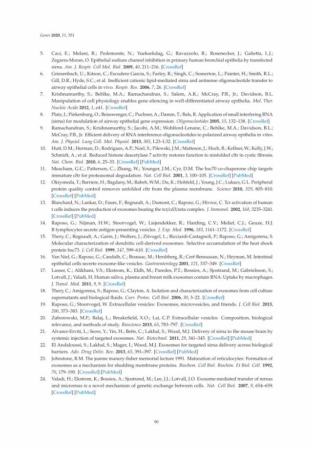
Genes 2020, 11, 351
5. Caci, E.; Melani, R.; Pedemonte, N.; Yueksekdag, G.; Ravazzolo, R.; Rosenecker, J.; Galietta, L.J.;Zegarra-Moran, O. Epithelial sodium channel inhibition in primary human bronchial epithelia by transfectedsirna. Am. J. Respir. Cell Mol. Biol. 2009, 40, 211–216. [CrossRef]
6. Griesenbach, U.; Kitson, C.; Escudero Garcia, S.; Farley, R.; Singh, C.; Somerton, L.; Painter, H.; Smith, R.L.;Gill, D.R.; Hyde, S.C.; et al. Inefficient cationic lipid-mediated sirna and antisense oligonucleotide transfer toairway epithelial cells in vivo. Respir. Res. 2006, 7, 26. [CrossRef]
7. Krishnamurthy, S.; Behlke, M.A.; Ramachandran, S.; Salem, A.K.; McCray, P.B., Jr.; Davidson, B.L.Manipulation of cell physiology enables gene silencing in well-differentiated airway epithelia. Mol. Ther.Nucleic Acids 2012, 1, e41. [CrossRef]
8. Platz, J.; Pinkenburg, O.; Beisswenger, C.; Puchner, A.; Damm, T.; Bals, R. Application of small interfering RNA(sirna) for modulation of airway epithelial gene expression. Oligonucleotides 2005, 15, 132–138. [CrossRef]
9. Ramachandran, S.; Krishnamurthy, S.; Jacobi, A.M.; Wohlford-Lenane, C.; Behlke, M.A.; Davidson, B.L.;McCray, P.B., Jr. Efficient delivery of RNA interference oligonucleotides to polarized airway epithelia in vitro.Am. J. Physiol. Lung Cell. Mol. Physiol. 2013, 305, L23–L32. [CrossRef]
10. Hutt, D.M.; Herman, D.; Rodrigues, A.P.; Noel, S.; Pilewski, J.M.; Matteson, J.; Hoch, B.; Kellner, W.; Kelly, J.W.;Schmidt, A.; et al. Reduced histone deacetylase 7 activity restores function to misfolded cftr in cystic fibrosis.Nat. Chem. Biol. 2010, 6, 25–33. [CrossRef] [PubMed]
11. Meacham, G.C.; Patterson, C.; Zhang, W.; Younger, J.M.; Cyr, D.M. The hsc70 co-chaperone chip targetsimmature cftr for proteasomal degradation. Nat. Cell Biol. 2001, 3, 100–105. [CrossRef] [PubMed]
12. Okiyoneda, T.; Barriere, H.; Bagdany, M.; Rabeh, W.M.; Du, K.; Hohfeld, J.; Young, J.C.; Lukacs, G.L. Peripheralprotein quality control removes unfolded cftr from the plasma membrane. Science 2010, 329, 805–810.[CrossRef] [PubMed]
13. Blanchard, N.; Lankar, D.; Faure, F.; Regnault, A.; Dumont, C.; Raposo, G.; Hivroz, C. Tcr activation of humant cells induces the production of exosomes bearing the tcr/cd3/zeta complex. J. Immunol. 2002, 168, 3235–3241.[CrossRef] [PubMed]
14. Raposo, G.; Nijman, H.W.; Stoorvogel, W.; Liejendekker, R.; Harding, C.V.; Melief, C.J.; Geuze, H.J.B lymphocytes secrete antigen-presenting vesicles. J. Exp. Med. 1996, 183, 1161–1172. [CrossRef]
15. Thery, C.; Regnault, A.; Garin, J.; Wolfers, J.; Zitvogel, L.; Ricciardi-Castagnoli, P.; Raposo, G.; Amigorena, S.Molecular characterization of dendritic cell-derived exosomes. Selective accumulation of the heat shockprotein hsc73. J. Cell Biol. 1999, 147, 599–610. [CrossRef]
16. Van Niel, G.; Raposo, G.; Candalh, C.; Boussac, M.; Hershberg, R.; Cerf-Bensussan, N.; Heyman, M. Intestinalepithelial cells secrete exosome-like vesicles. Gastroenterology 2001, 121, 337–349. [CrossRef]
17. Lasser, C.; Alikhani, V.S.; Ekstrom, K.; Eldh, M.; Paredes, P.T.; Bossios, A.; Sjostrand, M.; Gabrielsson, S.;Lotvall, J.; Valadi, H. Human saliva, plasma and breast milk exosomes contain RNA: Uptake by macrophages.J. Transl. Med. 2011, 9, 9. [CrossRef]
18. Thery, C.; Amigorena, S.; Raposo, G.; Clayton, A. Isolation and characterization of exosomes from cell culturesupernatants and biological fluids. Curr. Protoc. Cell Biol. 2006, 30, 3–22. [CrossRef]
19. Raposo, G.; Stoorvogel, W. Extracellular vesicles: Exosomes, microvesicles, and friends. J. Cell Biol. 2013,200, 373–383. [CrossRef]
20. Zaborowski, M.P.; Balaj, L.; Breakefield, X.O.; Lai, C.P. Extracellular vesicles: Composition, biologicalrelevance, and methods of study. Bioscience 2015, 65, 783–797. [CrossRef]
21. Alvarez-Erviti, L.; Seow, Y.; Yin, H.; Betts, C.; Lakhal, S.; Wood, M.J. Delivery of sirna to the mouse brain bysystemic injection of targeted exosomes. Nat. Biotechnol. 2011, 29, 341–345. [CrossRef] [PubMed]
22. El Andaloussi, S.; Lakhal, S.; Mager, I.; Wood, M.J. Exosomes for targeted sirna delivery across biologicalbarriers. Adv. Drug Deliv. Rev. 2013, 65, 391–397. [CrossRef] [PubMed]
23. Johnstone, R.M. The jeanne manery-fisher memorial lecture 1991. Maturation of reticulocytes: Formation ofexosomes as a mechanism for shedding membrane proteins. Biochem. Cell Biol. Biochim. Et Biol. Cell. 1992,70, 179–190. [CrossRef] [PubMed]
24. Valadi, H.; Ekstrom, K.; Bossios, A.; Sjostrand, M.; Lee, J.J.; Lotvall, J.O. Exosome-mediated transfer of mrnasand micrornas is a novel mechanism of genetic exchange between cells. Nat. Cell Biol. 2007, 9, 654–659.[CrossRef] [PubMed]
90

Genes 2020, 11, 351
25. Vlassov, A.V.; Magdaleno, S.; Setterquist, R.; Conrad, R. Exosomes: Current knowledge of their composition,biological functions, and diagnostic and therapeutic potentials. Biochim. Et Biophys. Acta 2012, 1820, 940–948.[CrossRef]
26. Zomer, A.; Vendrig, T.; Hopmans, E.S.; van Eijndhoven, M.; Middeldorp, J.M.; Pegtel, D.M. Exosomes: Fit todeliver small RNA. Commun. Integr. Biol. 2010, 3, 447–450. [CrossRef]
27. Thery, C.; Ostrowski, M.; Segura, E. Membrane vesicles as conveyors of immune responses. Nat. Rev. Immunol.2009, 9, 581–593. [CrossRef]
28. Doyle, L.M.; Wang, M.Z. Overview of extracellular vesicles, their origin, composition, purpose, and methodsfor exosome isolation and analysis. Cells 2019, 8, 727. [CrossRef]
29. Kanada, M.; Bachmann, M.H.; Hardy, J.W.; Frimannson, D.O.; Bronsart, L.; Wang, A.; Sylvester, M.D.;Schmidt, T.L.; Kaspar, R.L.; Butte, M.J.; et al. Differential fates of biomolecules delivered to target cells viaextracellular vesicles. Proc. Natl. Acad. Sci. USA 2015, 112, E1433–E1442. [CrossRef]
30. Sun, D.; Zhuang, X.; Zhang, S.; Deng, Z.B.; Grizzle, W.; Miller, D.; Zhang, H.G. Exosomes are endogenousnanoparticles that can deliver biological information between cells. Adv. Drug Deliv. Rev. 2013, 65, 342–347.[CrossRef] [PubMed]
31. Kalluri, R.; LeBleu, V.S. The biology, function, and biomedical applications of exosomes. Science 2020, 367.[CrossRef] [PubMed]
32. Konoshenko, M.Y.; Lekchnov, E.A.; Vlassov, A.V.; Laktionov, P.P. Isolation of extracellular vesicles: Generalmethodologies and latest trends. Biomed. Res. Int. 2018, 2018, 8545347. [CrossRef] [PubMed]
33. Reynolds, A.; Leake, D.; Boese, Q.; Scaringe, S.; Marshall, W.S.; Khvorova, A. Rational sirna design for RNAinterference. Nat. Biotechnol. 2004, 22, 326–330. [CrossRef]
34. Amarzguioui, M.; Lundberg, P.; Cantin, E.; Hagstrom, J.; Behlke, M.A.; Rossi, J.J. Rational design and in vitroand in vivo delivery of dicer substrate sirna. Nat. Protoc. 2006, 1, 508–517. [CrossRef] [PubMed]
35. Collingwood, M.A.; Rose, S.D.; Huang, L.; Hillier, C.; Amarzguioui, M.; Wiiger, M.T.; Soifer, H.S.; Rossi, J.J.;Behlke, M.A. Chemical modification patterns compatible with high potency dicer-substrate small interferingrnas. Oligonucleotides 2008, 18, 187–200. [CrossRef]
36. El-Andaloussi, S.; Lee, Y.; Lakhal-Littleton, S.; Li, J.; Seow, Y.; Gardiner, C.; Alvarez-Erviti, L.; Sargent, I.L.;Wood, M.J. Exosome-mediated delivery of sirna in vitro and in vivo. Nat. Protoc. 2012, 7, 2112–2126.[CrossRef]
37. Cooney, A.L.; Singh, B.K.; Sinn, P.L. Hybrid nonviral/viral vector systems for improved piggybac DNAtransposon in vivo delivery. Mol. Ther. J. Am. Soc. Gene Ther. 2015, 23, 667–674. [CrossRef]
38. Li, N.; Cooney, A.L.; Zhang, W.; Ehrhardt, A.; Sinn, P.L. Enhanced tropism of species b1 adenoviral-basedvectors for primary human airway epithelial cells. Mol. Methods Clin. Dev. 2019, 14, 228–236. [CrossRef]
39. Li, H.; Sheppard, D.N.; Hug, M.J. Transepithelial electrical measurements with the ussing chamber. J. Cyst.Fibros 2004, 3, 123–126. [CrossRef]
40. Cooney, A.L.; Abou Alaiwa, M.H.; Shah, V.S.; Bouzek, D.C.; Stroik, M.R.; Powers, L.S.; Gansemer, N.D.;Meyerholz, D.K.; Welsh, M.J.; Stoltz, D.A.; et al. Lentiviral-mediated phenotypic correction of cystic fibrosispigs. JCI Insight 2016, 1. [CrossRef]
41. Kowal, J.; Arras, G.; Colombo, M.; Jouve, M.; Morath, J.P.; Primdal-Bengtson, B.; Dingli, F.; Loew, D.;Tkach, M.; Thery, C. Proteomic comparison defines novel markers to characterize heterogeneous populationsof extracellular vesicle subtypes. Proc. Natl. Acad. Sci. USA 2016, 113, E968–E977. [CrossRef]
42. Stout, J.T.; Caskey, C.T. Hprt: Gene structure, expression, and mutation. Annu. Rev. Genet. 1985, 19, 127–148.[CrossRef]
43. Olsen, J.C.; Johnson, L.G.; Stutts, M.J.; Sarkadi, B.; Yankaskas, J.R.; Swanstrom, R.; Boucher, R.C. Correctionof the apical membrane chloride permeability defect in polarized cystic fibrosis airway epithelia followingretroviral-mediated gene transfer. Hum. Gene 1992, 3, 253–266. [CrossRef]
44. Cooney, A.L.; Singh, B.K.; Loza, L.M.; Thornell, I.M.; Hippee, C.E.; Powers, L.S.; Ostedgaard, L.S.;Meyerholz, D.K.; Wohlford-Lenane, C.; Stoltz, D.A.; et al. Widespread airway distribution and short-termphenotypic correction of cystic fibrosis pigs following aerosol delivery of piggybac/adenovirus. Nucleic AcidsRes. 2018, 46, 9591–9600. [CrossRef]
45. Sinn, P.L.; Cooney, A.L.; Oakland, M.; Dylla, D.E.; Wallen, T.J.; Pezzulo, A.A.; Chang, E.H.; McCray, P.B., Jr.Lentiviral vector gene transfer to porcine airways. Mol. Ther. Nucleic Acids 2012, 1, e56. [CrossRef]
91
Genes 2020, 11, 351
46. Pezzulo, A.A.; Starner, T.D.; Scheetz, T.E.; Traver, G.L.; Tilley, A.E.; Harvey, B.G.; Crystal, R.G.; McCray, P.B., Jr.;Zabner, J. The air-liquid interface and use of primary cell cultures are important to recapitulate thetranscriptional profile of in vivo airway epithelia. Am. J. Physiol. Lung Cell. Mol. Physiol. 2011, 300, L25–L31.[CrossRef]
47. Ramachandran, S.; Karp, P.H.; Jiang, P.; Ostedgaard, L.S.; Walz, A.E.; Fisher, J.T.; Keshavjee, S.; Lennox, K.A.;Jacobi, A.M.; Rose, S.D.; et al. A microrna network regulates expression and biosynthesis of wild-type anddeltaf508 mutant cystic fibrosis transmembrane conductance regulator. Proc. Natl. Acad. Sci. USA 2012,109, 13362–13367. [CrossRef]
48. Toumey, C. In the footsteps of biotech. Nat. Nanotechnol. 2010, 5, 475. [CrossRef]49. Bakand, S.; Hayes, A.; Dechsakulthorn, F. Nanoparticles: A review of particle toxicology following inhalation
exposure. Inhal. Toxicol. 2012, 24, 125–135. [CrossRef]50. Dobrovolskaia, M.A.; Aggarwal, P.; Hall, J.B.; McNeil, S.E. Preclinical studies to understand nanoparticle
interaction with the immune system and its potential effects on nanoparticle biodistribution. Mol. Pharm.2008, 5, 487–495. [CrossRef]
51. Iavicoli, I.; Leso, V.; Fontana, L.; Bergamaschi, A. Toxicological effects of titanium dioxide nanoparticles:A review of in vitro mammalian studies. Eur. Rev. Med Pharmacol. Sci. 2011, 15, 481–508. [PubMed]
52. Maynard, A.D. Don’t define nanomaterials. Nature 2011, 475, 31. [CrossRef] [PubMed]53. Johnsen, K.B.; Gudbergsson, J.M.; Skov, M.N.; Pilgaard, L.; Moos, T.; Duroux, M. A comprehensive overview
of exosomes as drug delivery vehicles—Endogenous nanocarriers for targeted cancer therapy. Biochim. EtBiophys. Acta 2014, 1846, 75–87. [CrossRef] [PubMed]
54. Chaput, N.; Thery, C. Exosomes: Immune properties and potential clinical implementations. Semin.Immunopathol. 2011, 33, 419–440. [CrossRef]
55. Marcus, M.E.; Leonard, J.N. Fedexosomes: Engineering therapeutic biological nanoparticles that truly deliver.Pharmaceuticals 2013, 6, 659–680. [CrossRef]
56. Chen, T.S.; Lim, S.K. Measurement of precursor mirna in exosomes from human esc-derived mesenchymalstem cells. Methods Mol. Biol. 2013, 1024, 69–86.
57. Munoz, J.L.; Bliss, S.A.; Greco, S.J.; Ramkissoon, S.H.; Ligon, K.L.; Rameshwar, P. Delivery of functionalanti-mir-9 by mesenchymal stem cell-derived exosomes to glioblastoma multiforme cells conferredchemosensitivity. Mol. Ther. Nucleic Acids 2013, 2, e126. [CrossRef]
58. Wahlgren, J.; De, L.K.T.; Brisslert, M.; Vaziri Sani, F.; Telemo, E.; Sunnerhagen, P.; Valadi, H. Plasma exosomescan deliver exogenous short interfering RNA to monocytes and lymphocytes. Nucleic Acids Res. 2012,40, e130. [CrossRef]
59. Tian, Y.; Li, S.; Song, J.; Ji, T.; Zhu, M.; Anderson, G.J.; Wei, J.; Nie, G. A doxorubicin delivery platform usingengineered natural membrane vesicle exosomes for targeted tumor therapy. Biomaterials 2014, 35, 2383–2390.[CrossRef]
60. Zhuang, X.; Xiang, X.; Grizzle, W.; Sun, D.; Zhang, S.; Axtell, R.C.; Ju, S.; Mu, J.; Zhang, L.; Steinman, L.; et al.Treatment of brain inflammatory diseases by delivering exosome encapsulated anti-inflammatory drugsfrom the nasal region to the brain. Mol. Ther. J. Am. Soc. Gene Ther. 2011, 19, 1769–1779. [CrossRef]
61. Mizrak, A.; Bolukbasi, M.F.; Ozdener, G.B.; Brenner, G.J.; Madlener, S.; Erkan, E.P.; Strobel, T.; Breakefield, X.O.;Saydam, O. Genetically engineered microvesicles carrying suicide mrna/protein inhibit schwannoma tumorgrowth. Mol. Ther. J. Am. Soc. Gene Ther. 2013, 21, 101–108. [CrossRef] [PubMed]
62. Sun, D.; Zhuang, X.; Xiang, X.; Liu, Y.; Zhang, S.; Liu, C.; Barnes, S.; Grizzle, W.; Miller, D.; Zhang, H.G.A novel nanoparticle drug delivery system: The anti-inflammatory activity of curcumin is enhanced whenencapsulated in exosomes. Mol. Ther. J. Am. Soc. Gene Ther. 2010, 18, 1606–1614. [CrossRef] [PubMed]
63. Haney, M.J.; Klyachko, N.L.; Zhao, Y.; Gupta, R.; Plotnikova, E.G.; He, Z.; Patel, T.; Piroyan, A.; Sokolsky, M.;Kabanov, A.V.; et al. Exosomes as drug delivery vehicles for parkinson’s disease therapy. J. Control. Release2015, 207, 18–30. [CrossRef] [PubMed]
64. Laakkonen, P.; Vuorinen, K. Homing peptides as targeted delivery vehicles. Integr. Biol. Quant. Biosci.Nano Macro 2010, 2, 326–337. [CrossRef]
65. Nakase, I.; Futaki, S. Combined treatment with a ph-sensitive fusogenic peptide and cationic lipids achievesenhanced cytosolic delivery of exosomes. Sci. Rep. 2015, 5, 10112. [CrossRef]
92

Genes 2020, 11, 351
66. Pathan, M.; Fonseka, P.; Chitti, S.V.; Kang, T.; Sanwlani, R.; Van Deun, J.; Hendrix, A.; Mathivanan, S.Vesiclepedia 2019: A compendium of RNA, proteins, lipids and metabolites in extracellular vesicles.Nucleic Acids Res. 2019, 47, D516–D519. [CrossRef]
67. Sinn, P.; Williams, G.; Vongpunsawad, S.; Cattaneo, R.; McCray, P. Measles virus preferentially transduces thebasolateral surface of well-differentiated human airway epithelia. J. Virol. 2002, 76, 2403–2409. [CrossRef]
68. Gupta, R.; Radicioni, G.; Abdelwahab, S.; Dang, H.; Carpenter, J.; Chua, M.; Mieczkowski, P.A.; Sheridan, J.T.;Randell, S.H.; Kesimer, M. Intercellular communication between airway epithelial cells is mediated byexosome-like vesicles. Am. J. Respir. Cell Mol. Biol. 2019, 60, 209–220. [CrossRef]
69. Kesimer, M.; Scull, M.; Brighton, B.; DeMaria, G.; Burns, K.; O’Neal, W.; Pickles, R.J.; Sheehan, J.K.Characterization of exosome-like vesicles released from human tracheobronchial ciliated epithelium:A possible role in innate defense. FASEB J. 2009, 23, 1858–1868. [CrossRef]
© 2020 by the authors. Licensee MDPI, Basel, Switzerland. This article is an open accessarticle distributed under the terms and conditions of the Creative Commons Attribution(CC BY) license (http://creativecommons.org/licenses/by/4.0/).
93


genesG C A T
T A C G
G C A T
Article
High Prevalence of Staphylococcus aureus EnterotoxinGene Cluster Superantigens in Cystic FibrosisClinical Isolates
Anthony J. Fischer 1,*, Samuel H. Kilgore 2, Sachinkumar B. Singh 1, Patrick D. Allen 1,
Alexis R. Hansen 1, Dominique H. Limoli 2 and Patrick M. Schlievert 2
1 Stead Family Department of Pediatrics, University of Iowa Carver College of Medicine, Iowa City, IA 52242,USA; [email protected] (S.B.S.); [email protected] (P.D.A.);[email protected] (A.R.H.)
2 Department of Microbiology and Immunology, University of Iowa Carver College of Medicine, Iowa City,IA 52242, USA; [email protected] (S.H.K.); [email protected] (D.H.L.);[email protected] (P.M.S.)
* Correspondence: [email protected]
Received: 7 November 2019; Accepted: 9 December 2019; Published: 12 December 2019
Abstract: Background: Staphylococcus aureus is a highly prevalent respiratory pathogen in cystic fibrosis(CF). It is unclear how this organism establishes chronic infections in CF airways. We hypothesizedthat S. aureus isolates from patients with CF would share common virulence properties that enablechronic infection. Methods: 77 S. aureus isolates were obtained from 45 de-identified patients withCF at the University of Iowa. We assessed isolates phenotypically and used genotyping assays todetermine the presence or absence of 18 superantigens (SAgs). Results: We observed phenotypicdiversity among S. aureus isolates from patients with CF. Genotypic analysis for SAgs revealed 79.8%of CF clinical isolates carried all six members of the enterotoxin gene cluster (EGC). MRSA and MSSAisolates had similar prevalence of SAgs. We additionally observed that EGC SAgs were prevalent inS. aureus isolated from two geographically distinct CF centers. Conclusions: S. aureus SAgs belongingto the EGC are highly prevalent in CF clinical isolates. The greater prevalence in these SAgs in CFairway specimens compared to skin isolates suggests that these toxins confer selective advantage inthe CF airway.
Keywords: cystic fibrosis; Staphylococcus aureus; superantigen; enterotoxin gene cluster; MRSA
1. Introduction
Cystic fibrosis (CF) is a common lethal genetic disease, which results in chronic airway infections,irreversible bronchiectasis, and respiratory failure. Staphylococcus aureus is the most prevalent bacterialpathogen in children with CF [1], and is present in ≈70% of all individuals with CF in the UnitedStates. Although Pseudomonas aeruginosa is the predominant pathogen in older patients, S. aureus is themost common bacterial species in patients with CF under age 24 [2]. Unlike P. aeruginosa, which hasmultiple effective treatments to eradicate early infections and control chronic infections [3–5], S. aureusinfections can be difficult to control with antibiotics [6]. CFTR modulator drugs may help preventincident infections with S. aureus, but they are unlikely to eliminate chronic S. aureus infections [7,8].Understanding how S. aureus infects and persists in the CF airway is critically important, as theseinfections may increase the risk of subsequent disease progression [9,10].
We hypothesized that S. aureus isolates in the CF airway would share common virulence properties.Some readily visible phenotypes such as hemolysis, pigmentation, and protease secretion could enableS. aureus to elude host defenses. People with CF are commonly treated with antibiotics; resistance
Genes 2019, 10, 1036; doi:10.3390/genes10121036 www.mdpi.com/journal/genes95

Genes 2019, 10, 1036
to antibiotics may be occur under the selective pressure of antibiotic exposure. Another potentialmechanism enabling S. aureus to establish chronic infection is the secretion of toxins that misdirectthe immune response. S. aureus produces a large number of secreted toxins that may be criticalfor establishing infections [11]. These include 18 unique superantigens (SAgs), secreted toxins thatbind both the T cell receptor and major histocompatibility complex molecules on antigen presentingcells [12,13].
Some SAgs are well known for their roles in acute infection. In the extreme example of toxic shocksyndrome, the SAg TSST-1 cross-links T cells and antigen presenting cells, stimulating massive cytokinerelease and blocking the immune system from developing lasting immunity [12,13]. By contrast,the enterotoxin gene cluster (EGC), an element encoding six staphylococcal enterotoxin (SE) andSE-like SAgs G, I, M, N, O, and U, is generally associated with long term mucosal colonization.The EGC is present in between 50% to 70% of isolates from individuals with nasal carriage of S.aureus [14,15]. These EGC toxins can stimulate T cell proliferation [16], yet neutralizing antibodyresponse to these toxins is surprisingly poor [17]. While these toxins have been associated withasymptomatic colonization, experimental studies in rabbits show that EGC SAgs may play crucial forinfections such as endocarditis [18].
It is not clear what role SAgs play in CF respiratory infections. EGC SAgs are prevalent in clinicalisolates of S. aureus; a recent European study showed that 57% of CF isolates harbored at least onegene belonging to the EGC [19]. This is similar to the prevalence of EGC in isolates from a cohort ofpatients with atopic dermatitis in the United States and Europe [20,21]. Our study had two goals: Todetermine whether these SAgs are as prevalent in CF isolates in the United States, and to determine ifthe superantigens were associated with methicillin resistance, which is common in the United Statesand has been linked to worse outcomes [10,22].
2. Materials and Methods
Ethics Statements: All bacterial isolates examined in this study were de-identified when theywere supplied to the research team. The University of Iowa Institutional Review Board (IRB)approved specimen collection after obtaining informed consent under approval numbers 200311016and 200803708.
Sources of Clinical Isolates: University of Iowa Cystic Fibrosis Biobank. 77 de-identified S. aureusclinical isolates were obtained from the University of Iowa Hospitals and Clinics clinical laboratoryfollowing CF clinic visits made between 12 December 2011 and 20 July 2012. Specimens were obtainedfrom both adult and pediatric patients.
CF Biospecimen Registry (CFBR) at Emory and Children’s Center for Cystic Fibrosis: 20 S. aureus isolatesfrom people with CF were collected between 1 January 2012 and 31 December 2013. These humansubject samples were provided by the CF Biospecimen Registry at the Children’s Healthcare of Atlantaand Emory University CF Discovery Core courtesy of Arlene Stecenko. The Emory University IRB hasapproved collecting and banking of these specimens after obtaining informed consent.
The Geisel School of Medicine at Dartmouth University: The Hogan laboratory at the Geisel Schoolof Medicine at Dartmouth University graciously provided 12 deidentified S. aureus isolates fromthe Dartmouth CF Translational Research Core. These isolates were obtained from adult patientswith CF between 1 January 2015 and 31 December 2017 with support by the CF Foundation RDPgrant STANTO15R0.
Bacterial Phenotypes: Clinical isolates of S. aureus were streaked onto tryptic soy agar (TSA)and blood agar to examine colony size, color, and hemolysis pattern. We streaked colonies ontomilk agar to score for secreted protease. Beta-toxin was scored by partial lysis on sheep blood agar;alpha-toxin by complete lysis on rabbit blood agar. Oxacillin resistance was determined by growthon Mueller–Hinton agar with 4% NaCl in the presence or absence of 6 μg/mL oxacillin at 33–35 ◦C.We examined for chloramphenicol, tetracycline, or erythromycin resistance by presence or absence ofgrowth with 10 μg/mL of each antibiotic.
96
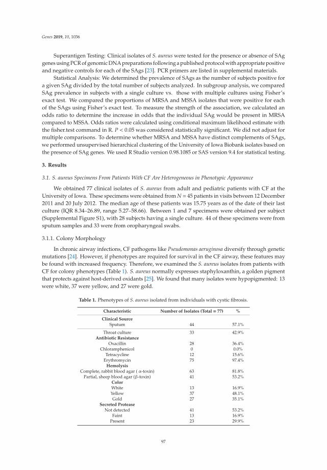
Genes 2019, 10, 1036
Superantigen Testing: Clinical isolates of S. aureus were tested for the presence or absence of SAggenes using PCR of genomic DNA preparations following a published protocol with appropriate positiveand negative controls for each of the SAgs [23]. PCR primers are listed in supplemental materials.
Statistical Analysis: We determined the prevalence of SAgs as the number of subjects positive fora given SAg divided by the total number of subjects analyzed. In subgroup analysis, we comparedSAg prevalence in subjects with a single culture vs. those with multiple cultures using Fisher’sexact test. We compared the proportions of MRSA and MSSA isolates that were positive for eachof the SAgs using Fisher’s exact test. To measure the strength of the association, we calculated anodds ratio to determine the increase in odds that the individual SAg would be present in MRSAcompared to MSSA. Odds ratios were calculated using conditional maximum likelihood estimate withthe fisher.test command in R. P < 0.05 was considered statistically significant. We did not adjust formultiple comparisons. To determine whether MRSA and MSSA have distinct complements of SAgs,we performed unsupervised hierarchical clustering of the University of Iowa Biobank isolates based onthe presence of SAg genes. We used R Studio version 0.98.1085 or SAS version 9.4 for statistical testing.
3. Results
3.1. S. aureus Specimens From Patients With CF Are Heterogeneous in Phenotypic Appearance
We obtained 77 clinical isolates of S. aureus from adult and pediatric patients with CF at theUniversity of Iowa. These specimens were obtained from N = 45 patients in visits between 12 December2011 and 20 July 2012. The median age of these patients was 15.75 years as of the date of their lastculture (IQR 8.34–26.89, range 5.27–58.66). Between 1 and 7 specimens were obtained per subject(Supplemental Figure S1), with 28 subjects having a single culture. 44 of these specimens were fromsputum samples and 33 were from oropharyngeal swabs.
3.1.1. Colony Morphology
In chronic airway infections, CF pathogens like Pseudomonas aeruginosa diversify through geneticmutations [24]. However, if phenotypes are required for survival in the CF airway, these features maybe found with increased frequency. Therefore, we examined the S. aureus isolates from patients withCF for colony phenotypes (Table 1). S. aureus normally expresses staphyloxanthin, a golden pigmentthat protects against host-derived oxidants [25]. We found that many isolates were hypopigmented: 13were white, 37 were yellow, and 27 were gold.
Table 1. Phenotypes of S. aureus isolated from individuals with cystic fibrosis.
Characteristic Number of Isolates (Total = 77) %
Clinical Source
Sputum 44 57.1%
Throat culture 33 42.9%Antibiotic Resistance
Oxacillin 28 36.4%Chloramphenicol 0 0.0%
Tetracycline 12 15.6%Erythromycin 75 97.4%
Hemolysis
Complete, rabbit blood agar ( α-toxin) 63 81.8%Partial, sheep blood agar (β-toxin) 41 53.2%
Color
White 13 16.9%Yellow 37 48.1%Gold 27 35.1%
Secreted Protease
Not detected 41 53.2%Faint 13 16.9%
Present 23 29.9%
97

Genes 2019, 10, 1036
Protease secretion is considered a virulence factor in skin infections [26], and protease productioncould be damaging to airways. Therefore, we tested for secreted protease by examining milk agarplates for zones of clearance. 23 of the isolates had distinct zones of clearance consistent with proteasesecretion, 13 had small or faint zones of clearance, and 41 isolates exhibited no clearance of milk agar inthis assay. There was a strong correlation between hypopigmentation and protease activity. Proteaseactivity, as determined by clearance of milk agar, was detected in 85% of white colonies but 7.5% ofgold colonies (P < 0.001).
Another characteristic of S. aureus is hemolysis, a phenotype that is linked to alpha and betahemolysin toxins. Previous studies of bacteria deficient in alpha toxin reveal its importance in cellularand animal models of CF [27,28]. We tested for the activity of these toxins by hemolysis patterns onsheep and rabbit blood agar plates. Alpha toxin (encoded by hla) activity was observed in the majorityof specimens, whereas beta-toxin (hlb) activity was observed in 40% of specimens.
3.1.2. Antibiotic Resistance
CF pathogens are under selective pressure from antibiotic treatment. We determined MRSA statusof these isolates by growth on Mueller–Hinton agar in the presence of 6 μg/mL of oxacillin. 28 isolatesderived from 19 individuals were phenotypically resistant to oxacillin. 49 isolates from 30 individualswere methicillin-susceptible S. aureus (MSSA). Four subjects had isolates of both MRSA and MSSA.Because macrolides and tetracyclines are commonly prescribed to patients with CF [2], we hypothesizedthat the S. aureus isolates would be resistant to these antibiotic classes, but remain susceptible toantibiotics that are not routinely given. Each isolate was grown on TSA containing either erythromycin,tetracycline, or chloramphenicol. Chronic azithromycin is routinely prescribed at the Universityof Iowa CF center [8]. The vast majority of isolates from the University of Iowa (75/77) exhibitederythromycin resistance, but tetracycline resistance was less common (12/77). Within the University ofIowa collection, the two isolates susceptible to erythromycin were obtained as oropharyngeal cultures.
3.2. High Prevalence of Enterotoxin Gene Cluster Genes in S. aureus Isolated From Patients WithCystic Fibrosis
S. aureus encodes a variety of secreted toxins, including bacterial superantigens (SAgs).We hypothesized that there would be similar heterogeneity in S. aureus secreted toxins. Using previouslydescribed methods [23], we assessed for the presence of 18 unique SAgs. Among these toxins, the mostcommon were genes belonging to the enterotoxin gene cluster (EGC), including seg, sel-i, sel-m, sel-n,sel-o, and sel-u. These genes were highly prevalent in isolates of S. aureus from patients with CF. All sixEGC genes were identified in 37 of the 45 patients examined. 97.8% of patients within this cohortgrew S. aureus that encoded at least one member of the EGC (Table 2). The genes encoding EGC toxinswere significantly more prevalent in CF specimens compared to the classic S. aureus SAg toxic shocksyndrome toxin-1(TSST-1; gene tstH), which was present in 11.7% of isolates.
Because some subjects within the University of Iowa cohort had multiple cultures, there may begreater opportunities to identify specific bacterial genes within these subjects. Therefore, we comparedthe prevalence of each toxin in subjects with multiple cultures vs. those with one culture. We identifiedsea and more frequently in patients with repeated cultures. Genes encoding EGC toxins were highlyprevalent in both groups. We observed no statistically significant differences in age or culture sourcebetween groups.
3.2.1. EGC Prevalence in MRSA and MSSA
We hypothesized that the genes encoding secreted toxins may be associated with either methicillinsusceptibility or resistance. To determine whether MRSA isolates had specific toxin signature(s), weperformed hierarchical clustering based on the presence or absence of toxins (Figure 1). MRSA isolateswere distributed widely in this analysis and often shared the same toxin profile as MSSA isolates.We separately tested whether individual toxins were associated with MRSA or MSSA (Table 3). None of
98

Genes 2019, 10, 1036
the MRSA isolates were positive for tstH, consistent with previous observations that TSST-1 is generallyassociated with MSSA [29]. MSSA was more likely than MRSA to be positive for sel-p (P = 0.03).While sel-p and tstH were more common in MSSA, sel-x was more common in MRSA. However, nocombination of SAgs was perfectly predictive of methicillin resistance, and genes encoding EGC toxinswere prevalent in both MSSA and MRSA isolates.
Table 2. Prevalence of S. aureus superantigen genes detected from individuals with cystic fibrosis.
Toxin
Iowa Subjects with CFTotal = 45
Single CultureTotal = 28
Multiple CulturesTotal = 17 P†
N % N % N %
sea 19 42.2% 8 28.6% 11 64.7% 0.03seb 2 4.4% 1 3.6% 1 5.9% 1.00sec 4 8.9% 2 7.1% 2 11.8% 0.63sed 6 13.3% 2 7.1% 4 23.5% 0.18see 3 6.7% 0 0.0% 3 17.6% 0.05seg 42 93.3% 25 89.3% 17 100.0% 0.28
sel-h 0 0.0% 0 0.0% 0 0.0% -sel-i 42 93.3% 25 89.3% 17 100.0% 0.28
sel-k 4 8.9% 2 7.1% 2 11.8% 0.63sel-l 3 6.7% 1 3.6% 2 11.8% 0.55
sel-m 39 86.7% 23 82.1% 16 94.1% 0.38
sel-n 41 91.1% 25 89.3% 16 94.1% 1.00
sel-o 43 95.6% 26 92.9% 17 100.0% 0.52
sel-p 30 66.7% 15 53.6% 15 88.2% 0.02sel-q 15 33.3% 8 28.6% 7 41.2% 0.52sel-u 41 91.1% 24 85.7% 17 100.0% 0.28
sel-x 39 86.7% 23 82.1% 16 94.1% 0.38tstH 8 17.8% 3 10.7% 5 29.4% 0.23≥1 of egc 44 97.8% 27 96.4% 17 100.0% 1.006 of egc 37 82.2% 21 75.0% 16 94.1% 0.13
†P values compare subjects with single culture to subjects with multiple cultures using Fisher’s exact test. Bold fontindicates genes belonging to the enterotoxin gene cluster.
Table 3. Association of S. aureus toxin genes with methicillin susceptibility or resistance.
S. aureusTotal = 77
MSSATotal = 49
MRSATotal = 28 OR * P†
Toxin N % N % N %
sea 25 32.5% 17 34.7% 8 28.6% 0.76 0.62seb 3 3.9% 3 6.1% 0 0.0% - 0.30sec 5 6.5% 5 10.2% 0 0.0% - 0.15sed 8 10.4% 3 6.1% 5 17.9% 3.28 0.13see 5 6.5% 4 8.2% 1 3.6% 0.42 0.65
seg 69 89.6% 45 91.8% 24 85.7% 0.54 0.45
sel-h 0 0.0% 0 0.0% 0 0.0% - -sel-i 69 89.6% 44 89.8% 25 89.3% 0.95 1.00
sel-k 5 6.5% 3 6.1% 2 7.1% 1.18 1.00sel-l 3 3.9% 3 6.1% 0 0.0% - 0.30
sel-m 67 87.0% 42 85.7% 25 89.3% 1.38 0.74
sel-n 68 88.3% 43 87.8% 25 89.3% 1.16 1.00
sel-o 71 92.2% 46 93.9% 25 89.3% 0.55 0.66
sel-p 46 59.7% 34 69.4% 12 42.9% 0.34 0.03sel-q 17 22.1% 11 22.4% 6 21.4% 0.94 1.00sel-u 69 89.6% 45 91.8% 24 85.7% 0.54 0.45
sel-x 61 79.2% 33 67.3% 28 100.0% - 0.0003tstH 9 11.7% 9 18.4% 0 0.0% - 0.02hla 63 81.8% 37 75.5% 26 92.9% 4.15 0.07hlb 41 53.2% 19 38.8% 22 78.6% 5.65 0.001
* OR = Odds ratio, values > 1 indicate increased odds of the toxin being encoded in MRSA versusmethicillin-susceptible S. aureus (MSSA). †P values calculated by Fisher’s exact test. Bold font indicates genesbelonging to the enterotoxin gene cluster.
99
Genes 2019, 10, 1036
Figure 1. Unsupervised hierarchical clustering of cystic fibrosis (CF) clinical isolates based on presenceof S. aureus toxin genes. Isolate numbers and subjects are indicated at right. Toxin genes are on thebottom margin, with members of the enterotoxin gene cluster (EGC) in blue. Letters A, B, C, D, E,and G are staphylococcal enterotoxin (SE) superantigens characterized as causing emesis after oraladministration. Letters H, I, K, L, M–Q, and X are SE-like superantigens. TSST is toxic shock syndrometoxin-1 superantigen. α and β are cytotoxins. Blue shading represents toxin presence. MRSA isolatesare indicated with red font. Dendrograms at left and top show relatedness of the isolates and toxins,respectively. The EGC was prevalent in both MRSA and MSSA isolates.
3.2.2. EGC Prevalence in S. aureus From Other U.S. CF Centers
We considered the possibility that the high prevalence of S. aureus encoding EGC was due togeographic sampling. To address this possibility, we obtained 12 S. aureus isolates from adults with CFat Dartmouth University and 20 S. aureus isolates from Emory University. We genotyped these isolatesfor the same set of toxins (Table 4). Notably, there were no significant differences between isolates
100

Genes 2019, 10, 1036
obtained in Iowa compared to Dartmouth or Emory, suggesting that the high prevalence of the EGC isnot related to geographic sampling of one region of the United States.
Table 4. S. aureus toxin genes identified in CF clinical isolates from geographically separate regions.Bold font indicates genes belonging to the enterotoxin gene cluster.
Toxin
IowaTotal = 77
EmoryTotal = 20
DartmouthTotal = 12
N % N % N %
sea 25 32.5% 0 0.0% 2 16.7%seb 3 3.9% 0 0.0% 0 0.0%sec 5 6.5% 1 5.0% 0 0.0%sed 8 10.4% 3 15.0% 0 0.0%see 5 6.5% 4 20.0% 0 0.0%seg 69 89.6% 16 80.0% 10 83.3%
sel-h 0 0.0% 0 0.0% 0 0.0%sel-i 69 89.6% 16 80.0% 10 83.3%
sel-k 5 6.5% 1 5.0% 2 16.7%sel-l 3 3.9% 1 5.0% 0 0.0%
sel-m 67 87.0% 15 75.0% 9 75.0%
sel-n 68 88.3% 16 80.0% 10 83.3%
sel-o 71 92.2% 15 75.0% 9 75.0%
sel-p 46 59.7% 5 25.0% 0 0.0%sel-q 17 22.1% 2 10.0% 2 16.7%sel-u 69 89.6% 16 80.0% 10 83.3%
sel-x 61 79.2% 19 95.0% 11 91.7%tstH 9 11.7% 1 5.0% 2 16.7%
4. Discussion
S. aureus isolates from patients with CF displayed heterogeneity of color and protease secretion.However, the majority of these diverse S. aureus isolates encoded the EGC. The heterogeneity ofS. aureus colony phenotypes suggests that putative virulence factors such as staphyloxanthin andprotease may not be under strong selective pressure to remain in the CF airway. By contrast, alpha-toxinmediated hemolysis was routinely observed. Chronic antibiotic exposure represents a strong selectivepressure; most of the isolates from the University of Iowa were resistant to erythromycin. The highprevalence of EGC toxins suggests that S. aureus is under pressure to maintain these genes duringinfection of the CF airway.
We considered the possibility that high prevalence of EGC was related to geographic exposure.Using three geographically distinct collections of S. aureus isolated from U.S. patients with cysticfibrosis, we found that EGC toxins had similarly high prevalence. This is similar to a recent studyof S. aureus isolates from Europe patients with CF, in which EGC toxins were present in ≈57% ofisolates [19]. The EGC prevalence within this U.S. collection is even higher, suggesting the continuedemergence of strains encoding this locus, possibly through the spread of one or more clones. S. aureusclonality is common in CF. In the European study, 5 spa types accounted for 25.6% of all S. aureusisolates. However, EGC-positive S. aureus isolates were not limited to a single clonal group [19].
Most of the CF isolates in the current study were obtained in 2011 and 2012, a time period similarto when specimens were collected for a study of atopic dermatitis that used the same genotypingmethodology [21]. We compared the prevalence of each of the SAgs in CF versus atopic dermatitis.All of the SAgs belonging to the EGC (seg, sel-i, sel-m, sel-n, sel-o, and sel-u) were significantly moreprevalent in CF as compared to atopic dermatitis collection. Among subjects with atopic dermatitis,three distinct genotypes of S. aureus were apparent. The first of these S. aureus genotypes, whichnearly always encoded all EGC toxins, appears highly similar to the prevailing S. aureus within the CF
101
Genes 2019, 10, 1036
population. Longitudinal studies are needed to determine whether or not the EGC associates withpersistence of S. aureus in diseases like CF and atopic dermatitis.
In comparing the prevalence of the EGC in these CF isolates to S. aureus isolates derived fromother anatomic sites, we find that CF has a uniquely high prevalence for this group of SAg toxins.The EGC is more prevalent in CF than in atopic dermatitis, diabetic foot ulcer, and significantly moreprevalent than in vaginal mucosa [30] and in patients with menstrual toxic shock syndrome (TSS) [12].The enrichment of EGC-positive isolates in these CF airway isolates suggests that the EGC may conferselective advantage for S. aureus strains in adapting to its role as a chronic pathogen in the airway.Typically, members of the EGC are considered colonization SAgs, due to low-level production [31].
In striking contrast to the CF, S. aureus isolated from acute inflammatory infections such as TSSand post-influenza necrotizing pneumonia have very high prevalence of tstH and produce SAgs inhigher concentration [12,18]. We observed that a minority of CF clinical isolates encode tstH; it isunknown what effect this SAg may have on progression of CF lung disease.
It is unclear whether patients with CF develop intact immune responses to S. aureus [32,33].Compared to P. aeruginosa, patients with CF may have attenuated antibody production [33].We hypothesize that this could be a consequence of immune misdirection by S. aureus SAgs. Moreover,these SAgs could facilitate increased inflammation, an important factor in CF disease progression.Future studies should examine the immune response to these prevalent S. aureus SAgs.
S. aureus SAgs represent a possible target for vaccination. Given the high prevalence of EGC SAgsin CF, future attempts at immunizing patients with CF against S. aureus may use these antigens asvaccine targets. Notably, a recent study has shown that immunization of rabbits against the SAgsTSST-1 and SEC, and the cytotoxin α-toxin protected 87/88 animals after intra-pulmonary challenge [34].This vaccination strategy depended on formation of cross-protective neutralizing antibodies. Forexample, antibodies raised against SEC protect against both SEB and the EGC SAg SEl-U. That studyalso suggested that vaccination against toxoids may be a more effective strategy than vaccinationagainst cell surface S. aureus virulence factors. In keeping with this notion, a group has recentlyperformed a first-in-humans vaccine trial against the TSST-1 toxoid [35].
4.1. Advantages
This study establishes that enterotoxin gene cluster members are highly prevalent in S. aureusisolates in American patients with CF, independent of methicillin resistance. Because we used consistentmethodology for genotyping, we can compare respiratory isolates of S. aureus from patients withCF to cutaneous isolates from patients with atopic dermatitis that were obtained at a similar time.This comparison reveals that genes encoding EGC toxins are enriched in respiratory isolates. We havealso confirmed the high prevalence of EGC toxins using S. aureus isolates from geographically distinctCF centers.
4.2. Limitations
Because these data are cross-sectional, it is unknown how long these strains have been present inthe CF airway. Many of these patients were sputum-producing. Thus, these infections may representchronic infection rather than initial infection. We are unable to correlate the presence or absenceof secreted toxins with pulmonary outcomes such as FEV1, since the specimens are de-identified.Although many strains encode secreted toxins, we cannot determine whether these genes are activelyexpressed in the CF airway. Because of de-identification, we are also unable to determine whetherthere is adaptive host response to presence or absence of these toxins. We intend to address theselimitations in future studies with a larger number of fully identified specimens.
5. Conclusions
S. aureus remains a prevalent pathogen in CF. Improvements in the prevention and treatment ofS. aureus infection remain a major goal in this disease. This study reveals the SAg ECG gene cluster
102
Genes 2019, 10, 1036
is highly prevalent in S. aureus CF isolates, revealing a potential vaccination target for this organismin CF.
Supplementary Materials: The following are available online at http://www.mdpi.com/2073-4425/10/12/1036/s1,Supplemental Methods: Genotyping assay. Figure S1: Frequency of repeated cultures within University ofIowa Biobank.
Author Contributions: Conceptualization, A.J.F. and P.M.S.; methodology, A.J.F., S.H.K., S.B.S., D.H.L., P.D.A.,A.R.H.; formal analysis, A.J.F., S.H.K., D.H.L., P.M.S.; resources, S.B.S. and D.H.L.; data curation, A.J.F., S.H.K.,S.B.S., P.M.S.; writing—original draft preparation, A.J.F.; writing—review and editing, A.J.F., D.H.L., S.B.S., P.M.S.;visualization, A.J.F.; supervision, A.J.F., P.M.S.; project administration, A.J.F. and P.M.S.; funding acquisition, A.J.F.and P.M.S.
Funding: A.J.F. was supported in part by the University of Iowa Department of Pediatrics, the Cystic FibrosisFoundation CFF FISCHE16I0, and NHLBI K08 HL136927. S.H.K. and P.M.S. were supported by University of Iowastart-up funds. D.H.L. is supported by CFF Post-doc to Faculty Transition Award LIMOLI18F5 and University ofIowa start-up funds.
Acknowledgments: We acknowledge Timothy Starner for assistance with University of Iowa CF Biobank isolates,Arlene Stecenko for assistance with Emory University isolates, and Deborah Hogan and Alex Crocker for assistancewith Dartmouth isolates.
Conflicts of Interest: P.M.S. has received patent protection regarding S. aureus secreted toxins, including methodsto induce immune response to S. aureus with novel toxoids. The remaining authors have no financial conflictsof interest. The funders of this study had no role in the design of the study; in the collection, analyses, orinterpretation of data; in the writing of the manuscript, or in the decision to publish the results.
References
1. Salsgiver, E.L.; Fink, A.K.; Knapp, E.A.; LiPuma, J.J.; Olivier, K.N.; Marshall, B.C.; Saiman, L. Changingepidemiology of the respiratory bacteriology of patients with cystic fibrosis. Chest 2016, 149, 390–400.[CrossRef] [PubMed]
2. Cystic Fibrosis Foundation 2016 Patient Registry Annual Data Report. 2017. Available online: https://www.cff.org/research/researcher-resources/patient-registry/2016-patient-registry-annual-data-report.pdf (accessedon 4 November 2019).
3. Ramsey, B.W.; Pepe, M.S.; Quan, J.M.; Otto, K.L.; Montgomery, A.B.; Williams-Warren, J.; Vasiljev, K.M.;Borowitz, D.; Bowman, C.M.; Marshall, B.C.; et al. Intermittent administration of inhaled tobramycin inpatients with cystic fibrosis. Cystic fibrosis inhaled tobramycin study group. N. Engl. J. Med. 1999, 340, 23–30.[CrossRef] [PubMed]
4. Gibson, R.L.; Emerson, J.; McNamara, S.; Burns, J.L.; Rosenfeld, M.; Yunker, A.; Hamblett, N.; Accurso, F.;Dovey, M.; Hiatt, P.; et al. Significant microbiological effect of inhaled tobramycin in young children withcystic fibrosis. Am. J. Respir. Crit. Care Med. 2003, 167, 841–849. [CrossRef] [PubMed]
5. Tiddens, H.A.; De Boeck, K.; Clancy, J.P.; Fayon, M.; Arets, H.G.M.; Bresnik, M.; Derchak, A.; Lewis, S.A.;Oermann, C.M. Open label study of inhaled aztreonam for Pseudomonas eradication in children with cysticfibrosis: The alpine study. J. Cyst. Fibros. 2015, 14, 111–119. [CrossRef]
6. Dezube, R.; Jennings, M.T.; Rykiel, M.; Diener-West, M.; Boyle, M.P.; Chmiel, J.F.; Dasenbrook, E.C. Eradicationof persistent methicillin-resistant Staphylococcus aureus infection in cystic fibrosis. J. Cyst. Fibros. 2018.[CrossRef]
7. Heltshe, S.L.; Mayer-Hamblett, N.; Burns, J.L.; Khan, U.; Baines, A.; Ramsey, B.W.; Rowe, S.M. Pseudomonasaeruginosa in cystic fibrosis patients with G551D-CFTR treated with ivacaftor. Clin. Infect. Dis. 2015,60, 703–712. [CrossRef]
8. Singh, S.B.; McLearn-Montz, A.J.; Milavetz, F.; Gates, L.K.; Fox, C.; Murry, L.T.; Sabus, A.; Porterfield, H.S.;Fischer, A.J. Pathogen acquisition in patients with cystic fibrosis receiving ivacaftor or lumacaftor/ivacaftor.Pediatr. Pulmonol. 2019. [CrossRef]
9. Caudri, D.; Turkovic, L.; Ng, J.; de Klerk, N.H.; Rosenow, T.; Hall, G.L.; Ranganathan, S.C.; Sly, P.D.; Stick, S.M.The association between Staphylococcus aureus and subsequent bronchiectasis in children with cystic fibrosis.J. Cyst. Fibros. 2018, 17, 462–469. [CrossRef]
103

Genes 2019, 10, 1036
10. Dasenbrook, E.C.; Merlo, C.A.; Diener-West, M.; Lechtzin, N.; Boyle, M.P. Persistent methicillin-resistantStaphylococcus aureus and rate of FEV1 decline in cystic fibrosis. Am. J. Respir. Crit. Care Med. 2008,178, 814–821. [CrossRef]
11. Dinges, M.M.; Orwin, P.M.; Schlievert, P.M. Exotoxins of Staphylococcus aureus. Clin. Microbiol. Rev. 2000, 13,16–34. [CrossRef]
12. Spaulding, A.R.; Salgado-Pabon, W.; Kohler, P.L.; Horswill, A.R.; Leung, D.Y.; Schlievert, P.M. Staphylococcaland streptococcal superantigen exotoxins. Clin. Microbiol. Rev. 2013, 26, 422–447. [CrossRef] [PubMed]
13. McCormick, J.K.; Yarwood, J.M.; Schlievert, P.M. Toxic shock syndrome and bacterial superantigens: Anupdate. Annu. Rev. Microbiol. 2001, 55, 77–104. [CrossRef] [PubMed]
14. Jarraud, S.; Peyrat, M.A.; Lim, A.; Tristan, A.; Bes, M.; Mougel, C.; Etienne, J.; Vandenesch, F.; Bonneville, M.;Lina, G. egc, a highly prevalent operon of enterotoxin gene, forms a putative nursery of superantigens inStaphylococcus aureus. J. Immunol. 2001, 166, 669–677. [CrossRef] [PubMed]
15. Becker, K.; Friedrich, A.W.; Peters, G.; von Eiff, C. Systematic survey on the prevalence of genes coding forstaphylococcal enterotoxins SELM, SELO, and SELN. Mol. Nutr. Food Res. 2004, 48, 488–495. [CrossRef][PubMed]
16. Grumann, D.; Scharf, S.S.; Holtfreter, S.; Kohler, C.; Steil, L.; Engelmann, S.; Hecker, M.; Volker, U.;Broker, B.M. Immune cell activation by enterotoxin gene cluster (egc)-encoded and non-egc superantigensfrom Staphylococcus aureus. J. Immunol. 2008, 181, 5054–5061. [CrossRef]
17. Holtfreter, S.; Bauer, K.; Thomas, D.; Feig, C.; Lorenz, V.; Roschack, K.; Friebe, E.; Selleng, K.; Lovenich, S.;Greve, T.; et al. egc-Encoded superantigens from Staphylococcus aureus are neutralized by human sera muchless efficiently than are classical staphylococcal enterotoxins or toxic shock syndrome toxin. Infect. Immun.2004, 72, 4061–4071. [CrossRef]
18. Stach, C.S.; Vu, B.G.; Merriman, J.A.; Herrera, A.; Cahill, M.P.; Schlievert, P.M.; Salgado-Pabon, W. Noveltissue level effects of the Staphylococcus aureus enterotoxin gene cluster are essential for infective endocarditis.PLoS ONE 2016, 11, e0154762. [CrossRef]
19. Garbacz, K.; Piechowicz, L.; Podkowik, M.; Mroczkowska, A.; Empel, J.; Bania, J. Emergence and spread ofworldwide Staphylococcus aureus clones among cystic fibrosis patients. Infect. Drug Res. 2018, 11, 247–255.[CrossRef]
20. Mempel, M.; Lina, G.; Hojka, M.; Schnopp, C.; Seidl, H.P.; Schafer, T.; Ring, J.; Vandenesch, F.; Abeck, D.High prevalence of superantigens associated with the egc locus in Staphylococcus aureus isolates from patientswith atopic eczema. Eur. J. Clin. Microbiol. Infect. Dis. 2003, 22, 306–309. [CrossRef]
21. Merriman, J.A.; Mueller, E.A.; Cahill, M.P.; Beck, L.A.; Paller, A.S.; Hanifin, J.M.; Ong, P.Y.; Schneider, L.;Babineau, D.C.; David, G.; et al. Temporal and racial differences associated with atopic dermatitisStaphylococcus aureus and encoded virulence factors. mSphere 2016, 1. [CrossRef]
22. Dasenbrook, E.C.; Checkley, W.; Merlo, C.A.; Konstan, M.W.; Lechtzin, N.; Boyle, M.P. Association betweenrespiratory tract methicillin-resistant Staphylococcus aureus and survival in cystic fibrosis. JAMA J. Am.Med. Assoc. 2010, 303, 2386–2392. [CrossRef] [PubMed]
23. Salgado-Pabon, W.; Case-Cook, L.C.; Schlievert, P.M. Molecular analysis of staphylococcal superantigens.Methods Mol. Biol. 2014, 1085, 169–185. [CrossRef] [PubMed]
24. Smith, E.E.; Buckley, D.G.; Wu, Z.; Saenphimmachak, C.; Hoffman, L.R.; D’Argenio, D.A.; Miller, S.I.;Ramsey, B.W.; Speert, D.P.; Moskowitz, S.M.; et al. Genetic adaptation by Pseudomonas aeruginosa to theairways of cystic fibrosis patients. Proc. Natl. Acad. Sci. USA 2006, 103, 8487–8492. [CrossRef] [PubMed]
25. Clauditz, A.; Resch, A.; Wieland, K.P.; Peschel, A.; Gotz, F. Staphyloxanthin plays a role in the fitnessof Staphylococcus aureus and its ability to cope with oxidative stress. Infect. Immun. 2006, 74, 4950–4953.[CrossRef]
26. Kolar, S.L.; Ibarra, J.A.; Rivera, F.E.; Mootz, J.M.; Davenport, J.E.; Stevens, S.M.; Horswill, A.R.; Shaw, L.N.Extracellular proteases are key mediators of Staphylococcus aureus virulence via the global modulation ofvirulence-determinant stability. MicrobiologyOpen 2013, 2, 18–34. [CrossRef]
27. Jarry, T.M.; Memmi, G.; Cheung, A.L. The expression of alpha-haemolysin is required for Staphylococcusaureus phagosomal escape after internalization in CFT-1 cells. Cell. Microbiol. 2008, 10, 1801–1814. [CrossRef]
28. Keitsch, S.; Riethmuller, J.; Soddemann, M.; Sehl, C.; Wilker, B.; Edwards, M.J.; Caldwell, C.C.; Fraunholz, M.;Gulbins, E.; Becker, K.A. Pulmonary infection of cystic fibrosis mice with Staphylococcus aureus requiresexpression of alpha-toxin. Biol. Chem. 2018, 399, 1203–1213. [CrossRef]
104

Genes 2019, 10, 1036
29. Schlievert, P.M.; Strandberg, K.L.; Lin, Y.C.; Peterson, M.L.; Leung, D.Y. Secreted virulence factor comparisonbetween methicillin-resistant and methicillin-sensitive Staphylococcus aureus, and its relevance to atopicdermatitis. J. Allergy Clin. Immunol. 2010, 125, 39–49. [CrossRef]
30. Vu, B.G.; Stach, C.S.; Salgado-Pabon, W.; Diekema, D.J.; Gardner, S.E.; Schlievert, P.M. Superantigens ofStaphylococcus aureus from patients with diabetic foot ulcers. J. Infect. Dis. 2014, 210, 1920–1927. [CrossRef]
31. Roetzer, A.; Gruener, C.S.; Haller, G.; Beyerly, J.; Model, N.; Eibl, M.M. Enterotoxin gene cluster-encoded seiand seln from Staphylococcus aureus isolates are crucial for the induction of human blood cell proliferationand pathogenicity in rabbits. Toxins 2016, 8, 314. [CrossRef]
32. Hollsing, A.E.; Granstrom, M.; Strandvik, B. Prospective study of serum staphylococcal antibodies in cysticfibrosis. Arch. Dis. Child. 1987, 62, 905–911. [CrossRef] [PubMed]
33. Moss, R.B.; Hsu, Y.P.; Lewiston, N.J. 125I-Clq-binding and specific antibodies as indicators of pulmonarydisease activity in cystic fibrosis. J. Pediatr. 1981, 99, 215–222. [CrossRef]
34. Spaulding, A.R.; Salgado-Pabon, W.; Merriman, J.A.; Stach, C.S.; Ji, Y.; Gillman, A.N.; Peterson, M.L.;Schlievert, P.M. Vaccination against Staphylococcus aureus pneumonia. J. Infect. Dis. 2014, 209, 1955–1962.[CrossRef] [PubMed]
35. Roetzer, A.; Jilma, B.; Eibl, M.M. Vaccine against toxic shock syndrome in a first-in-man clinical trial. ExpertRev. Vaccines 2017, 16, 81–83. [CrossRef]
© 2019 by the authors. Licensee MDPI, Basel, Switzerland. This article is an open accessarticle distributed under the terms and conditions of the Creative Commons Attribution(CC BY) license (http://creativecommons.org/licenses/by/4.0/).
105

genesG C A T
T A C G
G C A T
Review
Molecular Diagnosis and Genetic Counseling ofCystic Fibrosis and Related Disorders:New Challenges
Thierry Bienvenu, Maureen Lopez and Emmanuelle Girodon *
Molecular Genetics Laboratory, Cochin Hospital, APHP.Centre–Université de Paris, 75014 Paris, France;[email protected] (T.B.); [email protected] (M.L.)* Correspondence: [email protected]; Tel.: +33-015-841-1924
Received: 9 May 2020; Accepted: 2 June 2020; Published: 4 June 2020
Abstract: Identification of the cystic fibrosis transmembrane conductance regulator (CFTR) gene andits numerous variants opened the way to fantastic breakthroughs in diagnosis, research and treatmentof cystic fibrosis (CF). The current and future challenges of molecular diagnosis of CF and CFTR-relateddisorders and of genetic counseling are here reviewed. Technological advances have enabled to makea diagnosis of CF with a sensitivity of 99% by using next generation sequencing in a single step.The detection of heretofore unidentified variants and ethnic-specific variants remains challenging,especially for newborn screening (NBS), CF carrier testing and genotype-guided therapy. Among thecriteria for assessing the impact of variants, population genetics data are insufficiently taken intoaccount and the penetrance of CF associated with CFTR variants remains poorly known. The hugediversity of diagnostic and genetic counseling indications for CFTR studies makes assessment ofvariant disease-liability critical. This is especially discussed in the perspective of wide genomeanalyses for NBS and CF carrier screening in the general population, as future challenges.
Keywords: cystic fibrosis; CFTR; CFTR-related disorders; molecular diagnosis; CFTR variants; NextGeneration Sequencing (NGS); disease liability; interpretation; penetrance; genotype-guided therapy
1. Introduction
Cystic fibrosis (CF) is one of the most frequent life-limiting autosomal recessive diseases,characterized in its classical form by chronic pulmonary obstruction and infections, pancreaticinsufficiency, male infertility, sweat chloride concentrations ≥60 mmol/L and two loss-of-functionvariants in the cystic fibrosis transmembrane conductance regulator (CFTR) gene (NM_000492.4;LRG_663t1) [1]. With the implementation of newborn screening (NBS) for CF, an increasing number ofdiagnoses are now made in asymptomatic patients. CFTR variants have diverse effects on expressionand function of the CFTR protein, which principally acts as an ATP-gated chloride channel. Its absenceor dysfunction leads to ion flux perturbations in the epithelial cells of various organs involved in CF [2].
Since the discovery of the CFTR gene more than thirty years ago, considerable scientific advanceshave made CF a model in terms of comprehensive knowledge of a genetic disease, molecular diagnosis,genetic counseling and personalized medicine. Technical milestones have led to identify a huge numberof CFTR gene variants and a variety of molecular mechanisms responsible for CF [3], which contributeto the wide phenotype variability, and to achieve one of the highest sensitivities in the diagnosis of ahereditary disease, more than 99% of CF-causing variants being identified in newborns with CF [4].The advent of next generation sequencing (NGS)-based technologies has deeply modified laboratory’spractice, improved genotyping coverage and questioned screening policy. Specific molecular tools havealso been developed for preimplantation genetic diagnosis [5] and non-invasive prenatal diagnosisof CF [6]. Other clinical entities related to CFTR variants have been described since the early 90s,
Genes 2020, 11, 619; doi:10.3390/genes11060619 www.mdpi.com/journal/genes107
Genes 2020, 11, 619
with a continuum of CFTR dysfunction, from CFTR-related disorders (CFTR-RD), which are definedby evidence of CFTR dysfunction but do not meet the criteria for a CF diagnosis [7], to a number ofconditions associated with a higher proportion of CF carriers compared to the general population,such as asthma or bronchopulmonary allergic aspergillosis [8]. Very recently, Miller et al. reported anincreased risk for 57 CF-related symptoms in CF carriers in a study that questions the relevance ofdetecting CF carriers for preventive care [9]. CFTR-RDs principally include isolated male infertility bycongenital bilateral absence of the vas deferens, idiopathic pancreatitis, disseminated bronchiectasisand chronic rhinosinusitis. The contribution of CFTR variants however varies from one conditionto another, and may act in a multifactorial context, with other genes being potentially involved,such as ADGRG2 in male infertility by the absence of vas deferens [10] or PRSS1, SPINK1 and CTRC inpancreatitis [11]. There is thus a huge diversity of diagnostic and genetic counseling indications forsearching CFTR variants, and appropriate tools should be used to answer clinical questions [1,12,13].A great challenge in 2020 remains to accurately assess the disease liability of CFTR variants in theappropriate clinical context and to determine whether a variant should be reported as disease-causing,whether a genotype is compatible with the phenotype or which phenotype is compatible with thegenotype identified in the context of NBS for CF, and whether a variant should be considered forgenetic counseling purposes. The availability of genotype-guided therapy has also brought a renewedinterest in deciphering the impact of CFTR variants.
In the present article, we review the current and future challenges of molecular diagnosis andgenetic counseling. Despite a very high sensitivity of molecular tools, characterization of heretoforeunidentified disease-causing variants in patients remains a technical challenge. We will also focuson ethnic-specific variants, the detection of which being challenging for NBS, CF carrier testing andgenotype-guided therapy. We will then emphasize on the importance to consider population geneticsand penetrance data in the process to evaluate the impact of variants. These data will eventually bediscussed in the perspective of implementation of wide genome analyses for NBS and preconceptionCF carrier screening in the general population in an increasing number of countries.
2. Molecular Diagnosis
The following section reviews the process to achieve a molecular diagnosis of CF or CFTR-RD, byusing a panel of tools in successive stages and also covers the indications of prenatal diagnosis andcarrier testing.
2.1. Molecular Diagnosis of CF and CFTR-RD
A CF diagnosis is suggested by characteristic symptoms, a family history of CF (most often in asibling) or a positive CF NBS result and is confirmed by evidence of CFTR dysfunction, most oftenby abnormal sweat chloride test results or by identification of two CF-causing alleles, one on eachparental gene [1]. Diagnostic algorithms also include other CFTR functional investigations such astrans-epithelial nasal potential difference measurement or intestinal current measurement on rectalbiopsies [1]. The diagnosis of CFTR-RD is established in the event of symptoms suggestive for CFTRdysfunction but when the biological criteria for CF are not met. These clinical entities essentiallyinclude monosymptomatic disorders in adults but the distinction between a mild CF and a CFTR-RDwith multiorgan involvement may be artificial. Despite limitations, as for all tests, and the number ofinconclusive situations, CFTR genetic analysis is a cornerstone for the diagnosis of CF and CFTR-RD.In adult patients having a monosymptomatic disease suggestive of a CFTR-RD, the proportion ofhomozygotes or compound heterozygotes is lower than in CF patients. It was shown to be as high as82% in male infertility by absence of vas deferens [14] but low in other entities [7], e.g., 6.3% in aquagenicpalmoplantar keratoderma [15]. Diagnostic sensitivities also vary between studies, depending oninclusion criteria and investigations for other causes, such as for disseminated bronchiectasis orpancreatitis. Provision of clinical and biological information prior to CFTR testing is thus essential toensure that appropriate studies are carried out and that accurate interpretation is given.
108
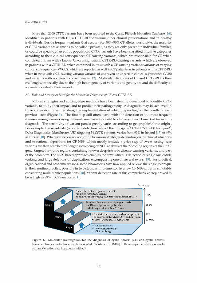
Genes 2020, 11, 619
More than 2000 CFTR variants have been reported to the Cystic Fibrosis Mutation Database [16],identified in patients with CF, a CFTR-RD or various other clinical presentations and in healthyindividuals. Beside frequent variants that account for 50%–90% CF alleles worldwide, the majorityof CFTR variants are as rare as to be called “private”, as they are only present in individual families,or could be specific of an ethnic population. CFTR variants have been classified into five categoriesaccording to their clinical consequence: CF-causing variants, which are responsible for CF whencombined in trans with a known CF-causing variant; CFTR-RD-causing variants, which are observedin patients with a CFTR-RD when combined in trans with a CF-causing variant; variants of varyingclinical consequences (VVCC), which are reported as well in CF patients as in patients with a CFTR-RDwhen in trans with a CF-causing variant; variants of unproven or uncertain clinical significance (VUS)and variants with no clinical consequences [12]. Molecular diagnosis of CF and CFTR-RD is thuschallenging especially due to the high heterogeneity of variants and genotypes and the difficulty toaccurately evaluate their impact.
2.2. Tools and Strategies Used for the Molecular Diagnosis of CF and CFTR-RD
Robust strategies and cutting-edge methods have been steadily developed to identify CFTRvariants, to study their impact and to predict their pathogenicity. A diagnosis may be achieved inthree successive molecular steps, the implementation of which depending on the results of eachprevious step (Figure 1). The first step still often starts with the detection of the most frequentdisease-causing variants using different commercially available kits, very often CE-marked for in vitrodiagnosis. The sensitivity of variant panels greatly varies according to geographic/ethnic origins.For example, the sensitivity (or variant detection rate) of the Elucigene® CF-EU2v1 kit (Elucigene®,Delta Diagnostics, Manchester, UK) targeting 51 CFTR variants, varies from 93% in Ireland [17] to 49%in Turkey [18]. Whenever necessary, according to various strategies depending on the clinical situationsand to national algorithms for CF NBS, which mostly include a prior step of sweat testing, rarevariants are then searched by Sanger sequencing or NGS analysis of the 27 coding regions of the CFTRgene, targeted intronic regions containing known deep-intronic disease-causing variants, and partof the promoter. The NGS-based approach enables the simultaneous detection of single nucleotidevariants and large deletions or duplications encompassing one or several exons [19]. For practical,organizational and economic reasons, some laboratories have now applied NGS as the single techniquein their routine practice, possibly in two steps, as implemented in a few CF NBS programs, notablyconsidering multi-ethnic populations [20]. Variant detection rate of this comprehensive step proved tobe as high as 99% in CF newborns [4].
Figure 1. Molecular investigation for the diagnosis of cystic fibrosis (CF) and cystic fibrosistransmembrane conductance regulator related disorders (CFTR-RD) in three steps. Sensitivity refers tovariant detection rate in patients with CF.
109
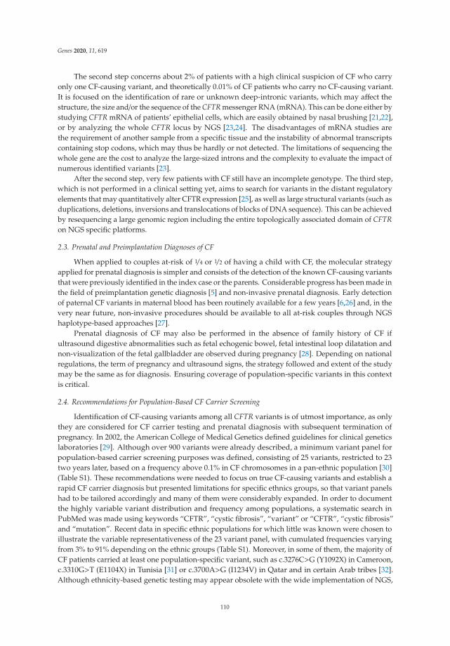
Genes 2020, 11, 619
The second step concerns about 2% of patients with a high clinical suspicion of CF who carryonly one CF-causing variant, and theoretically 0.01% of CF patients who carry no CF-causing variant.It is focused on the identification of rare or unknown deep-intronic variants, which may affect thestructure, the size and/or the sequence of the CFTR messenger RNA (mRNA). This can be done either bystudying CFTR mRNA of patients’ epithelial cells, which are easily obtained by nasal brushing [21,22],or by analyzing the whole CFTR locus by NGS [23,24]. The disadvantages of mRNA studies arethe requirement of another sample from a specific tissue and the instability of abnormal transcriptscontaining stop codons, which may thus be hardly or not detected. The limitations of sequencing thewhole gene are the cost to analyze the large-sized introns and the complexity to evaluate the impact ofnumerous identified variants [23].
After the second step, very few patients with CF still have an incomplete genotype. The third step,which is not performed in a clinical setting yet, aims to search for variants in the distant regulatoryelements that may quantitatively alter CFTR expression [25], as well as large structural variants (such asduplications, deletions, inversions and translocations of blocks of DNA sequence). This can be achievedby resequencing a large genomic region including the entire topologically associated domain of CFTRon NGS specific platforms.
2.3. Prenatal and Preimplantation Diagnoses of CF
When applied to couples at-risk of 1/4 or 1/2 of having a child with CF, the molecular strategyapplied for prenatal diagnosis is simpler and consists of the detection of the known CF-causing variantsthat were previously identified in the index case or the parents. Considerable progress has been made inthe field of preimplantation genetic diagnosis [5] and non-invasive prenatal diagnosis. Early detectionof paternal CF variants in maternal blood has been routinely available for a few years [6,26] and, in thevery near future, non-invasive procedures should be available to all at-risk couples through NGShaplotype-based approaches [27].
Prenatal diagnosis of CF may also be performed in the absence of family history of CF ifultrasound digestive abnormalities such as fetal echogenic bowel, fetal intestinal loop dilatation andnon-visualization of the fetal gallbladder are observed during pregnancy [28]. Depending on nationalregulations, the term of pregnancy and ultrasound signs, the strategy followed and extent of the studymay be the same as for diagnosis. Ensuring coverage of population-specific variants in this contextis critical.
2.4. Recommendations for Population-Based CF Carrier Screening
Identification of CF-causing variants among all CFTR variants is of utmost importance, as onlythey are considered for CF carrier testing and prenatal diagnosis with subsequent termination ofpregnancy. In 2002, the American College of Medical Genetics defined guidelines for clinical geneticslaboratories [29]. Although over 900 variants were already described, a minimum variant panel forpopulation-based carrier screening purposes was defined, consisting of 25 variants, restricted to 23two years later, based on a frequency above 0.1% in CF chromosomes in a pan-ethnic population [30](Table S1). These recommendations were needed to focus on true CF-causing variants and establish arapid CF carrier diagnosis but presented limitations for specific ethnics groups, so that variant panelshad to be tailored accordingly and many of them were considerably expanded. In order to documentthe highly variable variant distribution and frequency among populations, a systematic search inPubMed was made using keywords “CFTR”, “cystic fibrosis”, “variant” or “CFTR”, “cystic fibrosis”and “mutation”. Recent data in specific ethnic populations for which little was known were chosen toillustrate the variable representativeness of the 23 variant panel, with cumulated frequencies varyingfrom 3% to 91% depending on the ethnic groups (Table S1). Moreover, in some of them, the majority ofCF patients carried at least one population-specific variant, such as c.3276C>G (Y1092X) in Cameroon,c.3310G>T (E1104X) in Tunisia [31] or c.3700A>G (I1234V) in Qatar and in certain Arab tribes [32].Although ethnicity-based genetic testing may appear obsolete with the wide implementation of NGS,
110

Genes 2020, 11, 619
the challenge remains to ensure the coverage of population-specific CF-causing variants whereverappropriate, especially as the NGS-based approach is not affordable in all countries.
2.5. Impact of Variant Heterogeneity on Personalized Medicine
Development of drugs to correct, enhance and stabilize the CFTR protein has made CFTRgenotyping crucial to optimize therapy. It has renewed interest for the original variant classificationthat was based on functional data, six classes being recognized according to the CFTR protein defect in:I. synthesis, II. processing and maturation, III. gating, IV. conductance, V. abundance due to reducedamount of normal mRNA and VI. stability at the membrane [33–35]. However, clinical trials haveshowed that numerous variants caused pleiotropic defects, such as the most frequent CF-causing variantworldwide, c.1521_1523del (F508del) [36], thus justifying the use of drug combinations. Ivacaftor wasthe first drug to be US Food and Drug Administration approved for the treatment of CF, efficientlytargeting the gating defect of class III variants, and it was further extended to class IV variants (Table S2).The list may however differ with that approved by the European Medicines Agency. These variantsrepresent only 2%–10% of CF-causing variants in several populations and are totally absent in otherethnic groups (Table S2). Moreover, the clinical significance of several of them is questionable. Sincethen, combinations of ivacaftor with other molecules that aim to increase CFTR protein traffickingto the plasma membrane have been approved for specific variants: lumacaftor/ivacaftor in F508delhomozygous patients; tezacaftor/ivacaftor in patients carrying F508del and a variant associated withresidual CFTR function and elexacaftor/tezacaftor/ivacaftor in patients carrying at least one copy ofF508del. The monumental interest of the triple-combination is that a large proportion of patients withCF are eligible for treatment in numerous populations. However, in specific ethnic groups, F508delis absent (such as Iraq, Sudan, Qatar, Japan and China) or extremely rare (Jordan 6%–7%, Bahrain8%) [37] (Table S1). The challenge is thus to make personalized therapy accessible to all patients.Other strategies are still under development, such as amplifiers that increase the amount of CFTRavailable for modulators, readthrough agents targeting in frame premature termination codons andgene therapy [38].
3. How to Assess the Impact of CFTR Variants—The Challenge of Penetrance
Assessing the impact of CFTR variants is a comprehensive process, which allows one to answerclinical questions that cover diagnostic, genetic counseling and therapeutic issues. It is a daily challengein specialized CF molecular laboratories, especially when dealing with rare or unknown variants.It relies on a good dialogue between clinicians, electrophysiologists and molecular geneticists, withappropriate clinical information provided by the clinicians, and occasionally requires the expertise ofresearchers (Figure 2). In 2015, international recommendations for interpretation and classification ofsequence variations were published [39], based on diverse criteria including population, computational,segregation and functional data. They are reviewed in the present section. The recommendedphenotypic classification into five categories, that is, “pathogenic”, “likely pathogenic”, “uncertainsignificance”, “likely benign” and “benign” is however not completely equivalent as that used forCFTR variants and does not take into account phenotypic diversity, as “pathogenic” may be used forCF-causing and CFTR-RD-causing variants. As an example, variant c.1210-34TG[12]T[5] (TG12T5)may be considered pathogenic in the context of male infertility by absence of vas deferens but not inthe context of CF.
3.1. Population Data (Clinical)
Assessing the impact and potential clinical consequence of a CFTR variant starts with search forclinical observations related to this variant in laboratory’s own database, publicly available databasesand the literature. Considering the class of variants in trans is essential in the context of a recessivedisorder as CF. Beside the original CF Mutation Database, which most often describes the originalobservation and provides links to literature in PubMed, two other locus specific databases provide
111
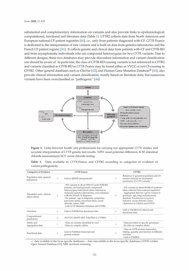
Genes 2020, 11, 619
substantial and complementary information on variants and also provide links to epidemiological,computational, functional and literature data (Table 1). CFTR2 collects data from North American andEuropean national CF patient registries [40], i.e., only from patients diagnosed with CF. CFTR-Franceis dedicated to the interpretation of rare variants and is built on data from genetics laboratories and theFrench CF patient registry [41]. It collects genetic and clinical data from patients with CF and CFTR-RDand from asymptomatic individuals who are compound heterozygous for two CFTR variants. Due todifferent designs, these two databases may provide discordant information and variant classificationone should be aware of. In particular, the class of CFTR-RD-causing variants is not referenced in CFTR2and variants classified as CFTR-RD in CFTR-France may be found either as VVCC or not CF-causing inCFTR2. Other general databases such as ClinVar [42], and Human Gene Mutation Database® [43], alsoprovide clinical information and variant classification, mostly based on literature data, but numerousvariants have been overclassified as “pathogenic” [44].
Figure 2. Links between health care professionals for carrying out appropriate CFTR studies andaccurate interpretation of CFTR genetic test results. NPD: nasal potential difference; ICM: intestinalchloride measurement; SCT: sweat chloride testing.
Table 1. Data available in CFTR-France and CFTR2 according to categories of evidence ofvariant pathogenicity.
Categories of Evidence CFTR-France CFTR2
Population data: generalpopulation + Link to dbSNP and gnomAD +
Reference to general population and CFcarriers analysis for incompletepenetrance of CFTR variants
Population data: clinicalobservations +
- 853 variants in about 5000 CF and CFTR-RDpatients, and asymptomatic compoundheterozygous individuals (data collected inmolecular genetics laboratories, cross-referencewith the French CF Registry)- Per patient: Age at diagnosis, symptoms,pancreatic status, meconium ileus, sweatchloride values, NBS- Link to CF Mutation Database and CFTR2
+
- 432 variants in about 89,000 CF patients(data collected from national registries)- Aggregated data for a given variant orgenotype: age, lung and pancreaticfunction, Pseudomonas aeruginosainfection, sweat chloride values- Reference to ClinVar and LOVD
Literature + Link to PubMed for functional data +Link to PubMed for clinical andfunctional data
Computationalpredictions + AGVGD, MAPP, SIFT, PolyPhen-2, CYSMA -
Allelic andsegregation data +
- Data on variants identified in trans- Data on complex alleles +
- Data provided on specific genotypes- No data on complex alleles
Functional data +Link to PubMed (transcript andprotein studies) +
- Data on CFTR protein maturation,folding, quantity and function in differentcell lines- Link to PubMed
+: data available in the locus specific databases; -: data unavailable in the locus specific databases; LOVD: LeidenOpen Variant Database [45]; NBS: newborn screening.
112

Genes 2020, 11, 619
3.2. Literature Data
The literature review may provide valuable information for clinical, population genetics andfunctional data.
3.3. Computational and Predictive Data
In silico bioinformatics tools help predict the impact of variants at the mRNA level or the proteinlevel, based on conservation of amino acids within different proteins of the same family, evolutionaryconservation within species and biochemical distance between amino acids. Importantly, any intronicor exonic variant, even synonymous, may alter splicing. There are some limitations with using insilico tools, notably because of challenges in prioritizing one tool over the other and the lack ofreliable standard interpretation guidelines [19]. Cystic fibrosis missense analysis (CYSMA) is a recentlydeveloped website dedicated to CFTR missense variants based on integrated in-house bioinformaticstools, which proved efficient to predict the impact of CFTR variants [46].
With the advent of wide genome analysis, aggregators that combine multiple evaluation tools havebeen implemented, such as Varsome [47] or Intervar [48]. They may be of help but exhibit significantlimitations for CFTR variants, displaying a high degree of uncertainty for numerous variants [44].
3.4. Allelic and Segregation Data
Genetic studies in the parents of a patient are necessary to confirm the status of homozygous for avariant or compound heterozygous for two variants. Inheritance of two variants from the same parent(in cis) indicates the presence of a complex allele and numerous frequent or rare complex alleles havebeen described in the CFTR gene [41,49,50]. Complex alleles increase the complexity of CFTR variantclassification, as illustrated for the c.445G>A (G149R) CF-causing variant and the c.1327G>T (D443Y)CFTR-RD-causing variant, each of them being described in cis with an already frequent complexallele c.[1727G>C;2002C>T] (G576A;R668C) [51]. Cis-variants may also affect the response to CFTRmodulators, which impacts on reporting genetic test results [52]. De novo occurrence of variants isextremely rare in CF but has been described [53], also justifying parents’ study. Abnormal segregationduring parents’ study may also unmask the presence of a deletion that would have escaped detection,depending on the techniques used [54].
3.5. Functional Data
Evidence of CFTR dysfunction may be brought by different categories of investigation, includingin vivo, ex vivo and in vitro tests. While sweat testing is most often performed before comprehensivegenetic testing in children, it may be performed after identification of CFTR variants in newborns,depending on NBS programs, or in adults with a possible CFTR-RD or CF. Nasal potential differenceor intestinal current measurements are often used in the second line in case of inconclusive sweat testresults or to help variant interpretation for patients carrying VUS. Likewise, other tests may be ofhelp, such as sweat secretion after β-adrenergic stimulation, also called evaporimetry [55]. Ex vivoassessment of CFTR function on miniaturized versions of organs called organoids, from minimallyinvasive rectal biopsies [56] or on bronchial or nasal cells [57,58], is based on sophisticated andcomprehensive techniques implemented in a few expert laboratories, which help diagnose, understandmechanistic defects and better predict organ-specific drug responses [59].
In vitro assays implemented to evaluate the impact of variants on CFTR mRNA or protein are alsousually performed in a research setting. Minigene systems most often reproduce one or several exonsin cloned plasmids, which are then transfected in human cells. They interrogate the impact of intronicor exonic variants on splicing [60,61] and are useful alternative tools when patients’ epithelial cells arenot available for mRNA study. Importantly, they have allowed demonstration of a splicing impact ofvariants heretofore considered as missense, such c.2908G>C (G970R), which escapes CFTR modulatortherapy [62] or c.3700A>G (I1234V) [63], or as nonsense, like c.2491G>T (E831X) [64]. Functional in vitro
113
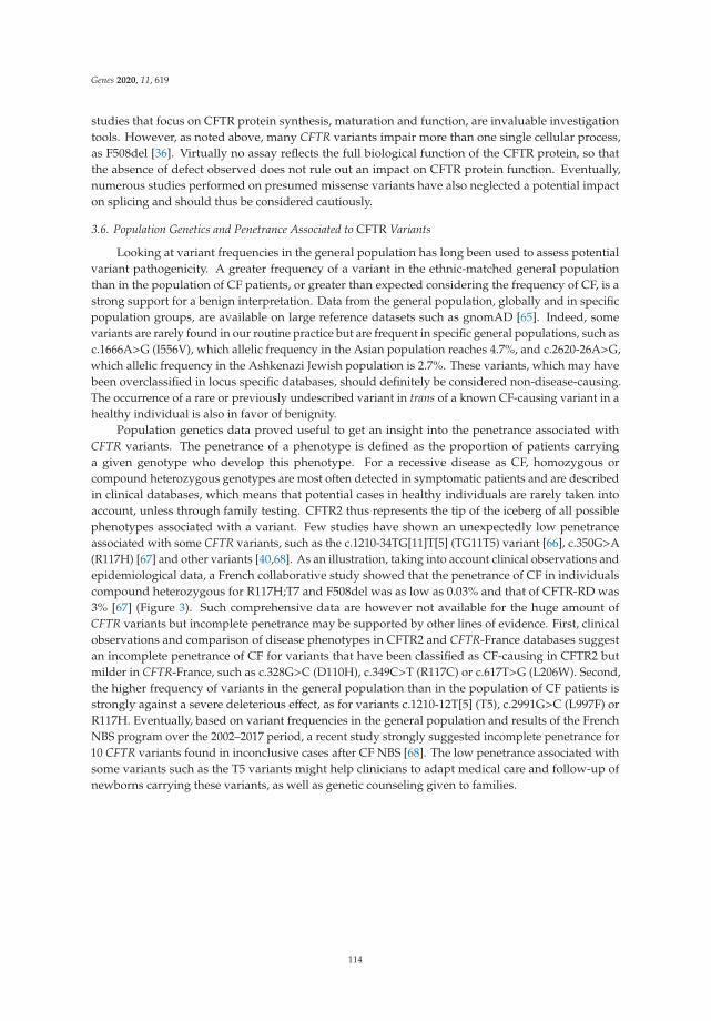
Genes 2020, 11, 619
studies that focus on CFTR protein synthesis, maturation and function, are invaluable investigationtools. However, as noted above, many CFTR variants impair more than one single cellular process,as F508del [36]. Virtually no assay reflects the full biological function of the CFTR protein, so thatthe absence of defect observed does not rule out an impact on CFTR protein function. Eventually,numerous studies performed on presumed missense variants have also neglected a potential impacton splicing and should thus be considered cautiously.
3.6. Population Genetics and Penetrance Associated to CFTR Variants
Looking at variant frequencies in the general population has long been used to assess potentialvariant pathogenicity. A greater frequency of a variant in the ethnic-matched general populationthan in the population of CF patients, or greater than expected considering the frequency of CF, is astrong support for a benign interpretation. Data from the general population, globally and in specificpopulation groups, are available on large reference datasets such as gnomAD [65]. Indeed, somevariants are rarely found in our routine practice but are frequent in specific general populations, such asc.1666A>G (I556V), which allelic frequency in the Asian population reaches 4.7%, and c.2620-26A>G,which allelic frequency in the Ashkenazi Jewish population is 2.7%. These variants, which may havebeen overclassified in locus specific databases, should definitely be considered non-disease-causing.The occurrence of a rare or previously undescribed variant in trans of a known CF-causing variant in ahealthy individual is also in favor of benignity.
Population genetics data proved useful to get an insight into the penetrance associated withCFTR variants. The penetrance of a phenotype is defined as the proportion of patients carryinga given genotype who develop this phenotype. For a recessive disease as CF, homozygous orcompound heterozygous genotypes are most often detected in symptomatic patients and are describedin clinical databases, which means that potential cases in healthy individuals are rarely taken intoaccount, unless through family testing. CFTR2 thus represents the tip of the iceberg of all possiblephenotypes associated with a variant. Few studies have shown an unexpectedly low penetranceassociated with some CFTR variants, such as the c.1210-34TG[11]T[5] (TG11T5) variant [66], c.350G>A(R117H) [67] and other variants [40,68]. As an illustration, taking into account clinical observations andepidemiological data, a French collaborative study showed that the penetrance of CF in individualscompound heterozygous for R117H;T7 and F508del was as low as 0.03% and that of CFTR-RD was3% [67] (Figure 3). Such comprehensive data are however not available for the huge amount ofCFTR variants but incomplete penetrance may be supported by other lines of evidence. First, clinicalobservations and comparison of disease phenotypes in CFTR2 and CFTR-France databases suggestan incomplete penetrance of CF for variants that have been classified as CF-causing in CFTR2 butmilder in CFTR-France, such as c.328G>C (D110H), c.349C>T (R117C) or c.617T>G (L206W). Second,the higher frequency of variants in the general population than in the population of CF patients isstrongly against a severe deleterious effect, as for variants c.1210-12T[5] (T5), c.2991G>C (L997F) orR117H. Eventually, based on variant frequencies in the general population and results of the FrenchNBS program over the 2002–2017 period, a recent study strongly suggested incomplete penetrance for10 CFTR variants found in inconclusive cases after CF NBS [68]. The low penetrance associated withsome variants such as the T5 variants might help clinicians to adapt medical care and follow-up ofnewborns carrying these variants, as well as genetic counseling given to families.
114

Genes 2020, 11, 619
Figure 3. Penetrance of phenotypes in individuals who are compound heterozygous forc.[350G>A;1210-12T[7]];[1521_1523del] (R117H;T7/F508del) from Thauvin et al. [67].
4. The Challenges of Genetic Counseling—When Genetic Counseling Meets Diagnosis
Genetic counseling for CF is a very important part of medical consultation and laboratory activities,which has become complex over the years (Figure 4). It has long been focused on the identification ofCF carriers in the family of patients having CF and on provision of counseling services to couples inorder to ensure informed reproductive decision-making [69]. Once a CF carrier is identified, testingfor the most frequent CF-causing variants according to his/her geographic origins is recommendedin the partner in a prenatal or preconception setting [13]. Extended CFTR sequencing analysis in thepartners is however more and more performed, at least in partners of CF patients, because of theprior risk for the couple of having a child with CF (1/70, provided a CF carrier frequency of 1/35) [69].Genetic counseling should also be considered for all symptomatic patients who carry CFTR variants,due to the potential risk of CF in the patients’ offspring and relatives. The identification of a CF variantleads to recommend CF carrier testing in the relatives and the partner in case of a parental project.While cascade testing for known CFTR-RD variants is not recommended, the identification of VVCCor VUS makes genetic counseling delicate and may be discussed on a case-by-case basis. Occasionally,CFTR testing in healthy adult siblings of a patient leads to identify the same genotype as in the patient.These individuals might develop symptoms related to a CFTR-RD or a mild form of CF, and thusneed clinical investigation. On the other hand, such findings contribute to documenting the variablepenetrance associated with some CFTR variants.
Identification of CF carriers may also occur in the absence of any family history of CF, duringthe process of NBS, or during preconception carrier screening or during wide genome analysis as anunsolicited or secondary finding (Figure 4). Expanded preconception carrier screening for CF and otherrecessive disorders is also under consideration in countries according to an overall positive attitude ofthe general population [70,71]. Preconception CF carrier screening has already been implemented inthe US, Israël and Northeast Italy [72]. In most countries however, for practical reasons variant panelsused for diagnostic purposes are also used for carrier testing. Again, it is important to discriminate trueCF-causing variants from those that are CFTR-RD-causing, VVCC or non disease-causing. The wideimplementation of NGS-based CFTR analysis in various clinical settings has increased this concern,with the identification of rare VUS or variants for which discrepant interpretation is found in databasesor the literature. For genetic counseling purposes, the question of penetrance associated with variantsis even more critical. Contrary to the diagnostic setting where the main question to answer is “does thegenotype account for the phenotype?”, which may already be difficult with inconclusive genotypes,
115

Genes 2020, 11, 619
a challenging question in genetic counseling is to predict the phenotype resulting from the combinationof a VUS or a VVCC with a known CF-causing variant.
Figure 4. Genetic counseling situations (in orange), with potential identification of CF carriers, accordingto variable practices of molecular analysis (in blue). The number of variants tested is indicated inbrackets. CFTR-RD: CFTR-related disorder; NBS: newborn screening; NIPD: non-invasive prenataldiagnosis; PND: prenatal diagnosis; PGD: preimplantation genetic diagnosis. Hatched lines: expectedpractice in the near future.
Identification of carriers of CF-causing variants is also of particular concern in the context of CFNBS. On one hand, testing the parents may lead to identify another CFTR variant, possibly outsidethe NBS panel depending on laboratory’s practice. This result then raises the question of looking forthis second variant in the infant who has a negative sweat test result. On the other hand, extendedCFTR gene sequencing is being considered as part of the core strategy in an increasing number ofprograms [73,74]. Moreover, the relevance of implementing extended NBS for numerous geneticdiseases is currently debated [74–77], also taking into account economical aspects. The introductionof NGS, with or without prior immunoreactive trypsinogen measurement and without filtering CFvariants would lead to detect not only a higher number of carriers of known CF-causing variantsbut a much higher number of carriers of VVCC and VUS. The risk would be to consider neonates ascarriers of a CF-causing variant and to offer inappropriate genetic counseling and testing in the family,and eventually inappropriate prenatal diagnosis.
The face of genetic counseling for CF will inevitably deeply change in the coming years. Healthpublic policies of CF carrier screening in the general population aim to detect most CF carrier couplesand prenatal requests may increase, especially with the availability of non-invasive procedures.This would ineluctably impact on the prevalence of CF births, which then would raise the question ofthe relevance of NBS if the incidence of CF is getting very low. In other respects, due to formidableprogress in genotype-guided therapy, parents at risk of having a child with CF could prefer the optionof continuation of pregnancy over that of termination. Prediction of changing attitudes and practices isa delicate business.
Very recently, a study conducted on 19,802 CF carriers who were matched each with five controls,reported a higher prevalence of 57 out of 59 CF-related symptoms or conditions in CF carriers thanin the general population. These conditions included already known CFTR-RDs, conditions where ahigher prevalence of CFTR variants has already been reported, such as allergic bronchopulmonaryaspergillosis, asthma, primary sclerosing cholangitis [8] or pancreatic cancer [78] and others that werenot previously described associated with CFTR dysfunction, such as cirrhosis or intestina atresia [9].Despite a number of limitations of this study, notably the absence of any data about CFTR testing
116
Genes 2020, 11, 619
(the number and kind of variants is not known, a number of CF carriers might have a CFTR-RDor bear a second CFTR variant), and the low absolute risk for a carrier to develop each condition,the results of the study, if confirmed, would challenge the status of “healthy carrier” and open a newera in personalized preventive medicine. This would also lead to dramatically modify the messagegiven to the parents of a so-called “healthy” carrier detected through NBS, both for the child and thecarrier parent, as well as to all carriers identified through family cascade testing or preconceptioncarrier screening. Genetic counseling should be very cautious with such data, which should also bediscussed keeping in mind the presumed heterozygote selective advantage, at least for carriers ofF508del [79]. Especially in the perspective of expanded carrier screening in the general population,the risk is again to overestimate CFTR variants as CF-causing, hence overpredict healthy individuals atrisk for developing a number of diseases. As long as the penetrance associated with CFTR variants isnot known, implementation of genomic analysis for CF NBS and genetic counseling purposes appeardetrimental. An optimal compromise would be to perform NGS with bioinformatics targeting a widepanel of fully penetrant CF-causing variants, as recently implemented for CF NBS [73,74]. It seems weare moving from a technological challenge towards a societal, political and ethical challenge.
Supplementary Materials: The following are available online at http://www.mdpi.com/2073-4425/11/6/619/s1,Table S1. Sensitivity of the ACMG recommended CF-causing variant panel for CF carrier screening in differentcountries and ethnic populations. ACMG: American College of Medical Genetics. Allelic frequencies amongCF alleles are indicated in percentage. 0 (cells in grey) indicates that the variant was absent in the populationtested. Table S2: Frequency in different countries of non-F508del variants approved for CFTR modulator therapy.* Variants approved by Food and Drug Administration (FDA) for ivacaftor in a first list and ** in the secondlist. Variants approved by European Medicines Agency (EMA) are in grey; £ Variants approved by FDA fortezacaftor/ivacaftor when in combination with F508del; § Variants approved by EMA for tezacaftor/ivacaftor; 0indicates that the variant was absent in the population tested; nd*: frequency < 0.01%. The total does not reflectthe approval in each country.
Author Contributions: Conception and design of the study, T.B. and E.G.; Review of literature data, writing ofthe manuscript, T.B., M.L. and E.G. All authors have read and agreed to the published version of the manuscript.
Funding: This research received no external funding.
Acknowledgments: Vaincre La Mucoviscidose is acknowledged for supporting our activities.
Conflicts of Interest: The authors declare no conflict of interest.
References
1. Farrell, P.M.; White, T.B.; Ren, C.L.; Hempstead, S.E.; Accurso, F.; Derichs, N.; Howenstine, M.; McColley, S.A.;Rock, M.; Rosenfeld, M.; et al. Diagnosis of cystic fibrosis: Consensus guidelines from the cystic fibrosisfoundation. J. Pediatr. 2017, 181, S4–S15. [CrossRef] [PubMed]
2. Schwiebert, E.M.; Egan, M.E.; Hwang, T.H.; Fulmer, S.B.; Allen, S.S.; Cutting, G.R.; Guggino, W.B. CFTRregulates outwardly rectifying chloride channels through an autocrine mechanism involving ATP. Cell 1995,81, 1063–1073. [CrossRef]
3. Bareil, C.; Bergougnoux, A. CFTR gene variants, epidemiology and molecular pathology. Arch. Pédiatrie2020, 27, eS8–eS12. [CrossRef]
4. Audrézet, M.P.; Munck, A.; Scotet, V.; Claustres, M.; Roussey, M.; Delmas, D.; Férec, C.; Desgeorges, M.Comprehensive CFTR gene analysis of the French cystic fibrosis screened newborn cohort: Implicationsfor diagnosis, genetic counseling, and mutation-specific therapy. Genet. Med. 2015, 17, 108–116. [CrossRef][PubMed]
5. Girardet, A.; Viart, V.; Plaza, S.; Daina, G.; De Rycke, M.; Des Georges, M.; Fiorentino, F.; Harton, G.;Ishmukhametova, A.; Navarro, J.; et al. The improvement of the best practice guidelines for preimplantationgenetic diagnosis of cystic fibrosis: Toward an international consensus. Eur. J. Hum. Genet. 2016, 24, 469–478.[CrossRef]
6. Gruber, A.; Pacault, M.; El Khattabi, L.A.; Vaucouleur, N.; Orhant, L.; Bienvenu, T.; Girodon, E.; Vidaud, D.;Leturcq, F.; Costa, C.; et al. Non-invasive prenatal diagnosis of paternally inherited disorders from maternalplasma: Detection of NF1 and CFTR mutations using droplet digital PCR. Clin. Chem. Lab. Med. 2018, 56,728–738. [CrossRef]
117

Genes 2020, 11, 619
7. Bombieri, C.; Claustres, M.; De Boeck, K.; Derichs, N.; Dodge, J.; Girodon, E.; Sermet, I.; Schwarz, M.;Tzetis, M.; Wilschanski, M.; et al. Recommendations for the classification of diseases as CFTR-relateddisorders. J. Cyst. Fibros. 2011, 10, S86–S102. [CrossRef]
8. Pagin, A.; Sermet-Gaudelus, I.; Burgel, P.-R. Genetic diagnosis in practice: From cystic fibrosis to CFTR-relateddisorders. Arch. Pédiatrie 2020, 27, eS25–eS29. [CrossRef]
9. Miller, A.C.; Comellas, A.P.; Hornick, D.B.; Stoltz, D.A.; Cavanaugh, J.E.; Gerke, A.K.; Welsh, M.J.; Zabner, J.;Polgreen, P.M. Cystic fibrosis carriers are at increased risk for a wide range of cystic fibrosis-related conditions.Proc. Natl. Acad. Sci. USA 2020, 117, 1621–1627. [CrossRef]
10. Pagin, A.; Bergougnoux, A.; Girodon, E.; Reboul, M.; Willoquaux, C.; Kesteloot, M.; Raynal, C.; Bienvenu, T.;Humbert, M.; Lalau, G.; et al. Novel ADGRG2 truncating variants in patients with X-linked congenitalabsence of vas deferens. Andrology 2020, 8, 618–624. [CrossRef]
11. Masson, E.; Chen, J.-M.; Audrézet, M.-P.; Cooper, D.N.; Férec, C. A conservative assessment of the majorgenetic causes of idiopathic chronic pancreatitis: Data from a comprehensive analysis of PRSS1, SPINK1,CTRC and CFTR genes in 253 young French patients. PLoS ONE 2013, 8, e73522. [CrossRef] [PubMed]
12. Castellani, C.; Cuppens, H.; Macek, M.; Cassiman, J.J.; Kerem, E.; Durie, P.; Tullis, E.; Assael, B.M.; Bombieri, C.;Brown, A.; et al. Consensus on the use and interpretation of cystic fibrosis mutation analysis in clinicalpractice. J. Cyst. Fibros. 2008, 7, 179–196. [CrossRef] [PubMed]
13. Dequeker, E.; Stuhrmann, M.; Morris, M.A.; Casals, T.; Castellani, C.; Claustres, M.; Cuppens, H.;des Georges, M.; Ferec, C.; Macek, M.; et al. Best practice guidelines for molecular genetic diagnosisof cystic fibrosis and CFTR-related disorders—Updated European recommendations. Eur. J. Hum. Genet.2009, 17, 51–65. [CrossRef] [PubMed]
14. Ratbi, I.; Legendre, M.; Niel, F.; Martin, J.; Soufir, J.-C.; Izard, V.; Costes, B.; Costa, C.; Goossens, M.; Girodon, E.Detection of cystic fibrosis transmembrane conductance regulator (CFTR) gene rearrangements enriches themutation spectrum in congenital bilateral absence of the vas deferens and impacts on genetic counselling.Hum. Reprod. 2007, 22, 1285–1291. [CrossRef] [PubMed]
15. Raynal, C.; Girodon, E.; Audrezet, M.P.; Cabet, F.; Pagin, A.; Reboul, M.P.; Dufernez, F.; Fergelot, P.;Bergougnoux, A.; Fanen, P.; et al. CFTR gene variants: A predisposition factor to aquagenic palmoplantarkeratoderma. Br. J. Dermatol. 2019, 181, 1097–1099. [CrossRef] [PubMed]
16. Cystic Fibrosis Mutation Data Base (CFMDB). Available online: http://www.genet.sickkids.on.ca/ (accessed on7 May 2020).
17. The Molecular Genetic Epidemiology of Cystic Fibrosis: Report of a Joint Meeting ofWHO/ECFTN/ICF(M)A/ECFS, Genoa, Italy, 19 June 2002. Available online: https://apps.who.int/iris/handle/10665/68702 (accessed on 7 May 2020).
18. Kılınç, M.O.; Ninis, V.N.; Daglı, E.; Demirkol, M.; Özkınay, F.; Arıkan, Z.; Çogulu, Ö.; Hüner, G.; Karakoç, F.;Tolun, A. Highest heterogeneity for cystic fibrosis: 36 mutations account for 75% of all CF chromosomes inTurkish patients. Am. J. Med. Genet. 2002, 113, 250–257. [CrossRef]
19. Bergougnoux, A.; Taulan-Cadars, M.; Claustres, M.; Raynal, C. Current and future molecular approaches inthe diagnosis of cystic fibrosis. Expert Rev. Respir. Med. 2018, 12, 415–426. [CrossRef]
20. Kharrazi, M.; Yang, J.; Bishop, T.; Lessing, S.; Young, S.; Graham, S.; Pearl, M.; Chow, H.; Ho, T.; Currier, R.;et al. Newborn screening for cystic fibrosis in California. Pediatrics 2015, 136, 1062–1072. [CrossRef]
21. Faà, V.; Incani, F.; Meloni, A.; Corda, D.; Masala, M.; Baffico, A.M.; Seia, M.; Cao, A.; Rosatelli, M.C.Characterization of a disease-associated mutation affecting a putative splicing regulatory element in intron6b of the Cystic Fibrosis Transmembrane Conductance Regulator (CFTR) Gene. J. Biol. Chem. 2009, 284,30024–30031. [CrossRef]
22. Costa, C.; Pruliere-Escabasse, V.; de Becdelievre, A.; Gameiro, C.; Golmard, L.; Guittard, C.; Bassinet, L.;Bienvenu, T.; Georges, M.D.; Epaud, R.; et al. A recurrent deep-intronic splicing CF mutation emphasizes theimportance of mRNA studies in clinical practice. J. Cyst. Fibros. 2011, 10, 479–482. [CrossRef]
23. Bonini, J.; Varilh, J.; Raynal, C.; Thèze, C.; Beyne, E.; Audrezet, M.-P.; Ferec, C.; Bienvenu, T.; Girodon, E.;Tuffery-Giraud, S.; et al. Small-scale high-throughput sequencing–based identification of new therapeutictools in cystic fibrosis. Genet. Med. 2015, 17, 796–806. [CrossRef] [PubMed]
24. Bergougnoux, A.; Délétang, K.; Pommier, A.; Varilh, J.; Houriez, F.; Altieri, J.P.; Koenig, M.; Férec, C.;Claustres, M.; Lalau, G.; et al. Functional characterization and phenotypic spectrum of three recurrentdisease-causing deep intronic variants of the CFTR gene. J. Cyst. Fibros. 2019, 18, 468–475. [CrossRef]
118

Genes 2020, 11, 619
25. Moisan, S.; Berlivet, S.; Ka, C.; Gac, G.L.; Dostie, J.; Férec, C. Analysis of long-range interactions in primaryhuman cells identifies cooperative CFTR regulatory elements. Nucleic Acids Res. 2016, 44, 2564–2576.[CrossRef]
26. Guissart, C.; Dubucs, C.; Raynal, C.; Girardet, A.; Tran Mau Them, F.; Debant, V.; Rouzier, C.;Boureau-Wirth, A.; Haquet, E.; Puechberty, J.; et al. Non-invasive prenatal diagnosis (NIPD) of cystic fibrosis:An optimized protocol using MEMO fluorescent PCR to detect the p.Phe508del mutation. J. Cyst. Fibros.2017, 16, 198–206. [CrossRef]
27. Guissart, C.; Them, F.T.M.; Debant, V.; Viart, V.; Dubucs, C.; Pritchard, V.; Rouzier, C.; Boureau-Wirth, A.;Haquet, E.; Puechberty, J.; et al. A broad test based on fluorescent-multiplex PCR for noninvasive prenataldiagnosis of Cystic Fibrosis. Fetal Diagn. Ther. 2019, 45, 403–412. [CrossRef] [PubMed]
28. Bergougnoux, A.; Jouannic, J.-M.; Verneau, F.; Bienvenu, T.; Gaitch, N.; Raynal, C.; Girodon, E. Isolatednonvisualization of the fetal gallbladder should be considered for the prenatal diagnosis of Cystic Fibrosis.Fetal Diagn. Ther. 2019, 45, 312–316. [CrossRef]
29. Richards, C.S.; Bradley, L.A.; Amos, J.; Allitto, B.; Grody, W.W.; Maddalena, A.; McGinnis, M.J.; Prior, T.W.;Popovich, B.W.; Watson, M.S. Standards and Guidelines for CFTR Mutation Testing. Genet. Med. 2002, 4,379–391. [CrossRef] [PubMed]
30. Watson, M.S.; Cutting, G.R.; Desnick, R.J.; Driscoll, D.A.; Klinger, K.; Mennuti, M.; Palomaki, G.E.;Popovich, B.W.; Pratt, V.M.; Rohlfs, E.M.; et al. Cystic fibrosis population carrier screening: 2004 revision ofAmerican college of medical genetics mutation panel. Genet. Med. 2004, 6, 387–391. [CrossRef] [PubMed]
31. Stewart, C.; Pepper, M.S. Cystic fibrosis on the African continent. Genet. Med. 2016, 18, 653–662. [CrossRef][PubMed]
32. Hammoudeh, S.; Gadelhak, W.; AbdulWahab, A.; Al-Langawi, M.; Janahi, I.A. Approaching two decadesof cystic fibrosis research in Qatar: A historical perspective and future directions. Multidiscip. Respir. Med.2019, 14, 29. [CrossRef]
33. Welsh, M.J.; Smith, A.E. Molecular mechanisms of CFTR chloride channel dysfunction in cystic fibrosis. Cell1993, 73, 1251–1254. [CrossRef]
34. Highsmith, W.E.; Burch, L.H.; Zhou, Z.; Olsen, J.C.; Boat, T.E.; Spock, A.; Gorvoy, J.D.; Quittell, L.;Friedman, K.J.; Silverman, L.M.; et al. A novel mutation in the cystic fibrosis gene in patients with pulmonarydisease but normal sweat chloride concentrations. N. Engl. J. Med. 1994, 331, 974–980. [CrossRef]
35. Haardt, M.; Benharouga, M.; Lechardeur, D.; Kartner, N.; Lukacs, G.L. C-terminal truncations destabilize theCystic Fibrosis Transmembrane Conductance Regulator without impairing its biogenesis. A novel class ofmutation. J. Biol. Chem. 1999, 274, 21873–21877. [CrossRef] [PubMed]
36. Veit, G.; Avramescu, R.G.; Chiang, A.N.; Houck, S.A.; Cai, Z.; Peters, K.W.; Hong, J.S.; Pollard, H.B.;Guggino, W.B.; Balch, W.E.; et al. From CFTR biology toward combinatorial pharmacotherapy: Expandedclassification of cystic fibrosis mutations. Mol. Biol. Cell 2016, 27, 424–433. [CrossRef] [PubMed]
37. Al-Sadeq, D.; Abunada, T.; Dalloul, R.; Fahad, S.; Taleb, S.; Aljassim, K.; Al Hamed, F.A.; Zayed, H. Spectrumof mutations of cystic fibrosis in the 22 Arab countries: A systematic review. Respirology 2019, 24, 127–136.[CrossRef] [PubMed]
38. Fajac, I.; Girodon, E. Genomically-guided therapies: A new era for cystic fibrosis. Arch. Pédiatrie 2020, 27,eS41–eS44. [CrossRef]
39. Richards, S.; Aziz, N.; Bale, S.; Bick, D.; Das, S.; Gastier-Foster, J.; Grody, W.W.; Hegde, M.; Lyon, E.;Spector, E.; et al. Standards and guidelines for the interpretation of sequence variants: A joint consensusrecommendation of the American college of medical genetics and genomics and the association for molecularpathology. Genet. Med. 2015, 17, 405–423. [CrossRef]
40. Sosnay, P.R.; Siklosi, K.R.; Van Goor, F.; Kaniecki, K.; Yu, H.; Sharma, N.; Ramalho, A.S.; Amaral, M.D.;Dorfman, R.; Zielenski, J.; et al. Defining the disease liability of variants in the cystic fibrosis transmembraneconductance regulator gene. Nat. Genet. 2013, 45, 1160–1167, CFTR2. Available online: https://www.cftr2.org/(accessed on 7 May 2020). [CrossRef]
41. Claustres, M.; Thèze, C.; des Georges, M.; Baux, D.; Girodon, E.; Bienvenu, T.; Audrezet, M.-P.; Dugueperoux, I.;Férec, C.; Lalau, G.; et al. CFTR-France, a national relational patient database for sharing genetic andphenotypic data associated with rare CFTR variants. Hum. Mutat. 2017, 38, 1297–1315. Available online:https://cftr.iurc.montp.inserm.fr/cgi-bin/home.cgi (accessed on 7 May 2020). [CrossRef]
119

Genes 2020, 11, 619
42. Landrum, M.J.; Lee, J.M.; Benson, M.; Brown, G.; Chao, C.; Chitipiralla, S.; Gu, B.; Hart, J.; Hoffman, D.;Hoover, J.; et al. ClinVar: Public archive of interpretations of clinically relevant variants. Nucleic Acids Res.2016, 44, D862–D868. Available online: https://www.ncbi.nlm.nih.gov/clinvar/ (accessed on 7 May 2020).[CrossRef]
43. Stenson, P.D.; Mort, M.; Ball, E.V.; Evans, K.; Hayden, M.; Heywood, S.; Hussain, M.; Phillips, A.D.;Cooper, D.N. The Human Gene Mutation Database: Towards a comprehensive repository of inheritedmutation data for medical research, genetic diagnosis and next-generation sequencing studies. Hum. Genet.2017, 136, 665–677. Available online: http://www.hgmd.cf.ac.uk/ac/index.php (accessed on 7 May 2020).[CrossRef] [PubMed]
44. Boussaroque, A.; Bergougnoux, A.; Raynal, C.; Audrézet, M.-P.; Sasorith, S.; Férec, C.; Bienvenu, T.; Girodon, E.Pitfalls in the interpretation of CFTR variants in the context of incidental findings. Hum. Mutat. 2019, 40,2239–2246. [CrossRef]
45. Leiden Open Version Database (LOVD). Available online: https://www.lovd.nl/ (accessed on 7 May 2020).46. Sasorith, S.; Baux, D.; Bergougnoux, A.; Paulet, D.; Lahure, A.; Bareil, C.; Taulan-Cadars, M.; Roux, A.;
Koenig, M.; Claustres, M.; et al. The CYSMA web server: An example of integrative tool for in silico analysisof missense variants identified in Mendelian disorders. Hum. Mutat. 2020, 41, 375–386. [CrossRef] [PubMed]
47. Kopanos, C.; Tsiolkas, V.; Kouris, A.; Chapple, C.E.; Albarca Aguilera, M.; Meyer, R.; Massouras, A.VarSome: The human genomic variant search engine. Bioinformatics 2019, 35, 1978–1980. Available online:http://varsome.com/ (accessed on 7 May 2020). [CrossRef] [PubMed]
48. Li, Q.; Wang, K. InterVar: Clinical interpretation of genetic variants by the 2015 ACMG-AMP Guidelines.Am. J. Hum. Genet. 2017, 100, 267–280. Available online: http://wintervar.wglab.org/ (accessed on 7 May2020). [CrossRef] [PubMed]
49. Vecchio-Pagán, B.; Blackman, S.M.; Lee, M.; Atalar, M.; Pellicore, M.J.; Pace, R.G.; Franca, A.L.; Raraigh, K.S.;Sharma, N.; Knowles, M.R.; et al. Deep resequencing of CFTR in 762 F508del homozygotes reveals clusters ofnon-coding variants associated with cystic fibrosis disease traits. Hum. Genome Var. 2016, 3, 16038. [CrossRef]
50. Bergougnoux, A.; Boureau-Wirth, A.; Rouzier, C.; Altieri, J.-P.; Verneau, F.; Larrieu, L.; Koenig, M.;Claustres, M.; Raynal, C. A false positive newborn screening result due to a complex allele carrying twofrequent CF-causing variants. J. Cyst. Fibros. 2016, 15, 309–312. [CrossRef]
51. El-Seedy, A.; Girodon, E.; Norez, C.; Pajaud, J.; Pasquet, M.-C.; de Becdelièvre, A.; Bienvenu, T.; desGeorges, M.; Cabet, F.; Lalau, G.; et al. CFTR mutation combinations producing frequent complex alleleswith different clinical and functional outcomes. Hum. Mutat. 2012, 33, 1557–1565. [CrossRef]
52. Chevalier, B.; Hinzpeter, A. The influence of CFTR complex alleles on precision therapy of cystic fibrosis.J. Cyst. Fibros. 2020, 19, S15–S18. [CrossRef]
53. Krenková, P.; Piskácková, T.; Holubová, A.; Balašcaková, M.; Krulišová, V.; Camajová, J.; Turnovec, M.;Libik, M.; Norambuena, P.; Štambergová, A.; et al. Distribution of CFTR mutations in the Czech population:Positive impact of integrated clinical and laboratory expertise, detection of novel/de novo alleles andrelevance for related/derived populations. J. Cyst. Fibros. 2013, 12, 532–537. [CrossRef]
54. Niel, F.; Legendre, M.; Bienvenu, T.; Bieth, E.; Lalau, G.; Sermet, I.; Bondeux, D.; Boukari, R.; Derelle, J.;Levy, P.; et al. A new large CFTR rearrangement illustrates the importance of searching for complex alleles.Hum. Mutat. 2006, 27, 716–717. [CrossRef] [PubMed]
55. Quinton, P.; Molyneux, L.; Ip, W.; Dupuis, A.; Avolio, J.; Tullis, E.; Conrad, D.; Shamsuddin, A.K.; Durie, P.;Gonska, T. β-Adrenergic sweat secretion as a diagnostic test for Cystic Fibrosis. Am. J. Respir. Crit. Care Med.2012, 186, 732–739. [CrossRef] [PubMed]
56. Dekkers, J.F.; Wiegerinck, C.L.; de Jonge, H.R.; Bronsveld, I.; Janssens, H.M.; de Winter-de Groot, K.M.;Brandsma, A.M.; de Jong, N.W.M.; Bijvelds, M.J.C.; Scholte, B.J.; et al. A functional CFTR assay using primarycystic fibrosis intestinal organoids. Nat. Med. 2013, 19, 939–945. [CrossRef] [PubMed]
57. Awatade, N.T.; Wong, S.L.; Hewson, C.K.; Fawcett, L.K.; Kicic, A.; Jaffe, A.; Waters, S.A. Human primaryepithelial cell models: Promising tools in the era of cystic fibrosis personalized medicine. Front. Pharmacol.2018, 9, 1429. [CrossRef]
58. Pranke, I.M.; Hatton, A.; Simonin, J.; Jais, J.P.; Le Pimpec-Barthes, F.; Carsin, A.; Bonnette, P.; Fayon, M.;Stremler-Le Bel, N.; Grenet, D.; et al. Correction of CFTR function in nasal epithelial cells from cysticfibrosis patients predicts improvement of respiratory function by CFTR modulators. Sci. Rep. 2017, 7, 7375.[CrossRef]
120
Genes 2020, 11, 619
59. Amaral, M.D.; de Boeck, K.; Amaral, M.; Davies, J.C.; de Boeck, K.; Drevinek, P.; Elborn, S.; Kerem, E.; Lee, T.Theranostics by testing CFTR modulators in patient-derived materials: The current status and a proposal forsubjects with rare CFTR mutations. J. Cyst. Fibros. 2019, 18, 685–692. [CrossRef]
60. Raynal, C.; Baux, D.; Theze, C.; Bareil, C.; Taulan, M.; Roux, A.-F.; Claustres, M.; Tuffery-Giraud, S.;des Georges, M. A classification model relative to splicing for variants of unknown clinical significance:Application to the CFTR gene. Hum. Mutat. 2013, 34, 774–784. [CrossRef]
61. Sharma, N.; Sosnay, P.R.; Ramalho, A.S.; Douville, C.; Franca, A.; Gottschalk, L.B.; Park, J.; Lee, M.;Vecchio-Pagan, B.; Raraigh, K.S.; et al. Experimental assessment of splicing variants using expressionMinigenes and comparison with in Silico predictions. Hum. Mutat. 2014, 35, 1249–1259. [CrossRef]
62. Amato, F.; Scudieri, P.; Musante, I.; Tomati, V.; Caci, E.; Comegna, M.; Maietta, S.; Manzoni, F.; Di Lullo, A.M.;Wachter, E.; et al. Two CFTR mutations within codon 970 differently impact on the chloride channelfunctionality. Hum. Mutat. 2019, 40, 742–748. [CrossRef]
63. Ramalho, A.S.; Clarke, L.A.; Sousa, M.; Felicio, V.; Barreto, C.; Lopes, C.; Amaral, M.D. Comparative ex vivo,in vitro and in silico analyses of a CFTR splicing mutation: Importance of functional studies to establishdisease liability of mutations. J. Cyst. Fibros. 2016, 15, 21–33. [CrossRef]
64. Hinzpeter, A.; Aissat, A.; Sondo, E.; Costa, C.; Arous, N.; Gameiro, C.; Martin, N.; Tarze, A.; Weiss, L.;de Becdelièvre, A.; et al. Alternative splicing at a NAGNAG acceptor site as a novel phenotype modifier.PLoS Genet. 2010, 6, e1001153. [CrossRef]
65. Karczewski, K.J.; Francioli, L.C.; Tiao, G.; Cummings, B.B.; Alföldi, J.; Wang, Q.; Collins, R.L.; Laricchia, K.M.;Ganna, A.; Birnbaum, D.P.; et al. The mutational constraint spectrum quantified from variation in 141,456humans. Nature 2020, 581, 434–443. [CrossRef]
66. Groman, J.D.; Hefferon, T.W.; Casals, T.; Bassas, L.; Estivill, X.; Des Georges, M.; Guittard, C.; Koudova, M.;Fallin, M.D.; Nemeth, K.; et al. Variation in a repeat sequence determines whether a common variant of thecystic fibrosis transmembrane conductance regulator gene is pathogenic or benign. Am. J. Hum. Genet. 2004,74, 176–179. [CrossRef] [PubMed]
67. Thauvin-Robinet, C.; Munck, A.; Huet, F.; Génin, E.; Bellis, G.; Gautier, E.; Audrézet, M.-P.; Férec, C.; Lalau, G.;Des Georges, M.; et al. The very low penetrance of cystic fibrosis for the R117H mutation: A reappraisal forgenetic counselling and newborn screening. J. Med. Genet. 2009, 46, 752–758. [CrossRef] [PubMed]
68. Boussaroque, A.; Audrézet, M.-P.; Raynal, C.; Sermet-Gaudelus, I.; Bienvenu, T.; Férec, C.; Bergougnoux, A.;Lopez, M.; Scotet, V.; Munck, A.; et al. Penetrance is a critical parameter for assessing the disease liability ofCFTR variants. J. Cyst. Fibros. 2020. [CrossRef] [PubMed]
69. Bieth, E.; Nectoux, J.; Girardet, A.; Gruchy, N.; Mittre, H.; Laurans, M.; Guenet, D.; Brouard, J.; Gerard, M.Genetic counseling for cystic fibrosis: A basic model with new challenges. Arch. Pédiatrie 2020, 27, eS30–eS34.[CrossRef]
70. Nijmeijer, S.C.M.; Conijn, T.; Lakeman, P.; Henneman, L.; Wijburg, F.A.; Haverman, L. Attitudes of thegeneral population towards preconception expanded carrier screening for autosomal recessive disordersincluding inborn errors of metabolism. Mol. Genet. Metab. 2019, 126, 14–22. [CrossRef] [PubMed]
71. Van Steijvoort, E.; Chokoshvili, D.; Cannon, J.W.; Peeters, H.; Peeraer, K.; Matthijs, G.; Borry, P. Interest inexpanded carrier screening among individuals and couples in the general population: Systematic review ofthe literature. Hum. Reprod. Update 2020, 26, 335–355. [CrossRef]
72. Delatycki, M.B.; Alkuraya, F.; Archibald, A.; Castellani, C.; Cornel, M.; Grody, W.W.; Henneman, L.;Ioannides, A.S.; Kirk, E.; Laing, N.; et al. International perspectives on the implementation of reproductivecarrier screening. Prenat. Diagn. 2020, 40, 301–310. [CrossRef]
73. Bergougnoux, A.; Lopez, M.; Girodon, E. The role of extended CFTR gene sequencing in newborn screeningfor Cystic Fibrosis. Int. J. Neonatal Screen. 2020, 6, 23. [CrossRef]
74. Farrell, P.M.; Rock, M.J.; Baker, M.W. The impact of the CFTR gene discovery on Cystic Fibrosis diagnosis,counseling, and preventive therapy. Genes 2020, 11, 401. [CrossRef] [PubMed]
75. Ceyhan-Birsoy, O.; Murry, J.B.; Machini, K.; Lebo, M.S.; Yu, T.W.; Fayer, S.; Genetti, C.A.; Schwartz, T.S.;Agrawal, P.B.; Parad, R.B.; et al. Interpretation of genomic sequencing results in healthy and ill newborns:Results from the BabySeq Project. Am. J. Hum. Genet. 2019, 104, 76–93. [CrossRef]
76. Kingsmore, S.F. Newborn testing and screening by whole-genome sequencing. Genet. Med. 2016, 18, 214–216.[CrossRef] [PubMed]
121

Genes 2020, 11, 619
77. Wilcken, B.; Wiley, V. Fifty years of newborn screening. J. Paediatr. Child Health 2015, 51, 103–107. [CrossRef][PubMed]
78. Cazacu, I.M.; Farkas, N.; Garami, A.; Balaskó, M.; Mosdósi, B.; Alizadeh, H.; Gyöngyi, Z.; Rakonczay, Z.;Vigh, É.; Habon, T.; et al. Pancreatitis-Associated Genes and pancreatic cancer risk: A systematic review andmeta-analysis. Pancreas 2018, 47, 1078–1086. [CrossRef] [PubMed]
79. Farrell, P.; Férec, C.; Macek, M.; Frischer, T.; Renner, S.; Riss, K.; Barton, D.; Repetto, T.; Tzetis, M.; Giteau, K.;et al. Estimating the age of p.(Phe508del) with family studies of geographically distinct European populationsand the early spread of cystic fibrosis. Eur. J. Hum. Genet. 2018, 26, 1832–1839. [CrossRef] [PubMed]
© 2020 by the authors. Licensee MDPI, Basel, Switzerland. This article is an open accessarticle distributed under the terms and conditions of the Creative Commons Attribution(CC BY) license (http://creativecommons.org/licenses/by/4.0/).
122

genesG C A T
T A C G
G C A T
Review
The Changing Epidemiology of Cystic Fibrosis:Incidence, Survival and Impact of the CFTRGene Discovery
Virginie Scotet 1,*, Carine L’Hostis 1 and Claude Férec 1,2
1 Inserm, University of Brest, EFS, UMR 1078, GGB, F-29200 Brest, France; [email protected] (C.L.);[email protected] (C.F.)
2 Department of Molecular Genetics, University Hospital of Brest, F-29200 Brest, France* Correspondence: [email protected]; Tel.: +33-298017281
Received: 25 April 2020; Accepted: 18 May 2020; Published: 26 May 2020
Abstract: Significant advances in the management of cystic fibrosis (CF) in recent decades havedramatically changed the epidemiology and prognosis of this serious disease, which is no longeran exclusively pediatric disease. This paper aims to review the changes in the incidence and survivalof CF and to assess the impact of the discovery of the responsible gene (the CFTR gene) on thesechanges. The incidence of CF appears to be decreasing in most countries and patient survival,which can be monitored by various indicators, has improved substantially, with an estimated medianage of survival of approximately50 years today. Cloning of the CFTR gene 30 years ago and efforts toidentify its many mutations have greatly improved the management of CF. Implementation of geneticscreening policies has enabled earlier diagnosis (via newborn screening), in addition to preventionwithin families or in the general population in some areas (via prenatal diagnosis, family testingor population carrier screening). In the past decade, in-depth knowledge of the molecular bases ofCF has also enabled the emergence of CFTR modulator therapies which have led to major clinicaladvances in the treatment of CF. All of these phenomena have contributed to changing the face of CF.The advent of targeted therapies has paved the way for precision medicine and is expected to furtherimprove survival in the coming years.
Keywords: cystic fibrosis; CFTR gene; incidence; survival; genotype-phenotype correlations; healthpolicies; newborn screening; CFTR modulators
1. Introduction
Cystic fibrosis (CF) has traditionally been defined as the most common life-threatening inheriteddisorder of children in Caucasian populations, with an incidence of 1/2500 live births [1]. This definitionis no longer appropriate today. Although CF remains a serious disorder, advances in the treatmentand management of the disease have remarkably changed the characteristics of the CF population [2–7].
Epidemiological changes have occurred both in the incidence of CF, which seems to be decreasingin most countries, and in the survival of CF patients, which has greatly improved in recent decades [4–8].When CF was first described by Dorothy H. Anderson in 1938 [9], the patients usually died in theirfirst year of life. Nowadays, the proportion of adult patients exceeds that of children in developedcountries and the estimated median age of survival is close to 50 years [10–12], which means that halfof the babies born today with CF may expect to survive into their fifth decade. From an exclusivelypediatric disease, CF has gradually also become a disease of the adult, with new associated pathologiesto be managed. Such epidemiological changes can be tracked by reliable tools such as CF patientregistries, which monitor the demographical and clinical characteristics of the CF population.
Genes 2020, 11, 589; doi:10.3390/genes11060589 www.mdpi.com/journal/genes123

Genes 2020, 11, 589
The discovery of the gene responsible for CF—the CFTR gene—30 years ago [13–15] markedan important milestone in the history of CF. It has upset our knowledge of the pathophysiology ofthe disease, contributed to improving the diagnosis and treatment of CF patients and paved the wayfor novel therapeutic approaches and the advent of targeted therapies [16]. This discovery hascontributed and will continue to contribute to the epidemiological changes observed in CF, throughthe implementation of genetic-based health policies that allow early diagnosis or prevention withinfamilies and/or populations, and the emergence of CFTR modulator therapies [17].
This paper reviews the changes that have occurred in the epidemiology of CF, focusing onincidence and survival, and reports the impact of the discovery of the CFTR gene on these changes.
2. The Incidence of Cystic Fibrosis
2.1. Estimates of the Incidence of CF Worldwide
The incidence of CF has traditionally been estimated at 1/2500 live births in a populations ofEuropean descent [1]. However, data from newborn screening (NBS) programs for CF reveal thatthe incidence appears to be lower than in the past. Today, the incidence of CF is estimated, on average,between 1/3000 and 1/6000 in such populations [18,19], which corresponds to carrier rates of 1/28and 1/40, respectively.
Previously, the incidence of CF was generally estimated from epidemiological studies, which mayhave been biased by under diagnosis and/or underreporting of cases. Incidence estimates have becomemore reliable since the implementation of NBS for CF, which has rapidly expanded worldwide inthe last decade [19]. The complete registration of cases at birth has led to an accurate measurement ofthe incidence and better monitoring of its time trends. Nevertheless, even with NBS data, it remainsof upmost importance to ensure that studies include the same type of patients when comparingincidence data between countries (such as inclusion or not of false negatives and of patients withmeconium ileus).
In a very recent paper describing why NBS for CF is worthwhile, we reviewed the latest data onthe incidence of CF worldwide [19]. In Europe, the incidence ranges from 1/1353 in Ireland [20] to1/25,000 in Finland [21] and is on average 1/4500 in Western Europe [22–24] and 1/6000 in Northernand Central Europe [25–27]. In Australasia, where a NBS program has been implemented for a long time,the incidence is well established and is on average 1/3000 [28]. The incidence is also approximately1/3300in Canada [29] and 1/4000 in the USA, where large ethnical variations are observed [30,31].
Very high incidences of CF, probably due to genetic drift and founding effects, have beenreported in small isolated populations, such as in the Amish population in Ohio (1/569) [32] or inSaguenay-Lac-Saint-Jean in Quebec (1/902) [33].
The disease is much rarer in other parts of the world. In Latin and South America, the incidence ofCF remains difficult to estimate in most countries due to the lack of registries and NBS programs as wellas the high ethnic admixture of the population. The average incidence seems to be approximately1/8000to 1/10,000, ranging from 1/6100 in Argentina to 1/15,000 in Costa Rica [19]. In Asian populations,the existence of CF is now better established, but the incidence remains underestimated in mostcountries. It appears higher in the Middle East (where consanguinity is common) than in EastAsia. The incidence ranges from 1/2560 in Jordan [34] to 1/350,000 in Japan [35] and is estimatedbetween 1/10,000 and 1/100,000 in the Indian population [36,37]. Few data are available in Africanpopulations [38].
2.2. Time Trends in the Incidence of CF
Time trends in the incidence of CF have been investigated in several studies, most of which reporteda decline [23,39–43] but not all [44,45]. This was the case for example in two American states: in Coloradowhich observed no decline in incidence over a 24 year period (1983–2006) [44] and in Wisconsin,which analyzed time trends over an 18 year period (1994–2011) and even observed a trending (but not
124

Genes 2020, 11, 589
significant) increase in incidence in recent years (in different genotype groups and in all ethnicgroups) [45]. In Brittany (western France), we analyzed time trends in the incidence of CF over a 35 yearperiod and observed a significant decline (from 1/1983 in the late 1970s to 1/3268 over the 2005–2009period) but also a clear breakpoint at the end of the 1980s, which seemed consecutive to the availabilityof prenatal diagnosis [42].
The temporal trends observed in incidence result from the combination of many factors.They stem from demographic changes (such as larger population admixtures, decreasing consanguinity,and decreasing fertility rates), from implementation of genetic-based health policies allowing preventionwithin families or populations (such as prenatal diagnosis, genetic preimplantation diagnosis, familytesting, prenatal screening, and population carrier screening), but also from cultural behaviors towarduse of genetic testing, prenatal diagnosis and pregnancy terminations. The causes of the changesobserved in incidence therefore vary by region and by population. Our team has shown that CF healthpolicies implemented in Brittany have reduced the incidence of CF by approximately one-third overthe study period [42,46].
The greatest declines in the incidence of CF have been observed in areas where prenatal screeningor population carrier screening (aiming at identifying all the couples with a one-in-four risk of havinga CF child in a population) is underway [23,39,40,43]. For example, in Massachusetts (USA), whereNBS for CF was implemented in 1999, Hale et al. reported a significant decrease in the number ofscreened CF patients since the publication of the US recommendations for population carrier screeningin 2003 [39]. The authors also observed a change in the structure of the screened cohort, with a lowerproportion of p.Phe508del homozygous patients. An Italian study, which analyzed time trends inthe incidence of CF in the Veneto/Trentino Alto Adige area over a 21 year period (1993–2013), alsoobserved a significant decline in incidence that was higher (~35%) in the eastern part of the areawhere population carrier screening is underway [23,40]. Similarly, Stafler et al. analyzed the impact ofpopulation carrier screening on the incidence of CF in Israel [43]. This country, which has not set upa NBS program for CF but which initiated population carrier screening in 1999, observed a markeddecline in the incidence of CF (~60%) over a 22 year period.
However, comparison of time trends in incidence between areas remains complex due to variabilityin study periods but also in public health policies that are implemented in those areas.
3. The Survival of Cystic Fibrosis Patients
3.1. The Changing Face of CF
Prognosis of CF patients has greatly improved in recent decades. One of the most strikingevidences of this change is the substantial growth in the proportion of adult patients, which currentlyexceeds 50% in most countries, and even 60% in Canada (Table 1) [10–12,47–51]. In this country,the proportion of adult patients has more than doubled in 35 years, increasing from 29.5% in 1984 to61.5% in 2018 [12]. This growth should continue, as illustrated by a study based on the European CFRegistry, which predicted that the number of adult patients living with CF in Europe was expected toincrease by 75% between 2010 and 2025 [52].
Cystic fibrosis, long qualified as a pediatric disease, has also gradually become a disease ofthe adult. The transition to adult care center is now a key step for patients and their family, and newelements have to be taken into account in disease management, such as employability, desire formarried life, and parenthood [53]. The number of pregnancies and paternities identified in CF patientshas therefore progressively increased and one of the challenges is now to assess the safety of CFTRmodulator therapies in pregnancy and breastfeeding [54,55]. As a result of the changing epidemiology,a growing number of studies has also been devoted to patients who reach the age of 40 [56,57].These “long survivors” now represent 11.9% of the CF population in France and 15.9% in Canada(Table 1). This population, which includes patients carrying mild genotypes but also patients diagnosedin adulthood, is particularly interesting for identifying the predictors of better survival [58].
125
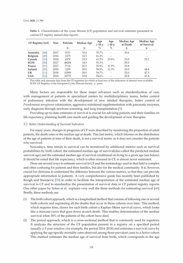
Genes 2020, 11, 589
Table 1. Characteristics of the cystic fibrosis (CF) population and survival estimates presented invarious CF registry annual data reports.
CF Registry [ref] Year Patients Median AgeAge≥ 18 y.
Age≥ 40 y.
Median Ageat Death
Median Ageof Survival
n y. % % y. y.
Australia [48] 2017 3151 19.6 53.7% - 35.6 -Belgium [49] 2016 1275 22.5 61.2% - - -Canada [10] 2018 4370 23.5 61.5% 15.9% 33.0 52.1ECFS [50] 2017 48204 18.5 51.3% - 29.0 -France [51] 2017 7114 20.3 55.9% 11.9% 33.8 -Ireland [52] 2018 1239 20.9 58.5% 11.3% 33.0 44.4UK [11] 2018 10509 20.0 54.7% - 32.0 47.3USA [12] 2018 30775 19.8 54.6% - 30.8 47.4
This table only presents data from the CF registries for which at least two of the indicators of interest were available.ECFS: CF Registry of the European Cystic Fibrosis Society. y.: years.
Many factors are responsible for these major advances such as standardization of care,with management of patients in specialized centers by multidisciplinary teams, better controlof pulmonary infection with the development of new inhaled therapies, better control ofPseudomonas aeruginosa colonization, aggressive nutritional supplementation with pancreatic enzymes,early diagnosis through newborn screening, and lung transplantation [5].
Providing up-to-date estimates of survival is crucial for advising patients and their families onlife expectancy, planning health care needs and guiding the development of new therapies.
3.2. Better Understanding of Survival Indicators
For many years, changes in prognosis of CF were described by monitoring the proportion of adultpatients, the death rates or the median age at death. This last metric, which informs on the distributionof the age of patients at time of their death, is not a survival metric as it does not consider the patientswho survived.
Nowadays, time trends in survival can be monitored by additional metrics such as survivalprobabilities by birth cohort, the estimated median age of survival(also called the predicted mediansurvival age) and the estimated median age of survival conditional on living to a given age (see below).It should be noted that life expectancy, which is often misused in CF, is almost never estimated.
There are several ways to estimate survival in CF and the terminology used in that field is complexand often confusing for patients and their families, but also for the medical community. It is, however,crucial for clinicians to understand the difference between the various metrics, so that they can provideappropriate information to patients. A very comprehensive guide has recently been published byKeogh and Stanojevic [59] in order to facilitate the interpretation of the estimated median age ofsurvival in CF and to standardize the presentation of survival data in CF patient registry reports.One other paper by Sykes et al. explains very well the three methods for estimating survival [60].Briefly, these methods are:
(1) The birth cohort approach, which is a longitudinal method that consists of following one or severalbirth cohorts and registering all the deaths that occur in those cohorts over time. This method,which requires time, draws for each birth cohort a Kaplan–Meier survival curve, which lookslike a staircase curve that goes down at each death. This enables determination of the mediansurvival when 50% of the patients of the cohort have died.
(2) The period approach, which is a cross-sectional method that is commonly used by registries.It analyzes the structure of the CF population present in a registry on a specified period(usually a 5 year window; for example, the period 2014–2018) and estimates a survival curve byapplying the age-specific mortality rates observed among those prevalent cases to a fictive cohort.This method estimates the median age of survival from birth, which corresponds to the age
126
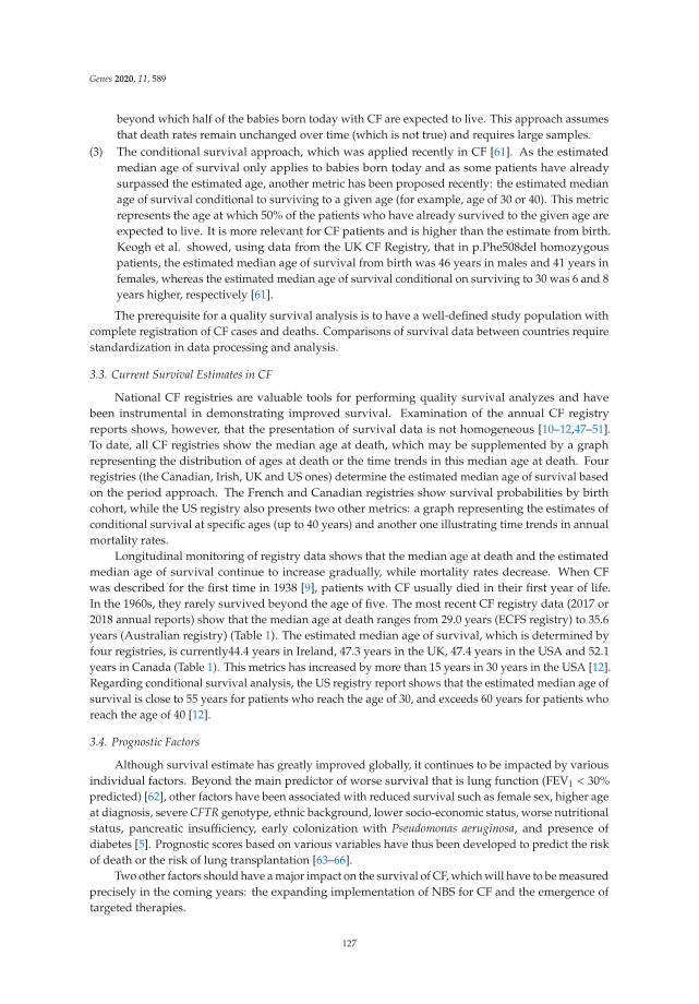
Genes 2020, 11, 589
beyond which half of the babies born today with CF are expected to live. This approach assumesthat death rates remain unchanged over time (which is not true) and requires large samples.
(3) The conditional survival approach, which was applied recently in CF [61]. As the estimatedmedian age of survival only applies to babies born today and as some patients have alreadysurpassed the estimated age, another metric has been proposed recently: the estimated medianage of survival conditional to surviving to a given age (for example, age of 30 or 40). This metricrepresents the age at which 50% of the patients who have already survived to the given age areexpected to live. It is more relevant for CF patients and is higher than the estimate from birth.Keogh et al. showed, using data from the UK CF Registry, that in p.Phe508del homozygouspatients, the estimated median age of survival from birth was 46 years in males and 41 years infemales, whereas the estimated median age of survival conditional on surviving to 30 was 6 and 8years higher, respectively [61].
The prerequisite for a quality survival analysis is to have a well-defined study population withcomplete registration of CF cases and deaths. Comparisons of survival data between countries requirestandardization in data processing and analysis.
3.3. Current Survival Estimates in CF
National CF registries are valuable tools for performing quality survival analyzes and havebeen instrumental in demonstrating improved survival. Examination of the annual CF registryreports shows, however, that the presentation of survival data is not homogeneous [10–12,47–51].To date, all CF registries show the median age at death, which may be supplemented by a graphrepresenting the distribution of ages at death or the time trends in this median age at death. Fourregistries (the Canadian, Irish, UK and US ones) determine the estimated median age of survival basedon the period approach. The French and Canadian registries show survival probabilities by birthcohort, while the US registry also presents two other metrics: a graph representing the estimates ofconditional survival at specific ages (up to 40 years) and another one illustrating time trends in annualmortality rates.
Longitudinal monitoring of registry data shows that the median age at death and the estimatedmedian age of survival continue to increase gradually, while mortality rates decrease. When CFwas described for the first time in 1938 [9], patients with CF usually died in their first year of life.In the 1960s, they rarely survived beyond the age of five. The most recent CF registry data (2017 or2018 annual reports) show that the median age at death ranges from 29.0 years (ECFS registry) to 35.6years (Australian registry) (Table 1). The estimated median age of survival, which is determined byfour registries, is currently44.4 years in Ireland, 47.3 years in the UK, 47.4 years in the USA and 52.1years in Canada (Table 1). This metrics has increased by more than 15 years in 30 years in the USA [12].Regarding conditional survival analysis, the US registry report shows that the estimated median age ofsurvival is close to 55 years for patients who reach the age of 30, and exceeds 60 years for patients whoreach the age of 40 [12].
3.4. Prognostic Factors
Although survival estimate has greatly improved globally, it continues to be impacted by variousindividual factors. Beyond the main predictor of worse survival that is lung function (FEV1 < 30%predicted) [62], other factors have been associated with reduced survival such as female sex, higher ageat diagnosis, severe CFTR genotype, ethnic background, lower socio-economic status, worse nutritionalstatus, pancreatic insufficiency, early colonization with Pseudomonas aeruginosa, and presence ofdiabetes [5]. Prognostic scores based on various variables have thus been developed to predict the riskof death or the risk of lung transplantation [63–66].
Two other factors should have a major impact on the survival of CF, which will have to be measuredprecisely in the coming years: the expanding implementation of NBS for CF and the emergence oftargeted therapies.
127

Genes 2020, 11, 589
3.5. New Statistical Developments and Future Trends in Survival
A survey that was recently carried out to decipher how CF patients access and use informationon life expectancy shows that most respondents wanted more personalized survival data [67].Mathematical developments are therefore underway in order to further improve the modelingof survival by providing personalized estimates. One of the challenges is to develop models based onlongitudinal data that are able to consider the current health status of the patients (and not only theirbaseline characteristics). A dynamic predictive model providing personalized estimates of survivalwas recently developed using data from the UK CF Registry [68]. This model, which integrates 16predictors, is able to predict survival up to 10 years for patients up to 50 years of age.
The estimated median age of survival of CF patients, which is close to 50 years today, is expectedto continue to improve in the future, with the rapid expanding of NBS for CF worldwide over the pastdecade and with the recent advent of CFTR modulator therapies. Further work is needed to assessthe effect of these factors on the survival of CF. Some studies have suggested that NBS for CF results ina prolonged survival, but few are yet able to assess its long-term effects. The studies performed in thatfield are presented in the next section.
The increase in survival estimates undeniably changes the population’s perception of the diseaseand leads to ethical reflection on the decisions to be made by couples when CF is diagnosed before birth.
4. The Impact of the Discovery of the CFTR Gene on the Epidemiology of the Disease
The discovery of the CFTR gene in 1989 marked an important milestone in the history of CFand raised tremendous hope in the medical and scientific community. An exemplary collaborationinvolving more than 100 laboratories worldwide and carried out through an international consortiumfor the study of the gene mutations (Cystic Fibrosis Genetic Analysis Consortium) has led tothe identification of more than 2000 different CFTR mutations to date [69]. The molecular explorationof this gene has enabled a better understanding of the genotype/phenotype correlations, improvedthe diagnosis and the management of CF patients and their families, and opened up the wayto the emergence of mutation-specific therapies, which contribute to modify the epidemiology ofCF [16,17]. In a very comprehensive article published in this special issue of Genes, Farrell et al.reviews the “impact of the CFTR gene discovery on CF diagnosis, genetic counseling and preventivetherapy” [16].
4.1. Study of Genotype/Phenotype Correlations
The growing number of mutations identified in the CFTR gene and the variability observed inthe phenotypic expression of CF have led the researchers to try to establish genotype/phenotypecorrelations [70,71].
Quickly after the gene discovery, the CFTR mutations could be classified into six classes accordingto their impact on the level of protein function [72]. Schematically, mutations in classes I, II, and III areusually associated with a classical form of CF (severe mutations), while those in classes IV, V, and VIare related to a milder phenotype (mild mutations) characterized by pancreatic sufficiency and laterbacterial colonization. The estimated median age of survival of patients carrying at least one mildmutation is generally ten years higher than that of patients with severe mutations.
Various tools are available to help identify the clinical impact of a CFTR variant: (1) internationalmutations databases such as the Cystic Fibrosis Mutation Database (http://www.genet.sickkids.on.ca/),CFTR2 database (https://cftr2.org/) or UMD-CFTR database (http://www.umd.be/CFTR/); (2) CFregistries, which may assist genetic counseling by providing aggregated clinical data associated witha given genotype; (3) bioinformatics prediction tools such as Polyphen or SIFT applications.
Despite these tools, it often remains difficult to predict with certainty the phenotype of a givengenotype, and it quickly became obvious that the CFTR genotype could not explain all the phenotypicvariability observed in CF, in particular in lung damage. Further research has been undertaken to
128

Genes 2020, 11, 589
identify other factors that influence the severity of the disease, including gene modifiers (such as genesinvolved in the immune response or in inflammation) [73,74], epigenetic factors and environmentalfactors (such as tobacco, pollution, socio-economic status, and adherence to therapies) [75].
The study of genotype/phenotype correlations also highlighted the existence of conditionsassociated with CFTR dysfunction that do not fulfill diagnostic criteria for CF. Those disorders, calledCFTR-related disorders (CFTR-RDs) [76], include congenital bilateral absence of vas deferens, acuterecurrent or chronic pancreatitis and disseminated bronchiectasis. Inclusion of patients with suchconditions in epidemiological studies dealing with incidence or survival of CF can bias the estimatesand the time trends.
4.2. Implementation of Genetic-Based Health Policies
In-depth knowledge of the molecular abnormalities of the CFTR gene [77] has enabled geneticscreening policies to be implemented, allowing prevention within families (such as prenatal diagnosisfor one-in-four at-risk couples or carrier testing in families) or in the general population in some areas(such as population carrier screening). As mentioned above, these health policies have contributed toreducing—in most countries and with varying degrees of importance—the incidence of CF throughthe identification of cases before birth [23,39–43,46].
The discovery of the CFTR gene has also increased the performance of NBS for CF by introducingDNA analysis into the screening protocol, and thus improved the diagnosis and management ofCF [16,78,79]. The coupling of the immunereactive trypsinogen assay to the search for CFTR mutationshas eliminated the need for a second blood sample. This has reduced the anxiety generated in families,leading to an earlier diagnosis and early access to specialized care centers, which prevents malnutritionand lung damage [80]. The positive effects of NBS for CF on short-term and long-term clinical outcomesare widely recognized [80–83] and include better nutritional status, lower pancreatic insufficiency,better lung function, lower infection rates, fewer and shorter hospitalizations. Some studies haveshown that NBS for CF results in prolonged survival [84–86], but it is still too early for most countriesto assess its long-term impact on survival. This could recently be measured in an Italian area inwhich NBS for CF has been running for over 40 years [87]. Tridello et al. reported a significantlyhigher survival probability at 20 years in the screened than in the non-screened group, both in patientswith severe (84.9% vs. 63.6%; difference: 21.3%; p = 0.007) and moderate disease (94.5% vs. 85.9%;difference: 8.6%; p = 0.016). A 9% difference was also observed in the survival probability at 30 years(80.1% vs. 71.0%) but was not significant [87]. Through early diagnosis, NBS for CF maximizes survivalin severe CF. The expanding of NBS worldwide in the past decade will inevitably continue to impactsurvival in the future.
4.3. Advent of CFTR Modulator Therapies
Deciphering of the molecular bases of CF has also led to the development of novel therapeuticapproaches and the search for pharmaceutical treatments aiming at correcting the defective CFTRprotein. These drugs, called CFTR modulators, search to improve the production, processing orexpression of the protein and include correctors, potentiators, stabilizers, amplifiers and read throughagents [88,89]. This approach is said to be “targeted” or “mutation specific” because the typeof molecules to be administered to patients depends on the type of CFTR mutations they carry.Many studies have been carried out in that field over the past decade and have led to major clinicaladvances in treatment, with significant improvements in biological and clinical endpoints of CF(as sweat chloride concentration orFEV1) [90].
It was almost 25 years after the discovery of the CFTR gene that the first CFTR modulator(ivacaftor—Kalydeco®) could be marketed. This potentialtor was approved in 2012 for the treatmentof CF patients aged ≥6 years carrying at least one G551D mutation [91]. This treatment was associatedwith significant improvements at day 28 in sweat chloride level, nasal potential difference and lungfunction (median increase of 8.7 points in the percentage of predicted FEV1). In 2015, the combination
129
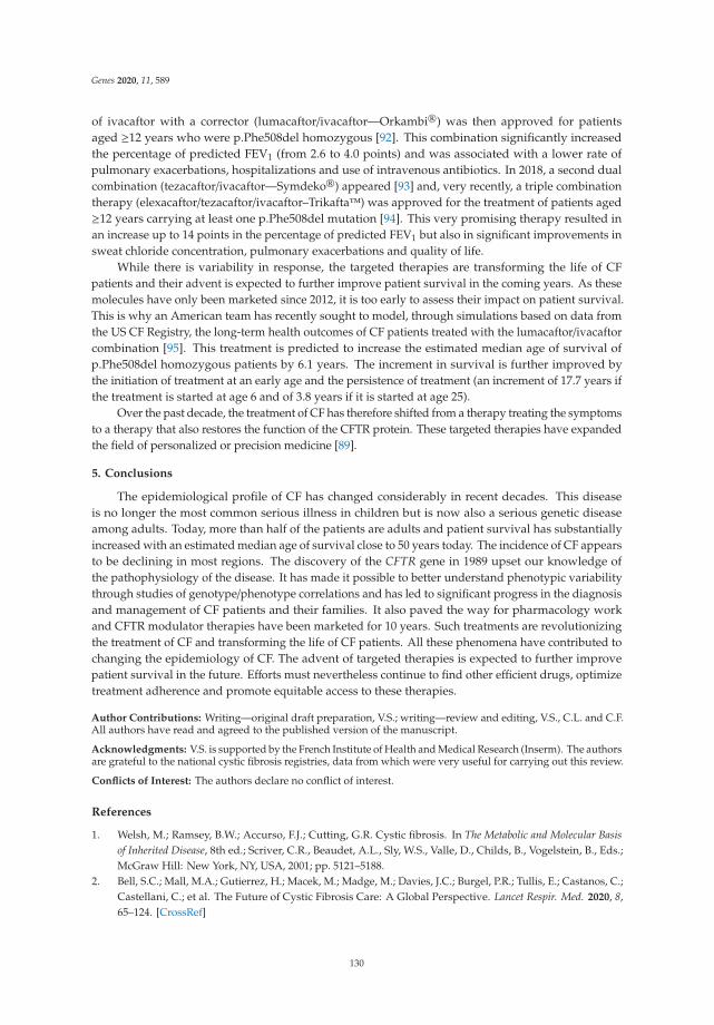
Genes 2020, 11, 589
of ivacaftor with a corrector (lumacaftor/ivacaftor—Orkambi®) was then approved for patientsaged ≥12 years who were p.Phe508del homozygous [92]. This combination significantly increasedthe percentage of predicted FEV1 (from 2.6 to 4.0 points) and was associated with a lower rate ofpulmonary exacerbations, hospitalizations and use of intravenous antibiotics. In 2018, a second dualcombination (tezacaftor/ivacaftor—Symdeko®) appeared [93] and, very recently, a triple combinationtherapy (elexacaftor/tezacaftor/ivacaftor–Trikafta™) was approved for the treatment of patients aged≥12 years carrying at least one p.Phe508del mutation [94]. This very promising therapy resulted inan increase up to 14 points in the percentage of predicted FEV1 but also in significant improvements insweat chloride concentration, pulmonary exacerbations and quality of life.
While there is variability in response, the targeted therapies are transforming the life of CFpatients and their advent is expected to further improve patient survival in the coming years. As thesemolecules have only been marketed since 2012, it is too early to assess their impact on patient survival.This is why an American team has recently sought to model, through simulations based on data fromthe US CF Registry, the long-term health outcomes of CF patients treated with the lumacaftor/ivacaftorcombination [95]. This treatment is predicted to increase the estimated median age of survival ofp.Phe508del homozygous patients by 6.1 years. The increment in survival is further improved bythe initiation of treatment at an early age and the persistence of treatment (an increment of 17.7 years ifthe treatment is started at age 6 and of 3.8 years if it is started at age 25).
Over the past decade, the treatment of CF has therefore shifted from a therapy treating the symptomsto a therapy that also restores the function of the CFTR protein. These targeted therapies have expandedthe field of personalized or precision medicine [89].
5. Conclusions
The epidemiological profile of CF has changed considerably in recent decades. This diseaseis no longer the most common serious illness in children but is now also a serious genetic diseaseamong adults. Today, more than half of the patients are adults and patient survival has substantiallyincreased with an estimated median age of survival close to 50 years today. The incidence of CF appearsto be declining in most regions. The discovery of the CFTR gene in 1989 upset our knowledge ofthe pathophysiology of the disease. It has made it possible to better understand phenotypic variabilitythrough studies of genotype/phenotype correlations and has led to significant progress in the diagnosisand management of CF patients and their families. It also paved the way for pharmacology workand CFTR modulator therapies have been marketed for 10 years. Such treatments are revolutionizingthe treatment of CF and transforming the life of CF patients. All these phenomena have contributed tochanging the epidemiology of CF. The advent of targeted therapies is expected to further improvepatient survival in the future. Efforts must nevertheless continue to find other efficient drugs, optimizetreatment adherence and promote equitable access to these therapies.
Author Contributions: Writing—original draft preparation, V.S.; writing—review and editing, V.S., C.L. and C.F.All authors have read and agreed to the published version of the manuscript.
Acknowledgments: V.S. is supported by the French Institute of Health and Medical Research (Inserm). The authorsare grateful to the national cystic fibrosis registries, data from which were very useful for carrying out this review.
Conflicts of Interest: The authors declare no conflict of interest.
References
1. Welsh, M.; Ramsey, B.W.; Accurso, F.J.; Cutting, G.R. Cystic fibrosis. In The Metabolic and Molecular Basisof Inherited Disease, 8th ed.; Scriver, C.R., Beaudet, A.L., Sly, W.S., Valle, D., Childs, B., Vogelstein, B., Eds.;McGraw Hill: New York, NY, USA, 2001; pp. 5121–5188.
2. Bell, S.C.; Mall, M.A.; Gutierrez, H.; Macek, M.; Madge, M.; Davies, J.C.; Burgel, P.R.; Tullis, E.; Castanos, C.;Castellani, C.; et al. The Future of Cystic Fibrosis Care: A Global Perspective. Lancet Respir. Med. 2020, 8,65–124. [CrossRef]
130
Genes 2020, 11, 589
3. Dodge, J.A.; Lewis, P.A. Cystic fibrosis is no longer an important cause of childhood death in the UK.Arch. Dis. Child. 2005, 90, 547.
4. Fajac, I.; Burgel, P.R. Demographic growth and targeted therapies: The changing face of cystic fibrosis.Rev. Mal. Respir. 2016, 33, 645–647. [CrossRef] [PubMed]
5. Stephenson, A.L.; Stanojevic, S.; Sykes, J.; Burgel, P.R. The changing epidemiology and demography of cysticfibrosis. Presse Med. 2017, 46, e87–e95. [CrossRef]
6. Corriveau, S.; Sykes, J.; Stephenson, A.L. Cystic fibrosis survival: The changing epidemiology. Curr. Opin.Pulm. Med. 2018, 24, 574–578. [CrossRef] [PubMed]
7. De Boeck, K. Cystic fibrosis in the year 2020: A disease with a new face. Acta Paediatr. 2020, in press.[CrossRef] [PubMed]
8. Stephenson, A.L.; Tom, M.; Berthiaume, Y.; Singer, L.G.; Aaron, S.D.; Whitmore, G.A.; Stanojevic, S.A contemporary survival analysis of individuals with cystic fibrosis: A cohort study. Eur. Respir. J. 2015, 45,670–679. [CrossRef] [PubMed]
9. Anderson, D.H. Cystic fibrosis of the pancreas and its relation to celiac disease. A clinical and pathologicstudy. Am. J. Dis. Child. 1938, 56, 344–399. [CrossRef]
10. Canadian Cystic Fibrosis Registry. Annual Data Report 2018. Available online: https://www.cysticfibrosis.ca/uploads/RegistryReport2018/2018RegistryAnnualDataReport.pdf (accessed on 20 April 2020).
11. UK Cystic Fibrosis Registry (Cystic Fibrosis Trust). Annual Data Report 2018. Available online: https://www.cysticfibrosis.org.uk/~{}/media/documents/the-work-we-do/uk-cf-registry/2018-registry-annual-data-report.ashx?la=en(accessed on 20 April 2020).
12. US Cystic Fibrosis Registry (Cystic Fibrosis Foundation). Annual Data Report 2018. Available online: https://www.cff.org/Research/Researcher-Resources/Patient-Registry/2018-Patient-Registry-Annual-Data-Report.pdf(accessed on 20 April 2020).
13. Rommens, J.M.; Iannuzzi, M.C.; Kerem, B.; Drumm, M.L.; Melmer, G.; Dean, M.; Rozmahel, R.; Cole, J.L.;Kennedy, D.; Hidaka, N.; et al. Identification of the cystic fibrosis gene: Chromosome walking and jumping.Science 1989, 245, 1059–1065. [CrossRef]
14. Riordan, J.R.; Rommens, J.M.; Kerem, B.; Alon, N.; Rozmahel, R.; Grzelczak, Z.; Zielenski, J.; Lok, S.;Plavsic, N.; Chou, J.L.; et al. Identification of the cystic fibrosis gene: Cloning and characterization ofcomplementary DNA. Science 1989, 245, 1066–1073. [CrossRef]
15. Kerem, B.; Rommens, J.M.; Buchanan, J.A.; Markiewicz, D.; Cox, T.K.; Chakravarti, A.; Buchwald, M.;Tsui, L.C. Identification of the cystic fibrosis gene: Genetic analysis. Science 1989, 245, 1073–1080. [CrossRef][PubMed]
16. Farrell, P.M.; Rock, M.J.; Baker, M.W. The Impact of the CFTR Gene Discovery on Cystic Fibrosis Diagnosis,Counseling, and Preventive Therapy. Genes 2020, 11, 401. [CrossRef]
17. Férec, C.; Scotet, V. Genetics ofcystic fibrosis: Basics. Arch. Pediatr. 2020, 27 (Suppl. 1), eS4–eS7.18. Southern, K.W.; Munck, A.; Pollitt, R.; Travert, G.; Zanolla, L.; Dankert-Roelse, J.; Castellani, C.; ECFS CF
Neonatal Screening Working Group. A survey of newborn screening for cystic fibrosis in Europe. J. Cyst.Fibros. 2007, 6, 57–65. [CrossRef] [PubMed]
19. Scotet, V.; Gutierrez, H.; Farrell, P.M. Newborn screening for CF across the globe —Where is it worthwhile?Int. J. Neonatal Screen. 2020, 6, 18. [CrossRef]
20. Farrell, P.M.; Joffe, S.; Foley, L.; Canny, G.J.; Mayne, P.; Rosenberg, M. Diagnosis of cystic fibrosis inthe Republic of Ireland: Epidemiology and costs. Ir. Med. J. 2007, 100, 557–560.
21. Kere, J.; Estivill, X.; Chillon, M.; Morral, N.; Nunes, V.; Norio, R.; Savilahti, E.; de la Chapelle, A. Cysticfibrosis in a low-incidence population: Two major mutations in Finland. Hum. Genet. 1994, 93, 162–166.[CrossRef]
22. Audrézet, M.P.; Munck, A.; Scotet, V.; Claustres, M.; Roussey, M.; Delmas, D.; Férec, C.; Desgeorges, M.Comprehensive CFTR gene analysis of the French cystic fibrosis screened newborn cohort: Implications fordiagnosis, genetic counseling, and mutation-specific therapy. Genet. Med. 2015, 17, 108–116. [CrossRef]
23. Castellani, C.; Picci, L.; Tridello, G.; Casati, E.; Tamanini, A.; Bartoloni, L.; Scarpa, M.; Assael, B.M.; Veneto CFLab Network. Cystic fibrosis carrier screening effects on birth prevalence and newborn screening. Genet. Med.2016, 18, 145–151. [CrossRef]
24. Dankert-Roelse, J.E.; Bouva, M.J.; Jakobs, B.S.; Janssens, H.M.; de Winter-de Groot, K.M.; Schonbeck, Y.;Gille, J.J.P.; Gulmans, V.A.M.; Verschoof-Puite, R.K.; Schielen, P.; et al. Newborn blood spot screening
131
Genes 2020, 11, 589
for cystic fibrosis with a four-step screening strategy in the Netherlands. J. Cyst. Fibros. 2019, 18, 54–63.[CrossRef]
25. Skov, M.; Baekvad-Hansen, M.; Hougaard, D.M.; Skogstrand, K.; Lund, A.M.; Pressler, T.; Olesen, H.V.;Duno, M. Cystic fibrosis newborn screening in Denmark: Experience from the first 2 years. Pediatr. Pulmonol.2020, 55, 549–555. [CrossRef] [PubMed]
26. Soltysova, A.; Tothova Tarova, E.; Ficek, A.; Baldovic, M.; Polakova, H.; Kayserova, H.; Kadasi, L.Comprehensive genetic study of cystic fibrosis in Slovak patients in 25 years of genetic diagnostics.Clin. Respir. J. 2018, 12, 1197–1206. [CrossRef] [PubMed]
27. David, J.; Chrastina, P.; Peskova, K.; Kozich, V.; Friedecky, D.; Adam, T.; Hlidkova, E.; Vinohradska, H.;Novotna, D.; Hedelova, M.; et al. Epidemiology of rare diseases detected by newborn screening in the CzechRepublic. Cent. Eur. J. Public Health 2019, 27, 153–159. [CrossRef] [PubMed]
28. Massie, R.J.; Curnow, L.; Glazner, J.; Armstrong, D.S.; Francis, I. Lessons learned from 20 years of newbornscreening for cystic fibrosis. Med. J. Aust. 2012, 196, 67–70. [CrossRef]
29. Lilley, M.; Christian, S.; Hume, S.; Scott, P.; Montgomery, M.; Semple, L.; Zuberbuhler, P.; Tabak, J.; Bamforth, F.;Somerville, M.J. Newborn screening forcystic fibrosisin Alberta: Two years of experience. Paediatr. Child.Health 2010, 15, 590–594. [CrossRef]
30. Kosorok, M.R.; Wei, W.H.; Farrell, P.M. The incidence ofcystic fibrosis. Stat. Med. 1996, 15, 449–462.[CrossRef]
31. Sullivan, B.P.; Freedman, S.D. Cystic fibrosis. Lancet 2009, 373, 1891–1904. [CrossRef]32. Klinger, K.W. Cystic fibrosisin the OhioAmish: Gene frequency and founder effect. Hum. Genet. 1983, 65,
94–98. [CrossRef]33. Daigneault, J.; Aubin, G.; Simard, F.; De Braekeleer, M. Genetic epidemiology of cystic fibrosis in
Saguenay-Lac-St-Jean (Quebec, Canada). Clin. Genet. 1991, 40, 298–303. [CrossRef]34. Nazer, H.M. Early diagnosis of cystic fibrosis in Jordanian children. J. Trop. Pediatr. 1992, 38, 113–115.
[CrossRef]35. Yamashiro, Y.; Shimizu, T.; Oguchi, S.; Shioya, T.; Nagata, S.; Ohtsuka, Y. The estimated incidence of cystic
fibrosis in Japan. J. Pediatr. Gastroenterol. Nutr. 1997, 24, 544–547. [CrossRef] [PubMed]36. Goodchild, M.C.; Insley, J.; Rushton, D.I.; Gaze, H. Cystic fibrosisin 3 Pakistani children. Arch. Dis. Child.
1974, 49, 739–741. [CrossRef] [PubMed]37. Kapoor, V.; Shastri, S.S.; Kabra, M.; Kabra, S.K.; Ramachandran, V.; Arora, S.; Balakrishnan, P.; Deorari, A.K.;
Paul, V.K. Carrier frequency of F508del mutation of cystic fibrosis in Indian population. J. Cyst. Fibros. 2006,5, 43–46. [CrossRef] [PubMed]
38. Kwarteng Owusu, S.; Morrow, B.M.; White, D.; Klugman, S.; Vanker, A.; Gray, D.; Zampoli, M. Cystic Fibrosisin Black African Children in South Africa: A Case Control Study. J. Cyst. Fibros. 2019. [CrossRef]
39. Hale, J.E.; Parad, R.B.; Comeau, A.M. Newborn screening showing decreasing incidence of cystic fibrosis.N. Engl. J. Med. 2008, 358, 973–974. [CrossRef]
40. Castellani, C.; Picci, L.; Tamanini, A.; Girardi, P.; Rizzotti, P.; Assael, B.M. Association between carrierscreening and incidence of cystic fibrosis. JAMA 2009, 302, 2573–2579. [CrossRef]
41. Massie, J.; Curnow, L.; Gaffney, L.; Carlin, J.; Francis, I. Declining prevalence of cystic fibrosis sincethe introduction of newborn screening. Arch. Dis. Child. 2010, 95, 531–533. [CrossRef]
42. Scotet, V.; Duguépéroux, I.; Saliou, P.; Rault, G.; Roussey, M.; Audrézet, M.P.; Férec, C. Evidence for declinein the incidence of cystic fibrosis: A 35-year observational study in Brittany, France. Orphanet J. Rare Dis.2012, 7, 14. [CrossRef]
43. Stafler, P.; Mei-Zahav, M.; Wilschanski, M.; Mussaffi, H.; Efrati, O.; Lavie, M.; Shoseyov, D.;Cohen-Cymberknoh, M.; Gur, M.; Bentur, L.; et al. The impact of a national population carrier screeningprogram on cystic fibrosis birth rate and age at diagnosis: Implications for newborn screening. J. Cyst. Fibros.2016, 15, 460–466. [CrossRef]
44. Sontag, M.L.; Wagener, J.S.; Accurso, F.; Sager, S.D. Consistent incidence of cystic fibrosis in a long-termnewborn screen population. In Proceedings of the 22nd AnnualNorth American Cystic Fibrosis ConferenceMeeting, Orlando, FL, USA, 23–26 October 2008.
45. Parker-McGill, K.; Nugent, M.; Bersie, R.; Hoffman, G.; Rock, M.; Baker, M.; Farrell, P.M.; Simpson, P.; Levy, H.Changing incidence of cystic fibrosis in Wisconsin, USA. Pediatr. Pulmonol. 2015, 50, 1065–1072. [CrossRef]
132

Genes 2020, 11, 589
46. Scotet, V.; Audrézet, M.P.; Roussey, M.; Rault, G.; Blayau, M.; De Braekeleer, M.; Férec, C. Impact of publichealth strategies on the birth prevalence of cystic fibrosis in Brittany, France. Hum. Genet. 2003, 113, 280–285.[CrossRef] [PubMed]
47. Australian Cystic Fibrosis Registry. Annual Data Report 2017. Available online: https://www.cysticfibrosis.org.au/getmedia/24e94d66-29fa-4e3f-8e65-21ee24ed2e5a/ACFDR-2017-Annual-Report_highres_singles.pdf.aspx(accessed on 20 April 2020).
48. Belgian Cystic Fibrosis Registry. Annual Data Report 2016. Available online: https://www.sciensano.be/sites/www.wiv-isp.be/files/report_belgian_cf_registry_2016_en_final_1.pdf (accessed on 20 April 2020).
49. European (ECFS) Cystic Fibrosis Registry. Annual Data Report 2018. Available online: https://www.ecfs.eu/sites/default/files/general-content-images/working-groups/ecfs-patient-registry/ECFSPR_Report2017_v1.3.pdf(accessed on 20 April 2020).
50. French Cystic Fibrosis Registry. Annual Data Report 2017. Available online: http://www.vaincrelamuco.org/sites/default/files/rapport_du_registre_-_donnees_2017.pdf (accessed on 20 April 2020).
51. Irish Cystic Fibrosis Registry. Annual Data Report 2018. Available online: https://cfri.ie/annual-reports/(accessed on 20 April 2020).
52. Burgel, P.R.; Bellis, G.; Olesen, H.V.; Viviani, L.; Zolin, A.; Blasi, F.; Elborn, J.S.; ERS/ECFS Task force onprovision of care for adults withcystic fibrosisin Europe. Future trends in cystic fibrosis demography in 34European countries. Eur. Respir. J. 2015, 46, 133–141. [CrossRef] [PubMed]
53. Coyne, I.; Sheehan, A.M.; Heery, E.; While, A.E. Improvingtransitiontoadulthealthcare for young peoplewithcysticfibrosis: A systematic review. J. Child. Health Care 2017, 21, 312–330. [CrossRef] [PubMed]
54. Hughan, K.S.; Daley, T.; Rayas, M.S.; Kelly, A.; Roe, A. Female reproductive health in cystic fibrosis. J. Cyst.Fibros. 2019, 18 (Suppl. 2), S95–S104. [CrossRef]
55. Vekaria, S.; Popowicz, N.; White, S.W.; Mulrennan, S.J. To be or not to be on CFTR modulatorsduringpregnancy: Risks to be considered. J. Cyst. Fibros. 2019, S1569-1993, 30983-X.
56. Hodson, M.E.; Simmonds, N.J.; Warwick, W.J.; Tullis, E.; Castellani, C.; Assael, B.; Dodge, J.A.; Corey, M.;International study of aging in cystic fibrosis. An international/multicentre report on patients with cysticfibrosis (CF) over the age of 40 years. J. Cyst. Fibros. 2008, 7, 537–542. [CrossRef]
57. Nick, J.A.; Chacon, C.S.; Brayshaw, S.J.; Jones, M.C.; Barboa, C.M.; St Clair, C.G.; Young, R.L.; Nichols, D.P.;Janssen, J.S.; Huitt, G.A.; et al. Effects of gender and age at diagnosis on disease progression in long-termsurvivors of cystic fibrosis. Am. J. Respir. Crit. Care Med. 2010, 182, 614–626. [CrossRef]
58. Férec, C.; Verlingue, C.; Guillermit, H.; Quéré, I.; Raguénès, O.; Feigelson, J.; Audrézet, M.P.; Moullier, P.;Mercier, B. Genotype Analysis of Adult Cystic Fibrosis Patients. Hum. Mol. Genet. 1993, 2, 1557–1560.[CrossRef]
59. Keogh, R.H.; Stanojevic, S. A guide to interpreting estimated median age of survival in cystic fibrosispatientregistry reports. J. Cyst. Fibros. 2018, 17, 213–217. [CrossRef]
60. Sykes, J.; Stanojevic, S.; Goss, C.H.; Quon, B.S.; Marshall, B.C.; Petren, K.; Ostrenga, J.; Fink, A.; Elbert, A.;Stephenson, A.L. A standardized approach to estimating survival statistics for population-basedcysticfibrosisregistry cohorts. J. Clin. Epidemiol. 2016, 70, 206–213. [CrossRef]
61. Keogh, R.H.; Szczesniak, R.; Taylor-Robinson, D.; Bilton, D. Up-to-date and projected estimates of survivalfor people with cystic fibrosis using baseline characteristics: A longitudinal study using UK patient registrydata. J. Cyst. Fibros. 2018, 17, 218–227. [CrossRef] [PubMed]
62. Kerem, E.; Reisman, J.; Corey, M.; Canny, G.J.; Levison, H. Prediction of mortality in patients with cysticfibrosis. N. Engl. J. Med. 1992, 326, 1187–1191. [CrossRef] [PubMed]
63. Liou, T.G.; Adler, F.R.; Fitzsimmons, S.C.; Cahill, B.C.; Hibbs, J.R.; Marshall, B.C. Predictive 5-year survivorshipmodel of cystic fibrosis. Am. J. Epidemiol. 2001, 153, 345–352. [CrossRef] [PubMed]
64. Aaron, S.D.; Stephenson, A.L.; Cameron, D.W.; Whitmore, G.A. A statistical model to predict one-year risk ofdeath in patients with cystic fibrosis. J. Clin. Epidemiol. 2015, 68, 1336–1345. [CrossRef]
65. Nkam, L.; Lambert, J.; Latouche, A.; Bellis, G.; Burgel, P.R.; Hocine, M.N. A 3-year Prognostic Score forAdults with Cystic Fibrosis. J. Cyst. Fibros. 2017, 16, 702–708. [CrossRef]
66. Stanojevic, S.; Sykes, J.; Stephenson, A.L.; Aaron, S.D.; Whitmore, G.A. Development and External Validationof 1- And 2-year Mortality Prediction Models in Cystic Fibrosis. Eur. Respir. J. 2019, 54, 1900224. [CrossRef]
133
Genes 2020, 11, 589
67. Keogh, R.H.; Bilton, D.; Cosgriff, R.; Kavanagh, D.; Rayner, O.; Sedgwick, P.M. Results from an online surveyof adults with cystic fibrosis: Accessing and using life expectancy information. PLoS ONE 2019, 14, e0213639.[CrossRef]
68. Keogh, R.H.; Seaman, S.R.; Barrett, J.K.; Taylor-Robinson, D.; Szczesniak, R. Dynamic Prediction of Survivalin Cystic Fibrosis: A Landmarking Analysis Using UK Patient Registry Data. Epidemiology 2019, 30, 29–37.[CrossRef]
69. Cystic Fibrosis Mutation Database. Available online: http://www.genet.sickkids.on.ca/app (accessed on20 April 2020).
70. Kerem, E.; Corey, M.; Kerem, B.S.; Rommens, J.; Markiewicz, D.; Levison, H.; Tsui, L.C.; Durie, P. The relationbetween genotype and phenotype in cystic fibrosis – analysis of the most common mutation (delta F508).N. Engl. J. Med. 1990, 323, 1517–1522. [CrossRef]
71. The Cystic Fibrosis Genotype-Phenotype Consortium. Correlation between genotype and phenotype inpatients with cystic fibrosis. N. Engl. J. Med. 1993, 329, 1308–1313. [CrossRef]
72. Welsh, M.J.; Smith, A.E. Molecular mechanisms of CFTR chloride channel dysfunction in cystic fibrosis. Cell1993, 73, 1251–1254. [CrossRef]
73. Drumm, M.L.; Konstan, M.W.; Schluchter, M.D.; Handler, A.; Pace, R.; Zou, F.; Zariwala, M.; Fargo, D.; Xu, A.;Dunn, J.M.; et al. Genetic modifiers of lung disease in cystic fibrosis. N. Engl. J. Med. 2005, 353, 1443–1453.[CrossRef] [PubMed]
74. Corvol, H.; Blackman, S.M.; Boelle, P.Y.; Gallins, P.J.; Pace, R.G.; Stonebraker, J.R.; Accurso, F.J.; Clement, A.;Collaco, J.M.; Dang, H.; et al. Genome-wide association meta-analysis identifies five modifier loci of lungdisease severity in cystic fibrosis. Nat. Commun. 2015, 6, 8382. [CrossRef] [PubMed]
75. Szczesniak, R.; Rice, J.L.; Brokamp, C.; Ryan, P.; Pestian, T.; Ni, Y.; Andrinopoulou, E.R.; Keogh, R.H.;Gecili, E.; Huang, R.; et al. Influences of environmental exposures on individuals living with cystic fibrosis.Expert Rev. Respir. Med. 2020, in press. [CrossRef] [PubMed]
76. Bombieri, C.; Claustres, M.; De Boeck, K.; Derichs, N.; Dodge, J.; Girodon, E.; Sermet, I.; Schwarz, M.;Tzetis, M.; Wilschanski, M.; et al. Recommendations for the Classification of Diseases as CFTR-relatedDisorders. J. Cyst. Fibros. 2011, 10 (Suppl. 2), S86–S102. [CrossRef]
77. Bareil, C.; Bergougnoux, A. CFTR gene variants, epidemiology and molecular pathology. Arch. Pediatr. 2020,27 (Suppl. 1), eS8–eS12. [CrossRef]
78. Farrell, P.M.; Mishler, E.H.; Fost, N.C.; Wilfond, B.S.; Tluczek, A.; Gregg, R.G.; Bruns, W.T.; Hassemer, D.J.;Laessig, R.H. Current issues in neonatal screening for cystic fibrosis and implications of the CF gene discovery.Pediatr. Pulmonol. 1991, 7, S11–S18. [CrossRef]
79. Scotet, V.; de Braekeleer, M.; Roussey, M.; Rault, G.; Parent, P.; Dagorne, M.; Journel, H.; Lemoigne, A.;Codet, J.P.; Catheline, M.; et al. Neonatal Screening for Cystic Fibrosis in Brittany, France: Assessment of 10Years’ Experience and Impact on Prenatal Diagnosis. Lancet 2000, 356, 789–794. [CrossRef]
80. Farrell, P.M.; Kosorok, M.R.; Laxova, A.; Shen, G.; Koscik, R.E.; Bruns, W.T.; Splaingard, M.; Mischler, E.H.Nutritional benefits of neonatal screening for cystic fibrosis. Wisconsin Cystic Fibrosis Neonatal ScreeningStudy Group. N. Engl. J. Med. 1997, 337, 963–969. [CrossRef]
81. Farrell, P.M.; Lai, H.J.; Li, Z.; Kosorok, M.R.; Laxova, A.; Green, C.G.; Collins, J.; Hoffman, G.; Laessig, R.;Rock, M.J.; et al. Evidence on improved outcomes with early diagnosis of cystic fibrosis through neonatalscreening: Enough is enough! J. Pediatr. 2005, 147, S30–S36. [CrossRef]
82. Mak, D.Y.; Sykes, J.; Stephenson, A.L.; Lands, L.C. The benefits of newborn screening for cystic fibrosis:The Canadian experience. J. Cyst. Fibros. 2016, 15, 302–308. [CrossRef] [PubMed]
83. Zhang, Z.; Lindstrom, M.J.; Farrell, P.M.; Lai, H.J.; Wisconsin Cystic Fibrosis Neonatal Screening Group.Pubertal Height Growth and Adult Height in Cystic Fibrosis After Newborn Screening. Pediatrics 2015, 137,e20152907. [CrossRef] [PubMed]
84. Mérelle, M.E.; Schouten, J.P.; Gerritsen, J.; Dankert-Roelse, J.E. Influence of neonatal screening and centralizedtreatment on long-term clinical outcome and survival of CF patients. Eur. Respir. J. 2001, 18, 306–315.[CrossRef]
85. Mastella, G.; Zanolla, L.; Castellani, C.; Altieri, S.; Furnari, M.; Giglio, L.; Lombardo, M.; Miano, A.; Sciuto, C.;Pardo, F.; et al. Neonatal screening for cystic fibrosis: Long-term clinical balance. Pancreatology 2001, 1,531–537. [CrossRef]
134
Genes 2020, 11, 589
86. Lai, H.J.; Cheng, Y.; Farrell, P.M. The survival advantage of patients with cystic fibrosis diagnosed throughneonatal screening: Evidence from the United States Cystic Fibrosis Foundation registry data. J. Pediatr.2005, 147, S57–S63. [CrossRef] [PubMed]
87. Tridello, G.; Castellani, C.; Meneghelli, I.; Tamanini, A.; Assael, B.M. Early Diagnosis from Newborn ScreeningMaximises Survival in Severe Cystic Fibrosis. ERJ Open Res. 2018, 4, 00109-2017. [CrossRef]
88. Clancy, J.P.; Cotton, C.U.; Donaldson, S.H.; Solomon, G.M.; Van Devanter, D.R.; Boyle, M.P.; Gentzsch, M.;Nick, J.A.; Illek, B.; Wallenburg, J.C.; et al. CFTR Modulator Theratyping: Current Status, Gaps and FutureDirections. J. Cyst. Fibros. 2019, 18, 22–34. [CrossRef]
89. Lopes-Pacheco, M. CFTR Modulators: The Changing Face of Cystic Fibrosis in the Era of Precision Medicine.Front. Pharmacol. 2020, 10, 1662. [CrossRef]
90. Shteinberg, M.; Taylor-Cousar, J.L. Impact of CFTR modulator use on outcomes in people with severe cysticfibrosis lung disease. Eur. Respir. Rev. 2020, 29, pii: 190112. [CrossRef]
91. Accurso, F.J.; Rowe, S.M.; Clancy, J.P.; Boyle, M.P.; Dunitz, J.M.; Durie, P.R.; Sagel, S.D.; Hornick, D.B.;Konstan, M.W.; Donaldson, S.H.; et al. Effect of VX-770 in persons with cystic fibrosis and the G551D-CFTRmutation. N. Engl. J. Med. 2010, 363, 1991–2003. [CrossRef]
92. Wainwright, C.E.; Elborn, J.S.; Ramsey, B.W.; Marigowda, G.; Huang, X.; Cipolli, M.; Colombo, C.; Davies, J.C.;De Boeck, K.; Flume, P.A.; et al. Lumacaftor-Ivacaftor in Patients with Cystic Fibrosis Homozygous forPhe508del CFTR. N. Engl. J. Med. 2015, 373, 220–231. [CrossRef] [PubMed]
93. Taylor-Cousar, J.L.; Munck, A.; McKone, E.F.; van der Ent, C.K.; Moeller, A.; Simard, C.; Wang, L.T.;Ingenito, E.P.; McKee, C.; Lu, Y.; et al. Tezacaftor-Ivacaftor in Patients with Cystic Fibrosis Homozygous forPhe508del. N. Engl. J. Med. 2017, 377, 2013–2023. [CrossRef] [PubMed]
94. Keating, D.; Marigowda, G.; Burr, L.; Daines, C.; Mall, M.A.; McKone, E.F.; Ramsey, B.W.; Rowe, S.M.;Sass, L.A.; Tullis, E.; et al. VX-445-Tezacaftor-Ivacaftor in Patients with Cystic Fibrosis and One or TwoPhe508del Alleles. N. Engl. J. Med. 2018, 379, 1612–1620. [CrossRef] [PubMed]
95. Rubin, J.L.; O’Callaghan, L.; Pelligra, C.; Konstan, M.W.; Ward, A.; Ishak, J.K.; Chandler, C.; Liou, T.G.Modeling long-term health outcomes of patients with cystic fibrosis homozygous for F508del-CFTR treatedwith lumacaftor/ivacaftor. Ther. Adv. Respir. Dis. 2019, 13, 1–23. [CrossRef]
© 2020 by the authors. Licensee MDPI, Basel, Switzerland. This article is an open accessarticle distributed under the terms and conditions of the Creative Commons Attribution(CC BY) license (http://creativecommons.org/licenses/by/4.0/).
135


genesG C A T
T A C G
G C A T
Review
Transcriptomic and Proteostasis Networks of CFTRand the Development of Small Molecule Modulatorsfor the Treatment of Cystic Fibrosis Lung Disease
Matthew D. Strub 1,2 and Paul B. McCray, Jr. 1,2,*
1 Interdisciplinary Graduate Program in Genetics, The University of Iowa, Iowa City, IA 52242, USA;[email protected]
2 Stead Family Department of Pediatrics, The University of Iowa, Iowa City, IA 52242, USA* Correspondence: [email protected]; Tel.: +1-(319)-335-6844
Received: 3 April 2020; Accepted: 8 May 2020; Published: 13 May 2020
Abstract: Cystic fibrosis (CF) is a lethal autosomal recessive disease caused by mutations in the CFtransmembrane conductance regulator (CFTR) gene. The diversity of mutations and the multipleways by which the protein is affected present challenges for therapeutic development. The observationthat the Phe508del-CFTR mutant protein is temperature sensitive provided proof of principle thatmutant CFTR could escape proteosomal degradation and retain partial function. Several specificprotein interactors and quality control checkpoints encountered by CFTR during its proteostasishave been investigated for therapeutic purposes, but remain incompletely understood. Furthermore,pharmacological manipulation of many CFTR interactors has not been thoroughly investigated forthe rescue of Phe508del-CFTR. However, high-throughput screening technologies helped identifyseveral small molecule modulators that rescue CFTR from proteosomal degradation and restorepartial function to the protein. Here, we discuss the current state of CFTR transcriptomic andbiogenesis research and small molecule therapy development. We also review recent progressin CFTR proteostasis modulators and discuss how such treatments could complement currentFDA-approved small molecules.
Keywords: Cystic fibrosis; CFTR; transcriptomics; proteostasis; small molecules; drug development
1. Cystic Fibrosis
Cystic Fibrosis (CF) is the most common lethal autosomal recessive disease in Caucasianpopulations, with approximately 75,000 individuals worldwide suffering from the condition [1,2].In the United States, CF was first identified as a clinical syndrome in 1938 by Dr. Dorothy Andersen,who observed fluid-filled cysts and scars within the pancreas and similar tissue damage in the lungsof deceased children that had experienced digestive and respiratory problems [3]. Dr. Andersentermed the disease “cystic fibrosis of the pancreas”. CF negatively affects multiple organ systemsand can cause meconium ileus, cholestasis, biliary cirrhosis, increased sweat chloride concentrations,infertility, diabetes, and growth failure, among other symptoms [4–11]. However, the majority ofmorbidity and mortality associated with CF results from chronic and progressive lung dysfunction,characterized by altered airway surface liquid pH, decreased host defenses at the airway surface,impaired mucociliary transport resulting in chronic bacterial infections, bronchiectasis, irreversibletissue remodeling, and respiratory failure. Following the identification of elevated levels of chloride inthe sweat of CF patients, it was hypothesized that the sweat ducts of these patients were impermeableto chloride [12]. Subsequent patch-clamp analyses of nasal and airway epithelial cells confirmed thedefect in chloride permeability of the plasma membranes [13–16]. In 1989 Choi, Collins, and colleagues
Genes 2020, 11, 546; doi:10.3390/genes11050546 www.mdpi.com/journal/genes137
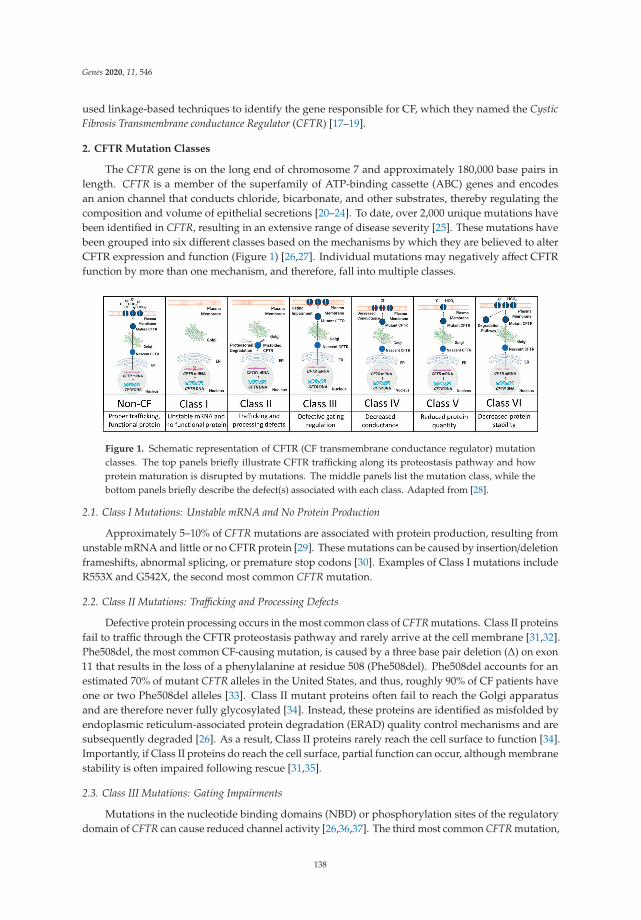
Genes 2020, 11, 546
used linkage-based techniques to identify the gene responsible for CF, which they named the CysticFibrosis Transmembrane conductance Regulator (CFTR) [17–19].
2. CFTR Mutation Classes
The CFTR gene is on the long end of chromosome 7 and approximately 180,000 base pairs inlength. CFTR is a member of the superfamily of ATP-binding cassette (ABC) genes and encodesan anion channel that conducts chloride, bicarbonate, and other substrates, thereby regulating thecomposition and volume of epithelial secretions [20–24]. To date, over 2,000 unique mutations havebeen identified in CFTR, resulting in an extensive range of disease severity [25]. These mutations havebeen grouped into six different classes based on the mechanisms by which they are believed to alterCFTR expression and function (Figure 1) [26,27]. Individual mutations may negatively affect CFTRfunction by more than one mechanism, and therefore, fall into multiple classes.
Figure 1. Schematic representation of CFTR (CF transmembrane conductance regulator) mutationclasses. The top panels briefly illustrate CFTR trafficking along its proteostasis pathway and howprotein maturation is disrupted by mutations. The middle panels list the mutation class, while thebottom panels briefly describe the defect(s) associated with each class. Adapted from [28].
2.1. Class I Mutations: Unstable mRNA and No Protein Production
Approximately 5–10% of CFTR mutations are associated with protein production, resulting fromunstable mRNA and little or no CFTR protein [29]. These mutations can be caused by insertion/deletionframeshifts, abnormal splicing, or premature stop codons [30]. Examples of Class I mutations includeR553X and G542X, the second most common CFTR mutation.
2.2. Class II Mutations: Trafficking and Processing Defects
Defective protein processing occurs in the most common class of CFTR mutations. Class II proteinsfail to traffic through the CFTR proteostasis pathway and rarely arrive at the cell membrane [31,32].Phe508del, the most common CF-causing mutation, is caused by a three base pair deletion (Δ) on exon11 that results in the loss of a phenylalanine at residue 508 (Phe508del). Phe508del accounts for anestimated 70% of mutant CFTR alleles in the United States, and thus, roughly 90% of CF patients haveone or two Phe508del alleles [33]. Class II mutant proteins often fail to reach the Golgi apparatusand are therefore never fully glycosylated [34]. Instead, these proteins are identified as misfolded byendoplasmic reticulum-associated protein degradation (ERAD) quality control mechanisms and aresubsequently degraded [26]. As a result, Class II proteins rarely reach the cell surface to function [34].Importantly, if Class II proteins do reach the cell surface, partial function can occur, although membranestability is often impaired following rescue [31,35].
2.3. Class III Mutations: Gating Impairments
Mutations in the nucleotide binding domains (NBD) or phosphorylation sites of the regulatorydomain of CFTR can cause reduced channel activity [26,36,37]. The third most common CFTR mutation,
138
Genes 2020, 11, 546
G551D, produces a protein that, despite reaching the cell membrane, has approximately 100-foldlower open probability than that of wild-type (wt) [38]. Dimerization of the NBDs of CFTR formstwo ATP binding pockets, termed ABP1 and ABP2 [39]. Whereas binding of ATP to ABP1 helps tostabilize the open channel conformation of CFTR, channel opening is dependent on ATP binding ofABP2 [40]. The G551D mutation, located in ABP2, prevents ATP binding, thus inhibiting opening ofthe CFTR channel [38]. The S1255P mutation, found in NBD2, also does not disrupt CFTR maturation,but instead alters the ATP-binding pocket, resulting in gating instability [41].
2.4. Class IV Mutations: Decreased Conductance
Class IV proteins achieve proper processing and gating, but mutations in their membrane spanningdomains cause a misshapen protein that restricts anion transport [26]. This results in a decreased rateof ion flow through each open channel and an overall decrease in current conducted by CFTR [42].Mutations affecting channel pore activity often arise in arginine residues (e.g., R117H, R347P, R334W).Some Class IV mutations, including R117H, also decrease the open probability of CFTR [43].
2.5. Class V Mutations: Reduced Protein Quantity
Inefficient protein maturation can be caused by alternative splicing, amino acid substitutions,or promoter mutations [26]. Class V mutations often produce incorrectly spliced versions of the CFTRmRNA in variable proportions, and the resultant proteins rarely transit to the cell membrane, resultingin a decreased number of functioning CFTR channels [44,45]. The most prevalent examples of Class Vmutations include c.3717+12191C>T and c.3140-26A>G [46,47].
2.6. Class VI mutations: Unstable Protein
Class VI mutants can act as functional proteins at the cell surface. However, instability inthe protein structure results in reduced residency at the cell surface, more rapid protein turnover,and therefore, less ion conductance [33,45]. Examples include c.120del123 and Phe508del when rescuedby low temperature or correctors (rPhe508del) [48].
3. CFTR Structure and Function
CFTR is a 1,480 amino acid transmembrane glycoprotein containing two homologous halves,each consisting of six transmembrane alpha helices (termed TMD or transmembrane domain) that forman anion conduction pore, and a nucleotide-binding domain that serves as the binding site for ATPhydrolysis (Figure 2). These halves are connected by a regulatory (R) domain that contains multiplephosphorylation sites and regulates channel activity. The R domain is intrinsically unstructuredand adapts its conformation upon binding to the NBDs and the CFTR N-terminus [49,50]. Recently,the cryo-EM structure of full length human CFTR was published, highlighting several key structuralelements required for a fully functional protein [51,52]. First, an unphosphorylated R domain preventsthe dimerization of NBD1 and NBD2, resulting in a closed channel. Secondly, a small inhibitoryhelix exists in the R domain that is docked inside the intracellular vestibule between the nucleotidebinding domains, which precludes channel opening. It is believed that the disruption of the interactionbetween this inhibitory helix and the nucleotide binding domains would allow for protein kinase A(PKA)-mediated phosphorylation of the R domain, resulting in NBD dimerization and subsequentopening of the ion channel. Third, the authors sought to explain why CFTR acts as an ion channel,whereas other ABC transporters function as pumps that move ions against electrochemical gradients.When comparing the structures of CFTR and other ABC proteins, differences in two transmembranehelices (TM7 and TM8) were identified, leading to the hypothesis that these helices affect ion conductionand gating.
CFTR is neither isolated from neighboring proteins nor does it act alone. Instead, CFTR is part ofa multiprotein assembly at the apical membrane surface, and is anchored via PDZ domains commonlyfound in plasma membrane proteins and other intracellular signaling proteins [53,54]. When at the apical
139

Genes 2020, 11, 546
membrane, CFTR is spatially located near other ion channels, membrane receptors, and cytoskeletalproteins. Additionally, transcriptomic studies in human CF cells suggest that significant changes ingene expression may result from the absence of functional CFTR [55]. Affected genes include membersof the protein processing and inflammatory response functional families, among others. Likewise,proteomic analyses of the wt and Phe508del-CFTR interactomes by Pankow et al. identified noveleffectors belonging to mRNA decay, co-translational control, endocytic recycling, ER quality controland folding, and protein degradation networks [56]. Such relationships between CFTR and other genessuggest that CFTR does not act solely as an ion channel, but instead, may have various roles throughoutits biogenesis. Interactions between CFTR and genes that influence its processing or maturation couldhelp to explain the wide range of phenotypes and severities in CF patients with identical mutations [33].A better understanding of such interactions could also lead to the development of small moleculemodulators for CF lung disease.
Figure 2. Cryo-EM structure of dephosphorylated, ATP-free CFTR. A. CFTR contains twotransmembrane domains (TMD1 in orange, TMD2 in blue), two nucleotide binding domains (NBD1 inpurple, NBD2 in yellow), and a regulatory (R) domain (cyan). CFTR is activated by phosphorylation ofthe R domain and ATP hydrolysis by the NBDs. Note that the structural flexibility of the R domainlimits its visibility by Cryo-EM. Instead, 19 alanines are shown that correspond to the C-terminalregion of the R domain. B. Magnified view of transmembrane helices (TM) 7 (brown) and 8 (gray).CFTR differs from other ABC transporters in that TM7 is displaced from its usual position and TM8breaks into three short helices, rather than being a continuous helix as seen in other ABC transporters.TM7 and TM8 are found in TMD2. PBD ID: 5UAK.
4. Cystic Fibrosis Transcriptome
4.1. mRNA Profiling
The first transcriptomic profiling of well-differentiated primary cultures of human airway epitheliafrom Phe508del/Phe508del donors was performed by Zabner et al. in 2005 [57]. Of the approximately22,000 genes represented on the Affymetrix U133A GeneChip, 18 were observed to be significantlyupregulated in CF, while 6 were downregulated. The KCl cotransporter KCC4 was identified aselevated in CF and was deemed a candidate for further studies. Interestingly, this profiling concludedthat the level of CFTR mRNA was not significantly different in Phe508del/Phe508del cells compared
140
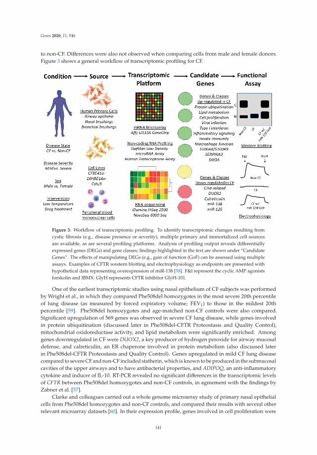
Genes 2020, 11, 546
to non-CF. Differences were also not observed when comparing cells from male and female donors.Figure 3 shows a general workflow of transcriptomic profiling for CF.
Figure 3. Workflow of transcriptomic profiling. To identify transcriptomic changes resulting fromcystic fibrosis (e.g., disease presence or severity), multiple primary and immortalized cell sourcesare available, as are several profiling platforms. Analysis of profiling output reveals differentiallyexpressed genes (DEGs) and gene classes; findings highlighted in the text are shown under “CandidateGenes”. The effects of manipulating DEGs (e.g., gain of function (GoF) can be assessed using multipleassays. Examples of CFTR western blotting and electrophysiology as endpoints are presented withhypothetical data representing overexpression of miR-138 [58]. F&I represent the cyclic AMP agonistsforskolin and IBMX. GlyH represents CFTR inhibitor GlyH-101.
One of the earliest transcriptomic studies using nasal epithelium of CF subjects was performedby Wright et al., in which they compared Phe508del homozygotes in the most severe 20th percentileof lung disease (as measured by forced expiratory volume; FEV1) to those in the mildest 20thpercentile [59]. Phe508del homozygotes and age-matched non-CF controls were also compared.Significant upregulation of 569 genes was observed in severe CF lung disease, while genes involvedin protein ubiquitination (discussed later in Phe508del-CFTR Proteostasis and Quality Control),mitochondrial oxidoreductase activity, and lipid metabolism were significantly enriched. Amonggenes downregulated in CF were DUOX2, a key producer of hydrogen peroxide for airway mucosaldefense, and calreticulin, an ER chaperone involved in protein metabolism (also discussed laterin Phe508del-CFTR Proteostasis and Quality Control). Genes upregulated in mild CF lung diseasecompared to severe CF and non-CF included statherin, which is known to be produced in the submucosalcavities of the upper airways and to have antibacterial properties, and ADIPOQ, an anti-inflammatorycytokine and inducer of IL-10. RT-PCR revealed no significant differences in the transcriptomic levelsof CFTR between Phe508del homozygotes and non-CF controls, in agreement with the findings byZabner et al. [57].
Clarke and colleagues carried out a whole genome microarray study of primary nasal epithelialcells from Phe508del homozygotes and non-CF controls, and compared their results with several otherrelevant microarray datasets [60]. In their expression profile, genes involved in cell proliferation were
141
Genes 2020, 11, 546
significantly upregulated in CF, while cilia-related genes were downregulated. Due to great variabilityin the gene expression profiles across the independent studies, the meta-analysis comparing this studyto five other microarray experiments (including the Zabner, Wright, and Ogilvie studies described in thissection) yielded few common dysregulated genes across at least three experiments. However, when theauthors compared their microarray results with the Ogilvie microarray, a molecular signature of nativeCF airway epithelial cells was observed, consisting of 21 common upregulated genes and 9 commondownregulated genes [61]. A significant number of these genes were involved in inflammation anddefense, including the upregulated CXCR4, FOS, S100A8, S100A9, and SERPINA3 transcripts.
Although several gene expression studies have used nasal epithelial brushings from CF donors,Ogilvie et al. concluded that transcriptomics of the CF nasal epithelium is not representative of geneexpression in the lung respiratory epithelium. Following bead array profiling of CF and non-CF nasaland bronchial epithelium, 863 genes were found to be significantly dysregulated in the bronchial cells,whereas only 15 genes were identified as dysregulated in nasal cells [61].
Polineni et al. performed RNA-sequencing of nasal mucosal cells from 134 CF subjects withvarying genotypes and disease severities, as assessed by cytokine levels in nasal lavages [62]. Pathwayanalysis of the gene expression data highlighted the positive correlation between CF disease severityand viral infection, inflammatory signaling, lipid metabolism, macrophage function, and innateimmunity. Multiple human leukocyte antigen (HLA) genes robustly contributed to the enrichedpathways and several were also observed at the intersection of the gene expression profiling andpreviously identified CF GWAS risk alleles. The authors concluded that HLA genes may serve astargets for interventions aiming to improve CF lung health.
A meta-analysis of 13 microarray experiments was performed by Clarke and colleagues, comparingCF with similar disorders (e.g., chronic obstructive pulmonary disease, asthma, and idiopathicpulmonary fibrosis), environmental factors (e.g., smoking), relevant cellular processes (e.g., epithelialregeneration), and non-respiratory controls (e.g., schizophrenia) [63]. Genes whose expression wasinversely related with CFTR across samples expressing Phe508del were subjected to an siRNAknockdown assay to identify potential negative regulators of CFTR. Nine genes, including SNX6,PSEN1, and RCN2, produced an appreciable increase in CFTR trafficking to the cell membrane. Whilethe siRNA knockdown experiments were considered preliminary by the authors, these genes mayserve as intriguing leads for therapeutic targets.
An additional transcriptomic study comparing the peripheral blood leukocytes of CF subjects withmild and severe lung disease was performed by Kormann et al. [64] Enrichment analyses identifiedgenes of the type I interferon response, as well as ribosomal stalk proteins, as upregulated in milddisease. Such modifiers of CF lung disease may have implications as new biomarkers or targetsfor intervention.
4.2. Non-coding RNA Profiling
While most CF-related transcriptomic studies have focused on mRNA profiling, McCray andcolleagues profiled global microRNA expression in well-differentiated primary cultures of humanairway epithelia by qPCR and identified 31 highly expressed microRNAs in CF [58]. Further analysesof these microRNAs identified SIN3A as a highly conserved target of miR-138. As SIN3A has conservedmotifs that bind to the transcriptional repressor CTCF and the CFTR locus contains functionalCTCF-binding sites, the authors hypothesized that miR-138 and SIN3A regulate CFTR. Functionalassays determined that overexpression of miR-138 or knockdown of SIN3A partially restored thematuration, trafficking, and function of Phe508del-CFTR. Oglesby et al. also identified miR-126 asdownregulated in CF airway epithelial cells [65]. Overexpression of miR-126 resulted in downregulatedTOM1 protein production. Furthermore, knockdown of TOM1 mRNA significantly increased NF-κBregulated IL-8 secretion, linking miR-126 to innate immune responses in CF. Additionally, miR-145has been shown to mediate TGF-β inhibition of CFTR function and knockdown of miR-145 restoredPhe508del function in human primary epithelial cells [66]. Likewise, miR-200b reduces CFTR during
142

Genes 2020, 11, 546
prolonged hypoxia, although inhibition of miR-200b also rescues CFTR mRNA levels in primarybronchial epithelial cells [67]. These studies lend further support to the strategy of manipulatingmicroRNAs or their target genes to enhance CFTR expression or alleviate symptoms associated with CF.
Likewise, Kamei and colleagues analyzed the expression of non-coding genes, or functionalRNAs with no protein-coding capacity [68]. Using the Human Transcriptome Array, 91 dysregulatednon-coding RNAs were identified in the CFBE41o- cell line. Linc-SUMF1-2, an intergenic non-codingRNA with no known function, was found to be inversely correlated with wild-type CFTR. Furtheranalyses identified eight dysregulated genes, including CXCL10, MYC, and LAMB3, as both CFTR-and linc-SUMF-1-2-dependent in CF airway epithelial cells, uncovering a novel regulatory pathway ofCF-associated gene regulation.
5. Phe508del-CFTR Proteostasis and Quality Control
CFTR was among the first membrane proteins identified as being regulated by the ERADpathway [69]. Wild-type CFTR undergoes co-translation and N-glycosylation in the ER before beingpacked into COPII vesicles at ER exit sites and trafficked to the Golgi apparatus. Upon reaching theGolgi, glycan processing and modification occurs, rendering a complex, mature form of the protein.CFTR is then trafficked to the cell surface, where its stability is tightly regulated by protein interactors.Upon removal from the plasma membrane, CFTR can undergo endocytosis or be recycled back to thecell surface [70]. Despite its proteostasis pathway being incompletely understood, CFTR interacts withseveral classes of proteins and must pass multiple quality control checkpoints during trafficking fromthe ER to the cell surface (Figure 4).
5.1. Chaperones and Protein Folding
Heat shock proteins (Hsps) serve as the first quality control constituents of CFTR biogenesis, asthese molecular chaperones co-translationally interact directly with CFTR. Hsp90, the constitutivelyexpressed isoform of Hsp70 (often referred to as Hsc70), and the stress-induced isoform of Hsp70bind CFTR during translation and assist in proper folding via ATP hydrolysis [71–75]. Smallmolecule-induced inhibition of Hsp90 in cultured human cells prevents proper CFTR folding, leadingto protein degradation [71]. However, both Hsp90 and Hsp70 can recruit channel folding andmaturation antagonizers, such as the Hsp70/Hsp90 organizing protein (HOP) [76,77]. HOP directsCFTR towards the degradation pathway by recruiting the E3 ubiquitin ligase CHIP (sometimes referredto as STUB1) to the CFTR-Hsp70/90 complex [78,79]. CHIP tags CFTR with ubiquitin (discussedin E3 Ubiquitin Ligases) and the CFTR protein is ushered toward the proteasome for degradation.The pro-degradation effects of CHIP can be reversed, however, by the Hsp/Hsc70 nucleotide exchangefactor HspBP1, which inhibits CHIP and results in the continuation of CFTR along the pro-foldingpathway [78,79]. The Hsp90 co-chaperone Aha1 is believed to prevent Hsp90 from properly interactingwith CFTR, resulting in degradation of nascent protein [80,81]. An additional nucleotide exchangefactor for Hsc70, Hsp105, has been observed to both promote the post-translational maturation ofCFTR, while also at times assisting in the co-translational degradation of CFTR [82,83].
Hsp40 co-chaperones, often referred to as J proteins, have also been shown to interact withCFTR during its initial translation stages. DNAJA1 (Hsp40/Hdj2) and DNAJB1 (Hsp40/Hdj1)interact with Hsc70 to promote folding of the NBD1 of CFTR and assist in rescuing wtCFTR fromendoplasmic reticulum retention [73,84,85]. However, these DNAJ proteins have been unable to rescuePhe508del-CFTR from being degraded and sometimes actually serve as pro-degradation componentsof the quality control machinery [86]. DNAJA1 has been shown to promote CHIP ubiquitin ligaseactivity and DNAJC5 (Hsp40 cysteine string protein; sometimes referred to as Csp) independentlyrecruits CHIP to the CFTR-Hsp90 complex [87–89]. Similarly, DNAJB12 recruits the E3 ubiquitin ligaseRMA1 to the CFTR-Hsc70 complex, promoting degradation of both Phe508del-CFTR and immaturewtCFTR [90,91].
143
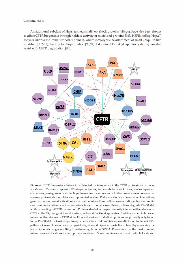
Genes 2020, 11, 546
An additional subclass of Hsps, termed small heat shock proteins (sHsps), have also been shownto affect CFTR biogenesis through holdase activity of misfolded proteins [92]. HSPB1 (sHsp Hsp27)recruits Ubc9 to the immature NBD1 domain, where it catalyzes the attachment of small ubiquitin-likemodifier (SUMO), leading to ubiquitination [93,94]. Likewise, HSPB4 (sHsp αA-crystallin) can alsoassist with CFTR degradation [93].
Figure 4. CFTR Proteostasis Interactors. Selected proteins active in the CFTR proteostasis pathwayare shown. Octagons represent E3 ubiquitin ligases; trapezoids indicate kinases; circles representchaperones; pentagons indicate deubiquitinases; co-chaperones and all other proteins are represented assquares; proteostasis modulators are represented as stars. Red arrows indicate degradation interactions;green arrows represent activation or maturation interactions; yellow arrows indicate that the proteincan have degradative or activation interactions. In most cases, these proteins degrade Phe508del,while promoting wtCFTR maturation. Proteins shaded in purple primarily interact with co-factors orCFTR at the ER; orange at the cell surface; yellow at the Golgi apparatus. Proteins shaded in blue caninteract with co-factors or CFTR at the ER or cell surface. Underlined proteins are primarily only foundin the Phe508del proteostasis pathway, whereas italicized proteins are usually found in the wtCFTRpathway. Curved lines indicate that pyridostigmine and biperiden are believed to act by mimicking thetranscriptional changes resulting from downregulation of SIN3A. Please note that the most commoninteractions and locations for each protein are shown. Some proteins are active at multiple locations.
144
Genes 2020, 11, 546
Calnexin (CNX) is a membrane chaperone found in the ER that affects the folding of CFTRtransmembrane domains [95]. While the binding of CNX to CFTR can prevent pro-degradation qualitycontrol proteins from binding immaturely folded CFTR, studies also suggest that CNX can obstructchannel maturation [96,97]. In fact, inhibition of CNX improves trafficking of wtCFTR from the ERto the cell membrane [96]. However, such inhibition has little effect on Phe508del-CFTR, perhapsindicating that Phe508del is targeted for degradation prior to the role of CNX in the quality controlpathway. Calreticulin (CRT), found in the ER lumen, does not contribute to CFTR folding, but insteadincreases the length of time that CFTR remains in the ER, resulting in increased CFTR turnover [98].
5.2. E3 Ubiquitin Ligases and Protein Degradation
The complex quality control mechanisms responsible for CFTR folding, maturation, and processingresult in a high rate of protein turnover, even for wtCFTR. In fact, current estimates indicate that only onefourth of wtCFTR is folded correctly and trafficked from the ER, whereas virtually no Phe508del-CFTRmanages to escape [99]. Proteins unreleased from the ER enter the ubiquitin-proteasome ERADpathway [34]. Proteins targeted for ERAD by chaperones like CHIP and RMA1 (discussed in Heat-shockproteins) are tagged with ubiquitin, a 76-amino acid polypeptide that signals the release of CFTR fromthe ER membrane. However, rather than trafficking to the cell membrane, ubiquitin-tagged CFTR ishydrolyzed by the proteolytic chymotrypsin-like activity of the proteasome [100].
The first step of the ubiquitin-proteasome ERAD pathway requires the ATP-dependent binding ofubiquitin to an E1 ubiquitin activating enzyme. The E1 enzyme catalyzes the C-terminus of ubiquitinand then transfers ubiquitin to an E1 active site cysteine residue. UBA1 and UBA6 are the onlyknown E1 ubiquitin activating enzymes in humans [101]. Next, E2 ubiquitin-conjugating enzymes arerecruited to E1-ubiquitin complex and catalyze the transfer of ubiquitin to the active site cysteine of E2.Currently, 35 unique E2 conjugating enzymes have been identified in the human genome [102]. Lastly,E3 ubiquitin ligases function as substrate identification molecules and bind both E2 enzymes andsubstrates while transferring ubiquitin from the E2 to the substrate. E3 ubiquitin ligases often transfermultiple ubiquitin polypeptides to a substrate, creating a polyubiquitin chain. Ubiquitinated proteinsare then trafficked to the proteasome for degradation. E3 ubiquitin ligases have been sorted intomultiple classes, including HECT and RING, depending on their active domains. Over 600 unique E3ubiquitin ligases have been identified thus far, and most E3s can target multiple substrates. Likewise,individual substrates may be ubiquitinated by multiple E3s [103,104].
To date, several E3 ubiquitin ligases have been shown to ubiquitinate CFTR. RMA1 (sometimescalled RNF5) and RNF185, a highly conserved homologue of RMA1, ubiquitinate misfolded CFTRfollowing NBD1 translation [90,105,106]. An additional E3 ubiquitin ligase, gp78, acts by elongatingthe polyubiquitin chains initiated by RMA1 and RNF185 [107]. Unlike RMA1 and RNF185, the E3ubiquitin ligase CHIP only acts on fully translated CFTR [78,79,86]. As RMA1, RNF185, and CHIP areunable to directly bind to CFTR, these proteins ubiquitinate misfolded CFTR through adaptor proteins.Specifically, RMA1 and RNF185 require Derlin-1, whereas CHIP binds Hsc70 or Hsp70 [108–110].Interestingly, whereas Hsc70/Hsp70 often promote CFTR folding, CHIP is able to “hijack” thesechaperones to trigger ERAD [111]. CHIP and E3 ubiquitin ligase RFFL can ubiquitinate CFTR at thecell periphery. Unlike CHIP, RFFL binds directly to CFTR and is independent of molecular chaperones.RFFL does not affect turnover of wtCFTR, but instead targets only misfolded protein on the cellsurface [112].
Additional E3 ubiquitin enzymes that target CFTR include MARCH2, NEDD4-2, SYVN1,and FBXO2. MARCH2 ubiquitinates CFTR through adaptor proteins CAL and STX6 [113]. NEDD4-2is a HECT E3 ubiquitin ligase that binds both wtCFTR and Phe508del-CFTR [114]. SYVN1 regulatesCFTR ubiquitination through the RNF5/AMFR pathway, whereas FBX02 binds directly to CFTR viathe SCF complex [115]. Knockdown of MARCH2, NEDD4-2, and SYVN1 has been demonstrated toimprove Phe508del-CFTR maturation and trafficking and restore partial function to the mutant protein.
145

Genes 2020, 11, 546
Lastly, the deubiquitinase USP10 has been shown to interact with wtCFTR in endosomes, reducingpolyubiquitination and improving rates of endocytic recycling of wtCFTR [116,117].
5.3. ER Stress and Anterograde Trafficking
Whereas properly folded CFTR usually enters COPII-coated vesicles budding from the ERand subsequently trafficks to the Golgi en route to the cell surface, mutant CFTR can also undergounconventional anterograde trafficking. Such trafficking is commonly induced by ER stress andinvolves the bypassing of the ER-to-Golgi transport, resulting in CFTR trafficking from the ER straightto the cell periphery [118,119]. During ER stress, IRE1 initiates the unfolded protein response (UPR),which increases both the expression of Sec16a and the number of possible exit sites in the ER. Sec16aacts as a secretory protein at such exit sites and facilitates scaffolding of COPII-coated vesicles.ER stress also causes GRASP55, usually found in the Golgi, to traffic to the ER, where it interacts withSec16a. Although the mechanism by which GRASP55/Sec16a aids in the trafficking of CFTR to the cellsurface is not currently understood, the resultant membrane bound CFTR lacks complex glycosylation,indicating that the protein bypassed the Golgi [119–121]. Despite being misfolded and incompletelyglycosylated, Phe508del-CFTR protein that reaches the cell surface through unconventional anterogradetrafficking retains partial function, suggesting that the GRASP55 pathway may serve as an interestingtherapeutic target.
5.4. Protein Kinases and Membrane Stability
Membrane stability of CFTR is partially regulated by protein kinases. PKA and protein kinaseC (PKC) have been shown to phosphorylate CFTR predominately at the R domain, although NBD1and C-terminal residues can also be phosphorylated [122–124]. While phosphorylation generallysuppresses endocytosis, AMP-activated protein kinase (AMPK) and spleen tyrosine kinase (SYK) havebeen shown to decrease CFTR plasma membrane stability [124,125]. In the ER, mixed-lineage kinase 3(MLK3) is believed to promote degradation by interacting with HOP [126]. Additionally, inhibition ofthe phosphatidylinositol 3-kinase (PI3K) pathway can increase CFTR stability and expression [127,128].
5.5. Tethering Factors and Endocytosis Adaptors
Also contributing to CFTR’s plasma membrane stability are endocytosis adaptors and tetheringfactors. Knockdown of the endocytosis factor DAB2 has been shown to stabilize Phe508del-CFTR at thecell surface by inhibiting endocytosis [129,130]. CFTR has a PDZ binding motif at the C-terminus thattethers to the PDZ domain of NHERF1, supporting channel activation and CFTR membrane stabilityfor both wild-type and mutant proteins [131]. The exchange protein EPAC1 strengthens this interactionand further suppresses endocytosis [132]. However, the CFTR-associated ligand (CAL) decreases thestability of CFTR at the cell membrane through its PDZ domain. Knockdown of CAL has been shownto improve function and stability of Phe508del-CFTR, suggesting that inhibition of the protein may bea therapeutic option [113,133,134].
6. Small Molecule Modulators
The observation by Welsh and colleagues that Phe508del-CFTR could be rescued and traffic to thecell surface via low temperature (27◦ C) incubation was transformative because it demonstrated that ifPhe508del (and potentially other mutations) could escape the ERAD pathway and traffic to the cellmembrane, they retained partial function [31]. While low temperature treatment is not a therapeuticallyviable option for CF patients, this observation encouraged researchers to target genes affecting theprocessing and maturation of CFTR [135]. Furthermore, through the use of high-throughput screeningtechnology, several small molecules that interact directly with CFTR and positively affect processing orfunction have been identified. Following lead optimization and clinical trials, four small molecules arenow FDA-approved for CF, providing potentially 90% of patients with at least one modulator option.Several other small molecules are currently being investigated in clinical trials (Table 1).
146

Genes 2020, 11, 546
Ta
ble
1.
Sele
cted
clin
ical
tria
lsus
ing
smal
lmol
ecul
em
odul
ator
sfo
rcy
stic
fibro
sis.
Sm
all
mo
lecu
le(s
)T
rad
eN
am
eC
om
pan
yP
hase
Mo
du
lato
rT
yp
eM
uta
tio
ns
Cli
nic
al
Tri
al
Ivac
afto
rK
alyd
eco
Vert
exPh
arm
aceu
tica
lsA
ppro
ved
Pote
ntia
tor
G55
1D,3
7ot
hers
*N
CT0
2725
567
Lum
acaf
tor+
Ivac
afto
rO
rkam
biVe
rtex
Phar
mac
euti
cals
App
rove
dLu
mac
afto
r=
Cor
rect
or;I
vaca
ftor
=Po
tent
iato
rPh
e508
del/P
he50
8del
NC
T036
0163
7
Teza
caft
or+
Ivac
afto
rSy
mde
koVe
rtex
Phar
mac
euti
cals
App
rove
dTe
zaca
ftor=
Cor
rect
or;I
vaca
ftor=
Pote
ntia
tor
Phe5
08de
l/Phe
508d
el,2
6ot
hers
**N
CT0
2412
111
Elex
acaf
tor+
Teza
caft
or+
Ivac
afto
rTr
ikaf
taVe
rtex
Phar
mac
euti
cals
App
rove
dEl
exac
afto
r,Te
zaca
ftor=
Cor
rect
ors;
Ivac
afto
r=
Pote
ntia
tor
Phe5
08de
l+an
yot
her
mut
atio
nN
CT0
4183
790
PTC
124+
Ivac
afto
rA
talu
ren
PTC
Ther
apeu
tics
Phas
e4
Prem
atur
est
opco
don
read
thro
ugh
Cla
ssIm
utat
ions
NC
T032
5696
8
VX
-561
—Ve
rtex
Phar
mac
euti
cals
Phas
e2
Pote
ntia
tor
G55
1D,8
othe
rs**
*N
CT0
3911
713
ABB
V-2
222
—A
bbV
iePh
ase
2C
orre
ctor
Phe5
08de
l/Phe
508d
elN
CT0
3969
888
ABB
V-3
067
—A
bbV
iePh
ase
2Po
tent
iato
rPh
e508
del/P
he50
8del
NC
T039
6988
8
ELX
-02
—El
oxx
Phar
mac
euti
cals
Phas
e2
Non
sens
em
utat
ion
read
thro
ugh
agen
tG
542X
NC
T041
3549
5
FDL1
69—
Flat
ley
Dis
cove
ryLa
bPh
ase
2C
orre
ctor
Phe5
08de
l/Phe
508d
elN
CT0
2767
297
PTI-
428+
Ivac
afto
r—
Prot
eost
asis
Ther
apeu
tics
Phas
e2
Am
plifi
erSa
me
asIv
acaf
tor
NC
T032
5842
4
PTI-
801
—Pr
oteo
stas
isTh
erap
euti
csPh
ase
2C
orre
ctor
Phe5
08de
l/Phe
508d
elN
CT0
3140
527
PTI-
808
—Pr
oteo
stas
isTh
erap
euti
csPh
ase
2Po
tent
iato
rPh
e508
del/P
he50
8del
NC
T032
5109
2
VX
-121
—Ve
rtex
Phar
mac
euti
cals
Phas
e2
Cor
rect
orPh
e508
del+
min
imal
func
tion
(MF)
mut
atio
nN
CT0
3912
233
MR
T500
5—
Tran
slat
eBi
oPh
ase
1m
RN
Ade
liver
yC
lass
Ior
IIm
utat
ions
NC
T033
7504
7
*M
utat
ions
A45
5E,A
1067
T,D
100E
,D11
0H,D
579G
,D11
52H
,D12
70N
,E56
K,E
193K
,E83
1X,F
1052
V,F1
074L
,G17
8R,G
551S
,G10
69R
,G12
44E,
G13
49D
,K10
60T,
L206
W,P
67L,
R74
W,R
117C
,R
347H
,R35
2Q,R
1070
Q,R
1070
W,S
549N
,S54
9R,S
945L
,S97
7F,S
1251
N,S
1255
P,R
117H
,c.5
79+
3A>
G,c
.265
7+5G>
A,c
.314
0-26
A>
G,c
.371
7+12
191C>
T.*
*M
uta
tion
sA
455E
,A10
67T,
D10
0E,D
110H
,D57
9G,D
1152
H,D
1270
N,E
56K
,E19
3K,E
831X
,F10
52V,
F107
4L,K
1060
T,L2
06W
,P67
L,R
74W
,R11
7C,R
347H
,R35
2Q,R
1070
W,S
945L
,S97
7F,c
.579+
3A>
G,c
.265
7+5G>
A,
c.31
40-2
6A>
G,c
.371
7+12
191C>
T.**
*G
178R
,G55
1S,G
1244
E,G
1349
D,S
549N
,S54
9R,S
1251
N,S
1255
P.
147

Genes 2020, 11, 546
6.1. CFTR Potentiators
Potentiators are a class of small molecules that increase anion transport via CFTR at the cellmembrane by increasing the channel open probability. As discussed previously, Class III CFTRmutations have gating defects, whereas Class IV CFTR mutations exhibit abnormal conductance.However, in both mutation classes, CFTR is trafficked to the cell membrane and partial function can berestored with the use of potentiators. Furthermore, clinical benefits have been observed in patientswith G551D and related mutations when receiving monotherapy with a single potentiator [136–141].As will be discussed later, potentiators can also help to restore function in Class II mutations, includingPhe508del-CFTR, when coupled with one or more correctors.
Vertex Pharmaceuticals (Boston, MA, USA) identified the first FDA-approved CFTR potentiator.Ivacaftor (trade name Kalydeco) was approved in 2012 for CF patients with the G551D mutationand was later approved for additional Class III mutations, including G1244E, G1349D, S549R, amongothers [140,142–146]. Kalydeco has also been approved for the Class IV mutation R117H and relatedmutations [141,147,148]. Ivacaftor was identified through a high throughput screen using NIH-3T3mouse fibroblast cells expressing Phe508del-CFTR [149]. These cells were first incubated at 27 ◦Cto rescue the mutant protein to the cell surface and then treated with small molecule candidates,with a fluorescent signal being detected when CFTR-mediated chloride transport occurred. Vertexscreened approximately 300,000 compounds using the NIH-3T3 fluorescence assay and identifiedfour scaffolds that had significant potentiating activity. Following medicinal chemistry to optimizethese scaffolds, investigators concluded that a singular scaffold was the most efficacious and leadoptimization of the scaffold resulted in the testing of an additional 70 small molecules. Of these, VX-770,which would later be named ivacaftor, showed superior function. In human bronchial epithelial(HBE) cells derived from G551D/Phe508del subjects, ivacaftor increased chloride secretion 10-fold,reaching 50% of wtCFTR levels [136]. In vivo studies in G551D patients followed, and two randomized,double-blind, placebo-controlled studies demonstrated a 10.5% increase in FEV1 compared to placeboand markedly reduced sweat chloride levels [143]. These groundbreaking results were the first inwhich a small molecule acted as a clinical modulator of CFTR. Patch clamp studies of ivacaftor-treatedcells expressing G551D concluded that ivacaftor increased the open probability of G551D-CFTRsix-fold [136]. Additionally, ivacaftor increased the open probability of Phe508del-CFTR five-fold andeven wtCFTR two-fold [136,139,150]. Further clinical trials of ivacaftor in Phe508del patients did notyield efficacious results [151].
The flavonoids genistein and curcumin have also been shown to have potentiating effects,especially when combined with lumacaftor (see CFTR Correctors), as these compounds enhanceforskolin-induced swelling in rectal organoids with Phe508del and G551D mutations [152]. Rattlesnakephospholipase A2 and several aminoarylthiazoles are also being investigated as potentiators inCFBE41o- and Phe508del-A549 cells [153–155]. Furthermore, several pharmaceutical companiesand research groups have small molecule potentiators in clinical trials, including Vertex, AbbVie,and Proteostasis Therapeutics (Table 1).
6.2. CFTR Correctors
While potentiators like ivacaftor have had significant clinical benefits for patients with Class IIIand Class IV mutations, little effect was seen in patients with Class II mutations, including those withPhe508del. As approximately 90% of CF patients have at least one Phe508del allele, there is substantialinterest in identifying small molecule correctors that can restore function to misfolded proteins withprocessing defects.
Corr-4a was the first corrector discovered that restored function to Phe508del-CFTR-transfectedepithelial cells at 37 ◦C to the same level as low temperature incubation [156]. High throughput screeninghas aided researchers in the quest to discover small molecule correctors and such experiments led VertexPharmaceuticals to discover VRT-422 and VRT-325 [137]. These compounds restored CFTR-mediatedchloride conductance to 10% of wtCFTR levels in HBE cells. Despite this modest restoration of
148
Genes 2020, 11, 546
conductance, these compounds served as an important proof-of-concept for small molecule correctionat a clinically relevant level. Further medicinal chemistry and lead optimization by Vertex led to thediscovery of VX-809, which reportedly rescued up to 30% of Phe508del-CFTR from degradation andrestored chloride conductance to approximately 15% of wtCFTR levels [157]. VX-809 was later namedlumacaftor. Interestingly, lumacaftor shows added efficacy when combined with low temperature,Corr-4a, and VRT-325, indicating that the misfolding defect(s) caused by the Phe508del-CFTR mutationis not entirely corrected by the individual compound [158–160]. This strongly supports a therapeuticstrategy of combining more than one corrector compound.
Following the identification of lumacaftor and ivacaftor, the corrector-potentiator combinationentered clinical trials. Once approved for Phe508del homozygous patients, the combination, marketedas Orkambi, gave up to 45% of CF patients a small molecule modulator option [161]. However,the clinical effect seen in Phe508del patients treated with Orkambi was modest, as homozygouspatients experienced an average increase in FEV1 of only 4% [162]. Additionally, an antagonistic effectbetween ivacaftor and lumacaftor was seen in several studies, leaving many patients without clinicalimprovement [163,164]. An estimated 15% of patients discontinued Orkambi within three months ofuse [165].
Recognizing the need for improved correctors, as well as the potential benefits of multi-correctortreatments, Vertex developed VX-661, later named tezacaftor, and VX-445, renamed elexacaftor. Whiletezacaftor is structurally related to and shares a mechanism with lumacaftor, elexacaftor is thoughtto act at a second site on CFTR, making it a corrector 2 (or C2) molecule [166]. Clinical trials with atriple-combination of tezacaftor, elexacaftor, and ivacaftor in patients with at least one Phe508del alleleresulted in an average increase in FEV1 of approximately 10%, as well as reduced sweat chloride andfrequency of pulmonary exacerbations [167,168]. This triple-combination therapy was subsequentlyFDA-approved in 2019 for patients with at least one Phe508del allele and marketed as Trikafta.
Vertex has also completed clinical trials of VX-440 and VX-152 in combination withtezacaftor/ivacaftor, but pursued elexacaftor as the third element of their triple-combinationstrategy [169]. Additional correctors in clinical trials have been reported by Vertex, AbbVie, FlatleyDiscovery Lab, and Proteostasis Therapeutics (Table 1) [170,171].
6.3. Premature Stop Codon Readthrough Agents
Although Kalydeco and Trikafta provide up to 90% of CF patients with modulator treatments,such small molecules are not therapeutic for patients with Class I mutations, which cause unstablemRNA and often no protein production. An estimated 9% of CF-causing mutations fall in Class I andapproximately half of all Israeli CF patients have such mutations [172,173]. As most Class I mutationsare caused by a premature stop codon, “readthrough” of these stop codons would theoreticallyallow for proper translation to the normal transcript termination site. This effect has been seen inR553X- and G542X-CFTR-expressing HeLa cells treated with aminoglycoside antibiotics, such asgentamicin [174,175]. However, preclinical studies of gentamicin treatment in patients with Class Imutations showed no clinical benefit [176,177].
High-throughput screens identified ataluren as a potentially efficacious readthrough agent.In subsequent experiments in transgenic mice harboring the G542X mutation, CFTR expression atthe plasma membrane was partially restored by ataluren treatment [178–180]. However, whileataluren progressed to phase III clinical trials, little benefit was observed [181]. Currently, syntheticaminoglycosides, ataluren derivatives, and escin, the FDA-approved active component of horsechestnut seed, are being investigated as readthrough agents in W1282X/Phe508del-CFBE and humanprimary epithelial cells [182–185]. It is important to note that such agents may cause the insertion ofnon-native amino acids at the site of readthrough, which may reduce channel function [186]. Currently,ELX-02, a eukaryotic ribosomal selective glycoside developed by Eloxx Pharmaceuticals (Waltham,MA, USA), is in Phase 2 clinical trials as a premature stop codon readthrough agent (NCT04135495).
149
Genes 2020, 11, 546
6.4. CFTR Stabilizers
While CFTR correctors can rescue mutant protein to the cell surface, long-term stability ofPhe508del-CFTR at the plasma membrane has not been observed following solo corrector treatment [187].Likewise, while low temperature treatment rescues CFTR to the cell surface, the protein’s half-lifeis still reduced and it experiences increased endocytosis and decreased recycling [188,189]. Class VICFTR mutations result in unstable protein configurations that lead to reduced residency of CFTR atthe cell surface and, therefore, less anion conductance. As correctors are currently unable to addressthis class of mutations, researchers have searched for small molecules to stabilize the mutant CFTRprotein at the plasma membrane for longer periods.
To date, several CFTR stabilizers have been identified. Although not a small molecule, hepatocytegrowth factor (HGF) has been shown to activate Rac1 signaling and resultingly stabilize CFTR throughits interaction with NHERF-1 [190]. While lumacaftor can increase CFTR plasma membrane stabilityto a modest degree, co-treatment of lumacaftor with HGF further enhanced the anchoring of CFTR toNHERF-1 in mouse small intestine organoids [131,191]. Treatment with vasoactive intestinal peptidealso stabilized interactions between CFTR and NHERF-1 by decreasing the rate of endocytosis [192].Lastly, cavosonstat, an inhibitor of S-nitrosoglutathione reductase, helps to stabilize CFTR by preventingits interaction with HOP [76,77]. Interestingly, cavosonstat is the only CFTR stabilizer to be tested inclinical trials (NCT02589236). It is currently being administered to Phe508del/Phe508del patients usingOrkambi and patients with Class III mutations using Kalydeco.
6.5. Splicing Correctors
Approximately 10% of CFTR mutations are caused by aberrant mRNA splicing that often resultsin immature protein that rarely trafficks to the cell membrane. Such mutations can be found acrossmultiple classes but are particularly common in Class V. Modulators able to correct splicing and restorefull-length CFTR mRNA could rescue CFTR protein function. Currently, antisense oligonucleotidesare being investigated as therapeutic options for splicing mutations [193].
6.6. CFTR Amplifiers
Amplifiers increase the amount of CFTR mRNA production and subsequent proteinproduction [194]. As the mRNA still contains a mutation, amplifiers do not directly correct processingor restore function to the protein. Instead, an increased amount of protein substrate is available formodulators to act upon. Therefore, amplifiers are always investigated as a component of a multi-drugtherapy. Phase 2 clinical trials were recently completed for the amplifier PTI-428, or nesolifcaftor,in patients using tezacaftor/ivacaftor (NTC03591094).
6.7. mRNA Delivery Agents
Delivery of CFTR-encoding mRNA to the lungs would allow epithelial cells to create wtCFTRprotein in a mutation-agnostic manner. Robinson and colleagues used lipid-based nanoparticles(LNPs) for delivery of chemically modified CFTR mRNA (cmCFTR) to CFTR knockout mice [195].Approximately 55% of net chloride efflux of normal mice was observed 3 days post-transfection.Translate Bio is currently testing MRT5005, an agent designed to deliver CFTR mRNA, in Phase Iclinical trials (NCT03375047).
6.8. Proteostasis Modulators
Glycerol and trimethylamine N-oxide (TMAO), when added to NIH 3T3 cells expressing Phe508del,were found to restore partial processing and function to Phe508del-CFTR [196–198]. High concentrationsof 4-phenylbutrate (4PBA) emerged as a candidate CFTR modulator, as it restored function toPhe508del-CFTR by interfering with Hsc70 in HEK293 cells expressing Phe508del. However, clinicaltrials of 4PBA showed little improvement in respiratory function [199–202]. Balch and colleagues tested
150
Genes 2020, 11, 546
HDAC inhibitors for the rescue of Phe508del-CFTR and identified suberoylanilide hydroxamic acid(SAHA) as efficacious in primary human bronchial epithelial cells [203,204]. The combination treatmentof cysteamine and epigallocatechin gallate has been shown to rescue CFTR trafficking, function,and plasma membrane stability through the correction of Beclin-1 autophagy flux in primary nasalepithelial cells [205,206]. A phase II clinical trial of the cysteamine-epigallocatechin gallate combinationreported decreased sweat chloride levels and modest increases in FEV1 in Phe508del/Phe508delpatients [207].
McCray and colleagues used a transcriptomic-based strategy to identify candidate correctors ofCFTR. By querying the genomic signature of miR-138-mediated CFTR rescue in the Connectivity Map,a catalogue of gene expression profiles of various cell lines treated with bioactive small molecules, thegroup was able to identify molecules whose genomic signatures closely resembled that of miR-138overexpression or SIN3A knockdown. After testing 27 small molecules, four were identified thatpartially rescued maturation and function of Phe508del-CFTR in primary human airway epithelia (HAE),including biperiden, pizotifen, pyridostigmine, and valproic acid. Of these, pyridostigmine showedcooperativity with corrector compound C18 (an analogue of lumacaftor) in improving Phe508del-CFTRfunction [208].
Likewise, Galietta and colleagues used connectivity mapping to identify drugs having a similarmode of action at the gene expression level as CFBE41o- and primary bronchial epithelial cells treated at27 ◦C for 24 h [209]. Several anti-inflammatory glucocorticoids were found to increase Phe508del-CFTRfunction in the cell line, but the activity could not be confirmed in primary cells. Sondo et al. alsoidentified 9-aminoacridine and ciclopirox as proteostasis regulators able to restore partial function toPhe508del-CFTR in cell lines [210]. However, these small molecules did not increase chloride secretionin primary bronchial epithelial cells from CF patients and subsequent microarray profiling revealeddifferent gene expression signatures generated by the treatments in cell lines and primary cells.
Additional investigations of the repurposing of drugs currently FDA-approved for non-CFdisorders yielded compounds that are efficacious in vitro. Miglustat (marketed under the trade nameZavesca and used to treat Gaucher disease) and sildenafil (marketed under the trade name Viagara andused to treat erectile dysfunction and pulmonary hypertension) treatments partially restored functionto Phe508del-CFTR in human nasal epithelial cells [211,212].
7. Conclusions
A golden age of CFTR small molecule modulators has arrived, as approximately 90% of CFpatients could receive clinical benefits from the use of one or more FDA-approved drugs. Clinicalstudies have reported improved lung function, reduced pulmonary exacerbations, increased weight,and improved quality of life measures. However, despite the profound impact that these drugs arehaving on patients, there are major areas that must be considered in the future of CF drug development.First, it is crucial that therapies be identified for all CFTR mutation classes. Currently, there are noapproved treatments for mutations causing premature stop codons, frameshifts, or nonsense mutations.Fortunately, small molecules to address some of these mutations are currently progressing throughclinical trials. However, it is possible that small molecule therapeutics will not provide clinical benefitsto all mutations. For such situations, the development of gene therapy or gene editing approaches maybe crucial [213,214]. Secondly, there are still patients in age ranges that are not approved to receive theFDA-approved small molecules. CFTR modulators are likely to have their greatest benefit if patientsare treated before irreversible tissue remodeling of the lung occurs, presumably shortly after birth,or even in utero. Orkambi is currently being tested in patients 12-24 months old, whereas Trikafta isunder investigation in patients 6-11 years of age. Lastly, the long-term effects of the current treatmentsare unknown, as the drugs only recently became available. Additionally, they are currently quiteexpensive, which limits their widespread availability worldwide, and places burdens on healthcaresystems. It will be important for healthcare professionals to continually monitor the efficacy and anypotential side effects of these compounds.
151

Genes 2020, 11, 546
While the advancements in small molecule treatments for CF in the last decade have beenmonumental and are acknowledged, it is imperative that improved treatments continue to be developed.As seen with Trikafta, the co-treatment of two or more small molecules may present opportunities toimprove the efficacy of pharmacological therapies. Tezacaftor and elexacaftor act synergistically, likelydue to the fact that elexacator acts on a different site in CFTR than tezacaftor. An alternative strategy isto pair a corrector that interacts directly with CFTR, such as tezacaftor, which a small molecule thatmanipulates the CFTR proteostasis pathway. Significant additive effects have been observed in humanprimary airway epithelial co-treated with pyridostigmine and corrector compound C18, as well asin SAHA paired with corrector compound C3. While these small molecules have not yet advancedto clinical trials, they lend support to the strategy of targeting proteostasis interactors. Furthermore,although several modulators of CFTR proteostasis have not shown significant efficacy in clinical trials,it would be unwise to abandon investigations of such therapies. Some failed candidates, such asglycerol and 9-aminoacridine, have incompletely understood mechanisms, making lead optimizationdifficult. Others, such as 4PBA, target proteins that can act in both degradation and maturationpathways, further complicating the already delicate process of rescuing Phe508del. However, as seenin Figure 4, many proteins involved in the CFTR proteostasis pathway have not been targetedpharmaceutically for correction of Phe508del and may be therapeutic targets. Lastly, as transcriptomicand proteomic studies of CFTR proteostasis continue to uncover new interactors, it is important forresearchers to investigate whether such interactors can be targeted therapeutically. Modulators of theCFTR proteostasis pathway could serve as pharmaceutical leads and complement the already existingdrugs discovered via high throughput screening.
Author Contributions: M.D.S., P.B.M.J. writing, review, figure creation, and editing. All authors have read andagreed to the published version of the manuscript.
Funding: This work was supported by National Institutes of Health Predoctoral Training Grant T32GM008629(PI Daniel Eberl). We also acknowledge support from the NIH (UG3 HL-147366, P01 HL51670, P01 HL091842), theCystic Fibrosis Foundation, the University of Iowa Center for Gene Therapy (DK54759), and the Roy J. CarverChair in Pulmonary Research (PBM).
Acknowledgments: We thank Jennifer A. Bartlett and Ashley L. Cooney for their critical review of the manuscript.
Conflicts of Interest: The authors have no conflicts of interest to declare.
References
1. Ratjen, F.; Doring, G. Cystic fibrosis. Lancet 2003, 361, 681–689. [CrossRef]2. Brown, S.D.; White, R.; Tobin, P. Keep them breathing: Cystic fibrosis pathophysiology, diagnosis,
and treatment. JAAPA 2017, 30, 23–27. [CrossRef] [PubMed]3. Clague, S. Dorothy Hansine Andersen. Lancet Respir. Med. 2014, 2, 184–185. [CrossRef]4. Fakhoury, K.; Durie, P.R.; Levison, H.; Canny, G.J. Meconium ileus in the absence of cystic fibrosis. Arch. Dis.
Child. 1992, 67, 1204–1206. [CrossRef] [PubMed]5. Kelly, A.; Moran, A. Update on cystic fibrosis-related diabetes. J. Cyst. Fibros. 2013, 12, 318–331. [CrossRef]
[PubMed]6. Kobelska-Dubiel, N.; Klincewicz, B.; Cichy, W. Liver disease in cystic fibrosis. Prz. Gastroenterol. 2014, 9,
136–141. [CrossRef]7. Quinton, P.M. Missing Cl conductance in cystic fibrosis. Am. J. Physiol. 1986, 251, C649–C652. [CrossRef]8. Sokol, R.Z. Infertility in men with cystic fibrosis. Curr. Opin. Pulm. Med. 2001, 7, 421–426. [CrossRef]9. Thalhammer, G.H.; Eber, E.; Uranus, S.; Pfeifer, J.; Zach, M.S. Partial splenectomy in cystic fibrosis patients
with hypersplenism. Arch. Dis. Child. 2003, 88, 143–146. [CrossRef]10. Chesdachai, S.; Tangpricha, V. Treatment of vitamin D deficiency in cystic fibrosis. J. Steroid Biochem. Mol. Biol.
2016, 164, 36–39. [CrossRef]11. Scaparrotta, A.; Di Pillo, S.; Attanasi, M.; Consilvio, N.P.; Cingolani, A.; Rapino, D.; Mohn, A.; Chiarelli, F.
Growth failure in children with cystic fibrosis. J. Pediatr. Endocrinol. Metab. 2012, 25, 393–405. [CrossRef][PubMed]
152
Genes 2020, 11, 546
12. Quinton, P.M. Chloride impermeability in cystic fibrosis. Nature 1983, 301, 421–422. [CrossRef]13. Knowles, M.; Gatzy, J.; Boucher, R. Relative ion permeability of normal and cystic fibrosis nasal epithelium.
J. Clin. Investig. 1983, 71, 1410–1417. [CrossRef] [PubMed]14. Welsh, M.J. An apical-membrane chloride channel in human tracheal epithelium. Science 1986, 232, 1648–1650.
[CrossRef] [PubMed]15. Welsh, M.J.; Liedtke, C.M. Chloride and potassium channels in cystic fibrosis airway epithelia. Nature 1986,
322, 467–470. [CrossRef] [PubMed]16. Schoumacher, R.A.; Shoemaker, R.L.; Halm, D.R.; Tallant, E.A.; Wallace, R.W.; Frizzell, R.A. Phosphorylation
fails to activate chloride channels from cystic fibrosis airway cells. Nature 1987, 330, 752–754. [CrossRef]17. Riordan, J.R.; Rommens, J.M.; Kerem, B.; Alon, N.; Rozmahel, R.; Grzelczak, Z.; Zielenski, J.; Lok, S.;
Plavsic, N.; Chou, J.L.; et al. Identification of the cystic fibrosis gene: Cloning and characterization ofcomplementary DNA. Science 1989, 245, 1066–1073. [CrossRef]
18. Kerem, B.; Rommens, J.M.; Buchanan, J.A.; Markiewicz, D.; Cox, T.K.; Chakravarti, A.; Buchwald, M.;Tsui, L.C. Identification of the cystic fibrosis gene: Genetic analysis. Science 1989, 245, 1073–1080. [CrossRef]
19. Rommens, J.M.; Iannuzzi, M.C.; Kerem, B.; Drumm, M.L.; Melmer, G.; Dean, M.; Rozmahel, R.; Cole, J.L.;Kennedy, D.; Hidaka, N.; et al. Identification of the cystic fibrosis gene: Chromosome walking and jumping.Science 1989, 245, 1059–1065. [CrossRef]
20. Anderson, M.P.; Gregory, R.J.; Thompson, S.; Souza, D.W.; Paul, S.; Mulligan, R.C.; Smith, A.E.; Welsh, M.J.Demonstration that CFTR is a chloride channel by alteration of its anion selectivity. Science 1991, 253, 202–205.[CrossRef]
21. Anderson, M.P.; Rich, D.P.; Gregory, R.J.; Smith, A.E.; Welsh, M.J. Generation of cAMP-activated chloridecurrents by expression of CFTR. Science 1991, 251, 679–682. [CrossRef] [PubMed]
22. Bear, C.E.; Li, C.H.; Kartner, N.; Bridges, R.J.; Jensen, T.J.; Ramjeesingh, M.; Riordan, J.R. Purification andfunctional reconstitution of the cystic fibrosis transmembrane conductance regulator (CFTR). Cell 1992, 68,809–818. [CrossRef]
23. Kunzelmann, K.; Schreiber, R.; Nitschke, R.; Mall, M. Control of epithelial Na+ conductance by the cysticfibrosis transmembrane conductance regulator. Pflugers Arch. 2000, 440, 193–201. [CrossRef]
24. Rich, D.P.; Anderson, M.P.; Gregory, R.J.; Cheng, S.H.; Paul, S.; Jefferson, D.M.; McCann, J.D.; Klinger, K.W.;Smith, A.E.; Welsh, M.J. Expression of cystic fibrosis transmembrane conductance regulator corrects defectivechloride channel regulation in cystic fibrosis airway epithelial cells. Nature 1990, 347, 358–363. [CrossRef][PubMed]
25. Castellani, C.; CFTR2 team. CFTR2: How will it help care? Paediatr. Respir. Rev. 2013, 14 (Suppl. 1), 2–5.[CrossRef] [PubMed]
26. Welsh, M.J.; Smith, A.E. Molecular mechanisms of CFTR chloride channel dysfunction in cystic fibrosis. Cell1993, 73, 1251–1254. [CrossRef]
27. Veit, G.; Avramescu, R.G.; Chiang, A.N.; Houck, S.A.; Cai, Z.; Peters, K.W.; Hong, J.S.; Pollard, H.B.;Guggino, W.B.; Balch, W.E.; et al. From CFTR biology toward combinatorial pharmacotherapy: Expandedclassification of cystic fibrosis mutations. Mol. Biol. Cell 2016, 27, 424–433. [CrossRef] [PubMed]
28. Boyle, M.P.; De Boeck, K. A new era in the treatment of cystic fibrosis: Correction of the underlying CFTRdefect. Lancet Respir. Med. 2013, 1, 158–163. [CrossRef]
29. Hamosh, A.; Trapnell, B.C.; Zeitlin, P.L.; Montrose-Rafizadeh, C.; Rosenstein, B.J.; Crystal, R.G.; Cutting, G.R.Severe deficiency of cystic fibrosis transmembrane conductance regulator messenger RNA carrying nonsensemutations R553X and W1316X in respiratory epithelial cells of patients with cystic fibrosis. J. Clin. Investig.1991, 88, 1880–1885. [CrossRef] [PubMed]
30. Tsui, L.C. Mutations and sequence variations detected in the cystic fibrosis transmembrane conductanceregulator (CFTR) gene: A report from the Cystic Fibrosis Genetic Analysis Consortium. Hum. Mutat. 1992, 1,197–203. [CrossRef] [PubMed]
31. Denning, G.M.; Ostedgaard, L.S.; Welsh, M.J. Abnormal localization of cystic fibrosis transmembraneconductance regulator in primary cultures of cystic fibrosis airway epithelia. J. Cell Biol. 1992, 118, 551–559.[CrossRef] [PubMed]
32. Kartner, N.; Augustinas, O.; Jensen, T.J.; Naismith, A.L.; Riordan, J.R. Mislocalization of delta F508 CFTR incystic fibrosis sweat gland. Nat. Genet. 1992, 1, 321–327. [CrossRef] [PubMed]
153
Genes 2020, 11, 546
33. Rowe, S.M.; Miller, S.; Sorscher, E.J. Cystic fibrosis. N. Engl. J. Med. 2005, 352, 1992–2001. [CrossRef][PubMed]
34. Cheng, S.H.; Gregory, R.J.; Marshall, J.; Paul, S.; Souza, D.W.; White, G.A.; O’Riordan, C.R.; Smith, A.E.Defective intracellular transport and processing of CFTR is the molecular basis of most cystic fibrosis. Cell1990, 63, 827–834. [CrossRef]
35. Lukacs, G.L.; Chang, X.B.; Bear, C.; Kartner, N.; Mohamed, A.; Riordan, J.R.; Grinstein, S. The delta F508mutation decreases the stability of cystic fibrosis transmembrane conductance regulator in the plasmamembrane. Determination of functional half-lives on transfected cells. J. Biol. Chem. 1993, 268, 21592–21598.
36. Drumm, M.L.; Wilkinson, D.J.; Smit, L.S.; Worrell, R.T.; Strong, T.V.; Frizzell, R.A.; Dawson, D.C.; Collins, F.S.Chloride conductance expressed by delta F508 and other mutant CFTRs in Xenopus oocytes. Science 1991,254, 1797–1799. [CrossRef]
37. Anderson, M.P.; Welsh, M.J. Regulation by ATP and ADP of CFTR chloride channels that contain mutantnucleotide-binding domains. Science 1992, 257, 1701–1704. [CrossRef]
38. Bompadre, S.G.; Li, M.; Hwang, T.C. Mechanism of G551D-CFTR (cystic fibrosis transmembrane conductanceregulator) potentiation by a high affinity ATP analog. J. Biol. Chem. 2008, 283, 5364–5369. [CrossRef]
39. Zhou, Z.; Wang, X.; Liu, H.Y.; Zou, X.; Li, M.; Hwang, T.C. The two ATP binding sites of cystic fibrosistransmembrane conductance regulator (CFTR) play distinct roles in gating kinetics and energetics. J. Gen.Physiol. 2006, 128, 413–422. [CrossRef]
40. Bompadre, S.G.; Sohma, Y.; Li, M.; Hwang, T.C. G551D and G1349D, two CF-associated mutations in thesignature sequences of CFTR, exhibit distinct gating defects. J. Gen. Physiol. 2007, 129, 285–298. [CrossRef]
41. Zeitlin, P.L. Novel pharmacologic therapies for cystic fibrosis. J. Clin. Investig. 1999, 103, 447–452. [CrossRef]42. Sheppard, D.N.; Rich, D.P.; Ostedgaard, L.S.; Gregory, R.J.; Smith, A.E.; Welsh, M.J. Mutations in CFTR
associated with mild-disease-form Cl- channels with altered pore properties. Nature 1993, 362, 160–164.[CrossRef] [PubMed]
43. Yu, Y.C.; Sohma, Y.; Hwang, T.C. On the mechanism of gating defects caused by the R117H mutation in cysticfibrosis transmembrane conductance regulator. J. Physiol. 2016, 594, 3227–3244. [CrossRef] [PubMed]
44. Zielenski, J.; Tsui, L.C. Cystic fibrosis: Genotypic and phenotypic variations. Annu. Rev. Genet. 1995, 29,777–807. [CrossRef] [PubMed]
45. Haardt, M.; Benharouga, M.; Lechardeur, D.; Kartner, N.; Lukacs, G.L. C-terminal truncations destabilizethe cystic fibrosis transmembrane conductance regulator without impairing its biogenesis. A novel class ofmutation. J. Biol. Chem. 1999, 274, 21873–21877. [CrossRef] [PubMed]
46. Stern, R.C.; Doershuk, C.F.; Drumm, M.L. 3849+10 kb C–>T mutation and disease severity in cystic fibrosis.Lancet 1995, 346, 274–276. [CrossRef]
47. Beck, S.; Penque, D.; Garcia, S.; Gomes, A.; Farinha, C.; Mata, L.; Gulbenkian, S.; Gil-Ferreira, K.; Duarte, A.;Pacheco, P.; et al. Cystic fibrosis patients with the 3272-26A–>G mutation have mild disease, leaky alternativemRNA splicing, and CFTR protein at the cell membrane. Hum. Mutat. 1999, 14, 133–144. [CrossRef]
48. Marson, F.A.L.; Bertuzzo, C.S.; Ribeiro, J.D. Classification of CFTR mutation classes. Lancet Respir. Med. 2016,4, e37–e38. [CrossRef]
49. Ma, J.; Zhao, J.; Drumm, M.L.; Xie, J.; Davis, P.B. Function of the R domain in the cystic fibrosis transmembraneconductance regulator chloride channel. J. Biol. Chem. 1997, 272, 28133–28141. [CrossRef]
50. Ostedgaard, L.S.; Baldursson, O.; Vermeer, D.W.; Welsh, M.J.; Robertson, A.D. A functional R domain fromcystic fibrosis transmembrane conductance regulator is predominantly unstructured in solution. Proc. Natl.Acad. Sci. USA 2000, 97, 5657–5662. [CrossRef]
51. Zhang, Z.; Liu, F.; Chen, J. Conformational changes of CFTR upon phosphorylation and ATP binding. Cell2017, 170, 483–491. [CrossRef] [PubMed]
52. Liu, F.; Zhang, Z.; Csanady, L.; Gadsby, D.C.; Chen, J. Molecular structure of the human CFTR ion channel.Cell 2017, 169, 85–95. [CrossRef] [PubMed]
53. Short, D.B.; Trotter, K.W.; Reczek, D.; Kreda, S.M.; Bretscher, A.; Boucher, R.C.; Stutts, M.J.; Milgram, S.L.An apical PDZ protein anchors the cystic fibrosis transmembrane conductance regulator to the cytoskeleton.J. Biol. Chem. 1998, 273, 19797–19801. [CrossRef] [PubMed]
154
Genes 2020, 11, 546
54. Moyer, B.D.; Duhaime, M.; Shaw, C.; Denton, J.; Reynolds, D.; Karlson, K.H.; Pfeiffer, J.; Wang, S.; Mickle, J.E.;Milewski, M.; et al. The PDZ-interacting domain of cystic fibrosis transmembrane conductance regulator isrequired for functional expression in the apical plasma membrane. J. Biol. Chem. 2000, 275, 27069–27074.[CrossRef]
55. Xu, Y.; Clark, J.C.; Aronow, B.J.; Dey, C.R.; Liu, C.; Wooldridge, J.L.; Whitsett, J.A. Transcriptional adaptationto cystic fibrosis transmembrane conductance regulator deficiency. J. Biol. Chem. 2003, 278, 7674–7682.[CrossRef]
56. Pankow, S.; Bamberger, C.; Calzolari, D.; Martinez-Bartolome, S.; Lavallee-Adam, M.; Balch, W.E.;Yates, J.R., 3rd. F508 CFTR interactome remodelling promotes rescue of cystic fibrosis. Nature 2015,528, 510–516. [CrossRef]
57. Zabner, J.; Scheetz, T.E.; Almabrazi, H.G.; Casavant, T.L.; Huang, J.; Keshavjee, S.; McCray, P.B., Jr. CFTRDeltaF508 mutation has minimal effect on the gene expression profile of differentiated human airwayepithelia. Am. J. Physiol. Lung Cell Mol. Physiol. 2005, 289, L545–L553. [CrossRef]
58. Ramachandran, S.; Karp, P.H.; Jiang, P.; Ostedgaard, L.S.; Walz, A.E.; Fisher, J.T.; Keshavjee, S.; Lennox, K.A.;Jacobi, A.M.; Rose, S.D.; et al. A microRNA network regulates expression and biosynthesis of wild-type andDeltaF508 mutant cystic fibrosis transmembrane conductance regulator. Proc. Natl. Acad. Sci. USA 2012, 109,13362–13367. [CrossRef]
59. Wright, J.M.; Merlo, C.A.; Reynolds, J.B.; Zeitlin, P.L.; Garcia, J.G.; Guggino, W.B.; Boyle, M.P. Respiratoryepithelial gene expression in patients with mild and severe cystic fibrosis lung disease. Am. J. Respir. CellMol. Biol. 2006, 35, 327–336. [CrossRef]
60. Clarke, L.A.; Sousa, L.; Barreto, C.; Amaral, M.D. Changes in transcriptome of native nasal epitheliumexpressing F508del-CFTR and intersecting data from comparable studies. Respir. Res. 2013, 14, 38. [CrossRef]
61. Ogilvie, V.; Passmore, M.; Hyndman, L.; Jones, L.; Stevenson, B.; Wilson, A.; Davidson, H.; Kitchen, R.R.;Gray, R.D.; Shah, P.; et al. Differential global gene expression in cystic fibrosis nasal and bronchial epithelium.Genomics 2011, 98, 327–336. [CrossRef]
62. Polineni, D.; Dang, H.; Gallins, P.J.; Jones, L.C.; Pace, R.G.; Stonebraker, J.R.; Commander, L.A.; Krenicky, J.E.;Zhou, Y.H.; Corvol, H.; et al. Airway mucosal host defense is key to genomic regulation of cystic fibrosislung disease severity. Am. J. Respir. Crit. Care Med. 2018, 197, 79–93. [CrossRef] [PubMed]
63. Clarke, L.A.; Botelho, H.M.; Sousa, L.; Falcao, A.O.; Amaral, M.D. Transcriptome meta-analysis revealscommon differential and global gene expression profiles in cystic fibrosis and other respiratory disordersand identifies CFTR regulators. Genomics 2015, 106, 268–277. [CrossRef] [PubMed]
64. Kormann, M.S.D.; Dewerth, A.; Eichner, F.; Baskaran, P.; Hector, A.; Regamey, N.; Hartl, D.; Handgretinger, R.;Antony, J.S. Transcriptomic profile of cystic fibrosis patients identifies type I interferon response and ribosomalstalk proteins as potential modifiers of disease severity. PLoS ONE 2017, 12, e0183526. [CrossRef] [PubMed]
65. Oglesby, I.K.; Bray, I.M.; Chotirmall, S.H.; Stallings, R.L.; O’Neill, S.J.; McElvaney, N.G.; Greene, C.M. miR-126is downregulated in cystic fibrosis airway epithelial cells and regulates TOM1 expression. J. Immunol. 2010,184, 1702–1709. [CrossRef]
66. Lutful Kabir, F.; Ambalavanan, N.; Liu, G.; Li, P.; Solomon, G.M.; Lal, C.V.; Mazur, M.; Halloran, B.; Szul, T.;Gerthoffer, W.T.; et al. MicroRNA-145 antagonism reverses TGF-beta inhibition of F508del CFTR correctionin airway epithelia. Am. J. Respir. Crit. Care Med. 2018, 197, 632–643. [CrossRef]
67. Bartoszewska, S.; Kamysz, W.; Jakiela, B.; Sanak, M.; Kroliczewski, J.; Bebok, Z.; Bartoszewski, R.; Collawn, J.F.miR-200b downregulates CFTR during hypoxia in human lung epithelial cells. Cell Mol. Biol. Lett. 2017,22, 23. [CrossRef]
68. Kamei, S.; Maruta, K.; Fujikawa, H.; Nohara, H.; Ueno-Shuto, K.; Tasaki, Y.; Nakashima, R.; Kawakami, T.;Eto, Y.; Suico, M.A.; et al. Integrative expression analysis identifies a novel interplay between CFTR andlinc-SUMF1-2 that involves CF-associated gene dysregulation. Biochem. Biophys. Res. Commun. 2019, 509,521–528. [CrossRef]
69. Jensen, T.J.; Loo, M.A.; Pind, S.; Williams, D.B.; Goldberg, A.L.; Riordan, J.R. Multiple proteolytic systems,including the proteasome, contribute to CFTR processing. Cell 1995, 83, 129–135. [CrossRef]
70. Farinha, C.M.; Matos, P.; Amaral, M.D. Control of cystic fibrosis transmembrane conductance regulatormembrane trafficking: Not just from the endoplasmic reticulum to the Golgi. FEBS J. 2013, 280, 4396–4406.[CrossRef]
155
Genes 2020, 11, 546
71. Loo, M.A.; Jensen, T.J.; Cui, L.; Hou, Y.; Chang, X.B.; Riordan, J.R. Perturbation of Hsp90 interaction withnascent CFTR prevents its maturation and accelerates its degradation by the proteasome. EMBO J. 1998, 17,6879–6887. [CrossRef] [PubMed]
72. Scott-Ward, T.S.; Amaral, M.D. Deletion of Phe508 in the first nucleotide-binding domain of the cystic fibrosistransmembrane conductance regulator increases its affinity for the heat shock cognate 70 chaperone. FEBS J.2009, 276, 7097–7109. [CrossRef] [PubMed]
73. Meacham, G.C.; Lu, Z.; King, S.; Sorscher, E.; Tousson, A.; Cyr, D.M. The Hdj-2/Hsc70 chaperone pairfacilitates early steps in CFTR biogenesis. EMBO J. 1999, 18, 1492–1505. [CrossRef]
74. Bagdany, M.; Veit, G.; Fukuda, R.; Avramescu, R.G.; Okiyoneda, T.; Baaklini, I.; Singh, J.; Sovak, G.; Xu, H.;Apaja, P.M.; et al. Chaperones rescue the energetic landscape of mutant CFTR at single molecule and in cell.Nat. Commun. 2017, 8, 398. [CrossRef] [PubMed]
75. Matsumura, Y.; David, L.L.; Skach, W.R. Role of Hsc70 binding cycle in CFTR folding and endoplasmicreticulum-associated degradation. Mol. Biol. Cell 2011, 22, 2797–2809. [CrossRef]
76. Marozkina, N.V.; Yemen, S.; Borowitz, M.; Liu, L.; Plapp, M.; Sun, F.; Islam, R.; Erdmann-Gilmore, P.;Townsend, R.R.; Lichti, C.F.; et al. Hsp 70/Hsp 90 organizing protein as a nitrosylation target in cystic fibrosistherapy. Proc. Natl. Acad. Sci. USA 2010, 107, 11393–11398. [CrossRef]
77. Zaman, K.; Sawczak, V.; Zaidi, A.; Butler, M.; Bennett, D.; Getsy, P.; Zeinomar, M.; Greenberg, Z.; Forbes, M.;Rehman, S.; et al. Augmentation of CFTR maturation by S-nitrosoglutathione reductase. Am. J. Physiol. LungCell Mol. Physiol. 2016, 310, L263–L270. [CrossRef] [PubMed]
78. Kabani, M.; McLellan, C.; Raynes, D.A.; Guerriero, V.; Brodsky, J.L. HspBP1, a homologue of the yeast Fes1and Sls1 proteins, is an Hsc70 nucleotide exchange factor. FEBS Lett. 2002, 531, 339–342. [CrossRef]
79. Alberti, S.; Bohse, K.; Arndt, V.; Schmitz, A.; Hohfeld, J. The cochaperone HspBP1 inhibits the CHIP ubiquitinligase and stimulates the maturation of the cystic fibrosis transmembrane conductance regulator. Mol. Biol.Cell 2004, 15, 4003–4010. [CrossRef]
80. Koulov, A.V.; LaPointe, P.; Lu, B.; Razvi, A.; Coppinger, J.; Dong, M.Q.; Matteson, J.; Laister, R.; Arrowsmith, C.;Yates, J.R., 3rd; et al. Biological and structural basis for Aha1 regulation of Hsp90 ATPase activity inmaintaining proteostasis in the human disease cystic fibrosis. Mol. Biol. Cell 2010, 21, 871–884. [CrossRef]
81. Wang, X.; Venable, J.; LaPointe, P.; Hutt, D.M.; Koulov, A.V.; Coppinger, J.; Gurkan, C.; Kellner, W.; Matteson, J.;Plutner, H.; et al. Hsp90 cochaperone Aha1 downregulation rescues misfolding of CFTR in cystic fibrosis.Cell 2006, 127, 803–815. [CrossRef] [PubMed]
82. Saxena, A.; Banasavadi-Siddegowda, Y.K.; Fan, Y.; Bhattacharya, S.; Roy, G.; Giovannucci, D.R.; Frizzell, R.A.;Wang, X. Human heat shock protein 105/110 kDa (Hsp105/110) regulates biogenesis and quality control ofmisfolded cystic fibrosis transmembrane conductance regulator at multiple levels. J. Biol. Chem. 2012, 287,19158–19170. [CrossRef] [PubMed]
83. Bracher, A.; Verghese, J. The nucleotide exchange factors of Hsp70 molecular chaperones. Front. Mol. Biosci.2015, 2, 10. [CrossRef] [PubMed]
84. Strickland, E.; Qu, B.H.; Millen, L.; Thomas, P.J. The molecular chaperone Hsc70 assists the in vitro foldingof the N-terminal nucleotide-binding domain of the cystic fibrosis transmembrane conductance regulator.J. Biol. Chem. 1997, 272, 25421–25424. [CrossRef] [PubMed]
85. Farinha, C.M.; Nogueira, P.; Mendes, F.; Penque, D.; Amaral, M.D. The human DnaJ homologue(Hdj)-1/heat-shock protein (Hsp) 40 co-chaperone is required for the in vivo stabilization of the cysticfibrosis transmembrane conductance regulator by Hsp70. Biochem. J. 2002, 366, 797–806. [CrossRef]
86. Younger, J.M.; Ren, H.Y.; Chen, L.; Fan, C.Y.; Fields, A.; Patterson, C.; Cyr, D.M. A foldable CFTR{Delta}F508biogenic intermediate accumulates upon inhibition of the Hsc70-CHIP E3 ubiquitin ligase. J. Cell Biol. 2004,167, 1075–1085. [CrossRef]
87. Schmidt, B.Z.; Watts, R.J.; Aridor, M.; Frizzell, R.A. Cysteine string protein promotes proteasomal degradationof the cystic fibrosis transmembrane conductance regulator (CFTR) by increasing its interaction with theC terminus of Hsp70-interacting protein and promoting CFTR ubiquitylation. J. Biol. Chem. 2009, 284,4168–4178. [CrossRef]
88. Zhang, H.; Peters, K.W.; Sun, F.; Marino, C.R.; Lang, J.; Burgoyne, R.D.; Frizzell, R.A. Cysteine string proteininteracts with and modulates the maturation of the cystic fibrosis transmembrane conductance regulator.J. Biol. Chem. 2002, 277, 28948–28958. [CrossRef]
156

Genes 2020, 11, 546
89. Zhang, H.; Schmidt, B.Z.; Sun, F.; Condliffe, S.B.; Butterworth, M.B.; Youker, R.T.; Brodsky, J.L.; Aridor, M.;Frizzell, R.A. Cysteine string protein monitors late steps in cystic fibrosis transmembrane conductanceregulator biogenesis. J. Biol. Chem. 2006, 281, 11312–11321. [CrossRef]
90. Grove, D.E.; Fan, C.Y.; Ren, H.Y.; Cyr, D.M. The endoplasmic reticulum-associated Hsp40 DNAJB12 andHsc70 cooperate to facilitate RMA1 E3-dependent degradation of nascent CFTRDeltaF508. Mol. Biol. Cell2011, 22, 301–314. [CrossRef]
91. Yamamoto, Y.H.; Kimura, T.; Momohara, S.; Takeuchi, M.; Tani, T.; Kimata, Y.; Kadokura, H.; Kohno, K.A novel ER J-protein DNAJB12 accelerates ER-associated degradation of membrane proteins including CFTR.Cell Struct. Funct. 2010, 35, 107–116. [CrossRef] [PubMed]
92. Mogk, A.; Ruger-Herreros, C.; Bukau, B. Cellular functions and mechanisms of action of small heat shockproteins. Annu. Rev. Microbiol. 2019, 73, 89–110. [CrossRef] [PubMed]
93. Ahner, A.; Gong, X.; Schmidt, B.Z.; Peters, K.W.; Rabeh, W.M.; Thibodeau, P.H.; Lukacs, G.L.; Frizzell, R.A.Small heat shock proteins target mutant cystic fibrosis transmembrane conductance regulator for degradationvia a small ubiquitin-like modifier-dependent pathway. Mol. Biol. Cell 2013, 24, 74–84. [CrossRef] [PubMed]
94. Gong, X.; Ahner, A.; Roldan, A.; Lukacs, G.L.; Thibodeau, P.H.; Frizzell, R.A. Non-native conformers of cysticfibrosis transmembrane conductance regulator NBD1 are recognized by Hsp27 and conjugated to SUMO-2for degradation. J. Biol. Chem. 2016, 291, 2004–2017. [CrossRef] [PubMed]
95. Rosser, M.F.; Grove, D.E.; Chen, L.; Cyr, D.M. Assembly and misassembly of cystic fibrosis transmembraneconductance regulator: Folding defects caused by deletion of F508 occur before and after thecalnexin-dependent association of membrane spanning domain (MSD) 1 and MSD2. Mol. Biol. Cell2008, 19, 4570–4579. [CrossRef]
96. Farinha, C.M.; Amaral, M.D. Most F508del-CFTR is targeted to degradation at an early folding checkpointand independently of calnexin. Mol. Cell Biol. 2005, 25, 5242–5252. [CrossRef]
97. Okiyoneda, T.; Niibori, A.; Harada, K.; Kohno, T.; Michalak, M.; Duszyk, M.; Wada, I.; Ikawa, M.; Shuto, T.;Suico, M.A.; et al. Role of calnexin in the ER quality control and productive folding of CFTR; differentialeffect of calnexin knockout on wild-type and DeltaF508 CFTR. Biochim. Biophys. Acta 2008, 1783, 1585–1594.[CrossRef]
98. Harada, K.; Okiyoneda, T.; Hashimoto, Y.; Ueno, K.; Nakamura, K.; Yamahira, K.; Sugahara, T.; Shuto, T.;Wada, I.; Suico, M.A.; et al. Calreticulin negatively regulates the cell surface expression of cystic fibrosistransmembrane conductance regulator. J. Biol. Chem. 2006, 281, 12841–12848. [CrossRef]
99. Ward, C.L.; Kopito, R.R. Intracellular turnover of cystic fibrosis transmembrane conductance regulator.Inefficient processing and rapid degradation of wild-type and mutant proteins. J. Biol. Chem. 1994, 269,25710–25718.
100. Oberdorf, J.; Carlson, E.J.; Skach, W.R. Redundancy of mammalian proteasome beta subunit function duringendoplasmic reticulum associated degradation. Biochemistry 2001, 40, 13397–13405. [CrossRef]
101. Mulder, M.P.; Witting, K.; Berlin, I.; Pruneda, J.N.; Wu, K.P.; Chang, J.G.; Merkx, R.; Bialas, J.; Groettrup, M.;Vertegaal, A.C.; et al. A cascading activity-based probe sequentially targets E1-E2-E3 ubiquitin enzymes.Nat. Chem. Biol. 2016, 12, 523–530. [CrossRef] [PubMed]
102. Ardley, H.C.; Robinson, P.A. E3 ubiquitin ligases. Essays Biochem. 2005, 41, 15–30. [CrossRef] [PubMed]103. Nakayama, K.I.; Nakayama, K. Ubiquitin ligases: Cell-cycle control and cancer. Nat. Rev. Cancer 2006, 6,
369–381. [CrossRef]104. Li, W.; Bengtson, M.H.; Ulbrich, A.; Matsuda, A.; Reddy, V.A.; Orth, A.; Chanda, S.K.; Batalov, S.; Joazeiro, C.A.
Genome-wide and functional annotation of human E3 ubiquitin ligases identifies MULAN, a mitochondrialE3 that regulates the organelle’s dynamics and signaling. PLoS ONE 2008, 3, e1487. [CrossRef]
105. Younger, J.M.; Chen, L.; Ren, H.Y.; Rosser, M.F.; Turnbull, E.L.; Fan, C.Y.; Patterson, C.; Cyr, D.M. Sequentialquality-control checkpoints triage misfolded cystic fibrosis transmembrane conductance regulator. Cell 2006,126, 571–582. [CrossRef] [PubMed]
106. El Khouri, E.; Le Pavec, G.; Toledano, M.B.; Delaunay-Moisan, A. RNF185 is a novel E3 ligase of endoplasmicreticulum-associated degradation (ERAD) that targets cystic fibrosis transmembrane conductance regulator(CFTR). J. Biol. Chem. 2013, 288, 31177–31191. [CrossRef]
107. Morito, D.; Hirao, K.; Oda, Y.; Hosokawa, N.; Tokunaga, F.; Cyr, D.M.; Tanaka, K.; Iwai, K.; Nagata, K. Gp78cooperates with RMA1 in endoplasmic reticulum-associated degradation of CFTRDeltaF508. Mol. Biol. Cell2008, 19, 1328–1336. [CrossRef]
157

Genes 2020, 11, 546
108. Mehnert, M.; Sommer, T.; Jarosch, E. Der1 promotes movement of misfolded proteins through the endoplasmicreticulum membrane. Nat. Cell Biol. 2014, 16, 77–86. [CrossRef]
109. Mehnert, M.; Sommermeyer, F.; Berger, M.; Kumar Lakshmipathy, S.; Gauss, R.; Aebi, M.; Jarosch, E.;Sommer, T. The interplay of Hrd3 and the molecular chaperone system ensures efficient degradation ofmalfolded secretory proteins. Mol. Biol. Cell 2015, 26, 185–194. [CrossRef]
110. Carvalho, P.; Stanley, A.M.; Rapoport, T.A. Retrotranslocation of a misfolded luminal ER protein by theubiquitin-ligase Hrd1p. Cell 2010, 143, 579–591. [CrossRef]
111. Meacham, G.C.; Patterson, C.; Zhang, W.; Younger, J.M.; Cyr, D.M. The Hsc70 co-chaperone CHIP targetsimmature CFTR for proteasomal degradation. Nat. Cell Biol. 2001, 3, 100–105. [CrossRef] [PubMed]
112. Okiyoneda, T.; Veit, G.; Sakai, R.; Aki, M.; Fujihara, T.; Higashi, M.; Susuki-Miyata, S.; Miyata, M.; Fukuda, N.;Yoshida, A.; et al. Chaperone-independent peripheral quality control of CFTR by RFFL E3 ligase. Dev. Cell2018, 44, 694–708. [CrossRef] [PubMed]
113. Cheng, J.; Guggino, W. Ubiquitination and degradation of CFTR by the E3 ubiquitin ligase MARCH2 throughits association with adaptor proteins CAL and STX6. PLoS ONE 2013, 8, e68001. [CrossRef] [PubMed]
114. Caohuy, H.; Jozwik, C.; Pollard, H.B. Rescue of DeltaF508-CFTR by the SGK1/Nedd4-2 signaling pathway.J. Biol. Chem. 2009, 284, 25241–25253. [CrossRef] [PubMed]
115. Ramachandran, S.; Osterhaus, S.R.; Parekh, K.R.; Jacobi, A.M.; Behlke, M.A.; McCray, P.B., Jr. SYVN1,NEDD8, and FBXO2 proteins regulate DeltaF508 cystic fibrosis transmembrane conductance regulator(CFTR) ubiquitin-mediated proteasomal degradation. J. Biol. Chem. 2016, 291, 25489–25504. [CrossRef]
116. Bomberger, J.M.; Barnaby, R.L.; Stanton, B.A. The deubiquitinating enzyme USP10 regulates the post-endocyticsorting of cystic fibrosis transmembrane conductance regulator in airway epithelial cells. J. Biol. Chem. 2009,284, 18778–18789. [CrossRef] [PubMed]
117. Bomberger, J.M.; Barnaby, R.L.; Stanton, B.A. The deubiquitinating enzyme USP10 regulates the endocyticrecycling of CFTR in airway epithelial cells. Channels (Austin) 2010, 4, 150–154. [CrossRef]
118. Farhan, H.; Weiss, M.; Tani, K.; Kaufman, R.J.; Hauri, H.P. Adaptation of endoplasmic reticulum exit sites toacute and chronic increases in cargo load. EMBO J. 2008, 27, 2043–2054. [CrossRef]
119. Piao, H.; Kim, J.; Noh, S.H.; Kweon, H.S.; Kim, J.Y.; Lee, M.G. Sec16A is critical for both conventional andunconventional secretion of CFTR. Sci. Rep. 2017, 7, 39887. [CrossRef]
120. Gee, H.Y.; Noh, S.H.; Tang, B.L.; Kim, K.H.; Lee, M.G. Rescue of DeltaF508-CFTR trafficking via aGRASP-dependent unconventional secretion pathway. Cell 2011, 146, 746–760. [CrossRef]
121. Kim, J.; Noh, S.H.; Piao, H.; Kim, D.H.; Kim, K.; Cha, J.S.; Chung, W.Y.; Cho, H.S.; Kim, J.Y.; Lee, M.G.Monomerization and ER Relocalization of GRASP Is a Requisite for Unconventional Secretion of CFTR.Traffic 2016, 17, 733–753. [CrossRef] [PubMed]
122. Chappe, V.; Irvine, T.; Liao, J.; Evagelidis, A.; Hanrahan, J.W. Phosphorylation of CFTR by PKA promotesbinding of the regulatory domain. EMBO J. 2005, 24, 2730–2740. [CrossRef] [PubMed]
123. Jia, Y.; Mathews, C.J.; Hanrahan, J.W. Phosphorylation by protein kinase C is required for acute activation ofcystic fibrosis transmembrane conductance regulator by protein kinase A. J. Biol. Chem. 1997, 272, 4978–4984.[CrossRef] [PubMed]
124. Hallows, K.R.; Raghuram, V.; Kemp, B.E.; Witters, L.A.; Foskett, J.K. Inhibition of cystic fibrosis transmembraneconductance regulator by novel interaction with the metabolic sensor AMP-activated protein kinase. J. Clin.Investig. 2000, 105, 1711–1721. [CrossRef] [PubMed]
125. Farinha, C.M.; Swiatecka-Urban, A.; Brautigan, D.L.; Jordan, P. Regulatory crosstalk by protein kinases onCFTR trafficking and activity. Front. Chem. 2016, 4, 1. [CrossRef]
126. Hegde, R.N.; Parashuraman, S.; Iorio, F.; Ciciriello, F.; Capuani, F.; Carissimo, A.; Carrella, D.; Belcastro, V.;Subramanian, A.; Bounti, L.; et al. Unravelling druggable signalling networks that control F508del-CFTRproteostasis. eLife 2015, 4. [CrossRef]
127. Tuo, B.; Wen, G.; Zhang, Y.; Liu, X.; Wang, X.; Liu, X.; Dong, H. Involvement of phosphatidylinositol 3-kinasein cAMP- and cGMP-induced duodenal epithelial CFTR activation in mice. Am. J. Physiol. Cell Physiol. 2009,297, C503–C515. [CrossRef]
128. Reilly, R.; Mroz, M.S.; Dempsey, E.; Wynne, K.; Keely, S.J.; McKone, E.F.; Hiebel, C.; Behl, C.; Coppinger, J.A.Targeting the PI3K/Akt/mTOR signalling pathway in cystic fibrosis. Sci. Rep. 2017, 7, 7642. [CrossRef]
158
Genes 2020, 11, 546
129. Fu, L.; Rab, A.; Tang, L.P.; Rowe, S.M.; Bebok, Z.; Collawn, J.F. Dab2 is a key regulator of endocytosis andpost-endocytic trafficking of the cystic fibrosis transmembrane conductance regulator. Biochem. J. 2012, 441,633–643. [CrossRef]
130. Madden, D.R.; Swiatecka-Urban, A. Tissue-specific control of CFTR endocytosis by Dab2: Cargo recruitmentas a therapeutic target. Commun. Integr. Biol. 2012, 5, 473–476. [CrossRef]
131. Loureiro, C.A.; Matos, A.M.; Dias-Alves, A.; Pereira, J.F.; Uliyakina, I.; Barros, P.; Amaral, M.D.; Matos, P.A molecular switch in the scaffold NHERF1 enables misfolded CFTR to evade the peripheral quality controlcheckpoint. Sci. Signal. 2015, 8, ra48. [CrossRef]
132. Lobo, M.J.; Amaral, M.D.; Zaccolo, M.; Farinha, C.M. EPAC1 activation by cAMP stabilizes CFTR at themembrane by promoting its interaction with NHERF1. J. Cell Sci. 2016, 129, 2599–2612. [CrossRef] [PubMed]
133. Cheng, J.; Cebotaru, V.; Cebotaru, L.; Guggino, W.B. Syntaxin 6 and CAL mediate the degradation of thecystic fibrosis transmembrane conductance regulator. Mol. Biol. Cell 2010, 21, 1178–1187. [CrossRef]
134. Cheng, J.; Wang, H.; Guggino, W.B. Modulation of mature cystic fibrosis transmembrane regulator proteinby the PDZ domain protein CAL. J. Biol. Chem. 2004, 279, 1892–1898. [CrossRef] [PubMed]
135. Lopes-Pacheco, M.; Boinot, C.; Sabirzhanova, I.; Rapino, D.; Cebotaru, L. Combination of correctors rescuesCFTR transmembrane-domain mutants by mitigating their interactions with proteostasis. Cell Physiol.Biochem. 2017, 41, 2194–2210. [CrossRef] [PubMed]
136. Van Goor, F.; Hadida, S.; Grootenhuis, P.D.; Burton, B.; Cao, D.; Neuberger, T.; Turnbull, A.; Singh, A.;Joubran, J.; Hazlewood, A.; et al. Rescue of CF airway epithelial cell function in vitro by a CFTR potentiator,VX-770. Proc. Natl. Acad. Sci. USA 2009, 106, 18825–18830. [CrossRef]
137. Van Goor, F.; Straley, K.S.; Cao, D.; Gonzalez, J.; Hadida, S.; Hazlewood, A.; Joubran, J.; Knapp, T.;Makings, L.R.; Miller, M.; et al. Rescue of DeltaF508-CFTR trafficking and gating in human cystic fibrosisairway primary cultures by small molecules. Am. J. Physiol. Lung Cell Mol. Physiol. 2006, 290, L1117–L1130.[CrossRef]
138. Van Goor, F.; Yu, H.; Burton, B.; Hoffman, B.J. Effect of ivacaftor on CFTR forms with missense mutationsassociated with defects in protein processing or function. J. Cyst. Fibros. 2014, 13, 29–36. [CrossRef]
139. Eckford, P.D.; Li, C.; Ramjeesingh, M.; Bear, C.E. Cystic fibrosis transmembrane conductanceregulator (CFTR) potentiator VX-770 (ivacaftor) opens the defective channel gate of mutant CFTR ina phosphorylation-dependent but ATP-independent manner. J. Biol. Chem. 2012, 287, 36639–36649.[CrossRef]
140. De Boeck, K.; Munck, A.; Walker, S.; Faro, A.; Hiatt, P.; Gilmartin, G.; Higgins, M. Efficacy and safety ofivacaftor in patients with cystic fibrosis and a non-G551D gating mutation. J. Cyst. Fibros 2014, 13, 674–680.[CrossRef]
141. Moss, R.B.; Flume, P.A.; Elborn, J.S.; Cooke, J.; Rowe, S.M.; McColley, S.A.; Rubenstein, R.C.; Higgins, M.;Group, V.X.S. Efficacy and safety of ivacaftor in patients with cystic fibrosis who have an Arg117His-CFTRmutation: A double-blind, randomised controlled trial. Lancet Respir. Med. 2015, 3, 524–533. [CrossRef]
142. Accurso, F.J.; Rowe, S.M.; Clancy, J.P.; Boyle, M.P.; Dunitz, J.M.; Durie, P.R.; Sagel, S.D.; Hornick, D.B.;Konstan, M.W.; Donaldson, S.H.; et al. Effect of VX-770 in persons with cystic fibrosis and the G551D-CFTRmutation. N. Engl. J. Med. 2010, 363, 1991–2003. [CrossRef] [PubMed]
143. Ramsey, B.W.; Davies, J.; McElvaney, N.G.; Tullis, E.; Bell, S.C.; Drevinek, P.; Griese, M.; McKone, E.F.;Wainwright, C.E.; Konstan, M.W.; et al. A CFTR potentiator in patients with cystic fibrosis and the G551Dmutation. N. Engl. J. Med. 2011, 365, 1663–1672. [CrossRef] [PubMed]
144. Davies, J.; Sheridan, H.; Bell, N.; Cunningham, S.; Davis, S.D.; Elborn, J.S.; Milla, C.E.; Starner, T.D.;Weiner, D.J.; Lee, P.S.; et al. Assessment of clinical response to ivacaftor with lung clearance index in cysticfibrosis patients with a G551D-CFTR mutation and preserved spirometry: A randomised controlled trial.Lancet Respir. Med. 2013, 1, 630–638. [CrossRef]
145. Davies, J.C.; Wainwright, C.E.; Canny, G.J.; Chilvers, M.A.; Howenstine, M.S.; Munck, A.; Mainz, J.G.;Rodriguez, S.; Li, H.; Yen, K.; et al. Efficacy and safety of ivacaftor in patients aged 6 to 11 years with cysticfibrosis with a G551D mutation. Am. J. Respir. Crit. Care Med. 2013, 187, 1219–1225. [CrossRef]
146. McKone, E.F.; Borowitz, D.; Drevinek, P.; Griese, M.; Konstan, M.W.; Wainwright, C.; Ratjen, F.;Sermet-Gaudelus, I.; Plant, B.; Munck, A.; et al. Long-term safety and efficacy of ivacaftor in patients withcystic fibrosis who have the Gly551Asp-CFTR mutation: A phase 3, open-label extension study (PERSIST).Lancet Respir. Med. 2014, 2, 902–910. [CrossRef]
159
Genes 2020, 11, 546
147. Carter, S.; Kelly, S.; Caples, E.; Grogan, B.; Doyle, J.; Gallagher, C.G.; McKone, E.F. Ivacaftor as salvage therapyin a patient with cystic fibrosis genotype F508del/R117H/IVS8-5T. J. Cyst. Fibros. 2015, 14, e4–e5. [CrossRef]
148. Ronan, N.J.; Fleming, C.; O’Callaghan, G.; Maher, M.M.; Murphy, D.M.; Plant, B.J. The role of Ivacaftor insevere cystic fibrosis in a patient with the R117H mutation. Chest 2015, 148, e72–e75. [CrossRef]
149. Hadida, S.; Van Goor, F.; Zhou, J.; Arumugam, V.; McCartney, J.;Hazlewood, A.; Decker, C.; Negulescu, P.; Grootenhuis, P.D. Discovery ofN-(2,4-di-tert-butyl-5-hydroxyphenyl)-4-oxo-1,4-dihydroquinoline-3-carboxamide (VX-770, ivacaftor), apotent and orally bioavailable CFTR potentiator. J. Med. Chem. 2014, 57, 9776–9795. [CrossRef]
150. Jih, K.Y.; Hwang, T.C. Vx-770 potentiates CFTR function by promoting decoupling between the gating cycleand ATP hydrolysis cycle. Proc. Natl. Acad. Sci. USA 2013, 110, 4404–4409. [CrossRef]
151. Flume, P.A.; Liou, T.G.; Borowitz, D.S.; Li, H.; Yen, K.; Ordonez, C.L.; Geller, D.E.; Group, V.X.S. Ivacaftor insubjects with cystic fibrosis who are homozygous for the F508del-CFTR mutation. Chest 2012, 142, 718–724.[CrossRef] [PubMed]
152. Dekkers, J.F.; Van Mourik, P.; Vonk, A.M.; Kruisselbrink, E.; Berkers, G.; de Winter-de Groot, K.M.;Janssens, H.M.; Bronsveld, I.; van der Ent, C.K.; de Jonge, H.R.; et al. Potentiator synergy in rectal organoidscarrying S1251N, G551D, or F508del CFTR mutations. J. Cyst. Fibros. 2016, 15, 568–578. [CrossRef] [PubMed]
153. Pedemonte, N.; Tomati, V.; Sondo, E.; Caci, E.; Millo, E.; Armirotti, A.; Damonte, G.; Zegarra-Moran, O.;Galietta, L.J. Dual activity of aminoarylthiazoles on the trafficking and gating defects of the cystic fibrosistransmembrane conductance regulator chloride channel caused by cystic fibrosis mutations. J. Biol. Chem.2011, 286, 15215–15226. [CrossRef] [PubMed]
154. Pesce, E.; Bellotti, M.; Liessi, N.; Guariento, S.; Damonte, G.; Cichero, E.; Galatini, A.; Salis, A.; Gianotti, A.;Pedemonte, N.; et al. Synthesis and structure-activity relationship of aminoarylthiazole derivatives ascorrectors of the chloride transport defect in cystic fibrosis. Eur. J. Med. Chem. 2015, 99, 14–35. [CrossRef][PubMed]
155. Faure, G.; Bakouh, N.; Lourdel, S.; Odolczyk, N.; Premchandar, A.; Servel, N.; Hatton, A.; Ostrowski, M.K.;Xu, H.; Saul, F.A.; et al. Rattlesnake phospholipase A2 increases CFTR-chloride channel current and correctsF508CFTR dysfunction: Impact in cystic fibrosis. J. Mol. Biol. 2016, 428, 2898–2915. [CrossRef]
156. Pedemonte, N.; Lukacs, G.L.; Du, K.; Caci, E.; Zegarra-Moran, O.; Galietta, L.J.; Verkman, A.S. Small-moleculecorrectors of defective DeltaF508-CFTR cellular processing identified by high-throughput screening. J. Clin.Investig. 2005, 115, 2564–2571. [CrossRef]
157. Van Goor, F.; Hadida, S.; Grootenhuis, P.D.; Burton, B.; Stack, J.H.; Straley, K.S.; Decker, C.J.; Miller, M.;McCartney, J.; Olson, E.R.; et al. Correction of the F508del-CFTR protein processing defect in vitro by theinvestigational drug VX-809. Proc. Natl. Acad. Sci. USA 2011, 108, 18843–18848. [CrossRef]
158. Loo, T.W.; Bartlett, M.C.; Clarke, D.M. Corrector VX-809 stabilizes the first transmembrane domain of CFTR.Biochem. Pharmacol. 2013, 86, 612–619. [CrossRef]
159. Ren, H.Y.; Grove, D.E.; De La Rosa, O.; Houck, S.A.; Sopha, P.; Van Goor, F.; Hoffman, B.J.; Cyr, D.M. VX-809corrects folding defects in cystic fibrosis transmembrane conductance regulator protein through action onmembrane-spanning domain 1. Mol. Biol. Cell 2013, 24, 3016–3024. [CrossRef]
160. Farinha, C.M.; King-Underwood, J.; Sousa, M.; Correia, A.R.; Henriques, B.J.; Roxo-Rosa, M.; Da Paula, A.C.;Williams, J.; Hirst, S.; Gomes, C.M.; et al. Revertants, low temperature, and correctors reveal the mechanismof F508del-CFTR rescue by VX-809 and suggest multiple agents for full correction. Chem. Biol. 2013, 20,943–955. [CrossRef]
161. Cholon, D.M.; Esther, C.R., Jr.; Gentzsch, M. Efficacy of lumacaftor-ivacaftor for the treatment of cysticfibrosis patients homozygous for the F508del-CFTR mutation. Expert Rev. Precis. Med. Drug Dev. 2016, 1,235–243. [CrossRef] [PubMed]
162. Wainwright, C.E.; Elborn, J.S.; Ramsey, B.W. Lumacaftor-Ivacaftor in patients with cystic fibrosis homozygousfor Phe508del CFTR. N. Engl. J. Med. 2015, 373, 1783–1784. [CrossRef] [PubMed]
163. Veit, G.; Avramescu, R.G.; Perdomo, D.; Phuan, P.W.; Bagdany, M.; Apaja, P.M.; Borot, F.; Szollosi, D.; Wu, Y.S.;Finkbeiner, W.E.; et al. Some gating potentiators, including VX-770, diminish DeltaF508-CFTR functionalexpression. Sci. Transl. Med. 2014, 6, 246ra297. [CrossRef]
164. Cholon, D.M.; Quinney, N.L.; Fulcher, M.L.; Esther, C.R., Jr.; Das, J.; Dokholyan, N.V.; Randell, S.H.;Boucher, R.C.; Gentzsch, M. Potentiator ivacaftor abrogates pharmacological correction of DeltaF508 CFTRin cystic fibrosis. Sci. Transl. Med. 2014, 6, 246ra296. [CrossRef]
160
Genes 2020, 11, 546
165. Walker, J. Vertex Pharmaceuticals’ Cystic Fibrosis Drug Disappoints. Wall Street J. 2016. Available online:https://www.wsj.com/articles/vertex-pharmaceuticals-loss-narrows-as-sales-surge-1461792448 (accessed on12 May 2020).
166. Keating, D.; Marigowda, G.; Burr, L.; Daines, C.; Mall, M.A.; McKone, E.F.; Ramsey, B.W.; Rowe, S.M.;Sass, L.A.; Tullis, E.; et al. VX-445-Tezacaftor-Ivacaftor in patients with cystic fibrosis and one or twoPhe508del alleles. N. Engl. J. Med. 2018, 379, 1612–1620. [CrossRef] [PubMed]
167. Heijerman, H.G.M.; McKone, E.F.; Downey, D.G.; Van Braeckel, E.; Rowe, S.M.; Tullis, E.; Mall, M.A.;Welter, J.J.; Ramsey, B.W.; McKee, C.M.; et al. Efficacy and safety of the elexacaftor plus tezacaftorplus ivacaftor combination regimen in people with cystic fibrosis homozygous for the F508del mutation:A double-blind, randomised, phase 3 trial. Lancet 2019, 394, 1940–1948. [CrossRef]
168. Middleton, P.G.; Mall, M.A.; Drevinek, P.; Lands, L.C.; McKone, E.F.; Polineni, D.; Ramsey, B.W.;Taylor-Cousar, J.L.; Tullis, E.; Vermeulen, F.; et al. Elexacaftor-Tezacaftor-Ivacaftor for cystic fibrosiswith a single Phe508del allele. N. Engl. J. Med. 2019, 381, 1809–1819. [CrossRef]
169. Vertex. Vertex Announces Positive Phase 1 & Phase 2 Data from Three Different Triple Combination Regimensin People with Cystic Fibrosis Who Have One F508del Mutation and One Minimal Function Mutation(F508del/Min). 2017. Available online: investors.vrtx.com (accessed on 12 May 2020).
170. Wang, X.; Liu, B.; Searle, X.; Yeung, C.; Bogdan, A.; Greszler, S.; Singh, A.; Fan, Y.; Swensen, A.M.; Vortherms, T.;et al. Discovery of 4-[(2R,4R)-4-({[1-(2,2-Difluoro-1,3-benzodioxol-5-yl)cyclopropyl]carbonyl}amino)-7-(difluoromethoxy)-3,4-dihydro-2H-chromen-2-yl]benzoic Acid (ABBV/GLPG-2222), a potent cystic fibrosistransmembrane conductance regulator (CFTR) corrector for the treatment of cystic fibrosis. J. Med. Chem.2018, 61, 1436–1449. [CrossRef]
171. Kym, P.R.; Wang, X.; Pizzonero, M.; Van der Plas, S.E. Recent progress in the discovery and development ofsmall-molecule modulators of CFTR. Prog. Med. Chem. 2018, 57, 235–276. [CrossRef]
172. Kerem, B.; Chiba-Falek, O.; Kerem, E. Cystic fibrosis in Jews: Frequency and mutation distribution. Genet. Test.1997, 1, 35–39. [CrossRef]
173. Bobadilla, J.L.; Macek, M., Jr.; Fine, J.P.; Farrell, P.M. Cystic fibrosis: A worldwide analysis of CFTRmutations–correlation with incidence data and application to screening. Hum. Mutat 2002, 19, 575–606.[CrossRef]
174. Howard, M.; Frizzell, R.A.; Bedwell, D.M. Aminoglycoside antibiotics restore CFTR function by overcomingpremature stop mutations. Nat. Med. 1996, 2, 467–469. [CrossRef]
175. Wilschanski, M.; Yahav, Y.; Yaacov, Y.; Blau, H.; Bentur, L.; Rivlin, J.; Aviram, M.; Bdolah-Abram, T.; Bebok, Z.;Shushi, L.; et al. Gentamicin-induced correction of CFTR function in patients with cystic fibrosis and CFTRstop mutations. N. Engl. J. Med. 2003, 349, 1433–1441. [CrossRef]
176. Clancy, J.P.; Rowe, S.M.; Bebok, Z.; Aitken, M.L.; Gibson, R.; Zeitlin, P.; Berclaz, P.; Moss, R.; Knowles, M.R.;Oster, R.A.; et al. No detectable improvements in cystic fibrosis transmembrane conductance regulator bynasal aminoglycosides in patients with cystic fibrosis with stop mutations. Am. J. Respir. Cell Mol. Biol. 2007,37, 57–66. [CrossRef] [PubMed]
177. Prayle, A.; Watson, A.; Fortnum, H.; Smyth, A. Side effects of aminoglycosides on the kidney, ear and balancein cystic fibrosis. Thorax 2010, 65, 654–658. [CrossRef] [PubMed]
178. Du, M.; Liu, X.; Welch, E.M.; Hirawat, S.; Peltz, S.W.; Bedwell, D.M. PTC124 is an orally bioavailablecompound that promotes suppression of the human CFTR-G542X nonsense allele in a CF mouse model.Proc. Natl. Acad. Sci. USA 2008, 105, 2064–2069. [CrossRef] [PubMed]
179. Kerem, E.; Hirawat, S.; Armoni, S.; Yaakov, Y.; Shoseyov, D.; Cohen, M.; Nissim-Rafinia, M.; Blau, H.;Rivlin, J.; Aviram, M.; et al. Effectiveness of PTC124 treatment of cystic fibrosis caused by nonsense mutations:A prospective phase II trial. Lancet 2008, 372, 719–727. [CrossRef]
180. Wilschanski, M.; Miller, L.L.; Shoseyov, D.; Blau, H.; Rivlin, J.; Aviram, M.; Cohen, M.; Armoni, S.; Yaakov, Y.;Pugatsch, T.; et al. Chronic ataluren (PTC124) treatment of nonsense mutation cystic fibrosis. Eur. Respir. J.2011, 38, 59–69. [CrossRef]
181. Kerem, E.; Konstan, M.W.; De Boeck, K.; Accurso, F.J.; Sermet-Gaudelus, I.; Wilschanski, M.; Elborn, J.S.;Melotti, P.; Bronsveld, I.; Fajac, I.; et al. Ataluren for the treatment of nonsense-mutation cystic fibrosis:A randomised, double-blind, placebo-controlled phase 3 trial. Lancet Respir. Med. 2014, 2, 539–547. [CrossRef]
161

Genes 2020, 11, 546
182. Rowe, S.M.; Sloane, P.; Tang, L.P.; Backer, K.; Mazur, M.; Buckley-Lanier, J.; Nudelman, I.; Belakhov, V.;Bebok, Z.; Schwiebert, E.; et al. Suppression of CFTR premature termination codons and rescue of CFTRprotein and function by the synthetic aminoglycoside NB54. J. Mol. Med. 2011, 89, 1149–1161. [CrossRef]
183. Pibiri, I.; Lentini, L.; Melfi, R.; Gallucci, G.; Pace, A.; Spinello, A.; Barone, G.; Di Leonardo, A. Enhancementof premature stop codon readthrough in the CFTR gene by Ataluren (PTC124) derivatives. Eur. J. Med. Chem.2015, 101, 236–244. [CrossRef]
184. Pibiri, I.; Lentini, L.; Tutone, M.; Melfi, R.; Pace, A.; Di Leonardo, A. Exploring the readthrough of nonsensemutations by non-acidic Ataluren analogues selected by ligand-based virtual screening. Eur. J. Med. Chem.2016, 122, 429–435. [CrossRef]
185. Mutyam, V.; Du, M.; Xue, X.; Keeling, K.M.; White, E.L.; Bostwick, J.R.; Rasmussen, L.; Liu, B.; Mazur, M.;Hong, J.S.; et al. Discovery of clinically approved agents that promote suppression of cystic fibrosistransmembrane conductance regulator nonsense mutations. Am. J. Respir. Crit. Care Med. 2016, 194,1092–1103. [CrossRef] [PubMed]
186. Lueck, J.D.; Yoon, J.S.; Perales-Puchalt, A.; Mackey, A.L.; Infield, D.T.; Behlke, M.A.; Pope, M.R.; Weiner, D.B.;Skach, W.R.; McCray, P.B., Jr.; et al. Engineered transfer RNAs for suppression of premature terminationcodons. Nat. Commun. 2019, 10, 822. [CrossRef] [PubMed]
187. He, L.; Kota, P.; Aleksandrov, A.A.; Cui, L.; Jensen, T.; Dokholyan, N.V.; Riordan, J.R. Correctors of DeltaF508CFTR restore global conformational maturation without thermally stabilizing the mutant protein. FASEB J.2013, 27, 536–545. [CrossRef] [PubMed]
188. Sharma, M.; Pampinella, F.; Nemes, C.; Benharouga, M.; So, J.; Du, K.; Bache, K.G.; Papsin, B.; Zerangue, N.;Stenmark, H.; et al. Misfolding diverts CFTR from recycling to degradation: Quality control at earlyendosomes. J. Cell Biol. 2004, 164, 923–933. [CrossRef]
189. Swiatecka-Urban, A.; Brown, A.; Moreau-Marquis, S.; Renuka, J.; Coutermarsh, B.; Barnaby, R.; Karlson, K.H.;Flotte, T.R.; Fukuda, M.; Langford, G.M.; et al. The short apical membrane half-life of rescued{Delta}F508-cystic fibrosis transmembrane conductance regulator (CFTR) results from accelerated endocytosisof {Delta}F508-CFTR in polarized human airway epithelial cells. J. Biol. Chem. 2005, 280, 36762–36772.[CrossRef] [PubMed]
190. Moniz, S.; Sousa, M.; Moraes, B.J.; Mendes, A.I.; Palma, M.; Barreto, C.; Fragata, J.I.; Amaral, M.D.; Matos, P.HGF stimulation of Rac1 signaling enhances pharmacological correction of the most prevalent cystic fibrosismutant F508del-CFTR. ACS Chem. Biol. 2013, 8, 432–442. [CrossRef]
191. Arora, K.; Moon, C.; Zhang, W.; Yarlagadda, S.; Penmatsa, H.; Ren, A.; Sinha, C.; Naren, A.P. Stabilizingrescued surface-localized deltaf508 CFTR by potentiation of its interaction with Na(+)/H(+) exchangerregulatory factor 1. Biochemistry 2014, 53, 4169–4179. [CrossRef]
192. Rafferty, S.; Alcolado, N.; Norez, C.; Chappe, F.; Pelzer, S.; Becq, F.; Chappe, V. Rescue of functional F508delcystic fibrosis transmembrane conductance regulator by vasoactive intestinal peptide in the human nasalepithelial cell line JME/CF15. J. Pharmacol. Exp. Ther. 2009, 331, 2–13. [CrossRef]
193. Igreja, S.; Clarke, L.A.; Botelho, H.M.; Marques, L.; Amaral, M.D. Correction of a cystic fibrosis splicingmutation by antisense oligonucleotides. Hum. Mutat. 2016, 37, 209–215. [CrossRef] [PubMed]
194. Molinski, S.V.; Ahmadi, S.; Ip, W.; Ouyang, H.; Villella, A.; Miller, J.P.; Lee, P.S.; Kulleperuma, K.; Du, K.;Di Paola, M.; et al. Orkambi(R) and amplifier co-therapy improves function from a rare CFTR mutation ingene-edited cells and patient tissue. EMBO Mol. Med. 2017, 9, 1224–1243. [CrossRef] [PubMed]
195. Robinson, E.; MacDonald, K.D.; Slaughter, K.; McKinney, M.; Patel, S.; Sun, C.; Sahay, G. Lipidnanoparticle-delivered chemically modified mRNA restores chloride secretion in cystic fibrosis. Mol. Ther.2018, 26, 2034–2046. [CrossRef]
196. Brown, C.R.; Hong-Brown, L.Q.; Biwersi, J.; Verkman, A.S.; Welch, W.J. Chemical chaperones correct themutant phenotype of the delta F508 cystic fibrosis transmembrane conductance regulator protein. Cell StressChaperones 1996, 1, 117–125. [CrossRef]
197. Brown, C.R.; Hong-Brown, L.Q.; Welch, W.J. Correcting temperature-sensitive protein folding defects. J. Clin.Investig. 1997, 99, 1432–1444. [CrossRef] [PubMed]
198. Sato, S.; Ward, C.L.; Krouse, M.E.; Wine, J.J.; Kopito, R.R. Glycerol reverses the misfolding phenotype of themost common cystic fibrosis mutation. J. Biol. Chem. 1996, 271, 635–638. [CrossRef] [PubMed]
162
Genes 2020, 11, 546
199. Rubenstein, R.C.; Egan, M.E.; Zeitlin, P.L. In vitro pharmacologic restoration of CFTR-mediated chloridetransport with sodium 4-phenylbutyrate in cystic fibrosis epithelial cells containing delta F508-CFTR. J. Clin.Investig. 1997, 100, 2457–2465. [CrossRef]
200. Rubenstein, R.C.; Zeitlin, P.L. Sodium 4-phenylbutyrate downregulates Hsc70: Implications for intracellulartrafficking of DeltaF508-CFTR. Am. J. Physiol. Cell Physiol. 2000, 278, C259–C267. [CrossRef]
201. Rubenstein, R.C.; Zeitlin, P.L. A pilot clinical trial of oral sodium 4-phenylbutyrate (Buphenyl) indeltaF508-homozygous cystic fibrosis patients: Partial restoration of nasal epithelial CFTR function. Am. J.Respir. Crit. Care Med. 1998, 157, 484–490. [CrossRef]
202. Zeitlin, P.L.; Diener-West, M.; Rubenstein, R.C.; Boyle, M.P.; Lee, C.K.; Brass-Ernst, L. Evidence of CFTRfunction in cystic fibrosis after systemic administration of 4-phenylbutyrate. Mol. Ther. 2002, 6, 119–126.[CrossRef] [PubMed]
203. Bouchecareilh, M.; Hutt, D.M.; Szajner, P.; Flotte, T.R.; Balch, W.E. Histone deacetylase inhibitor (HDACi)suberoylanilide hydroxamic acid (SAHA)-mediated correction of alpha1-antitrypsin deficiency. J. Biol. Chem.2012, 287, 38265–38278. [CrossRef] [PubMed]
204. Hutt, D.M.; Herman, D.; Rodrigues, A.P.; Noel, S.; Pilewski, J.M.; Matteson, J.; Hoch, B.; Kellner, W.; Kelly, J.W.;Schmidt, A.; et al. Reduced histone deacetylase 7 activity restores function to misfolded CFTR in cysticfibrosis. Nat. Chem. Biol. 2010, 6, 25–33. [CrossRef] [PubMed]
205. De Stefano, D.; Villella, V.R.; Esposito, S.; Tosco, A.; Sepe, A.; De Gregorio, F.; Salvadori, L.; Grassia, R.;Leone, C.A.; De Rosa, G.; et al. Restoration of CFTR function in patients with cystic fibrosis carrying theF508del-CFTR mutation. Autophagy 2014, 10, 2053–2074. [CrossRef] [PubMed]
206. Tosco, A.; De Gregorio, F.; Esposito, S.; De Stefano, D.; Sana, I.; Ferrari, E.; Sepe, A.; Salvadori, L.;Buonpensiero, P.; Di Pasqua, A.; et al. A novel treatment of cystic fibrosis acting on-target: Cysteamine plusepigallocatechin gallate for the autophagy-dependent rescue of class II-mutated CFTR. Cell Death Differ. 2017,24, 1305. [CrossRef] [PubMed]
207. Izzo, V.; Pietrocola, F.; Sica, V.; Durand, S.; Lachkar, S.; Enot, D.; Bravo-San Pedro, J.M.; Chery, A.; Esposito, S.;Raia, V.; et al. Metabolic interactions between cysteamine and epigallocatechin gallate. Cell Cycle 2017, 16,271–279. [CrossRef]
208. Ramachandran, S.; Osterhaus, S.R.; Karp, P.H.; Welsh, M.J.; McCray, P.B., Jr. A genomic signature approachto rescue DeltaF508-cystic fibrosis transmembrane conductance regulator biosynthesis and function. Am. J.Respir. Cell Mol. Biol. 2014, 51, 354–362. [CrossRef]
209. Pesce, E.; Gorrieri, G.; Sirci, F.; Napolitano, F.; Carrella, D.; Caci, E.; Tomati, V.; Zegarra-Moran, O.;di Bernardo, D.; Galietta, L.J. Evaluation of a systems biology approach to identify pharmacological correctorsof the mutant CFTR chloride channel. J. Cyst. Fibros. 2016, 15, 425–435. [CrossRef]
210. Sondo, E.; Tomati, V.; Caci, E.; Esposito, A.I.; Pfeffer, U.; Pedemonte, N.; Galietta, L.J. Rescue of the mutantCFTR chloride channel by pharmacological correctors and low temperature analyzed by gene expressionprofiling. Am. J. Physiol. Cell Physiol. 2011, 301, C872–C885. [CrossRef]
211. Dormer, R.L.; Harris, C.M.; Clark, Z.; Pereira, M.M.; Doull, I.J.; Norez, C.; Becq, F.; McPherson, M.A. Sildenafil(Viagra) corrects DeltaF508-CFTR location in nasal epithelial cells from patients with cystic fibrosis. Thorax2005, 60, 55–59. [CrossRef]
212. Norez, C.; Antigny, F.; Noel, S.; Vandebrouck, C.; Becq, F. A cystic fibrosis respiratory epithelial cell chronicallytreated by miglustat acquires a non-cystic fibrosis-like phenotype. Am. J. Respir. Cell Mol. Biol. 2009, 41,217–225. [CrossRef]
213. Krishnamurthy, S.; Wohlford-Lenane, C.; Kandimalla, S.; Sartre, G.; Meyerholz, D.K.; Theberge, V.; Hallee, S.;Duperre, A.M.; Del’Guidice, T.; Lepetit-Stoffaes, J.P.; et al. Engineered amphiphilic peptides enable deliveryof proteins and CRISPR-associated nucleases to airway epithelia. Nat. Commun. 2019, 10, 4906. [CrossRef][PubMed]
214. Yan, Z.; McCray, P.B., Jr.; Engelhardt, J.F. Advances in gene therapy for cystic fibrosis lung disease.Hum. Mol. Genet. 2019, 28, R88–R94. [CrossRef] [PubMed]
© 2020 by the authors. Licensee MDPI, Basel, Switzerland. This article is an open accessarticle distributed under the terms and conditions of the Creative Commons Attribution(CC BY) license (http://creativecommons.org/licenses/by/4.0/).
163


genesG C A T
T A C G
G C A T
Review
The Microbiome in Cystic Fibrosis Pulmonary Disease
Alice Françoise 1 and Geneviève Héry-Arnaud 1,2,*
1 UMR 1078 GGB, University of Brest, Inserm, EFS, F-29200 Brest, France; [email protected] Unité de Bactériologie, Pôle de Biologie-Pathologie, Centre Hospitalier Régional et Universitaire de Brest,
Hôpital de la Cavale Blanche, Boulevard Tanguy Prigent, 29200 Brest, France* Correspondence: [email protected]; Tel.: +33-298145102; Fax: +33-298145149
Received: 30 March 2020; Accepted: 8 May 2020; Published: 11 May 2020
Abstract: Cystic fibrosis (CF) is a genetic disease with mutational changes leading to profounddysbiosis, both pulmonary and intestinal, from a very young age. This dysbiosis plays an importantrole in clinical manifestations, particularly in the lungs, affected by chronic infection. The rangeof microbiological tools has recently been enriched by metagenomics based on next-generationsequencing (NGS). Currently applied essentially in a gene-targeted manner, metagenomics hasenabled very exhaustive description of bacterial communities in the CF lung niche and, to a lesserextent, the fungi. Aided by progress in bioinformatics, this now makes it possible to envisage shotgunsequencing and opens the door to other areas of the microbial world, the virome, and the archaeome,for which almost everything remains to be described in cystic fibrosis. Paradoxically, applying NGS inmicrobiology has seen a rebirth of bacterial culture, but in an extended manner (culturomics), whichhas proved to be a perfectly complementary approach to NGS. Animal models have also provedindispensable for validating microbiome pathophysiological hypotheses. Description of pathologicalmicrobiomes and correlation with clinical status and therapeutics (antibiotic therapy, cystic fibrosistransmembrane conductance regulator (CFTR) modulators) revealed the richness of microbiomedata, enabling description of predictive and follow-up biomarkers. Although monogenic, CF is amultifactorial disease, and both genotype and microbiome profiles are crucial interconnected factorsin disease progression. Microbiome-genome interactions are thus important to decipher.
Keywords: cystic fibrosis; lung microbiome; metagenomics; gut–lung axis
1. Introduction
Gene discovery and progress in genetics and genomics have dramatically modified our viewof precision medicine [1,2]. Cystic fibrosis (CF) is a monogenic disease implicating mutations ofboth copies of the gene coding the cystic fibrosis transmembrane conductance regulator (CFTR)protein, thus inherited in an autosomal recessive manner. The cftr gene has been known for morethan 30 years and mutation screening for CF is now routine [3]. However, CF shows great andincompletely understood clinical heterogeneity, which wide allelic heterogeneity and functionalclassification of clinical mutations fail to explain. Several studies over the last five years explored cftrgenotype–phenotype relationships [4,5], establishing that the disease depends on a balance between cftrmutations and the combined influence of modifier genes and other poorly characterized factors [6,7].
CF is thus a multifactorial monogenic disease, whose pathophysiology remains to be explained,particularly concerning infectious pulmonary disease. Chronic lung infections are the primary causeof morbidity-mortality in CF. The CF respiratory tract is colonized by numerous bacteria from anearly age [8]. Despite tremendous progress, CF patients still die from lung infection. Discoveringfactors for airway infection could help identify mechanisms for increased susceptibility to infection,with subpopulations for aggressive screening and therapy. Many studies explored the link between cftrgenotype and respiratory phenotype [9–12]. While p.F508del mutation was associated with Pseudomonas
Genes 2020, 11, 536; doi:10.3390/genes11050536 www.mdpi.com/journal/genes165
Genes 2020, 11, 536
aeruginosa colonization [12], the most threatening microbial pathogen in CF [13], the correlations thatcan be established between cftr mutations and the progression of lung disease do not fully explain thelung phenotypes of CF patients. For example, patients with the same cftr genotype may have a clinicaldiscordance, including siblings with CF [14].
Until recently, CF-related lung disease research focused on major pathogens such as P. aeruginosa.However, just as genetics has been interested in genes other than cftr [4,15], microbiology is alsoundergoing a paradigm shift, considering the whole microbial environment and not just one pathogen.In both fields, this shift was enabled by new technology: next-generation sequencing (NGS).
This review aims to describe the modalities and value of microbiome exploration in CF pulmonarydisease, complementing genetic data. The development of metagenomics tools and of “-omics” ingeneral provides decisive new knowledge about microbial communities associated with humans andtheir interactions with host and environmental factors. This review will focus mainly on describing theairways microbiome, but it will also address the gut microbiome through the gut–lung axis, which isvery important to decipher the respiratory disease.
2. Deciphering the Microbiome
2.1. New Technology, New Vocabulary
The term “microbiota” refers to all the microorganisms (bacteria, viruses, fungi, archaea, protists)present in an ecosystem [16]. It can be explored by genomic mapping of all microorganisms in thestudied environment, leading to the description of the microbiome (microbi-ome, i.e., “-ome” part ofthe microbes) [16,17]. In microbial ecology, the term “microbiome” also refers to the entire habitat:microorganisms, their genomes, and microscopic environmental conditions (micro-biome) [16,17].Complete microbiome study further includes intracellular mechanisms and interactions betweenmicroorganisms or between microorganisms and their host and environment; this is the aim ofcomplementary approaches such as transcriptomics or metabolomics [18,19]. Disease-associatedmicrobiome alterations are often referred to as a “dysbiosis”, a term that is widely used in themicrobiome field but remains vaguely defined and is often misused. However, in chronic conditionssuch as CF, the term is relevant. Dysbiosis can be analyzed at different levels (taxonomic, functional),but most often it is assessed at the taxonomic level; dysbiosis is defined as the loss or gain of bacteriathat promotes health or disease, respectively [18,19].
Most microbiome studies actually concern only a fraction of it: bacterial communities, but themicrobiome also comprises all the genetic material provided by viruses, fungi and archaea: virome,mycobiome, and archaeome; however, data remain scant, and “microbiome” implicitly still refers tobacteria. All microbiome data are based on taxonomy enabling predictions and hypotheses basedon knowledge of identical microorganisms. The most commonly used ranks, in ascending order,are species, genera, families, orders, classes, phyla, and domains (Table 1) [20–23]. With the emergenceof genomics, other dimensions have been added. Operational taxonomic units (OTUs) are clusters ofsimilar sequence variants recovered from high-throughput marker gene analysis (usually rrs gene thatencodes bacterial 16S rRNA). Each cluster represents a taxonomic unit (species or genus depending onsequence similarity threshold and type of bacterium). Typically, a 97% 16S gene sequence identitythreshold defines OTUs. Amplicon sequence variant (ASV) is a new term referring to individualDNA sequences recovered after removing spurious sequences generated during amplification andsequencing [24]. ASVs use a method resolving individual sequences without clustering. ASVs arethus inferred sequences of true biological origin. Given the high diversity of human microbiomes,simplifying methods are proposed, classifying the microbiome into clusters based on OTU abundance,first applied to the gut microbiome: three human enterotypes were described worldwide, independentof age, gender, body weight, or ethnic group, but diet-dependent in the long-term [25]. This methodwas then applied to other niches (pulmotypes, vaginotypes, etc.).
166

Genes 2020, 11, 536
NGS boosted analysis of human microbial communities, but without making traditional bacterialculture redundant if throughput is high. The era of metagenomics is also the era of high-throughputculture-based approach. We will see how these two complementary approaches are practiced.
2.2. Molecular-Based Strategies
2.2.1. Sampling and Pre-Analytical Consideration
In CF, the two main microbiomes are gut and lung, being the most affected [26]. For the gutmicrobiome, most studies use stool samples, easy to collect non-invasively. In addition, feces show lesseukaryotic contamination, facilitating pre-analytical processing, especially since bacterial load is high(1011 colony forming unit (CFU)/gram feces). Conversely, lung microbiome samples must be retrievedfrom the lower respiratory tract and bacterial load is lower [17]. However, pulmonary colonizationdensity is much higher. This allows pulmonary microbiome study in sputum, where contamination isminimal in CF patients expectorating spontaneously [27–29]. Bronchoalveolar lavage (BAL) used to bethe only method for non-expectorating patients, but induced sputum has been validated as reflectingCF bronchopulmonary bacterial communities, and is far less invasive, allowing iterative sampling forclose monitoring [30,31].
For molecular methods, there are many points of vigilance; two must be monitored as theygreatly influence outcome [26]: nucleic acid extraction, because many species are difficult to lyse,and contamination risk, as bacteria are ubiquitous, including in the DNA extraction or amplificationkits (“contaminome” or “kitome”) [32].
2.2.2. Targeted or Shotgun Metagenomics
The study of microbial communities in clinical niches focuses on two key questions:Who is there? This is addressed by ribosomal RNA gene profiling (targeted metagenomics
or metagenetics) [2], resolving the richness (number of OTUs per sample), evenness (similarity ofproportions of the different OTUs in a sample) and diversity (number of OTUs per sample and theirabundance) of the community (bacteria, fungi) up to OTU or ASV level. For bacteria, the target isthe 16S rRNA gene, common to all bacteria, with nine variable regions (V1–9) enabling taxonomicaffiliation interspaced by constant regions, allowing primer hybridization. For fungi, the target isITS1, ITS2, or 18S rRNA genes [33], and for archaea, selected 16S rRNA gene domains; however, thislast domain has not been extensively studied yet [34]. Viruses lack any universal gene, precluding atargeted-metagenomic approach [35]. Choice of library preparation and sequencing method largelydepends on local facilities. Illumina technology is the most widely applied worldwide in metagenomics.The MiSeq Illumina platform has short reading lengths (50–300 nt), that can be extended to 2 × 300 ntby reading amplified DNA in two directions. This technology provides only a partial view of genes,preventing taxonomic affiliation down to species level for all reads, and describing ecosystems atbest at genus level. Conversely, long-read sequencing (e.g., real-time sequencing, Pacific Biosciences;nanopore sequencing, Oxford Nanopore Technologies) can determine genes’ full-length, allowing finemicrobiome resolution and use of bioinformatic tools such as Picrust software, designed to predictmetagenome functional content from marker genes [36].
167

Genes 2020, 11, 536
Ta
ble
1.
Prin
cipa
ltax
ain
guta
ndlu
ngcy
stic
fibro
sis
(CF)
mic
robi
omes
,pre
sent
edac
cord
ing
toba
cter
ialt
axon
omy
[20–
23].
Do
ma
inP
hy
lum
Cla
ssO
rde
rF
am
ily
Ge
nu
sS
pe
cie
s
CF
lun
gm
icro
bio
me
Bact
eria
Bact
eroi
dete
sBa
cter
oidi
aBa
cter
oida
les
Prev
otel
lace
aePr
evot
ella
P.de
ntic
ola
Porp
hyro
mon
asP.
cato
niae
Firm
icut
esBa
cilli
Lact
obac
illal
esSt
rept
ococ
cace
aeSt
rept
ococ
cus
S.or
alis
Gra
nulic
atel
laG
.adi
acen
sG
emel
laG
.hae
mol
ysan
sSt
aphy
loco
ccus
S.au
reus
Vei
llone
llaV.
parv
ula
Prot
eoba
cter
iaG
amm
apro
teob
acte
ria
Pseu
dom
onal
esPs
eudo
mon
adac
eae
Pseu
dom
onas
P.ae
rugi
nosa
Burk
hold
eria
S.m
alto
phili
aA
chro
mob
acte
rB.
ceno
cepa
cia
Sten
otro
phom
onas
A.x
ylos
oxid
ans
Nei
sser
iaN
.muc
osa
Hae
mop
hilu
sH
.infl
uenz
ae
Act
inob
acte
ria
Act
inob
acte
ria
Act
inom
ycet
ales
Act
inom
ycet
acea
eA
ctin
omyc
esA
.odo
ntol
ytic
usR
othi
aR
.muc
ilagi
nosa
Ato
pobi
umA
.par
vulu
m
Fuso
bact
eria
Fuso
bact
eriia
Fuso
bact
eria
les
Fuso
bact
eria
ceae
Fuso
bact
eriu
mF.
nucl
eatu
m
CF
gu
tm
icro
bio
me
Bact
eria
Bact
eroi
dete
sBa
cter
oidi
aBa
cter
oida
les
Tann
erel
lace
aePa
raba
cter
oide
sP.
dist
ason
isPr
evot
ella
P.co
prii
Vei
llone
llaV.
parv
ula
Bact
eroi
des
B.fr
agili
s
Firm
icut
esC
lost
ridi
aC
lost
ridi
ales
Rum
inoc
occa
ceae
Faec
alib
acte
rium
F.pr
ausn
itzii
Blau
tiaB.
faec
is
Act
inob
acte
ria
Act
inob
acte
ria
Bifid
obac
teri
ales
Bifid
obac
teri
acea
eBi
fidob
acte
rium
B.lo
ngum
Cla
ss,o
rder
,and
fam
ilyar
em
enti
oned
only
for
the
first
spec
ies
liste
dfo
rea
chph
ylum
.
168
Genes 2020, 11, 536
What are they doing? This is addressed by whole metagenome shotgun sequencing, facilitatedas high-throughput technologies become more affordable, and consisting of untargeted sequencingof all microbial genomes directly after extraction, without amplification, limiting bias inducedby primers. It provides complete information whatever the microorganism (bacteria, phages,archaea, eukaryotic parasites): taxonomic composition, microbial community functional potential,and epidemiology [37]. As whole genome sequences may be reconstructed, metagenomics may elucidatecommunity composition up to clonal complex level, reconstructing metabolic pathways [38,39]. In CF,shotgun metagenomics generated unbiased quantitative diversity data in lung, discerning more speciesthan targeted metagenomics [35,37]. It is essential for virome study. Multiplex PCR kits detect mostairway-invading viruses but do not provide quantification data or detect the entire virus population.Metagenomics offers a precious alternative for exploring the lung virome, and also the CF archaeomein years to come [35,37].
Other “-omics” approaches complete community analysis. Transcriptomics and proteomicsestimate the degree of expression of previously identified genomes [26,40]. As several bacterial metabolicpathways influence many ecosystem parameters, metabolomics may extend our understanding ofmicrobial functions in CF lung [19,41,42].
2.3. Culture-Based Strategy
Studies have shown the quantitative and qualitative importance of non-cultivable orhard-to-cultivate bacteria such as anaerobes, unable to grow or even killed by oxygen. Thus, speciesimportant in the pathophysiology of bowel disease, like Faecalibacterium prausnitzii, were revealedby NGS [43]. Anaerobes were expected in the gut microbiome, but their level in the lungs wassurprising [44]. These NGS data encouraged a return to culture, but with high throughput bymultiplying culture conditions (enriched media, strict anaerobic atmosphere, extended incubation time,etc.) and systematic identification of each colony morphotype on MALDI-TOF mass spectrometry.Many improvements in culture media broaden the spectrum of cultivable bacteria. Artificialmedia mimic natural conditions, recreating macromolecular composition and abiotic conditions(pH, electrolytes concentration, etc.): artificial sputum mimicking bronchopulmonary mucus [45];or creating new culture facilities: fermenters mimicking the gastrointestinal tract [46] or artificialmucus-clogged bronchiole [47]. The “culturomics” extended-culture approach can culture bacteriapreviously considered “uncultivable” [48]. It also explores potential microbial interactions identifiedin meta-genetic studies and characterizes bacterial metabolites of interest [49–51]. In CF, extensiveculture-enriched airway microbiome profiling identified bacterial families, such as Ruminococcaceae orBacteriovoracaceae, in CF sputa not detected by 16S rDNA sequencing alone [52].
2.4. Animal Models
The microbiome is very sensitive to environmental factors such as diet, antibiotics, age, sex,etc. In animal models, these confounding factors can be better controlled (although cage effectswere reported) [53,54]. In CF, there are several models, with CF mouse models being the mostcommon, although not optimal for studying pulmonary disease [55]. As previously reviewed [55,56],CF ferret [57], rabbit [58], pig [59], sheep [60], or rat [61] models could be future alternatives for thestudy of microbiome as they show closer anatomy or pulmonary phenotype with humans than mice.Metagenomic studies have yet to be done. Analysis of CF mouse intestinal microbiota highlightedbacterial overgrowth as well as a decrease in microbiome richness and diversity [62–64]. This wasreplicated, but dysbiosis intensity seems model-dependent [64].
Different conditions can be chosen for animal microbiome experiments. Antibiotics can be usedto study the effect of microbiome disruption on a function of interest, for example, to test how CFpatients may react to the cocktails they receive. Lynch et al. demonstrated that changes in CF andnon-CF mouse microbiome under antibiotics were greater than the pre-treatment difference betweenthe two types of mice [65]. Germ-free animals [66] or animals under different diets [67] are other
169

Genes 2020, 11, 536
ways to explore microbiome function. Finally, animal models can explore the gut–lung axis or specificmicrobial interactions identified as pathophysiologically critical by -omics studies [68]. A major issueis that animal and human microbiomes are of different composition; indeed, results in mice are oftennot seen in humans. Humanized microbiome mouse models might overcome this [56], but have notyet been applied in CF.
3. CF Microbiome Landscape
3.1. Airway Microbiome
The respiratory tract measures approximately 50–75 m2 and is an open door to our environment.Its anatomical diversity (trachea, bronchi, bronchioles, alveolar sacs) corresponds to pulmonarygeography (biogeography) [17,19]. The main pulmonary bacterial phyla are Bacteroidetes andFirmicutes and to a lesser extent Proteobacteria and Actinobacteria [17,69]. High-throughput 16SrRNA gene sequencing highlighted a "core microbiome" of taxa present in most individuals [17,69].In healthy subjects, it mainly comprises Streptococcus, Prevotella, Fusobacterium, Veillonella, Porphyromonas,Haemophilus and Neisseria [69]. Interestingly, this organ dedicated to oxygenation hosts abundantstrictly anaerobic bacteria, such as Prevotella, Fusobacterium, Veillonella, and Porphyromonas.
3.1.1. CF Airways Microbiome Ecology
In CF, the absence or dysfunction of CFTR protein significantly impacts mucus rheology [70],particularly at the respiratory level, conferring hyperviscosity and promoting polymicrobialproliferation and microbial imbalance (dysbiosis) along the respiratory tract. More than 1000 specieswere identified in CF airways by shotgun metagenome sequencing of induced sputum [71,72]. On nasalmicrobiota analysis [71], while healthy subjects displayed a continuum in upper and lower airwaymicrobiomes [73], graduated sample analysis of the CF respiratory tract (nasal, nasopharyngeal, oral,and lung samples) demonstrated dissimilarities between the two [71,74,75]. The more advanced thedisease, the more pronounced the difference [74]. Analysis of CF sputum and BAL samples revealedcomplex microbial communities where all parts of the living microscopic domains could be described:bacterial microbiome, virome, and archaeome.
Bacterial microbiome: The complexity of the CF pulmonary microbiome is such thatclassical culture cannot provide an exhaustive bacterial inventory. NGS has greatly advancedunderstanding of CF pathophysiology. Actinobacteria, Bacteroidetes, Firmicutes, Fusobacteria,and Proteobacteria constitute >99% of the CF airway community. The CF pulmonary microbiomeshows overrepresentation of Proteobacteria and Actinobacteria [72]. The core microbiome comprisesStreptococcus, Prevotella, Veillonella, Rothia, Actinomyces, Gemella, Granulicatella, Fusobacterium, Neisseria,Atopobium, and Porphyromonas [23,76–78], with variations in other taxa [22]. Notably, anaerobicsare fewer than in non-CF lung microbiomes, which may be significant for CF pulmonaryphysiopathology [44]. The microbiome perspective also better deciphers the multidrug-resistancegene determinants by predicting the ecosystem “resistome” (i.e., all antibiotic-resistance genes in bothpathogenic and non-pathogenic bacteria) [79].
Virome: The CF lung virome is strongly affected by the mucosal environment and impairedimmunity [35]. Common respiratory viruses are found in 60% of CF patients (more than in thegeneral population) and cause greater morbidity than in non-CF subjects [80]. Presence correlates withinflammation, as they interfere with IFN and NF-kappaB pathways, and with bacterial co-infection(including P. aeruginosa), inducing exacerbation and impaired lung function [35,80]. In addition toeukaryotic viruses, the CF lung microbiome contains phages adapted to this particular environment [35,72,81], and known to impact the microbiome, driving pathogen adaptation and antibiotic resistance [81].
Mycobiome: Fungi such as Aspergillus fumigatus are long-known opportunistic pathogens for CFpatients, detected in sputum [82]. However, most CF airway fungi belong to the Candida or Malasseziafamilies and are mostly transient [33]. Fungal species may interact with the bacteriome and/or virome
170

Genes 2020, 11, 536
and be a cofactor in inflammation and immune response [83]. Deciphering the inter-kingdom networkmay elucidate CF pulmonary disease [84].
Archaeome: Archaea are a group of single-cell prokaryotic organisms, previously classified asbacteria but now distinguished in a specific domain, beside bacteria and eukaryotes. They are found inanaerobic environments, including human. Exploration of human-associated archaea is still very newbut has demonstrated diversity between anatomical niches [85]. In the CF lung, all archaeal phylashow <0.1% abundance [72,85].
3.1.2. CF Airway Microbiome Dynamics Throughout Disease Course
Disease course can be seen through the prism of the lung microbiome. Microbiome progressionis individual-specific, requiring personalized medical follow-up [20]; nevertheless, trends emerge.The intestinal microenvironment predisposes young CF children to intestinal and respiratory dysbiosis,possibly from birth [8]. Up to 11 years of age, microbial diversity is high [86], then, as P. aeruginosacolonization becomes chronic, richness and diversity is lost with age, disease progression,and dominance of pathogens [29,87]. Diversity is a marker of lung function. In long-term follow-up(10 years), diversity was maintained in patients with stable respiratory function, and decreased inpatients with impaired function on FEV1 [87]. This decrease correlates with the establishment of adominant pathogen [88], usually P. aeruginosa, whose prevalence increases with age [20,22]. Other taxaassociated with CF pathogenicity (Staphylococcus, Haemophilus, Burkholderia) are also more prevalent inolder patients [20,74]. Other pathogens of increasing concern (non-fermentative Gram-negative bacilli:Achromobacter, Stenotrophomonas) show similar colonization patterns, leading to persistent infection [89].
Variations in microbiome profile were also described in patients with pulmonary exacerbations(PEx). P. aeruginosa or other pathogens are systematic in sputum of CF PEx patients [90], but anaerobesare key components in PEx [91]. Variations in several anaerobic genera (e.g., Prevotella) account morefor variability in respiratory function after treatment and in the metabolic environmental shift duringPEx than the dominant opportunistic genera Pseudomonas [40,90]; thus, anaerobes may be better PExbiomarkers than the commonly used diversity, which shows no difference or diminution [20,92].Long-course antibiotics also impact microbiome maturation and evolution. During exacerbations,antibiotherapy modulates the microbiome, decreasing diversity and richness; long-term effects includereduced commensal bacterial population not corrected after wash-out [92].
3.2. Gut–Lung Connection
CF gut–lung dialogue is interesting, as gut and lung microbiomes are disrupted by the sameetiology (loss of CFTR function), making their interactions more complex. Dysbiosis of the twosites is partially independent. Altered microbial communities in gut and lung is governed byorgan-specific micro-environmental conditions (viscous mucus, hyperinflammation, etc.). However,the two microbiomes also interact. The intestinal microbiome especially impacts pulmonary microbiomeconstitution via microbial metabolite exchange [69]. In CF, the gut–lung axis is disrupted bydecreased abundance of bacteria producing short-chain fatty acids (SCFAs) [93,94], which haveimmunomodulatory properties, so that the gut dysbiosis correlates with pulmonary immunehomeostasis defects [93,94]. Close interaction between intestinal and pulmonary microbiotes wasshown in a murine CF model; Bazett et al. [63] revealed pulmonary hyper-reactivity in response toantibiotic-induced intestinal dysbiosis. Therefore, loss of gut microbiome diversity and functionalpotential under repeated antibiotic treatment (often started at an early age) may exacerbate pulmonarydisease in CF patients [95].
171
Genes 2020, 11, 536
4. Deciphering Genome–Microbiome Interactions
4.1. Influence of Cftr Mutation on Pulmotypes and Enterotypes
Human genetic variation is a factor in interpersonal differences in microbiomes. Genes directlyinfluence health by promoting a beneficial microbiome [96]. Studies of intestinal microbiome heritabilityrevealed a subset of microbes whose abundance is partly genetically determined by the host [97].Microbiomes are more similar for monozygotic twins than for dizygotic twins [97]. One of the mosthereditary taxa is the Christensenellaceae, a family of bacteria that has been shown to promote a leanhost phenotype. It is estimated that the host genotype influences 30–60% of the variation in the relativeabundance of Christensenellaceae [97]. However, genome-wide association studies to identify humangenetic variants associated with microbiome phenotypes is proving difficult. What about cystic fibrosis?
In mice, loss of cftr gene function causes intestinal dysbiosis. A close relationship was demonstratedbetween cftr genotype and microbiome constitution [66]. CF mice initially germ-free and transplantedwith fecal microbiota from non-CF mice had a different microbiological profile than non-CF controls [66].However, the exact mechanisms of microorganism selection by genotype are unknown. In humanstoo, CFTR protein functional impairment alters the gut microbiome [98]. Studies of the link betweenthe type of cftr mutation and effect on the microbiome showed conflicting results. Microbiomesdiffered depending on whether the patient had one or two alleles with p.F508del mutation or else twoalleles with other mutations [11,95], but further analyses found no such significant differences [99,100].This may be explained by two factors. The first is the possible involvement of many modulator genes,in addition to the cftr gene, in microbial community selection [9,101]. In CF gut samples, abundance ofActinobacteria depends on the number of p.F508del alleles, but the cftr mutation profile does not explainthe modulated bacterial metabolic pathways whereas more than 1000 genes can be otherwise over- orunder-expressed [9]. The second factor is the mutual influence of genotypes and microbiome patterns(e.g., enterotypes for the gut microbiome and pulmotypes for the lung microbiome). Microbiomedisruption, by antibiotics [101] or diet [67], also affects the level of expression of essential intestinalgenes and even CF modifier genes such as Slc6a14 [101].
4.2. Effects of CFTR-Modulating Therapies on the Microbiome
CFTR modulators, including ivacaftor, have CFTR-dependent and CFTR-independent effects onthe microbiome [102]. In the intestinal microbiome [103], ivacaftor increases Akkermansia, a beneficialbacterium involved in mucosal protection, and decreases Enterobacteriaceae, which correlates withdecreased fecal calprotectin, an inflammation marker. In the lung microbiome, significant positivechanges occurred within 48 h of initiation of ivacaftor and lasted for the first year; it reducedrelative abundance of Pseudomonas [104,105], and increased relative abundance of endogenous species(Streptococcus, anaerobes) [76]. This shift to a more diverse microbiome is the hallmark of a “healthier”CF microbiome. Studies showed a negative association between microbial diversity and respiratorytract inflammation [20], and positive correlation between increased taxa count and FEV1 [64]. However,neither gut nor lung microbiome changes were sustained in the second year [76,105–107].
5. Toward A New Microbiome-Based Medicine
5.1. A Source of New Prognosis and Diagnosis Biomarkers
Global microbiome parameters such as diversity, richness, or dominant populations are potentialprognostic factors to be monitored [108–110]. Microbiome diversity in particular is a major predictivemarker of disease progression in young adults, correlating with risk of subsequent lung transplantationand death [109]. In a decade-long study of the CF lung microbiome, community diversity decreasedsignificantly over time in patients with typically progressive lung disease but remained relativelystable in mild lung disease phenotypes [87]. This rethinking of CF-associated airway infection inthe light of microbiome analysis may be useful for clinicians making the often complicated decision
172
Genes 2020, 11, 536
about what antibiotic(s) to use in these complex infections [111]. That is the goal of the CF-MATTERSstudy, the first randomized controlled trial to compare microbiome-directed versus standard antibiotictherapy for CF patients with respiratory infections (https://www.cfmatters.eu/).
Similarly, clinical trial designs may need a baseline microbiome study to stratify patients accordingto dominant microbe. The efficacy of inhaled aztreonam, an antibiotic targeting P. aeruginosa in theCF airway, was evaluated using alternative outcomes according to microbiome effect [112]; benefitdepended essentially on impact on species other than P. aeruginosa.
In the era of predictive medicine, the microbiome may be a source of new biomarkers forfollow-up and early intervention. Risk of P. aeruginosa early colonization may be assessed on predictivebiomarkers within the microbiome. Porphyromonas is a candidate biomarker in the lungs (BEACHstudy; ClinicalTrials.gov Identifier: NCT03947957) [78], and Parabacteroides in the gut [8]. The predictivepotential of the microbiome for exacerbation was studied to adapt antibiotic therapeutic strategies.Three genera (Streptococcus, Haemophilus, Staphylococcus) emerged as predictive markers of antibioticresponse [112].
5.2. A Source of Innovative Therapies
5.2.1. Identification of Beneficial Microbes
Identifying potentially beneficial bacteria in CF consists first in comparing patients’ microbiomesversus healthy subjects to detect significant differences in abundance of well-known beneficialmicrobes such as Bifidobacterium or Lactobacillus, or new-generation probiotics such as F. prausnitzii [98].For the lung microbiome, larger genetic screening is needed, as the concept of lung probiotics isnew, and beneficial microbes maybe different from those in the gut [78,113,114]. Candidate probioticproperties must then be confirmed in vitro and in vivo. The immuno-modulatory potential of Bacteroidesfrom CF patients has been assessed in vitro [115], which should be followed by in vivo safety andefficacy experiments [116–118].
5.2.2. Other Innovative Therapies for the Gut Microbiota
In the gut–lung axis, dietary involvement offers a microbiome-based therapeutic perspectivefor preventing lung disease by manipulating the gut microbiome. Diet fortification with certain fattyacids [119] or carbohydrates is of interest, as these regulate production of SCFAs, which have apositive impact on lung function in CF patients [93,120]. Vitamin D supplementation, essential for thedevelopment of a healthy intestinal microbiota, could also be beneficial for patients who are generallydeficient due to malabsorption and dysbiosis [121]. Ultimately, knowledge acquired on the “gut–lung”axis may guide fecal microbiota transplantation in respiratory pathologies; only randomized controlledtrials can enable progress on this therapeutic track.
6. Conclusions
In conclusion, in the era of NGS, it seems just as fundamental to establish the microbial profile of aCF patient as to establish his/her genotype in order to understand the unique disease progression of eachpatient, particularly in respiratory sites. Although essentially based on DNA analysis, the microbiomeprovides the indispensable complement to interpret genotype: the phenotype. The microbiomecomprises an extremely rich sum of data, enabling precise individual assessment, and is now anessential key to improving precision in CF management by providing prognostic and monitoringbiomarkers, and possibly innovative therapeutic solutions. In the future, machine learning integratingdata from the joint efforts of geneticists and microbiologists will be crucial for better understanding ofthis infectious genetic disease (Figure 1).
173

Genes 2020, 11, 536
Figure 1. Contribution of microbiome science to cystic fibrosis (CF) research. This figure summarizesthe interplay/complementarity between metagenomics and genetics in deciphering CF lung disease,and the combined tools in microbiome research. The genotype profile is stable and fixed since birth,whereas the phenotype provided by the microbiome profiles from both niches, lungs and gut changeswith age. Both sets of data are necessary for precision medicine in CF.
Author Contributions: A.F. and G.H.-A. contributed equally to writing of the manuscript and design of the figure.All authors have read and agreed to the published version of the manuscript.
Funding: Work in the GGB Inserm Unit UMR1078-Microbiota axis is funded by by the French Ministry ofHealth (grant number PHRCI-2018/B/072), by Inserm Transfer, and by the associations Vaincre la Mucoviscidose(grant number RC20180502218) and Gaetan Saleün.
Acknowledgments: We thank Iain McGill for revision of the English manuscript.
Conflicts of Interest: The authors declare no conflict of interest.
References
1. Pranke, I.; Golec, A.; Hinzpeter, A.; Edelman, A.; Sermet-Gaudelus, I. Emerging therapeutic approaches forcystic fibrosis. from gene editing to personalized medicine. Front. Pharm. 2019, 10, 121. [CrossRef]
2. Corvol, H.; Thompson, K.E.; Tabary, O.; le Rouzic, P.; Guillot, L. Translating the genetics of cystic fibrosis topersonalized medicine. Transl. Res. 2016, 168, 40–49. [CrossRef] [PubMed]
3. Travert, G.; Heeley, M.; Heeley, A. history of newborn screening for cystic fibrosis—The early years. IJNS2020, 6, 8. [CrossRef]
4. O’Neal, W.K.; Knowles, M.R. Cystic fibrosis disease modifiers: Complex genetics defines the phenotypicdiversity in a monogenic disease. Annu. Rev. Genom. Hum. Genet. 2018, 19, 201–222. [CrossRef] [PubMed]
5. Sosnay, P.R.; Raraigh, K.S.; Gibson, R.L. Molecular genetics of cystic fibrosis transmembrane conductanceregulator. Pedia. Clin. N. Am. 2016, 63, 585–598. [CrossRef] [PubMed]
6. Brennan, M.-L.; Schrijver, I. Cystic Fibrosis. J. Mol. Diagn. 2016, 18, 3–14. [CrossRef]7. Bell, S.C.; Mall, M.A.; Gutierrez, H.; Macek, M.; Madge, S.; Davies, J.C.; Burgel, P.-R.; Tullis, E.; Castaños, C.;
Castellani, C.; et al. The future of cystic fibrosis care: A global perspective. Lancet Respir. Med. 2020, 8,65–124. [CrossRef]
8. Hoen, A.G.; Li, J.; Moulton, L.A.; O’Toole, G.A.; Housman, M.L.; Koestler, D.C.; Guill, M.F.; Moore, J.H.;Hibberd, P.L.; Morrison, H.G.; et al. Associations between gut microbial colonization in early life andrespiratory outcomes in cystic fibrosis. J. Pediatrics 2015, 167, 138–147.e3. [CrossRef]
9. Dayama, G.; Priya, S.; Niccum, D.E.; Khoruts, A.; Blekhman, R. Interactions between the gut microbiomeand host gene regulation in cystic fibrosis. Genome Med. 2020, 12, 12. [CrossRef]
174
Genes 2020, 11, 536
10. McCague, A.F.; Raraigh, K.S.; Pellicore, M.J.; Davis-Marcisak, E.F.; Evans, T.A.; Han, S.T.; Lu, Z.; Joynt, A.T.;Sharma, N.; Castellani, C.; et al. Correlating cystic fibrosis transmembrane conductance regulator functionwith clinical features to inform precision treatment of cystic fibrosis. Am. J. Respir Crit Care Med. 2019, 199,1116–1126. [CrossRef]
11. Schippa, S.; Iebba, V.; Santangelo, F.; Gagliardi, A.; De Biase, R.V.; Stamato, A.; Bertasi, S.; Lucarelli, M.;Conte, M.P.; Quattrucci, S. Cystic fibrosis transmembrane conductance regulator (CFTR) allelic variantsrelate to shifts in faecal microbiota of cystic fibrosis patients. PLoS ONE 2013, 8, e61176. [CrossRef]
12. Vongthilath, R.; Richaud Thiriez, B.; Dehillotte, C.; Lemonnier, L.; Guillien, A.; Degano, B.; Dalphin, M.-L.;Dalphin, J.-C.; Plésiat, P. Clinical and microbiological characteristics of cystic fibrosis adults never colonizedby Pseudomonas aeruginosa: Analysis of the French CF registry. PLoS ONE 2019, 14, e0210201. [CrossRef]
13. Elborn, J.S. Cystic fibrosis. Lancet 2016, 388, 2519–2531. [CrossRef]14. Mekus, F.; Ballmann, M.; Bronsveld, I.; Bijman, J.; Veeze, H.; Tümmler, B. Categories of ΔF508 homozygous
cystic fibrosis twin and sibling pairs with distinct phenotypic characteristics. Twin Res. 2000, 3, 277–293.[CrossRef] [PubMed]
15. Raynal, C.; Corvol, H. Variant classifications, databases and genotype-phenotype correlations. Arch. Pédiatrie2020, 27, eS13–eS18. [CrossRef]
16. Marchesi, J.R.; Ravel, J. The vocabulary of microbiome research: A proposal. Microbiome 2015, 3, 31. [CrossRef][PubMed]
17. Dickson, R.P.; Erb-Downward, J.R.; Martinez, F.J.; Huffnagle, G.B. The microbiome and the respiratory tract.Annu. Rev. Physiol. 2016, 78, 481–504. [CrossRef]
18. Cao, L.; Shcherbin, E.; Mohimani, H. A metabolome- and metagenome-wide association network revealsmicrobial natural products and microbial biotransformation products from the human microbiota. mSystems2019, 4, e00387-19. [CrossRef]
19. Garg, N.; Wang, M.; Hyde, E.; da Silva, R.R.; Melnik, A.V.; Protsyuk, I.; Bouslimani, A.; Lim, Y.W.; Wong, R.;Humphrey, G.; et al. Three-dimensional microbiome and metabolome cartography of a diseased humanlung. Cell Host Microbe. 2017, 22, 705–716.e4. [CrossRef]
20. Zemanick, E.T.; Wagner, B.D.; Robertson, C.E.; Ahrens, R.C.; Chmiel, J.F.; Clancy, J.P.; Gibson, R.L.; Harris, W.T.;Kurland, G.; Laguna, T.A.; et al. Airway microbiota across age and disease spectrum in cystic fibrosis.Eur. Respir. J. 2017, 50, 1700832. [CrossRef]
21. Frayman, K.B.; Wylie, K.M.; Armstrong, D.S.; Carzino, R.; Davis, S.D.; Ferkol, T.W.; Grimwood, K.;Storch, G.A.; Ranganathan, S.C. Differences in the lower airway microbiota of infants with and without cysticfibrosis. J. Cyst. Fibros. 2019, 18, 646–652. [CrossRef] [PubMed]
22. Frayman, K.B.; Armstrong, D.S.; Carzino, R.; Ferkol, T.W.; Grimwood, K.; Storch, G.A.; Teo, S.M.; Wylie, K.M.;Ranganathan, S.C. The lower airway microbiota in early cystic fibrosis lung disease: A longitudinal analysis.Thorax 2017, 72, 1104–1112. [CrossRef]
23. van der Gast, C.J.; Walker, A.W.; Stressmann, F.A.; Rogers, G.B.; Scott, P.; Daniels, T.W.; Carroll, M.P.; Parkhill, J.;Bruce, K.D. Partitioning core and satellite taxa from within cystic fibrosis lung bacterial communities. ISME J.2011, 5, 780–791. [CrossRef] [PubMed]
24. Caruso, V.; Song, X.; Asquith, M.; Karstens, L. Performance of microbiome sequence inference methods inenvironments with varying biomass. mSystems 2019, 4, e00163-18. [CrossRef]
25. MetaHIT Consortium (additional members); Arumugam, M.; Raes, J.; Pelletier, E.; Le Paslier, D.; Yamada, T.;Mende, D.R.; Fernandes, G.R.; Tap, J.; Bruls, T.; et al. Enterotypes of the human gut microbiome. Nature 2011,473, 174–180. [CrossRef]
26. Héry-Arnaud, G.; Boutin, S.; Cuthbertson, L.; Elborn, S.J.; Tunney, M.M. The lung and gut microbiome: Whathas to be taken into consideration for cystic fibrosis? J. Cyst. Fibros. 2019, 18, 13–21. [CrossRef] [PubMed]
27. Hogan, D.A.; Willger, S.D.; Dolben, E.L.; Hampton, T.H.; Stanton, B.A.; Morrison, H.G.; Sogin, M.L.; Czum, J.;Ashare, A. Analysis of lung microbiota in bronchoalveolar lavage, protected brush and sputum samplesfrom subjects with mild-to-moderate cystic fibrosis lung disease. PLoS ONE 2016, 11, e0149998. [CrossRef][PubMed]
28. Rogers, G.B.; Carroll, M.P.; Serisier, D.J.; Hockey, P.M.; Jones, G.; Kehagia, V.; Connett, G.J.; Bruce, K.D. Use of16S rRNA gene profiling by terminal restriction fragment length polymorphism analysis to compare bacterialcommunities in sputum and mouthwash samples from patients with cystic fibrosis. J. Clin. Microbiol. 2006,44, 2601–2604. [CrossRef] [PubMed]
175

Genes 2020, 11, 536
29. Cuthbertson, L.; Walker, A.W.; Oliver, A.E.; Rogers, G.B.; Rivett, D.W.; Hampton, T.H.; Ashare, A.; Elborn, J.S.;De Soyza, A.; Carroll, M.P.; et al. Lung function and microbiota diversity in cystic fibrosis. Microbiome 2020,8, 45. [CrossRef]
30. Ronchetti, K.; Tame, J.-D.; Paisey, C.; Thia, L.P.; Doull, I.; Howe, R.; Mahenthiralingam, E.; Forton, J.T.The CF-Sputum Induction Trial (CF-SpIT) to assess lower airway bacterial sampling in young children withcystic fibrosis: A prospective internally controlled interventional trial. Lancet Respir. Med. 2018, 6, 461–471.[CrossRef]
31. Klepac-Ceraj, V.; Lemon, K.P.; Martin, T.R.; Allgaier, M.; Kembel, S.W.; Knapp, A.A.; Lory, S.; Brodie, E.L.;Lynch, S.V.; Bohannan, B.J.M.; et al. Relationship between cystic fibrosis respiratory tract bacterialcommunities and age, genotype, antibiotics and Pseudomonas aeruginosa: Ecology of the cystic fibrosisrespiratory tract bacterial community. Environ. Microbiol. 2010, 12, 1293–1303. [CrossRef] [PubMed]
32. Kim, D.; Hofstaedter, C.E.; Zhao, C.; Mattei, L.; Tanes, C.; Clarke, E.; Lauder, A.; Sherrill-Mix, S.; Chehoud, C.;Kelsen, J.; et al. Optimizing methods and dodging pitfalls in microbiome research. Microbiome 2017, 5, 52.[CrossRef] [PubMed]
33. Willger, S.D.; Grim, S.L.; Dolben, E.L.; Shipunova, A.; Hampton, T.H.; Morrison, H.G.; Filkins, L.M.;O‘Toole, G.A.; Moulton, L.A.; Ashare, A.; et al. Characterization and quantification of the fungal microbiomein serial samples from individuals with cystic fibrosis. Microbiome 2014, 2, 40. [CrossRef] [PubMed]
34. Pausan, M.R.; Csorba, C.; Singer, G.; Till, H.; Schöpf, V.; Santigli, E.; Klug, B.; Högenauer, C.; Blohs, M.;Moissl-Eichinger, C. Exploring the archaeome: Detection of archaeal signatures in the human body.Front. Microbiol. 2019, 10, 2796. [CrossRef]
35. Billard, L.; Le Berre, R.; Pilorgé, L.; Payan, C.; Héry-Arnaud, G.; Vallet, S. Viruses in cystic fibrosis patients’airways. Crit. Rev. Microbiol. 2017, 43, 690–708. [CrossRef]
36. Wang, Z.; Liu, H.; Wang, F.; Yang, Y.; Wang, X.; Chen, B.; Stampfli, M.R.; Zhou, H.; Shu, W.; Brightling, C.E.;et al. A refined view of airway microbiome in chronic obstructive pulmonary disease at species andstrain-levels. Microbiology 2020. [CrossRef]
37. Pienkowska, K.; Wiehlmann, L.; Tümmler, B. Airway microbial metagenomics. Microbes Infect. 2018, 20,536–542. [CrossRef]
38. Bacci, G.; Mengoni, A.; Fiscarelli, E.; Segata, N.; Taccetti, G.; Dolce, D.; Paganin, P.; Morelli, P.; Tuccio, V.;De Alessandri, A.; et al. A different microbiome gene repertoire in the airways of cystic fibrosis patients withsevere lung disease. IJMS 2017, 18, 1654. [CrossRef]
39. Shi, X.; Gao, Z.; Lin, Q.; Zhao, L.; Ma, Q.; Kang, Y.; Yu, J. Meta-analysis reveals potential influence ofoxidative stress on the airway microbiomes of cystic fibrosis patients. Genom. Proteom. Bioinform. 2020,S1672022920300231. [CrossRef]
40. Twomey, K.B.; Alston, M.; An, S.-Q.; O’Connell, O.J.; McCarthy, Y.; Swarbreck, D.; Febrer, M.; Dow, J.M.;Plant, B.J.; Ryan, R.P. Microbiota and metabolite profiling reveal specific alterations in bacterial communitystructure and environment in the cystic fibrosis airway during exacerbation. PLoS ONE 2013, 8, e82432.[CrossRef]
41. Sharon, G.; Garg, N.; Debelius, J.; Knight, R.; Dorrestein, P.C.; Mazmanian, S.K. Specialized metabolites fromthe microbiome in health and disease. Cell Metab. 2014, 20, 719–730. [CrossRef] [PubMed]
42. Quinn, R.A.; Lim, Y.W.; Mak, T.D.; Whiteson, K.; Furlan, M.; Conrad, D.; Rohwer, F.; Dorrestein, P.Metabolomics of pulmonary exacerbations reveals the personalized nature of cystic fibrosis disease. PeerJ2016, 4, e2174. [CrossRef] [PubMed]
43. Sokol, H.; Pigneur, B.; Watterlot, L.; Lakhdari, O.; Bermudez-Humaran, L.G.; Gratadoux, J.-J.; Blugeon, S.;Bridonneau, C.; Furet, J.-P.; Corthier, G.; et al. Faecalibacterium prausnitzii is an anti-inflammatory commensalbacterium identified by gut microbiota analysis of Crohn disease patients. Proc. Natl. Acad. Sci. USA 2008,105, 16731–16736. [CrossRef] [PubMed]
44. Lamoureux, C.; Guilloux, C.-A.; Beauruelle, C.; Jolivet-Gougeon, A.; Héry-Arnaud, G. Anaerobes in cysticfibrosis patients’ airways. Crit. Rev. Microbiol. 2019, 45, 103–117. [CrossRef] [PubMed]
45. Kirchner, S.; Fothergill, J.L.; Wright, E.A.; James, C.E.; Mowat, E.; Winstanley, C. Use of artificial sputummedium to test antibiotic efficacy against Pseudomonas aeruginosa in conditions more relevant to the cysticfibrosis lung. JoVE 2012, 3857. [CrossRef] [PubMed]
176

Genes 2020, 11, 536
46. Verhoeckx, K.; Cotter, P.; López-Expósito, I.; Kleiveland, C.; Lea, T.; Mackie, A.; Requena, T.; Swiatecka, D.;Wichers, H. (Eds.) The Impact of Food Bioactives on Health; Springer International Publishing: Cham,Switzerland, 2015; pp. 45–52.
47. Comstock, W.J.; Huh, E.; Weekes, R.; Watson, C.; Xu, T.; Dorrestein, P.C.; Quinn, R.A. The WinCF Model—Aninexpensive and tractable microcosm of a mucus plugged bronchiole to study the microbiology of lunginfections. JoVE 2017, 55532. [CrossRef]
48. Lagier, J.-C.; Armougom, F.; Million, M.; Hugon, P.; Pagnier, I.; Robert, C.; Bittar, F.; Fournous, G.;Gimenez, G.; Maraninchi, M.; et al. Microbial culturomics: Paradigm shift in the human gut microbiomestudy. Clin. Microbiol. Infect. 2012, 18, 1185–1193. [CrossRef]
49. Vandeplassche, E.; Sass, A.; Lemarcq, A.; Dandekar, A.A.; Coenye, T.; Crabbé, A. In vitro evolution ofPseudomonas aeruginosa AA2 biofilms in the presence of cystic fibrosis lung microbiome members. Sci. Rep.2019, 9, 12859. [CrossRef]
50. Scott, J.E.; O’Toole, G.A. The Yin and Yang of Streptococcus Lung infections in cystic fibrosis: A model forstudying polymicrobial interactions. J. Bacteriol. 2019, 201, e00115-19. [CrossRef]
51. Lightly, T.J.; Phung, R.R.; Sorensen, J.L.; Cardona, S.T. Synthetic cystic fibrosis sputum medium diminishesBurkholderia cenocepacia antifungal activity against Aspergillus fumigatus independently of phenylacetic acidproduction. Can. J. Microbiol. 2017, 63, 427–438. [CrossRef]
52. Sibley, C.D.; Grinwis, M.E.; Field, T.R.; Eshaghurshan, C.S.; Faria, M.M.; Dowd, S.E.; Parkins, M.D.;Rabin, H.R.; Surette, M.G. Culture enriched molecular profiling of the cystic fibrosis airway microbiome.PLoS ONE 2011, 6, e22702. [CrossRef] [PubMed]
53. Dickson, R.P.; Erb-Downward, J.R.; Falkowski, N.R.; Hunter, E.M.; Ashley, S.L.; Huffnagle, G.B. The lungmicrobiota of healthy mice are highly variable, cluster by environment, and reflect variation in baseline lunginnate immunity. Am. J. Respir. Crit Care Med. 2018, 198, 497–508. [CrossRef] [PubMed]
54. Laukens, D.; Brinkman, B.M.; Raes, J.; De Vos, M.; Vandenabeele, P. Heterogeneity of the gut microbiome inmice: Guidelines for optimizing experimental design. FEMS Microbiol. Rev. 2016, 40, 117–132. [CrossRef][PubMed]
55. McCarron, A.; Donnelley, M.; Parsons, D. Airway disease phenotypes in animal models of cystic fibrosis.Respir. Res. 2018, 19, 54. [CrossRef]
56. Fiorotto, R.; Amenduni, M.; Mariotti, V.; Cadamuro, M.; Fabris, L.; Spirli, C.; Strazzabosco, M. Animal modelsfor cystic fibrosis liver disease (CFLD). Biochim. Biophys. Acta (BBA) Mol. Basis Dis. 2019, 1865, 965–969.[CrossRef] [PubMed]
57. Sun, X.; Olivier, A.K.; Liang, B.; Yi, Y.; Sui, H.; Evans, T.I.; Zhang, Y.; Zhou, W.; Tyler, S.R.;Fisher, J.T.; et al. Gastrointestinal pathology in juvenile and adult cystic fibrosis transmembrane conductanceregulator-knockout ferrets. Am. J.Respir. Cell. Mol. Biol. 2014, 50, 502–512. [CrossRef] [PubMed]
58. Cho, D.-Y.; Mackey, C.; Van Der Pol, W.J.; Skinner, D.; Morrow, C.D.; Schoeb, T.R.; Rowe, S.M.; Swords, W.E.;Tearney, G.J.; Woodworth, B.A. Sinus microanatomy and microbiota in a rabbit model of rhinosinusitis.Front. Cell. Infect. Microbiol. 2018, 7, 540. [CrossRef]
59. Stoltz, D.A.; Meyerholz, D.K.; Welsh, M.J. Origins of cystic fibrosis lung disease. N. Engl. J. Med. 2015, 372,351–362. [CrossRef]
60. Fan, Z.; Perisse, I.V.; Cotton, C.U.; Regouski, M.; Meng, Q.; Domb, C.; Van Wettere, A.J.; Wang, Z.; Harris, A.;White, K.L.; et al. A sheep model of cystic fibrosis generated by CRISPR/Cas9 disruption of the CFTR gene.JCI Insight 2018, 3, e123529. [CrossRef]
61. Birket, S.E.; Davis, J.M.; Fernandez, C.M.; Tuggle, K.L.; Oden, A.M.; Chu, K.K.; Tearney, G.J.; Fanucchi, M.V.;Sorscher, E.J.; Rowe, S.M. Development of an airway mucus defect in the cystic fibrosis rat. JCI Insight 2018,3, e97199. [CrossRef]
62. Norkina, O.; Burnett, T.G.; De Lisle, R.C. Bacterial overgrowth in the cystic fibrosis transmembraneconductance regulator null mouse small intestine. Infect. Immun. 2004, 72, 6040–6049. [CrossRef] [PubMed]
63. Bazett, M.; Bergeron, M.-E.; Haston, C.K. Streptomycin treatment alters the intestinal microbiome, pulmonaryT cell profile and airway hyperresponsiveness in a cystic fibrosis mouse model. Sci. Rep. 2016, 6, 19189.[CrossRef] [PubMed]
64. Bazett, M.; Honeyman, L.; Stefanov, A.N.; Pope, C.E.; Hoffman, L.R.; Haston, C.K. Cystic fibrosis mousemodel-dependent intestinal structure and gut microbiome. Mamm Genome 2015, 26, 222–234. [CrossRef][PubMed]
177
Genes 2020, 11, 536
65. Lynch, S.V.; Goldfarb, K.C.; Wild, Y.K.; Kong, W.; De Lisle, R.C.; Brodie, E.L. Cystic fibrosis transmembraneconductance regulator knockout mice exhibit aberrant gastrointestinal microbiota. Gut Microbes 2013, 4,41–47. [CrossRef]
66. Meeker, S.M.; Mears, K.S.; Sangwan, N.; Brittnacher, M.J.; Weiss, E.J.; Treuting, P.M.; Tolley, N.; Pope, C.E.;Hager, K.R.; Vo, A.T.; et al. CFTR dysregulation drives active selection of the gut microbiome. PLoS Pathog.2020, 16, e1008251. [CrossRef] [PubMed]
67. Debray, D.; El Mourabit, H.; Merabtene, F.; Brot, L.; Ulveling, D.; Chrétien, Y.; Rainteau, D.; Moszer, I.;Wendum, D.; Sokol, H.; et al. Diet-induced dysbiosis and genetic background synergize with cystic fibrosistransmembrane conductance regulator deficiency to promote cholangiopathy in mice. Hepatol. Commun.2018, 2, 1533–1549. [CrossRef] [PubMed]
68. Millette, G.; Langlois, J.-P.; Brouillette, E.; Frost, E.H.; Cantin, A.M.; Malouin, F. Despite antagonismin vitro, Pseudomonas aeruginosa enhances Staphylococcus aureus colonization in a murine lung infection model.Front. Microbiol. 2019, 10, 2880. [CrossRef]
69. Marsland, B.J.; Gollwitzer, E.S. Host–microorganism interactions in lung diseases. Nat. Rev Immunol. 2014,14, 827–835. [CrossRef]
70. Esther, C.R.; Muhlebach, M.S.; Ehre, C.; Hill, D.B.; Wolfgang, M.C.; Kesimer, M.; Ramsey, K.A.;Markovetz, M.R.; Garbarine, I.C.; Forest, M.G.; et al. Mucus accumulation in the lungs precedes structuralchanges and infection in children with cystic fibrosis. Sci. Transl. Med. 2019, 11, eaav3488. [CrossRef]
71. Mika, M.; Korten, I.; Qi, W.; Regamey, N.; Frey, U.; Casaulta, C.; Latzin, P.; Hilty, M. The nasal microbiota ininfants with cystic fibrosis in the first year of life: A prospective cohort study. Lancet Respir. Med. 2016, 4,627–635. [CrossRef]
72. Moran Losada, P.; Chouvarine, P.; Dorda, M.; Hedtfeld, S.; Mielke, S.; Schulz, A.; Wiehlmann, L.; Tümmler, B.The cystic fibrosis lower airways microbial metagenome. ERJ Open Res. 2016, 2, 00096–02015. [CrossRef][PubMed]
73. Charlson, E.S.; Bittinger, K.; Haas, A.R.; Fitzgerald, A.S.; Frank, I.; Yadav, A.; Bushman, F.D.; Collman, R.G.Topographical continuity of bacterial populations in the healthy human respiratory tract. Am. J. Respir CritCare Med. 2011, 184, 957–963. [CrossRef] [PubMed]
74. Boutin, S.; Graeber, S.Y.; Weitnauer, M.; Panitz, J.; Stahl, M.; Clausznitzer, D.; Kaderali, L.; Einarsson, G.;Tunney, M.M.; Elborn, J.S.; et al. Comparison of microbiomes from different niches of upper and lowerairways in children and adolescents with cystic fibrosis. PLoS ONE 2015, 10, e0116029. [CrossRef] [PubMed]
75. Prevaes, S.M.P.J.; de Steenhuijsen Piters, W.A.A.; de Winter-de Groot, K.M.; Janssens, H.M.;Tramper-Stranders, G.A.; Chu, M.L.J.N.; Tiddens, H.A.; van Westreenen, M.; van der Ent, C.K.;Sanders, E.A.M.; et al. Concordance between upper and lower airway microbiota in infants with cysticfibrosis. Eur. Respir. J. 2017, 49, 1602235. [CrossRef] [PubMed]
76. Bernarde, C.; Keravec, M.; Mounier, J.; Gouriou, S.; Rault, G.; Férec, C.; Barbier, G.; Héry-Arnaud, G. Impactof the CFTR-potentiator ivacaftor on airway microbiota in cystic fibrosis patients carrying a G551D mutation.PLoS ONE 2015, 10, e0124124. [CrossRef] [PubMed]
77. Keravec, M.; Mounier, J.; Prestat, E.; Vallet, S.; Jansson, J.K.; Burgaud, G.; Rosec, S.; Gouriou, S.; Rault, G.;Coton, E.; et al. Insights into the respiratory tract microbiota of patients with cystic fibrosis during earlyPseudomonas aeruginosa colonization. SpringerPlus 2015, 4, 405. [CrossRef]
78. Keravec, M.; Mounier, J.; Guilloux, C.-A.; Fangous, M.-S.; Mondot, S.; Vallet, S.; Gouriou, S.; Le Berre, R.;Rault, G.; Férec, C.; et al. Porphyromonas, a potential predictive biomarker of Pseudomonas aeruginosa pulmonaryinfection in cystic fibrosis. BMJ Open Resp. Res. 2019, 6, e000374. [CrossRef]
79. Ruppé, E.; Ghozlane, A.; Tap, J.; Pons, N.; Alvarez, A.-S.; Maziers, N.; Cuesta, T.; Hernando-Amado, S.;Clares, I.; Martínez, J.L.; et al. Prediction of the intestinal resistome by a three-dimensional structure-basedmethod. Nat. Microbiol. 2019, 4, 112–123. [CrossRef]
80. Jankauskaite, L.; Miseviciene, V.; Vaideliene, L.; Kevalas, R. Lower airway virology in health anddisease—From invaders to symbionts. Medicina 2018, 54, 72. [CrossRef]
81. Rolain, J.M.; Fancello, L.; Desnues, C.; Raoult, D. Bacteriophages as vehicles of the resistome in cystic fibrosis.J. Antimicrob. Chemother. 2011, 66, 2444–2447. [CrossRef]
178
Genes 2020, 11, 536
82. Kramer, R.; Sauer-Heilborn, A.; Welte, T.; Guzman, C.A.; Abraham, W.-R.; Höfle, M.G. Cohort study ofairway mycobiome in adult cystic fibrosis patients: Differences in community structure between fungi andbacteria reveal predominance of transient fungal elements. J. Clin. Microbiol. 2015, 53, 2900–2907. [CrossRef][PubMed]
83. Delhaes, L.; Monchy, S.; Fréalle, E.; Hubans, C.; Salleron, J.; Leroy, S.; Prevotat, A.; Wallet, F.;Wallaert, B.; Dei-Cas, E.; et al. The airway microbiota in cystic fibrosis: A complex fungal and bacterialcommunity—Implications for therapeutic management. PLoS ONE 2012, 7, e36313. [CrossRef] [PubMed]
84. The Mucofong Investigation Group; Soret, P.; Vandenborght, L.-E.; Francis, F.; Coron, N.; Enaud, R.;Avalos, M.; Schaeverbeke, T.; Berger, P.; Fayon, M.; et al. Respiratory mycobiome and suggestion ofinter-kingdom network during acute pulmonary exacerbation in cystic fibrosis. Sci. Rep. 2020, 10, 3589.[CrossRef]
85. Koskinen, K.; Pausan, M.R.; Perras, A.K.; Beck, M.; Bang, C.; Mora, M.; Schilhabel, A.; Schmitz, R.;Moissl-Eichinger, C. First insights into the diverse human archaeome: Specific detection of archaea in thegastrointestinal tract, lung, and nose and on skin. mBio 2017, 8, e00824-17. [CrossRef]
86. Cox, M.J.; Allgaier, M.; Taylor, B.; Baek, M.S.; Huang, Y.J.; Daly, R.A.; Karaoz, U.; Andersen, G.L.; Brown, R.;Fujimura, K.E.; et al. Airway microbiota and pathogen abundance in age-stratified cystic fibrosis patients.PLoS ONE 2010, 5, e11044. [CrossRef]
87. Zhao, J.; Schloss, P.D.; Kalikin, L.M.; Carmody, L.A.; Foster, B.K.; Petrosino, J.F.; Cavalcoli, J.D.;VanDevanter, D.R.; Murray, S.; Li, J.Z.; et al. Decade-long bacterial community dynamics in cystic fibrosisairways. Proc. Natl. Acad. Sci. USA 2012, 109, 5809–5814. [CrossRef]
88. Jorth, P.; Ehsan, Z.; Rezayat, A.; Caldwell, E.; Pope, C.; Brewington, J.J.; Goss, C.H.; Benscoter, D.; Clancy, J.P.;Singh, P.K. Direct lung sampling indicates that established pathogens dominate early infections in childrenwith cystic fibrosis. Cell Rep. 2019, 27, 1190–1204.e3. [CrossRef]
89. De Koff, E.M.; de Groot, K.M.W.; Bogaert, D. Development of the respiratory tract microbiota in cystic fibrosis.Curr. Opin. Pulm. Med. 2016, 22, 623–628. [CrossRef]
90. Zemanick, E.T.; Harris, J.K.; Wagner, B.D.; Robertson, C.E.; Sagel, S.D.; Stevens, M.J.; Accurso, F.J.; Laguna, T.A.Inflammation and airway microbiota during cystic fibrosis pulmonary exacerbations. PLoS ONE 2013, 8,e62917. [CrossRef]
91. Cuthbertson, L.; Rogers, G.B.; Walker, A.W.; Oliver, A.; Green, L.E.; Daniels, T.W.V.; Carroll, M.P.; Parkhill, J.;Bruce, K.D.; van der Gast, C.J. Respiratory microbiota resistance and resilience to pulmonary exacerbationand subsequent antimicrobial intervention. ISME J. 2016, 10, 1081–1091. [CrossRef]
92. Li, J.; Hao, C.; Ren, L.; Xiao, Y.; Wang, J.; Qin, X. Data mining of lung microbiota in cystic fibrosis patients.PLoS ONE 2016, 11, e0164510. [CrossRef] [PubMed]
93. Anand, S.; Mande, S.S. Diet, microbiota and gut-lung connection. Front. Microbiol. 2018, 9, 2147. [CrossRef][PubMed]
94. Zhang, D.; Li, S.; Wang, N.; Tan, H.-Y.; Zhang, Z.; Feng, Y. The cross-talk between gut microbiota and lungsin common lung diseases. Front. Microbiol. 2020, 11, 301. [CrossRef]
95. Nielsen, S.; Needham, B.; Leach, S.T.; Day, A.S.; Jaffe, A.; Thomas, T.; Ooi, C.Y. Disrupted progression of theintestinal microbiota with age in children with cystic fibrosis. Sci. Rep. 2016, 6, 24857. [CrossRef] [PubMed]
96. Goodrich, J.K.; Davenport, E.R.; Clark, A.G.; Ley, R.E. The relationship between the human genome andmicrobiome comes into view. Annu. Rev. Genet. 2017, 51, 413–433. [CrossRef] [PubMed]
97. Goodrich, J.K.; Waters, J.L.; Poole, A.C.; Sutter, J.L.; Koren, O.; Blekhman, R.; Beaumont, M.; Van Treuren, W.;Knight, R.; Bell, J.T.; et al. Human genetics shape the gut microbiome. Cell 2014, 159, 789–799. [CrossRef]
98. Vernocchi, P.; Del Chierico, F.; Quagliariello, A.; Ercolini, D.; Lucidi, V.; Putignani, L. A Metagenomic andin silico functional prediction of gut microbiota profiles may concur in discovering new cystic fibrosispatient-targeted probiotics. Nutrients 2017, 9, 1342. [CrossRef]
99. Muhlebach, M.S.; Hatch, J.E.; Einarsson, G.G.; McGrath, S.J.; Gilipin, D.F.; Lavelle, G.; Mirkovic, B.;Murray, M.A.; McNally, P.; Gotman, N.; et al. Anaerobic bacteria cultured from cystic fibrosis airwayscorrelate to milder disease: A multisite study. Eur. Respir. J. 2018, 52, 1800242. [CrossRef]
100. Miragoli, F.; Federici, S.; Ferrari, S.; Minuti, A.; Rebecchi, A.; Bruzzese, E.; Buccigrossi, V.; Guarino, A.;Callegari, M.L. Impact of cystic fibrosis disease on archaea and bacteria composition of gut microbiota.FEMS Microbiol. Ecol. 2017, 93, fiw230. [CrossRef]
179

Genes 2020, 11, 536
101. Ikpa, P.T.; Meijsen, K.F.; Nieuwenhuijze, N.D.A.; Dulla, K.; de Jonge, H.R.; Bijvelds, M.J.C. Transcriptomeanalysis of the distal small intestine of Cftr null mice. Genomics 2020, 112, 1139–1150. [CrossRef]
102. Rogers, G.B.; Taylor, S.L.; Hoffman, L.R.; Burr, L.D. The impact of CFTR modulator therapies on CF airwaymicrobiology. J. Cyst. Fibros. 2019, S156919931930829X. [CrossRef] [PubMed]
103. Ooi, C.Y.; Syed, S.A.; Rossi, L.; Garg, M.; Needham, B.; Avolio, J.; Young, K.; Surette, M.G.; Gonska, T. Impactof CFTR modulation with ivacaftor on gut microbiota and intestinal inflammation. Sci. Rep. 2018, 8, 17834.[CrossRef] [PubMed]
104. Rowe, S.M.; Heltshe, S.L.; Gonska, T.; Donaldson, S.H.; Borowitz, D.; Gelfond, D.; Sagel, S.D.; Khan, U.;Mayer-Hamblett, N.; Van Dalfsen, J.M.; et al. Clinical mechanism of the cystic fibrosis transmembraneconductance regulator potentiator ivacaftor in G551D-mediated cystic fibrosis. Am. J. Respir Crit Care Med.2014, 190, 175–184. [CrossRef] [PubMed]
105. Peleg, A.Y.; Choo, J.M.; Langan, K.M.; Edgeworth, D.; Keating, D.; Wilson, J.; Rogers, G.B.; Kotsimbos, T.Antibiotic exposure and interpersonal variance mask the effect of ivacaftor on respiratory microbiotacomposition. J. Cyst. Fibros. 2018, 17, 50–56. [CrossRef]
106. Harris, J.K.; Wagner, B.D.; Zemanick, E.T.; Robertson, C.E.; Stevens, M.J.; Heltshe, S.L.; Rowe, S.M.; Sagel, S.D.Changes in airway microbiome and inflammation with ivacaftor treatment in patients with cystic fibrosisand the G551D mutation. Ann. ATS 2020, 17, 212–220. [CrossRef]
107. Duytschaever, G.; Huys, G.; Bekaert, M.; Boulanger, L.; De Boeck, K.; Vandamme, P. Dysbiosis of bifidobacteriaand Clostridium cluster XIVa in the cystic fibrosis fecal microbiota. J. Cyst. Fibros. 2013, 12, 206–215. [CrossRef]
108. Muhlebach, M.S.; Zorn, B.T.; Esther, C.R.; Hatch, J.E.; Murray, C.P.; Turkovic, L.; Ranganathan, S.C.;Boucher, R.C.; Stick, S.M.; Wolfgang, M.C. Initial acquisition and succession of the cystic fibrosis lungmicrobiome is associated with disease progression in infants and preschool children. PLoS Pathog. 2018, 14,e1006798. [CrossRef]
109. Acosta, N.; Heirali, A.; Somayaji, R.; Surette, M.G.; Workentine, M.L.; Sibley, C.D.; Rabin, H.R.; Parkins, M.D.Sputum microbiota is predictive of long-term clinical outcomes in young adults with cystic fibrosis. Thorax2018, 73, 1016–1025. [CrossRef]
110. Sherrard, L.J.; Bell, S.C. Lower airway microbiota for ‘biomarker’ measurements of cystic fibrosis diseaseprogression? Thorax 2018, 73, 1001–1003. [CrossRef]
111. Cobián Güemes, A.G.; Lim, Y.W.; Quinn, R.A.; Conrad, D.J.; Benler, S.; Maughan, H.; Edwards, R.; Brettin, T.;Cantú, V.A.; Cuevas, D.; et al. Cystic fibrosis rapid response: Translating multi-omics data into clinicallyrelevant information. mBio 2019, 10, e00431-19. [CrossRef]
112. Heirali, A.A.; Acosta, N.; Storey, D.G.; Workentine, M.L.; Somayaji, R.; Laforest-Lapointe, I.; Leung, W.;Quon, B.S.; Berthiaume, Y.; Rabin, H.R.; et al. The effects of cycled inhaled aztreonam on the cystic fibrosis(CF) lung microbiome. J. Cyst. Fibros. 2019, 18, 829–837. [CrossRef] [PubMed]
113. Alexandre, Y.; Le Blay, G.; Boisramé-Gastrin, S.; Le Gall, F.; Héry-Arnaud, G.; Gouriou, S.; Vallet, S.; Le Berre, R.Probiotics: A new way to fight bacterial pulmonary infections? Méd. Mal. Infect. 2014, 44, 9–17. [CrossRef][PubMed]
114. Cho, D.; Skinner, D.; Lim, D.J.; Mclemore, J.G.; Koch, C.G.; Zhang, S.; Swords, W.E.; Hunter, R.; Crossman, D.K.;Crowley, M.R.; et al. The impact of Lactococcus lactis (probiotic nasal rinse) co-culture on growth ofpatient-derived strains of Pseudomonas aeruginosa. Int. Forum. Allergy Rhinol. 2020, alr.22521. [CrossRef][PubMed]
115. Antosca, K.M.; Chernikova, D.A.; Price, C.E.; Ruoff, K.L.; Li, K.; Guill, M.F.; Sontag, N.R.; Morrison, H.G.;Hao, S.; Drumm, M.L.; et al. Altered stool microbiota of infants with cystic fibrosis shows a reduction ingenera associated with immune programming from birth. J. Bacteriol. 2019, 201, e00274-19. [CrossRef]
116. Alvarez, S.; Herrero, C.; Bru, E.; Perdigon, G. Effect of Lactobacillus casei and Yogurt Administration onPrevention of Pseudomonas aeruginosa Infection in Young Mice. J. Food Prot. 2001, 64, 1768–1774. [CrossRef]
117. Tan, H.; Wang, C.; Zhang, Q.; Tang, X.; Zhao, J.; Zhang, H.; Zhai, Q.; Chen, W. Preliminary safety assessmentof a new Bacteroides fragilis isolate. Food Chem. Toxicol. 2020, 135, 110934. [CrossRef]
118. Butler, R. Non-invasive tests in animal models and humans: A new paradigm for assessing efficacy ofbiologics including prebiotics and probiotics. CPD 2008, 14, 1341–1350. [CrossRef]
119. Portal, C.; Gouyer, V.; Léonard, R.; Husson, M.-O.; Gottrand, F.; Desseyn, J.-L. Long-term dietary (n-3)polyunsaturated fatty acids show benefits to the lungs of Cftr F508del mice. PLoS ONE 2018, 13, e0197808.[CrossRef]
180

Genes 2020, 11, 536
120. Paoli, A.; Mancin, L.; Bianco, A.; Thomas, E.; Mota, J.F.; Piccini, F. Ketogenic diet and microbiota: Friends orenemies? Genes 2019, 10, 534. [CrossRef]
121. Kanhere, M.; Chassaing, B.; Gewirtz, A.T.; Tangpricha, V. Role of vitamin D on gut microbiota in cysticfibrosis. J. Steroid Biochem. Mol. Biol. 2018, 175, 82–87. [CrossRef]
© 2020 by the authors. Licensee MDPI, Basel, Switzerland. This article is an open accessarticle distributed under the terms and conditions of the Creative Commons Attribution(CC BY) license (http://creativecommons.org/licenses/by/4.0/).
181

genesG C A T
T A C G
G C A T
Review
The Impact of the CFTR Gene Discovery on CysticFibrosis Diagnosis, Counseling,and Preventive Therapy
Philip M. Farrell 1,*, Michael J. Rock 2 and Mei W. Baker 2,3
1 Departments of Pediatrics and Population Health Sciences, University of Wisconsin School of Medicine andPublic Health, 600 Highland Madison, WI 53792, USA
2 Department of Pediatrics, University of Wisconsin School of Medicine and Public Health, 600 Highland Ave,Madison, WI 53792, USA; [email protected] (M.J.R.); [email protected] (M.W.B.)
3 Newborn Screening Laboratory, Wisconsin State Laboratory of Hygiene, University of Wisconsin–Madison,465 Henry Mall, Madison, WI 53706, USA
* Correspondence: [email protected]; Tel.: +1-608-345-2308
Received: 27 March 2020; Accepted: 7 April 2020; Published: 8 April 2020
Abstract: Discovery of the cystic fibrosis transmembrane conductance regulator (CFTR) gene was thelong-awaited scientific advance that dramatically improved the diagnosis and treatment of cysticfibrosis (CF). The combination of a first-tier biomarker, immunoreactive trypsinogen (IRT), and,if high, DNA analysis for CF-causing variants, has enabled regions where CF is prevalent to screenneonates and achieve diagnoses within 1–2 weeks of birth when most patients are asymptomatic.In addition, IRT/DNA (CFTR) screening protocols simultaneously contribute important genetic data todetermine genotype, prognosticate, and plan preventive therapies such as CFTR modulator selection.As the genomics era proceeds with affordable biotechnologies, the potential added value of wholegenome sequencing will probably enhance personalized, precision care that can begin during infancy.Issues remain, however, about the optimal size of CFTR panels in genetically diverse regions and howbest to deal with incidental findings. Because prospects for a primary DNA screening test are on thehorizon, the debate about detecting heterozygote carriers will likely intensify, especially as we learnmore about this relatively common genotype. Perhaps, at that time, concerns about CF heterozygotecarrier detection will subside, and it will become recognized as beneficial. We share new perspectiveson that issue in this article.
Keywords: cystic fibrosis; newborn screening; trypsinogen; CFTR gene; next generation sequencing;health policy
1. Introduction
Every physician’s first duty is to diagnose—accurately and promptly—because diagnosis isthe first step of treatment. In the case of cystic fibrosis (CF), accurate and prompt diagnosis wasimpossible before the newborn screening (NBS) era for the majority of patients [1]. In fact, children withCF typically experienced a diagnostic odyssey [2] and suffered irreversible malnutrition [3] and/orlung disease [4] before they had their diagnoses established through sweat chloride tests. Others,perhaps 5%–10% and certainly at least 1% [5], died undiagnosed; those unfortunate patients wouldhave had fatal hyponatremic/hypochloremic dehydration, protein-energy malnutrition, or catastrophiclung disease [3,4]. It is likely that these tragic fatalities continue in some regions of the world where CFNBS has not yet been implemented [1]. The advantages of early diagnosis are not only intuitive butwere identified in clinical research as early as 1970, when Shwachman et al. [6] reported a classic studyshowing that survival was much better when diagnosis occurred before three months of age.
Genes 2020, 11, 401; doi:10.3390/genes11040401 www.mdpi.com/journal/genes183
Genes 2020, 11, 401
Early NBS attempts using meconium, however, failed feasibility assessment [7]. Fortunately,the use of dried blood spot screening, which was immediately successful for phenylketonuria [8],became attractive for CF when Crossley et al. [9] discovered in New Zealand that high immunoreactivetrypsinogen (IRT) levels predicted a higher risk for CF. Because most regions had well-functioningsystems in place for universal collection of dried blood spot specimens and their analysis in a centrallaboratory, the stage was set for CF NBS using IRT as an initial and, about two weeks later, a “recall”test to confirm hypertrypsinogenemia [10]. Skepticism remained, however, because of concerns aboutthe IRT test per se, whether or not significant clinical benefits actually occurred with early diagnoses,and how much adverse impact was being imposed on parents of screened neonates, i.e., the degreeof psychosocial harm [11]. In retrospect, the major issue that initially limited CF NBS acceptanceconcerned the IRT/IRT screening strategy (a protocol with relatively low sensitivity) using a biomarkerwith variable reliability [12,13]. Consequently, it became clear that more research was needed on allaspects of CF NBS and that the IRT method of screening needed to be improved. In fact, a decade afterthe report from New Zealand, there was still worldwide debate among health policy decision-makerswhether or not CF NBS was worthwhile [1] and even doubt among organizations like the US CysticFibrosis Foundation (CFF)—expressed emphatically when it sponsored a negative but influentialcommentary [11]. Consequently, CF NBS implementation was slow in North America and Europe,and one country (France) even discontinued their national IRT-based program [1]. Suddenly, however,the situation changed dramatically, when the CFTR discovery was reported in September 1989 [14].This major advance forever changed the landscape with regard to both diagnosis and treatment.
2. Development of the IRT/DNA Screening Strategy
Dans les champs de l’observation le hasard ne favorise que les esprits prepares (in the field of observation,chance favors only the prepared mind), emphasized Louis Pasteur during his lecture at the Universityof Lille on 7 December 1854. The story surrounding transformation from IRT/IRT to IRT/DNA inWisconsin fulfills Pasteur’s admonition. Beginning in April 1985, after securing grants from the CFFand NIH, Wisconsin’s two CF Centers began a randomized clinical trial (RCT) of newborn screeningthat led to a conclusion by 1989 that the IRT/IRT test was good, but not good enough, for routine useby public health laboratories [15]. The results supporting that conclusion were in the process of beingreported in Pediatrics at a time when the delay from manuscript acceptance (in this case, 3 July 1989) topublication was typically at least a few months. Under Conclusions, it was stated that the IRT testwill not be adequate as a sole screening method but might be useful as an initial marker if followedby another tier as in the thyroxin/TSH combination for congenital hypothyroidism. However, asRock et al. [15] awaited the page proofs, the discovery of the cystic fibrosis transmembrane conductanceregulator (CFTR) gene and the p.Phe508del (c.1521_1523delCTT) variant was reported [14], and withthe Editor’s approval, the following comment was added under a new section, entitled Speculationand Relevance: With advances in technology and the recent identification of one of the cystic fibrosis mutationsand the identification of other mutations to soon follow, we believe that the strategy for cystic fibrosis newbornscreening will need to evolve into a true two-tier screening test. The first tier would be the IRT assay; if the IRTassay is positive, the second tier would be performed on the same original blood spot, and it would be a probe forthe cystic fibrosis mutations. The implementation of cystic fibrosis screening, however, should be delayed until aclear benefit of newborn screening has been identified.
While the Rock et al. [15] article was still in press, and thanks to applicable NIH grant support,Farrell recruited an excellent molecular geneticist, Ronald Gregg, and early in 1990 a reliable butlaborious method was developed [16–19] to incorporate into the RCT. In summary, using a methodjust described by Jinks et al. [20], DNA was isolated from a 5-mm blood-soaked filter paper circleobtained from a Guthrie card, denatured, PCR-amplified, and then analyzed on polyacrylamide gelwith the genotype determined according to the “rapid” method of Rommens et al. [21]. Prior toimplementation of the IRT /DNA screening protocol, a pilot phase was performed to evaluate thevalidity of the contemplated DNA testing procedure. The Wisconsin’s team’s main concern was
184
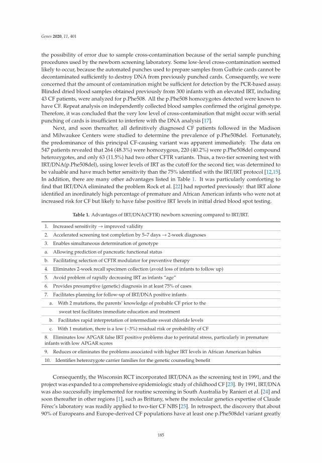
Genes 2020, 11, 401
the possibility of error due to sample cross-contamination because of the serial sample punchingprocedures used by the newborn screening laboratory. Some low-level cross-contamination seemedlikely to occur, because the automated punches used to prepare samples from Guthrie cards cannot bedecontaminated sufficiently to destroy DNA from previously punched cards. Consequently, we wereconcerned that the amount of contamination might be sufficient for detection by the PCR-based assay.Blinded dried blood samples obtained previously from 300 infants with an elevated IRT, including43 CF patients, were analyzed for p.Phe508. All the p.Phe508 homozygotes detected were known tohave CF. Repeat analysis on independently collected blood samples confirmed the original genotype.Therefore, it was concluded that the very low level of cross-contamination that might occur with serialpunching of cards is insufficient to interfere with the DNA analysis [17].
Next, and soon thereafter, all definitively diagnosed CF patients followed in the Madisonand Milwaukee Centers were studied to determine the prevalence of p.Phe508del. Fortunately,the predominance of this principal CF-causing variant was apparent immediately. The data on547 patients revealed that 264 (48.3%) were homozygous, 220 (40.2%) were p.Phe508del compoundheterozygotes, and only 63 (11.5%) had two other CFTR variants. Thus, a two-tier screening test withIRT/DNA(p.Phe508del), using lower levels of IRT as the cutoff for the second tier, was determined tobe valuable and have much better sensitivity than the 75% identified with the IRT/IRT protocol [12,15].In addition, there are many other advantages listed in Table 1. It was particularly comforting tofind that IRT/DNA eliminated the problem Rock et al. [22] had reported previously: that IRT aloneidentified an inordinately high percentage of premature and African American infants who were not atincreased risk for CF but likely to have false positive IRT levels in initial dried blood spot testing.
Table 1. Advantages of IRT/DNA(CFTR) newborn screening compared to IRT/IRT.
1. Increased sensitivity→ improved validity
2. Accelerated screening test completion by 5–7 days→ 2-week diagnoses
3. Enables simultaneous determination of genotype
a. Allowing prediction of pancreatic functional status
b. Facilitating selection of CFTR modulator for preventive therapy
4. Eliminates 2-week recall specimen collection (avoid loss of infants to follow up)
5. Avoid problem of rapidly decreasing IRT as infants “age”
6. Provides presumptive (genetic) diagnosis in at least 75% of cases
7. Facilitates planning for follow-up of IRT/DNA positive infants
a. With 2 mutations, the parents’ knowledge of probable CF prior to the
sweat test facilitates immediate education and treatment
b. Facilitates rapid interpretation of intermediate sweat chloride levels
c. With 1 mutation, there is a low (~3%) residual risk or probability of CF
8. Eliminates low APGAR false IRT positive problems due to perinatal stress, particularly in prematureinfants with low APGAR scores
9. Reduces or eliminates the problems associated with higher IRT levels in African American babies
10. Identifies heterozygote carrier families for the genetic counseling benefit
Consequently, the Wisconsin RCT incorporated IRT/DNA as the screening test in 1991, and theproject was expanded to a comprehensive epidemiologic study of childhood CF [23]. By 1991, IRT/DNAwas also successfully implemented for routine screening in South Australia by Ranieri et al. [24] andsoon thereafter in other regions [1], such as Brittany, where the molecular genetics expertise of ClaudeFérec’s laboratory was readily applied to two-tier CF NBS [25]. In retrospect, the discovery that about90% of Europeans and Europe-derived CF populations have at least one p.Phe508del variant greatly
185

Genes 2020, 11, 401
facilitated widespread implementation of the world’s first DNA-based population screening test—theIRT/DNA(p.Phe508del) method. Its advantages and demonstration of benefits [15,26] outweighing theinevitable harms of any screening test set the stage for the Centers for Disease Control and Prevention(CDC) and the US CFF to advocate for universal CF NBS in 2004 [2] and 2005 [27], respectively.The delay in approvals by these organizations can be attributed to the fact that such advocacy wasunprecedented, and both the CDC and CFF were concerned that the potential harm could outweighthe benefits [2,11,27]. These actions stimulated nationwide screening of 4 million American babiesannually in less than 5 years and now worldwide screening programs, as shown in Figure 1. Moreover,a complete transformation of the care strategy for children with CF [23,28–30] emerged as routineearly diagnosis facilitated preventive therapies, reflecting the view that prevention is far better thanintervention for chronic, potentially irreversible disease.
Figure 1. Cystic fibrosis newborn screening in 2020 around the world where the disease is prevalent.
3. Improvements in IRT/DNA Screening Protocols
Although the two-tier screening test with IRT/DNA and the p.Phe508del variant only using lowercutoff levels of IRT was advantageous [12,17], and needed to be continued for the RCT, the Wisconsinteam decided to change the protocol when randomization ended on 30 June 1994 after NBS-derivednutritional benefits were becoming evident [15,26]. Thus, the Wisconsin State Laboratory of Hygiene,led by Ron Laessig and Gary Hoffman, recognized that routine CF NBS should be introduced withtwo improvements in the screening algorithm. First, it had become clear during the RCT that thebiomarker IRT was a very challenging analyte with regard to setting reliable, stable cutoff values.In fact, there were both seasonal and kit-related variations that prevented equitable use of IRT andmitigation of false negative results [12,31]. This recognition led to the floating IRT cutoff value tactic,in which the highest 4%–5% of the daily specimens were reflexed to the DNA tier [12].
In addition, recognizing the important contribution of Comeau et al. [32], many regions expandedthe DNA tier to a CFTR multimutation panel and a sensitivity of >95% was achieved routinely (insome years, 100% sensitivity) [18]. In addition, the quality of screening improved significantly by notonly allowing test completion on the initial dried blood spot specimen, thus improving timeliness,but providing valuable information on most patients’ CFTR variants. It was quickly learned withIRT/DNA(CFTR) that the vast majority of CF cases can be presumptively (genetically) diagnosedwithin a week of birth from the initial blood specimen and valuable genetic data obtained to predictpancreatic functional status [33]. Later, the rapid genotyping capability of IRT/DNA (CFTR) screeningalso provided guidance for selection of CFTR modulators which can now be used to achieve organpreservation if begun early [34]. On the other hand, the CFTR multi-mutation tactic increases thenumber of incidental findings, particularly detection of CF heterozygote carriers, an increasingly
186
Genes 2020, 11, 401
important topic that is discussed in detail subsequently, and identification of those with CRMS/CFSPID(cystic fibrosis transmembrane conductance regulator-related metabolic syndrome/cystic fibrosis screenpositive, inconclusive diagnosis) [35,36], which is well reviewed elsewhere [37] and beyond the scopeof this article.
Concerns about the higher costs of DNA/CFTR analyses and increased carrier detection have led toanother change in regions wedded to collecting two dried blood specimens, namely the IRT/IRT/DNAprotocol [38]. This algorithm modification achieves those two objectives (lower costs and fewer carriersdetected) but delays diagnoses and does not improve sensitivity [39]. In view of the early risks of CF ininfants such as potentially fatal hyponatremic/hypochloremic dehydration [40], and the likelihood ofearly malnutrition [41], it has become clear that timeliness in diagnosing the disease is an imperative.Thus, more research is needed on comparing the impact of IRT/DNA with IRT/IRT/DNA protocols.Similarly, algorithms employing pancreatitis-associated protein (PAP) and other supplemental testsneed to be investigated further [1,35]. These algorithms may add potential advantages such asproviding a “safety net” [35].
4. Application of Next Generation Sequencing
Despite its great advantages and popularity, there are many imperfections of IRT/DNA screening.One of the biggest challenges is a lower-than-ideal positive predictive value (PPV), resulting in as manyas 10 heterozygote infant carriers (i.e., those with high IRT levels and single identified CFTR variants)for every CF case diagnosed after follow-up sweat testing. Although a 10:1 ratio may be disconcerting,other NBS tests lead to a higher proportion of false positive results such as screening for congenitaladrenal hyperplasia [42]. In CF, however, the situation is exacerbated by the need for confirmatorysweat testing aa well as the occurrence of an insufficient quantity of sweat collected or an inconclusivesweat test outcome in at least 10% of the screen-positive infants. In addition, the CFTR panels in generaluse during the past two decades have had insufficient CF-causing variants to allow the detection ofminority populations in many regions where uncommon CF-causing variants occur such as M1101K(c.3302T>A), found in Hutterite populations [43], and H199Y (c.595C>T) or S492F (c.1475C>T) seenin Hispanic populations [44]. As the genetic diversity of populations increases, the panels used forIRT/DNA screening will not adequately meet the goal of avoiding inequities, because missed casesare more likely among non-Caucasians. These challenges led CF NBS laboratories [45,46] to considerbiotechnologies that would greatly expand the CFTR panel after two major advances that facilitatedsuch a change—the advent of next generation sequencing (NGS) [47] and the knowledge gained fromthe CFTR2 project [48,49].
NGS, a deep, high-throughput, massive parallel DNA sequencing technology, has become popularduring the past decade for genetic and genomic sequencing. Rapid progress in NGS chemistry andinstrumentation coupled to the simultaneous development of advanced bioinformatics methods makesNGS feasible in public health laboratories that are responsible for NBS. Baker et al. [45] reportedtheir successful experience with using NGS to increase the detection capability of CFTR pathogenicvariants using DNA isolated from routine dried blood spot specimens. The NGS assay was designedto sequence all the coding regions, intron/exon boundaries, and selected intronic regions. The dataanalysis software was designed to mask much of the sequence data and reveal only the predeterminedCF-causing variants in the CFTR gene as characterized by the CFTR2 project [48,49]. The significanceof this study includes: (1) it was the first report from a public health NBS laboratory regarding thetechnical feasibility of applying NGS in routine NBS practice; and (2) the reported laboratory-developedDNA isolation method in this study is applicable to other molecular testing currently used in publichealth NBS laboratories, such as multiplexing real-time PCR assays to screen for severe combinedimmunodeficiency (SCID) [50] and spinal muscular atrophy (SMA) [51,52].
Built on the technical feasibility of applying NGS in routine NBS practice, the Wisconsin NBSprogram has, since April, 2016, further implemented the NGS CFTR variant panel as the routinesecond-tier testing method following first-tier IRT testing. Any newborn with one or more CF-causing
187

Genes 2020, 11, 401
variants identified is referred for a follow-up sweat chloride test as confirmatory testing. Recently,NGS data were reanalyzed on the infants with sweat chloride concentrations greater than 30 mmol/Lafter one CF-causing variant was identified through a regular NBS screening protocol. The NGS datareanalysis was performed by removing preset panel restrictions and viewing all variants. NGS datareanalysis is a cost-effective practice for identifying a second pathogenic or likely pathogenic CFTRvariant in infants with a likely CF or CRMS/ CFSPID diagnosis [37]. When a single, second CFTRvariant is detected with sequencing via NGS in a patient with a positive sweat test and/or symptoms ofCF, it is quite likely to be pathogenic. Moreover, mutations in some classes like a Class I prematuretermination codon (PTC) can be assumed to be pathogenic; as Sosnay et al. state, “the assumption thatvariants predicted to introduce a PTC are deleterious is commonly accepted practice” [48]. The currentCFTR variant panel in Wisconsin includes 328 CF-causing variants and the most common deletions(exon 2–3 deletion and exon 22–23 deletion) but not one deep intronic variant and other large deletionsand duplications; this may be increased further in the future as the more CF-causing variants areadded to the CFTR2 website. The State of New York also uses NGS and includes a third-tier CFTRsequence step before reporting all pathogenic and probable pathogenic variants prior to sweat testing,i.e., an “IRT/DNA/SEQ” algorithm [46], that markedly improves PPV.
5. The Potentially Added Value of Genomic Sequencing
The not-too-distant future holds many more opportunities for discovery of CF gene modifiers . . .whole-genome sequencing will expand GWAS-type studies to rare variant analyses [53]. NGS has made wholegenome sequencing (WGS) feasible and affordable with platforms that are commercially availableand proven reliable. Compared to whole exome sequencing, WGS is preferable, because the formeronly covers about 1.4% of the genome and will miss loci of potential clinical importance. Additionally,the modest cost of WGS now available is compelling. In fact, the cost of WGS is already similar to whatmany hospitals charge for bilateral sweat chloride tests. Clinical applications have therefore becomeattractive. Its use for diagnoses in critically ill newborns has already been established so that it willeventually become routine in neonatal intensive care units [54,55].
Although improvements in the diagnosis and treatment of CF have led to better outcomes for themajority of patients, the impact of pancreatic insufficiency, causing malnutrition and the occurrence ofearly obstructive lung disease with recurrent respiratory infections, can make identifying appropriatetherapies challenging in many patients. In fact, some children labeled “non-responders” do not respondwell to early GI/nutritional treatment, and they also have worse lung disease [56,57]. These observationsunderscore a gap in our understanding of what else will be needed to ensure successful therapeuticstrategies, especially preventive care. Although the development of CFTR modulators has begunto revolutionize the treatment of CF patients, variable responses to these expensive modalities hascreated another important gap that needs to be addressed. With universal NBS for CF identifyingpresymptomatic patients, better opportunities have emerged for closing these gaps. Thus, Wisconsinrecently launched a project entitled “Assessing the Added Value of Whole Genome Sequencing inCystic Fibrosis Newborn Screening” to address the hypothesis that identifying non-CFTR geneticvariants in individual patients could enlighten therapeutic decision-making [58]. Particular attentionis being given to potential genetic modifiers of lung disease and nutrigenomic/pharmacogenomicvariants related to common medications.
Exploring nutrigenomic and pharmacogenomic variants that are CF-relevant and can be readilyidentified with WGS appears to be fruitful according to our preliminary data [58,59]. A recentlypublished study of siblings with different lung disease manifestations but an identical CFTR genotypepredicting severe disease (p.Phe508del/CFTRdele2,3) generated WGS data that were informative [58].The patients’ variable phenotypes were accompanied by a surprising degree of differences in bothgenetic modifier and pharmacogenomics variants. In addition, in another study designed to gaininsights about the variable response of CF patients to vitamin D supplements, WGS data were obtainedand analyzed in 20 children whose 25-hydroxycholecalciferol levels varied during 4–24 months of
188

Genes 2020, 11, 401
treatment. Using a polygenic score technique to assess six informative loci, Lai et al. [59] found asignificant nutrigenomic correlation. This study is currently being expanded with inclusion of morechildren and also adults with CF. Further research will be needed and is likely to be completed in thenear future to determine if genomics added to DNA-based screening/genotyping will be valuable.
6. Prospects for a Primary DNA Screening Test
Many regions have implemented NBS for CF using the IRT/DNA two-tier protocol with a limitedpanel of variants [1]. According to 14 years of Wisconsin screening data, this protocol has hadapproximately 97% sensitivity, and the false negatives were due mainly to low IRT levels [31]. To avoidthe false negative results caused by low IRT levels, a primary DNA-based screening test could bea potential solution. From the technical perspective, DNA-based assays as primary screening testshave been done routinely in screening for SCID [50] and SMA [51,52]. For SCID, although measuringT-cell receptor excision circles (TRECs) uses molecular techniques, it does not provide specific geneticinformation about the individual. TRECs are small circles of DNA created in T cells during theirmaturation in the thymus. Their presence indicates maturation of T cells; TRECs are reduced inSCID [50]. For SMA, the screening assays are designed to assess for deletions in the survival of motorneuron 1 (SMN1) gene. Undetectable SMN1 by real-time PCR indicates a high risk for SMA due tohomozygous deletion of exon 7. This condition was recently added to the US Recommended UniformScreening Panel (RUSP) [52]. The screening test for SMA is genetic testing, but the strategy of onlytargeting SMN1 absence makes it possible to avoid SMA carrier identification in newborns. Successwith SMA primary DNA-based screening, however, has created interest in applying the same strategyfor CF NBS when analytical biotechnology advances enough to enable fast, affordable complete CFTRanalysis for CF-causing variants, as it inevitably will and perhaps not very far into the future. Evenif/when a primary DNA screening test for CF becomes technically feasible, unavoidable CF carrieridentification will continue to be a challenging situation.
7. Incidental Findings and Implications of Emerging Data for Genetic Counseling
An important impact of the CFTR gene discovery relates to CF heterozygote carriers who are nowbeing identified in countless numbers through prenatal and neonatal screening. One of the concernsabout expanding CFTR panels beyond the previously common 23 CF-causing variants [60] has beenthe increase in incidental findings, particularly heterozygous infants, i.e., false positives with highIRT levels, one CFTR variant, and negative sweat tests. The PPV of the IRT/DNA test ranges fromabout 10% to 63% depending on the algorithm and the number of CFTR variants in the panel [61].Detecting up to 10 carriers for every CF case diagnosed has been disconcerting for some, while othersconsider this an added benefit of CF NBS. Those in the former camp argue that that the purpose of CFNBS is identification of affected CF patients, and some have suggested that only more severely affectedchildren should be the target population [62]. On the other hand, CF specialists who readily tolerate,or even advocate for, carrier detection as a byproduct of NBS can point out the many parents who haveregarded that outcome as beneficial and appreciated the genetic counseling. In rare instances, an oldersibling with CF has also been found in association with a false positive NBS result, and in most casesbetter informed reproductive planning has ensued from the genetic counseling. With greatly expandedCFTR panels and the prospects of a primary DNA/CFTR screening test, the issue of detecting infantswho are carriers takes on greater importance and deserves detailed consideration.
There has been relatively little research done on the CF heterozygote carrier and no previousreview of the published data and their implications. In a recent international study, it was determinedthat the p.Phe508del variant arose in Western Europe at least 5000 years ago [63,64]. Data on the time tothe most recent common ancestor also revealed that it spread from west to east during the longue duréeof the Bronze Age [65] and into the Iron Age and beyond [64]. Hazardous environmental exposureswere numerous over that period [65]. Linking the p.Phe508del/wild type individual to a selectiveadvantage has not yet been possible, despite some efforts [66], but there can be no doubt that CF
189
Genes 2020, 11, 401
heterozygotes must have had a selective advantage [67]. The high prevalence of p.Phe508del amongnative Europeans implies that the p.Phe508del/wild-type individual has a heretofore undiscoveredhealth, survival, or fertility advantage. However, when parents ask questions in false-positive casesabout what CF carrier status might mean for their baby, we have lacked answers. Nevertheless,this situation seems to be changing, as some research has focused on the CF carrier.
Certainly, it has been known for many years that IRT levels in infants who prove to be carriers arehigher than the general population of screened babies [68,69]. This manifestation of CFTR variant/wildtype status was initially surprising but is now well accepted. It agrees with earlier observations thatCF carriers have higher sweat chloride levels than normal [70,71]. As long ago as 1962, a study by diSant’Agnese and Powell [70] revealed an impressive difference between 97 obligate CF carriers (parentsof patients with CF) and 117 “unselected adult controls” (mean sweat chloride values of 32 and 17mmol/L, respectively; p < 0.01), suggesting that the sweat electrolyte abnormality should be considereda subclinical phenotypic manifestation in the CF heterozygote. In addition, taking advantage of NBSfollow up data, Farrell and Koscik [72] showed conclusively that, after controlling the age factor,CF heterozygote carriers with the p.Phe508del variant have significantly increased sweat electrolyteconcentrations, although they were not high enough to be in the range diagnostic of the disease.In retrospect, this should not be surprising since as Miller et al. [73] point out “carriers have ∼50% asmuch CFTR anion channel activity as controls... and in some epithelia, anion transport is known to bereduced in carriers.”
With regard to symptoms and disease risk, Wang et al. [74] reported that adult obligate CFheterozygotes have a much higher prevalence of chronic rhinosinusitis than the general population.Others have reported a higher incidence of pancreatitis in genetically proven CF carrier adults [75,76].When disorders such as chronic pancreatitis occur in these patients, they might be better classified ashaving a CFTR-related disorder (CFTR-RD) [77], rather than simply being labeled CF carriers, but recentobservations [73,75,78] may stimulate reassessment of terminology. In addition to chronic sinusitis,three phenotypes are included in the CFTR-RD category: CBAVD (congenital bilateral absence of thevas deferens) causing male infertility, acute recurrent or chronic pancreatitis, and bronchiectasis [77].The prevalence of these and other conditions in CF carriers is surprisingly high as discussed below.
In 2011, Tluczek et al. [78] first identified a higher risk of disease in CF carriers. Specifically,based on evaluating children with a high IRT plus one CF-causing variant and a negative sweat test,Tluzcek et al. [78] observed that these carriers have more health system encounters for documentedillnesses during their first year than healthy infants with negative NBS results. More recently,Miller et al. [73] published a provocative study involving 18,902 carriers matched with controls andreported that those with one CFTR variant have a significantly increased risk for 57 of 59 CF-relateddiagnostic conditions based on odds ratio analyses. The relative risks were increased for someconditions previously associated with CF carriers (e.g., pancreatitis, male infertility, and bronchiectasis),but others were not previously suspected, such as diabetes, constipation and cholelithiasis. Althoughthe clinical significance of their findings remains to be determined, being identified as a CF carriermay promote attention to avoiding other disease-risk factors (e.g., the importance of avoidingheavy alcohol consumption in those who are intrinsically susceptible to pancreatitis). Moreover,Miller et al. [73] pointed out that identifying CF carriers may provide rational treatment options inthe future for symptomatic carriers using drugs designed to enhance CFTR function. On the otherhand, when applying these results to genetic counseling, it is important to keep in mind the differencesbetween relative and absolute risk. As Miller et al. [73] point out, the relative risk for chronic pancreatitisis high with an odds ratio of 6.76 [95% CI, 4.87–9.39] but the absolute risk is less than 1% (only 0.429 per100 carriers). However, when all the CFTR-RD conditions [74,77] are considered in male carriers basedon the data of Miller et al. [73], the cumulative absolute risk is 19.07 per 100 CF heterozygotes—not atrivial prevalence.
Thus, in addition to having a probable health benefit that explains their high frequency,CF carriers appear to be at increased risk for some diseases because of their partial CFTR dysfunction.
190
Genes 2020, 11, 401
To place this situation in perspective and learn from previous experiences, we need to considerthe situation with sickle hemoglobin heterozygotes—individuals labeled as SCT for sickle celltrait. For more than a half-century, they have been identified in a variety of screening programs.After NBS for hemoglobinopathies via cellulose acetate electrophoresis became routine in the USduring the late 1970s [79], numerous follow-up system problems occurred initially, particularlyamong African-American families. These included confusion, inappropriate labeling, stigmatization,and mistreatment. Over time, however, better follow-up communication practices have improvedoutcomes. Now, with data that clearly show serious health risks of SCT and benefits of earlyrecognition, the advantages of heterozygote detection are generally accepted [80,81]. Thus, recognizingthat SCT is actionable, health care systems have learned more about how to manage SCT-relatedcommunications and ensure effective, timely counseling [82]. Because the severe risks are delayedbeyond childhood, when precipitating factors may supervene, such as dehydration during strenuousathletic events [80,81], counseling for SCT risks that occurs immediately after NBS must be repeatedand target the heterozygous individual at an appropriate time [82]. The benefits are undeniable,however, especially the prevention of sudden death by simple hydration [80–83].
When considered collectively, the observations reviewed above suggest that we should not bedescribing CF carriers as completely healthy. Although providers who argue against incidental CFcarrier detection do not recognize its advantages and regard these CF NBS incidental findings as“unwanted,” the benefits of counseling parents of infant carriers are clear [84,85]. As more informationis learned about the CF heterozygote condition, we suggest that attitudes and practices may needto change and ensure truthful education and effective communications about the implications of CFcarrier status. Although the initial genetic counseling may eventually need to include the possibilitythat heterozygote infants have a higher risk of CFTR-RD conditions [73–77], incidental detection ofcarrier status in false-positive infants does not yet seem actionable for the child because of the lowabsolute risks and thus the expectation that most CF heterozygotes will be healthy—at least until laterin life. Consequently, as with SCT, the counseling will have to be timed appropriately and target thecarrier when risk factor mitigation could be valuable and CFTR modulators potentially useful forsevere disorders like pancreatitis [75,76].
8. The Opportunities for Preventive Therapies
Management of CF disease has traditionally relied on symptom-based treatments [86]. Thus, a “onesize fits all” philosophy has been common for many years, in which a plethora of drugs are beingused in standard doses. Malnutrition and growth faltering, obstructive lung disease with suppurativerespiratory infections, and unresponsive pulmonary exacerbations are challenging to treat, especiallyin “non-responders [56,57]. Thus, preventive therapies are increasingly attractive and feasible. Thereare four problems in CF that are amenable to prophylactic strategies, namely: (1) salt loss in sweat thatcan cause fatal hyponatremic/hypochloremic dehydration; (2) pancreatic insufficiency; (3) malnutrition;and (4) chronic obstructive lung disease with recurrent infections [4]. Although excessive salt loss mayseem minor and can be readily prevented with salt supplements, CF patients still die from this problem,and the breast-fed infant is especially vulnerable in hot weather. Treatment with a lifetime of daily saltsupplements is effective and essential, even when a patient is taking CFTR modulators [28]. Pancreaticinsufficiency and its sequelae (risk of malnutrition, recurrent pancreatitis, and diabetes mellitus) havealways been considered a permanent component of CF for the majority of patients with susceptiblegenotypes. To most CF specialists’ surprise, however, CFTR modulator therapy has been associatedwith at least partial preservation of pancreatic function in recent studies examining this issue [34,84–86].In fact, the weight gains of CF patients in the original trials of ivacaftor suggested its potential toreduce the detrimental impact of pancreatic insufficiency [28], and subsequent observations supportthis hypothesis [87–89]. Prevention of malnutrition in most children with CF diagnosed early in theWisconsin RCT was achieved with an aggressive approach to nutritional management, emphasizingthe combination of pancreatic enzyme replacement therapy, high caloric intake, and supplements of
191
Genes 2020, 11, 401
fat soluble vitamins plus essential fatty acid supplements [30,90]. On the other hand, some childrenwere found to be “non-responders” to this regimen [56,57,91], and this has led to more research [59].Lastly, the most important target for preventive therapy is the respiratory system, because the qualityand quantity of life in individual patients’ experience usually depends on the severity of lung disease.Thus, the most exciting aspect of CFTR modulator therapy is its potential for ameliorating lungdisease as discussed in detail elsewhere [28,88,89]. For CF patients, therefore, the transformation fromintervention to prevention, associated with early diagnosis and novel treatments, is clearly the mostimpactful result of the discovery of the CFTR gene.
9. Conclusions
The discovery of the CFTR gene was the scientific advance that improved our ability to diagnose CFrapidly, genotype patients simultaneously, predict pancreatic functional status immediately, and thenplan preventive care. The era of genetic/genomic medicine has brightened the outlook for all patientswith CF and especially children, who are now in most cases diagnosed while still asymptomatic.
Author Contributions: All three authors contributed substantially to the research and views reported herein.All authors have read and agreed to the published version of the manuscript.
Funding: Our research has been funded by the National Institutes of Health (DK34108), the Cystic FibrosisFoundation, and The Legacy of Angels Foundation.
Acknowledgments: We thank the late Ron Laessig, Gary Hoffman and Ronald Gregg for their essentialcontributions to the development of the Wisconsin CF newborn screening program and Anita Laxova forcoordinating the Wisconsin RCT of CF newborn screening. We are also grateful to Elinor Langfelder-Schwind,Karen Raraigh, and Michael Farrell for their review, revisions, and advice regarding Section 7 on incidentalfindings and to Erin Ryan for her help with the manuscript preparation.
Conflicts of Interest: The authors declare no conflict of interest.
References
1. Scotet, V.; Gutierrez, H.; Farrell, P.M. Newborn Screening for CF across the Globe—Where Is It Worthwhile?Int. J. Neonatal Screen. 2020, 6, 18. [CrossRef]
2. Grosse, S.D.; Boyle, C.A.; Botkin, J.R.; Comeau, A.M.; Kharrazi, M.; Rosenfeld, M.; Wilfond, B.S. NewbornScreening for Cystic Fibrosis: Evaluation of Benefits and Risks and Recommendations for State NewbornScreening Programs. In MMWR. Recommendations and Reports: Morbidity and Mortality Weekly Report.Recommendations and Reports; Centers for Disease Control and Prevention: Antlanta, GA, USA, 2004; VolumeVoume 53, pp. 1–36.
3. Farrell, P.; Gilbert-Barness, E.; Bell, J.; Gregg, R.; Mischler, E.; Odell, G.; Shahidi, N.; Robertson, I.; Evans, J.Progressive Malnutrition, Severe Anemia, Hepatic Dysfunction, and Respiratory Failure in a Three-Month-OldWhite Girl. Am. J. Med. Genet. 1993, 45, 725–738. [CrossRef] [PubMed]
4. Accurso, F.J.; Sontag, M.K.; Wagener, J.S. Complications Associated with Symptomatic Diagnosis in Infantswith Cystic Fibrosis. J. Pediatr. 2005, 147, S37–S41. [CrossRef] [PubMed]
5. Kharrazi, M.; Yang, J.; Bishop, T.; Lessing, S.; Young, S.; Graham, S.; Pearl, M.; Chow, H.; Ho, T.; Currier, R.; et al.Newborn Screening for Cystic Fibrosis in California. Pediatrics 2015, 136, 1062–1072. [CrossRef]
6. Shwachman, H.; Kulczycki, L.L. Long-Term Study of One Hundred Five Patients with Cystic Fibrosis:Studies Made Over a Five- to Fourteen-Year Period. AMA J. Dis. Child. 1958, 96, 6–15. [CrossRef]
7. Bruns, W.T.; Connell, T.R.; Lacey, J.A.; Whisler, K.E. Test Strip Meconium Screening for Cystic Fibrosis. Am. J.Dis. Child. 1977, 131, 71–73. [CrossRef]
8. Guthrie, R.; Susi, A. A Simple Phenylalanne Method for Detecting Phenylketonuria in Large Populations ofNewborn Infants. Pediatrics 1963, 32, 338–343.
9. Crossley, J.R.; Elliott, R.B.; Smith, P.A. Dried-Blood Spot Screening for Cystic Fibrosis in the Newborn.Lancet (Lond. Engl.) 1979, 1, 472–474. [CrossRef]
10. Hammond, K.B.; Abman, S.H.; Sokol, R.J.; Accurso, F.J. Efficacy of Statewide Neonatal Screening for CysticFibrosis by Assay of Trypsinogen Concentrations. N. Engl. J. Med. 1991, 325, 769–774. [CrossRef]
192
Genes 2020, 11, 401
11. Taussig, L.M.; Boat, T.F.; Dayton, D.; Fost, N.; Hammond, K.; Holtzman, N.; Johnson, W.; Kaback, M.M.;Kennel, J.; Rosenstein, B.J.; et al. Neonatal Screening for Cystic Fibrosis: Position Paper. Pediatrics 1983, 72,741–745.
12. Kloosterboer, M.; Hoffman, G.; Rock, M.; Gershan, W.; Laxova, A.; Li, Z.; Farrell, P.M. Clarification ofLaboratory and Clinical Variables That Influence Cystic Fibrosis Newborn Screening with Initial Analysis ofImmunoreactive Trypsinogen. Pediatrics 2009, 123, e338–e346. [CrossRef] [PubMed]
13. Therrell, B.L.; Hannon, W.H.; Hoffman, G.; Ojodu, J.; Farrell, P.M. Immunoreactive Trypsinogen (IRT) as aBiomarker for Cystic Fibrosis: Challenges in Newborn Dried Blood Spot Screening. Mol. Genet. Metab. 2012,106, 1–6. [CrossRef] [PubMed]
14. Kerem, B.S.; Rommens, J.M.; Buchanan, J.A.; Markiewicz, D.; Cox, T.K.; Chakravarti, A.; Buchwald, M.;Tsui, L.C. Identification of the Cystic Fibrosis Gene: Genetic Analysis. Science 1989, 245, 1073–1080. [CrossRef][PubMed]
15. Farrell, P.M.; Kosorok, M.R.; Rock, M.J.; Laxova, A.; Zeng, L.; Lai, H.C.; Hoffman, G.; Laessig, R.H.;Splaingard, M.L. Early Diagnosis of Cystic Fibrosis through Neonatal Screening Prevents Severe Malnutritionand Improves Long-Term Growth. Pediatrics 2001, 107, 1–13. [CrossRef]
16. Wilfond, B.; Gregg, R.G.; Laxova, A.; Hassemer, D.; Mischler, E.F.P. Mutation Analysis for CF NewbornScreening: A Two-Tiered Approach. Pediatr. Pulmonol. Suppl. 1991, 6, 238.
17. Gregg, R.G.; Wilfond, B.S.; Farrell, P.M.; Laxova, A.; Hassemer, D.; Mischler, E.H. Application of DNAAnalysis in a Population-Screening Program for Neonatal Diagnosis of Cystic Fibrosis (CF): Comparison ofScreening Protocols. Am. J. Hum. Genet. 1993, 52, 616–626.
18. Gregg, R.G.; Simantel, A.; Farrell, P.M.; Koscik, R.; Kosorok, M.R.; Laxova, A.; Laessig, R.; Hoffman, G.;Hassemer, D.; Mischler, E.H.; et al. Newborn Screening for Cystic Fibrosis in Wisconsin: Comparison ofBiochemical and Molecular Methods. Pediatrics 1997, 99, 819–824. [CrossRef]
19. Farrell, P.M.; Mischler, E.H.; Fost, N.C.; Wilfond, B.S.; Tluczek, A.; Gregg, R.G.; Bruns, W.T.; Hassemer, D.J.;Laessig, R.H. Current Issues in Neonatal Screening for Cystic Fibrosis and Implications of the CF GeneDiscovery. Pediatr. Pulmonol. 1991, 11 (Suppl. 7), 11–18. [CrossRef]
20. Jinks, D.C.; Minter, M.; Tarver, D.A.; Vanderford, M.; Hejtmancik, J.F.; McCabe, E.R.B. Molecular GeneticDiagnosis of Sickle Cell Disease Using Dried Blood Specimens on Blotters Used for Newborn Screening.Hum. Genet. 1989, 81, 363–366. [CrossRef]
21. Rommens, J.; Kerem, B.S.; Greer, W.; Chang, P.; Tsui, L.C.; Ray, P. Rapid Nonradioactive Detection of theMajor Cystic Fibrosis Mutation. Am. J. Hum. Genet. 1990, 46, 395–396.
22. Rock, M.J.; Mischler, E.H.; Farrell, P.M.; Wei, L.J.; Bruns, W.T.; Hassemer, D.J.; Laessig, R.H. NewbornScreening for Cystic Fibrosis Is Complicated by Age-Related Decline in Immunoreactive Trypsinogen Levels.Pediatrics 1990, 85, 1001–1007. [CrossRef] [PubMed]
23. Farrell, P.M. Improving the Health of Patients with Cystic Fibrosis through Newborn Screening. WisconsinCystic Fibrosis Neonatal Screening Study Group. Adv. Pediatr. 2000, 47, 79–115. [PubMed]
24. Ranieri, E.; Ryall, R.G.; Morris, C.P.; Nelson, P.V.; Carey, W.F.; Pollard, A.C.; Robertson, E.F. NeonatalScreening Strategy for Cystic Fibrosis Using Immunoreactive Trypsinogen and Direct Gene Anylysis. Br. Med.J. 1991, 302, 1237–1240. [CrossRef] [PubMed]
25. Férec, C.; Verlingue, C.; Parent, P.; Morin, J.F.; Codet, J.P.; Rault, G.; Dagorne, M.; Lemoigne, A.; Journel, H.;Roussey, M. Neonatal Screening for Cystic Fibrosis: Result of a Pilot Study Using Both ImmunoreactiveTrypsinogen and Cystic Fibrosis Gene Mutation Analyses. Hum. Genet. 1995, 96, 542–548. [CrossRef]
26. Farrell, P.M.; Lai, H.C.J.; Li, Z.; Kosorok, M.R.; Laxova, A.; Green, C.G.; Collins, J.; Hoffman, G.; Laessig, R.;Rock, M.J.; et al. Evidence on Improved Outcomes with Early Diagnosis of Cystic Fibrosis through NeonatalScreening: Enough Is Enough! J. Pediatr. 2005, 147, S30–S36. [CrossRef]
27. Campbell, P.W.; White, T.B. Newborn Screening for Cystic Fibrosis: An Opportunity to Improve Care andOutcomes. J. Pediatr. 2005, 147, S2–S5. [CrossRef]
28. Ramsey, B.W.; Davies, J.; McElvaney, N.G.; Tullis, E.; Bell, S.C.; Drevínek, P.; Griese, M.; McKone, E.F.;Wainwright, C.E.; Konstan, M.W.; et al. A CFTR Potentiator in Patients with Cystic Fibrosis and the G551DMutation. N. Engl. J. Med. 2011, 365, 1663–1672. [CrossRef]
29. Middleton, P.G.; Mall, M.A.; Drevínek, P.; Lands, L.C.; McKone, E.F.; Polineni, D.; Ramsey, B.W.;Taylor-Cousar, J.L.; Tullis, E.; Vermeulen, F.; et al. Elexacaftor-Tezacaftor-Ivacaftor for Cystic Fibrosiswith a Single Phe508del Allele. N. Engl. J. Med. 2019, 381, 1809–1819. [CrossRef]
193
Genes 2020, 11, 401
30. Borowitz, D.; Robinson, K.A.; Rosenfeld, M.; Davis, S.D.; Sabadosa, K.A.; Spear, S.L.; Michel, S.H.; Parad, R.B.;White, T.B.; Farrell, P.M.; et al. Cystic Fibrosis Foundation Evidence-Based Guidelines for Management ofInfants with Cystic Fibrosis. J. Pediatr. 2009, 155 (Suppl. 6), S73–S93. [CrossRef]
31. Rock, M.J.; Levy, H.; Zaleski, C.; Farrell, P.M. Factors Accounting for a Missed Diagnosis of Cystic Fibrosisafter Newborn Screening. Pediatr. Pulmonol. 2011, 46, 1166–1174. [CrossRef]
32. Comeau, A.M.; Parad, R.B.; Dorkin, H.L.; Dovey, M.; Gerstle, R.; Haver, K.; Lapey, A.; O’Sullivan, B.P.;Waltz, D.A.; Zwerdling, R.G.; et al. Population-Based Newborn Screening for Genetic Disorders WhenMultiple Mutation DNA Testing Is Incorporated: A Cystic Fibrosis Newborn Screening Model DemonstratingIncreased Sensitivity but More Carrier Detections. Pediatrics 2004, 113, 1573–1581. [CrossRef] [PubMed]
33. Kerem, E.; Kerem, B. Genotype-phenotype Correlations in Cystic Fibrosis. Pediatr. Pulmonol. 1996, 22,387–395. [CrossRef]
34. De Boeck, K. Cystic Fibrosis in the Year 2020: A Disease with a New Face. In Acta Paediatrica, InternationalJournal of Paediatrics; Blackwell Publishing Ltd.: Oxford, UK, 2020. [CrossRef]
35. Sommerburg, O.; Hammermann, J.; Lindner, M.; Stahl, M.; Muckenthaler, M.; Kohlmueller, D.; Happich, M.;Kulozik, A.E.; Stopsack, M.; Gahr, M.; et al. Five Years of Experience with Biochemical Cystic FibrosisNewborn Screening Based on IRT/PAP in Germany. Pediatr. Pulmonol. 2015, 50, 655–664. [CrossRef][PubMed]
36. Munck, A.; Mayell, S.J.; Winters, V.; Shawcross, A.; Derichs, N.; Parad, R.; Barben, J.; Southern, K.W.Cystic Fibrosis Screen Positive, Inconclusive Diagnosis (CFSPID): A New Designation and ManagementRecommendations for Infants with an Inconclusive Diagnosis Following Newborn Screening. J. Cyst. Fibros.2015, 14, 706–713. [CrossRef]
37. Ren, C.L.; Borowitz, D.S.; Gonska, T.; Howenstine, M.S.; Levy, H.; Massie, J.; Milla, C.; Munck, A.;Southern, K.W. Cystic Fibrosis Transmembrane Conductance Regulator-Related Metabolic Syndrome andCystic Fibrosis Screen Positive, Inconclusive Diagnosis. J. Pediatr. 2017, 181, S45–S51.e1. [CrossRef]
38. Sontag, M.K.; Wright, D.; Beebe, J.; Accurso, F.J.; Sagel, S.D. A New Cystic Fibrosis Newborn ScreeningAlgorithm: IRT/IRT1↑/DNA. J. Pediatr. 2009, 155, 618–622. [CrossRef]
39. CLSI. Newborn Screening for Cystic Fibrosis, 2nd ed.; CLSI guideline NBS05; Clinical and Laboratory StandardsInstitute: Wayne, PA, USA, 2019.
40. Di Sant’Agnese, P.A. Neonatal and General Aspects of Cystic Fibrosis. In Current Topics in ClinicalChemistry-Clinical Biochemistry of the Neonate; Young, D.C., Hicks, J.M., Eds.; John Wiley and Sons Inc.:New York, NY, USA, 1976.
41. Reardon, M.C.; Hammond, K.B.; Accurso, F.J.; Fisher, C.D.; McCabe, E.R.B.; Cotton, E.K.; Bowman, C.M.Nutritional Deficits Exist before 2 Months of Age in Some Infants with Cystic Fibrosis Identified by ScreeningTest. J. Pediatr. 1984, 105, 271–274. [CrossRef]
42. Allen, D.B.; Farrell, P.M. Newborn Screening: Principles and Practice. Adv. Pediatr. 1996, 43, 231–270.43. Zielenski, J.; Fujiwara, T.M.; Markiewicz, D.; Paradis, A.J.; Anacleto, A.I.; Richards, B.; Schwartz, R.H.;
Klinger, K.W.; Tsui, L.C.; Morgan, K. Identification of the M1101K Mutation in the Cystic FibrosisTransmembrane Conductance Regulator (CFTR) Gene and Complete Detection of Cystic Fibrosis Mutationsin the Hutterite Population. Am. J. Hum. Genet. 1993, 52, 609–615.
44. Watts, K.D.; Layne, B.; Harris, A.; McColley, S.A. Hispanic Infants with Cystic Fibrosis Show Low CFTRMutation Detection Rates in the Illinois Newborn Screening Program. J. Genet. Couns. 2012, 21, 671–675.[CrossRef]
45. Baker, M.W.; Atkins, A.E.; Cordovado, S.K.; Hendrix, M.; Earley, M.C.; Farrell, P.M. Improving NewbornScreening for Cystic Fibrosis Using Next-Generation Sequencing Technology: A Technical Feasibility Study.Genet. Med. 2016, 18, 231–238. [CrossRef] [PubMed]
46. Hughes, E.E.; Stevens, C.F.; Saavedra-Matiz, C.A.; Tavakoli, N.P.; Krein, L.M.; Parker, A.; Zhang, Z.;Maloney, B.; Vogel, B.; DeCelie-Germana, J.; et al. Clinical Sensitivity of Cystic Fibrosis Mutation Panels in aDiverse Population. Hum. Mutat. 2016, 37, 201–208. [CrossRef] [PubMed]
47. Metzker, M.L. Sequencing Technologies the next Generation. Nat. Rev. Genet. 2010, 31–46. [CrossRef][PubMed]
48. Sosnay, P.R.; Siklosi, K.R.; Van Goor, F.; Kaniecki, K.; Yu, H.; Sharma, N.; Ramalho, A.S.; Amaral, M.D.;Dorfman, R.; Zielenski, J.; et al. Defining the Disease Liability of Variants in the Cystic Fibrosis TransmembraneConductance Regulator Gene. Nat. Genet. 2013, 45, 1160–1167. [CrossRef] [PubMed]
194
Genes 2020, 11, 401
49. Sosnay, P.R.; Salinas, D.B.; White, T.B.; Ren, C.L.; Farrell, P.M.; Raraigh, K.S.; Girodon, E.; Castellani, C.Applying Cystic Fibrosis Transmembrane Conductance Regulator Genetics and CFTR2 Data to FacilitateDiagnoses. J. Pediatr. 2017, 181, S27.e1–S32.e1. [CrossRef]
50. Baker, M.W.; Grossman, W.J.; Laessig, R.H.; Hoffman, G.L.; Brokopp, C.D.; Kurtycz, D.F.; Cogley, M.F.;Litsheim, T.J.; Katcher, M.L.; Routes, J.M. Development of a Routine Newborn Screening Protocol for SevereCombined Immunodeficiency. J. Allergy Clin. Immunol. 2009, 124, 522–527. [CrossRef]
51. Vill, K.; Kölbel, H.; Schwartz, O.; Blaschek, A.; Olgemöller, B.; Harms, E.; Burggraf, S.; Röschinger, W.;Durner, J.; Gläser, D.; et al. One Year of Newborn Screening for SMA – Results of a German Pilot Project.J. Neuromuscul. Dis. 2019, 6, 503–515. [CrossRef]
52. Fabie, N.A.V.; Pappas, K.B.; Feldman, G.L. The Current State of Newborn Screening in the United States.In Pediatric Clinics of North America; W.B. Saunders: Philadelphia, PA, USA, 2019; pp. 369–386. [CrossRef]
53. O’Neal, W.K.; Knowles, M.R. Cystic Fibrosis Disease Modifiers: Complex Genetics Defines the PhenotypicDiversity in a Monogenic Disease. Annu. Rev. Genomics Hum. Genet. 2018, 19, 201–222. [CrossRef]
54. Petrikin, J.E.; Willig, L.K.; Smith, L.D.; Kingsmore, S.F. Rapid Whole Genome Sequencing and PrecisionNeonatology. In Seminars in Perinatology; W.B. Saunders: Philadelphia, PA, USA, 2015; pp. 623–631. [CrossRef]
55. Willig, L.K.; Petrikin, J.E.; Smith, L.D.; Saunders, C.J.; Thiffault, I.; Miller, N.A.; Soden, S.E.; Cakici, J.A.;Herd, S.M.; Twist, G.; et al. Whole-Genome Sequencing for Identification of Mendelian Disorders in CriticallyIll Infants: A Retrospective Analysis of Diagnostic and Clinical Findings. Lancet Respir. Med. 2015, 3, 377–387.[CrossRef]
56. Lai, H.J.; Shoff, S.M.; Farrell, P.M. Recovery of Birth Weight z Score within 2 Years of Diagnosis Is PositivelyAssociated with Pulmonary Status at 6 Years of Age in Children with Cystic Fibrosis. Pediatrics 2009, 123,714–722. [CrossRef]
57. Sanders, D.B.; Zhang, Z.; Farrell, P.M.; Lai, H.C.J. Early Life Growth Patterns Persist for 12 years and ImpactPulmonary Outcomes in Cystic Fibrosis. J. Cyst. Fibros. 2018, 17, 528–535. [CrossRef] [PubMed]
58. Wilk, M.A.; Braun, A.T.; Farrell, P.M.; Laxova, A.; Brown, D.M.; Holt, J.M.; Birch, C.L.; Sosonkina, N.;Wilk, B.M.; Worthey, E.A. Applying Whole-Genome Sequencing in Relation to Phenotype and Outcomes inSiblings with Cystic Fibrosis. Cold Spring Harb. Mol. Case Stud. 2020, 6. [CrossRef] [PubMed]
59. Lai, H.J.; Lu, Q.; Chin, L.H.; Wilk, M.; Worthey, E.; the FIRST Study Group. Whole-Genome SequencingReveals That Genetic Variations Predict Effectiveness of Vitamin D Supplemntation in Young Children withCF. Cyst. Fibros. Suppl. 2019, 18, S47. [CrossRef]
60. Watson, M.S.; Cutting, G.R.; Desnick, R.J.; Driscoll, D.A.; Klinger, K.; Mennuti, M.; Palomaki, G.E.;Popovich, B.W.; Pratt, V.M.; Rohlfs, E.M.; et al. Cystic Fibrosis Population Carrier Screening: 2004 Revisionof American College of Medical Genetics Mutation Panel. Genet. Med. 2004, 387–391. [CrossRef]
61. Dankert-Roelse, J.E.; Bouva, M.J.; Jakobs, B.S.; Janssens, H.M.; de Winter-de Groot, K.M.; Schönbeck, Y.;Gille, J.J.P.; Gulmans, V.A.M.; Verschoof-Puite, R.K.; Schielen, P.C.J.I.; et al. Newborn Blood Spot Screeningfor Cystic Fibrosis with a Four-Step Screening Strategy in The Netherlands. J. Cyst. Fibros. 2019, 18, 54–63.[CrossRef]
62. Scotet, V.; Audrézet, M.P.; Roussey, M.; Rault, G.; Dirou-Prigent, A.; Journel, H.; Moisan-Petit, V.; Storni, V.;Férec, C. Immunoreactive Trypsin/DNA Newborn Screening for Cystic Fibrosis: Should the R117H VariantBe Included in CFTR Mutation Panels? Pediatrics 2006, 118, e1523–e1529. [CrossRef]
63. Serre, J.L.; Simon-Bouy, B.; Mornet, E.; Jaume-Roig, B.; Balassopoulou, A.; Schwartz, M.; Taillandier, A.;Boué, J.; Boué, A. Studies of RFLP Closely Linked to the Cystic Fibrosis Locus throughout Europe Lead toNew Considerations in Populations Genetics. Hum. Genet. 1990, 84, 449–454. [CrossRef]
64. Farrell, P.; Férec, C.; Macek, M.; Frischer, T.; Renner, S.; Riss, K.; Barton, D.; Repetto, T.; Tzetis, M.;Giteau, K.; et al. Estimating the Age of p.(Phe508del) with Family Studies of Geographically DistinctEuropean Populations and the Early Spread of Cystic Fibrosis. Eur. J. Hum. Genet. 2018, 26, 1832–1839.[CrossRef]
65. Poolman, E.M.; Galvani, A.P. Evaluating Candidate Agents of Selective Pressure for Cystic Fibrosis. J. R.Soc. Interface 2007, 4, 91–98. [CrossRef]
66. Price, T. Europe before Rome; Oxford University Press: New York, NY, USA, 2013.67. Romeo, G.; Devoto, M.; Galietta, L.J. Why Is the Cystic Fibrosis Gene so Frequent? Hum. Genet. 1989, 84, 1–5.
[CrossRef]
195
Genes 2020, 11, 401
68. Scotet, V.; De Braekeleer, M.; Audrézet, M.P.; Lodé, L.; Verlingue, C.; Quéré, I.; Mercier, B.; Duguépéroux, I.;Codet, J.P.; Moineau, M.P.; et al. Prevalence of CFTR Mutations in Hypertrypsinaemia Detected throughNeonatal Screening for Cystic Fibrosis. Clin. Genet. 2001, 59, 42–47. [CrossRef] [PubMed]
69. Castellani, C.; Picci, L.; Scarpa, M.; Dechecchi, M.C.; Zanolla, L.; Assael, B.M.; Zacchello, F. Cystic FibrosisCarriers Have Higher Neonatal Immunoreactive Trypsinogen Values than Non-Carriers. Am. J. Med. Genet.2005, 135 A, 142–144. [CrossRef]
70. Sant’Agnese, P.A.; Powell, G.F. The Eccrine Sweat Defect in Cystic Fibrosis of the Pancrease (mucoviscidosis).Ann. N. Y. Acad. Sci. 1962, 93, 555–599. [CrossRef]
71. Sproul, A.; Huang, N. Diagnosis of Heterozygosity for Cystic Fibrosis by Discriminatory Analysis of SweatChloride Distribution. J. Pediatr. 1966, 69, 759–770. [CrossRef]
72. Farrell, P.M.; Koscik, R.E. Sweat Chloride Concentrations in Infants Homozygous or Heterozygous for F508Cystic Fibrosis. Pediatrics 1996, 97, 524–528. [PubMed]
73. Miller, A.C.; Comellas, A.P.; Hornick, D.B.; Stoltz, D.A.; Cavanaugh, J.E.; Gerke, A.K.; Welsh, M.J.; Zabner, J.;Polgreen, P.M. Cystic Fibrosis Carriers Are at Increased Risk for a Wide Range of Cystic Fibrosis-RelatedConditions. Proc. Natl. Acad. Sci. USA 2020, 117, 1621–1627. [CrossRef]
74. Wang, X.J.; Kim, J.; McWilliams, R.; Cutting, G.R. Increased Prevalence of Chronic Rhinosinusitis in Carriersof a Cystic Fibrosis Mutation. Arch. Otolaryngol.-Head Neck Surg. 2005, 131, 237–240. [CrossRef]
75. Cohn, J.A.; Neoptolemos, J.P.; Feng, J.; Yan, J.; Jiang, Z.; Greenhalf, W.; McFaul, C.; Mountford, R.; Sommer, S.S.Increased Risk of Idiopathic Chronic Pancreatitis in Cystic Fibrosis Carriers. Hum. Mutat. 2005, 26, 303–307.[CrossRef]
76. Sharer, N.; Schwarz, M.; Malone, G.; Howarth, A.; Painter, J.; Super, M.; Braganza, J. Mutations of the CysticFibrosis Gene in Patients with Chronic Pancreatitis. N. Engl. J. Med. 1998, 339, 645–652. [CrossRef]
77. Bombieri, C.; Claustres, M.; De Boeck, K.; Derichs, N.; Dodge, J.; Girodon, E.; Sermet, I.; Schwarz, M.;Tzetis, M.; Wilschanski, M.; et al. Recommendations for the Classification of Diseases as CFTR-RelatedDisorders. J. Cyst. Fibros. 2011, 10 (Suppl. 2), S86–S102. [CrossRef]
78. Tluczek, A.; McKechnie, A.C.; Brown, R.L. Factors Associated with Parental Perception of Child Vulnerability12 Months after Abnormal Newborn Screening Results. Res. Nurs. Heal. 2011, 34, 389–400. [CrossRef][PubMed]
79. Schmidt, R.M.; Brosious, E.M.; Holland, S.; Wright, J.M.; Serjeant, G.R. Use of Blood Specimens Collected onFilter Paper in Screening for Abnormal Hemoglobins. Clin. Chem. 1976, 22, 685–687. [CrossRef] [PubMed]
80. Naik, R.P.; Smith-Whitley, K.; Hassell, K.L.; Umeh, N.I.; De Montalembert, M.; Sahota, P.; Haywood, C.;Jenkins, J.; Lloyd-Puryear, M.A.; Joiner, C.H.; et al. Clinical Outcomes Associated with Sickle Cell Trait:A Systematic Review. Ann. Intern. Med. 2018, 619–627. [CrossRef] [PubMed]
81. Podduturi, V.; Guileyardo, J.M. Sickle Cell Trait as a Contributory Cause of Death in Natural Disease. J. Forens.Sci. 2015, 60, 807–811. [CrossRef]
82. Pecker, L.H.; Naik, R.P. The Current State of Sickle Cell Trait: Implications for Reproductive and GeneticCounseling. Blood 2018, 2331–2338. [CrossRef]
83. Shephard, R.J. Sickle Cell Trait: What Are the Costs and Benefits of Screening? In Journal of Sports Medicineand Physical Fitness; Edizioni Minerva Medica: Turin, Italy, 2016; pp. 1562–1573.
84. Wheeler, P.G.; Smith, R.; Dorkin, H.; Parad, R.B.; Comeau, A.M.; Bianchi, D.W. Genetic Counseling afterImplementation of Statewide Cystic Fibrosis Newborn Screening: Two Years’ Experience in One MedicalCenter. Genet. Med. 2001, 3, 411–415. [CrossRef]
85. Cavanagh, L.; Compton, C.J.; Tluczek, A.; Brown, R.L.; Farrell, P.M. Long-Term Evaluation of GeneticCounseling Following False-Positive Newborn Screen for Cystic Fibrosis. J. Genet. Couns. 2010, 19, 199–210.[CrossRef]
86. Clancy, J.P.; Cotton, C.U.; Donaldson, S.H.; Solomon, G.M.; VanDevanter, D.R.; Boyle, M.P.; Gentzsch, M.;Nick, J.A.; Illek, B.; Wallenburg, J.C.; et al. CFTR Modulator Theratyping: Current Status, Gaps and FutureDirections. J. Cyst. Fibrosis 2019, 22–34. [CrossRef]
87. Bellin, M.D.; Laguna, T.; Leschyshyn, J.; Regelmann, W.; Dunitz, J.; Billings, J.; Moran, A. Insulin SecretionImproves in Cystic Fibrosis Following Ivacaftor Correction of CFTR: A Small Pilot Study. Pediatr. Diabetes2013, 14, 417–421. [CrossRef]
196
Genes 2020, 11, 401
88. Rosenfeld, M.; Wainwright, C.E.; Higgins, M.; Wang, L.T.; McKee, C.; Campbell, D.; Tian, S.; Schneider, J.;Cunningham, S.; Davies, J.C.; et al. Ivacaftor Treatment of Cystic Fibrosis in Children Aged 12 to <24 Monthsand with a CFTR Gating Mutation (ARRIVAL): A Phase 3 Single-Arm Study. Lancet Respir. Med. 2018, 6,545–553. [CrossRef]
89. Rosenfeld, M.; Cunningham, S.; Harris, W.T.; Lapey, A.; Regelmann, W.E.; Sawicki, G.S.; Southern, K.W.;Chilvers, M.; Higgins, M.; Tian, S.; et al. An Open-Label Extension Study of Ivacaftor in Children with CFand a CFTR Gating Mutation Initiating Treatment at Age 2–5 years (KLIMB). J. Cyst. Fibros. 2019, 18, 838–843.[CrossRef] [PubMed]
90. Stallings, V.A.; Stark, L.J.; Robinson, K.A.; Feranchak, A.P.; Quinton, H. Evidence-Based PracticeRecommendations for Nutrition-Related Management of Children and Adults with Cystic Fibrosis andPancreatic Insufficiency: Results of a Systematic Review. J. Am. Diet. Assoc. 2008, 108, 832–839. [CrossRef][PubMed]
91. Shoff, S.M.; Ahn, H.Y.; Davis, L.; Lai, H.C.; Douglas, J.; Fost, N.; Green, C.; Gregg, R.; Kosorok, M.;Laessig, R.; et al. Temporal Associations among Energy Intake, Plasma Linoleic Acid, and GrowthImprovement in Response to Treatment Initiation after Diagnosis of Cystic Fibrosis. Pediatrics 2006, 117,391–400. [CrossRef] [PubMed]
© 2020 by the authors. Licensee MDPI, Basel, Switzerland. This article is an open accessarticle distributed under the terms and conditions of the Creative Commons Attribution(CC BY) license (http://creativecommons.org/licenses/by/4.0/).
197


MDPISt. Alban-Anlage 66
4052 BaselSwitzerland
Tel. +41 61 683 77 34Fax +41 61 302 89 18
www.mdpi.com
Genes Editorial OfficeE-mail: [email protected]
www.mdpi.com/journal/genes


MDPI St. Alban-Anlage 66 4052 Basel Switzerland
Tel: +41 61 683 77 34 Fax: +41 61 302 89 18
www.mdpi.com ISBN 978-3-03943-684-2