Intrathymic adeno-associated virus gene transfer rapidly ...
Upload
independentCategory
view
0download
0
JOURNAL OF VIROLOGY,0022-538X/98/$04.0010
Nov. 1998, p. 8568–8577 Vol. 72, No. 11
Copyright © 1998, American Society for Microbiology. All Rights Reserved.
Circular Intermediates of Recombinant Adeno-Associated Virus HaveDefined Structural Characteristics Responsible for Long-Term
Episomal Persistence in Muscle TissueDONGSHENG DUAN, PRERNA SHARMA, JUSAN YANG, YONGPING YUE, LORITA DUDUS,
YULONG ZHANG, KRISHNA J. FISHER, AND JOHN F. ENGELHARDT*
Department of Anatomy and Cell Biology and Department of Internal Medicine,University of Iowa Medical Center, Iowa City, Iowa
Received 20 May 1998/Accepted 16 July 1998
Adeno-associated viral (AAV) vectors have demonstrated great utility for long-term gene expression inmuscle tissue. However, the mechanisms by which recombinant AAV (rAAV) genomes persist in muscle tissueremain unclear. Using a recombinant shuttle vector, we have demonstrated that circularized rAAV interme-diates impart episomal persistence to rAAV genomes in muscle tissue. The majority of circular intermediateshad a consistent head-to-tail configuration consisting of monomer genomes which slowly converted to largemultimers of >12 kbp by 80 days postinfection. Importantly, long-term transgene expression was associatedwith prolonged (80-day) episomal persistence of these circular intermediates. Structural features of thesecircular intermediates responsible for increased persistence included a DNA element encompassing two viralinverted terminal repeats (ITRs) in a head-to-tail orientation, which confers a 10-fold increase in the stabilityof DNA following incorporation into plasmid-based vectors and transfection into HeLa cells. These studiessuggest that certain structural characteristics of AAV circular intermediates may explain long-term episomalpersistence with this vector. Such information may also aid in the development of nonviral gene deliverysystems with increased efficiency.
Adeno-associated virus type 2 (AAV) contains a single-stranded DNA genome of approximately 4.7 kb with two char-acteristic inverted terminal repeats (ITRs). Replication of AAVrequires an additional helper virus such as adenovirus or her-pesvirus. In the absence of a helper virus, wild-type AAV(wtAAV) establishes a latent infection by integrating into thehost genome in a site-specific manner at the AAVS1 locus onhuman chromosome 19 (21). Pivotal studies by Samulski andcolleagues demonstrated that ITR sequences are sufficient forpackaging of AAV genomes and paved the way for the gener-ation of recombinant vectors (28, 29). In contrast to wtAAV,recombinant AAV (rAAV) vectors do not appear to preferen-tially integrate at AAVS1 loci. Rather, these recombinant vec-tors can persist as episomes (1, 13) or alternatively can inte-grate into the cellular genome at sites other than chromosome19 AAVS1 (11, 18, 22, 26, 27, 35). To date, no concrete evi-dence has supported or disproved integration as the predom-inant mechanism of rAAV persistence in vivo.
Muscle-mediated gene transfer represents a very promisingapproach for the treatment of hereditary myopathies and sev-eral other metabolic disorders. Previous studies have demon-strated remarkably efficient and persistent transgene expres-sion to skeletal muscle in vivo with rAAV vectors. Applicationsin this model system include the treatment of several inheriteddisorders such as factor IX deficiency in hemophilia B anderythropoietin deficiencies (15, 19). Although the conversionof low-molecular-weight AAV genomes to high-molecular-weight concatemers has been inferred as evidence for integra-tion of proviral DNA in the host genome, no direct evidenceexists in this regard (5, 10, 34). Furthermore, the molecular
processes involved in establishing stable gene transduction innondividing mature myofibers remains a mystery. In the pres-ent study, we examined whether stability of rAAV genomes inmuscle might be conferred through the formation of circularintermediates. Circular intermediates have previously been hy-pothesized as preintegrating structures of AAV (22), but con-clusive identification has not yet been documented. To thisend, we sought to characterize the abundance and molecularstructure of AAV circular intermediates in muscle tissue byusing a rAAV shuttle vector capable of rescuing circular inter-mediates by bacterial transformation. This shuttle virus, calledAV.GFP3ori contained a green fluorescence protein (GFP) re-porter gene, an ampicillin resistance gene, and a bacterial ori-gin of replication. These studies have demonstrated that head-to-tail episomal circularized monomer and concatemer genomesrepresent a predominant molecular structure of rAAV follow-ing in vivo delivery to muscle tissue. Furthermore, circularintermediates demonstrated sustained episomal persistence inmuscle tissue as well as increased episomal stability in cell linesfollowing liposome-mediated transfection. Taken together, ourresults suggest that in vivo persistence of rAAV can occurthrough episomal circularized genomes which may representpreintegration intermediates with increased episomal stability.
MATERIALS AND METHODS
Production of rAAV shuttle vector. The cis-acting plasmid (pCisAV.GFP3ori)used for rAAV production was generated by subcloning the Bsp120I/NotI frag-ment (743 bp) of the GFP transgene from pEGFP-1 (Clontech) between thecytomegalovirus (CMV) enhancer/promoter and simian virus 40 poly(A) site byblunt-end ligation. The CMV enhancer/promoter and simian virus 40 poly(A)sequences were derived from pcDNA3.1 (Invitrogen). A 2.5-kb cassette contain-ing b-lactamase and bacterial replication origin from pUC19 was blunt-endligated downstream of the GFP reporter cassette. The ITR elements were de-rived from pSub201 (29). The entire plasmid contains a 4.7-kbp AAV componentflanked by a 2-kbp stuffer sequence which was derived from the luciferase geneof the pGL3-basic vector (Promega). The integrity of ITR sequences was con-firmed by restriction analysis with SmaI and PvuII and by direct sequencing using
* Corresponding author. Mailing address: Department of Anatomyand Cell Biology, University of Iowa School of Medicine, 51 NewtonRd., Room 1-101 BSB, Iowa City, IA 52242. Phone: (319) 335-7753.Fax: (319) 335-7198. E-mail: [email protected].
8568
on February 21, 2016 by guest
http://jvi.asm.org/
Dow
nloaded from
on February 21, 2016 by guest
http://jvi.asm.org/
Dow
nloaded from
on February 21, 2016 by guest
http://jvi.asm.org/
Dow
nloaded from

a modified dideoxy procedure which allowed for complete sequence throughboth 59 and 39 ITRs. rAAV stocks were generated by cotransfection ofpCisAV.GFP3ori and pRep/Cap (9) together with coinfection of recombinantAd.CMVlacZ (9) in 293 cells. The rAV.GFP3ori virus was subsequently purifiedthrough three rounds of CsCl banding as previously described (7). The typicalyields from this viral preparation were 1012 DNA molecules/ml. DNA titers weredetermined by viral DNA slot blot hybridization against GFP P32-labeled probewith copy number plasmid standards. The absence of helper adenovirus wasconfirmed by histochemical staining of rAAV-infected 293 cells for b-galactosi-dase, and no recombinant adenovirus was found in 1010 particles of purifiedrAAV stocks. The absence of significant wtAAV contamination was confirmedby immunocytochemical staining of AV.GFP3ori-Ad.CMVlacZ-coinfected 293cells with anti-Rep antibodies. Transfection with pRep/Cap was used to confirmthe specificity of immunocytochemical staining. No immunoreactive Rep stainingwas observed in 293 cells infected with 1010 rAAV particles.
Isolation of AAV circular intermediates from muscle tissue. The tibialis an-terior muscles of 4- to 5-week-old C57BL/6 mice were infected with AV.GFP3ori(3 3 1010 particles) in HEPES-buffered saline (30 ml). Animal care was carriedout in accordance with institutional guidelines of the University of Iowa. GFPexpression was analyzed by direct immunofluorescence of freshly excised tissuesand/or in formalin-fixed cryopreserved tissue sections in four independentlyinjected muscles harvested at 0, 5, 10, 16, 22, and 80 days postinfection. Tissuesections were counterstained with propidium iodide to identify nuclear DNA.Muscle Hirt DNA was prepared according to a previously published protocol(16), with several modifications. Specifically, 50 mg of muscle tissue was choppedinto small pieces and freeze-thawed three times in a dry ice–2-methylbutanebath. The sample was subsequently homogenized in 600 ml of buffer containing10 mM Tris, (pH 8.0), 10 mM EDTA, 1% sodium dodecyl sulfate, and 1 ml ofRNase (DNase free; 10-mg/ml stock; Boehringer Mannheim Biochemicals). Af-ter 30 min of incubation at 37°C, Pronase (Sigma catalog no. P0652) and pro-teinase K (Boehringer Mannheim catalog no. 745723) were added to final con-centrations of 0.5 and 1 mg/ml, respectively. The reaction was continued for 120min at 37°C, after which NaCl was added to a final concentration of 1.1 M.Following overnight precipitation at 4°C, samples were spun at 14,000 rpm in atabletop microcentrifuge for 20 min, and low-molecular-weight Hirt DNA waspurified from the supernatant by standard phenol-chloroform extraction (twice),chloroform extraction (twice), and ethanol precipitation. The final DNA pelletwas resuspended in 20 ml of water.
Hirt DNA was isolated from at least three independent muscle specimens foreach time point and used to transform Escherichia coli SURE cells, using 3 ml ofHirt DNA with 40 ml of electrocompetent bacterial (;109 CFU/mg of DNA;Stratagene Inc.). The resultant total number of bacterial colonies was quantifiedfor each time point; the abundance of head-to-tail circular intermediates wasevaluated for each time point (.20 bacterial clones analyzed) by PstI, AseI, SphI,and PstI/AseI digestion and confirmed by Southern blot analysis using ITR, GFP,and stuffer probes. The head-to-tail configurations in typical clones were alsoconfirmed by dideoxy sequencing using primers EL118 (59-CGGGGGTCGTTGGGCGGTCA-39) and EL230 (59-GGGCGGAGCCTATGGAAAA-39), whichare nested to 59 and 39 ITR sequences, respectively. Zero-hour controls weregenerated by mixing 3 3 1010 particles of AV.GFP3ori with control uninfectedmuscle lysates prior to Hirt DNA preparation. As described in Table 1, a numberof additional controls were performed to rule out nonspecific recombination oflinear AAV genomes in bacteria as a source for isolated circular intermediates.
Fractionation of muscle Hirt DNA preparations. Preparative-scale fraction-ation of the muscle Hirt DNA was performed by 1% agarose gel electrophoresisusing a Bio-Rad Mini Prep Cell (catalog no. 170-2908). A 4.5-ml (10.5-cm)tubular gel containing 13 Tris-borate-EDTA buffer was poured according to themanufacturer’s specification. A total of 20 ml of Hirt preparation from one entiremuscle sample was loaded on top of the gel. Electrophoresis was carried out ata constant current of 10 mA over a period of 5 h. Sample eluent was drawn fromthe preparative gel apparatus by a peristaltic pump at a rate of 100 ml/min andeluted into a fraction collector at 250 ml/fraction. The collected DNA wassubsequently concentrated by standard ethanol precipitation and used to trans-form E. coli SURE cells by electroporation as described above. Fractions weresized according to the migration of linear double-stranded DNA standards underidentical gel running conditions. At least 10 clones were analyzed for eachfraction by restriction digestion and Southern blotting to confirm the percentageof head-to-tail structures. Subsequently, the data from various fractions weregrouped into size ranges as indicated in Fig. 4 (greater than 50 clones analyzedfor structure from each size range and muscle sample).
In vitro persistence of AAV circular intermediates. Transgene expression andpersistence of AAV circular intermediate plasmid clones were evaluated fol-lowing transient transfection in HeLa and 293 cells. Two monomer circularintermediates, p81 and p87, which were structurally identical, by sequenceand Southern blot criteria, to p139 (Fig. 3; isolated from muscle tissue at 22 dayspostinfection) were evaluated. However, p81 and p87 were originally isolatedfrom AV.GFP3ori-infected HeLa cells. Subconfluent monolayers of cells in 24-well dishes were transfected with 0.5 mg of either an AAV circular intermediate(p81 or p87) or pCMVGFP, using Lipofectamine (Gibco BRL Inc.). The cultureswere then incubated for 5 h in serum-free Dulbecco modified Eagle mediumfollowed by incubation in Dulbecco modified Eagle medium supplemented with10% fetal bovine serum. All plasmid DNA samples used for transfections werespiked with pRSVlacZ (0.5 mg) as an internal control for transfection efficiency.At 48 h posttransfection, cells were passaged at a 1:10 dilution and allowed togrow to confluency (day 5), at which time GFP clones were quantified for sizeand abundance by direct fluorescence microscopy. The percentage of b-galacto-sidase-expressing cells was also quantified at this time point by 5-bromo-4-chloro-3-indolyl-b-D-galactopyranoside (X-Gal) staining. At 5 days, cells werepassaged an additional time (1:15 dilution); GFP clones were quantified again atday 10. The persistence of plasmid DNA at passage 5, 7, and 10 posttransfectionwas evaluated by Southern blot analysis of total cellular DNA using 32P-labeledGFP probes. To determine whether the head-to-tail ITR array within circularintermediates was responsible for increases in the persistence of GFP expression,the head-to-tail ITR DNA element was subcloned into the luciferase plasmidpGL3 to generate pGL3(ITR). The head-to-tail ITR DNA element was isolatedfrom a monomer circular intermediate (p81) by AatII and HaeII double digestionand subsequently inserted into the SalI site of pGL3 (Promega) by blunt-endligation. The resultant plasmid pGL3(ITR) contains the luciferase reporter andhead-to-tail ITR element 39 to the poly(A) site. The integrity of the ITR DNAelement within this plasmid was confirmed by partial sequencing and Southernblotting. The persistence of transgene expression from pGL3(ITR) was com-pared to that of pGL3 by luciferase assays on transiently transfected HeLa cellsas described above and analyzed at 10 days (passage 2). Transfection efficiencieswere normalized by using a dual renilla-luciferase reporter vector (pRLSV40;Promega).
TABLE 1. Control experiments for rescue of circular intermediates in bacteria
Type of input DNA Source of DNANo. of
molecules(1010)
No. of ampicillinresistant bacterial
colonies
Head-to-tail circularintermediates
present?e
Purified rAAV Hirt DNA from infected muscle (22 day) 3 ;5 3 103 YesVirus added to uninfected muscle Hirt DNAa 3 0 No
Linear ssDNA encompassing entireAAV genomeb
Isolated from purified virus 3 2 No
Linear dsDNA encompassing entirerAAV genome
Isolated from proviral plasmid (HindIII/PvuII)c 3 3 No
Linear dsDNA encompassing entirerAAV genome 1 ligased
Isolated from proviral plasmid (HindIII/PvuII) 3 ;6 3 103 Yes
a Purified virus was added to muscle homogenates prior to preparation of Hirt DNA.b Viral DNA predominantly contained single-stranded DNA (ssDNA) genomes, as evident by Southern blot analysis with the ITR probe (data not shown). However,
there was also a small amount of double-stranded DNA (dsDNA) AAV genomes, likely due to reannealing of single-stranded genomes during preparation. Purifiedviral DNA concentrations were determined by optical density at 260 nm, and 75 ng, representing approximately 3 3 1010 viral genomes, was used for transformationof bacteria.
c HindIII/PvuII digestion was used to remove the entire rAAV genome from pCisAV.GFP3ori. HindIII and PvuII leave 10 and 0 bp of flanking sequence outsidethe 59 and 39 ITRs, respectively. The linear dsDNA fragment (4.7 kbp) was gel isolated following blunting with T4 DNA polymerase, and the DNA concentration wasdetermined by optical density at 260 nm; 150 ng of linear fragment, representing approximately 3 3 1010 viral genomes, was used for transformation of bacteria.
d Linear dsDNA viral genomes (HindIII/PvuII-blunted fragment) were treated with T4 DNA ligase prior to transformation of bacteria.e Confirmed by restriction enzyme (AseI, PstI, and SphI) digestion and Southern blotting against the ITR probe.
VOL. 72, 1998 AAV CIRCULAR INTERMEDIATES 8569
on February 21, 2016 by guest
http://jvi.asm.org/
Dow
nloaded from

FIG. 1. Isolation of rAAV circular intermediates. With the aid of a rAAV cis-acting plasmid, pCisAV.GFP3ori, recombinant AAV virus (AV.GFP3ori) wasgenerated (A). This vector carries a CMV promoter/enhancer, GFP transgene cassette, ampicillin resistance gene (Amp), and bacterial replication origin (Ori). (B)Strategy for isolation of rAAV circular intermediates. ssDNA, single-stranded DNA; Rfm, replication form monomer; Rfd, replication form dimer.
8570 DUAN ET AL. J. VIROL.
on February 21, 2016 by guest
http://jvi.asm.org/
Dow
nloaded from

RESULTS
AAV circular intermediates represent stable episomal formsof viral DNA associated with long-term persistence of trans-gene expression in muscle tissue. To evaluate the molecularcharacteristics of rAAV genomes in muscle tissue, we useda rAAV shuttle viral vector (AV.GFP3ori) harboring an ampi-cillin resistance gene, bacterial origin of replication, and GFPreporter gene (Fig. 1A). This recombinant virus was used toevaluate the presence of circular intermediates by bacterial res-cue of replication-competent plasmids (Fig. 1B). In these stud-
ies, delivery of AV.GFP3ori (3 3 1010 particles) to the tibialismuscles of mice led to GFP transgene expression which peakedat 22 days and remained stable for at least 80 days (Fig. 2A).These results confirmed previous successes in rAAV-mediatedgene transfer to muscle tissue (5, 10, 15, 19, 34). The formationof circular intermediates was evaluated by E. coli transforma-tion of Hirt DNA harvested from muscle tissue at 0, 5, 10, 16,22, and 80 days after infection with AV.GFP3ori. In thesemuscle samples, circular intermediates were found to have acharacteristic head-to-tail structure. The most abundant form
FIG. 2. Formation of rAAV head-to-tail circular intermediates following in vivo transduction of muscle tissue. The tibialis anterior muscles of 4- to 5-week oldC57BL/6 mice were infected with AV.GFP3ori (3 3 1010 particles) in HEPES-buffered saline (30 ml). GFP expression (A) was analyzed by direct immunofluorescenceof freshly excised tissues and/or in formalin-fixed cryopreserved tissue sections in four independently injected muscle samples harvest at 0, 5, 10, 16, 22, and 80 days(dy) postinfection. GFP expression was detected at low levels beginning at 10 days and was maximum at 22 days postinfection. Expression remained stable to 80 days,at which time more than 50% of the tissue was positive (80-day tissue cross section counterstained with propidium iodide). Hirt DNA was isolated from muscle samplesat each of the various time points was used to transform E. coli. Rescued plasmids (p439, p16, and p17; all isolated from muscle tissue at 22 days postinfection) wereanalyzed by Southern blotting (B; agarose gel [left] and ITR-probed blot [right]). Lanes U, uncut; P, PstI cut; S, SphI cut. Positions of molecular weight DNA standards(lane L) are given at the left in base pairs. (C) Schematic drawing of the most predominant type of head-to-tail circular AAV intermediate plasmids rescued frombacteria, showing the structure of p17 as an example (SphI site flanking the 39 ITR designated the first base pair in the plasmid). Other typical clones included thosewith fewer than two ITRs as shown for p16. SphI digestion of p16 and p17 plasmids released ITR-hybridizing fragments of approximately 140 and 300 bp, respectively.The slightly lower than predicted apparent molecular size for these ITR fragments (364 bp for an ITR/ITR array) likely represents anomalous migration due to thehigh secondary structure of inverted repeats within ITRs. Additional restriction enzyme analyses used to determine these structures included double and single digestswith SphI, PstI, AseI, and/or SmaI (data not shown). Although sequence analysis of p17 and p16 using nested primers to 59 and 39 ITRs also confirmed the ITRorientations shown, complete sequence through the central regions of inverted ITR arrays in p17 and similar structures was not possible due to the high secondarystructure. Hence, at present it is impossible to rule out the possibility of small deletions in the central junctional regions of inverted ITRs. An example of an atypicalclone (p439) rescued from bacteria with unknown structure is also shown.
VOL. 72, 1998 AAV CIRCULAR INTERMEDIATES 8571
on February 21, 2016 by guest
http://jvi.asm.org/
Dow
nloaded from

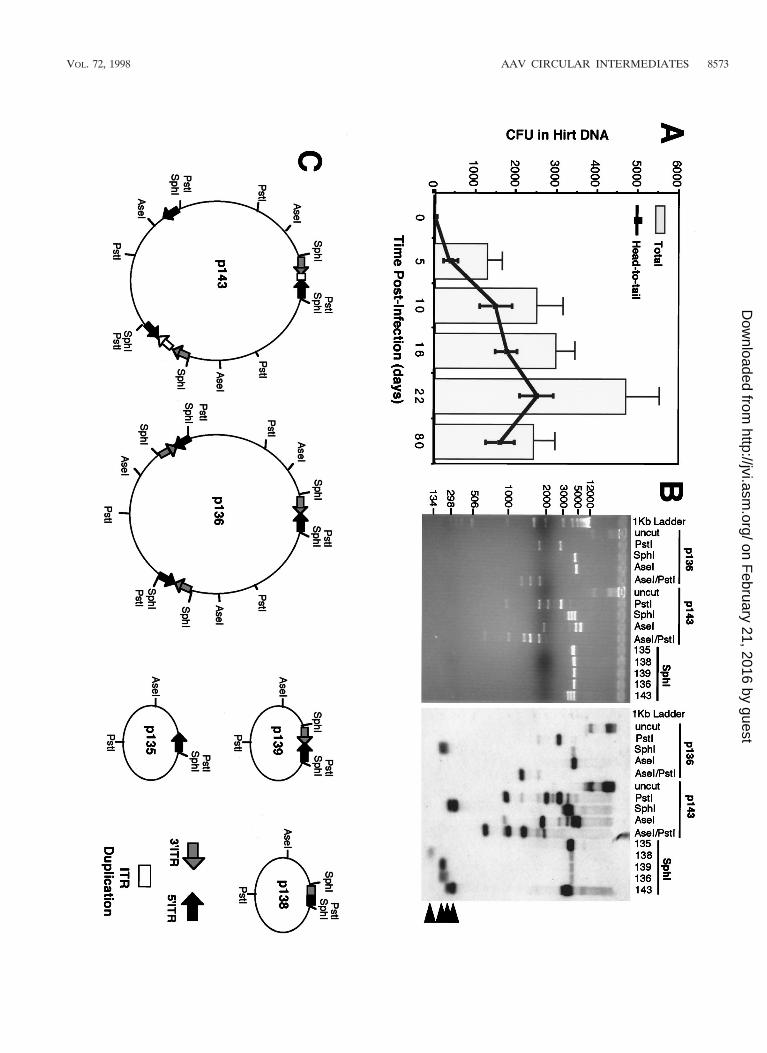
included two inverted ITRs within a circularized genome (Fig.2B, clone p17); also detected was a less frequent form (,5%)of circular intermediates, p439, with undetermined structure.When this type of replication-competent plasmid was seen, itwas not included in the quantification of head-to-tail circularintermediates since its structure could not be conclusively de-termined. The total abundance of muscle Hirt-derived head-to-tail circular intermediates (with one to two ITRs) demon-strated a time-dependent increase that peaked with transgeneexpression at 22 days and decreased slightly by day 80 (Fig.3A). Increased diversity in the length of ITR arrays within cir-cular intermediates was seen at longer time points. For exam-ple, Fig. 3B demonstrates several isolated circular intermedi-ates with one to three ITRs isolated from 80 days muscle Hirtsamples, in contrast to the more uniform structure of circularintermediates with two ITRs in a head-to-tail conformation at5 to 22 days postinfection (data not shown).
To evaluate the potential for artifactual rescue of linearrAAV genomes by recombination in bacteria, several controlexperiments were performed. First, uninfected control muscleHirt DNA preparations, spiked with an equal amount of rAAVused for in vivo infection of muscles, failed to give rise to rep-licating plasmids following transformation of E. coli. Second,when a blunted linear double-stranded HindIII/PvuII fragmentisolated from pCisAV.GFP3ori (encompassing the entire rAAVgenome) was used to transform bacteria, no ampicillin-resis-tant bacterial colonies were obtained. The addition of T4 ligaseto this fragment, however, led to significant numbers of bacte-rial colonies. Third, when purified single-stranded rAAV DNAwas used for transformation, no bacterial colonies were ob-tained. As summarized in Table 1, control experiments clearlydemonstrate that linear double-stranded or single-strandedDNA does not transform E. coli and that circularization isnecessary for transformation. Hence, in vivo circularization ofrAAV genomes is a prerequisite for rescuing autonomouslyreplicating plasmids in E. coli with this shuttle vector.
Molecular weight of circular intermediates suggest a con-version from monomer to multimer forms over time. To fur-ther characterize the circular intermediates isolated from mus-cles, Hirt samples from 22- and 80-day postinfection muscleswere size fractionated by continuous-flow gel electrophoresis(Bio-Rad) prior to amplification in bacteria. As shown in Fig.4, the majority of circular intermediates at 22 days postinfec-tion comigrated in their native unamplified form with lineardouble-stranded DNA standards of less than 3 kbp. Very fewclones were isolated from fractions of 3 to 5 kbp, and no cloneswere obtained from fractions larger than 5 kbp, at this timepoint. Furthermore, this size-fractionated molecular weight ofin vivo Hirt DNA-derived circular intermediates at 22 dayspostinfection correlated with that of head-to-tail monomer un-digested circular intermediate plasmids rescued in bacteria fromthis same time point (;2.5 kbp), suggesting that these nativecircular genomes may be supercoiled and/or single-strandedDNA molecules. However, it should be noted that although22-day circular AAV genomes migrated at a native molecularsize of ,3 kbp as unamplified Hirt DNA, the majority of res-cued circular intermediate plasmids from these fractions hadlinear molecular sizes of 4.7 kbp when digested to completionwith the single cutter AseI. In summary, these data suggest thatat early time points postinfection in muscle tissue, the predom-inant form of circular intermediates likely occurs as monomerhead-to-tail genomes. The lower apparent molecular size ofthis fraction compared to replication-form monomer (Rfm; 4.7kbp) and dimer (Rfd; 9.4 kbp) genomes provides indirect ev-idence that these forms are not responsible for rescued plas-mids in these Hirt DNA samples. Interestingly, when 80-day
FIG
.3.
Frequency
ofcircular
intermediate
formation
inm
uscletissue
following
transductionw
ithrA
AV
.Hirt
DN
As
isolatedfrom
rAA
V-infected
tibialism
usclesam
plesw
ereused
totransform
E.coli,
andthe
rescuedplasm
idsw
ereanalyzed
bySouthern
blottingas
previousdescribed
(more
than20
clonesw
ereanalyzed
fromatleasttw
oindependentm
usclesam
plesfor
eachtim
epoint).(A
)N
umbers
(mean
6standard
error)oftotalhead-to-tailcircular
intermediate
clones(line)
andtotalam
picillin-resistantbacteriaclones
(bar)isolated
fromeach
tibialisanterior
muscle
at0,5,10,16,22,and80
dayspostinfection.
Only
plasmids
which
containedone
totw
oIT
Rs
were
includedin
theestim
ationoftotalhead-to-tailcircular
intermediates.Plasm
idsw
hichdem
onstratedan
absenceofIT
R-hybridizing
SphIfragm
ents(betw
een150
to300
bp)w
ereom
ittedfrom
thecalculations.(B
)D
iversityof
ITR
arraysfound
inhead-to-tail
circularinterm
ediatesat
80days
postinfection.The
Southernblot
was
probedw
ithIT
Rsequences
andrepresents
circularinterm
ediatesw
ithone
tothree
ITR
s.SphIfragm
entsw
hichexcise
theinverted
ITR
arraysand
hybridizeto
ITR
probesare
marked
byarrow
headsat
theright.Sizes
ofm
olecularw
eightstandardsare
indicatedatthe
leftinbase
pairs.Additionalrestriction
enzyme
analysiswasused
todeterm
inethe
structureofm
onomerand
multim
ercircularinterm
ediates.Exam
plesareshow
nfortw
om
ultimer
circularinterm
ediates(p136
andp143)
which
containapproxim
atelythree
AA
Vgenom
es.Undigested
p136and
p143plasm
idsm
igrateat
.12
kbpw
hereasthe
predominant
forms
ofhead-to-tailundigested
circularinterm
ediatesat
22days
migrate
at2.5
kbp(data
notshow
n).The
digestionpattern
ofp136
isconsistent
with
auniform
head-to-tailconfigurationof
threegenom
esand
isindistinguishable
fromthe
digestionpattern
ofp139,which
containsone
circularizedgenom
e(undigested
p139m
igratesat2.5
kbp[data
notshown;also
seeresults
forp17
inF
ig.2]).Incontrast,p136
exhibitsa
more
complex
head-to-tailm
ultimer
circularinterm
ediatew
hichhas
variousdeletions
andduplications
within
theIT
Rarrays.(C
)Predicted
structuresof
fiverepresentative
intermediates
(complete
datafor
structuralanalysisare
notgiven).
8572 DUAN ET AL. J. VIROL.
on February 21, 2016 by guest
http://jvi.asm.org/
Dow
nloaded from
VOL. 72, 1998 AAV CIRCULAR INTERMEDIATES 8573
on February 21, 2016 by guest
http://jvi.asm.org/
Dow
nloaded from

muscle Hirt DNA samples were size fractionated, more cloneswere retrieved from larger fractions ranging from 3 to 12 kbp(Fig. 4). This shift in the molecular size of unamplified circularintermediates indicates the potential for recombination be-tween monomer forms in the generation of large circular mul-timer genomes. Such concatemerization has been previouslyobserved in muscle tissue and has usually been hypothesized toinvolve linear integrated forms of the AAV genome (5, 10, 15,34). Our present data shed new light on the molecular char-acteristics of these persistent AAV genomes and suggest thatthey are in fact circular and episomal. Based on yields of re-trievable circular plasmids reconstituted in Hirt DNA, the effi-ciency of bacterial transformation, and the initial inoculum ofvirus, we estimate that approximately 1 in 400 viral DNA par-ticles circularizes following infection in muscle (Table 2). Giventhe facts that (i) not all rAAV particles likely contain func-tional DNA molecules, (ii) circular intermediates may inte-grate, (iii) circular intermediates concatemerize over time, and(iv) transformation efficiencies compared supercoiled plasmidstandards to intermediates with an unknown relaxation state,these theoretical calculations may represent an underestima-tion.
AAV circular intermediates demonstrate increased persis-tence as plasmid-based vectors. Based on the finding thatcircular AAV intermediates were associated with long-termpersistence of transgene expression in muscle tissue, we hy-pothesized that rAAV circular head-to-tail intermediates maybe molecular structures of the AAV genome associated withthe latent life cycle and increased episomal stability. Severalaspects of the structure of AAV circular intermediates mayaccount for their increased stability in vivo. First, circulariza-tion of AAV genomes may create a nuclease-resistant confor-mation. Second, since the only viral sequences contained with-in circular intermediates are the head-to-tail ITR array, thesesequences might bind cellular factors capable of stabilizingthese structures in vivo. Several studies demonstrating in-creased persistence of transgene expression with plasmid DNAencoding viral ITRs lends support to this hypothesis (25,
32). We now propose a functional explanation for this find-ing through the association with circular intermediate forma-tion as part of the AAV life cycle.
To more closely evaluate the persistence of AAV head-to-tail circular intermediates, we performed several in vitro ex-periments by transfecting these intermediates into HeLa cellsand assessing the stability of plasmid DNA and transgene ex-pression by GFP clonal expansion. Results from HeLa celltransfection experiments demonstrated that two monomerhead-to-tail circular intermediates (p81 and p87, identical instructure to p139 [Fig. 4C]) studied gave rise to a 10-fold-higher number of 5- and 10-day transgene-expressing clonescompared to a control plasmid, pCMVGFP, lacking the ITRsequences (Fig. 5A and B). Additionally, the GFP-positivecolonies at 5 days posttransfection were threefold larger inHeLa cells transfected with p81 and p87 than in cells trans-fected with the pCMVGFP control vector (Fig. 5A and B).These studies suggest the AAV circular intermediates haveincreased stability of transgene expression and substantiatefindings for muscle tissue. However, from the above data, wecannot rule out potential ITR enhancer effects on transgeneexpression from circular intermediates. Of interest in this re-gard were two additional findings from these studies. First, theincreased transgene expression from AAV circular intermedi-ates p81 and p87 was also immediately apparent by 2 daysposttransfection in unpassaged HeLa cells (data not shown).Second, no differences in the persistence of transgene expres-sion were seen among any of the three constructs analyzed in293 cells (data not shown). These findings suggested that cell-specific factors may mediate increased transcription and/orincreased stability of AAV circular intermediates.
To determine whether increases in the persistence of trans-gene expression correlated with increased molecular persis-tence of head-to-tail circular intermediates following transfec-tion into HeLa cells, total DNA (low and high molecular weight)was isolated from cultures of pCMVGFP- and p81-transfectedHeLa cells at various passages posttransfection and analyzedby Southern blotting. Southern blots hybridized to 32P-labeledGFP probes demonstrated a significantly higher level of p81
FIG. 4. Molecular sizes of native unamplified circular intermediates in mus-cle tissue. Hirt DNA from AV.GFP3ori-infected muscle was size fractionated byelectrophoresis, and fractions of various molecular weights were transformedinto E. coli. Results demonstrate the abundance of head-to-tail circular inter-mediates at each of the given molecular sizes at 22 and 80 days after infectionwith the rAAV shuttle vector. Fractions were sized according to the migration oflinear double-stranded DNA standards under identical gel running conditions.The structures of circular intermediates were confirmed by Southern blot re-striction analysis (data not shown).
TABLE 2. Yield of circular intermediates isolated from Hirt DNA
Bacterialtransformation
Starting no.of plasmid orAAV genome
molecules
Actual no.of ampicillin-resistant CFU
Adjusted yield(CFU) for3 3 1010
molecules
Hirt DNA from rAAV-infected musclea
3 3 1010 5 3 103 5 3 105e
Hirt DNA 1 10 ng ofLacZ plasmidb
1.3 3 109 8.7 3 104c 2 3 108d
10 ng of LacZ plasmid 1.3 3 109 8.7 3 106 2 3 108d
a The actual amount of Hirt DNA used for transformation was 3/20 the entireHirt DNA. The numbers have been adjusted to reflect viral inoculum and yieldsfor the entire muscle.
b Plasmid DNA was spiked into mock-infected muscle homogenates prior toisolation of Hirt DNA. This reconstituted Hirt DNA was then used for trans-formation of bacteria.
c The average of several experiments indicates an approximate 100-fold re-duction in the number of CFU recovered from bacterial transformations withDNA isolated from Hirt extract spiked with plasmids compared to transforma-tion with an equivalent amount of plasmid DNA alone.
d The number of CFU in plasmid reconstitution studies have been adjusted tonormalize the number of plasmids genomes to that used in AAV experiments(3 3 1010). Control LacZ plasmid was approximately 7,000 bp with a molecularsize of 4.6 3 106 g/mol. Therefore, 230 ng of plasmid is equivalent to 3 3 1010
molecules (i.e., normalization factor of 23-fold).e Adjusted yield indicates that approximately 1 in 400 AAV genomes circular-
izes in vivo.
8574 DUAN ET AL. J. VIROL.
on February 21, 2016 by guest
http://jvi.asm.org/
Dow
nloaded from

plasmid DNA at passage 7 than that of the control vectorlacking the head-to-tail ITR sequence (Fig. 5C). The majorityof signal in undigested DNA samples was associated with a4.7-kb band migrating at the approximate size of the uncutmonomer plasmids. Together with the fact that the majority ofsignal from all cell cultures in Fig. 5C disappeared by passage10 (data not shown), these data suggest that these plasmidspredominantly remained episomal. Thus, in both muscle andHeLa cells, increased persistence of AAV circular intermedi-ates is correlated with stable transgene expression.
ITR arrays are responsible for increased persistence. Toinvestigate whether the head-to-tail ITR DNA element wasresponsible for the increased persistence of circular interme-diates, we cloned this DNA element into a secondary luciferasevector (pGL3) to give rise to pGL3(ITR). Transient transfec-tion experiments in HeLa cells demonstrated a fivefold in-crease in the persistence of luciferase expression from pGL3(ITR) compared to that of pGL3 in serially passaged 10 daycultures (Fig. 5D). These findings support the hypothesis thatthe head-to-tail ITR DNA element contained within circularintermediates is responsible for mediating the increased per-sistence of transgene expression and suggest a mechanism bywhich these molecular intermediates may confer stability toAAV genomes in vivo. Furthermore, increases in the stabilityof transgene expression conferred by this element appear to be
primarily context independent, since the head-to-tail ITR ele-ment was 39 to the luciferase gene in pGL3(ITR) and 59 to theGFP transgene in AAV circular intermediates.
DISCUSSION
Characterization of integrated proviral structures in differ-ent cell lines has demonstrated head-to-tail genomes as a pre-dominant structural form following rAAV infection (4, 7). Al-though head-to-head and tail-to-tail integrated structures havebeen noted for rAAV (23), these structures are traditionallythought to be associated with wtAAV lytic replication inter-mediates. These replication intermediates, termed Rfm andRfd, consist of genomes in duplex monomer and duplex dimerforms, respectively, with one covalently closed end (Fig. 1).Rfd genomes have a characteristic head-to-head or tail-to-tailorientation. Both Rfm and Rfd configurations have also beendemonstrated in rAAV-infected cells, and enhanced conver-
FIG. 5. Head-to-tail circular intermediates demonstrate increased stability ofGFP expression following transient transfection in HeLa cells. Subconfluentmonolayers of HeLa cells were cotransfected with p81, p87, or pCMVGFP andpRSVlacZ as an internal control for transfection efficiency as described in Ma-terials and Methods. (A) Expansion of GFP clones after one passage (arrow-heads). (B) Quantification of clone size (mean raw values) and clone numbers(normalized for transfection efficiency as determined by X-Gal staining forpRSVlacZ). Numbers above the bars represent quantification of GFP clonesafter passage 2 (also normalized for transfection efficiency). Results indicate themeans (6 standard errors) of duplicate experiments, with more than 20 fieldsquantified for each experimental point. The persistence of transfected p81 andpCMVGFP plasmid DNA at passage 7 posttransfection was evaluated bygenomic Southern blotting of total cellular DNA hybridized against a 32P-labeledGFP probe (C; results from two independent transfections are shown). In thesestudies, control cultures cotransfected with pRSVlacZ demonstrated a less than25% variation in transfection efficiency, as determined by X-Gal staining of cellsat 48 h posttransfection. Lanes: U, uncut; C, PstI cut. The migration of uncutdimer and monomer plasmids forms are marked on the left. PstI digestion of theplasmids results in bands at 4.7 kb (pCMVGFP; single PstI site in plasmid) and1.7 kb (p81; two PstI sites flanking the GFP gene). The band marked Genomicin undigested lanes was also seen with DNA from untransfected cells (data notshown) and hence is nonspecific hybridization of probe to the high concentrationof DNA in this area of the gel. To determine whether the head-to-tail ITR arraywithin circular intermediates was responsible for increases in the persistence ofGFP expression, the head-to-tail ITR DNA element was subcloned into theluciferase plasmid pGL3 to generate pGL3(ITR). (D) Luciferase transgeneexpression following transfection with pGL3 and pGL3(ITR) at 10 days (passage2) posttransfection. Results are the means (6 standard errors) for triplicateexperiments and are normalized for transfection efficiency by using a dual re-nilla-luciferase reporter vector (pRLSV40; Promega).
VOL. 72, 1998 AAV CIRCULAR INTERMEDIATES 8575
on February 21, 2016 by guest
http://jvi.asm.org/
Dow
nloaded from

sion of single-stranded AAV genomes to double-stranded Rfmand Rfd forms has been suggested as a mechanism for aug-mentation of rAAV transduction by adenovirus in cell lines (8,9). However, it is plausible that the mechanisms responsible forthe formation of Rfm and Rfd molecules are different frompathways which lead to long-term transgene expression. Insupport of this hypothesis is a recent study evaluating augmen-tation of rAAV transgene expression by adenovirus in the liver(30); the results demonstrated that coinfection of the liver withadenovirus and rAAV enhances short-term transgene expres-sion, while long-term expression was no different from thatafter infection with rAAV alone. The exact mechanism for theformation of head-to-tail circular intermediates is not clear;however, similar structures have been demonstrated to act aspreintegration intermediates for retrovirus (31). In this regard,circularized retroviral genomes with one and two viral longterminal repeats have been proposed. In addition, circularpreintegration intermediates have also been suggested by re-cent studies on wtAAV integration (22). The demonstrationthat circular intermediates exist in rAAV-infected muscle tis-sue explains several features of latent-phase infection withrAAV vectors, including proviral structure and stable episomalpersistence.
Previous studies have suggested that rAAV genomes deliv-ered to muscle tissue might persist as head-to-tail concatemers(5, 10, 15). However, it is not known whether these concate-mers exist as free episomes or as integrated proviruses in thehost genome. Our results demonstrating prolonged persistenceof head-to-tail circular intermediates at 80 days postinfectionsuggest that a large percentage of rAAV genomes may remainepisomal. The conversion of monomer circularized genomes tolarger circularized multimers appears to be an aspect associ-ated with long-term persistence and likely represents recom-binational events between monomer intermediates and/or roll-ing replication. Although the bacterial rescue strategy was notcapable of satisfactorily addressing the size of multimers, ourmodified approach to size fractionating Hirt DNA prior tobacterial rescue of intermediates lends support to this hypoth-esis. Additional supportive evidence for increased recombina-tion over time is the finding that greater variability in thelength of ITR arrays was observed at longer time points postin-fection. For example, at 5 to 22 days the majority of circularintermediates contained two ITRs in a head-to-tail fashion,whereas at 80 days the ITR arrays consisted of one to threeITRs. Such diversity of ITR arrays in muscle tissue infectedwith AAV has been previously found in studies using PCRapproaches (10, 15). In addition, the 30% decline in the abun-dance of circular intermediates in muscle tissue between 22and 80 days also supports a hypothesis that these molecularforms of AAV may represent preintegration complexes.
Given the fact that circular intermediates had long-term per-sistence in muscle tissue, we hypothesized that certain struc-tural features of these intermediates may affect episomalstability of DNA. Previous studies have noted increased per-sistence of transgene expression from plasmids encoding AAVITRs (25, 32). However, the physiologic significance of thisfinding has remained elusive. Our present study demonstratingthat the head-to-tail ITR arrays isolated from AAV circularintermediates can confer increased episomal persistence toplasmids following transfection in cell lines gives a mechanisticframework for ITR effects on plasmid persistence. Further-more, the correlation that AAV circular intermediates haveincreased persistence in cell lines in vitro lends support to thehypothesis that these structures represent stable episomalforms following rAAV transduction in muscle. Stability of cir-cular intermediates in vivo might be mediated by the binding of
cellular factors to Holliday-like junctions in ITR arrays whichstabilize or protect DNA from degradation.
However, several observations regarding the increased per-sistence of transgene expression conferred by circular interme-diates in vitro remain to be explained. First, increased persis-tence of transgene expression from circular intermediates wasobserved in HeLa but not 293 cells, which suggests that cell-specific factors may bind to the ITR arrays of circular inter-mediates to mediate functional effects. Second, increased trans-gene expression from circular intermediates expressing GFPand the ITR array containing luciferase plasmids was noted by48 h posttransfection in unpassaged HeLa cells, reminiscent ofprevious findings which suggest that the AAV ITRs have en-hancer-like properties (2). It is not known how an immediateincrease in transgene expression from AAV circular interme-diates could be linked to increased long-term persistence ofDNA. However, one hypothesis could posit the binding of cel-lular proteins to ITR arrays, which sequesters plasmids intosubcompartments of the nucleus which are more transcription-ally active and confer an increased level of DNA stability.Furthermore, we cannot rule out the possibility that the ITRarrays act as enhancers which by virtue of enhanced transcrip-tional activity and bound proteins at this element protect plas-mids from degradation or more evenly segregate plasmids tonuclei during mitosis. Despite the lack of a concrete mecha-nism for increased persistence of transgene expression fromcircular intermediates, the current data favor some combina-tion of pathways which involve both enhancer-like functionand increased DNA stability conferred by head-to-tail ITRelements.
rAAV has been shown to be an efficient vector for express-ing transgenes in various tissues and cell types in addition tomuscle, such as brain, retina, liver, lung, and hematopoeticcells (3, 6, 12, 14, 17, 20, 24, 30, 33). Despite these advances inthe application of rAAV, the mechanisms of in vivo rAAV-mediated transduction and persistence of transgene expressionremain unclear. Questions such as those relating to the molec-ular state of rAAV following in vivo delivery are highly rele-vant to the clinical application of this viral vector. For example,should rAAV primarily persist as an randomly integrated pro-virus, the potential for insertional mutagenesis could present amajor theoretical obstacle in the use of this vector due to thepotential for mutational oncogenesis. Our demonstration thatrAAV can persist as episomes suggests that random integra-tion and associated risks of malignancy may not be a majorconcern for this viral vector system. Additionally, the molecu-lar determinants of AAV circular intermediates associatedwith increased persistence in cell lines appear to be containedwithin the DNA elements encompassing the inverted ITRs.The isolation of this naturally occurring viral DNA element,which forms as part of the AAV life cycle and acts to stabilizecircular episomal DNA, may prove useful in increasing theefficacy of both viral and nonviral gene therapy vectors.
ACKNOWLEDGMENT
The work described here was supported by NIH grant R01 DK/HL58340 (J.F.E.).
REFERENCES
1. Afione, S. A., C. K. Conrad, W. G. Kearns, S. Chunduru, R. Adams, T. C.Reynolds, W. B. Guggino, G. R. Cutting, B. J. Carter, and T. R. Flotte. 1996.In vivo model of adeno-associated virus vector persistence and rescue. J. Vi-rol. 70:3235–3241.
2. Beaton, A., P. Palumbo, and K. I. Berns. 1989. Expression from the adeno-associated virus p5 and p19 promoters is negatively regulated in trans by theRep protein. J. Virol. 63:4450–4454.
3. Bennett, J., D. Duan, J. F. Engelhardt, and A. M. Maguire. 1997. Real-time,
8576 DUAN ET AL. J. VIROL.
on February 21, 2016 by guest
http://jvi.asm.org/
Dow
nloaded from

noninvasive in vivo assessment of adeno-associated virus-mediated retinaltransduction. Investig. Ophthalmol. Visual Sci. 38:2857–2863.
4. Cheung, A. K., M. D. Hoggan, W. W. Hauswirth, and K. I. Berns. 1980.Integration of the adeno-associated virus genome into cellular DNA inlatently infected human Detroit 6 cells. J. Virol. 33:739–748.
5. Clark, K. R., T. J. Sferra, and P. R. Johnson. 1997. Recombinant adeno-associated viral vectors mediate long-term transgene expression in muscle.Hum. Gene Ther. 8:659–669.
6. Conrad, C. K., S. S. Allen, S. A. Afione, T. C. Reynolds, S. E. Beck, M.Fee-Maki, X. Barrazza-Ortiz, R. Adams, F. B. Askin, B. J. Carter, W. B.Guggino, and T. R. Flotte. 1996. Safety of single-dose administration of anadeno-associated virus (AAV)-CFTR vector in the primate lung. Gene Ther.3:658–668.
7. Duan, D., K. J. Fisher, J. F. Burda, and J. F. Engelhardt. 1997. Structuraland functional heterogeneity of integrated recombinant AAV genomes. Vi-rus Res. 48:41–56.
8. Ferrari, F. K., T. Samulski, T. Shenk, and R. J. Samulski. 1996. Second-strand synthesis is a rate-limiting step for efficient transduction by recombi-nant adeno-associated virus vectors. J. Virol. 70:3227–3234.
9. Fisher, K. J., G. P. Gao, M. D. Weitzman, R. DeMatteo, J. F. Burda, andJ. M. Wilson. 1996. Transduction with recombinant adeno-associated virusfor gene therapy is limited by leading-strand synthesis. J. Virol. 70:520–532.
10. Fisher, K. J., K. Jooss, J. Alston, Y. Yang, S. E. Haecker, K. High, R. Pathak,S. E. Raper, and J. M. Wilson. 1997. Recombinant adeno-associated virus formuscle directed gene therapy. Nat. Med. 3:306–312.
11. Fisher-Adams, G., K. K. Wong, Jr., G. Podsakoff, S. J. Forman, and S.Chatterjee. 1996. Integration of adeno-associated virus vectors in CD341human hematopoietic progenitor cells after transduction. Blood 88:492–504.
12. Flannery, J. G., S. Zolotukhin, M. I. Vaquero, M. M. LaVail, N. Muzyczka,and W. W. Hauswirth. 1997. Efficient photoreceptor-targeted gene expres-sion in vivo by recombinant adeno-associated virus. Proc. Natl. Acad. Sci.USA 94:6916–6921.
13. Flotte, T. R., S. A. Afione, and P. L. Zeitlin. 1994. Adeno-associated virusvector gene expression occurs in nondividing cells in the absence of vectorDNA integration. Am. J. Respir. Cell Mol. Biol. 11:517–521.
14. Halbert, C. L., T. A. Standaert, M. L. Aitken, I. E. Alexander, D. W. Russell,and A. D. Miller. 1997. Transduction by adeno-associated virus vectors in therabbit airway: efficiency, persistence, and readministration. J. Virol. 71:5932–5941.
15. Herzog, R. W., J. N. Hagstrom, S. H. Kung, S. J. Tai, J. M. Wilson, K. J.Fisher, and K. A. High. 1997. Stable gene transfer and expression of humanblood coagulation factor IX after intramuscular injection of recombinantadeno-associated virus. Proc. Natl. Acad. Sci. USA 94:5804–5809.
16. Hirt, B. 1967. Selective extraction of polyoma DNA from infected mouse cellcultures. J. Mol. Biol. 26:365–369.
17. Kaplitt, M. G., P. Leone, R. J. Samulski, X. Xiao, D. W. Pfaff, K. L. O’Malley,and M. J. During. 1994. Long-term gene expression and phenotypic correc-tion using adeno-associated virus vectors in the mammalian brain. Nat.Genet. 8:148–154.
18. Kearns, W. G., S. A. Afione, S. B. Fulmer, M. C. Pang, D. Erikson, M. Egan,M. J. Landrum, T. R. Flotte, and G. R. Cutting. 1996. Recombinant adeno-associated virus (AAV-CFTR) vectors do not integrate in a site-specificfashion in an immortalized epithelial cell line. Gene Ther. 3:748–755.
19. Kessler, P. D., G. M. Podsakoff, X. Chen, S. A. McQuiston, P. C. Colosi, L. A.Matelis, G. J. Kurtzman, and B. J. Byrne. 1996. Gene delivery to skeletalmuscle results in sustained expression and systemic delivery of a therapeutic
protein. Proc. Natl. Acad. Sci. USA 93:14082–14087.20. Koeberl, D. D., I. E. Alexander, C. L. Halbert, D. W. Russell, and A. D.
Miller. 1997. Persistent expression of human clotting factor IX from mouseliver after intravenous injection of adeno-associated virus vectors. Proc. Natl.Acad. Sci. USA 94:1426–1431.
21. Kotin, R. M., R. M. Linden, and K. I. Berns. 1992. Characterization of apreferred site on human chromosome 19q for integration of adeno-associ-ated virus DNA by nonhomologous recombination. EMBO J. 11:5071–5078.
22. Linden, R. M., E. Winocour, and K. I. Berns. 1996. The recombinationsignals for adeno-associated virus site-specific integration. Proc. Natl. Acad.Sci. USA 93:7966–7972.
23. McLaughlin, S. K., P. Collis, P. L. Hermonat, and N. Muzyczka. 1988.Adeno-associated virus general transduction vectors: analysis of proviralstructures. J. Virol. 62:1963–1973.
24. Muzyczka, N. 1992. Use of adeno-associated virus as a general transductionvector for mammalian cells. Curr. Top. Microbiol. Immunol. 158:97–129.
25. Philip, R., E. Brunette, L. Kilinski, D. Murugesh, M. A. McNally, K. Ucar,J. Rosenblatt, T. B. Okarma, and J. S. Lebkowski. 1994. Efficient andsustained gene expression in primary T lymphocytes and primary and cul-tured tumor cells mediated by adeno-associated virus plasmid DNA com-plexed to cationic liposomes. Mol. Cell. Biol. 14:2411–2418.
26. Ponnazhagan, S., D. Erikson, W. G. Kearns, S. Z. Zhou, P. Nahreini, X. S.Wang, and A. Srivastava. 1997. Lack of site-specific integration of the re-combinant adeno-associated virus 2 genomes in human cells. Hum. GeneTher. 8:275–284.
27. Rutledge, E. A., and D. W. Russell. 1997. Adeno-associated virus vectorintegration junctions. J. Virol. 71:8429–8436.
28. Samulski, R. J., L. S. Chang, and T. Shenk. 1989. Helper-free stocks ofrecombinant adeno-associated viruses: normal integration does not requireviral gene expression. J. Virol. 63:3822–3828.
29. Samulski, R. J., L. S. Chang, and T. Shenk. 1987. A recombinant plasmidfrom which an infectious adeno-associated virus genome can be excised invitro and its use to study viral replication. J. Virol. 61:3096–3101.
30. Snyder, R. O., C. H. Miao, G. A. Patijn, S. K. Spratt, O. Danos, D. Nagy,A. M. Gown, B. Winther, L. Meuse, L. K. Cohen, A. R. Thompson, and M. A.Kay. 1997. Persistent and therapeutic concentrations of human factor IX inmice after hepatic gene transfer of recombinant AAV vectors. Nat. Genet.16:270–276.
31. Varmus, H. E. 1982. Form and function of retroviral proviruses. Science 216:812–820.
32. Vieweg, J., D. Boczkowski, K. M. Roberson, D. W. Edwards, M. Philip, R.Philip, T. Rudoll, C. Smith, C. Robertson, and E. Gilboa. 1995. Efficient genetransfer with adeno-associated virus-based plasmids complexed to cationicliposomes for gene therapy of human prostate cancer. Cancer Res. 55:2366–2372.
33. Walsh, C. E., A. W. Nienhuis, R. J. Samulski, M. G. Brown, J. L. Miller, N. S.Young, and J. M. Liu. 1994. Phenotypic correction of Fanconi anemia inhuman hematopoietic cells with a recombinant adeno-associated virus vec-tor. J. Clin. Investig. 94:1440–1448.
34. Xiao, X., J. Li, and R. J. Samulski. 1996. Efficient long-term gene transferinto muscle tissue of immunocompetent mice by adeno-associated virusvector. J. Virol. 70:8098–8108.
35. Yang, C. C., X. Xiao, X. Zhu, D. C. Ansardi, N. D. Epstein, M. R. Frey, A. G.Matera, and R. J. Samulski. 1997. Cellular recombination pathways and viralterminal repeat hairpin structures are sufficient for adeno-associated virusintegration in vivo and in vitro. J. Virol. 71:9231–9247.
VOL. 72, 1998 AAV CIRCULAR INTERMEDIATES 8577
on February 21, 2016 by guest
http://jvi.asm.org/
Dow
nloaded from

ERRATUM
Circular Intermediates of Recombinant Adeno-Associated Virus HaveDefined Structural Characteristics Responsible for Long-Term
Episomal Persistence in Muscle TissueDONGSHENG DUAN,1 PRERNA SHARMA,1 JUSAN YANG,1 YONGPING YUE,1 LORITA DUDUS,2
YULONG ZHANG,1 KRISHNA J. FISHER,2 AND JOHN F. ENGELHARDT1*
Department of Anatomy and Cell Biology and Department of Internal Medicine, University of Iowa Medical Center,Iowa City, Iowa,1 and Gene Therapy Program, Department of Pathology and Laboratory Medicine,
Tulane University Medical Center, New Orleans, Louisiana2
Volume 72, no. 11, p. 8568–8577, 1998. Page 8568: The byline and affiliation line should appear as shown above.
861
Copyright © 2022 FDOKUMEN