Vascularised Scaffolds for Cutaneous Wound Reconstruction ...
Upload
independentCategory
view
0download
0
Growth Factors, Cytokines, Cell Cycle Molecules
Improved Survival of Ischemic Cutaneous andMusculocutaneous Flaps after Vascular EndothelialGrowth Factor Gene Transfer UsingAdeno-Associated Virus Vectors
Serena Zacchigna,* Giovanni Papa,†
Andrea Antonini,† Federico Novati,†
Silvia Moimas,* Alessandro Carrer,* Nikola Arsic,*Lorena Zentilin,* Valentina Visintini,†
Michele Pascone,† and Mauro Giacca*From the Molecular Medicine Laboratory,* International Centre
for Genetic Engineering and Biotechnology, Trieste; and the
Plastic Surgery Unit,† Faculty of Medicine, University of Trieste,
Trieste, Italy
A major challenge in reconstructive surgery is flapischemia, which might benefit from induction oftherapeutic angiogenesis. Here we demonstrate theeffect of an adeno-associated virus (AAV) vector deliv-ering vascular endothelial growth factor (VEGF)165 intwo widely recognized in vivo flap models. For theepigastric flap model, animals were injected subcuta-neously with 1.5 � 1011 particles of AAV-VEGF at day0, 7, or 14 before flap dissection. In the transverserectus abdominis musculocutaneous flap model, AAV-VEGF was injected intramuscularly. The delivery ofAAV-VEGF significantly improved flap survival inboth models, reducing necrosis in all treatmentgroups compared to controls. The most notable re-sults were obtained by administering the vector 14days before flap dissection. In the transverse rectusabdominis musculocutaneous flap model, AAV-VEGFreduced the necrotic area by >50% at 1 week aftersurgery, with a highly significant improvement in thehealing process throughout the following 2 weeks.The therapeutic effect of AAV-VEGF on flap survivalwas confirmed by histological evidence of neoangio-genesis in the formation of large numbers of CD31-positive capillaries and �-smooth muscle actin-posi-tive arteriolae, particularly evident at the borderbetween viable and necrotic tissue. These results un-derscore the efficacy of VEGF-induced neovasculariza-tion for the prevention of tissue ischemia and the
improvement of flap survival in reconstructive sur-gery. (Am J Pathol 2005, 167:981–991)
Partial necrosis of skin flaps represents a major clinicalproblem in patients undergoing reconstructive proce-dures, with significant morbidity and no effective therapyavailable. The lack of oxygen and nutrients in the distalpart of the flap strongly compromises skin viability, re-sulting in flap necrosis, which often requires secondaryreconstructive interventions.1 The recent concept of ther-apeutic angiogenesis by local administration of angio-genic growth factors has emerged as an attractive ap-proach to enhance blood supply and perfusion incompromised tissues, thus improving flap survival.
The angiogenic response to tissue ischemia is a com-plex process involving the coordinated interplay of avariety of soluble factors, controlling new blood vesselformation. One of the fundamental molecules that arerequired in the angiogenic process is the vascular endo-thelial growth factor (VEGF). Four different isoforms ofVEGF have been described, with 121, 165, 189, and 206amino acids, arising by differential splicing of the primaryVEGF gene transcript. VEGF165 is the predominant formin all cells and tissues and its expression is promptlyinduced by tissue hypoxia. The main activity of VEGF is tostimulate the proliferation and migration of endothelialcells, with the constitution of a primitive capillary network,which subsequently matures to form larger vessels.
Research on therapeutic angiogenesis relating to plas-tic and reconstructive surgery is in its early stages. Sev-
Supported by grants from the Progetto Finalizzato “Genetica Molecolare”of the “Consiglio Nazionale delle Ricerche,” Italy; the Fondo Integrato perla Ricerca di Base program of the “Ministero dell’Istruzione, Universita’ eRicerca,” Italy; the “Fondazione Cassa di Risparmio” of Trieste, Italy; andthe “Fondo Trieste,” Italy.
S.Z. and G.P. contributed equally to this work.
Accepted for publication June 19, 2005.
Address reprint requests to Mauro Giacca, M.D. Ph.D., Director, ICGEBTrieste, Padriciano, 99, 34012 Trieste, Italy. E-mail: [email protected].
American Journal of Pathology, Vol. 167, No. 4, October 2005
Copyright © American Society for Investigative Pathology
981
eral laboratories have explored the possibility of promot-ing skin flap neovascularization by using recombinantVEGF proteins. In most of these studies, with some ex-ceptions,2 recombinant VEGF provided a beneficial ef-fect on the flap survival,3–9 although the exact mecha-nisms have not always been specifically addressed.Despite these encouraging findings, the use of recombi-nant proteins in a clinical setting is hampered by severalfactors, such as their short half-lives, poor bioavailability,and consequent need for frequent administrations to sus-tain long-lasting effects. Delivery of therapeutic genes toinduce neo-angiogenesis represents an appealing alter-native strategy that might overcome most of these prob-lems. Moreover, the easy accessibility of the flaps makesthem an ideal target for an in vivo gene transfer approach.
A variety of techniques allow for a coding DNA to betaken up by host cells, which then express the respectiveprotein. Many of them have been used for gene deliveryto the muscle and skin, including the use of naked plas-mid DNA and viral vectors.10–18 The use of viral vectorspresents a notable advantage over nonviral systems, byproviding a higher rate of transduction and expression.Among the novel approaches that hold promise to be-come a relevant therapeutic modality in humans is theuse of vectors based on the adeno-associated virus(AAV), a nonpathogenic and widespread parvovirus, in-capable of autonomous replication. Vectors based onAAV are able to transduce both dividing and nondividingcells and show a specific tropism for postmitotic cells,including skeletal and cardiac muscle,10,19 neurons,20
and liver.21,22 Because these vectors do not contain anyviral genes—which are transiently transfected in trans forthe packaging process—they elicit virtually no inflamma-tory or immune response.23,24 As a consequence, trans-gene expression from these vectors persists for severalmonths in a variety of animal tissues in vivo.25
Besides being able to transduce human keratinocytesin vitro,26 we and others have observed that the subcu-taneous delivery of AAV vectors results in the efficienttransduction of hair follicles, sweat gland ducts,27 andthe panniculus carnosus (the skeletal muscle layer withinthe dermal sheet in rodents).28,29 To our knowledge, thedata reported in this study are the first demonstration thatAAV can be successfully used in the plastic surgery fieldto deliver the VEGF165 gene as a tool to promote thera-peutic angiogenesis and skin flap survival in two differentin vivo flap models.
Materials and Methods
Recombinant AAV Vector Preparation andCharacterization
Two recombinant AAV vectors were obtained in thisstudy, expressing the LacZ reporter gene and the cDNAfor the 165 amino acid isoform of VEGF (VEGF165) underthe control of the constitutive cytomegalovirus immediateearly promoter. Infectious vector stocks were generatedin 293 cells and titrated by a competitive polymerasechain reaction procedure, as already described.30
Animals and Experimental Protocols
Animal care and treatment were conducted in conformitywith institutional guidelines in compliance with nationaland international laws and policies (European EconomicCommunity Council Directive 86/609, OJL 358, Decem-ber 12th, 1987). A total of 88 adult male Wistar ratsweighting 250 to 300 g were used for this study andhoused under controlled environmental conditions. Aftergeneral anesthesia with Avertin 2% (10 ml/kg), abdominalhair was removed and skin washed with sterile water. Aflap measuring 5 � 8 cm was outlined on the skin, ex-tending distally from the xiphoid process and bilaterallyfrom the midline (Figure 2).
In the first model, the skin flap was raised from thefascia on the inferior epigastric artery, by cutting all ofthe vessels from the left side of the abdomen, all of theperforators, and the right lateral thoracic artery. At theend of the procedure, the skin was immediately resuturedin place. In this model, 150 �l of either AAV-VEGF orplacebo were injected at 10 equally spaced subcutane-ous sites along the midline of the flap.
For the TRAM model, the entire left part of the flap wasraised on a plane between the panniculus carnosus andthe abdominal fascia, but on the right side the medialportion of the flap was left attached to the anterior rectussheet. In this way, the rectus abdominis represents themuscular component of the flap, providing the bloodsupply to the cutaneous component through the perfora-tor arteries. This time, AAV-VEGF or placebo was injectedintramuscularly in the same region where each perforatorartery arises from the rectus abdominis.
Each of the 12 experimental groups described in Fig-ure 2 was initially composed of four rats (an overview ofthe experimental groups is provided in Table 1). Rats ingroups 1, 2, 3, 7, 8, and 9 received AAV-VEGF at 0, 7,and 14 days before surgery, respectively, whereasgroups 4, 5, 6, 10, 11, and 12 were control groups, inwhich the same volume of AAV-LacZ or saline was ad-ministered at the same time points (in every controlgroup, two animals received AAV-LacZ and the other tworeceived saline). Based on the results of this first set ofexperiments, 40 additional rats were treated with the
Table 1. Experimental Groups
TreatmentTime of vector
deliveryGroup
no.
Epigastric flap (n � 24)AAV-VEGF During surgery 1
7 days before surgery 214 days before surgery 3
AAV-LacZ During surgery 47 days before surgery 514 days before surgery 6
TRAM flap (n � 24)AAV-VEGF During surgery 7
7 days before surgery 814 days before surgery 9
AAV-LacZ During surgery 107 days before surgery 1114 days before surgery 12
982 Zacchigna et alAJP October 2005, Vol. 167, No. 4
TRAM flap and injected with AAV-VEGF or AAV-LacZ(n � 20 per group) vectors 14 days before surgery. Flapsurvival was first assessed at day 7 after surgery bytaking digital images of the flaps using a Nikon E995camera followed by the measurement of the necrotic areausing the UTHSCSA Image Tool software. At the sametime point, 12 animals per group were sacrificed forhistological assessment. Flaps were harvested after thescars, as described in Figure 4A, and tissue biopsieswere fixed overnight in 10% buffered formalin. The otherrats (n � 8 per group) were followed for an additional 2weeks at 3-day intervals, by measuring the extent of thenecrotic area to assess the efficacy of the healingprocess.
Histological and ImmunohistochemicalEvaluation
Fixed samples were dehydrated with graded ethanol andembedded in paraffin. Five-�m sections were stainedwith hematoxylin for morphological analysis of tissues.For the histological score assignment, six slides of eachbiopsy were independently examined by three research-ers without knowledge of the previous treatment; biopsieswere harvested from at least six different animals perexperimental group. The inflammatory and adipose areaswere calculated by digital planimetry of tissue sections.
To visualize blood vessels by immunohistochemis-try, rehydrated serial paraffin sections were subjectedto antigen retrieval procedures. After inactivation ofendogenous peroxidase with 3% hydrogen peroxide,samples were rinsed in phosphate-buffered saline(PBS) and blocked with nonimmune horse serum fol-lowed by incubation with an anti-CD31 antibody (SantaCruz) or an anti-�-smooth muscle actin (�-SMA) anti-body (Sigma-Aldrich, St. Louis, MO). Slides wererinsed in PBS and then incubated with biotinylatedhorse secondary antibody (Vector Laboratories, Burlin-game, CA). After an additional washing in PBS, slideswere incubated in the presence of an avidin-biotincomplex and developed with 3,3�-diaminobenzidine(Lab Vision Corporation, Fremont, CA).
Statistical Analysis
The comparison of the efficacy of AAV vector delivery atdifferent time points with respect to flap surgery wasperformed by two-way factorial analysis of variance, fol-lowed by appropriate one-way analysis of variance andposthocs. Posthoc analysis was performed with Bonfer-roni/Dunn. Pair-wise comparison between groups wasperformed using the Student’s t-test.
Results
Efficacy of AAV-VEGF Gene Transfer in TwoSkin Flap Models in the Rat
This study is based on the use of two AAV vectors (AAV2serotype), delivering either the LacZ reporter gene (AAV-
LacZ) as a control, or the 165 amino acid isoform ofhuman VEGF (AAV-VEGF) under the control of the con-stitutive human cytomegalovirus immediate early pro-moter. Both vectors were obtained by standard co-trans-fection procedures at a titer of 1 � 1012 viral genomeparticles/ml in the International Centre for Genetic Engi-neering and Biotechnology AAV Vector Unit facility. Wehave already described the angiogenic effect of the AAV-VEGF vector in vivo after intramuscular injection in the ratskeletal muscle, in conditions of both normal perfusion30
or after ischemia,31 as well as in a rat skin woundmodel.29
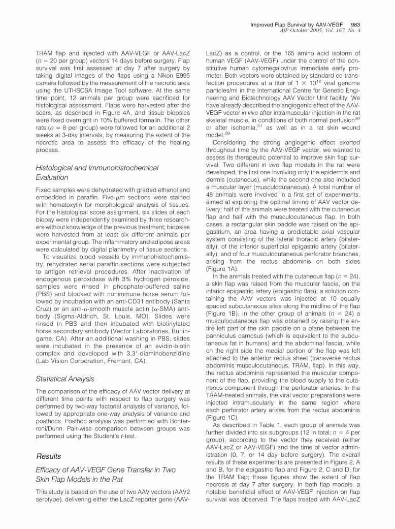
Considering the strong angiogenic effect exertedthroughout time by the AAV-VEGF vector, we wanted toassess its therapeutic potential to improve skin flap sur-vival. Two different in vivo flap models in the rat weredeveloped, the first one involving only the epidermis anddermis (cutaneous), while the second one also includeda muscular layer (musculocutaneous). A total number of48 animals were involved in a first set of experiments,aimed at exploring the optimal timing of AAV vector de-livery; half of the animals were treated with the cutaneousflap and half with the musculocutaneous flap. In bothcases, a rectangular skin paddle was raised on the epi-gastrium, an area having a predictable axial vascularsystem consisting of the lateral thoracic artery (bilater-ally), of the inferior superficial epigastric artery (bilater-ally), and of four musculocutaneous perforator branches,arising from the rectus abdominis on both sides(Figure 1A).
In the animals treated with the cutaneous flap (n � 24),a skin flap was raised from the muscular fascia, on theinferior epigastric artery (epigastric flap); a solution con-taining the AAV vectors was injected at 10 equallyspaced subcutaneous sites along the midline of the flap(Figure 1B). In the other group of animals (n � 24) amusculocutaneous flap was obtained by raising the en-tire left part of the skin paddle on a plane between thepanniculus carnosus (which is equivalent to the subcu-taneous fat in humans) and the abdominal fascia, whileon the right side the medial portion of the flap was leftattached to the anterior rectus sheet (transverse rectusabdominis musculocutaneous, TRAM, flap). In this way,the rectus abdominis represented the muscular compo-nent of the flap, providing the blood supply to the cuta-neous component through the perforator arteries. In theTRAM-treated animals, the viral vector preparations wereinjected intramuscularly in the same region whereeach perforator artery arises from the rectus abdominis(Figure 1C).
As described in Table 1, each group of animals wasfurther divided into six subgroups (12 in total; n � 4 pergroup), according to the vector they received (eitherAAV-LacZ or AAV-VEGF) and the time of vector admin-istration (0, 7, or 14 day before surgery). The overallresults of these experiments are presented in Figure 2, Aand B, for the epigastric flap and Figure 2, C and D, forthe TRAM flap; these figures show the extent of flapnecrosis at day 7 after surgery. In both flap models, anotable beneficial effect of AAV-VEGF injection on flapsurvival was observed. The flaps treated with AAV-LacZ
Improved Flap Survival by AAV-VEGF 983AJP October 2005, Vol. 167, No. 4
predictably underwent necrosis in their distal portion,similar to the untreated flaps, in which the extent of thenecrotic area reached 37.4 � 1.8% and 24.4 � 2.3% ofthe total flap surface for the cutaneous and the TRAMflaps, respectively (data not shown). As shown in Figure2B, the treatment with AAV-VEGF significantly reduced
epigastric flap necrosis in all of the three paired experi-mental groups (P � 0.01 by two-way analysis of vari-ance). The reduction in the necrotic area was 23.0%when the vector was injected during surgery, and 40.0%and 41.7% when injected 7 or 14 days before surgery,respectively. Similar results were also obtained for the
Figure 1. Schematic representation of the skin flaps and their vascularcomponents, with the indication of the vector injection sites. A: Thesurgical models of skin flap used in this study are based on a rectangularskin paddle measuring 5 � 8 cm, drawn on the abdomen of the animals.The predictable vascular system of the flap is composed of the lateralthoracic arteries, the inferior epigastric arteries, and the musculocutaneousperforator arteries arising from the rectus abdominis (usually four vesselson each side). B and C: The pictures schematically show the vascularcomponent providing the blood supply to each flap and the injection sites.In particular, the skin flap (B) was raised from the fascia on the inferiorepigastric artery, and the vector injected at 10 equally spaced subcutane-ous sites along the midline (inset in the top left part). The TRAM flap (C)was raised on a plane between the panniculus carnosus and the abdom-inal fascia, with the rectus abdominis as the only source of blood supplythrough the perforator arteries; in this model, the vector was administeredby intramuscular injection in the region where each perforator artery arisesfrom the rectus sheet (inset in the top left part).
984 Zacchigna et alAJP October 2005, Vol. 167, No. 4

TRAM model (P � 0.01), in which the reduction of flapnecrosis in the VEGF-treated animals versus LacZ con-trols was not evident when the vector was injected duringsurgery, and it was 38.1% and 50.0% when AAV-VEGFwas injected at 7 or 14 days before surgery, respectively(Figure 2D).
Taken together, these results indicate that the injectionof AAV-VEGF significantly increases flap survival. In par-ticular, posthoc statistical analysis in the TRAM flapmodel indicated that statistical significance wasachieved when the vector was administered at 14 daysbefore surgery (P � 0.05). Representative pictures offlaps treated with AAV-LacZ or AAV-VEGF 14 days beforesurgery are shown in Figure 2, A and C, for the epigastricand TRAM flap models, respectively.
AAV-VEGF Significantly Improves Flap Survival,Vascularization, and Healing in the TRAM Model
According to the results obtained by this first set of in vivoexperiments, a larger number or animals was used for asubsequent experiment in which the efficacy of AAV-VEGF delivery was assessed in the TRAM flap model,followed by histological and immunohistochemical eval-uation of the injected tissues. Rats received either AAV-LacZ or AAV-VEGF (n � 20 per group) 14 days beforesurgery, followed by the measurement of the necroticarea at day 7 after surgery. The results of this experimentare shown in Figure 3A. The percentage of the necroticarea was 22.8 � 2.7% and 8.9 � 2.8% in the animalstreated with AAV-LacZ and AAV-VEGF, respectively,resulting in almost 60% improvement in flap viability(P � 0.01).
Figure 2. Effect of VEGF on skin flap survival at different times of administration. At postoperative day 7, the regions of survival and necrosis were clearly definedin all of the flaps: the surviving skin appeared pink and tender, whereas the distal necrotic portion was black and rigid. A: The pictures show two selectedepigastric flaps, treated with AAV-LacZ (left) and AAV-VEGF (right), 14 days before flap elevation. The viability of the skin appears clearly improved afterAAV-VEGF administration. B: The effect of AAV-VEGF on epigastric flap survival was assessed by injecting the vector at different time points before surgery (0,7, and 14 days). The histograms represent the mean values and SD of the necrotic area relative to the total flap area, as measured by digital planimetry, for eachexperimental group. C: The pictures show two selected TRAM flaps, treated with AAV-LacZ (left) and AAV-VEGF (right), 14 days before flap elevation. Also inthis model, the treatment with AAV-VEGF resulted in a significant improvement in tissue viability. D: The effect of AAV-VEGF on TRAM flap survival was assessedby injecting the vector at different time points before surgery (0, 7, and 14 days). The histograms represent the mean values and SD of the necrotic area relativeto the total flap area, as measured by digital planimetry, for each experimental group.
Figure 3. Pretreatment with AAV-VEGF significantly improves TRAM flapsurvival and healing. A: The histogram shows the mean and SD of percentflap necrosis measured in 40 rats (20 per group) that were treated with theTRAM flap and injected with either AAV-LacZ or AAV-VEGF at day 14before flap elevation. Measurements were performed at day 7 after sur-gery. B: The healing process was monitored throughout time after surgeryup to 22 days, by measuring the extent of flap survival in animals treatedwith AAV-LacZ or AAV-VEGF (n � 8 per group). The means and SD ofsurvived flap areas are shown at each time point (expressed as a percent-age of the total flap area).
Improved Flap Survival by AAV-VEGF 985AJP October 2005, Vol. 167, No. 4
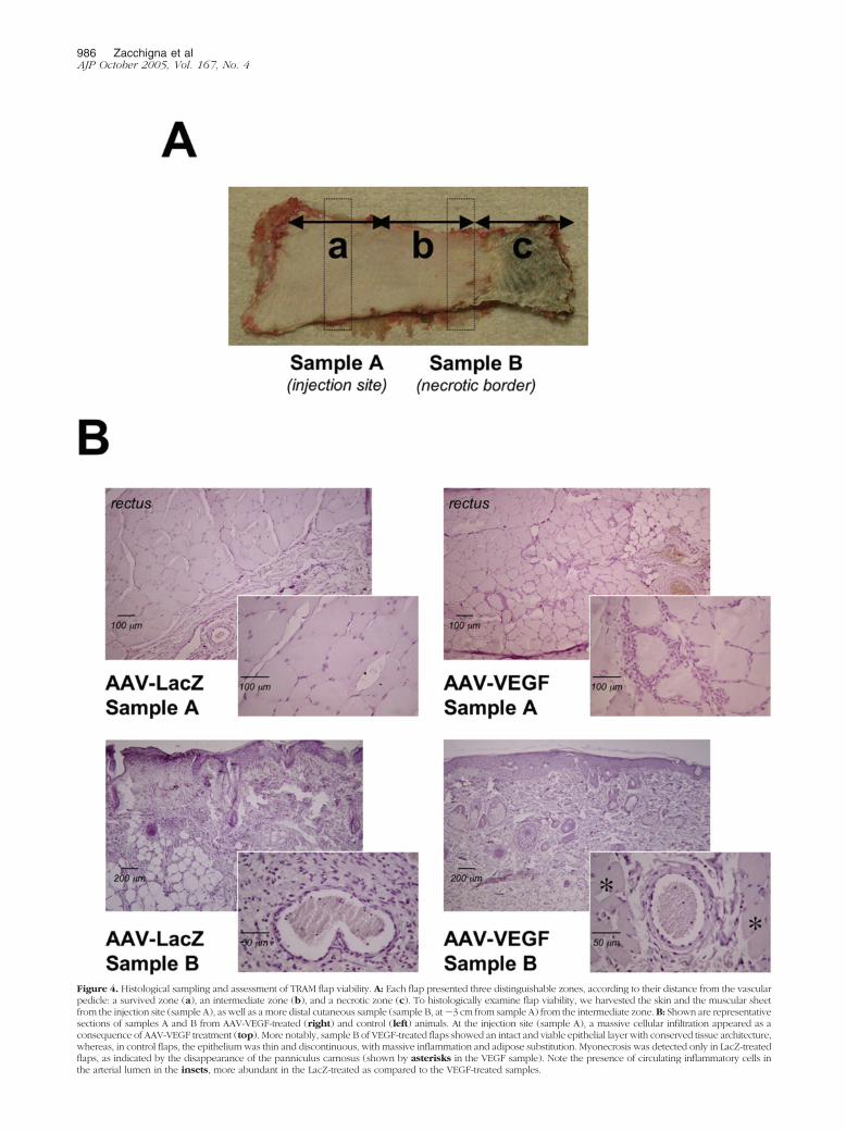
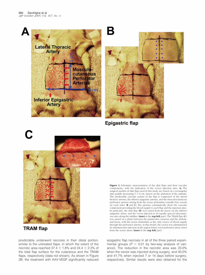
Figure 4. Histological sampling and assessment of TRAM flap viability. A: Each flap presented three distinguishable zones, according to their distance from the vascularpedicle: a survived zone (a), an intermediate zone (b), and a necrotic zone (c). To histologically examine flap viability, we harvested the skin and the muscular sheetfrom the injection site (sample A), as well as a more distal cutaneous sample (sample B, at �3 cm from sample A) from the intermediate zone. B: Shown are representativesections of samples A and B from AAV-VEGF-treated (right) and control (left) animals. At the injection site (sample A), a massive cellular infiltration appeared as aconsequence of AAV-VEGF treatment (top). More notably, sample B of VEGF-treated flaps showed an intact and viable epithelial layer with conserved tissue architecture,whereas, in control flaps, the epithelium was thin and discontinuous, with massive inflammation and adipose substitution. Myonecrosis was detected only in LacZ-treatedflaps, as indicated by the disappearance of the panniculus carnosus (shown by asterisks in the VEGF sample). Note the presence of circulating inflammatory cells inthe arterial lumen in the insets, more abundant in the LacZ-treated as compared to the VEGF-treated samples.
986 Zacchigna et alAJP October 2005, Vol. 167, No. 4
Flap healing was further assessed for an additional 15days by measuring the extension of the viable area of theflaps (n � 8 for each group). As shown in Figure 3B,treatment with AAV-VEGF determined a marked improve-ment in the healing process, resulting in �95% flap via-bility as early as 10 days after surgery and in the almostcomplete disappearance of the necrotic area at day 22.In contrast, healing was remarkably delayed in the ratstreated with AAV-LacZ (P � 0.01 by one-way analysis ofvariance).
To determine whether the improved tissue viability wasactually due to a sustained angiogenic response inducedby VEGF, representative sections from flap biopsies har-vested at day 7 after surgery were examined by histologyand immunohistochemistry. All flaps were designed tohave three zones (indicated as a, b, and c in Figure 4A),according to their distance from the vascular pedicle: asurvived zone (a), from which we took sample A, corre-sponding to the injection site; an intermediate zone (b),from which we took sample B at �3 cm from sample A, aregion corresponding to the border between viable andnecrotic skin; and a necrotic zone (c). Marked differ-ences were observed by histological analysis betweenAAV-VEGF- and AAV-LacZ-treated animals (Figure 4B).In sample A, the animals injected with AAV-VEGF showedmassive cellular infiltration close to the injection site. Thisfinding is consistent with our previous study, in which weinjected the same AAV-VEGF vector in the normoper-fused skeletal muscle, and observed the recruitment of alarge number of proliferating cells in close proximity tothe newly formed blood vessels, suggesting an importantrole of these cells in VEGF-induced neoangiogenesis.30
In sample B, a remarkable difference was also observedin terms of tissue viability. In the control, AAV-LacZ-treated animals, the epithelial layer was thin and imma-ture, with evidence of acute inflammation, adipose sub-stitution, and myonecrosis (as shown by the almostcomplete disappearance of the panniculus carnosus);the infiltrating inflammatory cells, mostly monocytes andneutrophils, were dispersed throughout the skin layers,concomitant with a severe disruption of the tissue archi-tecture. In contrast, the morphology was preserved in theAAV-VEGF-treated flaps, as revealed by the presence ofan intact epithelial layer and of skin appendages, withonly mild and focal inflammation, and poor accumulationof adipose tissue.
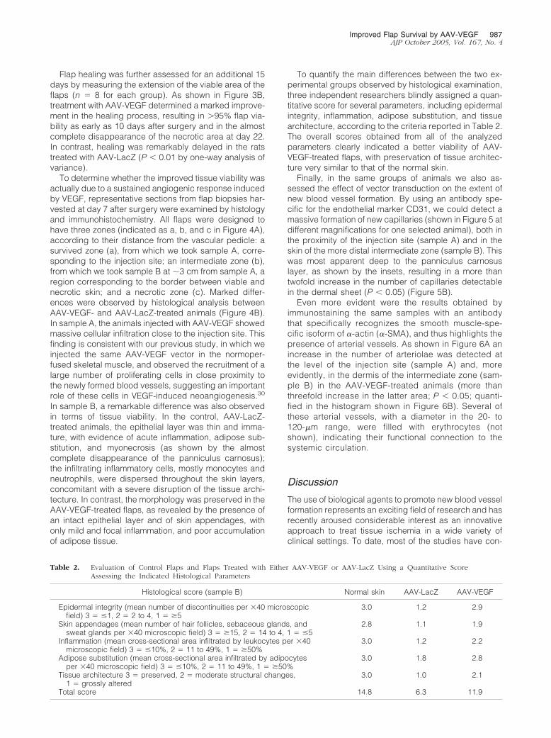
To quantify the main differences between the two ex-perimental groups observed by histological examination,three independent researchers blindly assigned a quan-titative score for several parameters, including epidermalintegrity, inflammation, adipose substitution, and tissuearchitecture, according to the criteria reported in Table 2.The overall scores obtained from all of the analyzedparameters clearly indicated a better viability of AAV-VEGF-treated flaps, with preservation of tissue architec-ture very similar to that of the normal skin.
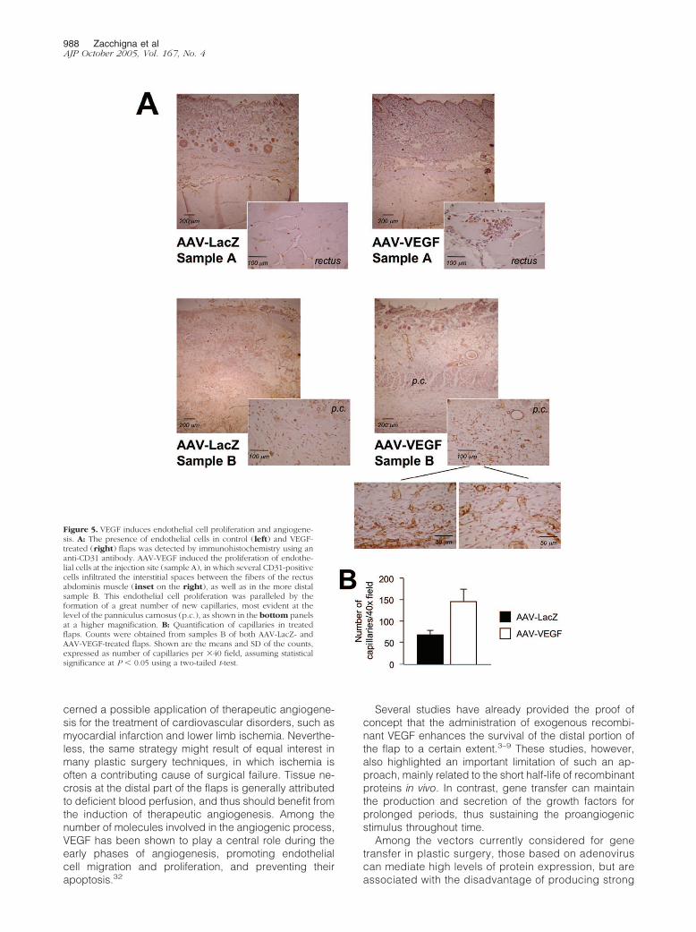
Finally, in the same groups of animals we also as-sessed the effect of vector transduction on the extent ofnew blood vessel formation. By using an antibody spe-cific for the endothelial marker CD31, we could detect amassive formation of new capillaries (shown in Figure 5 atdifferent magnifications for one selected animal), both inthe proximity of the injection site (sample A) and in theskin of the more distal intermediate zone (sample B). Thiswas most apparent deep to the panniculus carnosuslayer, as shown by the insets, resulting in a more thantwofold increase in the number of capillaries detectablein the dermal sheet (P � 0.05) (Figure 5B).
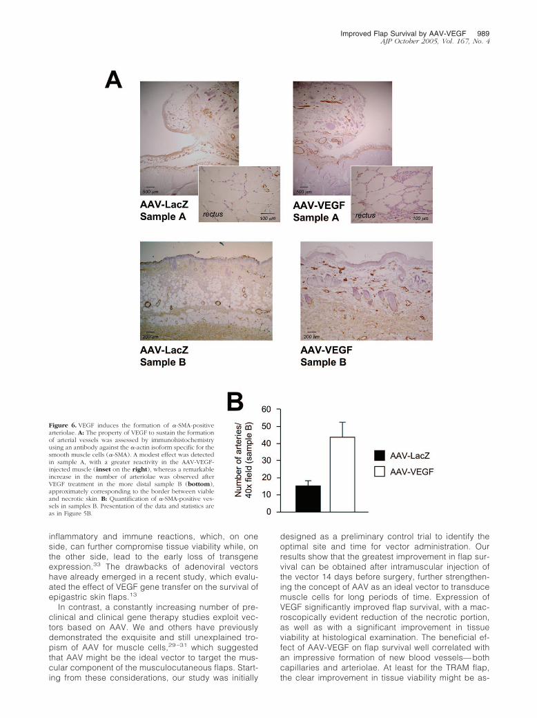
Even more evident were the results obtained byimmunostaining the same samples with an antibodythat specifically recognizes the smooth muscle-spe-cific isoform of �-actin (�-SMA), and thus highlights thepresence of arterial vessels. As shown in Figure 6A anincrease in the number of arteriolae was detected atthe level of the injection site (sample A) and, moreevidently, in the dermis of the intermediate zone (sam-ple B) in the AAV-VEGF-treated animals (more thanthreefold increase in the latter area; P � 0.05; quanti-fied in the histogram shown in Figure 6B). Several ofthese arterial vessels, with a diameter in the 20- to120-�m range, were filled with erythrocytes (notshown), indicating their functional connection to thesystemic circulation.
Discussion
The use of biological agents to promote new blood vesselformation represents an exciting field of research and hasrecently aroused considerable interest as an innovativeapproach to treat tissue ischemia in a wide variety ofclinical settings. To date, most of the studies have con-
Table 2. Evaluation of Control Flaps and Flaps Treated with Either AAV-VEGF or AAV-LacZ Using a Quantitative ScoreAssessing the Indicated Histological Parameters
Histological score (sample B) Normal skin AAV-LacZ AAV-VEGF
Epidermal integrity (mean number of discontinuities per �40 microscopicfield) 3 � �1, 2 � 2 to 4, 1 � �5
3.0 1.2 2.9
Skin appendages (mean number of hair follicles, sebaceous glands, andsweat glands per �40 microscopic field) 3 � �15, 2 � 14 to 4, 1 � �5
2.8 1.1 1.9
Inflammation (mean cross-sectional area infiltrated by leukocytes per �40microscopic field) 3 � �10%, 2 � 11 to 49%, 1 � �50%
3.0 1.2 2.2
Adipose substitution (mean cross-sectional area infiltrated by adipocytesper �40 microscopic field) 3 � �10%, 2 � 11 to 49%, 1 � �50%
3.0 1.8 2.8
Tissue architecture 3 � preserved, 2 � moderate structural changes,1 � grossly altered
3.0 1.0 2.1
Total score 14.8 6.3 11.9
Improved Flap Survival by AAV-VEGF 987AJP October 2005, Vol. 167, No. 4
cerned a possible application of therapeutic angiogene-sis for the treatment of cardiovascular disorders, such asmyocardial infarction and lower limb ischemia. Neverthe-less, the same strategy might result of equal interest inmany plastic surgery techniques, in which ischemia isoften a contributing cause of surgical failure. Tissue ne-crosis at the distal part of the flaps is generally attributedto deficient blood perfusion, and thus should benefit fromthe induction of therapeutic angiogenesis. Among thenumber of molecules involved in the angiogenic process,VEGF has been shown to play a central role during theearly phases of angiogenesis, promoting endothelialcell migration and proliferation, and preventing theirapoptosis.32
Several studies have already provided the proof ofconcept that the administration of exogenous recombi-nant VEGF enhances the survival of the distal portion ofthe flap to a certain extent.3–9 These studies, however,also highlighted an important limitation of such an ap-proach, mainly related to the short half-life of recombinantproteins in vivo. In contrast, gene transfer can maintainthe production and secretion of the growth factors forprolonged periods, thus sustaining the proangiogenicstimulus throughout time.
Among the vectors currently considered for genetransfer in plastic surgery, those based on adenoviruscan mediate high levels of protein expression, but areassociated with the disadvantage of producing strong
Figure 5. VEGF induces endothelial cell proliferation and angiogene-sis. A: The presence of endothelial cells in control (left) and VEGF-treated (right) flaps was detected by immunohistochemistry using ananti-CD31 antibody. AAV-VEGF induced the proliferation of endothe-lial cells at the injection site (sample A), in which several CD31-positivecells infiltrated the interstitial spaces between the fibers of the rectusabdominis muscle (inset on the right), as well as in the more distalsample B. This endothelial cell proliferation was paralleled by theformation of a great number of new capillaries, most evident at thelevel of the panniculus carnosus (p.c.), as shown in the bottom panelsat a higher magnification. B: Quantification of capillaries in treatedflaps. Counts were obtained from samples B of both AAV-LacZ- andAAV-VEGF-treated flaps. Shown are the means and SD of the counts,expressed as number of capillaries per �40 field, assuming statisticalsignificance at P � 0.05 using a two-tailed t-test.
988 Zacchigna et alAJP October 2005, Vol. 167, No. 4
inflammatory and immune reactions, which, on oneside, can further compromise tissue viability while, onthe other side, lead to the early loss of transgeneexpression.33 The drawbacks of adenoviral vectorshave already emerged in a recent study, which evalu-ated the effect of VEGF gene transfer on the survival ofepigastric skin flaps.13
In contrast, a constantly increasing number of pre-clinical and clinical gene therapy studies exploit vec-tors based on AAV. We and others have previouslydemonstrated the exquisite and still unexplained tro-pism of AAV for muscle cells,29 –31 which suggestedthat AAV might be the ideal vector to target the mus-cular component of the musculocutaneous flaps. Start-ing from these considerations, our study was initially
designed as a preliminary control trial to identify theoptimal site and time for vector administration. Ourresults show that the greatest improvement in flap sur-vival can be obtained after intramuscular injection ofthe vector 14 days before surgery, further strengthen-ing the concept of AAV as an ideal vector to transducemuscle cells for long periods of time. Expression ofVEGF significantly improved flap survival, with a mac-roscopically evident reduction of the necrotic portion,as well as with a significant improvement in tissueviability at histological examination. The beneficial ef-fect of AAV-VEGF on flap survival well correlated withan impressive formation of new blood vessels— bothcapillaries and arteriolae. At least for the TRAM flap,the clear improvement in tissue viability might be as-
Figure 6. VEGF induces the formation of �-SMA-positivearteriolae. A: The property of VEGF to sustain the formationof arterial vessels was assessed by immunohistochemistryusing an antibody against the �-actin isoform specific for thesmooth muscle cells (�-SMA). A modest effect was detectedin sample A, with a greater reactivity in the AAV-VEGF-injected muscle (inset on the right), whereas a remarkableincrease in the number of arteriolae was observed afterVEGF treatment in the more distal sample B (bottom),approximately corresponding to the border between viableand necrotic skin. B: Quantification of �-SMA-positive ves-sels in samples B. Presentation of the data and statistics areas in Figure 5B.
Improved Flap Survival by AAV-VEGF 989AJP October 2005, Vol. 167, No. 4
cribed to a better perfusion through the muscular layerdue to a local angiogenic effect of VEGF. Alternatively,secreted VEGF might have diffused from the muscle tothe skin layer of the more distal portion of the flap, thuspromoting angiogenesis within the derma, in agree-ment with a series of recent reports showing that AAVvectors delivered to the skeletal muscle are able todrive the expression and secretion of different mole-cules into the circulation.24,28 The increase in the den-sity of arteriolae in the skin distal to the injection site(sample B) in the TRAM model is strongly in favor of thesecond hypothesis, although we cannot exclude anindirect effect of VEGF through an enhancement of theblood supply.
In accordance with the marked improvement in flapviability after AAV-VEGF transduction, our time courseexperiment clearly indicates a persistent therapeuticeffect of the vector up to 3 weeks after surgery. Al-though the extent of flap necrosis is usually assessedat no longer that 7 days after flap elevation, reflectingthe timing by which the problem appears to be clini-cally relevant, an increased vascularization driven byVEGF might provide an additional benefit in the follow-ing healing process. Our results strongly support thishypothesis, and reinforce the notion that a deliverysystem able to sustain a prolonged expression of thetherapeutic gene is extremely desirable for this kind oftherapeutic application.
The observation that the administration of AAV-VEGF isparticularly effective when performed preoperatively alsosupports the usefulness of AAV-mediated therapeutic an-giogenesis in elective surgery conditions. Consideringthat the problem of flap breakdown is especially preva-lent in patients who have diabetes, obesity, and periph-eral vascular disease,34 as well as in smokers,35 it seemsreasonable that AAV-VEGF administration during the pre-operative phase might represent a useful therapeuticmethod to enhance blood flow in skin areas undergoingelevation and transposition in high-risk patients. In termsof clinical applications, the observation that the efficacyof AAV-mediated gene transfer is more evident in theTRAM model rather than in the epigastric flap model,renders AAV-VEGF gene transfer even more attractive. Infact, the results obtained from the epigastric flap model,in which the panniculus carnosus (present in rodents butnot in humans) represents the major target of AAV vectortransduction, cannot be directly extrapolated to the hu-man skin. In contrast, the TRAM flap, including a muscu-lar component in both species, is much more represen-tative of the human condition.
Taken together, our results reinforce the notion thatadequate blood supply is an essential requisite for flapsurvival, and indicate the feasibility of an angiogenicgene therapy approach in plastic and reconstructive sur-gery. In addition, considering the prolonged gene ex-pression driven by AAV vectors, this delivery systemmight become a novel and powerful experimental tool toinvestigate the biological role of other relevant growthfactors, as well as their combinations, to find new thera-peutic approaches for the treatment of tissue ischemia inplastic surgery. Few recent studies have reported about
the angiogenic potential of other factors, such as angio-poietin-1,36–38 platelet-derived growth factor,39 and fibro-blast growth factor,40–42 in different animal models ofskin flaps. Because AAV vectors enter the cells at highmultiplicity of infection, an attractive approach could bethe simultaneous administration of different vector prep-arations, expressing different proteins involved in theangiogenic process.30
Although it is the myocardium and the muscles of thelower limb that have been the intended sites of angio-genesis in the majority of studies to date, there isoptimism that plastic and reconstructive surgery andits patients could be major beneficiaries of the knowl-edge currently being accumulated. The various typesof flaps, from local skin flaps to complex free flapscould be helped by reinforcement of the natural angio-genic process occurring in the microvasculature. Oneof the major potential applications of a therapeuticangiogenesis approach using gene therapy will be thedesign of larger flaps than would otherwise be possi-ble, able to cover tissue defects previously consideredtoo large or complex.
Acknowledgments
We thank Marina Dapas and Maria Elena Lopez for ex-cellent technical support in AAV vector production, MariaCristina Prati and Matteo Dell’Omodarme for statisticalconsultancy, and Suzanne Kerbavcic for editorialassistance.
References
1. Hallock GG: Physiological studies using laser Doppler flowmetry tocompare blood flow to the zones of the free TRAM flap. Ann PlastSurg 2001, 47:229–233
2. Machens HG, Salehi J, Weich H, Munch S, Siemers F, Krapohl BD,Herter KH, Kruger S, Reichert B, Berger A, Vogt P, Mailander P:Angiogenic effects of injected VEGF165 and sVEGFR-1 (sFLT-1) in arat flap model. J Surg Res 2003, 111:136–142
3. Padubidri A, Browne Jr E: Effect of vascular endothelial growth factor(VEGF) on survival of random extension of axial pattern skin flaps inthe rat. Ann Plast Surg 1996, 37:604–611
4. Li QF, Reis ED, Zhang WX, Silver L, Fallon JT, Weinberg H: Acceler-ated flap prefabrication with vascular endothelial growth factor. JReconstr Microsurg 2000, 16:45–49
5. Kryger Z, Dogan T, Zhang F, Komorowska-Timek E, Shi DY, Cheng C,Lineaweaver WC, Buncke HJ: Effects of VEGF administration follow-ing ischemia on survival of the gracilis muscle flap in the rat. Ann PlastSurg 1999, 43:172–178
6. Kryger Z, Zhang F, Dogan T, Cheng C, Lineaweaver WC, Buncke HJ:The effects of VEGF on survival of a random flap in the rat: examina-tion of various routes of administration. Br J Plast Surg 2000,53:234–239
7. Banbury J, Siemionow M, Porvasnik S, Petras S, Browne E: Improvedperfusion after subcritical ischemia in muscle flaps treated with vas-cular endothelial growth factor. Plast Reconstr Surg 2000,106:1541–1546
8. Zhang F, Fischer K, Komorowska-Timek E, Guo M, Cui D, Dorsett-Martin W, Buncke HJ, Lineaweaver WC: Improvement of skin paddlesurvival by application of vascular endothelial growth factor in a ratTRAM flap model. Ann Plast Surg 2001, 46:314–319
9. Zhang F, Richards L, Angel MF, Zhang J, Liu H, Dorsett-Martin W,Lineaweaver WC: Accelerating flap maturation by vascular endothe-
990 Zacchigna et alAJP October 2005, Vol. 167, No. 4
lium growth factor in a rat tube flap model. Br J Plast Surg 2002,55:59–63
10. Snyder RO, Spratt SK, Lagarde C, Bohl D, Kaspar B, Sloan B, CohenLK, Danos O: Efficient and stable adeno-associated virus-mediatedtransduction in the skeletal muscle of adult immunocompetent mice.Hum Gene Ther 1997, 8:1891–1900
11. Greenhalgh DA, Rothnagel JA, Roop DR: Epidermis: an attractivetarget tissue for gene therapy. J Invest Dermatol 1994, 103:63S–69S
12. Cui L, Li FC, Zhang Q, Qian YL, Guan WX: Effect of adenovirus-mediated gene transfection of vascular endothelial growth factor onsurvival of random flaps in rats. Chin J Traumatol 2003, 6:199–204
13. Gurunluoglu R, Ozer K, Skugor B, Lubiatowski P, Carnevale K, Siemi-onow M: Effect of transfection time on the survival of epigastric skinflaps pretreated with adenovirus encoding the VEGF gene. Ann PlastSurg 2002, 49:161–169
14. Ghazizadeh S, Taichman LB: Virus-mediated gene transfer for cuta-neous gene therapy. Hum Gene Ther 2000, 11:2247–2251
15. O’Toole G, MacKenzie D, Lindeman R, Buckley MF, Marucci D,McCarthy N, Poole M: Vascular endothelial growth factor gene ther-apy in ischaemic rat skin flaps. Br J Plast Surg 2002, 55:55–58
16. Neumeister MW, Song YH, Mowlavi A, Suchy H, Mathur A: Effects ofliposome-mediated gene transfer of VEGF in ischemic rat gracilismuscle. Microsurgery 2001, 21:58–62
17. Lubiatowski P, Goldman CK, Gurunluoglu R, Carnevale K, SiemionowM: Enhancement of epigastric skin flap survival by adenovirus-medi-ated VEGF gene therapy. Plast Reconstr Surg 2002, 109:1986–1993
18. Liu PY, Tong W, Liu K, Han SH, Wang XT, Badiavas E, Rieger-ChristK, Summerhayes I: Liposome-mediated transfer of vascular endothe-lial growth factor cDNA augments survival of random-pattern skinflaps in the rat. Wound Repair Regen 2004, 12:80–85
19. Su H, Lu R, Kan YW: Adeno-associated viral vector-mediated vascu-lar endothelial growth factor gene transfer induces neovascular for-mation in ischemic heart. Proc Natl Acad Sci USA 2000,97:13801–13806
20. Kaplitt MG, Leone P, Samulski RJ, Xiao X, Pfaff DW, O’Malley KL,During MJ: Long-term gene expression and phenotypic correctionusing adeno-associated virus vectors in the mammalian brain. NatGenet 1994, 8:148–154
21. Nakai H, Herzog RW, Hagstrom JN, Walter J, Kung SH, Yang EY, TaiSJ, Iwaki Y, Kurtzman GJ, Fisher KJ, Colosi P, Couto LB, High KA:Adeno-associated viral vector-mediated gene transfer of humanblood coagulation factor IX into mouse liver. Blood 1998,91:4600–4607
22. Xiao W, Berta SC, Lu MM, Moscioni AD, Tazelaar J, Wilson JM:Adeno-associated virus as a vector for liver-directed gene therapy.J Virol 1998, 72:10222–10226
23. Chirmule N, Propert K, Magosin S, Qian Y, Qian R, Wilson J: Immuneresponses to adenovirus and adeno-associated virus in humans.Gene Ther 1999, 6:1574–1583
24. Kay MA, Manno CS, Ragni MV, Larson PJ, Couto LB, McClelland A,Glader B, Chew AJ, Tai SJ, Herzog RW, Arruda V, Johnson F, ScallanC, Skarsgard E, Flake AW, High KA: Evidence for gene transfer andexpression of factor IX in haemophilia B patients treated with an AAVvector. Nat Genet 2000, 24:257–261
25. Monahan PE, Samulski RJ: AAV vectors: is clinical success on thehorizon? Gene Ther 2000, 7:24–30
26. Descamps V, Blumenfeld N, Beuzard Y, Perricaudet M: Keratinocytesas a target for gene therapy. Sustained production of erythropoietin inmice by human keratinocytes transduced with an adenoassociatedvirus vector. Arch Dermatol 1996, 132:1207–1211
27. Hengge UR, Mirmohammadsadegh A: Adeno-associated virus ex-presses transgenes in hair follicles and epidermis. Mol Ther 2000,2:188–194
28. Donahue BA, McArthur JG, Spratt SK, Bohl D, Lagarde C, Sanchez L,Kaspar BA, Sloan BA, Lee YL, Danos O, Snyder RO: Selective uptakeand sustained expression of AAV vectors following subcutaneousdelivery. J Gene Med 1999, 1:31–42
29. Deodato B, Arsic N, Zentilin L, Galeano M, Santoro D, Torre V,Altavilla D, Valdembri D, Bussolino F, Squadrito F, Giacca M: Recom-binant AAV vector encoding human VEGF165 enhances wound heal-ing. Gene Ther 2002, 9:777–785
30. Arsic N, Zentilin L, Zacchigna S, Santoro D, Stanta G, Salvi A, SinagraG, Giacca M: Induction of functional neovascularization by combinedVEGF and angiopoietin-1 gene transfer using AAV vectors. Mol Ther2003, 7:450–459
31. Arsic N, Zacchigna S, Zentilin L, Ramirez-Correa G, Pattarini L, SalviA, Sinagra G, Giacca M: Vascular endothelial growth factor stimulatesskeletal muscle regeneration in vivo. Mol Ther 2004, 10:844–854
32. Ferrara N, Alitalo K: Clinical applications of angiogenic growth factorsand their inhibitors. Nat Med 1999, 5:1359–1364
33. Yang Y, Haecker SE, Su Q, Wilson JM: Immunology of gene therapywith adenoviral vectors in mouse skeletal muscle. Hum Mol Genet1996, 5:1703–1712
34. Hultman CS, Daiza S: Skin-sparing mastectomy flap complicationsafter breast reconstruction: review of incidence, management, andoutcome. Ann Plast Surg 2003, 50:249–255
35. Craig S, Rees TD: The effects of smoking on experimental skin flapsin hamsters. Plast Reconstr Surg 1985, 75:842–846
36. Gurunluoglu R, Lubiatowski P, Goldman CK, Carnevale K, SiemionowM: Enhancement of muscle flap hemodynamics by angiopoietin-1.Ann Plast Surg 2002, 48:401–409
37. Lubiatowski P, Gurunluoglu R, Goldman CK, Skugor B, Carnevale K,Siemionow M: Gene therapy by adenovirus-mediated vascular endo-thelial growth factor and angiopoietin-1 promotes perfusion of muscleflaps. Plast Reconstr Surg 2002, 110:149–159
38. Jung H, Gurunluoglu R, Scharpf J, Siemionow M: Adenovirus-medi-ated angiopoietin-1 gene therapy enhances skin flap survival. Micro-surgery 2003, 23:374–380
39. Carroll CM, Carroll SM, Schuschke DA, Barker JH: Augmentation ofskeletal muscle flap survival using platelet derived growth factor.Plast Reconstr Surg 1998, 102:407–415
40. Rashid MA, Akita S, Razzaque MS, Yoshimoto H, Ishihara H, Fujii T,Tanaka K, Taguchi T: Coadministration of basic fibroblast growthfactor and sucrose octasulfate (sucralfate) facilitates the rat dorsalflap survival and viability. Plast Reconstr Surg 1999, 103:941–948
41. Khouri RK, Brown DM, Leal-Khouri SM, Tark KC, Shaw WW: Theeffect of basic fibroblast growth factor on the neovascularisationprocess: skin flap survival and staged flap transfers. Br J Plast Surg1991, 44:585–588
42. Bayati S, Russell RC, Roth AC: Stimulation of angiogenesis to improvethe viability of prefabricated flaps. Plast Reconstr Surg 1998,101:1290–1295
Improved Flap Survival by AAV-VEGF 991AJP October 2005, Vol. 167, No. 4
Copyright © 2022 FDOKUMEN