Malignant, non-hodgkin's lymphomas in trypanosoma cruzi-infected rabbits treated with nitroarenes
Upload
independentCategory
view
3download
0
Review article
WHO-EORTC classification for cutaneous lymphomasRein Willemze, Elaine S. Jaffe, Gunter Burg, Lorenzo Cerroni, Emilio Berti, Steven H. Swerdlow, Elisabeth Ralfkiaer, Sergio Chimenti,Jose L. Diaz-Perez, Lyn M. Duncan, Florent Grange, Nancy Lee Harris, Werner Kempf, Helmut Kerl, Michael Kurrer, Robert Knobler,Nicola Pimpinelli, Christian Sander, Marco Santucci, Wolfram Sterry, Maarten H. Vermeer, Janine Wechsler, Sean Whittaker,and Chris J. L. M. Meijer
Primary cutaneous lymphomas are cur-rently classified by the European Organi-zation for Research and Treatment ofCancer (EORTC) classification or theWorld Health Organization (WHO) classifi-cation, but both systems have shortcom-ings. In particular, differences in the clas-sification of cutaneous T-cell lymphomasother than mycosis fungoides, Sezarysyndrome, and the group of primary cuta-neous CD30� lymphoproliferative disor-ders and the classification and terminol-
ogy of different types of cutaneous B-celllymphomas have resulted in consider-able debate and confusion. During recentconsensus meetings representatives ofboth systems reached agreement on anew classification, which is now calledthe WHO-EORTC classification. In thispaper we describe the characteristic fea-tures of the different primary cutaneouslymphomas and other hematologic neo-plasms frequently presenting in the skin,and discuss differences with the previous
classification schemes. In addition, therelative frequency and survival data of1905 patients with primary cutaneous lym-phomas derived from Dutch and Austrianregistries for primary cutaneous lympho-mas are presented to illustrate the clinicalsignificance of this new classification.(Blood. 2005;105:3768-3785)
© 2005 by The American Society of Hematology
Introduction
A variety of T- and B-cell neoplasms can involve the skin, eitherprimarily or secondarily. The term “primary cutaneous lymphoma”refers to cutaneous T-cell lymphomas (CTCLs) and cutaneousB-cell lymphomas (CBCLs) that present in the skin with noevidence of extracutaneous disease at the time of diagnosis. Afterthe gastrointestinal tract, the skin is the second most common siteof extranodal non-Hodgkin lymphoma, with an estimated annualincidence of 1:100,000.1
Primary cutaneous lymphomas often have a completely differ-ent clinical behavior and prognosis from histologically similarsystemic lymphomas, which may involve the skin secondarily, andtherefore require different types of treatment. For that reason,recent classification systems for non-Hodgkin lymphomas such asthe European Organization for Research and Treatment of Cancer(EORTC) classification for primary cutaneous lymphomas and theWorld Health Organization (WHO) classification for tumors ofhematopoietic and lymphoid tissues included primary cutaneouslymphomas as separate entities.2,3 In the EORTC classification,distinction was made between primary cutaneous lymphomas withan indolent, intermediate, or aggressive clinical behavior. The
clinical validity of this classification has been validated by severallarge studies, including follow-up data of more than 1300 patientswith a primary cutaneous lymphoma.2,4,5 Although there wasconsensus between the EORTC and WHO classifications on theclassification of most types of CTCLs, remaining differencesbetween the 2 classification systems, in particular the controversyon the definition and terminology of the different types of CBCLs,has resulted in considerable debate and confusion.6-9
During consensus meetings in Lyon, France (September 2003)and Zurich, Switzerland (January 2004), these differences wereresolved by representatives of both classification systems, and aconsensus classification was developed (Table 1). In this report wepresent the new WHO-EORTC classification for cutaneous lympho-mas. This review will focus on primary cutaneous lymphomas anda few other conditions that frequently first present in the skin, suchas CD4�/CD56� hematodermic neoplasm (formerly also known asblastic natural killer [NK] cell lymphoma) and adult T-cellleukemia/lymphoma. Other neoplasms that may also first present inthe skin in a minority of cases, such as precursor B-lymphoblasticleukemia/lymphoma and acute myeloid leukemia, and secondary
From the Department of Dermatology, Leiden University, Medical Center, theNetherlands; Laboratory of Pathology, National Cancer Institute, NationalInstitutes of Health, Bethesda, MD; Department of Dermatology, UniversityHospital Zurich, Switzerland; Department of Dermatology, University of Graz,Austria; Department of Dermatology, University of Milan-Biococca andOspedale Magiore, Milan, Italy; Department of Pathology, Division ofHematopathology, University of Pittsburgh School of Medicine, PA; Departmentof Pathology, University of Copenhagen, Denmark; Department ofDermatology, University of Rome, Italy; Department of Dermatology, CrucesHospital, Bilbao, Spain; Department of Pathology, Massachusetts GeneralHospital and Harvard Medical School, Boston, MA; Department ofDermatology, Hopital Pasteur, Colmar, France; Department of Pathology,University Hospital Zurich, Switzerland; Department of Dermatology, Universityof Vienna, Austria; Department of Dermatological Sciences, University ofFlorence, Italy; Department of Dermatology, Allgemeines Krankenhaus StGeorg, Hamburg, Germany; Department of Human Pathology and Oncology,
University of Florence, Italy; Department of Dermatology, Charite, HumboldtUniversity, Berlin, Germany; Department of Pathology, Hopital Henri Mondor,Creteil, France; Skin Tumour Unit, St John’s Institute of Dermatology, StThomas’ Hospital, London, England; and Department of Pathology, VrijeUniversiteit Medical Center, Amsterdam, the Netherlands.
Submitted September 10, 2004; accepted November 11, 2005. Prepublishedonline as Blood First Edition Paper, February 3, 2005; DOI 10.1182/blood-2004-09-3502.
An Inside Blood analysis of this article appears at the front of the issue.
Reprints: Rein Willemze, Department of Dermatology, B1-Q-93, LeidenUniversity Center, PO Box 9600, 2300 RC Leiden, the Netherlands; e-mail:[email protected].
© 2005 by The American Society of Hematology
3768 BLOOD, 15 MAY 2005 � VOLUME 105, NUMBER 10
cutaneous manifestations of systemic lymphomas, are not dis-cussed, but will be included in the monograph to be published inthe WHO Blue Book series in 2005. After a discussion of the 2most controversial groups of cutaneous lymphomas that were
defined differently in the original EORTC and WHO classificationschemes, the main features of the different types of primarycutaneous lymphoma are presented. In Table 2 the relative fre-quency and survival of 1905 patients with primary cutaneouslymphomas derived from Dutch and Austrian cutaneous lymphomaregistries are presented, to illustrate the clinical significance of theWHO-EORTC classification.
Classification of CTCLs other than mycosis fungoides, Sezarysyndrome, and primary cutaneous CD30� lymphoproliferations
The classification of this remaining group of CTCLs is difficult andconfusing, which is not surprising given the heterogeneity andrarity of these tumors. Together, they constitute less than 10% of allCTCLs.2,5 With few exceptions, these lymphomas are clinicallyaggressive and in most cases systemic chemotherapy is required. Inthe EORTC classification, most of these lymphomas were groupedas primary cutaneous CD30� large T-cell lymphoma or in theprovisional group of primary cutaneous small/medium-sizedpleomorphic CTCLs. Distinction between these 2 categories,which is based on the presence of more or less than 30% largeneoplastic T cells, was considered useful because several studiesdemonstrated a significant difference in survival between these 2groups.2,4,5,10 Recent studies suggest, however, that this favorableprognosis is restricted to small/medium-sized pleomorphic CTCLswith a CD4� T-cell phenotype, in particular those that present withlocalized disease, in contrast to those with a CD8� T-cell pheno-type.11 Moreover, the EORTC category of CD30� large-cellCTCLs has become quite heterogeneous through the recognition ofnew diagnostic categories, such as subcutaneous panniculitis-likeT-cell lymphoma (SPTL),12-17 extranodal NK/T-cell lymphoma,nasal type,18-20 CD4�/CD56� hematodermic neoplasm (blasticNK-cell lymphoma),21-24 aggressive epidermotropic CD8�
Table 1. WHO-EORTC classification of cutaneous lymphomaswith primary cutaneous manifestations
Cutaneous T-cell and NK-cell lymphomas
Mycosis fungoides
MF variants and subtypes
Folliculotropic MF
Pagetoid reticulosis
Granulomatous slack skin
Sezary syndrome
Adult T-cell leukemia/lymphoma
Primary cutaneous CD30� lymphoproliferative disorders
Primary cutaneous anaplastic large cell lymphoma
Lymphomatoid papulosis
Subcutaneous panniculitis-like T-cell lymphoma*
Extranodal NK/T-cell lymphoma, nasal type
Primary cutaneous peripheral T-cell lymphoma, unspecified
Primary cutaneous aggressive epidermotropic CD8� T-cell lymphoma
(provisional)
Cutaneous �/� T-cell lymphoma (provisional)
Primary cutaneous CD4� small/medium-sized pleomorphic T-cell lymphoma
(provisional)
Cutaneous B-cell lymphomas
Primary cutaneous marginal zone B-cell lymphoma
Primary cutaneous follicle center lymphoma
Primary cutaneous diffuse large B-cell lymphoma, leg type
Primary cutaneous diffuse large B-cell lymphoma, other
Intravascular large B-cell lymphoma
Precursor hematologic neoplasm
CD4�/CD56� hematodermic neoplasm (blastic NK-cell lymphoma)†
Table 2. Relative frequency and disease-specific 5-year survival of 1905 primary cutaneous lymphomas classifiedaccording to the WHO-EORTC classification
WHO-EORTC classification No. Frequency, %*Disease-specific5-year survival, %
Cutaneous T-cell lymphoma
Indolent clinical behavior
Mycosis fungoides 800 44 88
Folliculotropic MF 86 4 80
Pagetoid reticulosis 14 � 1 100
Granulomatous slack skin 4 � 1 100
Primary cutaneous anaplastic large cell lymphoma 146 8 95
Lymphomatoid papulosis 236 12 100
Subcutaneous panniculitis-like T-cell lymphoma 18 1 82
Primary cutaneous CD4� small/medium pleomorphic T-cell lymphoma† 39 2 75
Aggressive clinical behavior
Sezary syndrome 52 3 24
Primary cutaneous NK/T-cell lymphoma, nasal-type 7 � 1 NR
Primary cutaneous aggressive CD8� T-cell lymphoma† 14 � 1 18
Primary cutaneous �/� T-cell lymphoma† 13 � 1 NR
Primary cutaneous peripheral T-cell lymphoma, unspecified‡ 47 2 16
Cutaneous B-cell lymphoma
Indolent clinical behavior
Primary cutaneous marginal zone B-cell lymphoma 127 7 99
Primary cutaneous follicle center lymphoma 207 11 95
Intermediate clinical behavior
Primary cutaneous diffuse large B-cell lymphoma, leg type 85 4 55
Primary cutaneous diffuse large B-cell lymphoma, other 4 � 1 50
Primary cutaneous intravascular large B-cell lymphoma 6 � 1 65
NR indicates not reached.*Data are based on 1905 patients with a primary cutaneous lymphoma registered at the Dutch and Austrian Cutaneous Lymphoma Group between 1986 and 2002.†Primary cutaneous peripheral T-cell lymphoma, unspecified excluding the three provisional entities indicated with a double dagger (‡).
WHO-EORTC CLASSIFICATION FOR CUTANEOUS LYMPHOMAS 3769BLOOD, 15 MAY 2005 � VOLUME 105, NUMBER 10

CTCL,16,25,26 and cutaneous gamma/delta T-cell lymphoma.27-31 Inthe WHO classification, SPTL, nasal-type NK/T-cell lymphoma,and blastic NK-cell lymphoma were included as separate entities,whereas the other entities were part of the broad category ofperipheral T-cell lymphoma (PTL), unspecified.
In the WHO-EORTC classification, extranodal NK/T-cell lym-phoma, nasal type, and CD4�/CD56� hematodermic neoplasm(blastic NK-cell lymphoma), are defined as separate entities.Recent studies showed both clinical, histologic, and immunopheno-typical differences between cases of SPTL with an �/� T-cellphenotype and those with a �/� T-cell phenotype, suggesting thatthese may represent different entities. Whereas SPTLs with an �/�T-cell phenotype are homogeneous with a rather indolent clinicalbehavior in many patients, SPTLs with a �/� T-cell phenotypeoverlap with other types of �/�� T/NK-cell lymphoma andinvariably run a very aggressive clinical course.14,16,17,31,32
We therefore suggest that the term “SPTL” be restricted forSPTLs with an �/� T-cell phenotype.32 Recent studies havesuggested that some disorders can be separated out as provisionalentities from the broad group of “PTL, unspecified,” in the WHOclassification. These include aggressive epidermotropic CD8�
CTCL, cutaneous gamma-delta T-cell lymphoma (including casesformerly diagnosed as SPTL with a gamma/delta phenotype), andprimary cutaneous small-medium CD4� T-cell lymphoma. In theWHO-EORTC classification the term “PTL, unspecified,” is main-tained for remaining cases that do not fit into either of theseprovisional entities.
Primary cutaneous follicle-center cell lymphoma and primarycutaneous diffuse large B-cell lymphoma
In recent years, the EORTC categories of primary cutaneousfollicle center cell lymphoma (PCFCCL) and primary cutaneouslarge B-cell lymphoma of the leg (PCLBCL-leg) have been thesubject of much debate. The term PCFCCL was introduced in 1987as a term encompassing cutaneous lymphomas that were composedof cells with the morphology of follicle center cells (ie, centroblastsand [large] centrocytes), and that were classified as either centro-blastic/centrocytic or centroblastic lymphoma according to criteriaof the Kiel classification.33 Whereas small and early lesions mayshow both small and large neoplastic B cells and many admixedT cells, and may have a partly follicular growth pattern,tumorous lesions generally show a predominance of large Bcells, particularly large cleaved or multilobated cells, and lessfrequently a predominance of typical centroblasts and immuno-blasts.33-35 In contrast to nodal follicular lymphomas, PCFCCLsdo generally not express bcl-2 and are not typically associatedwith the t(14;18) translocation.36,37 Clinically, most patientspresent with localized skin lesions on the head or trunk and,regardless of the histologic subclassification on the basis ofgrowth pattern or number of blast cells, are highly responsive toradiotherapy and have an excellent prognosis.2,4,5,33,34,35,38
The WHO classification approached these lesions from adifferent perspective. PCFCCLs with a partly follicular growthpattern were included as a variant of follicular lymphoma anddesignated cutaneous follicle center lymphoma, while cases with adiffuse growth pattern and a predominance of large centrocytes orcentroblasts were generally classified as diffuse large B-celllymphoma. The designation of this latter group as diffuse largeB-cell lymphoma was controversial, since it could lead to overtreat-ment with muliagent chemotherapy rather than radiotherapy.
PCLBCL-leg was initially recognized as a subgroup of PCF-CCL with a somewhat different histology and a more unfavorable
prognosis.39 In the EORTC classification it was included as aseparate subgroup. PCLBCL-leg particularly affects elderly peopleand has a higher relapse rate and a more unfavorable prognosis thanPCFCCL with a diffuse large B-cell morphology.40 Histologically,they have a predominance of centroblasts and immunoblasts ratherthan large centrocytes, and consistently strongly express bcl-2protein.41 Although delineation of this subgroup based on site hasbeen criticized,6,8 recent clinicopathologic and genetic studiesfurther support that PCFCCL and PCLBCL-leg are distinct groupsof CBCL.42-46
During the consensus meeting in Zurich, histologic slides of alarge number of PCFCCLs and PCLBCL-leg’s were reviewed,together with the immunophenotype and clinical data. It wasrecognized that PCFCCLs as defined in the EORTC classificationindeed form a spectrum of disease that includes cases with afollicular, a follicular and diffuse, and a diffuse growth pattern, anda range of cellular composition from predominantly small centro-cytes to a predominance of large centrocytes with variable numbersof admixed centroblasts and immunoblasts. This entity will furtherbe referred to as “primary cutaneous follicle center lymphoma”(PCFCL). The group of PCLBCL-leg was also recognized as aseparate entity. According to the results of a recent multicenterstudy, it is also clear that cases with a similar morphology(predominance or cohesive sheets of centroblasts and immuno-blasts), immunophenotype (strong expression of bcl-2 and Mum-1/IRF4), and prognosis may arise at sites other than the leg.42 In theWHO-EORTC classification the term “PCLBCL, leg type” isproposed for both lesions on the legs and similar lesions at otherskin sites. In addition, the term “PCLBCL, other” is introduced forrare cases of PCLBCL not belonging to the group of PCLBCL, legtype, or PCFCL with a diffuse infiltration of large centrocytes.
Cutaneous T-cell lymphomas
Mycosis fungoides
Definition. Mycosis fungoides (MF) is a commonly epidermo-tropic CTCL characterized by a proliferation of small- to medium-sized T lymphocytes with cerebriform nuclei. The term MF shouldbe used only for the classical “Alibert-Bazin” type characterized bythe evolution of patches, plaques, and tumors, or for variantsshowing a similar clinical course. MF is the most common type ofCTCL and accounts for almost 50% of all primary cutaneouslymphomas (Table 2).
Clinical features. MF typically affects older adults (medianage at diagnosis: 55-60 years; male-to-female ratio: 1.6-2.0:1), butmay occur in children and adolescents.47-50 MF has an indolentclinical course with slow progression over years or sometimesdecades, from patches to more infiltrated plaques and eventually totumors (Figure 1A). In some patients, lymph nodes and visceralorgans may become involved in the later stages of the disease. Theinitial skin lesions have a predilection for the buttocks and othersun-protected areas. Patients with tumor-stage MF characteristi-cally show a combination of patches, plaques, and tumors, whichoften show ulceration. If only tumors are present, without preced-ing or concurrent patches or plaques, a diagnosis of MF is highlyunlikely and another type of CTCL should be considered.
Histopathology. Early patch lesions in MF show superficialbandlike or lichenoid infiltrates, mainly consisting of lymphocytesand histiocytes. Atypical cells with small- to medium-sized, highlyindented (cerebriform), and sometimes hyperchromatic nuclei arefew, and mostly confined to the epidermis (epidermotropism). They
3770 WILLEMZE et al BLOOD, 15 MAY 2005 � VOLUME 105, NUMBER 10

characteristically colonize the basal layer of the epidermis either assingle, often haloed cells, or in a linear configuration.51 In typicalplaques, epidermotropism is generally more pronounced than in thepatches (Figure 1B). The presence of intraepidermal collections ofatypical cells (Pautrier microabscesses) is a highly characteristicfeature, but is observed in only a minority of cases.52 Withprogression to tumor stage, the dermal infiltrates become morediffuse and epidermotropism may be lost. The tumor cells increasein number and size, showing variable proportions of small,medium-sized, to large cerebriform cells, blast cells with promi-nent nuclei, and intermediate forms. Transformation to a diffuselarge-cell lymphoma that may be either CD30� or CD30� mayoccur and is often associated with a poor prognosis.53
Immunophenotype. The neoplastic cells in MF have a matureCD3�, CD4�, CD45RO�, CD8� memory T-cell phenotype. In rarecases of otherwise classical, MF a CD4�, CD8� mature T-cellphenotype may be seen.25,26,54,55 Such cases have the same clinicalbehavior and prognosis as CD4� cases, and should not beconsidered separately. Demonstration of an aberrant phenotype (eg,loss of pan–T-cell antigens such as CD2, CD3, and CD5) is oftenseen and is an important adjunct in the diagnosis of MF.54
Expression of cytotoxic proteins (T-cell intracellular antigen-1[TIA-1], granzyme B) by the neoplastic CD4� T-cells has beendetected in 10% of MF plaques, but is much more common intumors showing blastic transformation.56
Genetic features. Clonal T-cell receptor gene rearrangementsare detected in most cases.51 Many structural and numericalchromosomal abnormalities have been described, in particular inthe advanced stages of MF, but recurrent, MF-specific chromo-somal translocations have not been identified.51,57 Chromosomalloss at 10q and abnormalities in p15, p16, and p53 tumorsuppressor genes are commonly found in patients with MF.51
Prognosis and predictive factors. The prognosis of patientswith MF is dependent on stage, and in particular the type and extentof skin lesions and the presence of extracutaneous disease.47-49
Patients with limited patch/plaque-stage MF have a similar lifeexpectancy to an age-, sex-, and race-matched control population.In recent studies, 10-year disease-specific survivals were 97%-98%for patients with limited patch/plaque disease (covering less than10% of the skin surface), 83% for patients with generalizedpatch/plaque disease (covering more than 10% of the skin surface),42% for patients with tumor stage disease, and about 20% forpatients with histologically documented lymph node involve-ment.47-49 Patients with effaced lymph nodes, visceral involvement,
and transformation into a large T-cell lymphoma have an aggres-sive clinical course. Patients usually die of systemic involvement orinfections.
Therapy. As long as the disease is confined to the skin,skin-targeted therapies as photo (chemo)–therapy (eg, psoralenplus ultraviolet A [PUVA]), topical application of nitrogen mustardor chlormustine (BCNU), or radiotherapy, including total skinelectron beam irradiation, are preferred.58-60 In patients with limitedpatch-stage disease topical steroids or bexarotene gel can be used.Biologicals such as interferon alpha and other cytokines (eg,interleukin-12 [IL-12]), traditional and new retinoids such asbexarotene, and receptor-targeted cytotoxic fusion proteins (eg,DAB389IL-2; denileukin diftitox), are increasingly used in thetreatment of MF.58,60-63 However, the exact place of these newtreatments, either as single-agent therapy or in combination withother therapies (eg, PUVA) in the treatment of MF remains to beestablished. Multiagent chemotherapy is generally used in case ofunequivocal lymph node or systemic involvement, or in cases withwidespread tumor-stage MF refractory to skin-targeted therapies,but should not be considered in early patch/plaque stage disease.64
Variants and subtypes of mycosis fungoides
Apart from the classical Alibert-Bazin type of MF, many clinicaland/or histologic variants have been reported. Clinical variants,such as bullous and hyper- or hypopigmented MF, have a clinicalbehavior similar to that of classical MF, and therefore are notconsidered separately. In contrast, folliculotropic MF (MF-associated follicular mucinosis), pagetoid reticulosis, and granulo-matous slack skin have distinctive clinicopathologic features, andare therefore considered separately.
Folliculotropic MF. Definition. Folliculotropic MF is a vari-ant of MF characterized by the presence of folliculotropic infil-trates, often with sparing of the epidermis, and preferentialinvolvement of the head and neck area. Most cases show mucinousdegeneration of the hair follicles (follicular mucinosis) and aretraditionally designated as MF-associated follicular mucinosis.Similar cases, but without follicular mucinosis, have been reportedas folliculocentric or pilotropic MF.65 Recent studies showed nodifferences in clinical presentation and clinical behavior betweencases of folliculotropic MF with or without associated follicularmucinosis, and suggested that cases with a preferential infiltrationof hair follicles with or without the presence of mucin should betermed “follicular MF” or “folliculotropic MF.”66-68 In the WHO-EORTC classification, folliculotropic MF is preferred as the mostappropriate term. From a biologic point of view, the most relevantfeature in both cases with and without associated follicular mucinosis isthe deep, follicular, and perifollicular localization of the neoplasticinfiltrates, which makes them less accessible to skin-targeted therapies.
Clinical features. Folliculotropic MF occurs mostly in adults,but may occasionally affect children and adolescents. Males aremore often affected than females. Patients may present withgrouped follicular papules, acneiform lesions, indurated plaques,and sometimes tumors, which preferentially involve and are mostpronounced in the head and neck area.68 The skin lesions are oftenassociated with alopecia, and sometimes with mucinorrhea. Infil-trated plaques in the eyebrows with concurrent alopecia are acommon and highly characteristic finding (Figure 2A). Unlike inclassical MF, pruritus is often severe, and may represent a goodparameter of disease progression. Secondary bacterial infectionsare frequently observed.
Histopathology. Characteristic findings include the primarilyperivascular and periadnexal localization of the dermal infiltrates
Figure 1. Mycosis fungoides. (A) Typical patches and plaques on the trunk. (B)Infiltration of atypical T cells into the epidermis with formation of Pautrier microab-scess (hematoxylin and eosin [H&E] staining; original magnification, � 200). Theimage in panel B was obtained through a Leica DM6000B microscope (Leica,Rijswijk, the Netherlands). Image acquisition was performed with a ProgResC10camera and software (JenaOptik, Jena, Germany). An HC Plan APO 40�/0.85objective was used.
WHO-EORTC CLASSIFICATION FOR CUTANEOUS LYMPHOMAS 3771BLOOD, 15 MAY 2005 � VOLUME 105, NUMBER 10

with variable infiltration of the follicular epithelium by small,medium-sized, or sometimes large hyperchromatic cells withcerebriform nuclei, and sparing of the epidermis (folliculotropisminstead of epidermotropism; Figure 2B). Most cases show muci-nous degeneration of the follicular epithelium (follicular mucino-sis), as assessed with Alcian blue staining. There is often aconsiderable admixture of eosinophils and sometimes plasma cells.In most cases the neoplastic T cells have a CD3�, CD4�, CD8�
phenotype as in classical MF. An admixture of CD30� blast cellsis common.
In some cases, prominent infiltration of both follicular epithe-lium and eccrine sweat glands may be observed.68 Similar caseswith prominent infiltration of eccrine sweat glands, often associ-ated with alopecia, have been designated as syringotropic MF.69,70
Prognosis. Recent studies described a disease-specific 5-yearsurvival of approximately 70%-80% in patients with folliculotropicMF (Table 2), which is similar to that of classical tumor-stage MF,but significantly worse than that of patients with classical plaquestage MF.68,71
Treatment. Because of the perifollicular localization of thedermal infiltrates, folliculotropic MF is often less responsive toskin-targeted therapies, such as PUVA and topical nitrogen mus-tard, than classical plaque-stage MF. In such cases total skinelectron beam irradiation is an effective treatment, but sustainedcomplete remissions are rarely achieved.68Alternatively, PUVAcombined with retinoids or interferon alpha may be considered,whereas persistent tumors can be effectively treated with localradiotherapy.
Pagetoid reticulosis. Definition. Pagetoid reticulosis is a vari-ant of MF characterized by the presence of localized patches orplaques with an intraepidermal proliferation of neoplastic T cells.The term pagetoid reticulosis should only be used for the localizedtype (Woringer-Kolopp type) and not for the disseminated type(Ketron-Goodman type). Generalized cases would currently likelybe classified as aggressive epidermotropic CD8� CTCL, cutaneousgamma/delta-positive T-cell lymphoma, or tumor-stage MF. 2,25,72
Clinical features. Patients present with a solitary psoriasiformor hyperkeratotic patch or plaque, which is usually localized on theextremities, and is slowly progressive. In contrast to classical MF,extracutaneous dissemination or disease-related deaths have neverbeen reported.
Histopathology. The typical histologic picture shows a hyper-plastic epidermis with marked infiltration by atypical pagetoidcells, singly or arranged in nests. The atypical cells have medium-sized or large, sometimes hyperchromatic and cerebriform nuclei,
and abundant, vacuolated cytoplasm. The upper dermis may show amixed infiltrate of lymphocytes or histiocytes, but does not containneoplastic T cells.
Immunophenotype. The neoplastic T cells may have either aCD3�, CD4�, CD8� or a CD3�, CD4�, CD8� phenotype. CD30 isoften expressed.73,74
Treatment. The preferred mode of treatment is radiotherapy orsurgical excision. In some instances topical nitrogen mustard ortopical steroids may be an acceptable alternative.
Granulomatous slack skin. Definition. Granulomatous slackskin (GSS) is an extremely rare subtype of CTCL characterizedby the slow development of folds of lax skin in the major skinfolds and histologically by a granulomatous infiltrate withclonal T cells.75
Clinical features. This condition shows circumscribed areas ofpendulous lax skin with a predilection for the axillae and groins. Inapproximately one-third of the reported patients, an associationwith Hodgkin lymphoma was observed, and association withclassical MF has also been reported.75-77 Most patients have anindolent clinical course (Table 2).
Histopathology. Fully developed lesions show dense granulo-matous dermal infiltrates containing atypical T cells with slightlyindented to cerebriform nuclei, macrophages and often manymultinucleated giant cells, and destruction of elastic tissue. Theepidermis may show focal infiltration by small atypical T cells. Theatypical T cells have a CD3�, CD4�, CD8� phenotype.
Therapy. Radiotherapy may be effective, but experience islimited. Rapid recurrences after surgical excision have been reported.
Sezary syndrome
Definition. Sezary syndrome (SS) is defined historically by thetriad of erythroderma, generalized lymphadenopathy, and thepresence of neoplastic T cells (Sezary cells) in skin, lymph nodes,and peripheral blood.78 In a recent report of the InternationalSociety for Cutaneous Lymphomas (ISCL), criteria recommendedfor the diagnosis of SS include one or more of the following: anabsolute Sezary cell count of least 1000 cells/mm3; demonstrationof immunophenotypical abnormalities (an expanded CD4� T-cellpopulation resulting in a CD4/CD8 ratio more than 10, loss of anyor all of the T-cell antigens CD2, CD3, CD4, and CD5, or both); orthe demonstration of a T-cell clone in the peripheral blood bymolecular or cytogenetic methods.79 It is acknowledged that SS ispart of a broader spectrum of erythrodermic CTCL, and thatalternative staging systems for assessment of the degree ofperipheral blood involvement in these erythrodermic CTCLs havebeen proposed.79,80 However, until the results of an ISCL studyinvestigating the clinical validity of these proposals are available,demonstration of a T-cell clone (preferably of the same T-cell clonein skin and peripheral blood) in combination with one of theabove-mentioned cytomorphologic or immunophenotypic criteriaare suggested as minimal criteria for the diagnosis of SS to excludepatients with benign inflammatory conditions simulating SS.
Clinical features. SS is a rare disease and occurs exclusively inadults. It is characterized by erythroderma, which may be associ-ated with marked exfoliation, edema, and lichenification, andwhich is intensely pruritic. Lymphadenopathy, alopecia, onychodys-trophy, and palmoplantar hyperkeratosis are common findings.78
Histopathology. The histological features in SS may be similarto those in MF. However, the cellular infiltrates in SS are moreoften monotonous, and epidermotropism may sometimes be absent.In up to one-third of biopsies from patients with otherwise classicalSS the histologic picture may be nonspecific.81,82 Involved lymph
Figure 2. Follicular mycosis fungoides. (A) Infiltrated plaques on the forehead andright eyebrow showing hair loss. (B) Diffuse dermal infiltrate surrounding follicularstructures. Note infiltrate-free zone beneath epidermis (no epidermotropism) (H&Estaining; original magnification, � 25). Image acquisition was performed as de-scribed for Figure 1B. An HC FLUOTAR 2.5�/0.07 objective was used.
3772 WILLEMZE et al BLOOD, 15 MAY 2005 � VOLUME 105, NUMBER 10

nodes characteristically show a dense, monotonous infiltrate ofSezary cells with effacement of the normal lymph node architec-ture.83 Bone marrow may be involved, but the infiltrates are oftensparse and mainly interstitial.84
Immunophenotype. The neoplastic T cells have a CD3�,CD4�, CD8� phenotype. In cases with a predominant CD3�,CD4�, CD8� T-cell population in the skin and peripheral blood, thediagnosis of actinic reticuloid should be considered.85 CirculatingSezary cells often show loss of CD7 and CD26.79
Genetic features. T-cell receptor genes are clonally rearranged.Demonstration of clonal T cells in the peripheral blood is consid-ered as an important diagnostic criterion allowing differentiationbetween SS and benign forms of erythroderma.2,79,80 Recurrentchromosomal translocations have not been detected in SS, butcomplex karyotypes are common.86,87 Several studies have identi-fied a consistent pattern of identical chromosomal abnormalities inSS, which was almost identical to that in MF, suggesting that bothconditions represent parts of the same spectrum of disease with asimilar pathogenesis.88,89 Chromosomal amplification of the JUNBgene, a member of the activator protein-1 (AP-1) transcriptionfactor complex involved in cell proliferation and T helper 2 (Th2)cytokine expression by T cells, has been identified in SS.90,91
Prognosis and predictive factors. The prognosis is generallypoor, with a median survival between 2 and 4 years, depending onthe exact definition used.2,80 The disease-specific 5-year survival of52 SS patients included in the Dutch and Austrian registries was24% (Table 2). Most patients die of opportunistic infections that aredue to immunosuppression.
Therapy. Extracorporeal photopheresis (ECP), either alone orin combination with other treatment modalities (eg, interferonalpha), has been reported as an effective treatment in SS anderythrodermic MF, with overall response rates of 30%-80%, andcomplete response rates of 14%-25%.92,93 This great variation inresponse rates may reflect differences in patient selection and/orconcurrent therapies. The suggested superiority of ECP over thetraditional low-dose chemotherapy regimens has not yet beensubstantiated by controlled randomized trials.93 Beneficial resultshave also reported of interferon alpha, either alone or in combina-tion with PUVA therapy, prolonged treatment with a combinationof low-dose chlorambucil (2-4 mg/d) and prednisone (10-20 mg/d)or with methotrexate (5-25 mg/wk), but complete responses areuncommon. Skin-directed therapies like PUVA or potent topicalsteroids may be used as adjuvant therapy. Recent studies reportbeneficial effects of bexarotene and alemtuzumab (anti-CD52) butthe long-term effects of these therapies remain to beestablished.58,62,94
Adult T-cell leukemia/lymphoma
Definition. Adult T-cell leukemia/lymphoma (ATLL) is a T-cellneoplasm etiologically associated with the human T-cell leukemiavirus 1 (HTLV-1). Skin lesions are generally a manifestation ofwidely disseminated disease. However, a slowly progressive formthat may have only skin lesions has been described (smolderingvariant).95
Clinical features. ATLL is endemic in areas with a highprevalence of HTLV-1 in the population, such as southwest Japan,the Caribbean islands, South America, and parts of Central Africa.ATLL develops in 1% to 5% of seropositive individuals after morethan 2 decades of viral persistence. Most patients present withacute ATLL characterized by the presence of leukemia, lymphad-enopathy, organomegaly, hypercalcemia, and in about 50% skinlesions, most commonly nodules or tumors (33%), generalized
papules (22%), or plaques (19%).96 Chronic and smolderingvariants frequently present with skin lesions, which may closelyresemble MF, whereas circulating neoplastic T cells are fewor absent.
Histopathology. Skin lesions show a superficial or more dif-fuse infiltration of medium-sized to large T cells with pleomorphicor polylobated nuclei, which often display marked epidermotro-pism. The histologic picture may be indistinguishable from MF.Skin lesions in the smoldering type may show sparse dermalinfiltrates with only slightly atypical cells. The neoplastic T cellsexpress a CD3�, CD4�, CD8� phenotype. CD25 is highlyexpressed.95,96
Genetic features. T-cell receptor genes are clonally rearranged.Clonally integrated HTLV-1 genes are found in all cases, and areuseful in differentiating between chronic or smoldering variants ofATLL and classical MF or SS.97
Prognosis and predictive factors. Clinical subtype is the mainprognostic factor. Survival in acute and lymphomatous variantsranges from 2 weeks to more than 1 year. Chronic and smolderingforms have a more protracted clinical course and a longer survival,but transformation into an acute phase with an aggressive coursemay occur.95,96
Treatment. In most cases systemic chemotherapy is re-quired.98,99 In chronic and smoldering cases mainly affecting theskin, skin-targeted therapies as in MF may be used.
Primary cutaneous CD30� lymphoproliferative disorders
Primary cutaneous CD30� lymphoproliferative disorders (LPDs)are the second most common group of CTCLs, accounting forapproximately 30% of CTCLs (Table 2). This group includesprimary cutaneous anaplastic large cell lymphoma (C-ALCL),lymphomatoid papulosis (LyP), and borderline cases. It is nowgenerally accepted that C-ALCL and LyP form a spectrum ofdisease, and that histologic criteria alone are often insufficient todifferentiate between these 2 ends of this spectrum.100 The clinicalappearance and course are used as decisive criteria for the definitediagnosis and choice of treatment. The term “borderline case”refers to cases in which, despite careful clinicopathologic correla-tion, a definite distinction between C-ALCL and LyP cannot bemade. Clinical examination during further follow-up will generallydisclose whether the patient has C-ALCL or LyP.101
Primary cutaneous anaplastic large-cell lymphoma. Defini-tion. Primary cutaneous anaplastic large cell lymphoma. (C-ALCL) is composed of large cells with an anaplastic, pleomor-phic, or immunoblastic cytomorphology and expression of theCD30 antigen by the majority (more than 75%) of tumor cells.100
There is no clinical evidence or history of LyP, MF, or anothertype of CTCL.
Clinical features. C-ALCL affects mainly adults with a male tofemale ratio of 2-3:1. Most patients present with solitary orlocalized nodules or tumors, and sometimes papules, and oftenshow ulceration (Figure 3).101,102 Multifocal lesions are seen inabout 20% of the patients. The skin lesions may show partial orcomplete spontaneous regression, as in LyP. These lymphomasfrequently relapse in the skin. Extracutaneous dissemination occursin approximately 10% of the patients, and mainly involves theregional lymph nodes.
Histopathology. There is a diffuse, nonepidermotropic infiltratewith cohesive sheets of large CD30� tumor cells. In most cases thetumor cells have the characteristic morphology of anaplastic cells,showing round, oval, or irregularly shaped nuclei, prominenteosinophilic nucleoli, and abundant cytoplasm (Figure 3). Less
WHO-EORTC CLASSIFICATION FOR CUTANEOUS LYMPHOMAS 3773BLOOD, 15 MAY 2005 � VOLUME 105, NUMBER 10
commonly (20%-25%), they have a nonanaplastic (pleomorphic orimmunoblastic) appearance.101,103 Reactive lymphocytes are oftenpresent at the periphery of the lesions. Ulcerating lesions may showa LyP-like histology with an abundant inflammatory infiltrate ofreactive T cells, histiocytes, eosinophils and/or neutrophils, andrelatively few CD30� cells. In such cases epidermal hyperplasiamay be prominent.
Immunophenotype. The neoplastic cells generally show anactivated CD4� T-cell phenotype with variable loss of CD2, CD5,and/or CD3, and frequent expression of cytotoxic proteins (gran-zyme B, TIA-1, perforin).104,105 Some cases (less than 5%) have aCD8� T-cell phenotype. CD30 must be expressed by the majority(more than 75%) of the neoplastic T cells.106 Unlike systemicCD30� lymphomas, most C-ALCLs express the cutaneous lympho-cyte antigen (CLA), but do not express epithelial membraneantigen (EMA) and anaplastic lymphoma kinase (ALK), indicativeof the 2;5 chromosomal translocation or its variants.2,107,108 UnlikeHodgkin and Reed-Sternberg cells in Hodgkin disease, staining forCD15 is generally negative. Coexpression of CD56 is observed inrare cases, but does not appear to be associated with an unfavorableprognosis.109
Genetic features. Most cases show clonal rearrangement ofT-cell receptor genes. The (2;5)(p23;q35) translocation and itsvariants, which is a characteristic feature of systemic ALCL, is notor rarely found in C-ALCL.108
Prognosis and predictive factors. The prognosis is usuallyfavorable with a 10-year disease-related survival exceeding90%.101,102 Patients presenting with multifocal skin lesions andpatients with involvement of only regional lymph nodes have asimilar prognosis to patients with only skin lesions.101 No differ-ence in clinical presentation, clinical behavior, or prognosis isfound between cases with an anaplastic morphology and cases witha nonanaplastic (pleomorphic or immunoblastic) morphology.101,103
Treatment. Radiotherapy or surgical excision is the first choiceof treatment in patients presenting with a solitary or few localizednodules or tumors. Patients presenting with multifocal skin lesionscan best be treated with radiotherapy in case of only a few lesions,or with low-dose methotrexate, as in LyP.101,110 Patients presenting
with or developing extracutaneous disease or rare patients withrapidly progressive skin disease should be treated with doxorubicin-based multiagent chemotherapy.
Lymphomatoid papulosis. Definition. Lymphomatoid papulo-sis (LyP) is defined as a chronic, recurrent, self-healing papulone-crotic or papulonodular skin disease with histologic featuressuggestive of a (CD30�) malignant lymphoma.
Clinical features. LyP generally occurs in adults (median age,45 years; male-to-female ratio, 1.5:1), but may occur in children aswell.101,102,111,112 LyP is characterized by the presence of papular,papulonecrotic, and/or nodular skin lesions at different stages ofdevelopment, predominantly on the trunk and limbs. Individualskin lesions disappear within 3 to 12 weeks, and may leave behindsuperficial scars (Figure 3). The duration of the disease mayvary from several months to more than 40 years. In up to 20% ofpatients LyP may be preceded by, associated with, or followedby another type of malignant (cutaneous) lymphoma, generallyMF, a (C-)ALCL, or Hodgkin lymphoma.101
Histopathology. The histologic picture of LyP is extremelyvariable, and in part correlates with the age of the biopsied skinlesion. Three histologic subtypes of LyP (types A, B, and C) havebeen described, which represent a spectrum with overlappingfeatures.100,101,112 In LyP type A lesions, scattered or small clustersof large, sometimes multinucleated or Reed-Sternberg–like, CD30�
cells are intermingled with numerous inflammatory cells, such ashistiocytes, small lymphocytes, neutrophils, and/or eosinophils.LyP type C lesions demonstrate a monotonous population or largeclusters of large CD30� T cells with relatively few admixedinflammatory cells. LyP type B is uncommon (less than 10%) and ischaracterized by an epidermotropic infiltrate of small atypical cellswith cerebriform nuclei similar to that observed in MF.
Immunophenotype. The large atypical cells in the LyP type Aand type C lesions have the same phenotype as the tumor cells inC-ALCL.113 The atypical cells with cerebriform nuclei in the LyPtype B lesions have a CD3�, CD4�, CD8� phenotype and do notexpress CD30 antigen.
Genetic features. Clonally rearranged T-cell receptor geneshave been detected in approximately 60%-70% of LyP lesions.114
Identical rearrangements have been demonstrated in LyP lesionsand associated lymphomas.115 The (2;5)(p23;q35) translocation isnot detected in LyP.108
Prognosis and predictive factors. LyP has an excellent progno-sis. In a recent study of 118 LyP patients only 5 (4%) patientsdeveloped a systemic lymphoma, and only 2 (2%) patients died ofsystemic disease over a median follow-up period of 77 months.101
Risk factors for the development of a systemic lymphomaare unknown.
Treatment. Since a curative therapy is not available and none ofthe available treatment modalities affects the natural course of thedisease, the short-term benefits of active treatment should bebalanced carefully against the potential side effects.101 Low-doseoral methotrexate (5-20 mg/wk) is the most effective therapy tosuppress the development of new skin lesions.110 Beneficial effectshave been reported of PUVA and topical chemotherapy. However,after discontinuation of treatment the disease generally relapseswithin weeks or months. Therefore, in patients with relatively fewand nonscarring lesions, long-term follow-up without active treat-ment should be considered.
Subcutaneous panniculitis-like T-cell lymphoma
Definition. Subcutaneous panniculitis-like T-cell lymphoma (SPTL)is defined as a cytotoxic T-cell lymphoma characterized by the
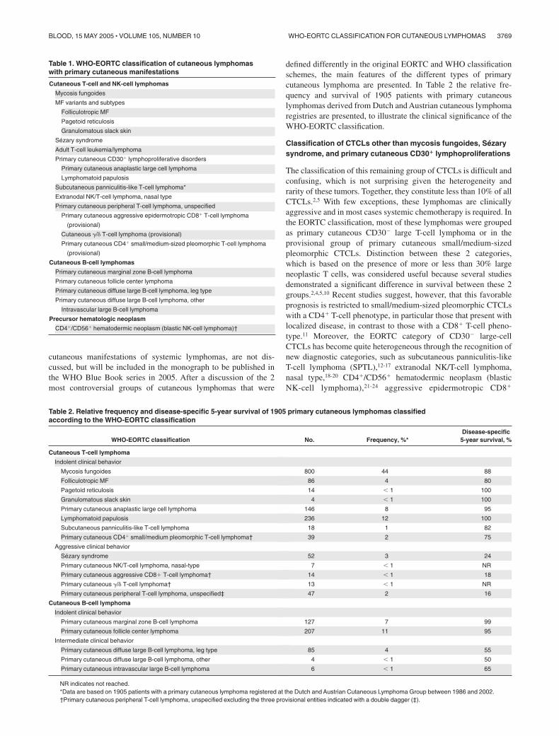
Figure 3. Primary cutaneous CD30� lymphoproliferative disease (pcCD30�
LPD). (A) Diffuse dermal infiltrate of large atypical cells admixed with smalllymphocytes. (H&E staining; original magnification, � 300). (B) The large atypicalcells are strongly positive for CD30. (C-D) The histologic picture in panels A and B canbe found both in C-ALC and in LyP. The final diagnosis depends on the clinicalpresentation. In combination with the solitary tumor of the patient shown in panel Cthe definite diagnosis will be C-ALC; in combination with recurrent, self-healingpapulonecrotic skin lesions (D), the final diagnosis is LyP. Image acquisition forpanels A and B was performed as described for Figure 1B. An HC Plan APO 20�/0.70objective was used.
3774 WILLEMZE et al BLOOD, 15 MAY 2005 � VOLUME 105, NUMBER 10

presence of primarily subcutaneous infiltrates of small, medium-sized, or large pleomorphic T cells and many macrophages,predominantly affecting the legs, and often complicated by ahemophagocytic syndrome.12 Recent studies suggest that at least 2groups of SPTL with a different histology, phenotype, and progno-sis can be distinguished. Cases with an �/�� T-cell phenotype areusually CD8�, are restricted to the subcutaneous tissue (no dermaland/or epidermal involvement), and often run an indolent clinicalcourse.14,16,17,32 In contrast, SPTL with a �/� T-cell phenotype,approximately 25% of all cases, are typically CD4�, CD8�, andoften coexpress CD56, the neoplastic infiltrates are not confined tothe subcutaneous tissue, but may involve the epidermis and/ordermis as well, and invariably have a very poor progno-sis.13,14,16,17,116 In the WHO-EORTC classification the term “SPTL”is only used for cases with an �/�� T-cell phenotype, whereas caseswith a �/�� T-cell phenotype are included in the category ofcutaneous �/� T-cell lymphomas.32
Clinical features. SPTL occurs in adults as well as in youngchildren, and both sexes are equally affected. Patients generallypresent with solitary or multiple nodules and plaques, whichmainly involve the legs, or may be more generalized (Figure 4A).Ulceration is uncommon. Systemic symptoms such as fever,fatigue, and weight loss may be present. The disease may becomplicated by a hemophagocytic syndrome, which is generallyassociated with a rapidly progressive course.117 However, a hemo-phagocytic syndrome is probably less common than in cutaneous�/� T-cell lymphomas with panniculitis-like lesions. Disseminationto extracutaneous sites is rare. SPTL may be preceded for years ordecades by an seemingly benign panniculitis.15,17,117
Histopathology. Histopathology reveals subcutaneous infil-trates simulating a panniculitis showing small, medium-sized, orsometimes large pleomorphic T cells with hyperchromatic nucleiand often many macrophages. The overlying epidermis and dermisare typically uninvolved.16,116 Rimming of individual fat cells byneoplastic T cells is a helpful, though not completely specific,diagnostic feature (Figure 4B,C).32 Necrosis, karyorrhexis, andcytophagocytosis are common findings. In the early stages theneoplastic infiltrates may lack significant atypia and a heavyinflammatory infiltrate may predominate.15,17,117
Immunophenotype. These lymphomas show a �/��, CD3�,CD4�, CD8� T-cell phenotype, with expression of cytotoxic
proteins (Figure 4D).14,16,17,32 CD30 and CD56 are rarely, ifever, expressed.
Genetic features. The neoplastic T cells show clonal T-cellreceptor (TCR) gene rearrangements. Specific genetic abnormali-ties have not been identified. Epstein-Barr virus (EBV) is absent.
Prognosis and predictive factors. In contrast to prior reportsindicating that patients with a SPTL have a rapidly fatal course,recent studies suggest that many patients with a SPTL (with aCD8�, �/�� T-cell phenotype) have a protracted clinical coursewith recurrent subcutaneous lesions but without extracutaneousdissemination or the development of a hemophagocytic syn-drome.17,32 Based on the few published reports in which appropri-ate phenotyping was performed, the 5-year survival of suchpatients may be more than 80%,32 which is consistent with the datapresented in Table 2.
Treatment. Patients have generally been treated with doxorubi-cin-based chemotherapy and radiotherapy.12-17 However, recentstudies suggest that many patients can be controlled for longperiods of time with systemic corticosteroids.17,32
Extranodal NK/T-cell lymphoma, nasal type
Definition. Extranodal NK/T-cell lymphoma, nasal type, is anearly always EBV� lymphoma of small, medium, or large cellsusually with an NK-cell, or more rarely a cytotoxic T-cell,phenotype. The skin is the second most common site of involve-ment after the nasal cavity/nasopharynx, and skin involvement maybe a primary or secondary manifestation of the disease. Since bothgroups show an aggressive clinical behavior and require the sametype of treatment, distinction between “primary” and “secondary”cutaneous involvement seems not useful for this category.16,19,32,118,119
Therefore, the WHO classification–derived term “extranodal NK/T-cell lymphoma, nasal type,” rather than “(primary) cutaneousNK/T-cell lymphoma, nasal type,” is preferred.
Clinical features. Patients are adults with a predominance ofmales. This lymphoma is more common in Asia, Central America,and South America. Patients generally present with multipleplaques or tumors preferentially on the trunk and extremities, or inthe case of nasal NK/T-cell lymphoma with a midfacial destructivetumor, previously also designated “lethal midline granu-loma.”18,19,32,119-122 Ulceration is common. Systemic symptomssuch as fever, malaise, and weight loss may be present, and somecases are accompanied by a hemophagocytic syndrome. Thedisease is closely related to aggressive NK-cell leukemia, whichalso may have cutaneous manifestations, and is also EBV associated.
Histopathology. These lymphomas show dense infiltrates in-volving the dermis and often the subcutis. Epidermotropism maybe present. Prominent angiocentricity and angiodestruction areoften accompanied by extensive necrosis.18,19 NK/T-cell lymphomahas a broad cytologic spectrum ranging from small to large cells,with most cases consisting of medium-sized cells. The cells mayhave irregular or oval nuclei, moderately dense chromatin, andpale cytoplasm. In some cases a heavy inflammatory infiltrate ofsmall lymphocytes, histiocytes, plasma cells, and eosinophilscan be seen.
Immunophenotype. The neoplastic cells express CD2, CD56,cytoplasmic CD3, and cytotoxic proteins (TIA-1, granzyme B,perforin), but lack surface CD3.122 In rare CD56� cases detectionof EBV by in situ hybridization and expression of cytotoxicproteins are required for diagnosis (Figure 5B).3 Latent membraneprotein-1 (LMP-1) is inconsistently expressed.
Genetic features. The T-cell receptor is usually in germlineconfiguration, but can be rearranged in rare tumors with a cytotoxic
Figure 4. Subcutaneous panniculitis-like T-cell lymphoma. (A) Deeply seatednodular skin lesions and residual lipodystrophy after disappearance of the skinlesions. (B) Infiltrates are almost exclusively localized in subcutaneous tissueresembling a lobular panniculitis (H&E staining; original magnification, � 25). (C)Rimming of individual fat cells by neoplastic T cells (H&E staining; original magnifica-tion, � 480). (D) Tumor cells show positive staining for CD8. Image acquisition forpanels B-D was performed as described for Figure 1B. An HC FLUOTAR 2.5�/0.07objective was used for panel B; an HC Plan APO 40�/0.85 objective was used forpanels C and D.
WHO-EORTC CLASSIFICATION FOR CUTANEOUS LYMPHOMAS 3775BLOOD, 15 MAY 2005 � VOLUME 105, NUMBER 10

T-cell phenotype. EBV is expressed almost in all cases, suggestinga pathogenetic role of this virus.18
Prognosis and predictive factors. Nasal-type NK/T-cell lym-phoma presenting in the skin is a highly aggressive tumor with amedian survival of less than 12 months.18,19,120,121 The mostimportant factor predicting poor outcome is the presence ofextracutaneous involvement at presentation. In patients presentingwith only skin lesions, a median survival of 27 months wasreported, compared with 5 months for patients presenting withcutaneous and extracutaneous disease.121 CD30�, CD56� casesreported to have a better prognosis may have been examples ofC-ALCL with coexpression of CD56.20
Therapy. Systemic chemotherapy is the first choice of treat-ment, but the results are disappointing.120,121
Variant. Hydroa vacciniforme-like CTCL is a rare type ofEBV-associated lymphoma of CD8� cytotoxic T cells, which affectschildren almost exclusively in Latin America and Asia.123-125 Patientspresent with a papulovesicular eruption clinically resembling hydroavacciniforme, particularly on the face and upper extremities (sun-exposed areas). The prognosis is poor.
Primary cutaneous peripheral T-cell lymphoma, unspecified
PTL, unspecified, in the WHO classification represent a heteroge-neous group which includes all T-cell neoplasms that do not fit intoany of the better defined subtypes of T-cell lymphoma/leukemia.Recent studies have suggested that primary cutaneous aggressiveepidermotropic CD8� cytotoxic T-cell lymphoma, cutaneousgamma-delta T-cell lymphoma, and primary cutaneous small-medium CD4� T-cell lymphoma can be separated out as provi-sional entities. For the remaining diseases that do not fit into eitherof these provisional entities the designation PTL, unspecified, ismaintained. In all cases a diagnosis of MF must be ruled out bycomplete clinical examination and an accurate clinical history.
Primary cutaneous aggressive epidermotropic CD8� cytotoxicT-cell lymphoma (provisional entity). Definition. Primary cuta-neous aggressive epidermotropic CD8� CTCL is characterized by aproliferation of epidermotropic CD8� cytotoxic T-cells and anaggressive clinical behavior.25,26 Differentiation from other types ofCTCL expressing a CD8� cytotoxic T-cell phenotype, as observedin more than 50% of patients with pagetoid reticulosis, and in rare
cases of MF, LyP, and C-ALCL, is based on the clinical presenta-tion and clinical behavior.25 In these latter conditions, no differencein clinical presentation or prognosis between CD4� and CD8�
cases is found.Clinical features. Clinically, these lymphomas are character-
ized by the presence of localized or disseminated eruptive papules,nodules, and tumors showing central ulceration and necrosis or bysuperficial, hyperkeratotic patches and plaques (Figure 5A).16,25
The clinical features are very similar to those observed in patientswith a cutaneous �/� T-cell lymphoma and cases described asgeneralized pagetoid reticulosis (Ketron-Goodman type) in thepast.32 These lymphomas may disseminate to other visceral sites(lung, testis, central nervous system, oral mucosa), but lymphnodes are often spared.25
Histopathology. Histologically, these lymphomas show an ac-anthotic or atrophic epidermis, necrotic keratinocytes, ulceration,and variable spongiosis, sometimes with blister formation.16,25
Epidermotropism is often pronounced ranging from a lineardistribution to a pagetoid pattern throughout the epidermis (Figure5B). Invasion and destruction of adnexal skin structures arecommonly seen. Angiocentricity and angioinvasion may be present.Tumor cells are small-medium or medium-large with pleomorphicor blastic nuclei.
Immunophenotype. The tumor cells have a betaF1�, CD3�,CD8�, granzyme B�, perforin�, TIA-1�, CD45RA�, CD45RO�,CD2�, CD4�, CD5�, CD7�/� phenotype (Figure 5C and5D).11,16,25,26,32 EBV is generally negative.
Genetic features. The neoplastic T cells show clonal TCRgene rearrangements. Specific genetic abnormalities have notbeen described.
Prognosis and predictive factors. These lymphomas often havean aggressive clinical course with a median survival of 32months.25 There is no difference in survival between cases with asmall- or large-cell morphology.11
Therapy. Patients are generally treated with doxorubicin-basedmultiagent chemotherapy.
Cutaneous gamma/delta T-cell lymphoma (provisional entity).Definition. Cutaneous gamma/delta T-cell lymphoma (CGD-TCL)is a lymphoma composed of a clonal proliferation of mature,activated gamma/delta T cells with a cytotoxic phenotype. Thisgroup includes cases previously known as SPTL with a gamma/delta phenotype. A similar and possibly related condition maypresent primarily in mucosal sites.28 Whether cutaneous andmucosal gamma/delta TCL are all part of a single disease (ie,muco-cutaneous gamma/delta TCL) is not yet clear.29,122 Distinc-tion between “primary” and “secondary” cutaneous cases is notuseful in this group, since both groups have a very grim prognosis.
Clinical features. Patients with CGD-TCL generally presentwith disseminated plaques and/or ulceronecrotic nodules or tu-mors, particularly on the extremities, but other sites may beaffected as well.27-31 Involvement of mucosal and other extranodalsites is frequently observed,28 but involvement of lymph nodes,spleen, or bone marrow is uncommon. A hemophagocytic syn-drome may occur in patients with panniculitis-like tumors.12,31
Histopathology. Three major histologic patterns of involve-ment can be present in the skin: epidermotropic, dermal, andsubcutaneous. Often more than one histologic pattern is present inthe same patient in different biopsy specimens or within a singlebiopsy specimen.27,31 The neoplastic cells are generally medium tolarge in size with coarsely clumped chromatin. Large blastic cellswith vesicular nuclei and prominent nucleoli are infrequent.Apoptosis and necrosis are common, often with angioinvasion. The
Figure 5. Primary cutaneous aggressive epidermotropic CD8� cytotoxic T-celllymphoma. (A) Typical presentation with nodules and tumors showing centralulceration. (B-D) Tumor cells show striking epidermotropism (H&E staining; originalmagnification, � 150), and strongly express CD8 (C) and TIA-1 (D). Image acquisi-tion for panels B-D was performed as described for Figure 1B. An HC Plan APO10�/0.40 objective lens was used.
3776 WILLEMZE et al BLOOD, 15 MAY 2005 � VOLUME 105, NUMBER 10

subcutaneous cases may show rimming of fat cells, similar to SPTLof alpha/beta origin.
Immunophenotype. The tumor cells characteristically have abetaF1�, CD3�, CD2�, CD5�, CD7�/�, CD56� phenotype withstrong expression of cytotoxic proteins. Most cases lack both CD4and CD8, though CD8 may be expressed in some cases.29,31 Infrozen sections the cells are strongly positive for TCR-delta. If onlyparaffin sections are available, the absence of betaF1 may be usedto infer a gamma/delta origin under appropriate circumstances.14,30
Genetic features. The cells show clonal rearrangement of theTCR gamma gene. TCR beta may be rearranged or deleted, but isnot expressed. EBV is generally negative.28,31
Prognosis and predictive factors. Most patients have aggres-sive disease resistant to multiagent chemotherapy and/or radia-tion.27-32 In a recent series of 33 patients, a median survival of 15months was noted.31 This study showed a trend for decreasedsurvival for patients who had subcutaneous fat involvementcompared with patients who had epidermal or dermal disease only.
Therapy. Patients should be treated with systemic chemo-therapy, but the results are often disappointing.
Primary cutaneous CD4� small/medium-sized pleomorphicT-cell lymphoma (provisional entity). Definition. Primary cuta-neous CD4� small/medium-sized pleomorphic T-cell lymphoma isa CTCL defined by a predominance of small- to medium-sizedCD4� pleomorphic T cells without (a history of) patches andplaques typical of MF, and in most cases, a favorable clinicalcourse.2 In contrast to the EORTC classification, in the WHO-EORTC classification the term “small/medium-sized pleomorphicCTCL” is restricted to cases with a CD4� T-cell phenotype. Caseswith a CD3�, CD4�, CD8� phenotype usually have a moreaggressive clinical course are included in the group of aggressiveepidermotropic CD8� CTCLs.11
Clinical features. Characteristically, these lymphomas presentwith a solitary plaque or tumor, generally on the face, the neck, orthe upper trunk (Figure 6A). Less commonly, they present with oneor several papules, nodules, or tumors.10,11,126-128
Histologic features. These lymphomas show dense, diffuse, ornodular infiltrates within the dermis with a tendency to infiltrate thesubcutis. Epidermotropism may be present focally. There is apredominance of small/medium-sized pleomorphic T cells (Figure6B). A small proportion (less than 30%) of large pleomorphic cellsmay be present.10 In some cases a considerable admixture withsmall reactive lymphocytes and histiocytes may be observed.
Immunophenotype. By definition these lymphomas have aCD3�, CD4�, CD8�, CD30� phenotype, sometimes with loss ofpan–T-cell markers. Cytotoxic proteins are generally not expressed.11
Genetic features. The TCR genes are clonally rearranged.126,128
No consistent cytogenetic abnormalities have yet been identified.Demonstration of an aberrant T-cell phenotype and clonality areuseful criteria to differentiate these small/medium-sized pleomor-
phic CTCLs from pseudo–T-cell lymphomas, which may alsopresent with a solitary plaque or nodule.129
Prognosis and predictive factors. These lymphomas have arather favorable prognosis, with an estimated 5-year survival ofapproximately 60% to 80% (Table 2).2,4,5,10,11,126-128 Particularly,cases presenting with a solitary or localized skin lesions seem tohave an excellent prognosis.11
Therapy. In patients with solitary localized skin lesions, surgi-cal excision or radiotherapy is the preferred mode of treatment.Cyclophosphamide as single-agent therapy and interferon alphahave been reported effective in patients with more generalized skindisease.128 However, the optimal treatment for this group has still tobe defined.
Primary cutaneous peripheral T-cell lymphoma, unspecified.Definition. The designation PTL, unspecified, is maintained forcutaneous T-cell lymphomas that do not fit into any of the betterdefined subtypes of CTCL. Hence, other categories of T-celllymphoma must be excluded. These include the 3 provisionalentities described earlier.
Clinical features. Patients are commonly adults, who presentwith solitary, localized, or more frequently generalized nodules ortumors.2,4,10,11 No sites of predilection have been recorded.
Histopathology. Skin lesions show nodular or diffuse infiltrateswith variable numbers of medium- to large-sized pleomorphic orimmunoblast-like T cells. Epidermotropism is generally mild orabsent. Large neoplastic cells represent at least 30% of the totaltumor cell population.10
Immunophenotype. Most cases show an aberrant CD4� T-cellphenotype with variable loss of pan–T-cell antigens. CD30 stainingis negative or restricted to a few scattered tumor cells. Rare casesmay show coexpression of CD56. Expression of cytotoxic proteinsis uncommon.11
Prognosis and predictive factors. The prognosis is generallypoor, with 5-year survival rates of less than 20% (Table 2).2,4,5,10,11
No statistical differences in survival were found between casespresenting with solitary/localized lesions and cases presenting withgeneralized skin lesions.11
Treatment. Patients should be treated with multiagentchemotherapy.
Precursor hematologic neoplasm
CD4�/CD56� hematodermic neoplasm (blasticNK-cell lymphoma)
Definition. In the WHO classification, blastic NK-cell lymphomawas included as a clinically aggressive neoplasm with a highincidence of cutaneous involvement and risk of leukemic dissemi-nation. The blastic cytologic appearance and CD56 expressioninitially suggested an NK-precursor origin.3 More recent studiessuggest derivation from a plasmacytoid dendritic cell precursor.23,24
“CD4�/CD56� hematodermic neoplasm”22 and “early plasmacy-toid dendritic cell leukemia/lymphoma”23 have been suggested asmore appropriate terms for this condition.
Clinical features. CD4�/CD56� hematodermic neoplasm com-monly presents in the skin with solitary or multiple nodules ortumors with or without concurrent extracutaneous localizations(Figure 7A).21-24,32 About half of the patients have nodal or bonemarrow involvement at presentation.23,121 Most patients presentingwith only skin lesions rapidly develop involvement of bonemarrow, peripheral blood, lymph nodes, and extranodal sites.23,121
CD4�/CD56� hematodermic neoplasm should be differentiated
Figure 6. Primary cutaneous CD4� small/medium-sized pleomorphic T-celllymphoma. (A) Presentation with a solitary tumor on the scalp. (B) Diffuse dermalinfiltrate mainly composed of small pleomorphic T cells with few scattered blasts cells(H&E staining; original magnification, � 750). Image acquisition was performed asdescribed for Figure 1B. An HC FLUOTAR 63�/0.90 objective lens was used.
WHO-EORTC CLASSIFICATION FOR CUTANEOUS LYMPHOMAS 3777BLOOD, 15 MAY 2005 � VOLUME 105, NUMBER 10

above all from myelomonocytic leukemia cutis, and are conceptu-ally similar to so-called “aleukemic leukemia cutis.”32
Histopathology. These lymphomas show nonepidermotropic,monotonous infiltrates of medium-sized cells with finely dispersedchromatin, and absent or indistinct nucleoli resembling lympho-blasts or myeloblasts (Figure 7B).3,21-24 The cells have sparsecytoplasm. Mitotic figures are frequent. Inflammatory cells areabsent. There is generally no necrosis or angioinvasion.
Immunophenotype. The tumor cells usually have a CD4�,CD56�, CD8�, CD7�/�, CD2�/�, CD45RA� phenotype, but do notexpress surface and cytoplasmic CD3 or cytotoxic proteins (Figure7C).3,23 TdT and CD68 may be positive. Since lymphoblastic andmyeloblastic neoplasms can also be positive for CD56, stains forCD3 and myeloperoxidase should always be performed in order toexclude these entities.3 The cells express CD123 and TCL1, both ofwhich support a relationship to plasmacytoid dendritic cells.23,24,130
Genetic features. T-cell receptor genes are in germline configu-ration. Tumor cells are negative for EBV.
Prognosis and predictive factors. CD4�/CD56� hematoder-mic neoplasm is an aggressive disease with a poor prognosis(median survival, 14 months).21-24,121 Systemic chemotherapy usu-ally results in a complete remission, but quick relapses unrespon-sive to further chemotherapy are the rule. No significant differencein survival is found between patients presenting with skin lesionswith or without concurrent extracutaneous disease.121
Recent studies suggest that patients can best be treated withregimens used in acute leukemias.121
Cutaneous B-cell lymphomas
Primary cutaneous marginal-zone B-cell lymphoma
Definition. Primary cutaneous marginal zone B-cell lymphoma(PCMZL) is an indolent lymphoma composed of small B cells,including marginal zone (centrocyte-like) cells, lymphoplasmacy-toid cells, and plasma cells. It includes cases previously designatedas primary cutaneous immunocytoma,131 and cases of cutaneousfollicular lymphoid hyperplasia with monotypic plasma cells.132
Exceptional cases of primary cutaneous plasmacytoma withoutunderlying multiple myeloma (extramedullary plasmacytoma ofthe skin) show considerable overlap with PCMZL and are thereforeincluded in this category.133 PCMZL is considered part of the broadgroup of extranodal marginal zone B-cell lymphomas commonlyinvolving mucosal sites, called MALT (mucosa-associated lym-phoid tissue) lymphomas.
Clinical features. In most instances patients with PCMZLpresent with red to violaceous papules, plaques, or nodules
localized preferentially on the trunk or extremities, especially thearms. In contrast to PCFCL, presentation with multifocal skinlesions is frequent (Figure 8). Ulceration is uncommon. PCMZLshave a tendency to recur in the skin, but dissemination toextracutaneous sites is exceedingly rare.131,134-136 In some casesspontaneous resolution of the skin lesions may be observed. Thedevelopment of anetoderma in spontaneously resolving lesions hasbeen observed.137 An association with Borrelia burgdorferi infec-tion has been reported in a significant minority of European casesof PCMZL, but not in Asian cases or cases from the UnitedStates.134,138-140 Associated autoimmune diseases are uncommon inPCMZL, but rather suggest secondary cutaneous involvement of asystemic lymphoma.131
Histopathology. These lymphomas show nodular to diffuseinfiltrates with sparing of the epidermis. The infiltrates are com-posed of small lymphocytes, marginal zone B cells (centrocyte-likecells), lymphoplasmacytoid cells, and plasma cells, admixed withsmall numbers of centroblast- or immunoblast-like cells and manyreactive T cells. Reactive germinal centers are frequently observed.They may be surrounded by a population of small- to medium-sized cells with irregular nuclei, inconspicuous nucleoli, andabundant pale cytoplasm (marginal zone B cells). Monotypicplasma cells are often located at the periphery of the infiltrates andin the superficial dermis beneath the epidermis.131,132,135,136 periodicacid-schiff (PAS)� intranuclear or intracytoplasmic inclusions maybe present in cases with a predominance of lymphoplasmacytoidcells. PCMZLs rarely show transformation into a diffuse largeB-cell lymphoma, but a relative increase in large transformedcells can be seen in some cases.
Immunophenotype. The marginal zone B cells express CD20,CD79a, and bcl-2, but are negative for CD5, CD10, and bcl-6,which may be useful in distinction from PCFCL.141,142 Reactivegerminal centers are typically bcl-6�, CD10�, and bcl-2�. Plasmacells express CD138 and CD79a, but generally not CD20, andshow monotypic cytoplasmic immunoglobulin light chain expres-sion on paraffin sections.
Genetic features. Immunoglobulin heavy chain (IgH) genesare clonally rearranged. Recent studies suggest the presence of thet(14;18)(q32;q21) involving the IGH gene on chromosome 14 andthe MLT gene on chromosome 18, and t(3;14)(p14.1;q32) involv-ing IGH and FOXP1 genes, in a proportion of PCMZLs.143,144
However, other translocations observed in gastric MALT lympho-mas, such as t(11;18)(q21;q21) and t(1;14)(p22;q32), have not beenfound in PCMZL.45,145
Prognosis and predictive factors. The prognosis of PCMZL isexcellent with a 5-year survival close to 100% (Table 2).131-136
Therapy. Patients with a solitary or a few lesions can be treatedwith radiotherapy or surgical excision. In patients with associated Bburgdorferi infection, systemic antibiotics should be tried first.146
For patients presenting with multifocal skin lesions, chlorambucilor intralesional or subcutaneous administration of interferon alpha
Figure 8. Primary cutaneous marginal zone lymphoma. Characteristic clinicalpresentation with multiple nodules and small tumors on the back and arms.
Figure 7. CD4�/CD56� hematodermic neoplasm (blastic NK-cell lymphoma). (A)Presentation with large tumor on the back. (B) Monotonous infiltration of medium-sized tumor cells (H&E staining; original magnification, � 480). (C) The tumor cellsstrongly express CD56. Image acquisition for panels B and C was performed asdescribed for Figure 1B. An HC Plan APO 40�/0.85 objective lens was used.
3778 WILLEMZE et al BLOOD, 15 MAY 2005 � VOLUME 105, NUMBER 10
may produce complete responses in approximately 50% of pa-tients.146 Very good results have also been obtained with the use ofsystemic or intralesional anti-CD20 antibody (rituximab).147 Inpatients showing frequent skin relapses, topical or intralesionalsteroids may be considered; alternatively, an expectant strategy canbe followed, similar to that used in other indolent B-cell lympho-mas and leukemias.
Primary cutaneous follicle-center lymphoma
Definition. Primary cutaneous follicle center lymphoma (PCFCL)is defined as a tumor of neoplastic follicle center cells, usually amixture of centrocytes (small and large cleaved follicle centercells) and variable numbers of centroblasts (large noncleavedfollicle center cells with prominent nucleoli), with a follicular, afollicular and diffuse, or a diffuse growth pattern, which generallypresent on the head or trunk. Lymphomas with a diffuse growthpattern and a monotonous proliferation of centroblasts and immu-noblasts are, irrespective of site, excluded and are classified asPCLBCL (Table 3).
Clinical features. PCFCL has a characteristic clinical presenta-tion with solitary or grouped plaques and tumors, preferentiallylocated on the scalp or forehead or on the trunk, and rarely on thelegs (Figure 9A-B).33-35 Particularly on the trunk, these tumors maybe surrounded by erythematous papules and slightly induratedplaques, which may precede the development of tumorous lesionsfor months or even many years. In the past, PCFCLs with such atypical presentation on the back were referred to as “reticulohistio-cytoma of the dorsum” or “Crosti lymphoma (Figure 9A).”34,148
Presentation with multifocal skin lesions is observed in a smallminority of patients, but is not associated with a more unfavorableprognosis.42,149 If left untreated, the skin lesions graduallyincrease in size over years, but dissemination to extracutaneoussites is uncommon.
Histopathology. PCFCLs show nodular to diffuse infiltrateswith almost constant sparing of the epidermis. The histologicpicture is variable, which relates primarily to the age and thegrowth rate of the biopsied skin lesion as well as the location.35,39 Aclear-cut follicular growth pattern is more commonly observed inlesions arising on the scalp than those presenting on the trunk.150
Small and early lesions contain a mixture of centrocytes, relativelyfew centroblasts, and many reactive T cells. Large centrocytes,often multilobated, are a common feature of PCFCL (Figure 9C).The large neoplastic B cells may have a fibroblast-like appearance.In small and/or early lesions a clear-cut follicular growth pattern or,more often, remnants of a follicular growth pattern may beobserved. If present, the abnormal follicles are composed ofmalignant bcl-6� follicle center cells enmeshed in a network ofCD21� or CD35� follicular dendritic cells. The follicles areill-defined, lack tingible body macrophages, and generally have areduced or absent mantle zone.150,151 With progression to tumorouslesions the neoplastic B cells increase both in number and size,whereas the number of reactive T cells steadily decreases.35,39
Follicular structures, if present before, are no longer visible exceptfor occasional scattered CD21� or CD35� follicular dendritic cells.Tumorous skin lesions generally show a monotonous population oflarge follicle center cells, generally large centrocytes and multilo-bated cells, and in rare cases spindle-shaped cells, with a variableadmixture of centroblasts and immunoblasts.34,35,39,42,152 Usually, aprominent stromal component is present.
Immunophenotype. The neoplastic cells express the B-cell–associated antigens CD20 and CD79a, and may show monotypicstaining for surface immunoglobulins (sIgs). However, absence ofdetectable sIg is common in tumorous lesions showing a diffusepopulation of large follicle center cells. PCFCLs consistentlyexpress bcl-6.141,142,151 CD10 expression is particularly observed incases with a follicular growth pattern, but is uncommon in PCFCLswith a diffuse growth pattern.142,153 Staining for CD5 and CD43 isnegative. Unlike nodal and secondary cutaneous follicular lympho-mas, PCFCLs do not express bcl-2 protein or show faint bcl-2staining in a minority of neoplastic B-cells.36,41,150 (Figure 9D,E).Staining for MUM-1/IRF4 is negative.46
Genetic features. Clonally rearranged immunoglobulin genesare present. Somatic hypermutation of variable heavy and lightchain genes has been demonstrated, which further supports thefollicle center cell origin of these lymphomas.154,155 In most studiesPCFCLs, including cases with a follicular growth pattern, do not
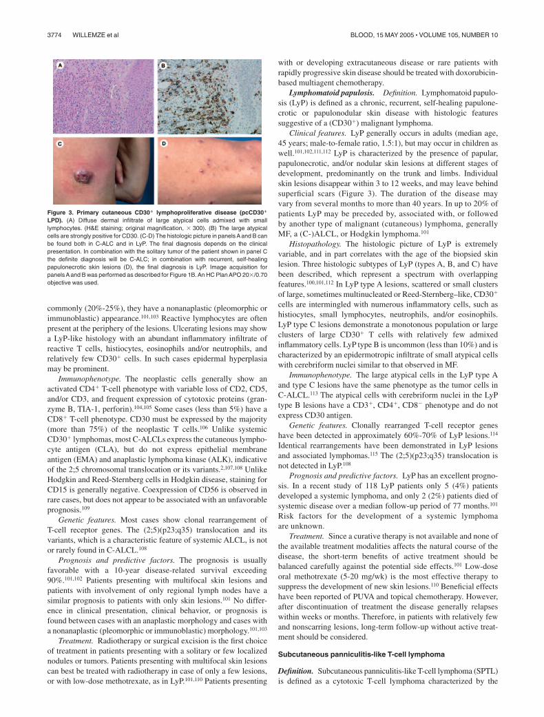
Figure 9. Primary cutaneous follicle center lymphoma. (A) Typical presentationwith tumors on the chest surrounded by less infiltrated erythematous skin lesions. (B)Presentation with multiple tumors confined to the scalp. (C) Diffuse dermal infiltratemainly consisting of large centrocytes and multilobated cells (H&E staining; originalmagnification, � 480). (D-E) Serial sections stained for CD20 (D) and bcl-2 (E). Bcl-2is expressed by perivascular T cells, but not by the neoplastic B cells. Imageacquisition for panels A and C-E was performed as described for Figure 1B. An HCPlan APO objective was used (40�/0.85 for panel C; 10�/0.40 for panels D and E).
Table 3. Characteristic features of PCFCL and PCLBCL, leg type
PCFCL PCLBCL, leg type
Morphology Predominance of centrocytes
that are often large,
especially in diffuse
lesions.
Predominance or confluent sheets
of medium-sized to large B cells
with round nuclei, prominent
nucleoli, and coarse chromatin
resembling centroblasts and/or
immunoblasts.
Centroblasts may be present,
but not in confluent sheets.
Diffuse growth pattern.
Growth pattern may be
follicular, follicular and
diffuse, or diffuse (a
continuum without distinct
categories or grades).
Phenotype Bcl-2: �/�* Bcl-2: ��‡
Bcl-6: � Bcl-6: �/�
CD10: �/�† CD10: �
Mum-1: � Mum-1: �
Clinical
features
Middle-aged adults. Elderly, especially females,
Localized lesions on head or
trunk (90%).
Lesions localized on leg(s), most
often below the knee.
Multifocal lesions in rare
cases.
Rare cases with lesions at other
sites than the leg (10%).
*Faint staining, when present; minority of cells (see comments in paragraph onPCFCL)
†Diffuse lesions, mostly CD10�.‡Strong staining; in most neoplastic cells.
WHO-EORTC CLASSIFICATION FOR CUTANEOUS LYMPHOMAS 3779BLOOD, 15 MAY 2005 � VOLUME 105, NUMBER 10

show the t(14;18), which is characteristically found in systemicfollicular lymphomas and a proportion of systemic diffuse largeB-cell lymphoma.37,41,151,153 (see “Comment” under “Primary cuta-neous follicle-center lymphoma”).
Inactivation of p15 and p16 tumor suppressor genes by promo-tor hypermethylation has been reported in about 10% and 30% ofPCFCLs, respectively.156 Chromosomal imbalances have beenidentified by comparative genomic hybridization (CGH) analysis ina minority of PCFCLs, but a consistent pattern has not yetemerged.44,157 In a recent study using interphase fluorescence insitu hybridization, no evidence for translocations involving IgH,myc, or bcl-6 loci were found.45 PCFCLs have the gene expressionprofile of germinal center–like large B-cell lymphomas.46
Prognosis and predictive factors. Regardless of the growthpattern (follicular or diffuse), the number of blast cells, or thepresence of either localized or multifocal skin disease, thesePCFCLs have an excellent prognosis with a 5-year survival of morethan 95% (Table 2).2,4,5,33-35,38,42,150,151 A recent study suggests thatstrong expression of bcl-2 in PCFCLs with a diffuse large-cellhistology is associated with a more unfavorable prognosis.158
Therapy. In patients with localized or few scattered skinlesions, radiotherapy is the preferred mode of treatment, even incases with a predominance of large “cleaved” cells.38,42,149,159-161
Cutaneous relapses, observed in approximately 20% of patients, donot indicate progressive disease and can be treated with radio-therapy as well. Anthracycline-based chemotherapy is requiredonly in patients with very extensive cutaneous disease and patientsdeveloping extracutaneous disease.42,150 Recent studies reportbeneficial effects of systemic or intralesional administration ofanti-CD20 antibody (rituximab) therapy in small series of PCFCLs,but the long-term effects of this approach have yet to bedetermined.162-164
Comment. Characteristically, PCFCLs do not express bcl-2protein, or show faint bcl-2 staining in a minority of tumor cells,and do not show the t(14;18).36,37,41 However, recent studies reportthe presence of t(14;18) and/or bcl-2 expression in a significantminority of PCFCLs.43,153,165-167 Whether these discrepant resultsare the result of differences in patient selection (eg, incompletestaging) or different definitions for bcl-2 positivity, or representregional differences is as yet unknown. Importantly, in PCFCLswith a follicular growth pattern there are no differences in clinicalpresentation and behavior between bcl-2 and/or t(14;18)-positiveand -negative cases.36,152,165-167 In contrast, recent studies suggestthat expression of bcl-2 protein by more than 50% of the neoplasticB cells in PCFCLs with a diffuse proliferation of large centrocytes,observed in approximately 15% of cases, is associated with a moreunfavorable prognosis.158 Further studies are warranted to definethe clinical and biological significance of bcl-2 expression and/orthe presence of t(14;18) observed in some cases of PCFCL.Notwithstanding, demonstration of bcl-2 expression and/or t(14;18) should always raise suspicion of a systemic lymphomainvolving the skin secondarily.
Primary cutaneous diffuse large B-cell lymphoma, leg type
Definition. Primary cutaneous diffuse large B-cell lymphoma, legtype, is a PCLBCL with a predominance or confluent sheets ofcentroblasts and immunoblasts, characteristically presenting withskin lesions on the (lower) legs. Uncommonly, skin lesions with asimilar morphology and phenotype can arise at sites other thanthe legs.
Clinical features. PCLBCL, leg type, predominantly affectselderly patients, particularly females.40,42,43 Patients present with
generally rapidly growing red or bluish-red tumors on one or both(lower) legs (Figure 10A). In contrast to the group of PCFCLs,these lymphomas more often disseminate to extracutaneous sitesand have a more unfavorable prognosis.40,42 Reports on PCLBCL,leg type, arising at sites other than the leg are few. In a recentEuropean multicenter study, 16 of 17 patients presented withsolitary or localized skin lesions either on the trunk or head, and 7of 17 patients developed extracutaneous disease.42
Histopathology. These lymphomas show diffuse infiltrates,which often extend into the subcutaneous tissue. These infiltratesgenerally show a monotonous population or confluent sheets ofcentroblasts and immunoblasts (Figure 10B).40,42 Mitotic figuresare frequently observed. Small B cells are lacking and reactive Tcells are relatively few and often confined to perivascular areas. Aprominent stromal reaction as in PCFCLs is not observed.
Immunophenotype. The neoplastic B cells express monotypicsIg and/or cIg and B-cell–associated antigens CD20 and CD79a. Incontrast to the group of PCFCLs, PCLBCL, leg type, shows strongbcl-2 expression, also in cases not located on the legs (Figure10C).41,43,142,158 Bcl-6 is expressed by most cases, whereas CD10staining is generally absent.142 Unlike PCFCL, most PCLBCL, legtypes, express MUM-1/IRF4 protein (Figure 10D).46,168
Genetic features. The t(14;18) is not found in PCLBCLs,although strong bcl-2 expression is common in this group.41,43 Insome cases bcl-2 overexpression may result from chromosomalamplification of the bcl-2 gene.157 Inactivation of p15 and p16tumor suppressor genes by promotor hypermethylation has beendetected in 11% and 44% of PCLBCLs, respectively.156 Chromo-somal imbalances have been identified in up to 85% of PCLBCLs,with gains in 18q and 7p and loss of 6q as most commonfindings.44,157 Recent studies demonstrated translocations involvingmyc, bcl-6, and IgH genes in 11 of 14 PCLBCL-leg cases, but not inpatients with a PCFCL with a diffuse infiltration of large centro-cytes.45 Recent studies suggest that patients with PCLBCL-leghave an activated B-cell gene expression profile.46
Prognosis and predictive features. The 5-year survival of 78cases included in the Dutch and Austrian registries was 55% (Table2). PCLBCLs on the leg have an inferior prognosis compared toPCLBCLs presenting at other sites.42 The presence of multiple skinlesions at diagnosis is a significant adverse risk factor. In a recentstudy, patients presenting with a single skin tumor on one leg had adisease-related 5-year survival of 100%, whereas patients present-ing with multiple skin lesions on one or both legs had adisease-related 5-year survival of 45% and 36%, respectively.42
Figure 10. Primary cutaneous diffuse large B-cell lymphoma, leg type. (A)Clinical presentation with multiple tumors on right lower leg. (B) Monotonousproliferation of centroblasts and immunoblasts (H&E staining; original magnification,� 480). (C-D) Characteristically, the neoplastic B cells strongly express bcl-2 (C) andMum-1/IRF-4 (D). Image acquisition for panels B-D was performed as described forFigure 1B. An HC Plan APO objective was used (40�/0.85 for panel B; 20�/0.70 forpanels C and D).
3780 WILLEMZE et al BLOOD, 15 MAY 2005 � VOLUME 105, NUMBER 10

Therapy. These lymphomas should be treated as systemicdiffuse large B-cell lymphomas with anthracycline-based chemo-therapy.40,42 In patients presenting with a single small skin tumor,radiotherapy may sometimes be considered.42 Systemic administra-tion of anti-CD20 antibody (rituximab) has proved effective insome patients, but long-term follow-up data are not available andthe place of rituximab in the treatment of PCLBCL, either as singleagent therapy or in combination with systemic chemotherapyremains to be established.162,169
Primary cutaneous diffuse large B-cell lymphoma, other
The term “PCLBCL, other,” refers to rare cases of large B-celllymphomas arising in the skin, which do not belong to the group ofPCLBCL, leg type, or the group of PCFCLs. These cases includemorphologic variants of diffuse large B-cell lymphoma, such asanaplastic or plasmablastic subtypes or T-cell/histiocyte rich largeB-cell lymphomas. Such cases are generally a skin manifestation ofa systemic lymphoma. Plasmablastic lymphomas are seen almostexclusively in the setting of HIV infection or other immunedeficiencies.170-172 Some of these cases had only skin lesions atpresentation. Rare cases of primary cutaneous T-cell/histiocyte-richB-cell lymphoma, characterized by the presence of large scatteredB cells in a background of numerous reactive T cells, have beenreported.173,174 Clinically, they show similarities with the groups ofPCFCL and PCMZL. These lymphomas commonly present withskin lesions on the head, the trunk or the extremities, and may infact represent an exaggerated T-cell infiltrate in association withother forms of CBCL. Unlike their nodal counterparts, they appearto have an excellent prognosis.173,174 In addition, rare cases ofprimary cutaneous intravascular large B-cell lymphoma may beincluded in this category.
Intravascular large B-cell lymphoma. Intravascular large B-cell lymphoma is a well-defined subtype of large B-cell lymphoma,defined by an accumulation of large neoplastic B cells within bloodvessels. These lymphomas preferentially affect the central nervoussystem, lungs, and skin and are generally associated with a poorprognosis.175
Patients often have widely disseminated disease, but cases withonly skin involvement may occur. Clinically, intravascular largeB-cell lymphoma may present with violaceous patches and plaquesor teleangiectatic skin lesions usually on the (lower) legs or thetrunk.175,176 Patients presenting with only skin lesions appear tohave a significantly better survival than patients with other clinicalpresentations (3-year overall survival: 56% versus 22%).176 Interest-ingly, colonization of cutaneous cherry hemangiomas by neoplasticcells as the only presenting sign of intravascular large B-celllymphoma has been reported.177-178 Histologically, dilated bloodvessels in the dermis and subcutis are filled and often extended by aproliferation of large neoplastic B cells. These cells may causevascular occlusion of venules, capillaries, and arterioles. In somecases small numbers of tumor cells can also be observed around
blood vessels. Multiagent chemotherapy is the preferred mode oftreatment, also in patients presenting with skin-limited disease.176
Conclusions and future directions
The WHO-EORTC classification for cutaneous lymphomas pre-sented herein may be considered as an important step forward.First, the development of this consensus classification will put anend to the ongoing discussion whether the EORTC or the WHOscheme can best be used, and is expected to contribute to a moreuniform diagnosis and hence a more uniform treatment of patientswith a cutaneous lymphoma. Second, major progress has beenmade in a better definition of some controversial groups ofcutaneous lymphoma, in particular the group of PCFCL/PCLBCLand the group of CTCL other than MF, SS, and the group ofprimary cutaneous CD30� LPD.
The new definitions of the groups of PCFCL, PCLBCL, legtype, and PCLBCL, other, will allow a more reliable distinctionbetween indolent and more aggressive types of CBCL, andfacilitate the decision whether radiotherapy or systemic chemo-therapy should be selected as first choice of treatment. Largemulticenter studies are now required to validate the currentproposals, and in particular to investigate the diagnostic andprognostic value of bcl-2 and Mum-1/IRF4 protein expression.
The classification of CTCL other than MF, SS, and the group ofprimary cutaneous CD30� LPD is still difficult, as it requiresaccurate clinicopathologic correlation and a number of complemen-tary techniques to arrive at a definite diagnosis. SPTL (with analpha/beta phenotype), extranodal NK/T-cell lymphoma, nasaltype, and CD4�/CD56� hematodermic neoplasm are now fairlywell-defined. However, considerable overlap is noted betweencutaneous (and mucosal) GD-TCL and aggressive epidermotropicCD8� CTCL. The similarities in clinical presentation and patternof dissemination between these conditions may reflect similaritiesin homing profile and biologic function of normal gamma/delta-positive T-cells and activated CD8� cytotoxic T cells, respectively.Apart from the group of SPTLs and the group of CD4� small/medium-sized pleomorphic CTCLs, these rare types of CTCL havea very poor prognosis and are generally resistant to conventionalchemotherapy. More aggressive regimens, including allogeneicbone marrow transplantation, for patients with aggressive types ofCTCL including advanced stages of MF and SS are currently underinvestigation.179,180
Recently, studies have started to investigate gene and proteinexpression profiles in different types of cutaneous lymphoma. It isexpected that these studies will not only contribute to a betterunderstanding of the molecular pathways involved in the develop-ment and progression of these lymphomas, but will also providenew molecular targets for diagnosis and therapeutic intervention,and ultimately to more refined classification schemes.
References
1. Groves FD, Linet MS, Travis LB, Devesa SS.Cancer surveillance series: non-Hodgkin’s lym-phoma incidence by histologic subtype in theUnited States from 1978 through 1995. J NatlCancer Inst. 2000;92:1240-1251.
2. Willemze R, Kerl H, Sterry W, et al. EORTC clas-sification for primary cutaneous lymphomas: aproposal from the Cutaneous Lymphoma StudyGroup of the European Organization for Re-search and Treatment of Cancer (EORTC).Blood. 1997;90:354-371.
3. Jaffe ES, Harris NL, Stein H, Vardiman JW (Eds).World Health Organization Classification of Tu-mors: Pathology and Genetics of Tumours of He-matopoietic and Lymphoid Tissues. Lyon, France:IARC Press;2001.
4. Grange F, Hedelin G, Joly P, et al. Prognostic fac-tors in primary cutaneous lymphomas other thanmycosis fungoides and the Sezary syndrome: theFrench study group on cutaneous lymphomas.Blood. 1999;93:3637-3642.
5. Fink-Puches R, Zenahlik P, Back B, Smolle J, Kerl
H, Cerroni L. Primary cutaneous lymphomas: appli-cability of current classification schemes (EORTC,WHO) based on clinicopathologic features observedin a large group of patients. Blood. 2002;99:800-805.
6. Norton AJ. Classification of cutaneous lym-phoma: a critical appraisal of recent proposals.Am J Dermatopathol. 1999;21:279-287.
7. Sander CA, Flaig MJ, Jaffe ES. Cutaneous mani-festations of lymphoma: a clinical guide based onthe WHO classification. Clin Lymphoma. 2001;2:86-100.
WHO-EORTC CLASSIFICATION FOR CUTANEOUS LYMPHOMAS 3781BLOOD, 15 MAY 2005 � VOLUME 105, NUMBER 10

8. Russell-Jones R. World Health Organization ofhematopoietic and lymphoid tissues: implicationsfor dermatology. J Am Acad Dermatol. 2003;48:93-102.
9. Willemze R, Meijer CJLM. EORTC classificationfor primary cutaneous lymphomas: the best guideto good clinical management. European Organi-zation for Research and Treatment of Cancer.Am J Dermatopathol. 1999;21:265-73.
10. Beljaards RC, Meijer CJLM, van der Putte SCJ,et al. Primary cutaneous T-cell lymphomas: clini-copathologic features and prognostic parametersof 35 cases other than mycosis fungoides andCD30-positive large cell lymphoma. J Pathol.1994;172:53-60.
11. Bekkenk MW, Vermeer MH, Jansen PM, et al.Peripheral T-cell lymphomas unspecified present-ing in the skin: analysis of prognostic factors in agroup of 82 patients. Blood. 2003;102:2213-2219.
12. Gonzalez CL, Medeiros LJ, Braziiel RM, Jaffe ES.T-cell lymphoma involving subcutaneous tissue: aclinicopathologic entity commonly associated withhemophagocytic syndrome. Am J Surg Pathol.1991;15:17-27.
13. Burg G, Dummer R, Wilhelm M, et al. A subcuta-neous delta-positive T-cell lymphoma that pro-duces interferon gamma. N Engl J Med. 1991;325:1078-1081.
14. Salhany KE, Macon WR, Choi JK, et al. Subcuta-neous panniculitis-like T-cell lymphoma: clinico-pathologic, immunophenotypic, and genotypicanalysis of alpha/beta and gamma/delta sub-types. Am J Surg Pathol. 1998;22:881-893.
15. Weenig RH, Ng CS, Perniciaro C. Subcutaneouspanniculitis-like T-cell lymphoma. Am J Dermato-pathol. 2001;23:206-215.
16. Santucci M, Pimpinelli N, Massi D, et al. Cyto-toxic/natural killer cell cutaneous lymphomas:report of EORTC Cutaneous Lymphoma TaskForce Workshop. Cancer. 2003;97:610-627.
17. Hoque SR, Child FJ, Whittaker SJ, et al. Subcuta-neous panniculitis-like T-cell lymphoma: a clinico-pathological, immunophenotypic and molecularanalysis of six patients. Br J Dermatol. 2003;148:516-525.
18. Chan JK, Sin VC, Wong KF, et al. Nonnasal lym-phoma expressing the natural killer cell markerCD56: a clinicopathologic study of 49 cases of anuncommon aggressive neoplasm. Blood. 1997;89:4501-4513.
19. Natkunam Y, Smoller BR, Zehnder JL, DorfmanRF, Warnke RA. Aggressive cutaneous NK andNK-like T-cell lymphomas: clinicopathologic, im-munohistochemical, and molecular analyses of12 cases. Am J Surg Pathol. 1999;23:571-581.
20. Mraz-Gernhard S, Natkunam Y, Hoppe RT, LeBoitP, Kohler S, Kim Y. Natural killer/natural killer-likeT-cell lymphoma, CD56�, presenting in the skin:an increasingly recognized entity with an aggres-sive course J Clin Oncol. 2001;19:2179-2188.
21. DiGiuseppe JA, Louie DC, Williams JE, et al.Blastic natural killer cell leukemia/lymphoma: aclinicopathologic study. Am J Surg Pathol. 1997;21:1223-1230.
22. Petrella T, Dalac S, Maynadie M, et al. CD4�CD56� cutaneous neoplasms: a distinct hemato-logical entity? Am J Surg Pathol. 1999;23:137-146.
23. Jacob MC, Chaperot C, Mossuz P, et al. CD4�CD56� lineage negative malignancies: a newentity developed from malignant early plasmacy-toid dendritic cells. Haematologica. 2003;88:941-955.
24. Petrella T, Comeau MR, Maynadie M, et al.“Agranular CD4� CD56� hematodermic neo-plasm” (blastic NK-cell lymphoma) originatesfrom a population of CD56� precursor cells re-lated to plasmacytoid monocytes. Am J SurgPathol. 2002;26:852-862.
25. Berti E, Tomasini D, Vermeer MH, Meijer CJLM,
Alessi E, Willemze R. Primary cutaneous CD8-positive epidermotropic cytotoxic T-cell lym-phoma: a distinct clinicopathologic entity with anaggressive clinical behaviour. Am J Pathol. 1999;155:483-492.
26. Agnarsson BA, Vonderheid EC, Kadin ME. Cuta-neous T-cell lymphoma with suppressor/cytotoxic(CD8) phenotype: identification of rapidly pro-gressive and chronic subtypes. J Am Acad Der-matol. 1990;22:569-577.
27. Berti E, Cerri A, Cavicchini S, et al. Primary cuta-neous gamma/delta lymphoma presenting as dis-seminated pagetoid reticulosis. J Invest Derma-tol. 1991;96:718-723.
28. Arnulf B, Copie-Bergman C, Delfau-Larue MH, etal. Nonhepatosplenic gamma-delta T-cell lym-phoma: a subset of cytotoxic lymphomas withmucosal or skin localization. Blood. 1998;91:1723-1731.
29. de Wolf-Peeters C, Achten R. Gamma-delta T-celllymphomas: a homogeneous entity? Histopathol-ogy. 2000;36:294-305.
30. Jones D, Vega F, Sarris A, Medeiros LJ. CD4-CD8- “Double-negative” cutaneous T-cell lympho-mas share common histologic features and anaggressive clinical course. Am J Surg Pathol.2002;26:225-231.
31. Toro JR, Liewehr DJ, Pabby N, et al. Gamma-delta-T-cell phenotype is associated with signifi-cantly decreased survival in cutaneous T-cell lym-phoma. Blood. 2003;101:3407-3412.
32. Massone C, Chott A, Metze D, et al. Subcutane-ous, blastic natural killer (NK), NK/T-cell andother cytotoxic lymphomas of the skin: a morpho-logic, immunophenotypic and molecular study of50 patients. Am J Surg Pathol. 2004;28:719-735.
33. Willemze R, Meijer CJLM, Sentis HJ, et al. Pri-mary cutaneous large cell lymphomas of follicularcenter cell origin. J Am Acad Dermatol. 1987;16:518-526.
34. Berti E, Alessi E, Caputo R, Gianotti R, Delia D,Vezzoni P. Reticulohistiocytoma of the dorsum.J Am Acad Dermatol. 1988;19:259-272.
35. Santucci M, Pimpinelli N, Arganini L. Primary cu-taneous B-cell lymphoma: a unique type of low-grade lymphoma. Cancer. 1991;67:2311-2326.
36. Cerroni L, Volkenandt M, Rieger E, Soyer HP,Kerl H. Bcl-2 protein expression and correlationwith the interchromosomal (14;18) translocationin cutaneous lymphomas and pseudolymphomas.J Invest Dermatol. 1994;102:231-235.
37. Child FJ, Russell-Jones R, Woolford AJ, et al.Absence of the t(14,18) chromosomal transloca-tion in primary cutaneous B-cell lymphoma. Br JDermatol. 2001;144:735-744.
38. Rijlaarsdam JU, Toonstra J, Meijer OW, NoordijkEM, Willemze R. Treatment of primary cutaneousB-cell lymphomas of follicular center cell origin: aclinical follow-up study of 55 patients treated withradiotherapy or polychemotherapy. J Clin Oncol.1996;14:549-555.
39. Willemze R, Meijer CJLM, Scheffer E, et al. Dif-fuse large cell lymphomas of follicle center cellorigin presenting in the skin: a clinicopathologicand immunologic study of 16 patients. Am JPathol. 1987;126:325-333.
40. Vermeer MH, Geelen FAMJ, van Haselen CW, etal. Primary cutaneous large B-cell lymphomas ofthe legs: a distinct type of cutaneous B-cell lym-phoma with an intermediate prognosis. Arch Der-matol. 1996;132:1304-1308.
41. Geelen FAMJ, Vermeer MH, Meijer CJLM, et al.Bcl-2 expression in primary cutaneous large B-cell lymphoma is site-related. J Clin Oncol. 1998;16:2080-2085.
42. Grange F, Bekkenk MW, Wechsler J, et al. Prog-nostic factors in primary cutaneous large B-celllymphomas: a European multicenter study. J ClinOncol. 2001;19:3602-3610.
43. Goodlad JR, Krajewski AS, Batstone PJ, et al.Primary cutaneous diffuse large B-cell lymphoma:
prognostic significance and clinicopathologic sub-types. Am J Surg Pathol. 2003;27:1538-1545.
44. Hallermann C, Kaune K, Siebert R, et al. Cytoge-netic aberration patterns differ in subtypes of pri-mary cutaneous B-cell lymphomas. J Invest Der-matol. 2004;122:1495-1502.
45. Hallermann C, Kaune KM, Gesk S, et al. Molecu-lar cytogenetic analysis of chromosomal break-points in the IGH, MYC, BCL6 and MALT1 geneloci in primary cutaneous B-cell lymphomas. J In-vest Dermatol. 2004;123:213-219.
46. Hoefnagel JJ, Dijkman R, Basso K, et al. Distincttypes of primary cutaneous large B-cell lym-phoma identified by gene expression profiling.Blood. 2005;105:3671-3678.
47. van Doorn R, van Haselen CW, van Voorst PC, etal. Mycosis fungoides: disease evolution andprognosis of 309 Dutch patients. Arch Dermatol.2000;136:504-510.
48. Zackheim H, Amin S, Kashani-Sabet M, McMillanA. Prognosis in cutaneous T-cell lymphoma byskin stage: long-term survival in 489 patients.J Am Acad Dermatol. 1999;40:418-425.
49. Kim YH, Liu HL, Mraz-Gernhard S, Varghese A,Hoppe RT. Long-term outcome of 525 patientswith mycosis fungoides and Sezary syndrome:clinical prognostic factors and risk for diseaseprogression. Arch Dermatol. 2003;139:857-866.
50. Wain EM, Orchard GE, Whittaker SJ, Spittle M,Russell-Jones R. Outcome in 34 patients withjuvenile-onset mycosis fungoides: a clinical, im-munophenotypic, and molecular study. Cancer.2003;98:2282-2290.
51. Smoller BR, Santucci M, Wood GS, WhittakerSJ. Histopathology and genetics of cutaneousT-cell lymphoma. Hematol Oncol Clin North Am.2003;17:1277-1311.
52. Nickoloff BJ. Light-microscopic assessment of100 patients with patch/plaque-stage mycosisfungoides. Am J Dermatopathol. 1988;10:469-477.
53. Diamandidou E, Colome-Grimmer M, Fayad L,Duvic M, Kurzrock R. Transformation of mycosisfungoides/Sezary syndrome: clinical characteris-tics and prognosis. Blood. 1998;92:1150-1159.
54. Ralfkiaer E. Controversies and discussion onearly diagnosis of cutaneous T-cell lymphoma:phenotyping. Dermatol Clin. 1994;12:329-334.
55. Whittam LR, Calonje E, Orchard G, Fraser-Andrews EA, Woolford A, Russell-Jones R. CD8-positive juvenile onset mycosis fungoides: an im-munohistochemical and genotypic analysis of sixcases. Br J Dermatol. 2000;143:1199-1204.
56. Vermeer MH, Geelen FAMJ, Kummer JA, et al.Expression of cytotoxic proteins by neoplasticT-cells in mycosis fungoides is associated withprogression from plaque stage to tumor stagedisease. Am J Pathol 1999;154:1203-1210.
57. Karenko L, Hyytinen E, Sarna S, Ranki A. Chro-mosomal abnormalities in cutaneous T-cell lym-phoma and its premalignant conditions as de-tected by G-banding and interphase cytogeneticmethods. J Invest Dermatol. 1997;108:22-29.
58. Whittaker SJ, Marsden JR, Spittle M, RussellJones R. Joint British Association of Dermatolo-gists and UK Cutaneous Lymphoma Groupguidelines for the management of primary cuta-neous T-cell lymphomas. Br J Dermatol. 2003;149:1095-1107.
59. Jones GW, Kacinski BM, Wilson LD, et al. Totalskin electron radiation in the management of my-cosis fungoides: consensus of the European Or-ganization for Research and Treatment of Cancer(EORTC) Cutaneous Lymphoma Project Group.J Am Acad Dermatol. 2002;47:364-370.
60. Dummer R, Kempf W, Hess Schmid M, et al.Therapy of cutaneous lymphoma: current prac-tice and future developments. Onkologie. 2003;26:366-372.
61. Olsen EA, Bunn PA. Interferon in the treatment of
3782 WILLEMZE et al BLOOD, 15 MAY 2005 � VOLUME 105, NUMBER 10
cutaneous T-cell lymphoma. Hematol Oncol ClinNorth Am. 1995;9:1089-1097.
62. Duvic M, Cather JC. Emerging new therapies forcutaneous T-cell lymphoma. Dermatology Clinics.2000;18:147-156.
63. Muche JM, Gellrich S, Sterry W. Treatment of cu-taneous T-cell lymphoma. Seminars in Cutane-ous Medicine and Surgery. 2000;19:142-148.
64. Kaye FJ, Bunn PA, Steinberg SM, et al. A ran-domized trial comparing combination electronbeam radiation and chemotherapy with topicaltherapy in the initial treatment of mycosis fun-goides. N Eng J Med. 1989;321:784-790.
65. Vergier B, Beylot-Barry M, Beylot C, et al. Pilo-tropic cutaneous T-cell lymphoma without muci-nosis: a variant of mycosis fungoides? Arch Der-matol. 1996;132:683-687.
66. Flaig MJ, Cerroni L, Schuhmann K, et al. Follicu-lar mycosis fungoides: a histopathologic analysisof nine cases. J Cutan Pathol. 2001;28:525-530.
67. Ke MS, Kamath NV, Nihal M, et al. Folliculotropicmycosis fungoides with central nervous systeminvolvement: demonstration of tumor clonality inintrafollicular T cells using laser capture microdis-section. J Am Acad Dermatol. 2003;48:238-243.
68. Van Doorn R, Scheffer E, Willemze R. Follicularmycosis fungoides: a distinct disease entity withor without associated follicular mucinosis. ArchDermatol 2001;138:191-198.
69. Burg G, Schmockel C. Syringolymphoid hyper-plasia with alopecia: a syringotropic cutaneousT-cell lymphoma? Dermatology. 1992;184:306-307.
70. Zelger B, Sepp N, Weyrer K, Grunewald K,Zelger B. Syringotropic cutaneous T-cell lym-phoma: a variant of mycosis fungoides? Br J Der-matol. 1994;130:765-769.
71. Bonta MD, Tannous ZS, Demierre MF, GonzalesE, Harris NL, Duncan LM. Rapidly progressingmycosis fungoides presenting as follicular muci-nosis. J Am Acad Dermatol. 2000;43:635-640.
72. Mielke V, Wolff HH, Winzer M, Sterry W. Local-ized and disseminated pagetoid reticulosis: diag-nostic immunophenotypical findings. Arch Derma-tol. 1989;125:402-406.
73. Burns MK, Chan LS, Cooper KD. Woringer-Kol-opp disease (localized pagetoid reticulosis) orunlesional mycosis fungoides? Arch Dermatol.1995;131:325-329.
74. Haghighi B, Smoller BR, LeBoit PE, Warnke RA,Sander CA, Kohler S. Pagetoid reticulosis (Wor-inger-Kolopp disease): an immunophenotypic,molecular and clinicopathologic study. ModPathol. 2000;13:502-510.
75. LeBoit PE. Granulomatous slack skin. DermatolClin. 1994;12:375-389.
76. van Haselen CW, Toonstra J, van der Putte SJ,van Dongen JJ, van Hees CL, van Vloten WA.Granulomatous slack skin: report of three pa-tients with an updated review of the literature.Dermatology. 1998;196:382-391.
77. Clarijs M, Poot F, Laka A, Pirard C, Bourlond A.Granulomatous slack skin: treatment with exten-sive surgery and review of the literature. Derma-tology. 2003;206:393-397.
78. Wieselthier JS, Koh HK. Sezary syndrome: diag-nosis, prognosis and critical review of treatmentoptions. J Am Acad Dermatol. 1990;22:381-401.
79. Vonderheid EC, Bernengo MG, Burg G, et al. Up-date on erythrodermic cutaneous T-cell lym-phoma: report of the International Society for Cu-taneous Lymphomas. J Am Acad Dermatol. 2002;46:95-106.
80. Scarisbrick JJ, Whittaker S, Evans AV, et al. Prog-nostic significance of tumor burden in the blood ofpatients with erythrodermic primary cutaneousT-cell lymphoma. Blood. 2001;97:624-630.
81. Sentis HJ, Willemze, Scheffer E. Histopathologicstudies in Sezary syndrome and erythrodermicmycosis fungoides: a comparison with benign
forms of erythroderma. J Am Acad Dermatol.1986;15:1217-1226.
82. Trotter MJ, Whittaker SJ, Orchard GE, Smith NP.Cutaneous histopathology of Sezary syndrome: astudy of 41 cases with a proven circulating T-cellclone. J Cutan Pathol. 1997;24:286-291.
83. Scheffer E, Meijer CJLM, van Vloten WA, Wil-lemze R. A histologic study of lymph nodes frompatients with the Sezary syndrome. Cancer.1986;57:2375-2380.
84. Sibaud V, Beylot-Barry M, Thiebaut R, et al. Bonemarrow histopathologic and molecular staging inepidermotropic T-cell lymphomas. Am J ClinPathol. 2003;119:414-423.
85. Toonstra J, Henquet CJM, van Weelden H, vander Putte SCJ, van Vloten WA. Actinic reticuloid:a clinical, photobiologic, histopathologic and fol-low-up study of 16 cases. J Am Acad Dermatol.1989;21:205-214.
86. Thangavelu M, Finn WG, Yelavarthi KK, et al. Re-curring structural chromosome abnormalities inperipheral blood lymphocytes of patients with my-cosis fungoides/Sezary syndrome. Blood. 1997;89:3371-3377.
87. Mao X, Lillington DM, Czepulkowski B, Russell-Jones R, Young BD, Whittaker S. Molecular cyto-genetic characterization of Sezary syndrome.Genes Chromosomes Cancer. 2003;36:250-260.
88. Karenko L, Kahkonen M, Hyytinen ER, Lindlof M,Ranki A. Notable losses at specific regions ofchromosomes 10q and 13q in the Sezary syn-drome detected by comparative genomic hybrid-ization. J Invest Dermatol. 1999;112:392-395.
89. Mao X, Lillington D, Scarisbrick JJ, et al. Molecularcytogenetic analysis of cutaneous T-cell lymphomas:identification of common genetic alterations in Sez-ary syndrome and mycosis fungoides. Br J Dermatol.2002;147:464-475.
90. Kari L, Loboda A, Nebozhyn M, et al. Classifica-tion and prediction of survival in patients with theleukemic phase of cutaneous T cell lymphoma.J Exp Med. 2003;197:1477-1488.
91. Mao X, Orchard G, Lillington DM, Russell-JonesR, Young BD, Whittaker SJ. Amplification andoverexpression of JUNB is associated with pri-mary cutaneous T-cell lymphomas. Blood. 2003;101:1513-1519.
92. Edelson R, Berger C, Gasparro F, et al. Treat-ment of cutaneous T-cell lymphoma by extracor-poreal photochemotherapy. N Eng J Med. 1987;316:297-303.
93. Russell-Jones R. Extracorporeal photopheresis incutaneous T-cell lymphoma: inconsistent dataunderline the need for randomized studies. Br JDermatol. 2000;142:16-21.
94. Lundin J, Hagberg H, Repp R, et al. Phase IIstudy of alemtuzumab (anti-CD52 monoclonalantibody, CAMPATH-1H) in patients with ad-vanced mycosis fungoides. Blood. 2003;101:4267-4272.
95. Shimoyama M. Diagnostic criteria and classifica-tion of clinical subtypes of adult T-cell leukemia-lymphoma: a report from the Lymphoma StudyGroup (1984-1987). Br J Haematol. 1991;79:428-437.
96. Setoyama M, Katahira Y, Kanzaki T. Clinicopatho-logic analysis of 124 cases of adult T-cell leuke-mia/lymphoma with cutaneous manifestations:the smouldering type with skin manifestationshas a poorer prognosis than previously thought.J Dermatol. 1999;26:785-790.
97. Oshima K, Suzumiya J, Sato K, et al. Nodal T-celllymphoma in an HTLV-I-endemic area: proviralHTLV-I DNA, histological classification and clini-cal evaluation. Br J Haematol. 1998;101:703-711.
98. Yamada Y, Tomonaga M. The current status oftherapy for adult T-cell leukemia-lymphoma inJapan. Leuk & Lymphoma. 2003;44:611-618.
99. Hermine O, Allard I, Levy V, Arnulf B, Gessain A,Bazarbachi A. A prospective phase II clinical trialwith the use of zidovudine and interferon-alpha in
the acute and lymphoma forms of adult T-cell leu-kemia/lymphoma. Hematol J. 2002;3:276-282.
100. Willemze R, Beljaards RC. Spectrum of primarycutaneous CD30� lymphoproliferative disorders:a proposal for classification and guidelines formanagement and treatment. J Am Acad Derma-tol. 1993; 28:973-980.
101. Bekkenk M, Geelen FAMJ, van Voorst Vader PC,et al. Primary and secondary cutaneous CD30-positive lymphoproliferative disorders: long termfollow-up data of 219 patients and guidelines fordiagnosis and treatment: a report from the DutchCutaneous Lymphoma Group. Blood. 2000;95:3653-3661.
102. Liu HL, Hoppe RT, Kohler S, Harvell JD, Reddy S,Kim YH. CD30� cutaneous lymphoproliferativedisorders: the Stanford experience in lymphoma-toid papulosis and primary cutaneous anaplasticlarge cell lymphoma. J Am Acad Dermatol. 2003;49:1049-1058.
103. Paulli M, Berti E, Rosso R, et al. CD30/Ki-1 posi-tive lymphoproliferative disorders of the skin:clinicopathologic correlation and and statisticalanalysis of 86 cases. J Clin Oncol. 1995;13:1343-1354.
104. Kaudewitz P, Stein H, Dallenbach F, et al. Primaryand secondary cutaneous Ki-1� (CD30�) ana-plastic large cell lymphomas: morphologic, immu-nohistologic, and clinical-characteristics. Am JPathol. 1989;135:359-367.
105. Kummer JA, Vermeer MH, Dukers DF, MeijerCJLM, Willemze R. Most primary cutaneousCD30-positive lymphoproliferative disorders havea CD4-positive cytotoxic T-cell phenotype. J In-vest Dermatol. 1997;109:636-640.
106. Beljaards RC, Meijer CJLM, Scheffer E, et al.Prognostic significance of CD30 (Ki-1/Ber-H2)expression in primary cutaneous large-cell lym-phomas of T-cell origin: a clinicopathologic andimmunohistochemical study in 20 patients. Am JPathol. 1989;135:1169-1178.
107. de Bruin PC, Beljaards RC, van Heerde P, et al.Differences in clinical behaviour and immunophe-notype between primary cutaneous and primarynodal anaplastic large cell lymphoma of T- or nullcell phenotype. Histopathol. 1993;23:127-135.
108. DeCouteau JF, Butmarc JR, Kinney MC, KadinME. The t(2;5) chromosomal translocation is nota common feature of primary cutaneous CD30�lymphoproliferative disorders: comparison withanaplastic large cell lymphoma of nodal origin.Blood. 1996;87:3437-3441.
109. Natkunam Y, Warnke RA, Haghighi B, et al. Co-expression of CD56 and CD30 in lymphomaswith primary presentation in the skin: clinico-pathologic, immunohistochemichal and molecularanalyses of seven cases. J Cut Pathol. 2000;27:392-399.
110. Vonderheid EC, Sajjadian A, Kadin ME. Metho-trexate is effective therapy for lymphomatoidpapulosis and other primary cutaneous CD30-positive lymphoproliferative disorders. J Am AcadDermatol. 1996;34:470-480.
111. Nijsten T, Curiel-Lewandrowski C, Kadin ME.Lymphomatoid papulosis in children: a retrospec-tive cohort study of 35 cases. Arch Dermatol.2004;140:306-312.
112. El Shabrawi-Caelen L, Kerl H, Cerroni L. Lym-phomatoid papulosis: reappraisal of clinicopatho-logic presentation and classification into subtypesA, B, and C. Arch Dermatol. 2004;140:441-447.
113. Kadin M, Nasu K, Sako D, Said J, Vonderheid E.Lymphomatoid papulosis: a cutaneous prolif-eration of activated helper T cells expressingHodgkin’s disease associated antigens. Am JPathol. 1985;119:315-325.
114. Weiss L, Wood G, Trela M, Warnke RA, Sklar J.Clonal T-cell populations in lymphomatoid papu-losis. N Engl J Med. 1986;315:475-479.
115. Davis TM, Morton CC, Miller-Cassman R, BalkSP, Kadin ME. Hodgkin’s disease, lymphomatoid
WHO-EORTC CLASSIFICATION FOR CUTANEOUS LYMPHOMAS 3783BLOOD, 15 MAY 2005 � VOLUME 105, NUMBER 10

papulosis and cutaneous T-cell lymphoma de-rived from a common T-cell clone. N Engl J Med.1992;326:1115-1122.
116. Takeshita M, Imayama S, Oshiro Y, et al. Clinico-pathologic analysis of 22 cases of subcutaneouspanniculitis-like CD56- and CD56� lymphomaand review of 44 other reported cases. Am J ClinPathol. 2004;121:408-416.
117. Marzano AV, Berti E, Paulli M, Caputo R. Cyto-phagocytic histiocytic panniculitis and subcutane-ous panniculitis-like T-cell lymphoma. Arch Der-matol. 2000;136:889-896.
118. El Shabrawi-Caelen L, Cerroni L, Kerl H. Theclinicopathologic spectrum of cytotoxic lympho-mas of the skin. Semin Cutan Med Surg 2000;19:118-123.
119. Miyamoto T, Yoshino T, Takehisa T, Hagari Y, Mi-hara M. Cutaneous presentation of nasal/nasaltype T/NK cell lymphoma: clinicopathological find-ings of four cases. Br J Dermatol. 1998;139:481-487.
120. Cheung MMC, Chan JKC, Lau WH, et al. Primarynon-Hodgkin lymphoma of the nose and naso-pharynx: clinical features, tumor immunopheno-type, and treatment outcome in 113 patients.J Clin Oncol. 1998;16:70-77.
121. Bekkenk MW, Jansen PM, Meijer CJLM, Wil-lemze R. CD56� hematological neoplasms pre-senting in the skin: a retrospective analysis of 23new cases and 130 cases from the literature. AnnOncol. 2004;15:1097-1108.
122. Jaffe ES, Krenacs L, Raffeld M. Classification ofcytotoxic T-cell and natural killer cell lymphomas.Semin Hematol. 2003;40:175-184.
123. Barrionueva C, Anderson VM, Zevallos-Giampi-etri E, et al. Hydroa-like cutaneous T-cell lym-phoma: a clinicopathologic and molecular geneticstudy of 16 pediatric cases from Peru. Appl Im-munohistochem Molec Morphol. 2002;10:7-14.
124. Chen HH, Hsiao CH, Chiu HC. Hydroa vaccini-forme-like primary cutaneous CD8-positive T-celllymphoma. Br J Dermatol. 2002;147:587-591.
125. Magana M, Sangueza P, Gil-Beristain J, et al. An-giocentric cutaneous T-cell lymphoma of child-hood (hydroa-like lymphoma): a distinctive type ofcutaneous T-cell lymphoma. J Am Acad Derma-tol. 1998;38:574-579.
126. von den Driesch P, Coors EA. Localized cutane-ous small to medium-sized pleomorphic T-celllymphoma: a report of 3 cases stable for years.J Am Acad Dermatol. 2002;46:531-535.
127. Sterry W, Siebel A, Mielke V. HTLV-1 negativepleomorphic T-cell lymphoma of the skin: theclinicopathologic correlations and natural historyof 15 patients. Br J Dermatol. 1992;126:456-462.
128. Friedmann D, Wechsler J, Delfau MH, et al. Pri-mary cutaneous pleomorphic small T-cell lym-phoma. Arch Dermatol. 1995;131:1009-1015.
129. Bakels V, van Oostveen JW, van der Putte CJ,Meijer CJLM, Willemze R. Immunophenotypingand gene rearrangement analysis provide addi-tional citeria to differentiate between cutaneousT-cell lymphomas and pseudo-T-cell lymphomas.Am J Pathol. 1997;150:1941-1949.
130. Herling M, Teittel MA, Shen RR, Medeiros LJ,Jones D. TCL1 expression in plasmacytoid den-dritic cells (DC2s) and the related CD4� CD56�blastic tumors of skin. Blood. 2003;101:5007-5009.
131. Rijlaarsdam JU, van der Putte SCJ, Berti E, et al.Cutaneous immunocytomas: a clinicopathologicstudy of 26 cases. Histopathology. 1993;23:119-125.
132. Schmid U, Eckert F, Griesser H, et al. Cutaneousfollicular lymphoid hyperplasia with monotypicplasma cells. A clinicopathologic study of 18 pa-tients. Am J Surg Pathol. 1995;19:12-20.
133. Torne R, Su WPD, Winkelmann RK, Smolle J,
Kerl H. Clinicopathologic study of cutaneous plas-macytoma. Int J Dermatol. 1990;29:562-566.
134. Li C, Inagaki H, Kuo TT, Huo S, Okabe M, EimotoT. Primary cutaneous marginal zone B-cell lym-phoma: a molecular and clinicopathologic studyof 24 Asian cases. Am J Surg Pathol. 2003;27:1061-1069.
135. Cerroni L, Signoretti S, Hofler G, et al. Primarycutaneous marginal zone B-cell lymphoma: a re-cently described entity of low-grade malignantcutaneous B-cell lymphoma. Am J Surg Pathol.1997;21:1307-1315.
136. Bailey EM, Ferry JA, Harris NL, Mihm MC, Jacob-son JO, Duncan LM. Marginal zone lymphoma(low-grade B-cell lymphoma of mucosa-associ-ated lymphoid tissue type) of skin and subcutane-ous tissue: a study of 15 patients. Am J SurgPathol. 1996;20:1011-1023.
137. Child FJ, Woollons A, Price ML, Calonje E, Rus-sell-Jones R. Multiple cutaneous immunocytomawith secondary anetoderma: report of two cases.Br J Dermatol. 2000;143:165-170.
138. Cerroni L, Zochling N, Putz B, Kerl H. Infection byBorrelia burgdorferi and cutaneous B-cell lym-phoma. J Cutan Pathol. 1997;24:457-461.
139. Goodlad JR, Davidson MM, Hollowood K, et al.Primary cutaneous B-cell lymphoma and Borreliaburgdorferi infection in patients from the High-lands of Scotland. Am J Surg Pathol. 2000;24:1279-1285.
140. Wood GS, Kamath NV, Guitart J, et al. Absenceof Borrelia burgdorferi DNA in cutaneous B-celllymphomas from the United States. J CutanPathol. 2001;28:502-507.
141. de Leval L, Harris NL, Longtine J, Ferry JA, Dun-can LM. Cutaneous B-cell lymphomas of follicularand marginal zone types: use of Bcl-6, CD10,Bcl-2, and CD21 in differential diagnosis andclassification. Am J Surg Pathol. 2001;25:732-741.
142. Hoefnagel JJ, Vermeer MH, Janssen PM, FleurenGJ, Meijer CJLM, Willemze R. Bcl-2, Bcl-6 andCD10 expression in cutaneous B-cell lymphoma:further support for a follicle centre cell origin anddifferential diagnostic significance. Br J Dermatol.2003;149:1183-1191.
143. Streubel B, Lamprecht A, Dierlamm J, et al. T(14;18)(q32;q21) involving IGH and MALT1 is a fre-quent chromosomal aberration in MALT lym-phoma. Blood. 2003;101:2335-2339.
144. Streubel B, Vinatzer U, Lamprecht A, Raderer M,Chott A. t(3;14)(p14.1;932) involving IGH andFOXP1 is a novel recurrent chromosomal aberra-tion in MALT lymphoma. Leukemia. 2005 Feb 10;[Epub ahead of print].
145. Gronbaek K, Ralfkiaer E, Kalla J, Skovgaard GL,Guldberg P. Infrequent somatic Fas mutations butno evidence of Bcl-10 mutations or t(11;18) in pri-mary cutaneous MALT-type lymphoma. J Pathol.2003;201:134-140.
146. Zenahlik P, Pink-Fuches R, Kapp KS, Kerl H, Cer-roni L. Therapy of primary cutaneous B-cell lym-phomas. Hautarzt. 2000;51:19-24.
147. Soda R, Constanzo A, Cantonetti M, Orlandi A,Bianchi L, Chimenti S. Systemic therapy of pri-mary cutaneous B-cell lymphoma, marginal zonetype, with rituximab, a chimeric anti-CD20 mono-clonal antibody. Acta Derm Venereol. 2001;81:207-208.
148. Crosti A. Micosi fungoide e reticuloistiocitomi cu-tanei maligni. Minerva Dermat. 1951;26:3-11.
149. Bekkenk M, Vermeer MH, Geerts ML, et al. Treat-ment of multifocal primary cutaneous B-cell lym-phoma: guidelines of the Dutch Cutaneous Lym-phoma Group. J Clin Oncol. 1999;17:2471-2478.
150. Cerroni L, Arzberger E, Putz B, et al. Primary cu-taneous follicular center cell lymphoma with fol-licular growth pattern. Blood. 2000;95:3922-3928.
151. Goodlad JR, Krajewski AS, Batstone PJ, et al.
Primary cutaneous follicular lymphoma: a clinico-pathologic and molecular study of 16 cases insupport of a distinct entity. Am J Surg Pathol.2002;26:733-741.
152. Cerroni L, El-Shabrawi-Caelen L, Pink-Fuches R,LeBoit PE, Kerl H. Cutaneous spindle-cell lym-phoma: a morphologic variant of cutaneous largeB-cell lymphoma. Am J Dermatopathol. 2000;22:299-309.
153. Kim BK, Surti U, Pandya AG, Swerdlow SH. Pri-mary and secondary cutaneous diffuse large B-cell lymphomas. Am J Surg Pathol 2003;27:356-364.
154. Aarts WM, Willemze R, Bende RJ, Meijer CJLM,Pals ST, van Noessel CJ. VH gene analysis ofprimary cutaneous B-cell lymphomas: evidencefor ongoing somatic hypermutation and isotypeswitching. Blood. 1998;92:3857-3864.
155. Gellrich S, Rutz S, Golembowski S, et al. Primarycutaneous follicle center cell lymphomas andlarge B-cell lymphomas of the leg descend fromgerminal center cells: a single cell polymerasechain reaction analysis. J Invest Dermatol. 2001;117:1512-1520.
156. Child FJ, Scarisbrick JJ, Calonje E, Orchard G,Russell-Jones R, Whittaker SJ. Inactivation oftumor suppressor genes p15(INK4b) andp16(INK4a) in primary cutaneous B cell lym-phoma. J Invest Dermatol. 2002;118:941-948.
157. Mao X, Lillington D, Child FJ, Russell-Jones R,Young B, Whittaker S. Comparative genomic hy-bridization analysis of primary cutaneous B-celllymphomas: identification of common genomicalterations in disease pathogenesis. Genes Chro-mosomes Cancer. 2002;35:144-155.
158. Grange F, Petrella T, Beylot-Barry M, et al. Bcl-2protein expression is the strongest independentprognostic factor of survival in primary cutaneouslarge B-cell lymphomas. Blood. 2004;103:3662-3668.
159. Pimpinelli N, Vallecchi C. Local orthovolt radio-therapy in primary cutaneous B-cell lymphomas:results in a series of 115 patients. Skin Cancer.1999;14:219-224.
160. Piccinno R, Caccialanza M, Berti E. Dermatologicradiotherapy of primary cutaneous follicle centercell lymphoma. Eur J Dermatol. 2003;13:49-52.
161. Smith BD, Glusac EJ, McNiff JM, et al. Primarycutaneous B-cell lymphoma treated with radio-therapy: a comparison of the European Organiza-tion for Research and Treatment of Cancer andthe WHO classification systems. J Clin Oncol.2004;22:634-639.
162. Heinzerling LM, Urbanek M, Funk JO, et al. Re-duction of tumor burden and stabilization of dis-ease by systemic therapy with anti-CD20 anti-body (rituximab) in patients with primarycutaneous B-cell lymphoma. Cancer. 2000;89:1835-1844.
163. Gellrich S, Muche JM, Pelzer K, Audring H, SterryW. Anti-CD20 antibodies in primary cutaneousB-cell lymphoma: initial results in dermatologicpatients. Hautarzt. 2001;52:205-210.
164. Paul T, Radny P, Krober SM, Paul A, Blaheta HJ,Garbe C. Intralesional rituximab for cutaneousB-cell lymphoma. Br J Dermatol. 2001;144:1239-1243.
165. Aguilera NS, Tomaszewski MM, Moad JC, BauerFA, Taubenberger JK, Abbondanzo SL. Cutane-ous follicle center lymphoma: a clinicopathologicstudy of 19 cases. Mod Pathol. 2001;14:828-835.
166. Mirza I, Macpherson S, Paproski S, et al. Primarycutaneous follicular lymphoma: an assessment ofclinical, histopathologic, immunophenotypic, and
3784 WILLEMZE et al BLOOD, 15 MAY 2005 � VOLUME 105, NUMBER 10
molecular features. J Clin Oncol. 2002;20:647-655.
167. Bergman R, Kurtin PJ, Gibson LE, Hull PR, Kim-linger TK, Schroeter AL. Clinicopathologic, immu-nophenotypic, and molecular characterization ofprimary cutaneous follicular B-cell lymphoma.Arch Dermatol. 2001;137:432-439.
168. Paulli M, Viglio A, Vivenza D, et al. Primary cuta-neous large B-cell lymphoma of the leg: histoge-netic analysis of a controversial clinicopathologicentity. Hum Pathol. 2002;33:937-943.
169. Brogan BL, Zic JA, Kinney MC, Hu JY, HamiltonKS, Greer JP. Large B-cell lymphoma of the leg:clinical and pathologic characteristics in a NorthAmerican series. J Am Acad Dermatol. 2003;49:223-228.
170. Nicol I, Boye T, Carsuzaa F, et al. Post-transplantplasmablastic lymphoma of the skin. Br J Derma-tol. 2003;149:889-891.
171. Hausermann P, Khanna N, Buess M, et al. Cuta-neous plasmablastic lymphoma in an HIV-posi-
tive male: an unrecognized cutaneous manifesta-tion. Dermatology. 2004;208:287-290.
172. Colomo L, Loong F, Rives S, et al. Diffuse largeB-cell lymphomas with plasmablastic differentia-tion represent a heterogeneous group of diseaseentities. Am J Surg Pathol. 2004;28:736-747.
173. Sander CA, Kaudewitz P, Kutzner H, et al. T-cellrich B-cell lymphoma presenting in the skin. Aclinicopathologic analysis of six cases. J CutanPathol. 1996;23:101-108.
174. Li S, Griffin CA, Mann RB, Borowitz MJ. Primarycutaneous T-cell rich B-cell lymphoma: clinicallydistinct from its nodal counterpart? Mod Pathol.2001;14:10-13.
175. Perniciaro C, Winkelmann RK, Daoud MS, SuWPD. Malignant angioendotheliomatosis is anangiotropic intravascular lymphoma: immunohis-tochemical, ultrastructural and molecular geneticstudies. Am J Dermatopathol. 1995;17:242-248.
176. Ferreri AJM, Campo E, Seymour JF, et al. Intra-vascular lymphoma: clinical presentation, natural
history, management and prognostic factors in aseries of 38 cases with special emphasis on the“cutaneous variant.” Br J Haematol. 2004;127:173-183.
177. Rubin MA, Cossman J, Freter CE, Azumi N. Intra-vascular large cell lymphoma coexisting withinhemangiomas of the skin. Am J Surg Pathol.1997;21:860-864.
178. Kobayashi T, Munakata S, Sugiura H, et al. An-giotropic lymphoma: proliferation of B-cells in thecapillaries of cutaneous angiomas. Br J Dermatol.2000;143:162-164.
179. Guitart J, Wickless SC, Oyama Y, et al. Long-termremission after allogeneic hematopoietic stemcell transplantation in refractory cutaneous T-celllymphoma. Arch Dermatol. 2002;138:1359-1365.
180. Soligo D, Ibatici A, Berti E, et al. Treatment of ad-vanced mycosis fungoides by allogeneic stem-cell transplantation with a nonmyeloablative regi-men. Bone Marrow Transplant. 2003;31:663-666.
Erratum
In the article by Nick et al entitled “Recombinant human activated protein Creduces human endotoxin-induced pulmonary inflammation via inhibition ofneutrophil chemotaxis,” which appeared in the December 15, 2004, issue ofBlood (Volume 104:3878-3885), the lipopolysaccharide (LPS) administeredto volunteers was incorrectly described: the wrong E coli strain wasdescribed. The LPS used in the study was actually derived from E coli strainO:113 (provided by the National Institutes of Health, Bethesda, MD).
WHO-EORTC CLASSIFICATION FOR CUTANEOUS LYMPHOMAS 3785BLOOD, 15 MAY 2005 � VOLUME 105, NUMBER 10
Copyright © 2022 FDOKUMEN