MyD88/CD40 Genetic Adjuvant Function in Cutaneous ... - PLOS
Upload
khangminh22Category
view
1download
0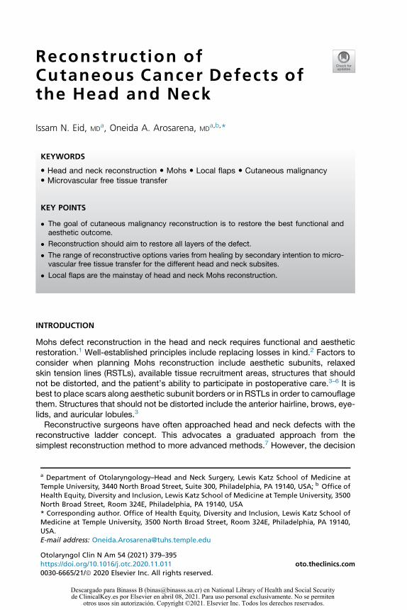
Reconstruction ofCutaneous Cancer Defects of
the Head and NeckIssam N. Eid, MDa, Oneida A. Arosarena, MDa,b,*
KEYWORDS
� Head and neck reconstruction � Mohs � Local flaps � Cutaneous malignancy� Microvascular free tissue transfer
KEY POINTS
� The goal of cutaneous malignancy reconstruction is to restore the best functional andaesthetic outcome.
� Reconstruction should aim to restore all layers of the defect.
� The range of reconstructive options varies from healing by secondary intention to micro-vascular free tissue transfer for the different head and neck subsites.
� Local flaps are the mainstay of head and neck Mohs reconstruction.
INTRODUCTION
Mohs defect reconstruction in the head and neck requires functional and aestheticrestoration.1 Well-established principles include replacing losses in kind.2 Factors toconsider when planning Mohs reconstruction include aesthetic subunits, relaxedskin tension lines (RSTLs), available tissue recruitment areas, structures that shouldnot be distorted, and the patient’s ability to participate in postoperative care.3–6 It isbest to place scars along aesthetic subunit borders or in RSTLs in order to camouflagethem. Structures that should not be distorted include the anterior hairline, brows, eye-lids, and auricular lobules.3
Reconstructive surgeons have often approached head and neck defects with thereconstructive ladder concept. This advocates a graduated approach from thesimplest reconstruction method to more advanced methods.7 However, the decision
a Department of Otolaryngology–Head and Neck Surgery, Lewis Katz School of Medicine atTemple University, 3440 North Broad Street, Suite 300, Philadelphia, PA 19140, USA; b Office ofHealth Equity, Diversity and Inclusion, Lewis Katz School of Medicine at Temple University, 3500North Broad Street, Room 324E, Philadelphia, PA 19140, USA* Corresponding author. Office of Health Equity, Diversity and Inclusion, Lewis Katz School ofMedicine at Temple University, 3500 North Broad Street, Room 324E, Philadelphia, PA 19140,USA.E-mail address: [email protected]
Otolaryngol Clin N Am 54 (2021) 379–395https://doi.org/10.1016/j.otc.2020.11.011 oto.theclinics.com0030-6665/21/ª 2020 Elsevier Inc. All rights reserved.
Descargado para Binasss B ([email protected]) en National Library of Health and Social Security de ClinicalKey.es por Elsevier en abril 08, 2021. Para uso personal exclusivamente. No se permiten
otros usos sin autorización. Copyright ©2021. Elsevier Inc. Todos los derechos reservados.

Eid & Arosarena380
on which reconstructive method to use should depend on defect characteristics andthe patient’s and surgeon’s preferences in order to achieve the best outcome.
HEALING BY SECONDARY INTENTION
Healing by secondary intention is a useful reconstructive method in patients with mul-tiple comorbidities that make a general anesthetic less desirable.2 This approach’s ad-vantages include ease of cancer surveillance, procedural cost avoidance, and lowerrisk of complications.2,8 The disadvantages include length of time and extent of woundcare needed. The wound should be kept moist with an occlusive dressing. Healing bysecondary intention is useful in scalp and forehead defects, even in cases of defectsdown to or with missing periosteum. It can be particularly advantageous in areas withabsence of surrounding skin laxity from prior resections.9 It can also be used for smalldefects of the cheek, medial canthal area, temple, and concave surfaces of the earand nose.2,4,8,9
SKIN AND COMPOSITE GRAFT PHYSIOLOGY
Optimizing the wound bed by removing nonvital tissue promotes graft survival. Full-thickness skin grafts (FTSGs) offer better color match, and decreased contractionand depression compared with split-thickness skin grafts (STSGs). STSGs arecommonly indicated for large scalp defects, for coverage of a wound bed being moni-tored for cancer recurrence, or for muscle free flap coverage. Delay of grafting by 12 to14 days with wound care allows granulation tissue in-growth, which in turn decreasesgraft loss, depression, and contracture.5,10 Composite graft cooling for 7 to 14 daysreduces metabolic requirements and has been shown to improve survival.5
SKIN FLAP PHYSIOLOGY
Local flaps have several advantages over healing by secondary intention and skingrafting, including better color and texture match and decreased wound contrac-tion.11–13 The arterial blood supply to the skin can be categorized as musculocutane-ous, direct cutaneous, and septocutaneous. Direct cutaneous arteries include thesuperficial temporal, posterior auricular, occipital, supratrochlear, and supraorbital ar-teries. Within the skin, at least 5 different vascular plexuses have been described:dermal, subdermal, subcutaneous, prefascial, and subfascial networks. These plex-uses are interconnected via anastomosing (choke) vessels, creating collateral bloodflow that allows cutaneous flap survival. Vascular delay enhances flap survival throughloss of sympathetic tone, and axial reorientation and dilatation of choke vessels.14
Flaps may be categorized according to movement and/or blood supply. The basicflap movement types are advancement, rotation, and transposition. Advancement flapsare designed by moving tissue adjacent to the defect in 1 linear direction, while rotationflaps are curvilinear and rotate about a pivot point into the defect.4,8,11,15 The simplestadvancement flap is incisional closure with undermining. Advancement flaps can besubcategorized as unipedicle (eg, U-plasty), bipedicle (O/T, A/T), V/Y, Y/V,and H-plasty.11 The ideal defect for a rotation flap is triangular in shape, with the trian-gle’s height to width ratio being 2 to 1. The curve’s radius should be 1 to 2 times the tri-angle’s height. Transposition flaps are lifted over an incomplete skin bridge into thedefect. Transposition flap examples include the rhombic, bilobe, and note flaps.15
Random pattern flaps rely on blood supply from surrounding reticular dermal ves-sels or perforating vessels from the subdermal plexus.4,8 If the perfusion pressure atany portion of the flap falls below the arteriole closing pressure, the distal soft tissues
Descargado para Binasss B ([email protected]) en National Library of Health and Social Security de ClinicalKey.es por Elsevier en abril 08, 2021. Para uso personal exclusivamente. No se permiten
otros usos sin autorización. Copyright ©2021. Elsevier Inc. Todos los derechos reservados.
Reconstruction of Cutaneous Cancer Defects 381
will necrose.14 Axial pattern flaps obtain their blood supply from a named artery.4,8
Reconstruction within the major facial aesthetic subunits will be described in therest of the article.
FOREHEAD AND SCALP
Considerations in scalp and forehead reconstruction include maintaining the hairlineand avoiding hair-bearing skin loss (Fig. 1). Scalp skin is immobile, making reconstruc-tion challenging.16
Primary closure is generally limited to defects that are less than 3 cm in diameter.16
Healing by secondary intention is appropriate for large forehead defects that have firstbeen reduced in size by advancement flaps. This may result in a better cosmetic resultthat skin grafting, and the resulting scar may be serially excised.3
Skin thickness of the scalp and forehead makes skin grafting a less desirable recon-structive option. Defects extending to the bone are better served by local advance-ment flaps. However, drilling the calvarium’s outer cortex will increase STSGsurvivability.16 Use of wound matrix material (Integra, Integra Life Sciences Corpora-tion, Princeton, New Jersey) can aid grafting and has been used in full-thickness de-fects in patients who are not microvascular reconstruction candidates.17
Advancement flaps can be incised in forehead rhytids.1,3,11 These are deal foreyebrow reconstruction.11 Bilateral advancement flaps can be designed in H-plasty,A/T, and O/T/O/Z closures (Figs. 2 and 3).3,8,11,15 Rotation flaps can bedesigned along the hairline.1
Healing by secondary intention or poor flap planning can lead to hairline andeyebrow position distortion in the forehead. Distortion can be minimized by securingthe galea at the hairline or eyebrow to periosteum. Injury to the facial nerve’s temporalbranch can be avoided by dissection in a subcutaneous plane or at the deep temporalfascia level.1
OCULAR ADNEXA
Periocular reconstruction goals are to restore eyelid form and function, including globeprotection.1,18 Several techniques are available for eyelid reconstruction that depend
Fig. 1. (A) Scalp defect involving hairline. (B) Defect repaired with advancement/rotationflap.
Descargado para Binasss B ([email protected]) en National Library of Health and Social Security de ClinicalKey.es por Elsevier en abril 08, 2021. Para uso personal exclusivamente. No se permiten
otros usos sin autorización. Copyright ©2021. Elsevier Inc. Todos los derechos reservados.

Fig. 2. Forehead Mohs reconstruction O/T flap. (A) Defect. (B) Immediate postoperativeclosure. (C) Several months postoperative result.
Eid & Arosarena382
on defect size. Healing by secondary intention is appropriate for small (<1 cm),shallow, upper eyelid defects involving the concave medial canthal area.8,12,18 Primaryclosure of small, nonmargin-involving, anterior lamella defects using an elliptical exci-sion, M-plasty, O/-Z-plasty, or double-S ellipse also provides acceptable re-sults.8,12,13 Ellipses in the periorbital area should be oriented perpendicular to theRSTLs to avoid vertical tension on the eyelids.13 For larger defects, local advancementand transposition (note, rhombic, V/Y, and bilobed flaps) can be designed from adja-cent tissue.12,13,18 FTSGs from the upper eyelid, pre- and postauricular area, supra-clavicular area, and inner arm provide hairless skin with acceptable color matchingfor anterior lamellar defects.8,12,13,18 Grafts should be oversized by up to 30% to pre-vent eyelid malposition.8,18
For larger defects involving the lower eyelid anterior lamella, a pedicled transposi-tion flap from the upper eyelid can provide defect closure up to two-thirds the lowereyelid width.8,12,13,19 Lower eyelid retraction can be prevented with lateral canthaltightening procedures and a suborbicularis fat lift.13 Posterior lamellar defects canbe repaired with nasal septal composite grafts, buccal mucosal grafts with cartilagesupport (eg, auricular cartilage), hard palate mucoperiosteum, or upper lid tarsocon-junctival grafts/flaps.12,13,18
Fig. 3. Scalp Mohs reconstruction with O/Z flap. (A) Defect. (B) Immediate postoperativeclosure.
Descargado para Binasss B ([email protected]) en National Library of Health and Social Security de ClinicalKey.es por Elsevier en abril 08, 2021. Para uso personal exclusivamente. No se permiten
otros usos sin autorización. Copyright ©2021. Elsevier Inc. Todos los derechos reservados.

Reconstruction of Cutaneous Cancer Defects 383
For full-thickness defects, end-to-end eyelid closure with wedge excision (with orwithout lateral canthotomy) is appropriate for defects less than 15 mm in length orless than 40% of the lid margin.8,12,13,18,20 For defects up to two-thirds of the eyelidlength, orbicularis flaps such as the Tenzel semicircular advancement flap, bolsteredby cartilage grafts, may be used.12,13,18 For larger, full-thickness defects, a cross-lidtarsoconjunctival flap may be used (Hughes procedure). When designing this flap,at least 4 mm of upper lid tarsus must be preserved to prevent eyelid malposition.The anterior lamella can be covered with a local flap, or a postauricular FTSG. Thesecond-stage separation is performed after 4 to 6 weeks.8,12,13,20 The Cutler-Beardflap is a cross-lid flap used to reconstruct the upper eyelid. This skin-muscle-conjunctival flap requires a cartilage graft between the lamellae to restore the uppereyelid tarsus.12,18
An alternative to the Hughes procedure is the cheek rotation (Mustarde) flap8 with anasal septal composite graft for posterior lamella reconstruction.18 Drawbacks to thismethod’s use include lower lid atonia and possible ectropion.20 Frost sutures can pre-vent lower lid retraction.8
Defects involving the medial canthus are approached by securing the upper andlower lid remnants to the medial canthal tendon’s posterior reflection or the lacrimalcrest with permanent suture. The skin defect can then be replaced with an FTSG,or, if the defect extends to bone, with a paramedian forehead transposition flap(PFF).8,12,18,20,21 Intubation of the remaining lacrimal canalicular system or conjuncti-vodacrocystorhinostomy can be performed at the time of reconstruction for defectsinvolving the lacrimal system.13,18 Lateral canthal reconstruction is directed towardthe lateral orbital rim periosteum at Whitnall tubercle. If the periosteum is absent inthis area, fixation may be performed with a drill hole placed at the Whitnall tubercle.18
Complications include epiphora, hypertrophic scarring, ectropion, edema, infection,exposure keratopathy, lagophthalmos, ptosis, corneal abrasion, trichiasis, andchange in visual acuity.1,13 Factors associated with complications are FTSG useand a defect more than one-half of the horizontal eyelid length repair. Webbing cancomplicate reconstruction in the medial canthal area and can be treated withZ-plasty.1 When the medial canthus has been excised for margin control, superioror inferior canthal displacement and angle deformity can result.20
NOSE
Nasal reconstruction goals are to restore contour and maintain the airway.1 Cartilag-inous alar batten or sidewall support grafts are used to prevent airway compromise,minimize scar contracture and restore contour.22–24
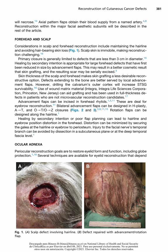
Healing by secondary intention is limited to defects less than 5 mm on concave sur-faces. Primary closure of defects less than 1 cm may be used for nasal dorsum andsidewall defects.22,25,26 FTSG use is most successful in patients with Fitzpatrick 1or 2 skin types and nasal skin that is not very sebaceous.3,22 Skin grafting shouldbe avoided when the perichondrium and periosteum are not intact. Conchal bowlcomposite grafts may be used for defects less than 1 cm (Fig. 4).22
Cheek advancement flaps can be used to reconstruct nasal sidewall defects.4 TheLemmo flap is a laterally based bipedicle advancement flap that advances skin fromthe upper nasal dorsum to reconstruct lower nasal dorsal defects. The glabellar defectis closed in a V/Y fashion, avoiding long dorsal scars for a more favorable glabellarscar.25
Transposition flaps commonly used for nasal skin reconstruction include the naso-labial, bilobed, rhombic and note flaps. These flaps are based on facial and angular
Descargado para Binasss B ([email protected]) en National Library of Health and Social Security de ClinicalKey.es por Elsevier en abril 08, 2021. Para uso personal exclusivamente. No se permiten
otros usos sin autorización. Copyright ©2021. Elsevier Inc. Todos los derechos reservados.
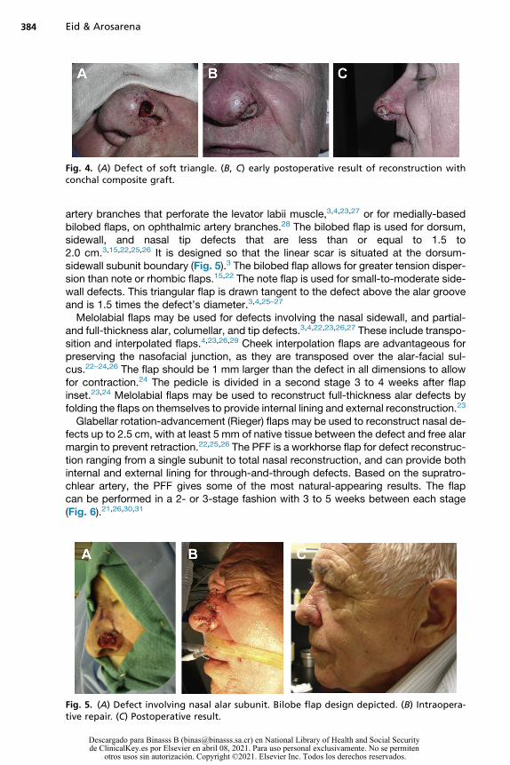
Fig. 4. (A) Defect of soft triangle. (B, C) early postoperative result of reconstruction withconchal composite graft.
Eid & Arosarena384
artery branches that perforate the levator labii muscle,3,4,23,27 or for medially-basedbilobed flaps, on ophthalmic artery branches.28 The bilobed flap is used for dorsum,sidewall, and nasal tip defects that are less than or equal to 1.5 to2.0 cm.3,15,22,25,26 It is designed so that the linear scar is situated at the dorsum-sidewall subunit boundary (Fig. 5).3 The bilobed flap allows for greater tension disper-sion than note or rhombic flaps.15,22 The note flap is used for small-to-moderate side-wall defects. This triangular flap is drawn tangent to the defect above the alar grooveand is 1.5 times the defect’s diameter.3,4,25–27
Melolabial flaps may be used for defects involving the nasal sidewall, and partial-and full-thickness alar, columellar, and tip defects.3,4,22,23,26,27 These include transpo-sition and interpolated flaps.4,23,26,29 Cheek interpolation flaps are advantageous forpreserving the nasofacial junction, as they are transposed over the alar-facial sul-cus.22–24,26 The flap should be 1 mm larger than the defect in all dimensions to allowfor contraction.24 The pedicle is divided in a second stage 3 to 4 weeks after flapinset.23,24 Melolabial flaps may be used to reconstruct full-thickness alar defects byfolding the flaps on themselves to provide internal lining and external reconstruction.23
Glabellar rotation-advancement (Rieger) flaps may be used to reconstruct nasal de-fects up to 2.5 cm, with at least 5 mm of native tissue between the defect and free alarmargin to prevent retraction.22,25,26 The PFF is a workhorse flap for defect reconstruc-tion ranging from a single subunit to total nasal reconstruction, and can provide bothinternal and external lining for through-and-through defects. Based on the supratro-chlear artery, the PFF gives some of the most natural-appearing results. The flapcan be performed in a 2- or 3-stage fashion with 3 to 5 weeks between each stage(Fig. 6).21,26,30,31
Fig. 5. (A) Defect involving nasal alar subunit. Bilobe flap design depicted. (B) Intraopera-tive repair. (C) Postoperative result.
Descargado para Binasss B ([email protected]) en National Library of Health and Social Security de ClinicalKey.es por Elsevier en abril 08, 2021. Para uso personal exclusivamente. No se permiten
otros usos sin autorización. Copyright ©2021. Elsevier Inc. Todos los derechos reservados.
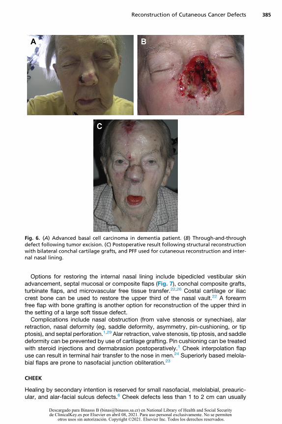
Fig. 6. (A) Advanced basal cell carcinoma in dementia patient. (B) Through-and-throughdefect following tumor excision. (C) Postoperative result following structural reconstructionwith bilateral conchal cartilage grafts, and PFF used for cutaneous reconstruction and inter-nal nasal lining.
Reconstruction of Cutaneous Cancer Defects 385
Options for restoring the internal nasal lining include bipedicled vestibular skinadvancement, septal mucosal or composite flaps (Fig. 7), conchal composite grafts,turbinate flaps, and microvascular free tissue transfer.22,26 Costal cartilage or iliaccrest bone can be used to restore the upper third of the nasal vault.22 A forearmfree flap with bone grafting is another option for reconstruction of the upper third inthe setting of a large soft tissue defect.Complications include nasal obstruction (from valve stenosis or synechiae), alar
retraction, nasal deformity (eg, saddle deformity, asymmetry, pin-cushioning, or tipptosis), and septal perforation.1,29 Alar retraction, valve stenosis, tip ptosis, and saddledeformity can be prevented by use of cartilage grafting. Pin cushioning can be treatedwith steroid injections and dermabrasion postoperatively.1 Cheek interpolation flapuse can result in terminal hair transfer to the nose in men.24 Superiorly based melola-bial flaps are prone to nasofacial junction obliteration.23
CHEEK
Healing by secondary intention is reserved for small nasofacial, melolabial, preauric-ular, and alar-facial sulcus defects.6 Cheek defects less than 1 to 2 cm can usually
Descargado para Binasss B ([email protected]) en National Library of Health and Social Security de ClinicalKey.es por Elsevier en abril 08, 2021. Para uso personal exclusivamente. No se permiten
otros usos sin autorización. Copyright ©2021. Elsevier Inc. Todos los derechos reservados.
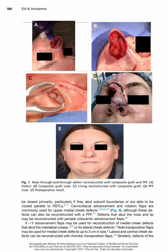
Fig. 7. Nose through-and-through defect reconstructed with composite graft and PFF. (A)Defect. (B) Composite graft inset. (C) Lining reconstructed with composite graft. (D) PFFinset. (E) Postoperative result.
Eid & Arosarena386
be closed primarily, particularly if they abut subunit boundaries or are able to beclosed parallel to RSTLs.6,11 Cervicofacial advancement and rotation flaps arecommonly used for upper medial cheek defects1,3,6,8,32 (Fig. 8), although these de-fects can also be reconstructed with a PFF.21 Defects that abut the nose and lipmay be reconstructed with perialar crescentic advancement flaps.32
V/Y advancement flaps may be used for reconstruction of medial cheek defectsthat abut themelolabial crease,3,32 or for lateral cheek defects.6 Note transposition flapsmay be used for medial cheek defects up to 3 cm in size.6 Lateral and central cheek de-fects can be reconstructed with rhombic transposition flaps.3,6 Similarly, defects of the
Descargado para Binasss B ([email protected]) en National Library of Health and Social Security de ClinicalKey.es por Elsevier en abril 08, 2021. Para uso personal exclusivamente. No se permiten
otros usos sin autorización. Copyright ©2021. Elsevier Inc. Todos los derechos reservados.

Fig. 8. (A) Medial cheek defect following excision of basal cell carcinoma. (B, C) Cervicofacialrotation flap repair.
Reconstruction of Cutaneous Cancer Defects 387
perilabial cheek that abut the chin may be reconstructed with bilobed flaps.6,32 O/Tand A/T closures may be used for defects abutting the preauricular sulcus.Microvascular free tissue transfer is indicated for combined cheek and eyelid de-
fects with exposed bone, or when soft tissue bulk is required (Figs. 9 and 10). FTSGsmay be used to reconstruct cheek defects in patients whose medical condition maynot allow for local flap reconstruction. Limitations of free flaps and FTSG includeinability to provide color-matched skin.32
PERIORAL RECONSTRUCTION
Perioral reconstruction goals are to maintain oral competence, motion, sensation, andcosmesis. Aesthetic subunits (ie, the vermillion border, philtral ridges, cupid’s bow and
Fig. 9. Cheek defect reconstructed with anterolateral thigh free flap.
Descargado para Binasss B ([email protected]) en National Library of Health and Social Security de ClinicalKey.es por Elsevier en abril 08, 2021. Para uso personal exclusivamente. No se permiten
otros usos sin autorización. Copyright ©2021. Elsevier Inc. Todos los derechos reservados.

Fig. 10. (A) Defect from excision of advanced squamous cell carcinoma necessitating orbitalexenteration, en-bloc ethmoidectomy and resection of cheek soft tissue; (B) reconstructionwith rectus abdominus free flap.
Eid & Arosarena388
labiomental crease) should be reconstructed while maintaining lip height andprojection.
Primary Closure of Small Cutaneous Defects
Primary, V-shaped closure may be used for defects up to 30% of the lip.2 This is donewith wide undermining and advancement for tension-free closure (Fig. 11).
Bilateral Mucosal Advancement Flaps for Isolated Red Lip Defects
Advancement flaps such as the Burow wedge flap can be used for partial-thickness,lateral upper lip defects.33 Rhombic flaps may be used for central and lateral upper lipdefects, allowing incisions to fall at the philtral ridges, vermilion-cutaneous border, andnasolabial fold.34 Central upper lip defects may also be reconstructed with a PFF.21
Fig. 11. Primary lip closure with advancement flaps. (A) Before surgery. (B) Shortly aftersurgery.
Descargado para Binasss B ([email protected]) en National Library of Health and Social Security de ClinicalKey.es por Elsevier en abril 08, 2021. Para uso personal exclusivamente. No se permiten
otros usos sin autorización. Copyright ©2021. Elsevier Inc. Todos los derechos reservados.
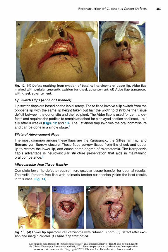
Fig. 12. (A) Defect resulting from excision of basal cell carcinoma of upper lip. Abbe flapmarked with perialar crescentic excision for cheek advancement. (B) Abbe flap transposedwith cheek advancement.
Reconstruction of Cutaneous Cancer Defects 389
Lip Switch Flaps (Abbe or Estlander)
Lip switch flaps are based on the labial artery. These flaps involve a lip switch from theopposite lip with the same lip height taken but half the width to distribute the tissuedeficit between the donor site and the recipient. The Abbe flap is used for central de-fects and requires the pedicle to remain attached for a delayed section and inset, usu-ally after 3 weeks (Figs. 12 and 13). The Estlander flap involves the oral commissureand can be done in a single stage.2
Bilateral Advancement Flaps
The most common among these flaps are the Karapanzic, the Gillies fan flap, andBernard-von Burrow closure. These flaps borrow tissue from the cheek and upperlip to restore the lower lip, and cause some degree of microstomia. The Karapanzicflap’s advantage is neurovascular structure preservation that aids in maintainingoral competence.2
Microvascular Free Tissue Transfer
Complete lower lip defects require microvascular tissue transfer for optimal results.The radial forearm free flap with palmaris tendon suspension yields the best resultsin this case (Fig. 14).
Fig. 13. (A) Lower lip squamous cell carcinoma with cutaneous horn. (B) Defect after exci-sion and margin control. (C) Abbe flap transposed.
Descargado para Binasss B ([email protected]) en National Library of Health and Social Security de ClinicalKey.es por Elsevier en abril 08, 2021. Para uso personal exclusivamente. No se permiten
otros usos sin autorización. Copyright ©2021. Elsevier Inc. Todos los derechos reservados.
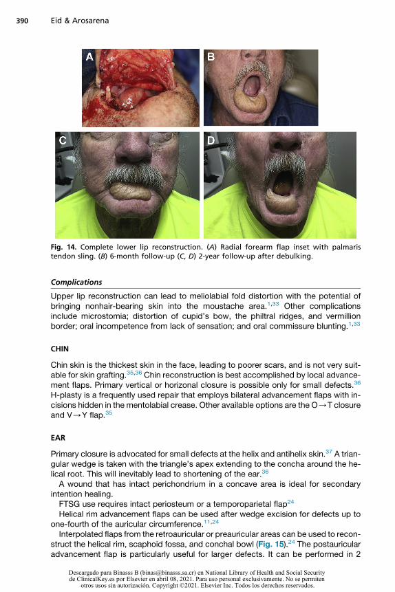
Fig. 14. Complete lower lip reconstruction. (A) Radial forearm flap inset with palmaristendon sling. (B) 6-month follow-up (C, D) 2-year follow-up after debulking.
Eid & Arosarena390
Complications
Upper lip reconstruction can lead to meliolabial fold distortion with the potential ofbringing nonhair-bearing skin into the moustache area.1,33 Other complicationsinclude microstomia; distortion of cupid’s bow, the philtral ridges, and vermillionborder; oral incompetence from lack of sensation; and oral commissure blunting.1,33
CHIN
Chin skin is the thickest skin in the face, leading to poorer scars, and is not very suit-able for skin grafting.35,36 Chin reconstruction is best accomplished by local advance-ment flaps. Primary vertical or horizonal closure is possible only for small defects.36
H-plasty is a frequently used repair that employs bilateral advancement flaps with in-cisions hidden in the mentolabial crease. Other available options are the O/T closureand V/Y flap.35
EAR
Primary closure is advocated for small defects at the helix and antihelix skin.37 A trian-gular wedge is taken with the triangle’s apex extending to the concha around the he-lical root. This will inevitably lead to shortening of the ear.36
A wound that has intact perichondrium in a concave area is ideal for secondaryintention healing.FTSG use requires intact periosteum or a temporoparietal flap24
Helical rim advancement flaps can be used after wedge excision for defects up toone-fourth of the auricular circumference.11,24
Interpolated flaps from the retroauricular or preauricular areas can be used to recon-struct the helical rim, scaphoid fossa, and conchal bowl (Fig. 15).24 The postauricularadvancement flap is particularly useful for larger defects. It can be performed in 2
Descargado para Binasss B ([email protected]) en National Library of Health and Social Security de ClinicalKey.es por Elsevier en abril 08, 2021. Para uso personal exclusivamente. No se permiten
otros usos sin autorización. Copyright ©2021. Elsevier Inc. Todos los derechos reservados.

Fig. 15. (A) Through-and-through defect of conchal bowl and anterior ear canal followingexcision of basal cell carcinoma. V/Y advancement flap marked. (B) Folded advancementflap inset. (C) 4-month postoperative result.
Reconstruction of Cutaneous Cancer Defects 391
stages with a possible cartilage harvest from the contralateral ear to give support andstructure.36
Costochondral cartilage is the ideal framework for larger auricular defects and re-quires good soft tissue coverage with a local or regional skin flap.37 Total auriculec-tomy defects can be addressed in several ways. Many patient factors come intoplay including patient aesthetic goals, while factoring in age and comorbidities. Inolder patients, an ear prosthetic is a viable option. Massive defects around the earrequire flaps ranging from a supraclavicular flap to free tissue transfer such as a radialforearm or anterolateral thigh free flap for coverage. Reconstruction with alloplasticframeworks is prone to implant exposure.Healing by secondary intention has a greater infection risk compared with other
reconstructive methods. Healing by secondary intention and FTSG use are associatedwith wound depression. Wedge excision of defects greater than one-fourth of theauricular circumference can lead to distortion and auricular cupping. Postauricular tis-sue advancement can result in placing hair-bearing skin on the ear and postauricularsulcus blunting, which can make wearing glasses difficult. Reconstruction adjacent toor in the external auditory canal can result in stenosis.
SKIN EXPANSION
When defects are greater than half the cheek aesthetic unit, greater than one-third ofthe forehead, or over 6 cm in the scalp, expansion techniques are indicated (Fig. 16).Forehead skin expansion may be indicated for complete nasal reconstruction.
Descargado para Binasss B ([email protected]) en National Library of Health and Social Security de ClinicalKey.es por Elsevier en abril 08, 2021. Para uso personal exclusivamente. No se permiten
otros usos sin autorización. Copyright ©2021. Elsevier Inc. Todos los derechos reservados.

Fig. 16. (A) Patient with dermatofibrosarcoma protuberans of left scalp and forehead. (B)Defect initially covered with STSG at the time of expander placement. (C) Intraoperativeimplant removal after expansion complete. (D) Immediate postoperative result followingadvancement of expanded scalp skin.
Eid & Arosarena392
Temporary cosmetic deformity and discomfort from expansion in the supraorbital re-gion are this technique’s disadvantages.38 Tissue expander use includes risks ofinfection, implant extrusion, mechanical failure, tissue necrosis, bony changes, hema-toma, and seroma formation.1,38
COMPLICATIONS
The incidence of complications following Mohs defect reconstruction varies from lessthan 0.5% in the glabella, jawline, and nasolabial folds, to greater than 45% in thenose. The complication rate also varies with the reconstructive method, ranging
Descargado para Binasss B ([email protected]) en National Library of Health and Social Security de ClinicalKey.es por Elsevier en abril 08, 2021. Para uso personal exclusivamente. No se permiten
otros usos sin autorización. Copyright ©2021. Elsevier Inc. Todos los derechos reservados.

Reconstruction of Cutaneous Cancer Defects 393
from less than 0.5% with primary closure and 27% with advancement flaps. Transpo-sition and interpolated flaps, particularly if superiorly based, are prone topincushioning.1,21,23
Complications increase in frequency with defect size. Excessive wound tension,vascular compromise, and infection can lead to complete or partial flap necrosis.1,14
Smoking, uncontrolled hypertension, collagen disorders, diabetes mellitus, and previ-ous radiation therapy can compromise flap and graft vascularity.1,4,15,21,23,26 Othercomplications include persistent scar erythema, hypopigmented scar, sensory neu-ropathies, telangectasias,1 incisional pain,4 hematoma, bleeding,4,21 and obscuringtumor recurrence.4
CLINICS CARE POINTS
� Healing by secondary intention is a useful reconstructive method in patients withcomorbidities that preclude general anesthesia, or when surveillance for aggres-sive malignancies is necessary.
� When considering skin grafting, delay of skin grafting for 12-14 days to promotegranulation tissue in-growth decreases the risk of graft loss, depression andcontracture.
� Reconstruction with local flaps provide the best color and texture match, anddecrease the risk of wound contracture.
� Smoking, uncontrolled hypertension, auto-immune disease, diabetes mellitusand previous radiation therapy can compromise flap and graft viability.
DISCLOSURE
The authors have no commercial or financial conflicts of interest, or any funding sour-ces to disclose.
REFERENCES
1. Berens AM, Akkina SR, Patel SA. Complications in facial Mohs defect reconstruc-tion. Curr Opin Otolaryngol Head Neck Surg 2017;25(4):258–64.
2. Becker GD, Adams LA, Levin BC. Secondary intention healing of exposed scalpand forehead bone after Mohs surgery. Otolaryngol Head Neck Surg 1999;121(6):751–4.
3. Joseph AW, Joseph SS. Mohs reconstruction and scar revision. Otolaryngol ClinNorth Am 2019;52(3):461–71.
4. Chen EH, Johnson TM, Ratner D. Introduction to flap movement: reconstruction offive similar nasal defects using different flaps. Dermatol Surg 2005;31(8 Pt 2):982–5.
5. Brenner MJ, Moyer JS. Skin and composite grafting techniques in facial recon-struction for skin cancer. Facial Plast Surg Clin North Am 2017;25(3):347–63.
6. Hanks JE, Moyer JS, Brenner MJ. Reconstruction of cheek defects secondary tomohs microsurgery or wide local excision. Facial Plast Surg Clin North Am 2017;25(3):443–61.
7. Ge NN, McGuire JF, Dyson S, et al. Nonmelanoma skin cancer of the head andneck II: surgical treatment and reconstruction. Am J Otolaryngol 2009;30(3):181–92.
8. Harvey DT, Taylor RS, Itani KM, et al. Mohs micrographic surgery of the eyelid: anoverview of anatomy, pathophysiology, and reconstruction options. DermatolSurg 2013;39(5):673–97.
Descargado para Binasss B ([email protected]) en National Library of Health and Social Security de ClinicalKey.es por Elsevier en abril 08, 2021. Para uso personal exclusivamente. No se permiten
otros usos sin autorización. Copyright ©2021. Elsevier Inc. Todos los derechos reservados.

Eid & Arosarena394
9. Deutsch BD, Becker FF. Secondary healing of Mohs defects of the forehead,temple, and lower eyelid. Arch Otolaryngol Head Neck Surg 1997;123(5):529–34.
10. Robinson JK, Dillig G. The advantages of delayed nasal full-thickness skin graft-ing after Mohs micrographic surgery. Dermatol Surg 2002;28(9):845–51.
11. Shew M, Kriet JD, Humphrey CD. Flap basics II: advancement flaps. Facial PlastSurg Clin North Am 2017;25(3):323–35.
12. Huggins AB, Latting MW, Marx DP, et al. Ocular adnexal reconstruction for cuta-neous periocular malignancies. Semin Plast Surg 2017;31(1):22–30.
13. Segal KL, Nelson CC. Periocular reconstruction. Facial Plast Surg Clin North Am2019;27(1):105–18.
14. Lucas JB. The physiology and biomechanics of skin flaps. Facial Plast Surg ClinNorth Am 2017;25(3):303–11.
15. Starkman SJ, Williams CT, Sherris DA. Flap Basics I: rotation and transpositionflaps. Facial Plast Surg Clin North Am 2017;25(3):313–21.
16. Olson MD, Hamilton GS 3rd. Scalp and forehead defects in the post-Mohs sur-gery patient. Facial Plast Surg Clin North Am 2017;25(3):365–75.
17. Richardson ML, Lange JP, Jordan JR. Reconstruction of full-thickness scalp de-fects using a dermal regeneration template. JAMA Facial Plast Surg 2016;18(1):62–7.
18. Lu GN, Pelton RW, Humphrey CD, et al. Defect of the Eyelids. Facial Plast SurgClin North Am 2017;25(3):377–92.
19. Perry MJ, Langtry J, Martin IC. Lower eyelid reconstruction using pedicled skinflap and palatal mucoperiosteum. Dermatol Surg 1997;23(5):395–7 [discussion:397–8].
20. Moy RL, Ashjian AA. Periorbital reconstruction. J Dermatol Surg Oncol 1991;17(2):153–9.
21. Reckley LK, Peck JJ, Roofe SB. Flap basics III: interpolated flaps. Facial PlastSurg Clin North Am 2017;25(3):337–46.
22. Dibelius GS, Toriumi DM. Reconstruction of cutaneous nasal defects. Facial PlastSurg Clin North Am 2017;25(3):409–26.
23. Carucci JA. Melolabial flap repair in nasal reconstruction. Dermatol Clin 2005;23(1):65–71, vi.
24. Nguyen TH. Staged cheek-to-nose and auricular interpolation flaps. DermatolSurg 2005;31(8 Pt 2):1034–45.
25. Lu GN, Kriet JD, Humphrey CD. Local cutaneous flaps in nasal reconstruction.Facial Plast Surg 2017;33(1):27–33.
26. Joseph AW, Truesdale C, Baker SR. Reconstruction of the nose. Facial Plast SurgClin North Am 2019;27(1):43–54.
27. Alam M, Goldberg LH. Oblique advancement flap for defects of the lateral nasalsupratip. Arch Dermatol 2003;139(8):1039–42.
28. Skaria AM. The medial based bi- or trilobed flap for repair of distal alar defects.Dermatology 2013;227(2):165–70.
29. Arden RL, Miguel GS. The subcutaneous melolabial island flap for nasal alarreconstruction: a clinical review with nuances in technique. Laryngoscope2012;122(8):1685–9.
30. Menick FJ. Nasal reconstruction. Plast Reconstr Surg 2010;125(4):138e–50e.
31. Correa BJ, Weathers WM, Wolfswinkel EM, et al. The forehead flap: the gold stan-dard of nasal soft tissue reconstruction. Semin Plast Surg 2013;27(02):096–103.
32. Rapstine ED, Knaus WJ 2nd, Thornton JF. Simplifying cheek reconstruction: a re-view of over 400 cases. Plast Reconstr Surg 2012;129(6):1291–9.
Descargado para Binasss B ([email protected]) en National Library of Health and Social Security de ClinicalKey.es por Elsevier en abril 08, 2021. Para uso personal exclusivamente. No se permiten
otros usos sin autorización. Copyright ©2021. Elsevier Inc. Todos los derechos reservados.

Reconstruction of Cutaneous Cancer Defects 395
33. Oberemok S, Eliezri Y, Desciak E. Burow’s wedge flap revisited. Dermatol Surg2005;31(2):210–6 [discussion: 216].
34. Skaria AM. The transposition advancement flap for repair of postsurgical defectson the upper lip. Dermatology 2011;223(3):203–6.
35. Larrabee YC, Moyer JS. Reconstruction of Mohs defects of the lips and chin.Facial Plast Surg Clin North Am 2017;25(3):427–42.
36. Badash I, Shauly O, Lui CG, et al. Nonmelanoma facial skin cancer: a review ofdiagnostic strategies, surgical treatment, and reconstructive techniques. ClinMed Insights Ear Nose Throat 2019;12. 1179550619865278.
37. Smith RM, Byrne PJ. Reconstruction of the Ear. Facial Plast Surg Clin North Am2019;27(1):95–104.
38. Hodgkinson DJ, Lam Q. Expansion techniques after Mohs’ surgery on the face.Australas J Dermatol 2001;42(1):9–14.
Descargado para Binasss B ([email protected]) en National Library of Health and Social Security de ClinicalKey.es por Elsevier en abril 08, 2021. Para uso personal exclusivamente. No se permiten
otros usos sin autorización. Copyright ©2021. Elsevier Inc. Todos los derechos reservados.
Copyright © 2022 FDOKUMEN