Vascularised Scaffolds for Cutaneous Wound Reconstruction ...
Upload
khangminh22Category
view
0download
0
COMPLICATIONS IN CUTANEOUS LASER SURGERY
This book is dedicated to my Research Assistant Mussarrat Hussain MDHe worked tirelessly in helping me organize and write this book
Without his gallant efforts this book would never have become a reality
COMPLICATIONS IN CUTANEOUS LASERSURGERY
DAVID J GOLDBERG MDClinical Professor of DermatologyDirector of Laser Research and Mohs SurgeryMount Sinai School of Medicine New YorkClinical Professor of DermatologyDirector of Dermatologic SurgeryUMDNJmdashNew Jersey Medical SchoolAdjunct Professor of LawFordham Law School New YorkDirector of Skin Laser amp Surgery Specialists of New York and New JerseyHackensack New JerseyUSA
LONDON AND NEW YORK
A MARTIN DUNITZ BOOK
copy 2004 Martin Dunitz an imprint of the Taylor amp Francis Group plc
First published in the United Kingdom in 2004by Taylor amp Francis an imprint of the Taylor amp Francis Group 2 Park Square Milton Park
Abingdon Oxfordshire OX14 4RNTel +44 (0) 1235 828600Fax +44 (0) 1235 829000E-mail infodunitzcouk
Website httpwwwdunitzcouk
This edition published in the Taylor amp Francis e-Library 2005
ldquoTo purchase your own copy of this or any of Taylor amp Francis or Routledgersquos collection of thousands of eBooks please go to wwweBookstoretandfcoukrdquo
All rights reserved No part of this publication may be reproduced stored in a retrieval system ortransmitted in any form or by any means electronic mechanical photocopying recording orotherwise without the prior permission of the publisher or in accordance with the provisions of
the Copyright Designs and Patents Act 1988 or under the terms of any licence permitting limitedcopying issued by the Copyright Licensing Agency 90 Tottenham Court Road London W1P 0LP
Although every effort has been made to ensure that all owners of copyright material have beenacknowledged in this publication we would be glad to acknowledge in subsequent reprints or
editions any omissions brought to our attention
A CIP record for this book is available from the British Library
Library of Congress Cataloging-in-Publication Data
Data available on application
ISBN 0-203-00217-2 Master e-book ISBN
ISBN 1 84184 245 1 (Print Edition)
Distributed in North and South America by
Taylor amp Francis2000 NW Corporate Blvd
Boca Raton FL 33431 USA
Within Continental USATel 800 272 7737 Fax 800 374 3401
Outside Continental USATel 561 994 0555 Fax 561 361 6018
E-mail orderscrcpresscom
Distributed in the rest of the world byThomson Publishing Services Cheriton House
North Way AndoverHampshire SP10 5BE UKTel +44 (0) 1264 332424
E-mail salesordertandfthomsonpublishingservicescouk
Composition by Tek-Art Croydon Surrey
CONTENTS
Preface vi
1 Laser complications laser-tissue interaction 1
2 Complications in laser resurfacing 19
3 Complications in laser treatment of tattoos and pigmented lesions 50
4 Complications in laser treatment of unwanted hair 77
5 Complications in laser treatment of vascular lesions 107
Index 133
PREFACE
The concept of selective photothermolysis conceived in 1983 revolutionized cutaneous laser therapy A new generation ofhighly selective pulsed lasers that conformed to these principles was developed in the late 1980s and early 1990s Newersystems are still evolving with each year These lasers have been successfully employed in clinical practice for the treatmentof various disorders Selective photothermolysis remains the basis of low-risk laser treatment of photoaging skin benignpigmented lesions and tattoos unwanted hair and the cutaneous vascular lesions seen within both medical and cosmetic laserdermatology The American Society for Dermatologic Surgery estimated that among its members 100 million skin laser andlight procedures were performed in 2003 These numbers continue to increase With the increasing worldwide availability ofmore powerful lasers and light sources complications are going to be seen with these technologies
A basic understanding of laser-tissue interaction as described in Chapter 1 of this book will lessen the incidence of suchcomplications However complications can and do happen Chapter 2 describes complications seen with laser resurfacingChapter 3 describes complications seen after laser treatment of pigmented lesions and tattoos Chapter 4 describescomplications seen after laser treatment of unwanted hair Finally Chapter 5 describes complications seen after laser treatmentof vascular lesions Each chapter begins with several key points These are followed by a discussion of the lasers used to treatthe entity described in each chapter The core of each chapter describes and illustrates the complications that can be seen withtodayrsquos cutaneous lasers
1LASER COMPLICATIONS LASER-TISSUE INTERACTION
KEY POINTS
(1) An understanding of basic laser-tissue interaction will lead to a lessened incidence of laser complications(2) Complications are more common after ablative laser resurfacing as compared to non-ablative techniques
However no dermal remodeling technique will have a zero incidence of complications(3) Complications from pigment-specific lasers are usually related to collateral tissue damage occurring
during treatment This will be more common from millisecond as compared to nanosecond pigmentedlesion and light sources
(4) Complications from laser hair removal systems occur because of either too much heat delivered into thedermis or alternatively not enough epidermal cooling
(5) Complications from vascular lasers are generally related to excess heat or insufficient cooling This is morecommon with longer wavelength systems
INTRODUCTION
Lasers represent the most precise and selective surgical tools ever made For years electromagnetic radiation (EMR) fromlasers lamps and other EMR sources has been used to treat a variety of medical conditions in ophthalmology dermatologyurology otolaryngology and other specialties
Dermatologic EMR sources have been used to perform a wide variety of procedures including hair removal treatment ofpigmented lesions removal of unwanted vascular lesions tattoo removal and skin resurfacing (Figure 11) For all thesetreatments a natural or artificial chromophore present in the body is heated by absorption of either monochromatic orbroadband EMR Typical natural chromophores include water melanin and hemoglobin Artificial chromophores include thevarious dyes and inks seen in tattoos
The entire skin is accessible to light At an optical wavelength of about 1200 nm about one quarter of the incident lightgoes entirely through the dermis1 In principle then every cell type and multicellular structure in the skin is a potential treatmenttarget However according to the Grotthus-Draper law selective photothermolysis and all associated light-activatedmechanisms must begin with light absorption of a penetrating light
Excellent progress has been seen when the EMR lsquotargetrsquo is the natural chromophores of hemoglobin or melanin In tattooremoval an exogenous pigment defines the target There is no single laser (wavelength) that will perform every kind of lasersurgery While performing laser surgery attention must be focused not only on the laserrsquos potential to perform precisetreatments but also on the destructive potential of that same EMR-emitting system It is this destructive potential that formsthe basis of potential complications that may be associated with cutaneous laser surgery
In the ultimate analysis current laser systems all heat an absorbing target A laser surgeon wishes to avoid excess thermalinjury to tissue There is generally a transitional zone between death and survival of any biologic system heated above itsnormal temperature Any combination of temperature and time corresponding to a point lying beyond the transition zonewill lead to the potential for excess tissue injury and resultant complications
BASIC LASER PRINCIPLES
The term LASER is an acronym for the words Light Amplification by the Stimulated Emission of Radiation (Figure 12)Radiation may be defined as the transmission of energy from one point in space to another with or without an interveningmaterial absorbing medium
Radiation can be
I Particulate radiation Particulate radiation is a stream of material particles such as electrons neutrons or other atomicfragments This kind of radiation needs no material medium for its transmission but can pass through various mediumsusually with some attenuation andor change of direction Particulate radiation requires a transfer of mass and the energytransmitted is the kinetic energy of the moving particles Dermatologic lasers do not fit into this category
II Mechanical radiation Mechanical radiation is the transmission of vibrations through a material medium Sound is anexample Mechanical radiation requires the presence of a material medium for its transmission However the mediumneed not move as a whole its particles merely oscillate elastically about fixed positions transmitting energy from one tothe next Current dermatologic lasers also do not fit into this category
Figure 11 Electromagnetic spectrum (EMR) utilized in dermatologic laser treatments
Figure 12 LASER (Light Amplification by the Stimulated Emission of Radiation)
2 COMPLICATIONS IN CUTANEOUS LASER SURGERY
III Electromagnetic radiation (EMR) EMR is what current dermatologic lasers produce Unlike mechanical radiationelectromagnetic radiation requires no medium for its transmission as it can travel through free space devoid of anymatter whatever It can also propagate through space-containing matter in the form of gases liquids or solids Uponentering such mediums electromagnetic radiation will in general be changed in direction and speed of propagation
ELECTROMAGNETIC SPECTRUM
The range of values in terms of frequency wavelength andor photonic energy encountered in the natural universe is knownas the electromagnetic spectrum In terms of any one of these parameters there may be a span of some times20 magnitude At thevery short end of the wavelength spectrum there are cosmic rays and at the very long end there are radio waves
In general lasers generate EMR ranging in wavelengths from 100 nm to 20000 nm (1 nm=1times10minus9 m) Although forsimplicity these wavelengths are all called lsquolightrsquo by strict definition light is only the interval from 400 nm to 700 nm This isthe light which our eyes can perceive in daylight (the visible light spectrum)
The amount of laser energy delivered per area is fluence sometimes called the dose usually given in Jcm2 (joules) Therate at which energy is delivered is called power measured in watts (W) Power delivered per unit area is therefore the rate ofenergy delivery per amount of skin surface This is called the irradiance usually given in Wcm2 Laser exposure duration(pulsewidth) for pulsed lasers is an extremely important term because this parameter defines the time over which energy isdelivered Finally the laser delivery system spot size may greatly affect intensity within the skin
BASIC ELEMENTS OF LASERS
Every laser has certain common elements of structure and function (Figure 13) These common elements are
I A material medium having the proper energy levels to produce the desired wavelengths of light The mediums availabletoday for lasers include hundreds of different materials grouped into three basic categories gases liquids and solids
II A resonant optical cavity shaped in the form of a cylinder whose length is much greater than its diameter and havingcoaxial mirrors at opposite ends of it
III An external source of energy to provide the excitation of the atoms or molecules of the medium by the process ofpumping
IV The delivery system which may be composed of fiberoptics or articulating mirrors Not all wavelengths of laser light canbe transmitted efficiently through a slender quartz optical fiber the most flexible and convenient device of all In non-invasive cosmetic laser dermatology where laser wavelengths lie in a range from 300 nm to 2100 nm the most
Figure 13 Laser structure
LASER COMPLICATIONS LASER-TISSUE INTERACTION 3
commonly used delivery system is the quartz optical fiber Wavelengths in the mid-to-far infrared (2500ndash20000 nmablative lasers) range of the spectrum must be transmitted via a series of articulating mirrors
GENERATION OF LASER LIGHT
Laser light is generated when the laser medium is pumped by the introduction of energy from an external source Some of theatoms or molecules in the medium will be excited into an upper level of energy From there one sees a possibledownward radiative transition to a lower level that is above the ground level The difference between this upper level and theoriginal status of the laser medium determines the frequency and wavelength of the emitted laser radiation
UNIQUESPECIAL PROPERTIES CHARACTERISTICS OF LASER LIGHT
It is now well established that laser light has three uniquespecial characteristics
1 Collimation2 Coherence3 Monochromaticity
Collimation Laser-emitted rays are collimated in that they emanate from a laser and all rays are parallel to each otherBecause of this property of laser light there is no divergence or convergence of laser-emitted radiation unless a lens or mirroris placed in front of the beam path
Coherence Laser-emitted irradiation is coherent in that the rays are in phase with each other in both space and timeSpatial coherence means that the crests and troughs of all the waves coincide along lines perpendicular to the rays Temporalcoherence means that the frequency wavelength and speed of travel are all constant
Monochromaticity Laser light is said to be monochromatic in that it consists of just one wavelength In reality no lightsource produces just a single wavelength but the bandwidth variation of dermatologic lasers is generally no more than 01 nm
LASER-TISSUE INTERACTION
Laser-tissue interaction leads to four fundamental optical phenomena
1 Reflection and backscattering from the surface at impact2 Transmission into or through the tissue3 Absorption by the tissue4 Scattering within and perhaps out of the tissue
Reflection
In general reflection shows pronounced variations within the spectral range of 400ndash1500 nm This reflection is stronglydependent on the pigments present in the absorbing tissue However in the ranges of 100ndash300 nm and 2000ndash40000nm reflectance is lsquocolorblindrsquo The most significant practical effect of reflection of laser light from living tissue is thereduction of power density of the laser irradiation once there is actual penetration into tissue
Transmission
Transmission is measured in terms of transmittance which is the ratio of the intensity of a transmitted ray as it emergesdistally from the absorbing tissue to that of the same ray immediately after entering the tissue
Absorption
Absorption of laser energy leads to the conversion of the radiant laser energy into other forms of energy such as thermalenergy (heat)
4 COMPLICATIONS IN CUTANEOUS LASER SURGERY
Scattering
Scattering is defined as a change in direction of a light in living tissue without a change in its wavelength Scattering isactually a composite of several distinct phenomena such as diffuse reflection refraction of light rays reflectant diffractionand resonant absorption In the end it is the two fundamental processes of absorption and scattering that govern allinteractions of light with matter The absorption spectra of major skin chromophores dominate most laser-tissue interactionsin dermatology When absorption occurs the laser photons surrender energy to an absorbing chromophore On absorption thephoton ceases to exist and the absorbing chromophores then become excited
Optical penetration in skin is governed by a combination of absorption and scattering From the ultraviolet (UV) throughthe near infrared (IR) spectrum both absorption and scattering tend to be stronger at shorter wavelengths In generalhowever a gradual increase occurs in the depth of penetration into skin with longer wavelengths The most penetratingwavelengths are in the 650ndash1500 nm red and near infrared region Far infrared (water-absorbing) wavelengths penetratesignificantly less
THEORY OF SELECTIVE PHOTOTHERMOLYSIS
The concept of selective photothermolysis (SP) was conceived in 19832 to explain the laser treatment of pediatric port winestains3 Initially laser wavelengths were chosen at the peak of oxyhemoglobinrsquos yellow absorption band (577 nm) Thismaximized light absorption in the superficial blood vessels relative to epidermal melanin
At the time dye lasers were the only technology producing 577 nm light They provided the first evidence for effective andsafe treatment of pediatric port wine stains4
This theory of SP has been proposed to explain the laser-induced injury that is confined to microscopic sites of selectivelight absorption in the skin with minimal damage to the adjacent tissues To achieve this selective effect lasers need to fulfillthree requirements
1 They should emit a wavelength that is highly absorbed by the targeted structure2 They should produce sufficiently high energies to inflict thermal damage to the target3 The time of tissue exposure to the laser should be short enough to limit the damage to the target without heat diffusion to
the surrounding tissues
This theory revolutionized cutaneous laser therapy A new generation of highly selective pulsed lasers that conformed to theseprinciples was developed in the late 1980s and early 1990s These lasers were successfully employed in clinical practice forthe treatment of various disorders SP remains the basis of low-risk laser treatment of photoaging skin benign pigmentedlesions and tattoos unwanted hair and the cutaneous vascular lesions seen within both medical and cosmetic laserdermatology However as will be seen in this book the risk of complications with all lasers will always remain
EXTENDED THEORY OF SELECTIVE PHOTOTHERMOLYSIS
The concept of selective photothermolysis (SP) emphasizes both the selective damage and minimal light energy requirementsseen with current laser technology2 However the use of such a short pulsewidth may become inapplicable when the targetabsorption is non-uniform over its area This may be seen when the actual target exhibits weak or no absorption yet othersurrounding portions of the target exhibit significant absorption If this is the case the weakly absorbing part of the targetchromophore has to be damaged by heat diffusion from the highly pigmentedstrongly absorbing portion of the chromophore(the heater or absorber) Such non-specific thermal damage evokes the concept of thermal damage time (TDT) The TDT of atarget is the time required for irreversible target damage with sparing of the surrounding tissue For a non-uniformly absorbingtarget structure the TDT is the time it takes for the outermost part of the target to reach a target damage temperature throughheat diffusion from the heated chromophore
According to the concept of extended SP target damage can still be selective even though the TDT is many times as longas the thermal relaxation time (TRT) of the actual target
This new extended theory of selective thermal damage of non-uniformly pigmented structures in biological tissuepostulates that the target is destroyed by heat diffusion from the absorbing chromophore to the target but not by direct heatingfrom laser irradiation as is seen with SP5 This theory has been applied to the treatment of both unwanted hair and some vascularlesions With the extra thermal effect needed to comply with this concept complications can arise
LASER COMPLICATIONS LASER-TISSUE INTERACTION 5
LASER COMPLICATIONS
Complications can arise after any laser procedure Each chapter in this book will evaluate the potential problems associatedwith distinct broad categories of cutaneous laser procedures
LASER RESURFACING
It is reasonable to assume that any means of depth-controlled destruction could be used to treat photoaged skin Like chemicalpeels and dermabrasion laser resurfacing is used to destroy the skin to a controlled depth One would therefore expect astrong correlation between the duration and extent of wound-healing and clinical efficacy Differences in efficacy between theapplications of different lasers should relate to factors affecting the wound-healing response Both efficacy and duration ofwound-healing are related to the total anatomic depth of necrosis including residual thermal damage6 With superficial injurythere will be faster healing but lower efficacy may be observed
The mechanisms of tissue damage during laser resurfacing include removal by vaporization of a layer near the surfacethermal coagulation of extracellular matrix proteins in a residual layer and a deeper zone of sub-lethal thermal injury Inaddition some photomechanical damage may occur in the case of Er YAG (Erbium Yttrium-Aluminum-Garnet) laserirradiation
Type I collagen fibrils are known to contract rapidly at temperatures of 55ndash60degC and to shrink to as much as one third oftheir original length78 It is hypothesized that the post-laser repair of this tightened collagen scaffolding results in preservationof the heat-induced collagen contraction Because new collagen is formed during the heating process the shortened collagenfibrils lead to a new tightened overall structure
The use of high-energy pulsed and scanning carbon dioxide (CO2 10600 nm) and Er YAG (2940 nm) lasers allowsclinicians to remove rhytides and other effects of photodamage (Figures 14ndash17) Too much thermal damage or any processthat interferes with the normal re-epithelialization process may lead to the complications described in Chapter 2 of thisbook
Carbon Dioxide Laser
The 10600 nm wavelength of the CO2 laser is preferentially absorbed by water having an extinction coefficient of about 30microm (1 microm=1times10ndash6 m)910 The thermal relaxation time of the absorbing layer of tissue has been calculated to be less than 1millisecond11 If the laser-tissue interaction time is confined to this time period or less by rapid pulsing of the laser or by
Figure 14 Before CO2 laser resurfacing
6 COMPLICATIONS IN CUTANEOUS LASER SURGERY
rapid scanning of a continuous beam then a layer of tissue will be rapidly vaporized leaving a layer of residual thermalnecrosis measuring only 50ndash100 microm10ndash12
When the CO2 laser interacts with tissue there are three distinct zones of tissue alteration correlating with the degree oftissue heating (1) The zone of direct impact results in vaporization of intracellular water and tissue ablation (2) Underlyingthis zone is a layer of irreversible thermal damage and denaturation resulting in tissue necrosis (3) Below this layer is a zoneof reversible non-lethal thermal damage It is in this last zone of reversible thermal damage in which collagen shrinkageoccurs This accounts for the visible tissue tightening observable as the CO2 laser interacts with the dermis
Er YAG Laser
The Er YAG laser wavelength of 2940 nm closely corresponds to an absorption peak of water This laser wavelength isapproximately 10-fold better absorbed by water than is the 10600 nm wavelength of the CO2 laser13 The efficient superficialabsorption when coupled with a short pulse duration (250ndash350 microseconds micros) allows Er YAG lasers to ablate even finelayers of tissue with significantly less Er YAG laser-induced collateral thermal damage than is seen with a CO2 laser Asingle pass with an Er YAG laser may ablate 10ndash30 microm and leave a zone of thermal necrosis of only 5ndash15 microm1415 With sucha small zone of thermal injury little tissue desiccation occurs and each subsequent pass produces very similar effectsTherefore unlike the CO2 laser there is not a significantly diminished return with each subsequent pass of the laser With lessnon-selective thermal damage the Er YAG laser when used superficially achieves more rapid healing and a significantlylower rate of complications However when this laser is aggressively used significant penetration into the skin can occurbecause of its minimal laser-induced thermal effect In this case the Er YAG laser can potentially induce the same or evenhigher risk of scarring than is seen with a CO2 laser
Non-ablative lasers and light sources do not remove the epidermis Their thermal effect is also markedly lessened as comparedto the CO2 laser However since their basic mechanism is still one of heat induction complications can also arise from thesesystems In addition because some of the non-ablative systems are used with protective epidermal cooling problems can arisewhen the cooling impacts on highly pigmented skin
Non-ablative Skin Systems
Recently skin cooling has been combined with mid-infrared lasers to produce an lsquoupside-downrsquo burnheating in which thedermis reaches a much higher temperature than the epidermis This technique has been employed to treat photoaged skinwithout inducing the obvious wound seen after ablative laser resurfacing
Figure 15 After CO2 laser resurfacing
LASER COMPLICATIONS LASER-TISSUE INTERACTION 7
Improvement after non-ablative treatment may result from
1 Photothermal heating that leads to fibroblast activation collagen remodeling and subsequent increased pro-collagen IIIexpression andor
2 Vascular activation with endothelial disruption leading to cytokine activation and subsequent collagen remodeling
Figure 16 Before Er YAG laser resurfacing
Figure 17 After Er YAG laser resurfacing
8 COMPLICATIONS IN CUTANEOUS LASER SURGERY
The mid-infrared lasers (1064ndash1540 nm) with deeply penetrating wavelengths can be coupled with surface cooling tostimulate new collagen production by gentle dermal heating This creates a thermal wound which may activate migration offibroblasts and neocollagenesis rather than inducing the immediate collagen contraction seen after CO2 laser resurfacing Theepidermal cooling prevents the thermal wound from also damaging the epidermis Lasers that remodel through vascularactivation show all the characteristics of the vascular lesion lasers described below
Non-ablative Radiofrequency
Recently non-ablative radiofrequency (RF) has been used to produce immediate collagen contraction with a single treatmentThis volumetric heat-inducing radiofrequency source uses simultaneous contact cooling for epidermal preservation Muchakin to the non-ablative lasers such RF sources can be associated with complications
LASER TREATMENT OF TATTOOS AND PIGMENTED LESIONS
The first pigment-specific laser a ruby laser was used in 1961 Since that time laser physicians have successfully treatedmany pigmentary abnormalities within the skin Properly chosen wavelengths of light used with appropriate pulse durationscan selectively alter pigmented cells and disrupt exogenous and endogenous pigment in a manner that leaves the adjacent skintotally intact
Targeting cutaneous pigmentation with lasers is dependent on several parameters such as the nature of the targetedpigment (endogenous or exogenous pigment) its absorption characteristics its distribution in tissue (intracellular orextracellular) and its anatomic location in the skin (epidermis dermis or both) Melanin the main chromophore in mostepidermal and dermal pigmented lesions has a broad absorption spectrum extending from the ultraviolet range through thevisible and near infrared spectra (Figure 18)16 Across this wide range of absorbing wavelengths any laser with sufficientenergy levels can target melanin To be most effective and avoid adverse effects it is necessary to use wavelengths that bothavoid absorption by other skin chromophores and penetrate to the desired depth An ideal lsquoselectiversquo window for targetingmelanin lies between 630 nm and 1100 nm where there is good skin penetration and preferential absorption of melanin overoxyhemoglobin
Pigment specificity of lasers is not only dependent on wavelength but also on pulsewidth Although the subcellular eventsthat characterize the interaction between lasers and pigmented cells are not entirely known the primary site of laser-inducedpigment damage is most likely the melanosome the intracellular organelle in which melanin is synthesized and stored17
Electron microscopic studies have demonstrated that melanosomes targeted by short-pulsed lasers exhibit membranedisruption and disorganization of their internal content18 With an estimated thermal relaxation time that ranges from 250nanoseconds to 1000 nanoseconds depending on their size melanosomes require sub-microsecond laser pulses (lt1microsecond) for their selective disruption Pulse durations of 40ndash750 nanoseconds (Q-switched lasers) are able to disruptmelanosomes but longer pulse durations in the millisecond domain do not appear to cause specific melanosome damage19
Action spectrum studies2021 comparing the effectiveness of different laser wavelengths in inducing pigment injury in guineapig skin have shown similar melanosome alterations to that seen in humans but with substantial differences in threshold dosesand depth of penetration Shorter wavelengths (lt600 nm) damage pigmented cells with lower energy fluences while longerwavelengths (gt600 nm) penetrate deeper into the skin yet need more energy to induce melanosome disruption In additionshorter wavelengths can damage only superficial pigmented lesions leaving deeper structures intact while longerwavelengths can target pigmented lesions in the dermis such as nevus of Ota and many tattoos
Mechanism of Laser-Tattoo Interaction
Tattoos consist of insoluble sub-micrometer-sized pigmented particles that are phagocytosed by dermal cells22 Treatmentwith short-pulsed lasers results in fragmentation of tattoo particles and selective death of pigment-containing cells23 Thereleased pigment is then removed through transepidermal elimination rephagocytosed by dermal macrophages or eliminatedthrough lymphatic drainage
The physical mechanisms of laser-tattoo interaction are not well understood2425 This is because the size of the tattoo particlesis too small and the laser duration too short to make direct observations of the tattoo pigment break-up process feasible
Laser-energy deposition in tattoo particles must be both heat- and stress-confined in order to fracture the tattoo particlesWhen these conditions are satisfied a strong stress (ie acoustic or pressure) wave is generated inside the tattoo particleFracture occurs when the strength of the tensile or compressive component of the stress wave exceeds the correspondingstrength limit of the particle The efficiency for causing fracture increases with decreasing laser pulselength However thisefficiency fails to further increase if the pulselength decreases below the stress confinement time
LASER COMPLICATIONS LASER-TISSUE INTERACTION 9
The maximal temperature reached inside a treated tattoo particle is about 900degC at the end of a 35 picosecond pulse This iswell below the melting point for graphite However because the temperature of the tattoo particle reaches a value well abovethe boiling point of water a cavitation bubble is formed in the tissue around the particle As the bubble expands the shearstress at the bubble surface increases and may cause collateral damage to the soft tissue immediately surrounding the tattooparticle This could be the cause of empty vacuoles in the ash-white lesions seen throughout the dermis after laser treatmentof tattoos26 Too much collateral damage may lead to the complications described in Chapter 3 of this book
Lasers with longer pulse durations (millisecond lasers and light sources) are less efficient in generating the required tensilestress for breaking tattoo particles They may also generate excessive heat with a resultant chance of complications
Lasers used in Pigmented LesionTattoo Treatments
Short-pulsed Q-switched lasers (pulse duration lt1 microsecond) have become the standard for the treatment of manypigmented skin disorders and tattoos (Figures 19ndash112) These Q-switched lasers operate through an extremely fastelectromagnetic switch that allows build-up of excessive energy in the laser cavity This energy is released in the form of apowerful pulse sufficiently short in duration to selectively target sub-cellular organelles such as melanosomes and tattooparticles There are three short-pulsed pigment selective lasers in clinical use today They are the Q-switched ruby laser (694nm 25ndash40 nanoseconds) the Q-switched alexandrite laser (755 nm 50ndash100 nanoseconds) and the Q-switched Nd YAG laser(1064 nm 5ndash10 nanoseconds) which can be frequently doubled to emit a green light at 532 nm of the same pulse duration
Pigment non-specific lasers such as the CO2 laser (10600 nm) and the Er YAG laser (2940 nm) are primarily used inlaser skin resurfacing but may also remove superficial pigmented lesions as a secondary event Continuous-wave andquasicontinuous-wave millisecond-emitting 511ndash694 nm laser irradiation produces selective pigment removal at thesewavelengths However in the absence of reproducible nanosecond-induced spatial confinement of thermal injury thesemillisecond systems carry a higher risk for textural and pigment changes compared with pulsed lasers Pigment removal canalso be achieved with a filtered flashlamp that generates an intense polychromatic visible to infrared millisecond-pulsedlight at variable pulsewidths and intervals With the placement of cut-off filters specific wavelengths are selected that cantarget a variety of skin disorders These systems do not usually generate the immediate post-treatment wound that is seen withQ-switched lasers but they are also not as user-friendly as the nanosecond Q-switched systems
LASER TREATMENT OF UNWANTED HAIR
Although the exact mechanism of action of laser hair removal is unknown the use of laser and light source devices is basedon the theory of selective photothermolysis2
Figure 18 Melanin absorption curve
10 COMPLICATIONS IN CUTANEOUS LASER SURGERY
Laser hair removal utilizes light to cause thermal or mechanical damage of hair follicles To achieve hair growth delay it issufficient to either damage matrix cells of anagen hair follicles coagulate blood vessels of the papilla or possibly to destroypart of the outer root sheath (ORS)27 For permanent hair follicle damage it is necessary to damage stem cells that are locatedin the bulge area at the interface of the ORS and the connective tissue sheath28 One can also irreversibly damage a hairfollicle at the level of the dermis by replacing it with connective tissue
The matrix cells produce the hair shaft The matrix cells also contain melanosomes that produce hair melanin (Figure 113)The concentration of melanin in the matrix cells is significantly higher than it is in the hair shaft This melanin is distributeduniformly and densely in the matrix cells Thus for a pulsewidth longer than the thermal relaxation time (TRT) of individualmelanosomes (1 microsecond) the matrix cells act as a uniformly pigmented target This is a typical example of a targetwhere the theory of selective photothermolysis (SP) theory is applicable (Figures 114 and 115) For selective and effectivetreatment the energy and pulsewidth have to be significantly shorter than the TRT of the matrix cells
Another theoretical method of halting hair shaft growth is to coagulate blood vessels in the papilla The loop of blood vesselsin the papilla is located in the center of the matrix cell dome Blood absorption is significantly lower than melanin absorptionin the neighboring matrix cells because of the small vessel size So the most effective method of papilla blood vesselcoagulation is to utilize heat diffusion from the matrix cells that absorb light In this scenario the extended theory of SP maybe applicable
Finally although stem cells in the basal cell layer of the lower isthmus ORS do not have pigment that can effectivelyabsorb light in the therapeutic hair removal window (600ndash1200 nm)27 these stem cells can be damaged by heat diffusion fromthe melanin-rich hair shaft or an artificial chromophore inside the internal root sheath (IRS)
Lasers used for Hair Removal
Ruby (694 nm) alexandrite (755 nm) diode (810 nm) Nd (Neodymium) YAG (1064 nm) lasers as well as intense pulsedlight systems (550ndash1100 nm) all emit light that is absorbed by hair containing melanin as well as epidermal melanin (Figures
Figure 19 Tattoo before treatment with Q-switched laser
Figure 110 Tattoo after eight treatments with Q-switched laser
LASER COMPLICATIONS LASER-TISSUE INTERACTION 11
116 and 117) The longer the wavelength the deeper is the penetration Conversely the shorter the wavelength the greater isthe melanin absorption Thus longer wavelength systems tend to be less absorbed by epidermal melanin and generally aresafer in darker complexioned individuals In addition hair removal systems are generally coupled with some sort of coolingsystem that is also epidermis-protective It is the heat that is required for hair removal or the overproduction of this thermaleffect that may lead to the complications described in Chapter 4 of this book
LASER TREATMENT OF VASCULAR LESIONS
Lasers have been used to treat vascular lesions since the early 1970s It was not until the development of the pulsed dye laserin the late 1980s that the first cosmetically reproducible results were easily achieved Recent development of longerwavelength longer pulse duration pulsed lasers and light sources have improved outcome significantly Basic requirementsfor a laser or light source to treat vascular lesions are
Figure 111 Solar lentigines before treatment with a Q-switched laser
Figure 112 Solar lentigines after treatment with a Q-switched laser
12 COMPLICATIONS IN CUTANEOUS LASER SURGERY
1 a wavelength that is proportionately better absorbed by the target (hemoglobin) than surrounding chromophores2 an ability to penetrate to the full depth of the target blood vessel3 sufficient energy to damage the vessel without damaging the overlying skin and4 an exposure duration long enough to slowly coagulate the vessel and its lining without damaging the surrounding tissue
The choice of laser wavelength(s) fluence determination and pulse duration of light exposure are all related to the size flowdepth and type of treated targeted vessel The correct choice of treatment parameters is aided by an understanding of the
Figure 113 Hair follicle containing melanin-absorbing chromophore
Figure 114 Selective photothermolysis (SP) of target tissue (hair) and resultant thermal diffusion
LASER COMPLICATIONS LASER-TISSUE INTERACTION 13
histology of target vessels Lasers with larger spot size-delivering handpieces penetrate deeper into tissue and optimizefluence delivery to the target
Over the last two decades there has been a steady progression toward longer wavelength and longer pulse durationvascular lasers29ndash31 The slower incorporation of energy emitted by these longer pulse duration lasers allows for a gradualdiffuse vascular necrosis as opposed to the early more rapid pulse duration vascular lasers that produce a more explosivephase change (vaporization) with a pressure wave phenomenon causing vessel wall rupture
For vascular lesions the exposure time should be long enough to conduct heat from the red blood cell-filled lumen to theentire blood vessel wall Merely coagulating the endothelium is insufficient because a direct correlation exists between thepermanent eradication of a vein and degree of vessel wall injury Greater success is expected as the depth of the vessel walldamage progresses from the endothelial cell layer intima and media to the adventitia32ndash35
Large-diameter vessels require a longer pulse duration to allow sufficient time for an even diffusion of heat throughout thecylindrical vessel lumen30 Consistent with the above findings the trend towards longer pulsewidths has been driven by the
Figure 115 SP and resultant vacuolization produced in laser-treated hair
Figure 116 Hair before laser hair removal
14 COMPLICATIONS IN CUTANEOUS LASER SURGERY
desire to eliminate purpura as an immediate complication of pulsed dye laser treatment Purpura results from a combination ofintravascular hemorrhage (immediate) thrombosis and vasculitis (delayed) Pulsewidths greater than 10 millisecondsproduce little or no immediate purpura However such long pulse duration lasers may still cause delayed vasculitis Patientsmust still be warned that lsquobruisingrsquo can appear several days after treatment Of note the millisecond non-laser pulsed lightsystems work in a similar manner to the millisecond vascular lasers
A new generation of longer wavelength and long-pulse duration near infrared lasers (755ndash1064 nm) has emerged for thetreatment of some telangiectasia and leg veins (Figure 118)36ndash38 Deeper vessels require a longer wavelength to allowpenetration to their depth However even with deeper penetrating wavelengths pulse durations must be matched to the vesselsize As the depth and size of the vessel changes so do the absorption characteristics
Several studies have demonstrated that heating induces hemoglobin modification3940 The change is due to oxidativereactions with formation of met-hemoglobin (Met-Hb) When blood is subject to heat the first observable event is Met-Hbformation followed by distorted heme protein formation and protein denaturation3941 Even under mild heating (50ndash54degC) Met-Hb is formed from oxygenated hemoglobin (HbO2) and hemoglobin (Hb) This phenomenon is responsible for the change inblood absorption after laser irradiation42 Met-Hb has an absorbance about times475 higher than that of HbO2 and Met-Hb has anabsorbance about times20 higher than that of Hb43
This phenomenon may explain why the longer wavelengths (eg infrared lasers such as Nd YAG) are efficient in treatingleg veins (Figures 119 and 120) Because the energy required to treat such vessels and the associated required depth ofpenetration complications may occur with the use of these lasers Complications seen with these vascular lasers are describedin Chapter 5
Figure 117 Decrease in hair after six treatments of laser hair removal
LASER COMPLICATIONS LASER-TISSUE INTERACTION 15
CONCLUSION
Cutaneous laser surgery has allowed physicians to treat photoaging skin pigmented lesions and tattoos unwanted hair and avariety of vascular lesions A thorough understanding of laser-tissue interaction should lead to a decreased likelihood of mostof the complications described in this book
Figure 118 Hemoglobin absorption curve
Figure 119 Leg veins before treatment with the 1064 nm Nd YAG laser
16 COMPLICATIONS IN CUTANEOUS LASER SURGERY
REFERENCES
1 Anderson RRLasers in dermatologymdashA critical update J Dermatol 2000 27700ndash7052 Anderson RR Parrish JASelective photothermolysis Precise microsurgery by selective absorption of pulsed radiation Science 1983
220524ndash5273 Anderson RR Parrish JAMicrovasculature can be selectively damaged using dye lasers a basic theory and experimental evidence in
human skin Lasers Surg Med 19811263ndash2664 Morelli JG Tan OT Garden J et al Tunable dye laser (577 nm) treatment of port wine stains Lasers Surg Med 1986694ndash995 Altshuler GB Anderson RR Smirnov MZ et al Extended theory of selective photothermolysis Lasers Surg Med 2001 29
416ndash4326 Khatri KA Ross V Grevelink JM Anderson RRComparison of erbium YAG and carbon dioxide lasers in resurfacing of facial
rhytides Arch Dermatol 1999 135391ndash3977 Gilchrest BASkin aging and photoaging Dermatol Nurs 1990 279ndash828 Goldfarb MT Ellis CN Weiss JS Voorheer JJTopical tretinoin therapy its use in photoaged skin J Am Acad Dermatol 1989 21
645ndash6509 Clark SD Kobayashi DK Welgus HG Regulation of the expression of tissue inhibitor of metalloproteinases and collagenase by
retinoids and glucocorticoids in human fibroblasts J Clin Invest 1987 801280ndash128810 Drake LA Dinehart SM Farmer ER et al Guidelines of care for photoagingphotodamage DermatologyWorldGuidelines 1995
November (Suppl)11ndash1311 Fisher GJ Datta SC Tauar HS et al Molecular basis of sun-induced premature skin aging and retinoid antagonism Nature 1996
379335ndash33912 Fitzpatrick RE Goldman MP Satur NM et al Pulsed carbon dioxide laser trichloracetic acid Baker-Gordon phenol and
dermabrasion A comparative clinical and histological study of cutaneous resurfacing in a porcine model Arch Dermatol 1996 132469ndash471
13 Hale GM Querry MR Optical constants of water in the 200 nm to 200 m wavelength region Appl Optics 1973 12555ndash56314 Hohenleutner U Hohenleutner S Saumier W et al Faster effective skin ablation with an Er YAG laser Determination of ablation
rates and thermal damage zones Lasers Surg Med 1997 20242ndash24715 Kaufman R Hibst R Pulsed erbium YAG laser ablation in cutaneous surgery Lasers Surg Med 1996 19324ndash33016 Anderson RR Parrish JA The optics of human skin J Invest Dermatol 1981 7713ndash1917 Polla LL Margolis RJ Dover JS et al Melanosomes are a primary target of Q-switched ruby laser irradiation in guinea pig skin J
Invest Dermatol 1987 89281ndash28618 Dover JS Polla LL Margolis RJ et al Pulse width dependence of pigment cell damage at 694 nm in guinea pig skin SPIE Lasers Med
1986 712200ndash20519 Watanabe S Flotte T Margolis R et al The effects of pulse duration on selective pigmented cell injury by dye lasers J Invest
Dermatol 1987 8853220 Anderson RR Margollis RJ Dover JS et al Selective photothermolysis of cutaneous pigmentation by Q-switched Nd YAG laser pulse
at 1064 532 and 355 nm J Invest Dermatol 1989 9328ndash3221 Sherwood K Murry S Kurban A Tan O Effects of wavelength of cutaneous pigment using pulsed irradiation J Invest Dermatol
1989 92717ndash720
Figure 120 Some improvement in leg veins after treatment with the Nd YAG laser
LASER COMPLICATIONS LASER-TISSUE INTERACTION 17
22 Ort RJ Tattoos and their treatments Med Surg Dermatol 1998 5265ndash26823 Taylor CR Anderson RR Gange RW et al Light and electron microscopic analysis of tattoos treated by Q-switched ruby laser J
Invest Dermatol 1991 97131ndash13624 Sheeban-Dare RA Cotterill JA Lasers in dermatology Br J Dermatol 1993 1291ndash825 Zelickson BD Laser tattoo removal Lasers in dermatology Bio-optics and treatment of human skin Optical Society of Am Tech
Digest Series 1997 1542ndash4726 Ferguson JE Andrew SM Jones CJP et al The Q-switched neodymium YAG laser and tattoos a microscopic analysis of laser
tattoo interactions Br J Dermatol 1997 137405ndash41027 Dierickx CC Grossman MC Farinelli WA Anderson RR Permanent hair removal by normal-mode ruby laser Arch Dermatol
1998 134837ndash84228 Sun T Cotsarelis G Lavker RM Hair follicle stem cells the bulge activation hypothesis J Invest Dermatol 1991 96 (suppl 5)
77Sndash78S29 Garden JM Tan OT Kerschmann R Anderson RR Parish JA Effects of dye laser pulse duration on selective cutaneous vascular
injury J Invest Dermatol 1986 87653ndash65730 Dierickx CC Casparian JM Venugopalan V Farinelli WA Anderson RR Thermal relaxation of port wine stains vessels probed in vivo
the need for 1ndash10 millisecond laser pulse treatment J Invest Dermatol 1995 105709ndash71431 Hsia J Lowry JA Zelickson B Treatment of leg telangiectasias using a long pulsed dye laser at 595 nm Lasers Surg Med 1997 20
1ndash532 Goldman MP Kaplan RP Oki LN Cavender PA Strick RA Bennett RG Sclerosing agents in the treatment of telangiectasias
comparison of the clinical and histologic effects of intravascular polidocanol sodium tetradecyl sulfate and hypertrophic saline inthe dorsal rabbit ear vein model Arch Dermatol 1987 1231196ndash1201
33 Hanschell HM Treatment of varicose veins Br Med J 1947 263034 Goldman MP A comparison of sclerosing agents clinical and histologic effects of intravascular sodium morrhuate ethanolamine
oleate hypertonic saline (117) and Sclerodex in the dorsal rabbit ear vein J Dermatol Surg Oncol 1991 17354ndash36235 Martin DE Goldman MP A comparison of sclerosing agents clinical and histologic effects of intravascular sodium tetradecyl
sulfate and chromated plycerine in the dorsal rabbit ear vein J Dermatol Surg Oncol 1990 1618ndash2236 McDaniel DH Ash K Lords J Newman J et al Laser treatment of spider leg veins clinical evaluation of a new long-pulsed
alexandrite laser Dermatol Surg 1999 2152ndash5837 Min RJ Navarro L 810 nm diode laser treatment of facial telangiectasias Lasers Surg Med Suppl 20001210438 Rogachefsky AS Silapunt S Goldberg DJ Nd YAG Laser (1064nm) irradiation for lower extremity telangiectases and small
reticular veins Efficacy as measured by vessel color and size Dermatol Surg 2002 28220ndash22339 Alves OC Wajnberg E Heat denaturation of metHb and HbNO epr evidence for the existence of a new hemichrome Int J Biol
1993 15(5)273ndash27940 Seto Y Kataoka M Tsuge K Stability of blood carbon monoxide and hemoglobins during heating Forensic Sci Int 2001 121(1ndash2)
144ndash15041 Barton JK Frangineas G Pummer H Black JF Cooperative phenomena in two-pulse two-color laser photocoagulation of cutaneous
blood vessels Photochem Photobiol 2001 73(6)642ndash65042 Randeberg L Daae Hagen A Svaasand L Optical properties of human blood as a function of temperature In Bartels KE ed Lasers
in Surgery Advanced Characterization Therapeutics and Systems XII Bellingham WA SPIE 200220ndash2843 Kuenstner JT Norris KH Spectrophotometry of human hemoglobin in near infrared region from 1000 to 2500 nm J Near Infrared
Spectroscopy 1994 259ndash65
18 COMPLICATIONS IN CUTANEOUS LASER SURGERY
References
1 LASER COMPLICATIONS LASER-TISSUEINTERACTION
1 Anderson RRLasers in dermatologymdashA critical update JDermatol 2000 27700ndash705
2 Anderson RR Parrish JASelective photothermolysisPrecise microsurgery by selective absorption of pulsedradiation Science 1983 220524ndash527
3 Anderson RR Parrish JAMicrovasculature can beselectively damaged using dye lasers a basic theory andexperimental evidence in human skin Lasers Surg Med19811263ndash266
4 Morelli JG Tan OT Garden J et al Tunable dye laser(577 nm) treatment of port wine stains Lasers Surg Med1986694ndash99
5 Altshuler GB Anderson RR Smirnov MZ et al Extendedtheory of selective photothermolysis Lasers Surg Med 200129 416ndash432
6 Khatri KA Ross V Grevelink JM Anderson RRComparisonof erbium YAG and carbon dioxide lasers in resurfacing offacial rhytides Arch Dermatol 1999 135391ndash397
7 Gilchrest BASkin aging and photoaging Dermatol Nurs1990 279ndash82
8 Goldfarb MT Ellis CN Weiss JS Voorheer JJTopicaltretinoin therapy its use in photoaged skin J Am AcadDermatol 1989 21 645ndash650
9 Clark SD Kobayashi DK Welgus HG Regulation of theexpression of tissue inhibitor of metalloproteinases andcollagenase by retinoids and glucocorticoids in humanfibroblasts J Clin Invest 1987 801280ndash1288
10 Drake LA Dinehart SM Farmer ER et al Guidelines ofcare for photoagingphotodamageDermatologyWorldGuidelines 1995 November (Suppl)11ndash13
11 Fisher GJ Datta SC Tauar HS et al Molecular basis ofsun-induced premature skin aging and retinoid antagonismNature 1996 379335ndash339
12 Fitzpatrick RE Goldman MP Satur NM et al Pulsed
carbon dioxide laser trichloracetic acid Baker-Gordonphenol and dermabrasion A comparative clinical andhistological study of cutaneous resurfacing in a porcinemodel Arch Dermatol 1996 132 469ndash471
13 Hale GM Querry MR Optical constants of water in the200 nm to 200 m wavelength region Appl Optics 197312555ndash563
14 Hohenleutner U Hohenleutner S Saumier W et al Fastereffective skin ablation with an Er YAG laserDetermination of ablation rates and thermal damage zonesLasers Surg Med 1997 20242ndash247
15 Kaufman R Hibst R Pulsed erbium YAG laser ablation incutaneous surgery Lasers Surg Med 1996 19324ndash330
16 Anderson RR Parrish JA The optics of human skin JInvest Dermatol 1981 7713ndash19
17 Polla LL Margolis RJ Dover JS et al Melanosomes area primary target of Q-switched ruby laser irradiation inguinea pig skin J Invest Dermatol 1987 89281ndash286
18 Dover JS Polla LL Margolis RJ et al Pulse widthdependence of pigment cell damage at 694 nm in guinea pigskin SPIE Lasers Med 1986 712200ndash205
19 Watanabe S Flotte T Margolis R et al The effects ofpulse duration on selective pigmented cell injury by dyelasers J Invest Dermatol 1987 88532
20 Anderson RR Margollis RJ Dover JS et al Selectivephotothermolysis of cutaneous pigmentation by Q-switchedNd YAG laser pulse at 1064 532 and 355 nm J InvestDermatol 1989 9328ndash32
21 Sherwood K Murry S Kurban A Tan O Effects ofwavelength of cutaneous pigment using pulsed irradiation JInvest Dermatol 1989 92717ndash720
Figure 120 Some improvement in leg veins after treatmentwith the Nd YAG laser
22 Ort RJ Tattoos and their treatments Med Surg Dermatol1998 5265ndash268
23 Taylor CR Anderson RR Gange RW et al Light andelectron microscopic analysis of tattoos treated byQ-switched ruby laser J Invest Dermatol 1991 97131ndash136
24 Sheeban-Dare RA Cotterill JA Lasers in dermatology BrJ Dermatol 1993 1291ndash8
25 Zelickson BD Laser tattoo removal Lasers indermatology Bio-optics and treatment of human skinOptical Society of Am Tech Digest Series 1997 1542ndash47
26 Ferguson JE Andrew SM Jones CJP et al The Q-switchedneodymium YAG laser and tattoos a microscopic analysis oflaser tattoo interactions Br J Dermatol 1997 137405ndash410
27 Dierickx CC Grossman MC Farinelli WA Anderson RRPermanent hair removal by normal-mode ruby laser ArchDermatol 1998 134837ndash842
28 Sun T Cotsarelis G Lavker RM Hair follicle stemcells the bulge activation hypothesis J Invest Dermatol1991 96 (suppl 5) 77Sndash78S
29 Garden JM Tan OT Kerschmann R Anderson RR Parish JAEffects of dye laser pulse duration on selective cutaneousvascular injury J Invest Dermatol 1986 87653ndash657
30 Dierickx CC Casparian JM Venugopalan V Farinelli WAAnderson RR Thermal relaxation of port wine stains vesselsprobed in vivo the need for 1ndash10 millisecond laser pulsetreatment J Invest Dermatol 1995 105709ndash714
31 Hsia J Lowry JA Zelickson B Treatment of legtelangiectasias using a long pulsed dye laser at 595 nmLasers Surg Med 1997 20 1ndash5
32 Goldman MP Kaplan RP Oki LN Cavender PA Strick RABennett RG Sclerosing agents in the treatment oftelangiectasias comparison of the clinical and histologiceffects of intravascular polidocanol sodium tetradecylsulfate and hypertrophic saline in the dorsal rabbit earvein model Arch Dermatol 1987 1231196ndash1201
33 Hanschell HM Treatment of varicose veins Br Med J1947 2630
34 Goldman MP A comparison of sclerosing agents clinicaland histologic effects of intravascular sodium morrhuateethanolamine oleate hypertonic saline (117) andSclerodex in the dorsal rabbit ear vein J Dermatol SurgOncol 1991 17354ndash362
35 Martin DE Goldman MP A comparison of sclerosing
agents clinical and histologic effects of intravascularsodium tetradecyl sulfate and chromated plycerine in thedorsal rabbit ear vein J Dermatol Surg Oncol 19901618ndash22
36 McDaniel DH Ash K Lords J Newman J et al Lasertreatment of spider leg veins clinical evaluation of a newlong-pulsed alexandrite laser Dermatol Surg 19992152ndash58
37 Min RJ Navarro L 810 nm diode laser treatment offacial telangiectasias Lasers Surg Med Suppl 200012104
38 Rogachefsky AS Silapunt S Goldberg DJ Nd YAG Laser(1064nm) irradiation for lower extremity telangiectases andsmall reticular veins Efficacy as measured by vessel colorand size Dermatol Surg 2002 28220ndash223
39 Alves OC Wajnberg E Heat denaturation of metHb andHbNO epr evidence for the existence of a newhemichrome Int J Biol 1993 15(5)273ndash279
40 Seto Y Kataoka M Tsuge K Stability of blood carbonmonoxide and hemoglobins during heating Forensic Sci Int2001 121(1ndash2) 144ndash150
41 Barton JK Frangineas G Pummer H Black JF Cooperativephenomena in two-pulse two-color laser photocoagulation ofcutaneous blood vessels Photochem Photobiol 200173(6)642ndash650
42 Randeberg L Daae Hagen A Svaasand L Opticalproperties of human blood as a function of temperature InBartels KE ed Lasers in Surgery AdvancedCharacterization Therapeutics and Systems XIIBellingham WA SPIE 200220ndash28
43 Kuenstner JT Norris KH Spectrophotometry of humanhemoglobin in near infrared region from 1000 to 2500 nm JNear Infrared Spectroscopy 1994 259ndash65
2 COMPLICATIONS IN LASER RESURFACING
1 Brody HJ Chemical peeling In Baxter S Brody HJ edsChemical Peeling and Resurfacing 2nd edn St Louis MOMosby 1997 1ndash256
2 Alt TH The value of effective therapeutic and cosmeticdermabrasion In Epstein E Epstein EJ eds Controversiesin Dermatology Philadelphia WB Saunders 1984439ndash446
3 Stanly RJ Roenigk RK Actinic cheilitis treatment withcarbon dioxide laser Mayo Clin Proc 1988 63230ndash235
4 Fitzpatrick RE Goldman MP Carbon dioxide laser surgeryIn Goldman MP Fitzpatrick RE eds Cutaneous LaserSurgery The Art and Science of Selective PhotothermolysisSt Louis Mosby 1994198ndash258
5 Alster TS Lewis AB Dermatologic laser surgery Areview Dermatol Surg 199622797ndash805
6 Gupta MA Schork NJ Ellis CN Aging-related concerns andbody image Possible future implications for eatingdisorders Int J Eat Disord 1993 14481ndash486
7 Gupta MA Schrok NJ Ellis CN Psychosocial correlates ofthe treatment of photodamaged skin with topical retinoicacid A prospective controlled study J Am Acad Dermatol1994 30969ndash972
8 Olsen EA Katz HI Levine N et al Tretinoin emollientcream A new therapy for photodamaged skin J Am AcadDermatol 1992 26215ndash224
Figure 242 Epidermal whitening immediately after intensepulsed light non-ablative treatment Expect to see somescarring
Figure 243 One week after intense pulsed lightnon-ablative treatment
9 Pearlman SF Late and mid-life astonishment Disruptionsto identity and self-esteem Women Ther 1993 141ndash12
10 Burks JW Wire Brush Surgery The Treatment of CertainCosmetic Defects and Diseases of the Skin Springfield ILCharles C Thomas 1956
11 Blau S Rein CR Dermabrasion of the acne pit AMA ArchDerm Syphilol 1954 70754ndash766
12 Epstein ES Dermabrasion In Epstein E Epstein EJeds Skin Surgery 5th edn Springfield IL Charles CThomas 1982593ndash614
13 Stuzin JM Baker TJ Gordon HL Treatment of photoagingFacial chemical peeling (phenol and trichloroacetic acid)and dermabrasion Clin Plast Surg 1993 209ndash25
14 Goldberg DJ Laser surgery of the skin Am Fam Physician198940109ndash116
15 Bernstein LJ Kauvar ANB Grossman MC Geronemus RG Theshort- and long-term side effects of carbon dioxide laserresurfacing Dermatol Surg 1997 23519ndash525
16 Waldorf HA Kauvar AN Geronemus RG Skin resurfacing offine to deep rhytides using a char-free CO 2 laser in 47patients Dermatol Surg 1995 21940ndash946
17 Nanni CA Alster TS Complications of carbon dioxidelaser resurfacing An evaluation of 500 patients DermatolSurg 1998 24 315ndash320
Figure 244 Two weeks after intense pulsed lightnon-ablative treatment
Figure 245 Four weeks after intense pulsed lightnon-ablative treatment
18 Schwartz RJ Burns AJ Rohrich RJ et al Long-termassessment of CO 2 facial laser resurfacing aestheticresults and complications Plast Reconstr Surg 1999103592ndash601
19 Alster TS Side effects and complications of lasersurgery In Alster TS ed Manual of Cutaneous LaserTechniques 2nd edn Philadelphia Lippincott Williams ampWilkins 2000175ndash187
20 Alster TS Cutaneous resurfacing with CO 2 and erbiumYAG lasers preoperative intraoperative and postoperativeconsiderations Plast Reconstr Surg 1999 103619ndash632
21 Alster TS Nanni CA Williams CM Comparison of fourcarbon dioxide resurfacing lasers a clinical andhistologic evaluation Dermatol Surg 1999 25153ndash159
22 Phipps A Skin healing Probl Gen Surg 1989 6235
23 Lowe NJ Lask G Griffin ME Laser skin resurfacing preand post treatment guidelines Dermatol Surg 1995211017ndash1019
24 Fitzpatrick RE Goldman MP Satur NM Tope WD Pulsedcarbon dioxide laser resurfacing of photoaged facial skinArch Dermatol 1996 32395ndash402
25 Lowe NJ Lask G Griffin ME et al Skin resurfacingwith UltraPulse carbon dioxide laser observations on 100patients Dermatol Surg 1995 211025ndash1029
Figure 246 Six weeks after intense pulsed lightnon-ablative treatment
Figure 247 One year after intense pulsed lightnon-ablative treatment Note persistent hypertrophicscarring
26 Alster TS West TB Resurfacing of atrophic facial scarswith a high-energy pulsed carbon dioxide laser DermatolSurg 1996 22 151ndash155
27 Alster TS Garg S Treatment of facial rhytides with theUltraPulse high-energy carbon dioxide laser Plast ReconstrSurg 1996 98 791ndash794
28 Lask G Keller G Lowe NJ et al Laser skin resurfacingwith the SilkTouch flashscanner for facial rhytidesDermatol Surg 1995 211021ndash1024
29 David LM Sarne A Unger WP Rapid laser scanning forfacial resurfacing Dermatol Surg 1995 211031ndash1033
30 Ho C Nguyen Q Lowe NJ et al Laser resurfacing inpigmented skin Dermatol Surg 1995 211035ndash1037
31 Weinstein C Why I abandoned CO 2 laser resurfacingThe dilemma of evolving technologies Aesthetic Surg J1999 67
32 Burns AJ Erbium laser resurfacing Current conceptsPlast Reconstr Surg 1999 103617
33 Laws RA Finley EM McCollough ML Grabski WJ Alabasterskin after carbon dioxide laser resurfacing with histologiccorrelation Dermatol Surg 1998 24633ndash636
34 Weinstein C Erbium laser resurfacing Current conceptsPlast Reconstr Surg 1999 103602ndash616
35 Weinstein C Roberts TL 3rd Aesthetic skin resurfacingwith the high-energy UltraPulse CO 2 laser Clin PlastSurg 1997 24 379ndash405
36 Olbricht SM Stern RS Tang SV et al Complications ofcutaneous laser surgery A survey Arch Dermatol 1987123345ndash349
Figure 248 Before intense pulsed light non-ablativetreatment
Figure 249 Epidermal whitening immediately after intensepulsed light non-ablative treatment
37 Abergel RP Meeker CA Oikarinen H et al Retinoidmodulation of connective tissue metabolism in keloidfibroblast cultures Arch Dermatol 1985 121632ndash635
38 Jetten MA Retinoids specifically enhance the number ofepidermal growth factor receptors Nature 1980284626ndash629
39 Kenny MC Shihh LM Labereir U et al Modulation ofrabbit keratinocyte production of collagen sulphatedglycosaminoglycans and fibronectin by retinol and retinoicacid Biochem Biophys Acta 1986 889156ndash162
40 Alster TS Side effects and complications of lasersurgery In Alster TS ed Manual of Cutaneous LaserTechniques Philadelphia Lippincott-Raven 1997142ndash151
41 Alster TS Preoperative preparation for CO 2 laserresurfacing In Coleman WP Lawrence N eds SkinResurfacing Baltimore Williams amp Wilkins 1998171ndash179
42 Apfelberg D Preoperative considerations in laserresurfacing Int J Aesth Restor Surg 1997521ndash28
43 Dover J Hruza G Laser resurfacing Semin Cutan MedSurg 1996 15177ndash188
44 Katz BE MacFarlane DF Atypical facial scarring afterisotretinoin therapy in a patient with a previousdermabrasion J Am Acad Dermatol 199430852ndash853
45 Munster AM Immunologic response of trauma and burns Anoverview Am J Med 198975142ndash145
46 Yurt RW In Mandell GL Bennett JE Dolin R eds
Mandell Douglas and Bennettrsquos Principles and Practice ofInfectious Diseases New York Churchill Livingstone19952761ndash2765
Figure 250 One week after intense pulsed lightnon-ablative treatment
Figure 251 Four weeks after intense pulsed lightnon-ablative treatment No scarring resulted
47 Alster TS Nanni CA Famciclovir prophylaxis of herpessimplex virus reactivation after laser skin resurfacingDermatol Surg 1999 25242ndash246
48 Monheit GD Facial resurfacing may trigger the herpessimplex virus Cosmetic Dermatol 199589ndash16
49 Sriprachya-anunts S Fitzpatrick RE Goldman MP SmithSR Infections complicating pulsed carbon dioxide laserresurfacing for photoaged facial skin Dermatol Surg199723527ndash535 [Discussion 535ndash536]
50 Ratner D Tse Y Marchell N et al Cutaneous laserresurfacing J Am Acad Dermatol 1999 41365ndash389
51 Nanni CA Postoperative management and complications ofcarbon dioxide laser resurfacing In Alster TS ApfelbergDB eds Cosmetic Laser Surgery A Practitionerrsquos Guide2nd edn New York NY Wiley-Liss 199937ndash55
52 Levy PM Salomon D Use of Biobrane after laserresurfacing Dermatol Surg 199824729ndash734
53 Maloney BP Millman B Monheit G McCollough EG Theetiology of prolonged erythema after chemical peelDermatol Surg 1998 24337ndash341
Figure 252 Herpetic infection after laser resurfacing
Figure 253 Bacterial infection one week after CO 2 laserresurfacing
54 Lanzafame RJ Naim JO Rogers DW et al Comparisons ofcontinuous-wave chop-wave and superpulsed laser woundsLasers Surg Med 19888119ndash124
55 Nanni CA Handling complications of laser treatmentDermatol Ther 200013127ndash139
56 Alster TS Nanni CA Complications of cutaneous laser
resurfacing In Biesman B ed Carbon Dioxide Laser inFacial Aesthetic and Reconstructive Surgery BaltimoreWilliams amp Wilkins 199981ndash90
57 Ruiz-Esparza J Gomez JMB De La Torre OLG et alErythema after laser skin resurfacing Dermatol Surg19982431ndash34
58 Goodman GJ Carbon dioxide laser resurfacing DermatolSurg 199824665ndash672
59 Trelles MA Mordon S Svaavand LO et al The origin androle of erythema after carbon dioxide laser resurfacing aclinical and histological study Dermatol Surg19982425ndash29
60 Massey R Jones D Diamond J et al The importance ofpatient selection in CO 2 laser skin resurfacing CosmeticDermatol 199710 9ndash14
Figure 254 Before Er YAG laser resurfacing
Figure 255 Bacterial infection one week after Er YAGlaser resurfacing
Figure 256 Candidiasis after laser resurfacing
Figure 257 Persistent erythema 1 year after CO 2 laserresurfacing
Figure 258 Milia beneath eyelids after CO 2 laserresurfacing
3 COMPLICATIONS IN LASER TREATMENT OFTATTOOS AND PIGMENTED LESIONS
1 Grumet GW Psychodynamic implications of tattoos Am JOrthopsychiatry 1983 53482ndash492
2 Scutt R Gotch C Art Sex and Symbol The Mystery ofTattooing South Brunswick NJ AS Barnes 1974
3 Goldstein N Psychological implications of tattoos JDermatol Surg Oncol 1979 5883ndash888
4 Scutt RWB The chemical removal of tattoos Br J PlastSurg 1972 25189ndash194
5 Goldberg HM Tattoo allergy Plast Reconstr Surg 1998981315ndash1316
6 Sulzberger MB Tattoo dermatitis AMA Arch Derm Syphilol1937 361265
7 Baumler W Eibler ET Hohenleutner U et al Q-switchlaser and tattoo pigments First results of the chemicaland photophysical analysis of 41 compounds Lasers Surg Med2000 2613ndash21
8 Caron GA Tattoo reactions Arch Dermatol 1968 97589
9 Nilles M Eckert F Pseudolymphoma following tattooingHautarzt 1990 41236ndash238
10 Zinberg M Heilman E Glickman F Cutaneouspseudolymphoma resulting from a tattoo J Dermatol SurgOncol 1982 8955ndash958
11 Blumenthal G Okun MR Ponitch JA Pseudolymphomatousreactions to tattoos Report of three cases J Am AcadDermatol 1982 6 485ndash488
12 Jones MS Malony ME Helm KF Systemic sarcoidosispresenting in the black dye of a tattoo Cutis 199759113ndash115
Figure 323 Nevus of Ota with overlying titanium dioxidecosmetic tattoo
Figure 324 Immediate darkening of titanium dioxidetattooed nevus of Ota after Q-switched laser treatment
13 Sowden JM Cartwright PH Smith AG et al Sarcoidosis
presenting with a granulomatous reaction confined to redtattoos Clin Exp Dermatol 1992 17446ndash448
14 Hindson C Foulds I Cotterill J Laser therapy oflichenoid red tattoo reaction Br J Dermatol 1992133665ndash666
15 Clarke J Black MM Lichenoid tattoo reactions Br JDermatol 1979 100451ndash454
16 Wiener DA Scher RK Basal cell carcinoma arising in atattoo Cutis 1987 39125ndash126
17 McQuarrie D Squamous cell carcinoma arising in atattoo Minn Med 1996 49799ndash801
18 Kircik L Armus S van den Broek H Malignant melanomain a tattoo Int J Dermatol 1993 32297ndash298
19 Soroush V Gurevitch AW Peng SK Malignant melanoma ina tattoo a case report and review of the literature Cutis1997 59 111ndash112
20 Saungueza OP Yadav S White CR Jr et al Evolution ofB-cell lymphoma from pseudo lymphoma A multidisciplinaryapproach using histology immunohistochemistry andSouthern blot analysis Am J Dermatopathol 199214408ndash413
21 Goldstein AP Histologic reaction in tattoos J DermatolSurg Oncol 1979 5896
22 Goldstein N Complications from tattoos J Dermatol SurgOncol 1979 5869ndash878
23 Hamberger E Tattooing as psychic defence mechanism IntJ Soc Psychiatry 1966 1260
24 Tazelaar DJ Hypersensitivity to chromium in alight-blue tattoo Dermatologica 1970 141282ndash287
25 Tindball JP Smith JG Jr Unusual reactions in yellowtattoos Microscopic studies on histologic sections SouthMed J 1962 55792
Figure 325 Cosmetic tattoo on lower eyelids No immediatedarkening after Q-switched laser treatment
Figure 326 Tattoo before Q-switched laser treatment
26 Taylor CR Gange RW Dover JS et al Treatment oftattoos by Q-switched ruby laser a dose-response studyArch Dermatol 1990 126893ndash899
27 Dupont C Decorative tattoos an analysis of 100 casesActa Derm Venereol (Stockh) 1994 74236
28 Verma S Lanigan SW Reasons for requesting laserremoval of unwanted tattoos Br J Dermatol 1999140483ndash485
29 Goldstein N Penoff J Price N et al Techniques ofremoval of tattoos J Dermatol Surg Oncol 1979 5901ndash910
30 DeCoste SD Anderson RR Comparison of Q-switched rubyand Q-switched Nd YAG laser treatment of tattoos LasersSurg Med (Suppl) 1991 364
31 Strong AMM Jackson IT The removal of amateur tattoosby salabrasion Br J Dermatol 1979 101693ndash696
32 Colver GB Dawber RPR Tattoo removal using a liquidnitrogen cryospray Clin Exp Dermatol 1984 9364ndash366
33 Dvir E Hirshowitz B Tattoo removal by cryosurgeryPlast Reconstr Surg 1980 66373ndash379
34 Boo-Chai K The decorative tattoo Its removal bydermabrasion Plast Reconstr Surg 1963 32559ndash563
35 Clabaugh WA Removal of tattoos by superficialdermabrasion Arch Dermatol 1968 98515ndash521
36 Clabaugh WA Tattoo removal by superficial dermabrasionPlast Reconstr Surg 1975 55401ndash405
37 Colver GB Jones RL Cherry GW Dawber RP Ryan TJPrecise dermal damage with an infrared coagulator Br JDermatol 1986 14603ndash608
Figure 327 Early hypopigmentation 2 months afterQ-switched laser treatment
Figure 328 Tattoo with small scar after test spot with CO2 laser and before Q-switched laser treatment
38 Groot DW Arlette JP Johnston PA Comparison of theinfrared coagulator and the carbon dioxide laser in removalof decorative tattoos J Am Acad Dermatol 1986 15518ndash522
39 Venning VA Colver GB Millard PR Ryan TJ Tattooremoval using infrared coagulation a dose comparison Br JDermatol 1987 1799ndash105
40 Penoff JH The office treatment of tattoos a simple andeffective method Plast Reconstr Surg 1987 79186ndash191
41 Buncke HJ Jr Conway H Surgery of decorative andtraumatic tattoos Plast Reconstr Surg 1957 2067ndash77
42 Wheeler ES Miller TA Tattoo removal by split thicknesstangential excision West J Med 1976 124272ndash275
43 Anderson RR Parrish JA Selective photothermolysisPrecise microsurgery by selective absorption of pulsedradiation Science 1983 220524
44 Goldman L Blaney DJ Kindel DJ Jr Franke EK Effect ofthe laser beam on the skin J Invest Dermatol 196340121ndash122
45 Goldman L Ingelman JM Richfield DF Impact of thelaser on nevi and melanosomes Arch Dermatol 19649071ndash75
46 Goldman L Wilson RG Hornby P et al Radiation from aQ-switched ruby laser Effect of repeated impacts of poweroutput of 10 megawatts on a tattoo of man J InvestDermatol 1965 4469
47 Goldman L Rockwell RJ Meyer R et al Laser treatmentof tattoos A preliminary survey of three years clinicalexperience JAMA 1967 201841
Figure 329 Hypopigmentation 3 months after Q-switchedlaser treatment
Figure 330 Tattoo after two superficial treatments withCO 2 laser and before Q-switched laser treatment
48 Apfelberg DB Maser MR Lash H Argon laser treatment ofdecorative tattoos Br J Plast Surg 1979 32141ndash144
49 Apfelberg DB Rivers J Masers MR Lash H Update onlaser usage in the treatment of decorative tattoos LasersSurg Med 1982 2 169ndash177
50 Reid R Muller S Tattoo removal by CO 2 laserdermabrasion Plast Reconstr Surg 1980 65717
51 Goldman MP Fitzpatrick RE Tattoo Removal in CutaneousLaser Surgery St Louis MO Mosby 1999
52 Ara G Anderson RR Mandell KG et al Irradiation ofpigmented melanoma cell with high intensity pulsedradiation generates acoustic waves and kills cells LasersSurg Med 1990 1052ndash59
53 Ort RJ Tattoos and their treatments Med Surg Dermatol1998 5265ndash269
54 Taylor CR Anderson RR Gange RW et al Light andelectron microscopic analysis of tattoo treated byQ-switched ruby laser J Invest Dermatol 1991 97131ndash136
55 Bailin PL Ratz JR Levine HL Removal of tattoos by CO2 laser J Dermatol Surg Oncol 1980 6997ndash1001
56 Fisher AA Contact Dermatitis Philadelphia Lea ampFebiger 1986713ndash714
57 Schwartz RA Mathias EG Miller CH et al Granulomatousreaction to purple tattoo pigment Contact Dermatitis 198716199ndash202
58 Ravits HG Allergic tattoo granuloma Arch Dermatol1962 86287ndash289
59 Biro L Klein WP Unusual complications of mercurial(cinnabar) tattoo Arch Dermatol 1967 96(2)165ndash167
Figure 331 Areas of permanent hypopigmentation from CO 2laser and temporary hypopigmentation induced by Q-switchedlaser
Figure 332 Post-inflammatory hyperpigmentation induced byQ-switched Nd YAG laser treatment of tattoo
60 Kyanko NE Pontasch MJ Brodell RT Red tattooreactions Treatment with carbon dioxide laser J DermatolSurg Oncol 1989 15 652ndash656
61 Bjornberg A Allergic reaction to cobalt in light bluetattoo markings Acta Venereol (Stockh) 1961 41259
62 Bjornberg A Reaction to light in yellow tattoos fromcadmium sulphide Arch Dermatol 1963 88267ndash271
63 Lowenthal LA Reactions in green tattoos Arch Dermatol1973 107101
64 Kaufman R Hibst R Pulsed erbium YAG laser ablation incutaneous surgery Lasers Surg Med 1996 19324ndash330
65 Anderson RR Geronemus R Kilmer SL et al Cosmetictattoo ink darkening a complication of Q-switched andpulsed-laser treatment Arch Dermatol 1993 1291010ndash1014
66 Lehmann G Pierchalla P Tattooing dyes [in German]Dermatosen Beruf Umwelt 1988 36152ndash156
67 Slater DN Durrant TE Tattoos Light and transmissionelectron microscopic studies with X-ray microanalysis ClinExp Dermatol 1984 9167ndash173
Figure 333 Q-switched ruby induced hypopigmentation andearly atrophy
Figure 334 Tattoo before Q-switched ruby laser treatment
68 Torimoto T Fox R III Fox M Photoelectricelectrochemical doping of TiO 2 particles and the effectof charged carrier density on the photocatalytic activityof microporous semiconductor electrode film J ElectrochemSoc 1996 1433712ndash717
69 Tope W Tsoukas M Farinelli W Anderson RR Tattoo inkdarkening The effect of wave length fluence and pulseduration Lasers Surg Med 1994 15364 [abstract]
70 Zelickson BD Mehregan DA Zarrin AA et al Clinicalhistologic ultrastructural evaluation of tattoos treatedwith three laser system Lasers Surg Med 1994 15364ndash372
71 Dover JS Margolis RJ Polla LL et al Pigmented guineapig skin irradiated with Q-switched ruby laser pulsesMorphologic and histologic findings Arch Dermatol 198912543ndash49
72 Polla LL Margolis RJ Dover JS et al Melanomas are aprimary target of Q-switched ruby laser irradiation inguinea pig skin J Invest Dermatol 1987 89281ndash286
73 Kilmer SL Casparian JM Wimberly JM et al Hazards ofQ-switched lasers Lasers Surg Med Suppl 1993 556
74 Grevelink JM Casparian JM Gonzalez E et alUndesirable effects associated with the treatment oftattoos and pigmented lesions with Q-switched lasers at1064 nm and 694 nm The MGH clinical experience Lasers
Surg Med Suppl 1993 555
75 Kilmer SL Anderson RR Clinical use of the Q-switchedruby laser and the Q-switched Nd YAG (1064 nm and 532 nm)lasers for treatment of tattoos J Dermatol Surg Oncol1993 19330ndash338
Figure 335 Mild atrophy induced by 10 Q-switched rubylaser treatments
Figure 336 Tattoo before treatment with Q-switched NdYAG laser treatment
76 Kilmer SL Lee MS Grevelink JM Flotte TO Anderson RRThe Q-switched Nd YAG laser (1064 nm) effectively treatstattoos A controlled dose response study Arch Dermatol1993 129971ndash978
77 Kilmer SL Garden JM Laser treatment of pigmentedlesions and tattoos Seminars in Cutaneous Med Surg 20004232ndash244
78 Levine PC Anderson RR Q-switched ruby laser treatmentof pigmented lesions and tattoos Clinical Dermatol 199513(1)75ndash79
79 Kilmer SL Farinelli W Tearney G et al Use of a largespot size for the treatment of tattoos enhances clinicalefficacy and decreases potential side effects Lasers SurgMed Suppl 1994 65
80 Alora MB Arndt KA Taylor CR Scarring followingQ-switched laser treatment of lsquoDouble Tattoosrsquo ArchDermatol 2000 136 269ndash270
81 Ferguson JE August PJ Evaluation of the Nd YAG laserfor the treatment of amateur and professional tattoos Br JDermatol 1996 135586ndash591
82 Taylor CR Laser ignition of traumatically embeddedfirework debris Lasers Surg Med 1998 22157ndash158
83 Fusade T Toubel G Grognard G Mazer JM Treatment ofgunpowder traumatic tattoo by Q-switched Nd YAG laser Anunusual adverse effect Dermatol Surg 2000 261057ndash1059
84 Taylor CR Gange RW Dover JS et al Treatment oftattoos by Q-switched ruby laser A dose response studyArch Dermatol 1990 126893ndash899
85 Apfelberg DB Manchester GH Decorative and traumatictattoo biophysics and removal Clin Plast Surg 198714243ndash251
Figure 337 Atrophy of skin after 15 Q-switched Nd YAGlaser treatments
Figure 338 Persistence of atrophy 6 months after 15Q-switched Nd YAG laser treatments
86 Hruza GJ Dover JS Flotte TJ Goetschkes M Watanabe SAnderson RR Q-switched ruby laser radiation of normalhuman skin Histological and ultrastructural findings ArchDermatol 1991 1271799ndash1805
87 Hruza GJ Margolis RJ Watanabe S et al Selectivephotothermolysis of pigmented cells by Q-switched Nd YAGlaser pulses at 1064 532 355 nm wave lengths J InvestDermatol 1987 88496
88 Murphy GF Shepard RS Paul BS et al Organellespecific injury to melanin-containing cells in human skinby pulsed laser irradiation Lab Invest 1987 49680
89 Yun PL Arndt KA Anderson RR Q-switched laser-inducedchrysiasis treated with long-pulsed laser Arch Dermatol2002 138 1012ndash1014
Figure 339 Traumatic tattoo of face prior to Q-switchedruby laser treatment
Figure 340 Hypopigmentation of skin after Q-switchedlaser treatment of post-inflammatory hyperpigmentation
Figure 341 Cafeacute-au-lait macule before Q-switched Nd YAGlaser treatment
Figure 342 Areas of both hyper- and hypopigmentation 4months after Q-switched Nd YAG laser treatment
Figure 343 Beckerrsquos nevus before Q-switched ruby lasertreatment
Figure 344 Some clearing on chest noted Mildhypopigmentation also seen
Figure 345 Erythema and hypopigmentation noted 4 monthsafter Q-switched ruby laser treatment of cafeacute-au-laitmacule
Figure 346 Beckerrsquos nevus on right cheek before treatmentwith Q-switched ruby laser
Figure 347 Mild hyerpigmentation noted at lateral portionof cheek after treatment with Q-switched ruby laser
Figure 348 Hypopigmentation at site of Q-switched lasertreatment of cafeacute-au-lait macule
4 COMPLICATIONS IN LASER TREATMENT OFUNWANTED HAIR
1 Ross EV Ladin Z Kreindel M Dierickx C Theoreticalconsiderations in laser hair removal Dermatol Clin 199917333ndash355
2 Liew SH Laser hair removal Guidelines for managementAm J Clin Dermatol 2002 3107ndash115
3 Welch A Wissler E Priebw L Significance of blood flowin calculations of temperature in laser irradiated tissueIEEE Trans Biomed Eng 1980 27164ndash166
4 Liew SH Ruby laser-assisted hair removal clinicalefficacy and evaluation of cutaneous changes [doctoralthesis] London University College London 2000
5 Haedersdal M Egekvist H Efsen J Bjerring P Skinpigmentation and texture changes after hair removal withthe normal-mode ruby laser Acta Derm Venereol 199979465ndash468
6 Anderson RR Laser-tissue interactions In Baxter SHed Cutaneous Laser Surgery The Art and Science ofSelective Photothermolysis St Louis MO Mosby19941ndash19
7 Grossman MC Dierickx C Farinelli W Flotte T AndersonRR Damage to hair follicles by normal-mode ruby laserpulses J Am Acad Dermatol 1996 35889ndash894
Figure 431 Epidermal whitening after Nd YAG laser hairremoval Generally this is seen with excessive fluencesand inadequate epidermal
cooling
Figure 432 Ulcerations seen 1 week after treatment ofpatient seen in Figure 430
8 Bjerring P Zachariae H Lybecker H Clement MEvaluation of the free-running ruby laser for hair removalActa Dermatol Venereol 1998 7848ndash51
9 Williams R Havoonjian H Isagholian K et al A clinicalstudy of hair removal using the long-pulsed ruby laserDermatol Surg 1998 24837ndash842
10 Liew SH Grobbelaar AO Gault DT et al Ruby laser hair
removal Clinical efficacy in Fitzpatrick skin type IndashV andhistological changes in epidermal melanocytes Br JDermatol 1999 1401105ndash1109
11 Liew SH Grobbelaar AO Gault DT et al Rubylaser-assisted hair removal Repeated treatments andclinical efficacy Eur J Plast Surg 2000 23121ndash126
12 McDaniel DH Clinical usefulness of the hexascan JDermatol Surg Oncol 1993 19312ndash317
13 Weeland RG Clinical uses of lasers in dermatologyLasers Surg Med 1995 162 [abstract]
14 Defrin R Ohry A Blumen N Urca G Sensory determinantsof thermal pain Brain 2002 125501ndash510
15 Drummond PD The effect of sympathetic activity onthermal hyperalgesia in capsaicin-treated skin during bodycooling and warming Eur J Pain 2001 559ndash67
16 Pieran FK Torrey P Carpenter DO Afferent nerve fiberactivity responding to temperature changes of scrotal skinof the rat J Neurophysiol 1975 38601ndash612
Figure 433 Ulcerations seen 2 weeks after treatment ofpatient seen in Figure 430
Figure 434 Ulcerations seen 3 weeks after treatment ofpatient seen in Figure 430
17 Eremia S Newman N Topical anesthesia for laser hairremoval Comparison of spot sizes and 755 nm vs 800 nmwavelengths Dermatol Surg 2000 26667ndash669
18 Garcia C Alamoudi H Nakib M Zimmo S Alexandritelaser hair removal is safe for Fitzpatrick skin typesIVndashVI Dermatol Surg 2000 26130ndash134
19 Waldorf HA Alster TS McMillan K et al Effects ofdynamic cooling on 585 nm pulsed dye laser treatment ofport-wine-stain birthmarks Dermatol Surg 1997 23657ndash662
20 Nanni CA Alster TS Laser-assisted hair removal Sideeffects of Q-switched Nd YAG long-pulsed ruby andalexandrite lasers J Am Acad Dermatol 1999 41165ndash171
21 Anvari B Tanenbaum BS Milner TE et al A theoreticalstudy of the thermal response of skin to cryogen spraycooling and pulsed laser irradiation Implications for
treatment of port-wine-stain birthmarks Phys Med Biol1995 401451ndash1465
22 Eremia S Li SY Umar SH Newman N Laser hair removallong term results with a 755 nm alexandrite laser DermatolSurg 2001 27920ndash924
23 Nahm WK Tsoukas MM Falanga V et al Preliminary studyof fine changes in the duration of dynamic cooling during755 nm laser hair removal on pain and epidermal damage inpatients with skin types IIIndashV Lasers Surg Med200231247ndash251
24 Majoran B Kimel S Verkruysse W et al Cryogen spraycooling in laser dermatology Effects of ambient humidityand frost formation Lasers Surg Med 2001 28469ndash476
Figure 435 Scarring seen 3 months after treatment ofpatient seen in Figure 430
Figure 436 Scarring seen 6 months after treatment ofpatient seen in Figure 430
25 Liew SH Cerio R Sarathchandra P et al Rubylaser-assisted hair removal An ultrastructural evaluationof cutaneous damage Br J Plast Surg 1999 52636ndash643
26 Anderson RR Laser-tissue interactions In Goldman MPFitzpatrick RE eds Cutaneous Laser Surgery The Art andScience of Selective Photothermolysis St Louis MOMosby 19841ndash18
27 Menon IR Persad S Ranadive NS et al Role ofsuperoxide and hydrogen peroxide in cell lysis duringirridiation of in-vitro Ehrlich ascitic carcinoma cells inthe presence of melanin Can J Biochem Cell Biol 198563278ndash283
28 Sarna T Sealy RC Free radicals from eumelanin Quantumyields and wavelength dependence Arch Biochem Biophys1984 232 574ndash583
29 Chekdekel MR Agin PP Sayre RM Photochemistry ofpheomelanin Action spectrum for superoxide productionPhotochem Photobiol 1980 34553ndash555
30 Marks F Furstenberger G Heinzelmann T et alMechanisms in tumour promotion Guidance for riskassessment and cancer chemoprevention Toxicol Lett 19958283907ndash917
31 Liew SH Grobbelaar AO Gault DT et al The effect ofruby laser light on cellular proliferation of epidermalcells Ann Plast Surg 1999 43519ndash522
Figure 437 Chin hairs before treatment with an Nd YAGlaser
Figure 438 Epidermal whitening after Nd YAG laser hairremoval Generally this is seen with excessive fluencesand inadequate epidermal
cooling
32 Logan ID McKenna PG Barnett YA An investigation ofthe cytotoxic and mutagenic potential of low intensitylaser irridiation in Friend erythroleukaemia cells MutatRes 1995 34767ndash71
33 Foly FD Pathology of cutaneous burns Surg Clin NorthAm 1970 501210ndash1220
34 Kennedy CTC Reactions to mechanical and thermal injuryIn Campion RH Burton JL Ebling FJG eds Textbook onDermatology Oxford Blackwell 1992808ndash818
35 Anderson RR Margolis RJ Watenabe S et al Selectivephotothermolysis of cutaneous pigmentation by Q-switchedNd YAG laser at 1064 532 and 355 nm J Invest Dermatol1989 9328ndash32
36 Radmanesh M Mostaghimi M Yousefi I et alLeukotrichia developed following application of intensepulsed light for hair removal Dermatol Surg 200228572ndash574
37 Nanni CA Alster TS A practical review oflaser-assisted hair removal using the Q-switched Nd YAGlong-pulsed ruby and longpulsed alexandrite lasersDermatol Surg 1998 241399ndash1405
38 Manuskiatti W Dierickx CC Gonzalez S et al Laserhair removal affects sebaceous glands and sebum excretionA pilot study J Am Acad Dermatol 1999 41 176ndash180
39 Montagna W The sebaceous glands in man In Montagna WEllis RA Silver AF eds The Sebaceous Glands IV OxfordPergamon 196319ndash31
Figure 439 Ulcerations seen 3 days after treatment of
patient seen in Figure 437
Figure 440 Three weeks after treatment of patient seen inFigure 437
40 Eisen AZ Holyoke JB Lobitz WC Responses of thesuperficial portion of the human pilosebaceous apparatus tocontrolled injury J Invest Dermatol 1955 25145ndash156
41 Moore GP Panaretto BA Carter NB Epidermal hyperplasiaand wool follicle regression in sheep infused withepidermal growth factor J Invest Dermatol 198584172ndash175
42 Goldberg DJ Laser Hair Removal London Martin Dunitz2000
Figure 441 Scarring of chin 3 months after treatment ofpatient seen in Figure 437
Figure 442 Scarring of chin 6 months after treatment ofpatient seen in Figure 437
Figure 443 Scarring of chin 9 months after treatment ofpatient seen in Figure 437
Figure 444 Chin hairs before treatment with an Nd YAGlaser
Figure 445 Epidermal whitening after Nd YAG laser hairremoval Generally this is seen with excessive fluencesand inadequate epidermal
cooling
Figure 446 Ulcerations seen 1 week after treatment ofpatient seen in Figure 444
Figure 447 Three months after treatment of patient seenin Figure 444
Figure 448 Scarring of chin 6 months after treatment ofpatient seen in Figure 444
Figure 449 Chin hairs before treatment with an Nd YAGlaser
Figure 450 Epidermal whitening after Nd YAG laser hairremoval Generally this is seen with excessive fluencesand inadequate epidermal
cooling
Figure 451 Ulcerations seen 1 week after treatment ofpatient seen in Figure 449
Figure 452 Three months after treatment of patient seenin Figure 449
Figure 453 Scarring of chin 6 months after treatment ofpatient seen in Figure 449
Figure 454 Facial hair before ruby laser hair removal
Figure 455 Facial hair immediately after treatment with aruby laser No cooling device was used
Figure 456 Ulceration leading to scarring 2 weeks afterpatient in Figure 454 was treated
Figure 457 Test spots using various Nd YAG laserfluences Note early blistering at site 3 This site mightlead to a scar
Figure 458 Purpura after treatment with a ruby laser
5 COMPLICATIONS IN LASER TREATMENT OFVASCULAR LESIONS
1 Owens N Stephenson KL Hemangioma An evaluation oftreatment by injection and surgery Plast Reconstr Surg19483109ndash123
2 McElfresh AE Robbins RR Radiation therapy ofhemangiomas J Pediatr 196159311ndash312
3 Brown SH Jr Neerhont RC Fonkalsrud EW Prednisonetherapy in the management of large hemangiomas in infantsand children Surgery 197271168ndash173
4 Zarem HA Edgerton MT Induced resolution of cavernoushemangiomas following prednisone therapy Plast ReconstrSurg 1967 3976ndash83
5 Goldman L Blaney DJ Kindel DJ et al Pathology of theeffect of the laser beam on the skin Nature1963197912ndash914
Figure 518 Persistent purpura 1 week after FLPDL treatment
Figure 519 Extensive purpura of leg immediately afterFLPDL treatment
6 Arndt KA Treatment technics in argon laser therapy J AmAcad Dermatol 1984 190ndash97
7 Noe JM Barsky SH Geer DE Rosen S Port-wine stains andthe response to argon laser therapy Successful treatmentand the predictive role of color age and biopsy PlastReconstr Surg 198065 130ndash136
8 Dixon JA Huether S Rotering R Hypertrophic scarring inargon laser treatment of port-wine stains Plast RecontrSurg 198473 771ndash779
9 Tan OT Carney JM Margolis R et al Histologicresponses of port-wine stains treated by argon carbondioxide and tunable dye lasers A preliminary report ArchDermatol 1986 1221016ndash1022
10 Cosman B Experience in the argon laser therapy ofport-wine stains Plast Reconstr Surg 198065119ndash129
11 Apfelberg DB Maser MR Lash H Extended clinical use ofthe argon laser for cutaneous lesions Arch Dermatol 197915719ndash721
12 Apfelberg DB Maser MR Lash H Rivers JL Progressreport on extended clinical use of the argon laser forcutaneous lesions Lasers Surg Med 1980 171ndash83
13 Apfelberg DB Maser MR Lash H Argon laser treatment ofcutaneous vascular abnormalities Progess report Ann PlastSurg 1981 114ndash18
14 Cosman B Role of retreatment in minimal-power argonlaser therapy for port-wine stains Lasers Surg Med1982243ndash57
Figure 520 Port wine stain before treatment with the FLPDL
Figure 521 Purpura immediately after treatment with theFLPDL
15 Alster TS Kurban AK Grove GL Grove MJ Tan OTAlteration of argon laser induced scars by the pulsed dyelaser Lasers Surg Med 199313368ndash373
16 Van Gemert MJC Hulsbergen-Henning JP A model approachto laser coagulation of dermal vascular lesions ArchDermatol Res 1981270429ndash439
17 A new ray of hope for port-wine stains Lancet19811480 [editorial]
18 Apfelberg DB Flores JT Maser MR Lash H Analysis ofcomplications of argon laser treatment for port-winehemangiomas with reference to striped technique LasersSurg Med 19832357ndash371
19 Gilchrest BA Rosen S Noe JM Chilling port wine stainsimproves the response to argon lasers therapy PlastReconstr Surg 1982 69278ndash283
20 Barsky SH Rosen S Geer DE Noe JM The nature andevolution of port-wine stains A computer-assisted study JInvest Dermatol 198074154ndash157
21 Finley JL Barsky SH Geer DE Karrat BR Noe JM RoenS Healing of port-wine stains after argon laser therapyArch Dermatol 1981117486ndash489
22 Fitzpatrick RE Feinberg JA Low dose argon lasertherapy of port-wine stains International Congress ofApplied Lasers and Electric Optics rsquo83 Proc 647 1981
Figure 522 Port wine stain before FLPDL treatment
Figure 523 Purpura immediately after treatment with theFLPDL
23 Ohmori S Huang CK Takada H Experiences upon theutilization of argon laser for treatment of port-winestains Transactions of the Seventh International CongressPlastic and Reconstructive Surgery Rio de Janeiro 1979
24 Scheibner A Wheeland RG Argon-pumped tunable dye lasertherapy for facial port-wine stain hemangiomas in adults Anew technique using small spot size and minimal power JDermatol Surg Oncol 198915277ndash282
25 Dixon JA Rotering RH Huether SE Patientrsquos evaluationof argon laser therapy of port-wine stains decorativetattoo and essential telangiectasia Lasers Surg Med19844181ndash190
26 Rottenleur G Mordon S Buys B et al Robotizedscanning laser handpiece for the treatment of port winestains and other angiodysplasias Lasers Surg Med19888283ndash287
27 Carruth JAS The establishment of precise physicalparameters for the treatment of port-wine stains with theargon laser Lasers Surg Med 1982237ndash42
28 West TB Alster TS Comparison of the long pulse dye(590ndash595 nm) and KTP (532 nm) lasers in the treatment offacial and leg telangiectasias Dermatol Surg199824221ndash226
29 Arndt KA Treatment techniques in argon laser therapyComparison of pulsed and continuous exposures J Am AcadDermatol 1984 1190ndash97
30 Apfelberg DB Maser MR Lash H Argon laser managementof cutaneous vascular deformities West J Med197612499ndash101
Figure 524 Port wine stain before FLPDL treatment
Figure 525 Purpura immediately after treatment with theFLPDL
31 Anderson RR Parish JA The optics of human skin JInvest Dermatol 19817713ndash19
32 Tonge AKF Tan OT Boll J Parrish JA Murphy GFUltrastructure effects of melanin pigment on targetspecificity using a pulsed dye laser (577 nm) J InvestDermatol 198788747ndash752
33 Patterson MS Wilson BC Wyman DR The propagation ofoptical properties of tissue and resulting fluencedistributions Lasers Med Sci 19916379ndash390
34 Swinehart JM Hypertrophic scarring resulting fromflashlamp-pumped pulsed dye laser surgery J Am AcadDermatol 199125 845ndash846
35 Bailin PL Treatment of port-wine stains with the CO 2laser Early results In Arndt KA Noe JM Rosen S edsCutaneous Laser Therapy Principles and Methods New YorkWiley 1983129ndash136
36 Anderson RR Parrish JA Microvasculature can beselectively damaged using lasers A basic theory andexperimental evidence in human skin Lasers Surg Med 19811263ndash276
37 Greenwald J Rosen S Anderson RR et al Comparativehistological studies of the tunable dye (at 577 nm) laserand argon laser Specific vascular effects of the dyelaser J Invest Dermatol 198177305ndash310
38 Garden JM Tan OT Kerschmann R et al Normal humanskin response to varying parameters of pulsed dye laserirradiation J Invest Dermatol 1982 78172ndash175
39 Nakagama H Tan OT Parrish JA Ultrastructural changesafter pulsed laser radiation J Invest Dermatol 198584396ndash400
Figure 526 Telangiectasias before FLPDL treatment
Figure 527 Purpura immediately after treatment with theFLPDL
40 Tan OT Kerschmann R Parrish JA The effects ofepidermal pigmentation on selective vascular effects ofpulsed laser Lasers Surg Med 1984 4365ndash374
41 Anderson RR Parrish JA Selective photothermolysisPrecise microsurgery by selective absorption of pulsedradiation Science 1983 220524ndash527
42 Tan OT Kerschmann R Parrish JA Effects of skin
temperature on selective vascular injury caused by pulsedlaser irradiation J Invest Dermatol 1985 85441ndash444
43 Goldman MP Fitzpatrick RE Treatment of cutaneousvascular lesions In Goldman MP Fitzpatrick RE edsCutaneous Laser Surgery The Art and Science of SelectiveThermolysis St Louis MO Mosby 199491ndash105
44 Ashinoff R Geronemus RG Treatment of a port-wine stainin a black patient with the pulsed dye laser J DermatolSurg Oncol 1992 18147ndash148
45 Tan OT Stafford TJ Treatment of port-wine stains at577 nm Clinical results Med Instrum 1987 21218ndash221
46 Polla LL Tan OT Garden JM Parrish JA Tunable pulseddye laser for treatment of benign cutaneous vascularectasia Dermatologica 1987 17411ndash17
47 Garden JM Tan OT Parrish JA The pulsed dye laser Itsuse at 577 nm wave length J Dermatol Surg Oncol 198713134ndash138
Figure 528 Rosacea before FLPDL treatment
Figure 529 Purpura immediately after treatment with theFLPDL
48 Tan OT Sherwood K Gilchrest BA Successful treatmentof children with port wine stains using theflashlamp-pulsed tunable dye laser N Engl J Med 1989320416ndash421
49 Lanigan SW Patient-reported morbidity followingflashlamp-pumped pulsed tunable dye laser treatment ofport-wine stains Br J Dermatol 1995 133423ndash425
50 Garden JM Polla LL Tan OT The treatment of port-winestains by pulsed dye laser Arch Dermatol 1988124889ndash896
51 Bernstein LJ Geronemus RG Keloid formation with the585 nm pulsed dye laser during isotretinoin treatment ArchDermatol 1997 133111ndash112
52 Zachariea H Delayed wound healing and keloid formationfollowing argon laser treatment or dermabrasion duringisotretinoin treatment Br J Dermatol 1988 118703ndash706
53 Rubenstein R Roenigk HH Stegman S Hanke CW Atypical
keloids after dermabrasion of patients taking isotretinoinJ Am Acad Dermatol 1986 15280ndash285
54 Bauer EA Seltzer JL Eisen AZ Inhibition of collagendegradative enzymes by retinoic acid in vitro J Am AcadDermatol 1982 6 603ndash607
55 Olbricht SM Stern RS Tang SV Noe JM Arndt KAComplications of cutaneous laser surgery A survey ArchDermatol 1987 123 345ndash349
Figure 530 Poikiloderma of the neck before FLPDL treatment
Figure 531 Purpura after FLPDL treatment of poikilodermaof the neck
56 Garden JM Bakus AD Clinical efficacy of the pulsed dyelaser in the treatment of vascular lesions J Dermatol SurgOncol 1993 19321ndash326
57 Reyes BA Geronemus RG Treatment of port-wine stainsduring childhood with the flashlamp-pumped pulsed dyelaser J Am Acad Dermatol 1990 231142ndash1148
58 Glassberg E Lask GP Tan EML Uitto J Theflashlamp-pumped 577 nm pulsed tunable dye laser Clinicalefficacy and in vitro studies J Dermatol Surg Oncol 1988141200ndash1208
59 Renfro L Geronemus RG Anatomical differences ofport-wine stains in response to treatment with the pulseddye laser Arch Dermatol 1993 129182ndash188
60 Tomita Y Maeda K Tegami H Mechanisms forhyperpigmentation in post inflammatory pigmentationurticaria pigmentosa and sunburn Dermatologica 1989 179(Suppl 1)49ndash53
61 Goldman MP Fitzpatrick RE Pulsed dye laser treatmentof leg telangiectasia with and without simultaneoussclerotherapy J Dermatol Surg Oncol 1990 16338ndash344
62 Archauer BM Vander Kam VM Padilla III JF Clinicalexperience with the tunable pulsed dye laser (585 nm) inthe treatment of capillary vascular malformations PlastReconstr Surg 1993 921233
Figure 532 Facial telangiectasia before treatment withthe FLPDL
Figure 533 Purpura and crusting after treatment with theFLPDL
63 Lavine VJ Geronemus RG Adverse effects associated withthe 577 and 585 nm pulsed dye laser in the treatment ofcutaneous vascular lesions A study of 500 patients J AmAcad Dermatol 1988 124889ndash896
64 Abd-Al-Raheem TA Hohenleutner U Landthaler MGranuloma pyogenicum as a complication of flashlamp-pumpedpulsed dye laser Dermatology 1994 189283ndash285
Figure 534 Resolution of purpura and crusting aftertreatment with the FLPDL
Figure 535 Post-inflammatory hyperpigmentation 6 monthsafter treatment with the FLPDL
Figure 536 Spider veins before treatment with the Nd YAGlaser
Figure 537 Post-inflammatory hyperpigmentation 2 monthsafter treatment with the Nd YAG laser
Figure 538 Post-inflammatory hyperpigmentation 3 monthsafter treatment with the Nd YAG laser
Figure 539 Post-inflammatory hyperpigmentation aftertreatment of spider veins with an intense pulsed lightsource
Figure 540 Post-inflammatory hyperpigmentation aftertreatment of spider veins with an intense pulsed lightsource
Figure 541 Spider veins before treatment with an intensepulsed light source
Figure 542 Post-inflammatory hyperpigmentation aftertreatment of spider veins with an intense pulsed lightsource
Figure 543 Port wine stain before treatment with theargon laser
Figure 544 Scarring and hypopigmentation after treatmentwith the argon laser
Figure 545 Hypopigmentation after treatment with theargon laser
Figure 546 Linear streaks of hypopigmentation induced byintense pulsed light treatment of port wine stain
Figure 547 Pyogenic granuloma arising in port wine stainafter FLPDL treatment
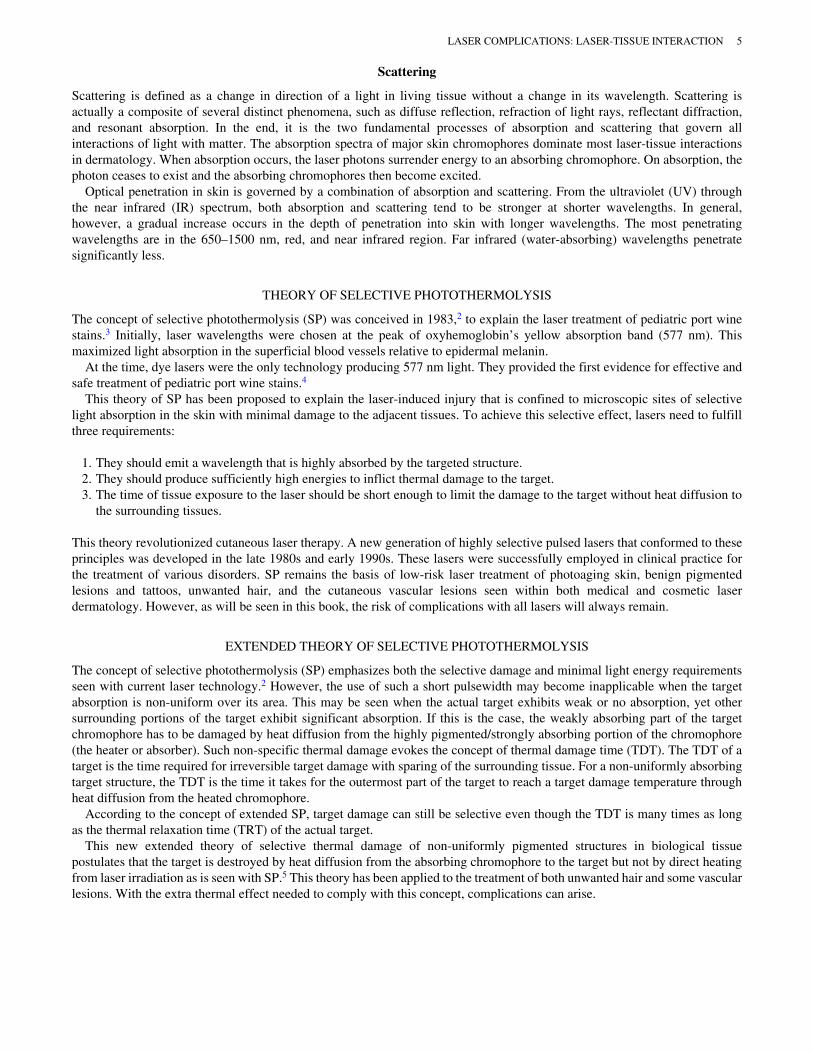

Figure 546 Linear streaks of hypopigmentation induced byintense pulsed light treatment of port wine stain
Figure 547 Pyogenic granuloma arising in port wine stainafter FLPDL treatment
Copyright © 2022 FDOKUMEN