Effects of decoy molecules targeting NF-kappaB transcription factors in Cystic fibrosis IB3–1 cells
Upload
khangminh22Category
view
3download
0
*For correspondence:
Competing interests: The
authors declare that no
competing interests exist.
Funding: See page 21
Received: 06 August 2020
Accepted: 28 January 2021
Published: 28 January 2021
Reviewing editor: Kiyoshi
Takeda, Osaka University, Japan
Copyright Kim et al. This
article is distributed under the
terms of the Creative Commons
Attribution License, which
permits unrestricted use and
redistribution provided that the
original author and source are
credited.
Induction of the IL-1RII decoy receptor byNFAT/FOXP3 blocks IL-1b-dependentresponse of Th17 cellsDong Hyun Kim1, Hee Young Kim1,2,3, Sunjung Cho2, Su-Jin Yoo4, Won-Ju Kim5,Hye Ran Yeon6, Kyungho Choi6, Je-Min Choi5, Seong Wook Kang4,Won-Woo Lee1,2,3,7*
1Laboratory of Autoimmunity and Inflammation (LAI), Department of BiomedicalSciences, Seoul National University College of Medicine, Seoul, Republic of Korea;2Department of Microbiology and Immunology, Seoul National University College ofMedicine, Seoul, Republic of Korea; 3Cancer Research Institute and Institute ofInfectious Diseases, Seoul National University College of Medicine, Seoul, Republicof Korea; 4Department of Internal Medicine, Chungnam National University Schoolof Medicine, 282 Munhwa-ro, Jung-gu, Daejeon, Republic of Korea; 5Department ofLife Science, College of Natural Sciences and Research Institute for NaturalSciences, Hanyang University, Seoul, Republic of Korea; 6Department ofBiochemistry and Molecular Biology, Department of Biomedical Sciences, andCancer Research Institute, Seoul National University College of Medicine, Seoul,Republic of Korea; 7Ischemic/Hypoxic Disease Institute, Seoul National UniversityCollege of Medicine; Seoul National University Hospital Biomedical ResearchInstitute, Seoul, Republic of Korea
Abstract Derived from a common precursor cell, the balance between Th17 and Treg cells must
be maintained within immune system to prevent autoimmune diseases. IL-1b-mediated IL-1
receptor (IL-1R) signaling is essential for Th17-cell biology. Fine-tuning of IL-1R signaling is
controlled by two receptors, IL-1RI and IL-RII, IL-1R accessory protein, and IL-1R antagonist. We
demonstrate that the decoy receptor, IL-1RII, is important for regulating IL-17 responses in TCR-
stimulated CD4+ T cells expressing functional IL-1RI via limiting IL-1b responsiveness. IL-1RII
expression is regulated by NFAT via its interaction with Foxp3. The NFAT/FOXP3 complex binds to
the IL-1RII promoter and is critical for its transcription. Additionally, IL-1RII expression is
dysregulated in CD4+ T cells from patients with rheumatoid arthritis. Thus, differential expression
of IL-1Rs on activated CD4+ T cells defines unique immunological features and a novel molecular
mechanism underlies IL-1RII expression. These findings shed light on the modulatory effects of IL-
1RII on Th17 responses.
IntroductionInterleukin-1 (IL-1) consists of two distinct forms (IL-1a and IL-1b) and is a master cytokine of local
and systemic inflammation (Dinarello et al., 2012). Primarily produced by monocytes, macrophages,
and dendritic cells (DCs) in response to stimulation, IL-1b is involved in a variety of biological activi-
ties such as cell proliferation, differentiation, and apoptosis through its binding to functional IL-1
receptor I (IL-1RI) expressed on virtually all cells. Thus, IL-1b plays a role as a critical pathogenic
mediator of many autoinflammatory, autoimmune, infectious, and degenerative
diseases (Dinarello, 2011).
Kim et al. eLife 2021;10:e61841. DOI: https://doi.org/10.7554/eLife.61841 1 of 29
RESEARCH ARTICLE
In human T-cell immunity, IL-1b has been identified as an essential cytokine with a role similar to
that played by IL-6 in mice for Th17 differentiation, and is also important for expansion, in vivo sur-
vival, and effector function of IL-17-producing T cells (Acosta-Rodriguez et al., 2007; Lee et al.,
2010). More recently, the importance of IL-1 signaling has been also emphasized in murine Th17
cells. Even though TGF-b and IL-6 are sufficient for murine Th17 differentiation, the IL-1RI knockout
(KO) mouse is resistant to experimental autoimmune encephalomyelitis due to a failure to generate
Th17 cells (Sutton et al., 2006; Chung et al., 2009). Further, IL-1b and IL-23 along with IL-6 are criti-
cal for generation of highly pathogenic Th17 cells in an autoimmune mouse model (Lee et al., 2012;
Ronchi et al., 2016). Recent studies have demonstrated that IL-1b functions as a proinflammatory
regulator of pathogenic human Th17 cells, which concomitantly produce IFN-g and GM-
CSF (Zielinski et al., 2012). Due to its potent effect as a proinflammatory cytokine, the biological
activity of IL-1b is tightly regulated at multiple levels by diverse mechanisms including receptor
antagonism through production of a decoy receptor and soluble forms of signaling receptors or
accessory proteins (Garlanda et al., 2013a).
The IL-1R signaling complex includes the functional IL-1RI subunit and a signaling subunit, inter-
leukin-1 receptor accessory protein (IL-1RAcP). Dimerization of the IL-1R1 and IL-1RAcP cytoplasmic
Toll/IL-1R (TIR) domains initiates a signaling cascade through recruitment of adapter
proteins (Garlanda et al., 2013b). The decoy IL-1RII has a short cytoplasmic tail lacking the signal-
transducing TIR domain and acts as a molecular trap, capturing IL-1 with high-affinity and exerting a
dominant-negative effect (Shirakawa et al., 1987). In contrast to ubiquitously expressed IL-1RI, the
expression of IL-1RII is restricted to cells including monocytes, neutrophils, and B cells (Martin et al.,
2017; Colotta et al., 1996; Colotta et al., 1993; McMahan et al., 1991). In addition, it was recently
demonstrated that human-activated Treg cells and follicular regulatory T (Tfr) cells upregulate IL-1RII
to attenuate their IL-1-dependent responses (Tran et al., 2009; Ritvo et al., 2017), suggesting a
physiological significance for IL-1RII expression in immune regulation.
The physiological relevance of IL-1RII expression was investigated in autoimmune arthritis animal
models using an IL-1RII conditional KO mouse. Deficiency of IL-1RII was found to exacerbate autoim-
mune arthritis by inhibiting IL-1 signaling in innate cells, such as macrophages and
neutrophils (Martin et al., 2017; Shimizu et al., 2015). Our previous study showed that IL-1RII
mRNA expression is markedly higher in IL-1RI+ memory CD4+ T cells compared to IL-1RI- memory
CD4+ T cells. Further, the negative regulatory role of IL-1RII was further evidenced by the significant
augmentation of IL-17 production by IL-1RI+ memory CD4+ T cells in response to IL-1b and TCR
stimulation after treatment with neutralizing IL-1RII antibody (Lee et al., 2010). Given its preferential
expression on IL-17-producing IL-1RI+ CD4+ T cells as well as Tregs, IL-1RII has been suggested to
play a pivotal role in maintaining the balance between Th17 and Treg cells. These cells are recipro-
cally interconnected in their development pathways and conversion between the two subsets is criti-
cal for pathogenesis and resolution of many inflammatory disorders (Zielinski et al., 2012;
Basu et al., 2015; Gagliani et al., 2015). However, little is known about the molecular mechanism
underlying IL-1RII expression by CD4+ T cells and its contribution to modulation of Th17 responses
in humans.
Here, we provide novel insight into the immune regulatory role of decoy IL-1RII expressed on
human CD4+ T cells by exploring its molecular mechanisms of action and analyzing its effect on
Th17 responses. TCR stimulation potently induces functional IL-1RI on memory CD4+ T cells and a
subset of IL-1RI+ CD4 T cells co-express decoy IL-1RII, conferring limited responsiveness to IL-1b. IL-
RII expression is mediated through binding of a cooperative complex of the transcription factors
NFAT and Foxp3 to its promoter. Differential expression of IL-1RI and IL-RII on activated CD4+ T
cells defines unique immunological features via modulation of IL-1 b responsiveness. Of note, de
novo expression of IL-1RI is aberrantly higher in synovial CD4+ T cells from RA patients, but TCR-
mediated induction of IL-1RII expression is impaired compared to peripheral CD4+ T cells from RA
patients or healthy controls. These findings underscore the regulatory role of decoy IL-1RII on IL-1b-
mediated IL-1R signaling affecting Th17 responses and contributing to pathogenesis of Th17-related
disorders.
Kim et al. eLife 2021;10:e61841. DOI: https://doi.org/10.7554/eLife.61841 2 of 29
Research article Immunology and Inflammation

Results
IL-1b is a crucial for promoting Th17 responses in humansTo investigate the role of IL-1b in promoting IL-17 production in human CD4+ T cells, purified mem-
ory CD4+ T cells from healthy donors were stimulated with anti-CD3/CD28-coated beads with or
without exogenous IL-1b. The frequency of IL-17-producing cells and their level of IL-17 production
were significantly increased by exogenous IL-1b treatment (Figure 1A and B) in a dose-dependent
manner (Figure 1—figure supplement 1A.). Consistent with previous reports (Lee et al., 2012;
Ronchi et al., 2016; Zielinski et al., 2012), IL-1b also significantly enhanced the frequency of IL-17/
IFN-g double producing cells, whereas IL-10-producing cells were decreased (Figure 1C and Fig-
ure 1—figure supplement 1B). Furthermore, IL-1b treatment led to an upregulated expression of
the genes which are crucial for pathogenic functions of Th17 cells such as IL-22, Casp1, and several
chemokines (Figure 1—figure supplement 1C). As seen in Figure 1D, Th17-polarizing cytokines,
such as IL-1b, IL-6 and IL-23, intensify the Th17 response of memory CD4+ T cells as observed by the
increased amount of IL-17 in the culture. This induction was substantially diminished in the absence
of IL-1b. The IL-17-promoting effect of IL-1b on human memory CD4+ T cells was not replaceable
with IL-6 or IL-23 (Figure 1D), indicating the essential role of this cytokine for the Th17 response in
memory CD4+ T cells. In contrast to this, Th17 differentiation from naive CD4+ T cells was efficiently
achieved in vitro by triggering the TCR in the presence of a mixture of Th17-polarizing cytokines,
although IL-17 was minimally produced in the absence of IL-1b (Figure 1E). Our findings demon-
strate that IL-1b is important for promoting Th17 responses of human memory CD4+ T cells and that
IL-1b-stimulated memory CD4+ T cells exhibit a major functional feature of pathogenic Th17 cells.
TCR stimulation induces the expression of IL-1 receptors by memoryCD4+ T cellsThe expression of receptors for IL-1b on CD4+ T cells is affected by various stimuli, which may play a
role in modulating responsiveness to IL-1b (Lee et al., 2010; Tran et al., 2009). We first examined
the expression profile of IL-1 receptors including functional IL-1RI and the decoy IL-1RII in ex vivo
and in vitro activated CD4+ T cells. Approximately 10% of ex vivo CD4+ T cells were found to
express IL-1RI, whereas no obvious expression of IL-1RII was observed on the same cells
(Figure 2A). These IL-1RI+ memory CD4+ T cells also had higher expression of IL-23R that is predom-
inantly expressed in IL-17-producing cells (Figure 2—figure supplement 1; Wilson et al., 2007). As
reported (Lee et al., 2010), IL-1RI-expressing CD4+ T cells are predominantly memory
CD45RA-CD4+ T cells (Figure 2B, p<0.001) and only a small fraction of naive CD4+ T cells express
IL-1RI. TCR stimulation strongly triggered expression of both IL1RI and IL-1RII by memory CD4+ T
cells, with rapid increase of their mRNA levels within 4 hr and sustained or even increased expression
until 24 hr post-stimulation (Figure 2—figure supplement 2A). Of interest, the kinetics of IL-1RI and
IL-1RII expression differ noticeably in that IL-1RI expression is significantly increased within 12 hr
after TCR stimulation, whereas induction of IL-1RII mainly occurs in IL-1RI-expressing cells after 36 hr
of stimulation (Figure 2C–D). To further examine the role of TCR stimulation on the expression of IL-
1Rs, CD4+ T cells were stimulated with different concentrations of anti-CD3 Ab for 48 hr. As seen in
Figure 2E, IL-1RI expression was maximal even at the lowest concentration of CD3 Ab, whereas IL-
1RII expression increased in a CD3 Ab concentration-dependent manner. Our data show that TCR-
stimulated memory CD4+ T cells can be subdivided into three subsets based on the expression of IL-
1RI and IL-1RII: IL-1RI+IL-1RII-, IL-1RI+IL-1RII+, and IL-1RI-IL-1RII- cells (Figure 2C). Around half of IL-
1R+ memory CD4+ T cells co-expressed decoy IL-1RII on day 2 when expression of the two receptors
peaked. In stark contrast with memory CD4+ T cells, no expression of IL-1RI or IL-1RII by naive CD4+
T cells was found to occur following TCR stimulation alone. Rather, Th17-polarizing cytokines were
required for induction of IL-1RI and IL-1RII by naive CD4+ T cells (Figure 2—figure supplement 2B).
We next tested whether induced IL-1RII could functionally modulate CD4+ T cell responsiveness to
IL-1b. As seen in Figure 2F, neutralizing antibody (Ab) to IL-1RII significantly enhanced the produc-
tion of IL-17 by memory CD4+ T cells in response to TCR and IL-1b co-stimulation. These findings
indicate that TCR triggering alone leads to induction of IL-1RI and IL-1RII by memory CD4+ T cells,
and both receptors are involved in regulating responsiveness to IL-1b.
Kim et al. eLife 2021;10:e61841. DOI: https://doi.org/10.7554/eLife.61841 3 of 29
Research article Immunology and Inflammation
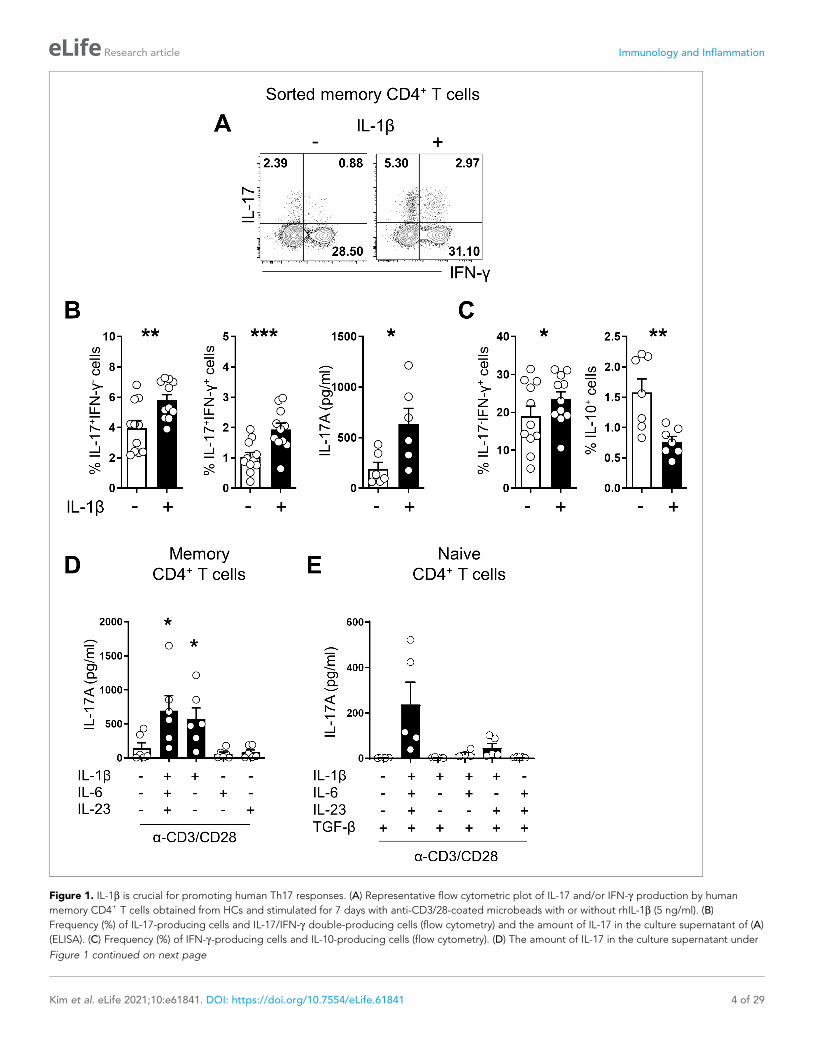
Figure 1. IL-1b is crucial for promoting human Th17 responses. (A) Representative flow cytometric plot of IL-17 and/or IFN-g production by human
memory CD4+ T cells obtained from HCs and stimulated for 7 days with anti-CD3/28-coated microbeads with or without rhIL-1b (5 ng/ml). (B)
Frequency (%) of IL-17-producing cells and IL-17/IFN-g double-producing cells (flow cytometry) and the amount of IL-17 in the culture supernatant of (A)
(ELISA). (C) Frequency (%) of IFN-g-producing cells and IL-10-producing cells (flow cytometry). (D) The amount of IL-17 in the culture supernatant under
Figure 1 continued on next page
Kim et al. eLife 2021;10:e61841. DOI: https://doi.org/10.7554/eLife.61841 4 of 29
Research article Immunology and Inflammation

Differential expression of IL-1Rs defines the immunological features ofCD4+ T cellsUpregulated IL-1RII expression has been reported in several types of Tregs in humans, indicating its
potential regulatory role in IL-1-dependent responses (Tran et al., 2009; Ritvo et al., 2017). Similar
to a recent report (Mercer et al., 2010), we found that CD4+ T cells expressing GARP, a marker of
activated Tregs, exhibit preferentially increased IL-1RII expression (Figure 3—figure supplement 1).
However, a subset of IL-1RI+ CD4+ T cells without GARP expression also exhibited IL-1RII expression
on cells in response to TCR stimulation (Figure 3—figure supplement 1A and B). Thus, this sug-
gests that IL-1RII expression is not strictly limited by activated Treg cells. To further investigate this
finding, IL1RII expression was analyzed on conventional memory CD4+ T cells from which
CD25hiCD127dim/- Tregs were removed. Although CD25hiCD127(dim/-) and foxp3+ Treg cells might
be not exactly overlapped, foxp3+ cells were markedly removed by our depletion protocol (Fig-
ure 3—figure supplement 2) and the residual foxp3+ T cells mainly belonged to non-suppressive
cytokine-producing Foxp3low T cells (III in Figure 3—figure supplement 2C) by the definition of the
Sakaguchi group. In fact, foxp3hi-activated Treg cells (II in Figure 3—figure supplement 2C) were
mostly removed by our Treg-depletion protocol. TCR triggering led to the induction of IL-1RII
expression even by Treg-depleted T cells (Figure 3A). Of note, these IL-1RII+ memory CD4+ T cells
have higher expression of Foxp3 that could be induced in Treg-depleted conventional CD4+ T cells
with Smad3- and NFAT-dependent manner (Figure 3—figure supplement 3) and other Treg-related
markers such as CD39, CD73, and CTLA-4 (Figure 3B) compared with the other two subsets. To
characterize the features of IL-1RI+IL-1RII+ memory CD4+ T cells, chemokine receptors and cytokine
expression was analyzed in three populations (IL-1RI+IL-1RII-, IL-1RI+IL-1RII+, and IL-1RI-IL-1RII- cells).
As seen in Figure 3C, around 50% of IL-1RI+IL-1RII- and IL-1RI+IL-1RII+ had a Th17-associlated phe-
notype (CD161+CCR6+), whereas IL-1RI-IL-1RII- cells mainly consist of Th1 (CD161-CXCR3+CCR6-)
and non-Th1/17 subsets. Moreover, a higher frequency of IL-1RI+IL-1RII+ cells showed a phenotype
typical of ex-Th17 cells (CD161+CXCR3+CCR6+), whereas the IL-1RI+IL-1RII- subset included more
cells with the Th17 phenotype (CD161+CXCR3-CCR6+)(Basdeo et al., 2017). Although the frequency
of IL-23R was comparable among three populations, its expression level was significantly higher in
IL-1RI+IL-1RII- cells than IL-1RI+IL-1RII+, and IL-1RI-IL-1RII- cells (Figure 3—figure supplement 4).
Intracellular cytokine staining (ICS) data revealed that IL-1RI+IL-1RII- and IL-1RI+IL-1RII+ cells had dif-
ferent cytokine production profiles, showing that IL-1RI+IL-1RII+ consists of fewer IL-17-producing,
but more IFN-g-producing, cells than IL-1RI+IL-1RII- cells (Figure 3D). Our findings demonstrate that
IL-1RII is inducible by activated memory CD4+ T cells, associated with Foxp3 expression, and IL-1RII+
CD4+ T cells have unique immunological features.
NFAT and Foxp3 are essential for IL-1RII expression in memory CD4+ TcellsSince TCR-induced Foxp3+ T cells preferentially express IL-1RII (Figure 3A), we hypothesized that
Treg-related transcriptional factors (TFs) are involved in IL-1RII gene expression. In preliminary
experiments using inhibitors for TFs related to Treg cells, we found that cyclosporin A (CsA), an
inhibitor of calcineurin, selectively represses the frequency of IL-1RII+ cells among TCR-induced IL-
1RI+ memory CD4+ T cells in a dose-dependent manner (Figure 4A and B). The immunosuppressive
Figure 1 continued
the indicated cytokine conditions (ELISA): rhIL-6 (25 ng/ml), rhIL-23 (25 ng/ml), and rhL-1b (5 ng/ml). (E) Purifed naive CD4+ T cells were stimulated for 7
days with anti-CD3/28-coated microbeads in serum-free X-VIVO 10 medium under the indicated cytokine conditions. IL-17 in the culture supernatant
(ELISA): rhIL-6 (25 ng/ml), rhIL-23 (25 ng/ml), rhL-1b (5 ng/ml), and rhTGF-b (10 ng/ml). Bar graphs show the mean ± SEM of six (A–D), and five (E)
independent experiments. * = p<0.05, ** = p<0.01, and *** = p<0.001 by two-tailed paired t-test.
The online version of this article includes the following source data and figure supplement(s) for figure 1:
Source data 1. Figure 1B IL-1b significantly enhance IL-17 & IFN-g producing memory CD4+ T cells.
Source data 2. Figure 1C IL-1b regulated IL-10 producing memory CD4+ T cells.
Source data 3. Figure 1D Th17-polarizing cytokines intensify the Th17 response of memory CD4+ T cells.
Source data 4. Figure 1E IL-1b is important for promoting Th17 differentiation form naıve CD4+ T cells.
Figure supplement 1. IL-1b is a crucial cytokine for promoting Th17 responses in humans.
Kim et al. eLife 2021;10:e61841. DOI: https://doi.org/10.7554/eLife.61841 5 of 29
Research article Immunology and Inflammation
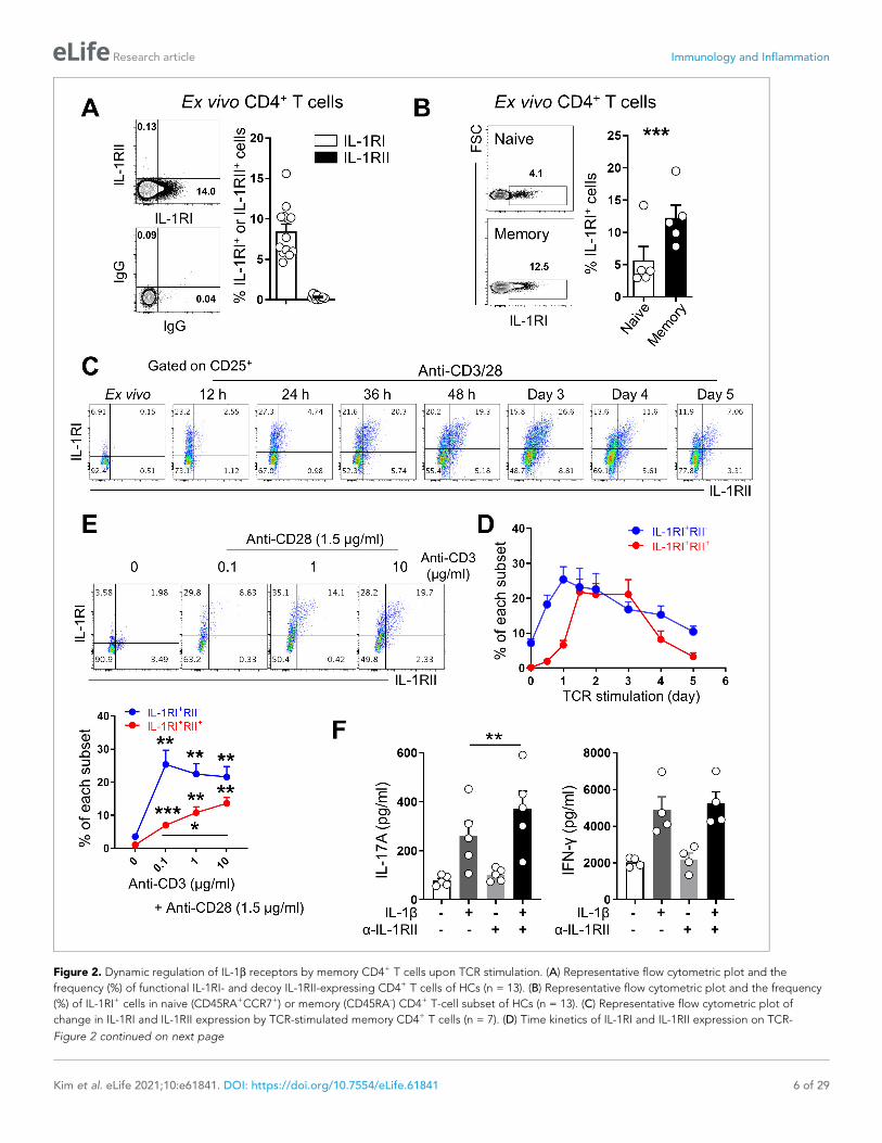
Figure 2. Dynamic regulation of IL-1b receptors by memory CD4+ T cells upon TCR stimulation. (A) Representative flow cytometric plot and the
frequency (%) of functional IL-1RI- and decoy IL-1RII-expressing CD4+ T cells of HCs (n = 13). (B) Representative flow cytometric plot and the frequency
(%) of IL-1RI+ cells in naive (CD45RA+CCR7+) or memory (CD45RA-) CD4+ T-cell subset of HCs (n = 13). (C) Representative flow cytometric plot of
change in IL-1RI and IL-1RII expression by TCR-stimulated memory CD4+ T cells (n = 7). (D) Time kinetics of IL-1RI and IL-1RII expression on TCR-
Figure 2 continued on next page
Kim et al. eLife 2021;10:e61841. DOI: https://doi.org/10.7554/eLife.61841 6 of 29
Research article Immunology and Inflammation
effect of CsA is mediated by inhibiting calcineurin-mediated dephosphorylation of the nuclear factor
of activated T-cells (NFATc), which plays a critical role in peripheral activation and differentiation of
Tregs (Kiani et al., 2000). To further investigate whether NFAT is directly involved in IL-1RII gene
expression, purified memory CD4+ T cells were treated with cell-permeable peptide (CPP)-conju-
gated VIVIT, a selective and potent inhibitor of calcineurin/NFAT interaction, and stimulated with
anti-CD3/CD28 mAbs-coated beads for 24 hr. As seen in Figure 4C and D, expression of IL-1RII
mRNA was completely abrogated and its protein level was also significantly diminished. We next
investigated if Foxp3, a master TF in Tregs, is required for IL-1RII gene expression. Consistent with a
previous report (Kang et al., 2012), 1,25-Dihydroxyvitamin D3 [1,25(OH)2VD3] promoted Foxp3
expression in memory CD4+ T cells following TCR stimulation in the presence of IL-2, and induction
of Foxp3 was clearly attenuated by CsA treatment (Figure 4E). Moreover, the expression of IL-1RII,
but not IL-1RI, was found to be significantly upregulated by treatment with 1,25(OH)2VD3 and IL-2 in
memory CD4+ T cells at day 2 post-stimulation and resultantly, the ratio of IL-1RII+ to IL-1RI+
increased two fold (Figure 4F–H). This suggests that Foxp3 upregulation is associated with induction
of IL-1RII. Furthermore, our findings demonstrate that NFAT and Foxp3 are essential for expression
of IL-1RII by memory CD4+ T cells in humans.
NFAT/FOXP3 interaction is responsible for expression of IL-1RII byhuman memory CD4+ T cellsIt has been demonstrated that Treg activity is modulated by a cooperative complex of NFAT and
FOXP3(27). Since both TFs are critical for IL-1RII expression (Figure 4), we next examined whether
formation of a cooperative NFAT/FOXP3 complex is responsible for upregulation of IL-1RII expres-
sion on memory CD4+ T cells upon TCR stimulation. To this end, peptide FOXP3 393–403, a specific
inhibitor of the NFAT/FOXP3 interaction, was used to treat TCR-stimulated memory CD4+ T cells for
48 hr. Although this peptide inhibitor was initially designed for murine Tregs, this same inhibitory
effect was verified for the human NFAT/FOXP3 interaction through inhibition of CD25 induction, a
target of this complex (Figure 5A), as described in a previous report (Lozano et al., 2015). Our data
show that peptide FOXP3 393–403 significantly inhibits IL-1RII expression by approximately 50%
compared with the control peptide, FOXP3 399A, (Figure 5B and C). Thus, this confirms the impor-
tant role of the NFAT/FOXP3 complex in IL-1RII expression.
To determine whether the NFAT/FOXP3 complex directly binds to the IL-1RII gene, in silico
screening was conducted for potential NFAT and FOXP3-binding sites in the minimal promoter of
the human IL-1RII gene using PROMO 3.0 software, an algorithm that predicts nuclear-receptor-
binding elements (Messeguer et al., 2002). Potential response elements for NFAT and FOXP3 were
closely located in the conserved non-coding sequence (CNS) region (�1309 to �1203) of the IL-1RII
gene (Figure 5—figure supplement 1), implying the importance of this region for IL-1RII expression.
Three luciferase reporter constructs (Figure 5D) containing 1.3 kb (pGL4/IL-1RII-a1414), 0.7 kb
(pGL4/IL-1RII-a814), and 0.1 kb (pGL4/IL-1RII-a215) of the IL-1RII promoter were transduced into the
Jurkat leukemic T-cell line. Due to a lack of Foxp3 expression in conventional Jurkat cells, we gener-
ated a Foxp3-expressing Jurkat cell line (Foxp3-Jurkat) using lentiviral vector transduction
Figure 2 continued
stimulated memory CD4+ T cells (n = 5). (E) Representative flow cytometric plot and the frequency (%) of IL-1RI and IL-1RII expression by memory CD4+ T cells in response to stimulation with different concentrations of anti-CD3 and 1.5 mg/ml of anti-CD28 Ab for 48 hr (n = 5). (F) The amounts of IL-17A
(left) and IFN-g (right) in culture supernatants of memory CD4+ T cells stimulated with anti-CD3/28-coated microbeads for 7 days with or without IL-1b
(n = 5). Anti-IL-1RII neutralizing Ab or control isotype Ab was added into the culture at day 2 post-stimulation. Bar graphs and line graphs show the
mean ± SEM. * = p<0.05, ** = p<0.01, and *** = p<0.001 by two-tailed paired t-test.
The online version of this article includes the following source data and figure supplement(s) for figure 2:
Source data 1. Figure 2A Ex vivo expression of IL-1RI & IL-1RII on CD4+ T cells.
Source data 2. Figure 2B Ex vivo expression of IL-1RI between naıve and memory CD4+ T cells.
Source data 3. Figure 2B Ex vivo expression of IL-1RI between naıve and memory CD4+ T cells.
Source data 4. Figure 2D Time kinetics of IL-1RI & IL-1RII.
Source data 5. Figure 2E Effect of TCR signaling strength on expression of IL-1RI & IL-1RII.
Figure supplement 1. The differential expression of IL-23R on IL-1RI+ and IL-1RI- memory CD4+ T cells in humans.
Figure supplement 2. The expression of receptors for IL-1b is dynamically changed by various stimulations of CD4+ T cells.
Kim et al. eLife 2021;10:e61841. DOI: https://doi.org/10.7554/eLife.61841 7 of 29
Research article Immunology and Inflammation
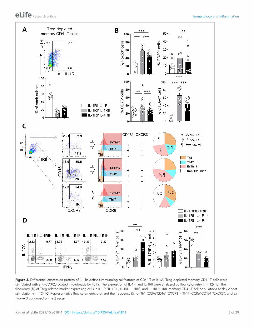
Figure 3. Differential expression pattern of IL-1Rs defines immunological features of CD4+ T cells. (A) Treg-depleted memory CD4+ T cells were
stimulated with anti-CD3/28-coated microbeads for 48 hr. The expression of IL-1RI and IL-1RII were analyzed by flow cytometry (n = 12). (B) The
frequency (%) of Treg-related marker-expressing cells in IL-1RI+IL-1RII-, IL-1RI+IL-1RII+, and IL-1RI-IL-1RII- memory CD4+ T cell populations at day 2 post-
stimulation (n = 12). (C) Representative flow cytometric plot and the frequency (%) of Th1 (CCR6-CD161-CXCR3+), Th17 (CCR6+CD161+CXCR3-), and ex-
Figure 3 continued on next page
Kim et al. eLife 2021;10:e61841. DOI: https://doi.org/10.7554/eLife.61841 8 of 29
Research article Immunology and Inflammation
(Figure 5—figure supplement 2A). IL-1RII expression was markedly enhanced in Foxp3-Jurkat cells
following stimulation with PMA and ionomycin (Figure 5—figure supplement 2B and C), which are
known to cause NFAT activation (Brignall et al., 2017). As seen in Figure 5D, Foxp3-Jurkat cells
transfected with pGL4/IL-1RII-a1414 including the CNS region exhibited significantly higher lucifer-
ase activity compared with the other two constructs. We determined the functional relevance of
these TF binding sites in the CNS region of the IL-1RII promoter by mutational analysis. Mutation of
three bases within each NFAT binding site in the CNS region resulted in a significant decrease in IL-
1RII promoter activity (Figure 5D), indicating the importance of these sites for promoter function.
To further investigate whether NFAT and Foxp3 bind the predicted binding sites in the IL-1RII
promoter region, we performed a CHIP assay with TCR-stimulated memory CD4+ T cells. NFAT
recruitment to the DNA response element (NFAT B1: �1209 to �1188) in the CNS region of IL-1RII
promoter was significantly enriched upon TCR stimulation, whereas Foxp3 was observed to bind a
slight distant from the CNS region (Foxp3 B2: �425 to �418) of IL-1RII promoter (Figure 5E and F).
To further confirm this finding, NFAT recruitment to the B1 site were evaluated in Jurkat cells with
or without foxp3 expression. NFAT recruitment to the NFAT BC (binding control), a binding site in
human IL-2 promoter region, were comparable between Jurkat cells with or without foxp3 expres-
sion, whereas NFAT recruitment to the B1 site were significantly enriched in Foxp3-expressing Jurkat
cells upon stimulation with PMA and ionomycin compared with Jurkat cells without foxp3 expression
(Figure 5—figure supplement 3). Collectively, these results suggest that NFAT and Foxp3 bind
directly to the IL-1RII promoter and regulate its transcription.
IL-1RII ameliorates IL-1b-mediated Th17 responses and confers featuresof Tregs to IL-1RI+ memory CD4+ T cellsOur data thus far demonstrate that the induction of IL-1RII on IL-1RI+ memory CD4+ T cells is con-
trolled by NFAT/FOXP3, which plays an important role in the induction of Tregs in humans, and this
induction leads to weakened IL-1b-mediated Th17 responses. Accumulating evidence demonstrates
that Th17 and Treg cells display instability and plasticity and can interconvert under specific condi-
tions to modulate immune responses (Gagliani et al., 2015; Obermajer et al., 2014). Therefore, we
next investigated how IL-1RII expression influences the balance between Th17 and Treg cells. To
address this question, Treg-depleted memory CD4+ T cells were stimulated for 48 hr to induce the
expression of IL-1RI and IL-1RII. Three different subsets, IL-1RI+IL-1RII-, IL-1RI+IL-1RII+, and IL-1RI-IL-
1RII- cells, were purified by cell sorting, and stimulated through their TCR for 5 days in the presence
of IL-1b. Following exposure to IL-1b, IL-1RI+ cells were the major IL-17 producing population
regardless of IL-1RII expression, whereas IL-1RI-IL-1RII- cells predominantly produced IFN-g
(Figure 6A and B). IL-1RII expression, however, attenuated the Th17 response of IL-1RI+ cells, as
shown by a reduced frequency of IL-17-producing cells, and in particular IL-17 and IFN-g dual pro-
ducing cells known as typical pathogenic Th17 cells (Figure 6A). Our qPCR assay showed several
pathogenic Th17 cell-associated genes including IL-22, CCL3, and CSF2 were upregulated in IL-1RI+
IL-1RII- cells than IL-1RI+ IL-1RII+ cells (Figure 6—figure supplement 1). In contrast, increased
expression of Foxp3 was observed in IL-17-producing IL-1RI+RII+ compared with IL-1RI+ILRI- cells,
Figure 3 continued
Th17 (CCR6+CD161+CXCR3+) cells in IL-1RI+IL-1RII-, IL-1RI+IL-1RII+, and IL-1RI-IL-1RII- memory CD4+ T cell populations (n = 9). (D) Intracellular cytokine
staining (ICS) of IL-17- and IFN-g in IL-1RI+IL-1RII-, IL-1RI+IL-1RII+, and IL-1RI-IL-1RII- memory CD4+ T cell populations (n = 4). Bar graphs and pie charts
show the mean ± SEM. * = p<0.05, ** = p<0.01, and *** = p<0.001 by two-tailed paired t-test. †, ‡, and ¶ = p<0.05: compared between indicated
subsets by two-tailed paired t-test (C).
The online version of this article includes the following source data and figure supplement(s) for figure 3:
Source data 1. Figure 3A Frequency of IL-1RI+IL-1RII-, IL-1RI+IL-1RII+, and IL-1RI-IL-1RII- subset of Treg-depleted memory CD4+ T cells.
Source data 2. Figure 3B Expression of Treg related markers on L-1RI+IL-1RII-, IL-1RI+IL-1RII+, and IL-1RI-IL-1RII- subsets.
Source data 3. Figure 3C Frequency of ex-Th17, Th17, and Th1 of L-1RI+IL-1RII-, IL-1RI+IL-1RII+, and IL-1RI-IL-1RII- subsets.
Source data 4. Figure 3D Expression of IL-17 & IFN-g in L-1RI+IL-1RII-, IL-1RI+IL-1RII+, and IL-1RI-IL-1RII- subsets.
Figure supplement 1. CD4+ T cells expressing GARP, a marker of activated Tregs, preferentially, but not exclusively, increase IL-1RII expression.
Figure supplement 2. Efficiency of sorting-based Treg depletion from CD4+ T cells.
Figure supplement 3. Critical roles of Smad3 and NFAT for Foxp3 expression in non-Treg memory CD4+ T cells.
Figure supplement 4. The correlation of IL-23R with IL-1RI+IL-1RII+ and IL-1RI+IL-1RII- subset of memory CD4+ T cells.
Kim et al. eLife 2021;10:e61841. DOI: https://doi.org/10.7554/eLife.61841 9 of 29
Research article Immunology and Inflammation

Figure 4. NFAT and Foxp3 are essential for IL-1RII expression in memory CD4+ T cells. (A and B) Representative flow cytometric plot (A) and the
frequency (B) of IL-1RI and IL-1RII expression on TCR-stimulated memory CD4+ T cells treated with the indicated concentration of CsA, a chemical
inhibitor of NFAT at day 2 post-stimulation (n = 5). (C) The expression of IL-1RII mRNA in TCR-stimulated or unstimulated memory CD4+ T cells in the
presence or absence of CPP-VIVIT (1 mM), NFAT-specific peptide inhibitor (n = 4). (D) The expression of CD25, IL-1RI, and IL-1RII on TCR-stimulated
Figure 4 continued on next page
Kim et al. eLife 2021;10:e61841. DOI: https://doi.org/10.7554/eLife.61841 10 of 29
Research article Immunology and Inflammation
implying that IL-1RII is associated with modulating the balance between Th17 cells and Tregs
(Figure 6C). Confirmatory FACS analysis revealed that CTLA -4 expression on IL-1RI+IL-1RII+ cells is
significantly higher than on IL-1RI+IL-1RII-, whereas IL-1RI+IL-1RII- cells had higher frequencies of cells
expressing CCR6 and CD161, Th17-related markers, than did IL-1RI+IL-1RII+ cells (Figure 6D).
Although the receptor of IL-23, which is a critical cytokine for the differentiation, commitment, and
survival of Th17 cells, was upregulated in IL-1RI+IL-1RII- cells (Figure 3—figure supplement 4), the
increase in IL-17 production by IL-23 was minimal in the in vitro system. This may be because IL-23
receptor expression on IL-1RI+ cells is limited and the induction kinetic of IL-23R is different from IL-
1Rs (Figure 6—figure supplement 2). These data demonstrate that the expression of IL-1RII plays a
critical role in maintaining the balance between Th17 and Treg cells in humans.
Aberrant expression of IL-1Rs in synovial CD4+ T cells in patients withrheumatoid arthritis (RA)It has been reported that the frequency of Foxp3+CD4+ T cells is markedly increased in the synovial
cavity of RA patients and these cells are able to produce IL-17 (Komatsu et al., 2014; Du et al.,
2014; Afzali et al., 2013). Furthermore, IL-1b is obviously elevated in synovial fluid (SF) of RA
patients (Kim et al., 2016). This finding prompted us to examine the expression profile of IL-1RI and
IL-1RII in synovial and peripheral CD4+ T cells in RA patients. The frequency of IL-1RI-expressing cells
was significantly higher in ex vivo memory CD4+ T cells derived from synovial fluid (16.35 ± 1.70%)
and peripheral blood (14.49 ± 1.20%) of RA patients compared with counterpart cells (7.11 ± 0.85%;
p<0.001) from age-match healthy controls (HCs) (Figure 7A and B). Like its expression on peripheral
CD4+ T cells of HCs, IL-1RII was minimally expressed by ex vivo CD4+ T cells of RA patients, with the
exception of a small population of synovial CD4+ T cells that expressed higher levels of IL-1RII, indi-
cating their activated state. Of importance, in IL-1RI+ cells TCR-mediated induction of IL-1RII was sig-
nificantly impaired in synovial CD4+ T cells compared with peripheral CD4+ T cells of HCs and even
of RA patients (Figure 7C and D). As a consequence, the ratio of IL-1RI+IL-1RII+ to IL-1RI+IL-1RII-
was lower in RA patients than HCs (Figure 7E). Given that the frequency of foxp3+ CD4+ T cells in
PBMC of HC was comparable to that in RA PBMC and was even significant lower than that in RA
SFMC, impaired expression of IL-1RII in RA memory CD4+ T cells was not likely to be caused by the
reduced frequency of Foxp3+ Treg cells (Figure 7—figure supplement 1). As seen in Figure 7F, the
reduced induction of IL-1RII on TCR-stimulated memory CD4+ T cells in RA patients is likely linked to
augmented induction (3.3 fold-increase) of IL-1b-mediated IL-17 production in response to TCR stim-
ulation compared with HCs (2.4 fold-increase) (Figure 7F) because this induction of RA patients was
observed at a comparable level to that of CD4+ T cells of HCs with anti-IL-1RII neutralizing Ab
(Figure 7F and G). This data suggests that TCR-induced IL-1RII is associated with modulation of IL-
17 production in human CD4+ T cells.
DiscussionTh17 cells are a unique subset of CD4+ effector T cells that elicit protective immune responses
against extracellular bacterial and fungal pathogens (Gaffen, 2009). Th17 cells have been also impli-
cated in the pathogenesis of many autoimmune disorders (Lee et al., 2012; Tabarkiewicz et al.,
Figure 4 continued
memory CD4 T cells in the presence or absence of CPP-VIVIT (n = 3). (E) Representative histogram plot of Foxp3 in TCR-stimulated memory CD4+ T
cells in the presence or absence of 1,25-dihydroxyvitamin D3 (VD3; 10 mM) and rhIL-2 (250 IU/ml) with or without CsA. MFI indicates mean fluorescent
intensity. (F) Flow cytometric analysis of IL-1RI and IL-1RII on memory CD4+ T cell under same conditions as in (E). (G and H) The frequencies (%) of IL-
1RI+ and IL-1RII+ cells and the ratio of IL-1RII+ to IL-1RI+ cells under same conditions in (E) (n = 6). Bar graphs show the mean ± SEM. * = p<0.05, ** =
p<0.01, and *** = p<0.001 by two-tailed paired t-test.
The online version of this article includes the following source data for figure 4:
Source data 1. Figure 4A NFAT inhibitor CsA selectivley repress the expression of IL-1RII.
Source data 2. Figure 4B NFAT inhibiton peptide VIVIT repress the expression of IL-1RII.
Source data 3. Figure 4G expression of IL-1RII significantly upregulated by treatment with 1,25(OH)2VD3 and IL-2 in memory CD4+ T cells.
Source data 4. Figure 4H Ratio of IL-1RII+/IL-1RI+.
Kim et al. eLife 2021;10:e61841. DOI: https://doi.org/10.7554/eLife.61841 11 of 29
Research article Immunology and Inflammation

Figure 5. NFAT/FOXP3 interaction is responsible for expression of IL-1RII by human memory CD4+ T cells. (A) The frequency (%) of CD25, a NFAT-
dependent molecule, on TCR-stimulated memory CD4+ T cells in the presence of Foxp3 393–403 peptide (100 mM), NFAT/FOXP3 interaction inhibition
peptide, or control Foxp3 399 peptide (100 mM) at day 2 post-stimulation (n = 4). (B and C) Representative flow cytometric plot (B) and the frequencies
(C) of IL-1RI and IL-1RII expression on TCR-stimulated memory CD4+ T cells in the presence of Foxp3 393–403 peptide or control Foxp3 399 peptide at
Figure 5 continued on next page
Kim et al. eLife 2021;10:e61841. DOI: https://doi.org/10.7554/eLife.61841 12 of 29
Research article Immunology and Inflammation
2015). Unlike conventional Th1 and Th2 cells, Th17 cells exhibit a substantial degree of instability,
heterogeneity, and plasticity (Stadhouders et al., 2018). Many molecular mediators are closely
involved in regulating plasticity of Th17 cells (Lee et al., 2012; Wang et al., 2015; Ichiyama et al.,
2016; Stockinger and Omenetti, 2017). Among these, cytokines are critical determinants in the
fate of Th17 cells. TGF-b and IL-6 give rise to nonpathogenic or beneficial Th17 cells, whereas IL-1b,
IL-6 and IL-23 are involved in generation of pathogenic Th17 cells (Lee et al., 2012). Further, cell-
fate mapping mouse models demonstrate that pathogenic Th17 cells can further convert into Th1
cells producing IFN-g, but not IL-17 (ex-Th17 Th1 cells), in an IL-23-dependent manner (Hirota et al.,
2011), whereas TGF-b is critical for transdifferentiation of Th17 cells into Treg cells (ex-Th17 Treg
cells) during the resolution of inflammation or in the cancer microenvironment (Gagliani et al., 2015;
Downs-Canner et al., 2017).
IL-1b is an essential cytokine for Th17 biology in humans that boosts Th17 production, induces
pathogenic Th17 cells, and impairs the Th17-Treg balance (Basu et al., 2015). Activities of IL-1 are
tightly controlled at different levels due to its potent proinflammatory effects. IL-1 receptor antago-
nist (IL-1Ra) and IL-1RII are negative regulators of the IL-1 system. IL-1Ra binds functional IL-1RI with
no recruitment of IL-1RAcP, resulting in the repression of IL-1 binding in a competitive manner,
whereas the decoy receptor, IL-1RII cannot transduce the signal initialized by the IL-1RI and IL-1Ra
complex (Schluter et al., 2018). We previously reported that the expression of IL-1RI and IL-1RII on
CD4+ T cells is dynamically regulated in response to environmental factors such as TCR or cytokine
stimulation, and such regulation influences Th17 cell responses (Lee et al., 2010). Furthermore, IL-
1RII mRNA is expressed at a higher level in IL-1RI+CD4+ T cells than in IL-1RI-CD4+ T cells and block-
ade by IL-1RII leads to reduced IL-17 production (Lee et al., 2010). However, the mechanism modu-
lating IL-1RII expression and its role during the immune response remains unclear.
The expression of IL-1 and IL-1RII is tightly restricted under steady-state conditions, but is upre-
gulated during inflammation (Schluter et al., 2018). In contrast to IL-1RI, which is ubiquitously
expressed at least at low levels, IL-1RII expression is confined to certain cell types, including mono-
cytes, macrophages, some T cells in humans and microglial cells, osteoclasts and neutrophils in
mice. Early studies showed that anti-inflammatory signals such as IL-4 and IL-13, as well as dexa-
methasone stimulate IL-1RII expression by monocytes and macrophages, suggesting its contribution
to the anti-inflammatory effect (Colotta et al., 1996; Colotta et al., 1993). IL-1RII can be co-
expressed with IL-1RI on the cell surface, albeit at a low copy number, and counterpoises the system
by secluding IL-1 and IL-1RAcP from the neighboring signaling receptor IL-1RI (Colotta et al., 1993;
Neumann et al., 2000). A recent study demonstrated that the cytosolic form of IL-1RII, expressed in
certain types of cells, interacts with pro-IL-1a preventing cleavage and its calpain-dependent
Figure 5 continued
day 2 post-stimulation (n = 4). (D) Foxp3-expressing Jurkat cells were transfected with the indicated pGL4.10 luciferase vectors (pGL4/IL-1RII-a1414,
pGL4/IL-1RII-a814, and pGL4/IL-1RII-a215) and pGL4.74 control renilla vector as internal control, followed by stimulation with PMA and ionomycin for 48
hr (upper panel: n = 4). Comparison of luciferase activity between pGL4/IL-1RII-a1414 and pGL4/IL-1RII-a1414mu (�1209 to �1188 NFAT-binding motif
mutant) transfected Foxp3-expressing Jurkat cells (lower panel: n = 4). Luciferase activity was determinded using dual lucifease assay system. (E and F)
Purified memory CD4+ T cells were stimulated with plate-bound anti-CD3/28 mAbs for 24 hr. ChIP qPCR was perfomed using anti-NFATc2 or anti-
Foxp3 Ab at the IL-1RII promoter region. Enrichment of NFAT within four putative NFAT binding motifs (E) and enrichment of Foxp3 within three
putative Foxp3-binding motifs (F) was analyzed by qPCR (n = 5 or 6). There are two (�1209 ~ �1188 of IL-1RII promoter), one (�649 ~ �639), and one
(+10 ~ +18) binding sites for NFAT B1, B2 and B3, respectively. NFAT BC (binding control) has a binding site in human IL-2 promoter region. There are
three (�4652 ~ �4611), one (�1236 ~ �1230), and one (�425 ~ �418) binding sites for Foxp3 B1, B2 and B3, respectively. Foxp3 BC (binding control)
has a binding site in the human IL-2Ra promoter region (Zhang et al., 2013). Bar graphs show the mean ± SEM. * = p<0.05, ** = p<0.01, and *** =
p<0.001 by two-tailed paired t-test.
The online version of this article includes the following source data and figure supplement(s) for figure 5:
Source data 1. Figure 5A The inhibitory effect of FOXP3 393–403, a specific inhibitor of the NFAT/FOXP3 interaction.
Source data 2. Figure 5C NFAT/Foxp3 interaction inhibitor significantly repress the IL-1RII expression.
Source data 3. Figure 5D IL-1RII promoer activity measured via luciferase assay.
Source data 4. Figure 5E and F Result of NFAT & Foxp3 ChIP-qPCR via IL-1RII promter.
Figure supplement 1. Analysis of potential binding sites of NFAT and Foxp3 in the minimal promoter of the human IL-1RII gene and schematic
diagram of three luciferase reporter constructs.
Figure supplement 2. Overexpression of Foxp3 in Jurkat cells leads to an increase in the expression of IL-1RII.
Figure supplement 3. Cooperation of NFAT/Foxp3 via binding of NFAT on NFAT B1 site.
Kim et al. eLife 2021;10:e61841. DOI: https://doi.org/10.7554/eLife.61841 13 of 29
Research article Immunology and Inflammation
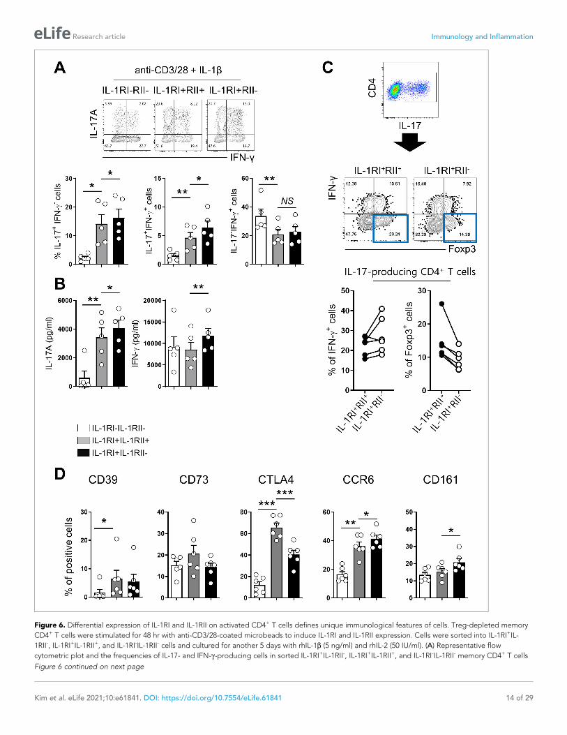
Figure 6. Differential expression of IL-1RI and IL-1RII on activated CD4+ T cells defines unique immunological features of cells. Treg-depleted memory
CD4+ T cells were stimulated for 48 hr with anti-CD3/28-coated microbeads to induce IL-1RI and IL-1RII expression. Cells were sorted into IL-1RI+IL-
1RII-, IL-1RI+IL-1RII+, and IL-1RI-IL-1RII- cells and cultured for another 5 days with rhIL-1b (5 ng/ml) and rhIL-2 (50 IU/ml). (A) Representative flow
cytometric plot and the frequencies of IL-17- and IFN-g-producing cells in sorted IL-1RI+IL-1RII-, IL-1RI+IL-1RII+, and IL-1RI-IL-1RII- memory CD4+ T cells
Figure 6 continued on next page
Kim et al. eLife 2021;10:e61841. DOI: https://doi.org/10.7554/eLife.61841 14 of 29
Research article Immunology and Inflammation

maturation and influences necrosis-induced sterile inflammation (Zheng et al., 2013). In a mouse
model, IL-1RII deficiency on neutrophils and macrophages was found to aggravate K/BxN serum
transfer-induced arthritis and collagen-induced arthritis by inhibiting IL-1 signals (Martin et al.,
2017; Shimizu et al., 2015). However, the role of IL-RII on T cells remains to be understood.
We found that TCR-mediated induction of IL-1RI kinetically precedes IL-1RII expression by 12–24
hr and the induction of IL-1RII is predominantly observed in IL-1RI-expressing cells (Figure 2C and
D). This suggests cis-regulation by IL-1RII involves its competitive binding to IL-1 and IL-1RAcP on
CD4+ T cells under inflammatory conditions. Furthermore, treatment with IL-1b, but not other cyto-
kines, further increased the expression of IL-1RII in TCR-stimulated CD4+ memory T cells, supporting
the idea that there is a negative feedback loop of IL-1 signaling in CD4+ T cells (Figure 2—figure
supplement 2B).
Of note, IL-1RII expression on memory CD4+ T cells was found to correlate with the dose of anti-
CD3 mAb used for stimulation, whereas IL-1RI expression was significantly increased following weak
anti-CD3 stimulation (100 ng/ml) (Figure 2E). Purvis et al., 2010 demonstrated that low-strength
stimulation of human CD4+ T cells in a Th17-polarizing cytokine milieu strongly favors Th17
responses. They proposed that low Ca2+ signaling in CD4+ T cells under low-strength stimulation is
preferential for the increase in binding of NFAT to the proximal region of the IL-17 promoter in
CD4+ T cells, thereby enhancing IL-17 production. It should be noted that this enhancement of Th17
responses is observed only under treatment with Th17-polarizing cytokine such as IL-1b, IL-23, and
TGF-b, suggesting a possible role of these cytokines in modulating IL-17 production (Purvis et al.,
2010). Our finding may give another explanation as to why low-strength TCR stimulation favors
Th17 responses in human CD4+ T cells, in that weak induction of IL-1RII leads to elevated IL-1b sig-
naling, which causes increased RORgt activity and IL-17 production. Thus, we suggest that high-
strength stimulation induces robust expression of IL-1RII on memory CD4+ T cells expressing IL-1RI
and suppresses excessive IL-17 production via limiting IL-1b responsiveness (Figure 2F).
Considering the preferential expression of IL-1RII by TCR-induced Foxp3+ T cells
(Figure 3; Tran et al., 2009; Mercer et al., 2010), it is reasonable to assume that Treg-related TFs
are involved in IL-1RII gene expression. Foxp3 forms a multiprotein complex with many protein part-
ners related to the regulation of transcription (Rudra et al., 2012). Among these Foxp3 partners,
NFAT plays a key role in regulation of major Treg-related molecules such as CTLA-4, IL-2, and CD25
via direct binding of the NFAT/FOXP3 complex to the promotors of these genes (Wu et al., 2006;
Hermann-Kleiter and Baier, 2010). Our findings allow the addition of IL-1RII to this list of genes. In
vitro assays using the CsA, a drug targeting NFAT, show selective inhibition of IL-1RII expression on
activating IL-1RI+ CD4+ T cells (Figure 4B). Furthermore, a VIVIT peptide derived from the calci-
neurin-NFAT-binding motif, PxIxIT, also significantly represses IL-1RII mRNA and protein expression
(Figure 4C and D). Repression of NFAT activation by the VIVIT peptide is attributable to inhibition
of NFAT binding to calcineurin affecting NFAT-dependent gene expression without affecting calci-
neurin phosphatase activity (Aramburu et al., 1998; Aramburu et al., 1999). On the contrary,
enhanced induction of Foxp3 following treatment with 1,25(OH)two vitamin D3 was found to primar-
ily augment the expression of IL-1RII and resultantly, the ratio of IL-1RII+ to IL-1RI+ activating CD4+ T
cells was significantly increased (Figure 4H). This implies that 1,25(OH)two vitamin D3 causes limited
IL-1b responsiveness.
Figure 6 continued
at day 7 post-stimulation (n = 6). (B) The amount of IL-17 and IFN-g in the culture supernatant (A) by ELISA (n = 6). (C) Representative flow cytometric
plot and the frequency of IFN-g+ cells and Foxp3+ cells in the IL-17 producing IL-1RI+IL-1RII- or IL-1RI+IL-1RII+ cells (n = 5). (D) The frequencies of Treg-
related marker- or Th17-related marker-expressing cells on sorted IL-1RI+IL-1RII-, IL-1RI+IL-1RII+, and IL-1RI-IL-1RII- memory CD4+ T cells at day 7 post-
stimulation (n = 4). Bar graphs show the mean ± SEM. * = p<0.05, ** = p<0.01, and *** = p<0.001 by two-tailed paired t-test.
The online version of this article includes the following source data and figure supplement(s) for figure 6:
Source data 1. Figure 6A Frequency of IL-17 & IFN-g producing sorted IL-1RI+IL-1RII-, IL-1RI+IL-1RII+, and IL-1RI-IL-1RII- cells.
Source data 2. Figure 6B Concentraion of IL-17 & IFN-g in the culture supernatant of sorted IL-1RI+IL-1RII-, IL-1RI+IL-1RII+, and IL-1RI-IL-1RII- cells.
Source data 3. Figure 6C Frequency of Foxp3 & IFN-g producing cells of IL-17 producing IL-1RI+IL-1RII- and IL-1RI+IL-1RII+ cells.
Source data 4. Figure 6D Expression of Treg related markers on sorted L-1RI+IL-1RII-, IL-1RI+IL-1RII+, and IL-1RI-IL-1RII- cells.
Figure supplement 1. IL-1b-mediated changes in pathogenic Th17-cell-associated gene signature of TCR-activated memory CD4+ T cells in humans.
Figure supplement 2. Effect of IL-23 on Th17 production in IL-1RI+IL-1RII- cells and IL-1RI+IL-1RII+ memory CD4 T cells.
Kim et al. eLife 2021;10:e61841. DOI: https://doi.org/10.7554/eLife.61841 15 of 29
Research article Immunology and Inflammation
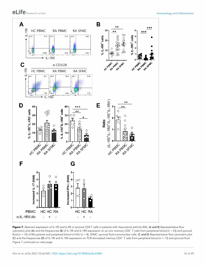
Figure 7. Aberrant expression of IL-1RI and IL-RII in synovial CD4 T cells in patients with rheumatoid arthritis (RA). (A and B) Representative flow
cytometric plot (A) and the frequencies (B) of IL-1RI and IL-1RII expression on ex vivo memory CD4+ T cells from peripheral blood (n = 23) and synovial
fluid (n = 15) of RA patients and peripheral blood of HCs (n = 8). SFMC: synovial fluid mononuclear cells. (C and D) Representative flow cytometric plot
(C) and the frequencies (D) of IL-1RI and IL-1RII expression on TCR-stimulated memory CD4+ T cells from peripheral blood (n = 13) and synovial fluid
Figure 7 continued on next page
Kim et al. eLife 2021;10:e61841. DOI: https://doi.org/10.7554/eLife.61841 16 of 29
Research article Immunology and Inflammation
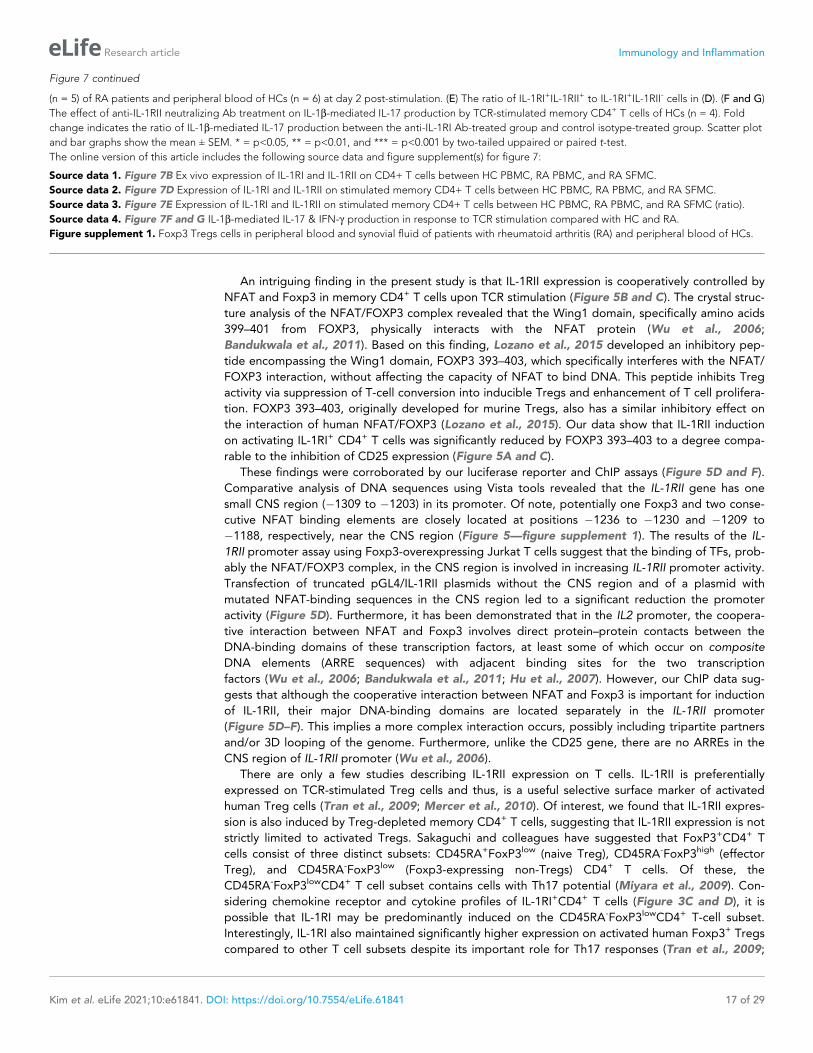
An intriguing finding in the present study is that IL-1RII expression is cooperatively controlled by
NFAT and Foxp3 in memory CD4+ T cells upon TCR stimulation (Figure 5B and C). The crystal struc-
ture analysis of the NFAT/FOXP3 complex revealed that the Wing1 domain, specifically amino acids
399–401 from FOXP3, physically interacts with the NFAT protein (Wu et al., 2006;
Bandukwala et al., 2011). Based on this finding, Lozano et al., 2015 developed an inhibitory pep-
tide encompassing the Wing1 domain, FOXP3 393–403, which specifically interferes with the NFAT/
FOXP3 interaction, without affecting the capacity of NFAT to bind DNA. This peptide inhibits Treg
activity via suppression of T-cell conversion into inducible Tregs and enhancement of T cell prolifera-
tion. FOXP3 393–403, originally developed for murine Tregs, also has a similar inhibitory effect on
the interaction of human NFAT/FOXP3 (Lozano et al., 2015). Our data show that IL-1RII induction
on activating IL-1RI+ CD4+ T cells was significantly reduced by FOXP3 393–403 to a degree compa-
rable to the inhibition of CD25 expression (Figure 5A and C).
These findings were corroborated by our luciferase reporter and ChIP assays (Figure 5D and F).
Comparative analysis of DNA sequences using Vista tools revealed that the IL-1RII gene has one
small CNS region (�1309 to �1203) in its promoter. Of note, potentially one Foxp3 and two conse-
cutive NFAT binding elements are closely located at positions �1236 to �1230 and �1209 to
�1188, respectively, near the CNS region (Figure 5—figure supplement 1). The results of the IL-
1RII promoter assay using Foxp3-overexpressing Jurkat T cells suggest that the binding of TFs, prob-
ably the NFAT/FOXP3 complex, in the CNS region is involved in increasing IL-1RII promoter activity.
Transfection of truncated pGL4/IL-1RII plasmids without the CNS region and of a plasmid with
mutated NFAT-binding sequences in the CNS region led to a significant reduction the promoter
activity (Figure 5D). Furthermore, it has been demonstrated that in the IL2 promoter, the coopera-
tive interaction between NFAT and Foxp3 involves direct protein–protein contacts between the
DNA-binding domains of these transcription factors, at least some of which occur on composite
DNA elements (ARRE sequences) with adjacent binding sites for the two transcription
factors (Wu et al., 2006; Bandukwala et al., 2011; Hu et al., 2007). However, our ChIP data sug-
gests that although the cooperative interaction between NFAT and Foxp3 is important for induction
of IL-1RII, their major DNA-binding domains are located separately in the IL-1RII promoter
(Figure 5D–F). This implies a more complex interaction occurs, possibly including tripartite partners
and/or 3D looping of the genome. Furthermore, unlike the CD25 gene, there are no ARREs in the
CNS region of IL-1RII promoter (Wu et al., 2006).
There are only a few studies describing IL-1RII expression on T cells. IL-1RII is preferentially
expressed on TCR-stimulated Treg cells and thus, is a useful selective surface marker of activated
human Treg cells (Tran et al., 2009; Mercer et al., 2010). Of interest, we found that IL-1RII expres-
sion is also induced by Treg-depleted memory CD4+ T cells, suggesting that IL-1RII expression is not
strictly limited to activated Tregs. Sakaguchi and colleagues have suggested that FoxP3+CD4+ T
cells consist of three distinct subsets: CD45RA+FoxP3low (naive Treg), CD45RA-FoxP3high (effector
Treg), and CD45RA-FoxP3low (Foxp3-expressing non-Tregs) CD4+ T cells. Of these, the
CD45RA-FoxP3lowCD4+ T cell subset contains cells with Th17 potential (Miyara et al., 2009). Con-
sidering chemokine receptor and cytokine profiles of IL-1RI+CD4+ T cells (Figure 3C and D), it is
possible that IL-1RI may be predominantly induced on the CD45RA-FoxP3lowCD4+ T-cell subset.
Interestingly, IL-1RI also maintained significantly higher expression on activated human Foxp3+ Tregs
compared to other T cell subsets despite its important role for Th17 responses (Tran et al., 2009;
Figure 7 continued
(n = 5) of RA patients and peripheral blood of HCs (n = 6) at day 2 post-stimulation. (E) The ratio of IL-1RI+IL-1RII+ to IL-1RI+IL-1RII- cells in (D). (F and G)
The effect of anti-IL-1RII neutralizing Ab treatment on IL-1b-mediated IL-17 production by TCR-stimulated memory CD4+ T cells of HCs (n = 4). Fold
change indicates the ratio of IL-1b-mediated IL-17 production between the anti-IL-1RI Ab-treated group and control isotype-treated group. Scatter plot
and bar graphs show the mean ± SEM. * = p<0.05, ** = p<0.01, and *** = p<0.001 by two-tailed uppaired or paired t-test.
The online version of this article includes the following source data and figure supplement(s) for figure 7:
Source data 1. Figure 7B Ex vivo expression of IL-1RI and IL-1RII on CD4+ T cells between HC PBMC, RA PBMC, and RA SFMC.
Source data 2. Figure 7D Expression of IL-1RI and IL-1RII on stimulated memory CD4+ T cells between HC PBMC, RA PBMC, and RA SFMC.
Source data 3. Figure 7E Expression of IL-1RI and IL-1RII on stimulated memory CD4+ T cells between HC PBMC, RA PBMC, and RA SFMC (ratio).
Source data 4. Figure 7F and G IL-1b-mediated IL-17 & IFN-g production in response to TCR stimulation compared with HC and RA.
Figure supplement 1. Foxp3 Tregs cells in peripheral blood and synovial fluid of patients with rheumatoid arthritis (RA) and peripheral blood of HCs.
Kim et al. eLife 2021;10:e61841. DOI: https://doi.org/10.7554/eLife.61841 17 of 29
Research article Immunology and Inflammation
Mercer et al., 2010). Since Foxp3 is generally induced by TCR stimulation even on Treg-depleted
conventional CD4+ T cells with Smad3 and NFAT dependent manner (Figure 3—figure supplement
3), strong TCR stimulation results in preferential expression of IL-RII, which limits IL-1b responsive-
ness and resultantly, reduces Th17 responses. In addition to the fact that the differentiation of Th17
and Treg cells is closely related (Zhou et al., 2008), both cells display instability and plasticity during
immune responses; Foxp3+ Treg cells can be converted into IL-17-producing cells in certain inflam-
matory environments and may potentially contribute to pathogenesis of autoimmune diseases,
whereas Th17 cells transdifferentiate into Treg cells during resolution of
inflammation (Gagliani et al., 2015; Obermajer et al., 2014). Several studies suggest that some
Th17 cells act like Tregs and this unique subset of Th17 cells are increased in tumor-bearing mice,
human cancer patients (Gagliani et al., 2015; Downs-Canner et al., 2017; Krummey et al., 2014;
Thibaudin et al., 2016; Jung et al., 2017), and during several inflammatory diseases. In our study,
IL-1RI+IL-1RII+ cells were found to be similar in phenotype to Treg cells with relatively high expres-
sion of CD39, CD73, and CTLA-4 following TCR stimulation (Figures 3B and 6D). Because of their
phenotype and potential modulatory function via CTLA-4 and IL-1RII, IL-1RI+IL-1RII+ cells may act as
pivotal regulators of the Th17-cell-mediated inflammatory environment. Thus, the function and
homeostatic capacity of IL-1RI+IL-1RII+ cells require further investigation.
Rheumatoid arthritis is a prototype autoimmune disease characterized by chronic inflammation of
the joint synovial membrane (McInnes and O’Dell, 2010; McInnes and Schett, 2011). The disturbed
Th17/Treg balance contributes to RA pathogenesis due to the strong pro-inflammatory response of
preponderant Th17 cells and impaired Treg cells lacking immunomodulatory capacity, partly due to
the cytokine circumstance (Niu et al., 2012; Wang et al., 2012; Noack and Miossec, 2014;
Nie et al., 2013). The interesting observation in this study was diminished TCR-mediated induction
of IL-1RII, and not IL-1RI, on memory CD4+ T cells from RA patients compared to the peripheral
counterpart from healthy controls (Figure 7D), causing a significantly reduced ratio of IL-1RI+IL-1RII+
to IL-1RI+IL-1RII- memory CD4+ T cells (Figure 7E). Of note, blocking of IL-1RII on memory CD4+ T
cells of HCs led to increased IL-1b-mediated induction of IL-17 at a level comparable to that of RA
patients (Figure 7F). Given the elevated level of IL-1b in RA synovial fluid and an important role of
Th17 responses in RA pathogenesis (Kim et al., 2016; Kay and Calabrese, 2004), impaired induc-
tion of IL-1RII may be associated with modulation of IL-17 production in CD4+ T cells of RA patients
under chronic autoreactive situations.
In summary, the current study provides new insight into the immune regulatory role of the decoy
receptor, IL-1RII, expressed by human CD4+ T cells in response to TCR stimulation. Here, we investi-
gated the molecular mechanisms underlying IL-1RII expression and analyzing its influence on Th17
responses and consequentially, the Th17/Treg balance. IL-RII expression is mediated molecularly by
a cooperative complex of the NFAT and Foxp3 TFs. Further, differential expression of IL-1RI and IL-
RII on activated CD4+ T cells imparts unique immunological features to these cells via modulation of
IL-1b responsiveness. Together, these findings increase understanding of the modulatory mecha-
nisms controlling the Th17 response, and of IL-1b-mediated IL-1R signaling and its contribution to
the pathogenesis of autoimmune diseases such as RA.
Materials and methods
Cell preparationThe study protocols were approved by the institutional review board (IRB) of Seoul National Univer-
sity Hospital and Chungnam National University Hospital (IRB No.1109-055-378, 1306-002-491, and
1403-049-564 for Seoul National University College of Medicine/Seoul National University Hospital
and IRB No.2012-01-024 for Chungnam National University Hospital). Peripheral blood of RA
patients and healthy controls (HCs) was drawn after obtaining written, informed consent. The meth-
ods were performed in accordance with the approved guidelines. Peripheral blood mononuclear
cells (PBMCs) were isolated from blood of RA patients and HCs by density gradient centrifugation
method (Bicoll separating solution; BIOCHROM Inc, Cambridge, UK). Naive and memory CD4+ T
cells were negatively purified from PBMC using human naive CD4+ T cells and human memory CD4+
T cells enrichment kit (both from STEMCELL, Vancouver, BC, Canada), respectively. In some experi-
ments, non-Treg memory CD4+ T cells were obtained by depletion of Tregs from previously purified
Kim et al. eLife 2021;10:e61841. DOI: https://doi.org/10.7554/eLife.61841 18 of 29
Research article Immunology and Inflammation
memory CD4+ T cells using CD4+CD25+CD127dim/- Regulatory T cells isolation kit II (Miltenyi Bio-
tech, Bergisch Gladbach, Germany).
Cell lineTo generate Foxp3-expressing Jurkat cell line, the lentiviral vector for expression of human FoxP3
(pLEF-puro-FoxP3) was prepared as follows. pLEF-puro was created from biscistronic lentiviral vec-
tor, pLECE3, which harbors EF1alpha promoter-driven expression cassette and CMV promoter-GFP
expression cassette (Lee et al., 2019) , by replacing GFP ORF with puroR ORF. Human FoxP3 ORF
was amplified by PCR from pCMV5-entry-FoxP3 (OriGene, Rockville, MD, USA) and cloned into
multi-cloning sites under the EF1alpha promoter in pLEF-puro. The lentivirus for FoxP3 expression
was produced by transfecting pLEF-puro-FoxP3 plasmid into 293FT cells (Invitrogen, Waltham, MA,
USA) along with packaging plasmids (pMDLg/pRRE, pRSVrev, and pMD.G). 48–72 hr after transfec-
tion, the culture supernatant containing the lentivirus was harvested and concentrated 20-fold using
a centrifugal ultrafiltration unit (Amicon Ultra-15, 100kD cut-off, Merck Millipore, Burlington, MA,
USA). Jurkat cells (Clone E6-1) were purchased from ATCC (Manassas, VA, USA), regularly tested to
exclude mycoplasma infections, and authenticated using Short Tandem Repeat (STR) profiling.Jurkat
cells were transduced with the concentrated lentivirus in the presence of polybrene (6 mg/ml, Sigma-
Aldrich, St. Louis, MO, USA) via spin-infection at 2,500 rpm for 90 min at room temperature. The
transduced Jurkat cells were selected in the media containing puromycin (2 mg/ml, Sigma-Aldrich,
St. Louis, MO, USA) and established as a cell line by limiting dilution.
Cell culturePurified naive and memory CD4+ T cells were cultured in serum-free X-VIVO 10 medium (Lonza,
Basel, Switzerland) and RPMI 1640 medium supplemented with 10% fetal bovine serum, 1% penicil-
lin/streptomycin, and 1% L-glutamine, respectively. Cells were seeded at 2.5 � 104 into 96-well U
bottom plate and stimulated with anti-CD3/CD28-coated microbeads (Dynabeads T-Activator CD3/
CD28; Thermo Fisher Scientific, Waltham, MA) for 5 or 7 days in the absence or presence of the indi-
cated cytokines or neutralizing antibodies (Abs); recombinant human (rh) IL-1b (5 ng/ml), rhIL-6 (25
ng/ml), rhIL-23 (25 ng/ml), rhTGF-b (10 ng/ml; all from R and D systems, Minneapolis, MN), rhIL-2
(100 IU/ml, PeproTech, Rocky Hill, NJ), anti-human IL-1RII Ab, or mouse IgG2A (20 mg/ml, both from
R and D systems). In some experiments, cells were stimulated with anti-CD3/CD28-coated microbe-
ads with peptide inhibitors, chemical inhibitors, or reagents: dNP2-VIVIT and dNP2-VEET peptides
were synthesized by using a solid-phase synthesis and purified by high performance liquid chroma-
tography (Anygen, Kwangju, Korea) as previously reported (Lee et al., 2019). The peptides were
synthesized with >95% purity. Peptide inhibitor Foxp3 393–403 (KCFVRVESEKG, 100 mM) and con-
trol peptide Foxp3 399A (KCFVRVASEKG, 100 mM) were manufactured at GenScript, Nanjing,
China (Lozano et al., 2015; Lim et al., 2015), Cyclosporin A (10 or 50 nM, Sigma-Aldrich, St. Louis,
MO) or 1, 25-dioxylvitamin D3 (100 mM, Sigma-Aldrich). To obtain IL-1RI+IL-1RII- and IL-1RI+IL-1RII+
T cells, Treg-depleted memory CD4+ T cells were stimulated with anti-CD3/CD28-coated microbe-
ads for 48 hr to induce the expressions of IL-1RI and IL-1RII. The stimulated cells were harvested and
stained with FITC-anti-IL-1RII, PE-anti-IL-1RI (both from R and D systems), PE-Cy7-anti-CD45RA, and
APC-anti-CD4 (all from BD Bioscience, San Jose, CA), followed by cell-sorting into IL-1RI+IL-1RII-, IL-
1RI+IL-1RII+, and IL-1RI-IL-1RII- cells using FACSAria II (BD Bioscience).
Flow cytometryPBMC or cultured T cells were stained for 30 min at 4˚C with following fluorochrome-conjugated
Abs: APC-Cy7-anti-CD3, v500-anti-CD4, PE-Cy5-anti-CD25, APC-anti-CD25, PE-Cy7-anti-CD45RA,
BV421-anti-CD161, (six from BD Bioscience), FITC-anti-GARP (Enzo Life Science, Farmingdale, NY),
PE-Cy7-anti-GITR, APC-Cy7-anti-CD73, BV421-anti-CD39, PerCP-Cy5.5-anti-CTLA-4, PE-Cy7-anti-
CXCR3 (five from BioLegend, San Diego, CA), PE-anti-IL-1RI, APC-anti-IL-1RII, and FITC-anti-IL-1RII
(three from R and D systems). For intracellular cytokine staining (ICS), cultured T cells were re-stimu-
lated for 6 hr with PMA (50 ng/ml) and ionomycin (1 mg/ml) in the presence of BFA for last 4 hr, fol-
lowed by staining with PE-anti-IL-1RI and FITC-anti-IL-1RII Abs. The stained cells were fixed and
permeabilized using Fix/Perm buffer (BioLegend), followed by staining with anti-IL-17A, anti-IFN-g
Kim et al. eLife 2021;10:e61841. DOI: https://doi.org/10.7554/eLife.61841 19 of 29
Research article Immunology and Inflammation
(both from eBioscience), and anti-Foxp3 (Biolegend) Abs. The cells were acquired using a BD
LSRFortessa (BD Bioscience) and analyzed using FlowJo software (Tree Star, Ashland, OR).
Enzyme-linked immunosorbent assay (ELISA)The amounts of IL-17, IFN-g , and IL-10 in culture supernatant were quantified using commercially
available human ELISA kits according to manufacturer’s instructions (Both IL-17A ELISA and IL-10
ELISA kits from eBioscience and Human IFN-g ELISA MAX Deluxe from BioLegend). Measurement of
OD (optical density) was performed using Infinite M200 Pro Multimode microplate reader (Tecan,
Mannedorf, Switzerland).
Quantitative real-time PCRTotal RNA was extracted from purified or cultured T cells with TRIzol reagent (Invitrogen) and cDNA
was synthesized using GoScript Reverse Transcription System (Promega, Madison, WI). Real-time
quantitative RT-PCR was performed on CFX system (Bio-Rad, Hercules, CA, USA) using SYBR green
master mix (Bio-Rad). Primers were designed with Primer designing tool-NCBI (https://www.ncbi.
nlm.nih.gov/tools/primer-blast/) or adopted from previously published primer sequences; Foxp3, 5’-
GCACCTTCCCAAATCCCAGT-3’ and 5’-GGCCACTTGCAGACACCAT-3’; IL-1RI, 5’- GTGATTG
TGAGCCCAGCTA-3’ and 5’-TGTTTGCAGGATTTTCCACA-3’; IL-1RII, 5’-CCTTGTCAACCTCTGGGG
TA-3, and 5’-ACAGCGGTAATAGCCAGCAT-3’; IL-10, 5’- AGCTCCAAGAGAAAGGCATCT-3, and
5’- TATAGAGTCGCCACCCTGATGT-3’; IL-22, 5’- GCTTGACAAGTCCAACTTCCA-3, and 5’- GC
TCACTCATACTGACTCCGTG-3’; IL-17, 5’- AACTCATCCATCCCCAGTTG-3, and 5’- GAGGACC
TTTTGGGATTGGT-3’; IFN-g, 5’- TTTGGGTTCTCTTGGCTGTT-3, and 5’- TCTTTTGGATGCTCTGG
TCA-3’. The levels of gene expression were normalized to the expression of ACTINB. The compara-
tive Ct method (DDCt) was used for quantification of gene expression.
Luciferase reporter assayPutative NFAT and Foxp3-binding sites were screened using Promo 3.0 software (ALGGEN, http://
alggen.lsi.upc.es). Conserved noncoding sequence (CNS) was analyzed using VISTA tool (http://
genome.lbl.gov/vista/index.shtml). The promoter region (�1311 to +100) of human IL-1RII (Gene ID:
7850) gene containing both putative NFAT/FOXP3 binding motif and CNS (�1306 to �1200) was
cloned into pGL4.10 basic luciferase reporter vector (pGL4/IL-1RII-a1414). Mutant IL-1RII reporter
plasmid (pGL4/IL-1RII-a1414mu; NFAT-binding motif ACAGTTTCCA into ACAGAAACCA in the
CNS region) was generated with QuikChange II Site-Directed Mutagenesis Kit (Agilent Technologies,
Santa Clara, CA). Two truncated reporter vectors, pGL4/IL-1RII-a814 and pGL4/IL-1RII-a215 includ-
ing �714 to +100 and �115 to +100 of the promoter, respectively, were also cloned. Plasmids were
transfected into Jurkat cells using NEON electroporation system (Life Technology, Carlsbad, CA),
followed manufacturer’s instructions. Foxp3-expressing Jurkat cells or control Jurkat cells were trans-
fected with the indicated IL-1RII promoter cloned luciferase vector and internal control Renilla
pGL4.74 by NEON electroporation system (Life Technology). After resting for 5 hr, transfected cells
were stimulated with PMA and Ionomycin for 48 hr. Cells were lysed and luminescence was detected
using Dual-Luciferase Reporter Assay system (Promega) followed manufacturer’s instructions.
Chromatin immunoprecipitation (ChIP)-qPCR assayPurified memory CD4+ T cells were stimulated with plate-bound anti-CD3 (10 mg/ml) and soluble
anti-CD28 (2 mg/ml; both from Life Technology) Ab for 24 hr. Cells were cross-linked using 1% form-
aldehyde for 10 min and lysed with 1% SDS-lysis buffer. ChIP assay were performed using commer-
cially available Ez-ChIP assay kit (Merck, Darmstadt, Germany) according to manufacturer’s
instructions. DNA-bound proteins were immunoprecipitated using anti-NFATc2 (Santa-Cruz Biotech-
nology, Heidelberg, Germany) or anti-Foxp3 (Thermo Fisher Scientific) Ab. Co-precipitated DNA
were purified through NucleoSpin gDNA clean-up kit (MACHEREY-NAGEL, Duren, Germany) and
used as a template for real-time qRT-PCR using primers as following; NFAT_B1, 5’- ATTTACCC
TTTGCTCAACAAACTCC- 3’ and 5’- AGTCCAGACATTTGAGGATGGGG-3’; NFAT_B2, 5’-
GAGAGGTCGCAGGGGATGAT- 3’ and 5’- TCTGAGAACTCATGGTCCTGG-3’; NFAT_B3, 5’- GG
TGATCATGTACTCAGACCCA- 3’ and 5’-GGGAGCTGGGATTTCCTAACC-3’; NFAT_BC (binding
control), 5’- TGCTCTAACTATGCAATGGCT- 3’ and 5’- TGATCAGAGCTCTCTTCTGATTATT-3’;
Kim et al. eLife 2021;10:e61841. DOI: https://doi.org/10.7554/eLife.61841 20 of 29
Research article Immunology and Inflammation
Foxp3_B1, 5’- TAGGGTGGGTGGTTGAGGG - 3’ and 5’- GAAGATAAAACCTCAGCGTCACC �3’;
Foxp3_B2, 5’- ATTTACCCTTTGCTCAACAAACTCC- 3’ and 5’- AGTCCAGACATTTGAGGATGGGG-
3’; Foxp3_B3, 5’- AGAGAGGAGCAACCAACAACA- 3’ and 5’-CTTCTGAAAGTGGGAATCCAGC-3’;
Foxp3_BC (binding control), 5’- AGATTCTACTAGACAAGGGCACAG- 3’ and 5’-CATACAAACCAAC
TGTTCTCTCCC-3’ (Zhang et al., 2013).
Statistical analysisTwo-tailed paired or unpaired t test was done to analyze data using Prism eight software (Software,
La Jolla, CA, USA) or and Microsoft Excel 2016 as indicated in the figure legends. p-Values of less
than 0.05 considered statistically significant.
AcknowledgementsThe authors thank Jiyeon Jang (Seoul National University College of Medicine) for assisting in the
recruitment of human subjects and thank Core Lab, Clinical Trials Center, Seoul National University
Hospital for drawing blood.
Additional information
Funding
Funder Grant reference number Author
National Research Foundationof Korea
2013R1A1A2012522 Won-Woo Lee
National Research Foundationof Korea
2018R1A2B2006310 Won-Woo Lee
Seoul National University Hos-pital
0320180220: 2018-1293 Won-Woo Lee
The funders had no role in study design, data collection and interpretation, or the
decision to submit the work for publication.
Author contributions
Dong Hyun Kim, Data curation, Software, Formal analysis, Investigation, Writing - original draft; Hee
Young Kim, Sunjung Cho, Data curation, Formal analysis, Investigation, Methodology; Su-Jin Yoo,
Resources, Formal analysis, Investigation; Won-Ju Kim, Formal analysis, Investigation, Methodology;
Hye Ran Yeon, Resources, Investigation, Methodology; Kyungho Choi, Conceptualization, Resources,
Formal analysis, Methodology, Writing - review and editing; Je-Min Choi, Conceptualization, Investi-
gation, Methodology, Writing - review and editing; Seong Wook Kang, Conceptualization, Resour-
ces, Data curation, Investigation; Won-Woo Lee, Conceptualization, Resources, Data curation,
Formal analysis, Supervision, Funding acquisition, Validation, Investigation, Writing - original draft,
Project administration, Writing - review and editing
Author ORCIDs
Dong Hyun Kim https://orcid.org/0000-0002-2765-4376
Won-Woo Lee https://orcid.org/0000-0002-5347-9591
Ethics
Human subjects: The study protocols were approved by the institutional review board (IRB) of Seoul
National University Hospital and Chungnam National University Hospital (IRB No.1109- 055-378,
1306-002-491, and 1403-049-564 for Seoul National University College of Medicine/Seoul National
University Hospital and IRB No.2012-01-024 for Chungnam National University Hospital). Peripheral
blood ofrheumatoid arthritispatients and healthy controls was drawn after obtaining written,
informed consent.
Kim et al. eLife 2021;10:e61841. DOI: https://doi.org/10.7554/eLife.61841 21 of 29
Research article Immunology and Inflammation

Decision letter and Author response
Decision letter https://doi.org/10.7554/eLife.61841.sa1
Author response https://doi.org/10.7554/eLife.61841.sa2
Additional filesSupplementary files. Transparent reporting form
Data availability
All data generated or analysed during this study are included in the manuscript and supporting files.
ReferencesAcosta-Rodriguez EV, Napolitani G, Lanzavecchia A, Sallusto F. 2007. Interleukins 1beta and 6 but nottransforming growth factor-beta are essential for the differentiation of interleukin 17-producing human T helpercells. Nature Immunology 8:942–949. DOI: https://doi.org/10.1038/ni1496, PMID: 17676045
Afzali B, Mitchell PJ, Edozie FC, Povoleri GA, Dowson SE, Demandt L, Walter G, Canavan JB, Scotta C, MenonB, Chana PS, Khamri W, Kordasti SY, Heck S, Grimbacher B, Tree T, Cope AP, Taams LS, Lechler RI, John S,et al. 2013. CD161 expression characterizes a subpopulation of human regulatory T cells that produces IL-17 ina STAT3-dependent manner. European Journal of Immunology 43:2043–2054. DOI: https://doi.org/10.1002/eji.201243296, PMID: 23677517
Aramburu J, Garcia-Cozar F, Raghavan A, Okamura H, Rao A, Hogan PG. 1998. Selective inhibition of NFATactivation by a peptide spanning the calcineurin targeting site of NFAT. Molecular Cell 1:627–637.DOI: https://doi.org/10.1016/S1097-2765(00)80063-5, PMID: 9660947
Aramburu J, Yaffe MB, Lopez-Rodrıguez C, Cantley LC, Hogan PG, Rao A. 1999. Affinity-driven peptideselection of an NFAT inhibitor more selective than cyclosporin A. Science 285:2129–2133. DOI: https://doi.org/10.1126/science.285.5436.2129, PMID: 10497131
Bandukwala HS, Wu Y, Feuerer M, Chen Y, Barboza B, Ghosh S, Stroud JC, Benoist C, Mathis D, Rao A, Chen L.2011. Structure of a domain-swapped FOXP3 dimer on DNA and its function in regulatory T cells. Immunity 34:479–491. DOI: https://doi.org/10.1016/j.immuni.2011.02.017, PMID: 21458306
Basdeo SA, Cluxton D, Sulaimani J, Moran B, Canavan M, Orr C, Veale DJ, Fearon U, Fletcher JM. 2017. Ex-Th17(Nonclassical Th1) Cells are functionally distinct from classical Th1 and Th17 cells and are not constrained byregulatory T cells. The Journal of Immunology 198:2249–2259. DOI: https://doi.org/10.4049/jimmunol.1600737, PMID: 28167631
Basu R, Whitley SK, Bhaumik S, Zindl CL, Schoeb TR, Benveniste EN, Pear WS, Hatton RD, Weaver CT. 2015. IL-1signaling modulates activation of STAT transcription factors to antagonize retinoic acid signaling and controlthe TH17 cell-iTreg cell balance. Nature Immunology 16:286–295. DOI: https://doi.org/10.1038/ni.3099,PMID: 25642823
Brignall R, Cauchy P, Bevington SL, Gorman B, Pisco AO, Bagnall J, Boddington C, Rowe W, England H, Rich K,Schmidt L, Dyer NP, Travis MA, Ott S, Jackson DA, Cockerill PN, Paszek P. 2017. Integration of kinase andcalcium signaling at the level of chromatin underlies inducible gene activation in T cells. The Journal ofImmunology 199:2652–2667. DOI: https://doi.org/10.4049/jimmunol.1602033, PMID: 28904128
Chung Y, Chang SH, Martinez GJ, Yang XO, Nurieva R, Kang HS, Ma L, Watowich SS, Jetten AM, Tian Q, DongC. 2009. Critical regulation of early Th17 cell differentiation by interleukin-1 signaling. Immunity 30:576–587.DOI: https://doi.org/10.1016/j.immuni.2009.02.007, PMID: 19362022
Colotta F, Re F, Muzio M, Bertini R, Polentarutti N, Sironi M, Giri JG, Dower SK, Sims JE, Mantovani A. 1993.Interleukin-1 type II receptor: a decoy target for IL-1 that is regulated by IL-4. Science 261:472–475.DOI: https://doi.org/10.1126/science.8332913, PMID: 8332913
Colotta F, Saccani S, Giri JG, Dower SK, Sims JE, Introna M, Mantovani A. 1996. Regulated expression andrelease of the IL-1 decoy receptor in human mononuclear phagocytes. Journal of Immunology 156:2534–2541.PMID: 8786316
Dinarello CA. 2011. A clinical perspective of IL-1b as the gatekeeper of inflammation. European Journal ofImmunology 41:1203–1217. DOI: https://doi.org/10.1002/eji.201141550, PMID: 21523780
Dinarello CA, Simon A, van der Meer JW. 2012. Treating inflammation by blocking interleukin-1 in a broadspectrum of diseases. Nature Reviews Drug Discovery 11:633–652. DOI: https://doi.org/10.1038/nrd3800,PMID: 22850787
Downs-Canner S, Berkey S, Delgoffe GM, Edwards RP, Curiel T, Odunsi K, Bartlett DL, Obermajer N. 2017.Suppressive IL-17A+Foxp3+ and ex-Th17 IL-17AnegFoxp3+ treg cells are a source of tumour-associated tregcells. Nature Communications 8:14649. DOI: https://doi.org/10.1038/ncomms14649
Kim et al. eLife 2021;10:e61841. DOI: https://doi.org/10.7554/eLife.61841 22 of 29
Research article Immunology and Inflammation
Du R, Zhao H, Yan F, Li H. 2014. IL-17+Foxp3+ T cells: an intermediate differentiation stage between Th17 cellsand regulatory T cells. Journal of Leukocyte Biology 96:39–48. DOI: https://doi.org/10.1189/jlb.1RU0114-010RR, PMID: 24744433
Gaffen SL. 2009. Structure and signalling in the IL-17 receptor family. Nature Reviews Immunology 9:556–567.DOI: https://doi.org/10.1038/nri2586
Gagliani N, Amezcua Vesely MC, Iseppon A, Brockmann L, Xu H, Palm NW, de Zoete MR, Licona-Limon P, PaivaRS, Ching T, Weaver C, Zi X, Pan X, Fan R, Garmire LX, Cotton MJ, Drier Y, Bernstein B, Geginat J, StockingerB, et al. 2015. Th17 cells transdifferentiate into regulatory T cells during resolution of inflammation. Nature523:221–225. DOI: https://doi.org/10.1038/nature14452, PMID: 25924064
Garlanda C, Dinarello CA, Mantovani A. 2013a. The Interleukin-1 family: back to the future. Immunity 39:1003–1018. DOI: https://doi.org/10.1016/j.immuni.2013.11.010
Garlanda C, Riva F, Bonavita E, Gentile S, Mantovani A. 2013b. Decoys and regulatory "Receptors" of the IL-1/Toll-Like Receptor Superfamily. Frontiers in Immunology 4:180. DOI: https://doi.org/10.3389/fimmu.2013.00180, PMID: 23847621
Hermann-Kleiter N, Baier G. 2010. NFAT pulls the strings during CD4+ T helper cell effector functions. Blood115:2989–2997. DOI: https://doi.org/10.1182/blood-2009-10-233585, PMID: 20103781
Hirota K, Duarte JH, Veldhoen M, Hornsby E, Li Y, Cua DJ, Ahlfors H, Wilhelm C, Tolaini M, Menzel U, GarefalakiA, Potocnik AJ, Stockinger B. 2011. Fate mapping of IL-17-producing T cells in inflammatory responses. NatureImmunology 12:255–263. DOI: https://doi.org/10.1038/ni.1993, PMID: 21278737
Hu H, Djuretic I, Sundrud MS, Rao A. 2007. Transcriptional partners in regulatory T cells: foxp3, runx and NFAT.Trends in Immunology 28:329–332. DOI: https://doi.org/10.1016/j.it.2007.06.006, PMID: 17618833
Ichiyama K, Gonzalez-Martin A, Kim BS, Jin HY, Jin W, Xu W, Sabouri-Ghomi M, Xu S, Zheng P, Xiao C, Dong C.2016. The MicroRNA-183-96-182 cluster promotes T helper 17 cell pathogenicity by negatively regulatingtranscription factor Foxo1 expression. Immunity 44:1284–1298. DOI: https://doi.org/10.1016/j.immuni.2016.05.015, PMID: 27332731
Jung MK, Kwak JE, Shin EC. 2017. IL-17A-Producing Foxp3+regulatory T cells and human diseases. ImmuneNetwork 17:276–286. DOI: https://doi.org/10.4110/in.2017.17.5.276, PMID: 29093649
Kang SW, Kim SH, Lee N, Lee WW, Hwang KA, Shin MS, Lee SH, Kim WU, Kang I. 2012. 1,25-DihyroxyvitaminD3 promotes FOXP3 expression via binding to vitamin D response elements in its conserved noncodingsequence region. Journal of Immunology 188:5276–5282. DOI: https://doi.org/10.4049/jimmunol.1101211,PMID: 22529297
Kay J, Calabrese L. 2004. The role of interleukin-1 in the pathogenesis of rheumatoid arthritis. Rheumatology 43Suppl 3:iii2–iii9. DOI: https://doi.org/10.1093/rheumatology/keh201, PMID: 15150426
Kiani A, Rao A, Aramburu J. 2000. Manipulating immune responses with immunosuppressive agents that targetNFAT. Immunity 12:359–372. DOI: https://doi.org/10.1016/S1074-7613(00)80188-0, PMID: 10795734
Kim BH, Yoon BR, Kim EK, Noh KH, Kwon SH, Yi EH, Lee HG, Choi JS, Kang SW, Park IC, Lee WW, Ye SK. 2016.Alleviation of collagen-induced arthritis by the benzoxathiole derivative BOT-4-one in mice: implication of theTh1- and Th17-cell-mediated immune responses. Biochemical Pharmacology 110-111:47–57. DOI: https://doi.org/10.1016/j.bcp.2016.03.018, PMID: 27005941
Komatsu N, Okamoto K, Sawa S, Nakashima T, Oh-hora M, Kodama T, Tanaka S, Bluestone JA, Takayanagi H.2014. Pathogenic conversion of Foxp3+ T cells into TH17 cells in autoimmune arthritis. Nature Medicine 20:62–68. DOI: https://doi.org/10.1038/nm.3432
Krummey SM, Floyd TL, Liu D, Wagener ME, Song M, Ford ML. 2014. Candida-elicited murine Th17 cellsexpress high Ctla-4 compared with Th1 cells and are resistant to costimulation blockade. The Journal ofImmunology 192:2495–2504. DOI: https://doi.org/10.4049/jimmunol.1301332, PMID: 24493820
Lee WW, Kang SW, Choi J, Lee SH, Shah K, Eynon EE, Flavell RA, Kang I. 2010. Regulating human Th17 cells viadifferential expression of IL-1 receptor. Blood 115:530–540. DOI: https://doi.org/10.1182/blood-2009-08-236521, PMID: 19965648
Lee Y, Awasthi A, Yosef N, Quintana FJ, Xiao S, Peters A, Wu C, Kleinewietfeld M, Kunder S, Hafler DA, SobelRA, Regev A, Kuchroo VK. 2012. Induction and molecular signature of pathogenic TH17 cells. NatureImmunology 13:991–999. DOI: https://doi.org/10.1038/ni.2416
Lee HG, Kim LK, Choi JM. 2019. NFAT-specific inhibition by dNP2-VIVIT ameliorates autoimmuneencephalomyelitis by regulation of Th1 and Th17. Molecular Therapy - Methods & Clinical Development 16:32–41. DOI: https://doi.org/10.1016/j.omtm.2019.10.006, PMID: 31737742
Lee SH, Soh H, Chung JH, Cho EH, Lee SJ, Ju JM, Sheen JH, Kim H, Oh SJ, Lee SJ, Chung J, Choi K, Kim SY,Ryu JS. 2020. Feasibility of real-time in vivo 89Zr-DFO-labeled CAR T-cell trafficking using PET imaging. PLOSONE 15:e0223814. DOI: https://doi.org/10.1371/journal.pone.0223814, PMID: 31910217
Lim S, Kim WJ, Kim YH, Lee S, Koo JH, Lee JA, Yoon H, Kim DH, Park HJ, Kim HM, Lee HG, Yun Kim J, Lee JU,Hun Shin J, Kyun Kim L, Doh J, Kim H, Lee SK, Bothwell ALM, Suh M, et al. 2015. dNP2 is a blood-brain barrier-permeable peptide enabling ctCTLA-4 protein delivery to ameliorate experimental autoimmuneencephalomyelitis. Nature Communications 6:8244. DOI: https://doi.org/10.1038/ncomms9244,PMID: 26372309
Lozano T, Villanueva L, Durantez M, Gorraiz M, Ruiz M, Belsue V, Riezu-Boj JI, Hervas-Stubbs S, Oyarzabal J,Bandukwala H, Lourenco AR, Coffer PJ, Sarobe P, Prieto J, Casares N, Lasarte JJ. 2015. Inhibition of FOXP3/NFAT interaction enhances T cell function after TCR stimulation. The Journal of Immunology 195:3180–3189.DOI: https://doi.org/10.4049/jimmunol.1402997, PMID: 26324768
Kim et al. eLife 2021;10:e61841. DOI: https://doi.org/10.7554/eLife.61841 23 of 29
Research article Immunology and Inflammation
Martin P, Palmer G, Rodriguez E, Seemayer CA, Palomo J, Talabot-Ayer D, Gabay C. 2017. Deficiency in IL-1receptor type 2 aggravates K/BxN serum Transfer-Induced arthritis in mice but has no impact on systemicinflammatory responses. The Journal of Immunology 198:2916–2926. DOI: https://doi.org/10.4049/jimmunol.1600855, PMID: 28235865
McInnes IB, O’Dell JR. 2010. State-of-the-art: rheumatoid arthritis. Annals of the Rheumatic Diseases 69:1898–1906. DOI: https://doi.org/10.1136/ard.2010.134684, PMID: 20959326
McInnes IB, Schett G. 2011. The pathogenesis of rheumatoid arthritis. New England Journal of Medicine 365:2205–2219. DOI: https://doi.org/10.1056/NEJMra1004965
McMahan CJ, Slack JL, Mosley B, Cosman D, Lupton SD, Brunton LL, Grubin CE, Wignall JM, Jenkins NA,Brannan CI. 1991. A novel IL-1 receptor, cloned from B cells by mammalian expression, is expressed in manycell types. The EMBO Journal 10:2821–2832. DOI: https://doi.org/10.1002/j.1460-2075.1991.tb07831.x,PMID: 1833184
Mercer F, Kozhaya L, Unutmaz D. 2010. Expression and function of TNF and IL-1 receptors on human regulatoryT cells. PLOS ONE 5:e8639. DOI: https://doi.org/10.1371/journal.pone.0008639, PMID: 20066156
Messeguer X, Escudero R, Farre D, Nunez O, Martinez J, Alba MM. 2002. PROMO: detection of knowntranscription regulatory elements using species-tailored searches. Bioinformatics 18:333–334. DOI: https://doi.org/10.1093/bioinformatics/18.2.333
Miyara M, Yoshioka Y, Kitoh A, Shima T, Wing K, Niwa A, Parizot C, Taflin C, Heike T, Valeyre D, Mathian A,Nakahata T, Yamaguchi T, Nomura T, Ono M, Amoura Z, Gorochov G, Sakaguchi S. 2009. Functionaldelineation and differentiation dynamics of human CD4+ T cells expressing the FoxP3 transcription factor.Immunity 30:899–911. DOI: https://doi.org/10.1016/j.immuni.2009.03.019, PMID: 19464196
Neumann D, Kollewe C, Martin MU, Boraschi D. 2000. The membrane form of the type II IL-1 receptor accountsfor inhibitory function. The Journal of Immunology 165:3350–3357. DOI: https://doi.org/10.4049/jimmunol.165.6.3350, PMID: 10975853
Nie H, Zheng Y, Li R, Guo TB, He D, Fang L, Liu X, Xiao L, Chen X, Wan B, Chin YE, Zhang JZ. 2013.Phosphorylation of FOXP3 controls regulatory T cell function and is inhibited by TNF-a in rheumatoid arthritis.Nature Medicine 19:322–328. DOI: https://doi.org/10.1038/nm.3085, PMID: 23396208
Niu Q, Cai B, Huang Z, Shi Y, Wang L. 2012. Disturbed Th17/Treg balance in patients with rheumatoid arthritis.Rheumatology International 32:2731–2736. DOI: https://doi.org/10.1007/s00296-011-1984-x
Noack M, Miossec P. 2014. Th17 and regulatory T cell balance in autoimmune and inflammatory diseases.Autoimmunity Reviews 13:668–677. DOI: https://doi.org/10.1016/j.autrev.2013.12.004
Obermajer N, Popp FC, Soeder Y, Haarer J, Geissler EK, Schlitt HJ, Dahlke MH. 2014. Conversion of Th17 intoIL-17A(neg) regulatory T cells: a novel mechanism in prolonged allograft survival promoted by mesenchymalstem cell-supported minimized immunosuppressive therapy. The Journal of Immunology 193:4988–4999.DOI: https://doi.org/10.4049/jimmunol.1401776, PMID: 25305313
Purvis HA, Stoop JN, Mann J, Woods S, Kozijn AE, Hambleton S, Robinson JH, Isaacs JD, Anderson AE, HilkensCM. 2010. Low-strength T-cell activation promotes Th17 responses. Blood 116:4829–4837. DOI: https://doi.org/10.1182/blood-2010-03-272153, PMID: 20713963
Ritvo PG, Churlaud G, Quiniou V, Florez L, Brimaud F, Fourcade G, Mariotti-Ferrandiz E, Klatzmann D. 2017. Tfrcells lack IL-2Ra but express decoy IL-1R2 and IL-1Ra and suppress the IL-1-dependent activation of Tfh cells.Science Immunology 2:eaan0368. DOI: https://doi.org/10.1126/sciimmunol.aan0368, PMID: 28887367
Ronchi F, Basso C, Preite S, Reboldi A, Baumjohann D, Perlini L, Lanzavecchia A, Sallusto F. 2016. Experimentalpriming of encephalitogenic Th1/Th17 cells requires pertussis toxin-driven IL-1b production by myeloid cells.Nature Communications 7:11541. DOI: https://doi.org/10.1038/ncomms11541, PMID: 27189410
Rudra D, deRoos P, Chaudhry A, Niec RE, Arvey A, Samstein RM, Leslie C, Shaffer SA, Goodlett DR, RudenskyAY. 2012. Transcription factor Foxp3 and its protein partners form a complex regulatory network. NatureImmunology 13:1010–1019. DOI: https://doi.org/10.1038/ni.2402, PMID: 22922362
Schluter T, Schelmbauer C, Karram K, Mufazalov IA. 2018. Regulation of IL-1 signaling by the decoy receptor IL-1R2. Journal of Molecular Medicine 96:983–992. DOI: https://doi.org/10.1007/s00109-018-1684-z, PMID: 30109367
Shimizu K, Nakajima A, Sudo K, Liu Y, Mizoroki A, Ikarashi T, Horai R, Kakuta S, Watanabe T, Iwakura Y. 2015. IL-1 receptor type 2 suppresses collagen-induced arthritis by inhibiting IL-1 signal on macrophages. The Journalof Immunology 194:3156–3168. DOI: https://doi.org/10.4049/jimmunol.1402155, PMID: 25725107
Shirakawa F, Tanaka Y, Ota T, Suzuki H, Eto S, Yamashita U. 1987. Expression of interleukin 1 receptors onhuman peripheral T cells. Journal of Immunology 138:4243–4248. PMID: 2953802
Stadhouders R, Lubberts E, Hendriks RW. 2018. A cellular and molecular view of T helper 17 cell plasticity inautoimmunity. Journal of Autoimmunity 87:1–15. DOI: https://doi.org/10.1016/j.jaut.2017.12.007
Stockinger B, Omenetti S. 2017. The dichotomous nature of T helper 17 cells. Nature Reviews Immunology 17:535–544. DOI: https://doi.org/10.1038/nri.2017.50
Sutton C, Brereton C, Keogh B, Mills KHG, Lavelle EC. 2006. A crucial role for interleukin (IL)-1 in the induction ofIL-17–producing T cells that mediate autoimmune encephalomyelitis. Journal of Experimental Medicine 203:1685–1691. DOI: https://doi.org/10.1084/jem.20060285
Tabarkiewicz J, Pogoda K, Karczmarczyk A, Pozarowski P, Giannopoulos K. 2015. The role of IL-17 and Th17lymphocytes in autoimmune diseases. Archivum Immunologiae Et Therapiae Experimentalis 63:435–449.DOI: https://doi.org/10.1007/s00005-015-0344-z, PMID: 26062902
Thibaudin M, Chaix M, Boidot R, Vegran F, Derangere V, Limagne E, Berger H, Ladoire S, Apetoh L, GhiringhelliF. 2016. Human ectonucleotidase-expressing CD25 high Th17 cells accumulate in breast cancer tumors and
Kim et al. eLife 2021;10:e61841. DOI: https://doi.org/10.7554/eLife.61841 24 of 29
Research article Immunology and Inflammation

exert immunosuppressive functions. OncoImmunology 5:e1055444. DOI: https://doi.org/10.1080/2162402X.2015.1055444
Tran DQ, Andersson J, Hardwick D, Bebris L, Illei GG, Shevach EM. 2009. Selective expression of latency-associated peptide (LAP) and IL-1 receptor type I/II (CD121a/CD121b) on activated human FOXP3+ regulatoryT cells allows for their purification from expansion cultures. Blood 113:5125–5133. DOI: https://doi.org/10.1182/blood-2009-01-199950
Wang W, Shao S, Jiao Z, Guo M, Xu H, Wang S. 2012. The Th17/Treg imbalance and cytokine environment inperipheral blood of patients with rheumatoid arthritis. Rheumatology International 32:887–893. DOI: https://doi.org/10.1007/s00296-010-1710-0
Wang C, Yosef N, Gaublomme J, Wu C, Lee Y, Clish CB, Kaminski J, Xiao S, Meyer Zu Horste G, Pawlak M, KishiY, Joller N, Karwacz K, Zhu C, Ordovas-Montanes M, Madi A, Wortman I, Miyazaki T, Sobel RA, Park H, et al.2015. CD5L/AIM regulates lipid biosynthesis and restrains Th17 cell pathogenicity. Cell 163:1413–1427.DOI: https://doi.org/10.1016/j.cell.2015.10.068, PMID: 26607793
Wilson NJ, Boniface K, Chan JR, McKenzie BS, Blumenschein WM, Mattson JD, Basham B, Smith K, Chen T,Morel F, Lecron JC, Kastelein RA, Cua DJ, McClanahan TK, Bowman EP, de Waal Malefyt R. 2007.Development, cytokine profile and function of human interleukin 17-producing helper T cells. NatureImmunology 8:950–957. DOI: https://doi.org/10.1038/ni1497, PMID: 17676044
Wu Y, Borde M, Heissmeyer V, Feuerer M, Lapan AD, Stroud JC, Bates DL, Guo L, Han A, Ziegler SF, Mathis D,Benoist C, Chen L, Rao A. 2006. FOXP3 controls regulatory T cell function through cooperation with NFAT. Cell126:375–387. DOI: https://doi.org/10.1016/j.cell.2006.05.042, PMID: 16873067
Zhang Y, Maksimovic J, Naselli G, Qian J, Chopin M, Blewitt ME, Oshlack A, Harrison LC. 2013. Genome-wideDNA methylation analysis identifies hypomethylated genes regulated by FOXP3 in human regulatory T cells.Blood 122:2823–2836. DOI: https://doi.org/10.1182/blood-2013-02-481788
Zheng Y, Humphry M, Maguire JJ, Bennett MR, Clarke MC. 2013. Intracellular interleukin-1 receptor 2 bindingprevents cleavage and activity of interleukin-1a, controlling necrosis-induced sterile inflammation. Immunity 38:285–295. DOI: https://doi.org/10.1016/j.immuni.2013.01.008, PMID: 23395675
Zhou L, Lopes JE, Chong MM, Ivanov II, Min R, Victora GD, Shen Y, Du J, Rubtsov YP, Rudensky AY, Ziegler SF,Littman DR. 2008. TGF-beta-induced Foxp3 inhibits T(H)17 cell differentiation by antagonizing RORgammatfunction. Nature 453:236–240. DOI: https://doi.org/10.1038/nature06878, PMID: 18368049
Zielinski CE, Mele F, Aschenbrenner D, Jarrossay D, Ronchi F, Gattorno M, Monticelli S, Lanzavecchia A, SallustoF. 2012. Pathogen-induced human TH17 cells produce IFN-g or IL-10 and are regulated by IL-1b. Nature 484:514–518. DOI: https://doi.org/10.1038/nature10957, PMID: 22466287
Kim et al. eLife 2021;10:e61841. DOI: https://doi.org/10.7554/eLife.61841 25 of 29
Research article Immunology and Inflammation

Appendix 1
Appendix 1—key resources table
Reagent type(species) orresource Designation
Source orreference Identifiers Additional information
Cell line (Homo-sapiens)
Jurkat (clone E6-1) ATCC Cat# TIB-152,RRID:CVCL_0367
Cell line (Homo-sapiens)
Foxp3+ Jurkat This paper Foxp3-expressing cell line
Transfectedconstruct (Homo-sapiens)
pLECE3 Lee et al., 2020 Lentiviral expressionvector
Biologicalsample (Homo-sapiens)
Rheumatoid Arthritis patient(Blood and Synovial fluidsample)
ChungnamNationalUniversityHospital
Department ofInternalMedicine
Antibody Anti-human-NFATc2 (mousemonoclonal)
Santacruz biotech Cat# Sc-7296,RRID:AB_628012
ChIP(2 mg/test)
Antibody Anti-human-GITR PE-Cy7(mouse monoclonal)
BioLegend Cat# 371223,RRID:AB_2687170
FACS(1:50)
Antibody Anti-human-CD73 APC-Cy7(mouse monoclonal)
BioLegend Cat# 344021,RRID:AB_2566755
FACS(1:25)
Antibody Anti-human-CD39 BV421(mouse monoclonal)
BioLegend Cat# 328213,RRID:AB_10933084
FACS(1:50)
Antibody Anti-human-CTLA-4 PerCP-Cy5.5 (mouse monoclonal)
BioLegend Cat# 369607,RRID:AB_2629673
FACS(1:25)
Antibody Anti-human-CXCR3 PE-Cy7(mouse monoclonal)
BioLegend Cat# 353719,RRID:AB_11218804
FACS(1:50)
Antibody Anti-human-CD127 BV421(mouse monoclonal)
BioLegend Cat# 351309,RRID:AB_10898326
FACS(1:50)
Antibody Anti-human-Foxp3 Alexa Fluor647 (mouse monoclonal)
BioLegend Cat# 320014,RRID:AB_439750
FACS(1:25)
Antibody Anti-human-IL-10 PE (mousemonoclonal)
eBioscience Cat# 12-7108-81,RRID:AB_466178
FACS(1:25)
Antibody Anti-human-IL-17 PE-Cy7(mouse monoclonal)
eBioscience Cat# 25-7179-41,RRID:AB_11042972
FACS(1:25)
Antibody Anti-human-IFN-g Alexa Fluor700 (mouse monoclonal)
eBioscience Cat# 56-7319-42,RRID:AB_2574509
FACS(1:200)
Antibody Anti- Human IL-23RBiotinylated (Goat polyclonal)
R and D systems Cat# BAF1400,RRID:AB_355982
FACS(1:50)
Antibody Anti-human IL-1 RI PE (Goatpolyclonal)
R and D systems Cat# FAB269P,RRID:AB_2124912
FACS(1:10)
Antibody Anti-Human IL-1 RIIFluorescein-conjugated(mouse monoclonal)
R and D systems Cat# FAB663F,RRID:AB_1964612
FACS(1:10)
Continued on next page
Kim et al. eLife 2021;10:e61841. DOI: https://doi.org/10.7554/eLife.61841 26 of 29
Research article Immunology and Inflammation

Appendix 1—key resources table continued
Reagent type(species) orresource Designation
Source orreference Identifiers Additional information
Antibody Anti- Human IL-1RII (mousemonoclonal)
R and D systems Cat# MAB263,RRID:AB_2125174
Neutralization(20 mg/ml)
Antibody Mouse IgG2A Isotype control(mouse monoclonal)
R and D systems Cat# MAB003,RRID:AB_357345
Neutralization control(20 mg/ml)
Antibody Anti-human-CD4 v500 (mousemonoclonal)
BD Bioscience Cat# 560768,RRID:AB_1937323
FACS(1:50)
Antibody Anti-human-CD4 APC (mousemonoclonal)
BD Bioscience Cat# 555349,RRID:AB_398593
FACS(1:25)
Antibody Anti-human-CD3 APC-Cy7(mouse monoclonal)
BD Bioscience Cat# 557832,RRID:AB_396890
FACS(1:50)
Antibody Anti-human-CD25 PE-Cy5(mouse monoclonal)
BD Bioscience Cat# 555433,RRID:AB_395827
FACS(1:25)
Antibody Anti-human-CD25 APC(mouse monoclonal)
BD Bioscience Cat# 561399,RRID:AB_10643029
FACS(1:25)
Antibody Anti-human-CD45RA PE-Cy7(mouse monoclonal)
BD Bioscience Cat# 560675,RRID:AB_1727498
FACS(1:50)
Antibody Anti-human-CD161 BV421(mouse monoclonal)
BD Bioscience Cat# 562615,RRID:AB_2737678
FACS(1:25)
Antibody Anti-human-CCR6 APC(mouse monoclonal)
BD Bioscience Cat# 560619,RRID:AB_1727439
FACS(1:25)
Antibody Streptavidin conjugatedPacific Blue
BD Bioscience Cat# 560797,RRID:AB_2033992
FACS(1:25)
Antibody Streptavidin conjugated AlexaFluor 488
Life technologies Cat#S11223
FACS(1:500)
Antibody anti-human CD3 functionalgrade purified(mousemonoclonal)
eBioscience Cat# 16-0037-81,RRID:AB_468854
Functional T cell assay(0.1 ~ 10 mg/ml)
Antibody Anti-human CD28 FunctionalGrade (mouse monoclonal)
eBioscience Cat# 16-0289-85,RRID:AB_468927
Functional T cell assay(1.5 mg/ml)
RecombinantDNA reagent
pGL4/IL-1RII-a1414 (luciferaseplasmid)
This paper Containing the IL-1RIIpromoter region (�1314to +100)
RecombinantDNA reagent
pGL4/IL-1RII-a814 (luciferaseplasmid)
This paper Containing the IL-1RIIpromoter region (�714 to+100)
RecombinantDNA reagent
pGL4/IL-1RII-a215 (luciferaseplasmid)
This paper Containing the IL-1RIIpromoter region (�115 to+100)
RecombinantDNA reagent
pGL4.74 (Renilla control) Promega Cat# E6921
RecombinantDNA reagent
pGL4.10 (luciferase plasmid) Promega Cat# E6651
RecombinantDNA reagent
pLEF-puro-FoxP3 This paper Foxp3 overexpressionplasmid
Continued on next page
Kim et al. eLife 2021;10:e61841. DOI: https://doi.org/10.7554/eLife.61841 27 of 29
Research article Immunology and Inflammation

Appendix 1—key resources table continued
Reagent type(species) orresource Designation
Source orreference Identifiers Additional information
Peptide,recombinantprotein
Recombinant human IL-1b R and D systems Cat# 201-LB-005
Peptide,recombinantprotein
Recombinant human IL-6 R and D systems Cat# 206-IL-010
Peptide,recombinantprotein
Recombinant human IL-23 R and D systems Cat# 1290-IL
Peptide,recombinantprotein
Recombinant human TGF-b1 R and D systems Cat# 240-B
Peptide,recombinantprotein
Recombinant human IL-2 PeproTech Cat# AF-200–02
Peptide,recombinantprotein
Foxp3 393–403 Lozano et al.,2015(PMID:26324768)
NFAT/Foxp3 interactioninhibition peptide
Peptide,recombinantprotein
Foxp3 399A Lozano et al.,2015(PMID:26324768)
Control peptide
Peptide,recombinantprotein
dNP2-VIVITLee et al., 2019(PMID:31737742)
NFAT inhibition peptide(1 mM)
Peptide,recombinantprotein
dNP2-VEET Lee et al., 2019(PMID:31737742)
Control peptide(1 mM)
Commercialassay or kit
QuikChange II XL Site-Directed Mutagenesis Kit, 10Rxn
genomics agilent Cat# 200523
Commercialassay or kit
EasySep Human memory CD4+ T cell enrichment kit
STEMCELL Cat# 19157
Commercialassay or kit
EasySep Human Naıve CD4+T Cell Isolation Kit
STEMCELL Cat# 19555
Commercialassay or kit
CD4+CD25+CD127dim/-Regulatory T Cell Isolation KitII, human
Miltenyi Biotec Cat# 130-094-775
Commercialassay or kit
CD3/CD28 activationDynabeads
Thermo Fisher Cat# 11161D
Commercialassay or kit
EZ-ChIP Merck Millipore Cat# 17–371
Commercialassay or kit
NucleoSpin gDNA clean-up kit MACHEREY-NAGEL
Cat# 740230.50
Commercialassay or kit
Dual-Luciferase ReporterAssay System
Promega Cat# E1910
Commercialassay or kit
GoScript ReverseTranscriptase
Promega Cat# A5001
Commercialassay or kit
Power SYBR Green Master Mix Bio-Rad Cat# 4367659
Commercialassay or kit
Human IL-17A ELISA Ready-set-go
eBioscience Cat# 88-7176-88,RRID:AB_2575036
Continued on next page
Kim et al. eLife 2021;10:e61841. DOI: https://doi.org/10.7554/eLife.61841 28 of 29
Research article Immunology and Inflammation
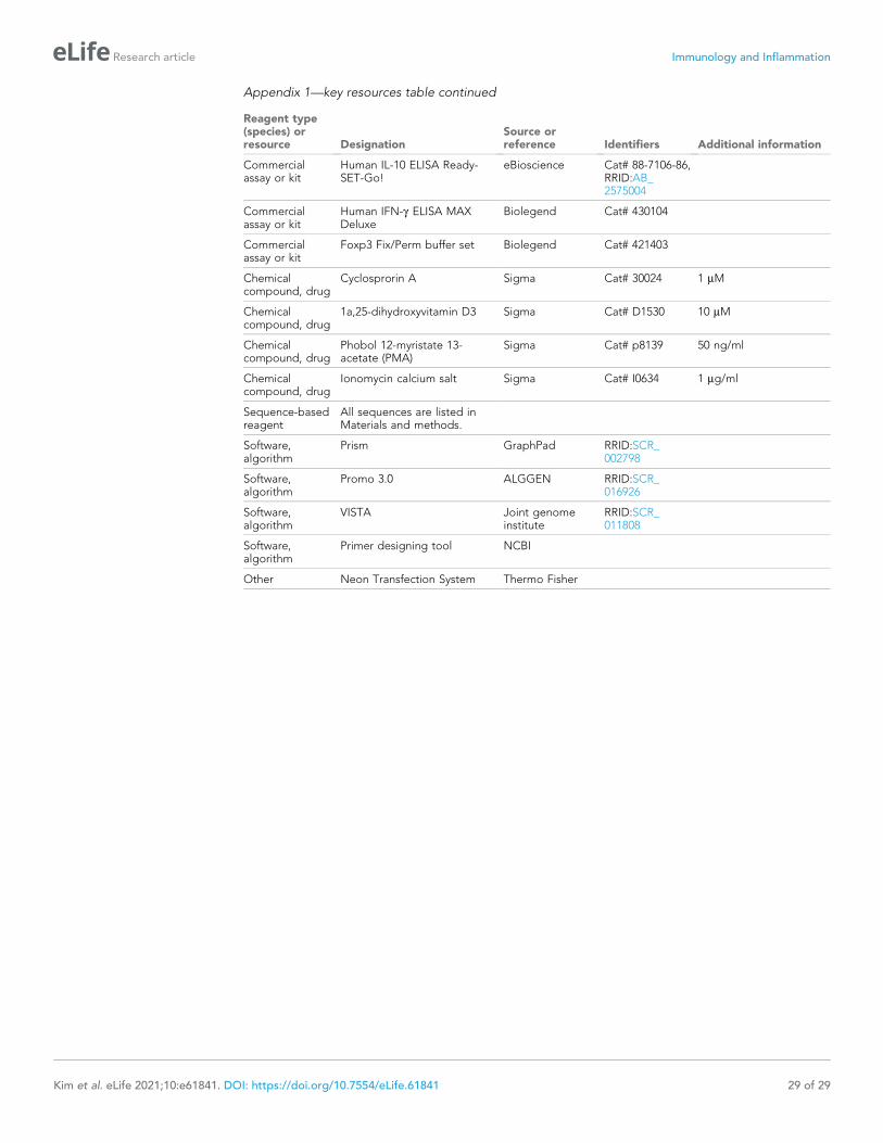
Appendix 1—key resources table continued
Reagent type(species) orresource Designation
Source orreference Identifiers Additional information
Commercialassay or kit
Human IL-10 ELISA Ready-SET-Go!
eBioscience Cat# 88-7106-86,RRID:AB_2575004
Commercialassay or kit
Human IFN-g ELISA MAXDeluxe
Biolegend Cat# 430104
Commercialassay or kit
Foxp3 Fix/Perm buffer set Biolegend Cat# 421403
Chemicalcompound, drug
Cyclosprorin A Sigma Cat# 30024 1 mM
Chemicalcompound, drug
1a,25-dihydroxyvitamin D3 Sigma Cat# D1530 10 mM
Chemicalcompound, drug
Phobol 12-myristate 13-acetate (PMA)
Sigma Cat# p8139 50 ng/ml
Chemicalcompound, drug
Ionomycin calcium salt Sigma Cat# I0634 1 mg/ml
Sequence-basedreagent
All sequences are listed inMaterials and methods.
Software,algorithm
Prism GraphPad RRID:SCR_002798
Software,algorithm
Promo 3.0 ALGGEN RRID:SCR_016926
Software,algorithm
VISTA Joint genomeinstitute
RRID:SCR_011808
Software,algorithm
Primer designing tool NCBI
Other Neon Transfection System Thermo Fisher
Kim et al. eLife 2021;10:e61841. DOI: https://doi.org/10.7554/eLife.61841 29 of 29
Research article Immunology and Inflammation
Copyright © 2022 FDOKUMEN