Brian & Brian (1952) The wasp, Vespula sylvestris Scopoli: feeding, foraging and colony development
Upload
independentCategory
view
1download
0
The
Journ
al o
f Exp
erim
enta
l M
edic
ine
ARTICLE
JEM © The Rockefeller University Press $15.00
Vol. 204, No. 2, February 19, 2007 369–380 www.jem.org/cgi/doi/10.1084/jem.20061334
369
Regulatory T cells play a key role in suppress-ing immune responses and in maintaining im-munological homeostasis. Regulatory T cells have been shown to prevent autoimmune dis-eases, to down-modulate immune response to allergens, pathogens, and cancer cells, and to mediate peripheral transplantation tolerance (1–7). The best characterized subset of regula-tory T cells are the CD4+CD25+FOXP3+ natural regulatory T (nTreg) cells, whose dif-ferentiation, maintenance, and function are controlled by TCR engagement (8, 9), CD28 engagement (10, 11), FOXP3 (12, 13), and IL-2
(14, 15). nTreg cells are generated in the thy-mus upon TCR–peptide–MHC interaction (9) as a cell population with a broad repertoire of high-affi nity TCRs, recognizing self-antigens (9, 16), tumor-associated antigens (17), allogeneic antigens (18), and possibly pathogen-derived antigens (19). Recent fi ndings indicate that nTreg cells derive from high-affi nity autoreac-tive T cells, which upon contact with a subset of activated DCs within the Hassal’s corpuscles in the thymic medulla (20) acquire regulatory function. nTreg cells are readily present in the thymus, blood, and secondary lymphoid organs of healthy individuals or mice and represent 5–10% of the total CD4+ T cells (2). The mechanisms by which nTreg cells are activated
WASP regulates suppressor activity of human and murine CD4+CD25+FOXP3+ natural regulatory T cells
Francesco Marangoni,1,2 Sara Trifari,1 Samantha Scaramuzza,1 Cristina Panaroni,1 Silvana Martino,3 Luigi D. Notarangelo,4 Zeina Baz,5 Ayse Metin,6 Federica Cattaneo,1 Anna Villa,1,7 Alessandro Aiuti,1 Manuela Battaglia,1 Maria-Grazia Roncarolo,1,2 and Loïc Dupré1
1San Raffaele Telethon Institute for Gene Therapy (HSR-TIGET), 20132 Milan, Italy2Vita-Salute San Raffaele University, 20132 Milan, Italy3Department of Pediatrics, University of Turin, 10126 Turin, Italy4Department of Pediatrics, University of Brescia, Spedali Civili, 25125 Brescia, Italy5Department of Pediatrics, St. George Hospital University Medical Center, 1100 2807 Beirut, Lebanon6SB Ankara Diskapi Children’s Hospital, 06500 Ankara, Turkey7Human Genome Department, Istituto di Tecnologie Biomediche, CNR ITB, 20090 Segrate, Milan, Italy
A large proportion of Wiskott-Aldrich syndrome (WAS) patients develop autoimmunity and
allergy. CD4+CD25+FOXP3+ natural regulatory T (nTreg) cells play a key role in peripheral
tolerance to prevent immune responses to self-antigens and allergens. Therefore, we inves-
tigated the effect of WAS protein (WASP) defi ciency on the distribution and suppressor
function of nTreg cells. In WAS−/− mice, the steady-state distribution and phenotype of
nTreg cells in the thymus and spleen were normal. However, WAS−/− nTreg cells engrafted
poorly in immunized mice, indicating perturbed homeostasis. Moreover, WAS−/− nTreg cells
failed to proliferate and to produce transforming growth factor 𝛃 upon T cell receptor
(TCR)/CD28 triggering. WASP-dependent F-actin polarization to the site of TCR triggering
might not be involved in WAS−/− nTreg cell defects because this process was also ineffi -
cient in wild-type (WT) nTreg cells. Compared with WT nTreg cells, WAS−/− nTreg cells
showed reduced in vitro suppressor activity on both WT and WAS−/− effector T cells. Simi-
larly, peripheral nTreg cells were present at normal levels in WAS patients but failed to
suppress proliferation of autologous and allogeneic CD4+ effector T cells in vitro. Thus,
WASP appears to play an important role in the activation and suppressor function of nTreg
cells, and a dysfunction or incorrect localization of nTreg cells may contribute to the devel-
opment of autoimmunity in WAS patients.
CORRESPONDENCE
Maria-Grazia Roncarolo:
Abbreviations used: HD,
healthy donor; MFI, mean
fl uorescence intensity; nTreg,
CD4+CD25+FOXP3+ natural
regulatory T; WAS, Wiskott-
Aldrich syndrome; WASP,
WAS protein.
L. Dupré’s present address is INSERM U563, Purpan
University Hospital, 31024 Toulouse Cedex 3, France.

370 ROLE OF WASP IN CD4+CD25+FOXP3+ NTREG CELLS | Marangoni et al.
and mediate suppression of eff ector CD4+ and CD8+ T cells are still under investigation. Activation of nTreg cells is trig-gered by engagement of TCR and requires IL-2 (8, 21). In vitro suppressor activity of nTreg cells is mediated predomi-nantly by cell–cell contact mechanisms. It remains to be clari-fi ed which are the molecules responsible for this process. A role for membrane-bound TGF-β (22) and CTLA-4 (23) in mediating nTreg cell suppressor function was hypothe-sized, but it appears to be limited to specifi c in vitro settings and was not confi rmed by follow-up studies (24, 25). A pos-sible additional mechanism of suppression could be via the release of perforin and granzyme by nTreg cells, resulting in apoptosis of eff ector T cells (26, 27). In addition, secretion of TGF-β (28, 29) and IL-10 (30) can contribute to the sup-pressor activity of nTreg cells in vivo. Quantitative and quali-tative defects in regulatory T cells may result in pathology. Indeed, there is evidence that skewing of antigen-specifi c T cells toward a regulatory instead of a Th1 or Th2 phenotype is key in the maintenance of homeostasis and in the preven-tion of autoimmunity (31) and allergy (32).
Wiskott-Aldrich syndrome (WAS) is a primary human X-linked immunodefi ciency caused by mutations in the WAS protein (WASP) (33) and characterized by recurrent infections and thrombocytopenia. A high proportion of WAS patients develop eczema and have high IgE levels (34), sug-gesting a Th2 unbalance. This is in line with the recent fi nd-ing that WAS T cell lines have a selective defect in Th1 cytokine production (35) and that WAS−/− mice develop a skewed Th2 phenotype (Snapper, S.B., personal communi-cation). Moreover, WAS patients lacking WASP expression have an increased risk of developing autoimmune diseases, including hemolytic anemia, cutaneous vasculitis, IgA nephropa-thy, infl ammatory bowel disease, and arthritis (34, 36). WASP is a tightly regulated hematopoietic-specifi c protein, which controls actin nucleation (37), immunological synapse assembly (38), and migration to secondary lymphoid organs (39). In addition, WASP regulates apoptosis (40), proliferation, and IL-2 production in eff ector T cells (41–44). Although these defects contribute to the immunodefi ciency, it cannot be ex-cluded that defects in regulatory T cell populations and func-tion are associated with the development of autoimmunity and with the unbalanced Th2 response in WAS patients. To determine whether the absence of WASP is associated with nTreg cell dysfunction, we characterized nTreg cells isolated from WAS−/− mice and WAS patients. In WAS−/− mice, the diff erentiation of nTreg cells and their steady-state distri-bution to secondary lymphoid organs were not signifi cantly impaired. However, nTreg cell activation, in vitro suppres-sive activity, and in vivo engraftment were strongly reduced. Similarly, in WAS patients the frequency of peripheral nTreg cells was normal, but they displayed poor suppressor activity toward normal and WASP-defi cient eff ector T cells in vitro. These data demonstrate that WASP is a major regulator of nTreg cell activation and function, suggesting that the auto-immune and atopic pathological manifestations in WAS may result from this impairment.
RESULTS
WASP and F-actin fail to polarize in murine nTreg cells
after TCR triggering
To investigate whether WASP plays a role in the function of nTreg cells, we fi rst analyzed WASP expression and localiza-tion in mouse WT nTreg cells. nTreg cells from the spleens of C57BL/6 mice express WASP at levels comparable to that of CD4+CD25− T cells, CD8+ T cells (Fig. 1 a), B cells, NK cells, and granulocytes (not depicted). We previously showed that WASP promotes actin polarization at the immunologi-cal synapse of eff ector T cells (38). Accordingly, after activa-tion of WT CD4+CD25− eff ector T cells with WT APCs in the presence of anti-CD3 mAbs, F-actin and WASP polar-ized to the APC contact site in �50% of the conjugates (Fig. 1, b and c). In contrast, <10% of CD4+CD25+ WT nTreg
Figure 1. WASP expression in murine nTreg cells. (a) WASP expres-
sion in CD4+CD25+, CD4+CD25−, and CD8+ T cells. Splenocytes from WT
mice were stained with anti-WASP mAbs (fi lled histogram) or isotype
matched control antibodies (empty histogram) and gated on the indicated
T cell subset. Numbers indicate percentages and MFI of WASP+ cells.
(b) F-actin and WASP localization in murine splenic WT CD4+CD25− and
CD4+CD25+ T cells upon stimulation. CD4+CD25− effector T cells (left)
and CD4+CD25+ nTreg cells (right) were stimulated with WT APCs
(stained in orange) in the absence or presence of 10 μg/ml anti-CD3
mAbs. Images showing F-actin staining (left column), WASP staining
(middle column), and their bright fi eld overlay (right column) are shown.
These images are representative of at least 100 T cell–APC conjugates that
were evaluated for each experimental condition. White arrows indicate
F-actin and WASP polarization at the T cell–APC interface. (c) Percentage of
WT CD4+CD25+ nTreg cells and WT CD4+CD25− effector T cells showing
F-actin polarization at the T cell–APC interface in the presence of APCs
and in the absence or presence of the indicated amount of anti-CD3
mAbs. Histogram bars represent the average percentage (± SD) of F-actin
polarization in T cells from three independent experiments. *, P < 0.05,
Student’s t test.

JEM VOL. 204, February 19, 2007 371
ARTICLE
cells polarized F-actin and WASP at the contact of APCs. In most nTreg cells, a ring-like distribution of F-actin and WASP beneath the plasma membrane was observed (Fig. 1 b). The reduced polarization of F-actin and WASP in nTreg cells was not due to their defective ability to form conjugates with APCs because similar proportions of nTreg cells and eff ector T cells were in contact with APCs (not depicted). These data indicate that murine nTreg cells express levels of WASP similar to those of CD4+CD25− eff ector T cells, but they do not effi ciently polarize F-actin and WASP to the APC contact site upon TCR triggering, suggesting that WASP-dependent actin polarization at the immuno-logical synapse does not occur in nTreg cells under these in vitro conditions.
Normal differentiation and steady-state tissue distribution
of nTreg cells in WAS−/− mice
To determine whether WASP is required for nTreg cell development, Foxp3 expression was analyzed in double nega-tive, double positive, CD4+ single positive, and CD8+ single
positive thymocytes of WAS−/− mice. Comparable propor-tions of Foxp3-expressing CD4+ single positive and double positive thymocytes were observed in WT and WAS−/− mice. The vast majority of CD24loFoxp3+ mature thymo-cytes were contained in the CD4+ single positive T cells in both WT and WAS−/− mice (Fig. 2 a). In addition, the over-all thymic cellularity was similar in WT and WAS−/− mice (not depicted). Moreover, the relative percentages and abso-lute counts of CD4+CD8−CD25+ T cells, as well as the mean fl uorescence intensity (MFI) of CD25, were also com-parable (Fig. 2, b and c). A raise in the absolute count of CD4+CD25−Foxp3+ and CD4+CD25+Foxp3+ T cells was found in the thymi of WAS−/− mice as compared with WT controls (Fig. 2, d and e). Collectively, these data indicate that thymic diff erentiation of nTreg cells can effi ciently occur in WAS−/− mice.
We next investigated if WASP defi ciency may infl uence the steady-state distribution of nTreg cells into the spleen. The overall splenocyte count was comparable in WT and WAS−/− mice (not depicted). Normal percentages and numbers
Figure 2. Thymic generation of nTreg cells in WAS−/− mice.
(a) Thymic development of nTreg cells in WT and WAS−/− mice. CD4/CD8
fl ow cytometric plots were gated on live thymocytes, total Foxp3+ cells, and
mature CD24loFoxp3+ cells. Numbers indicate the percentage of cells in
the respective region. Data are representative of 12 mice per group ana-
lyzed in three independent experiments. (b) Immunophenotype of CD4+
single positive thymocytes from representative WT and WAS−/− mice.
Numbers indicate the percentages and MFI of CD25+ cells. (c) Percentage,
absolute count, and MFI of CD25+ cells among CD4+ single positive thy-
mocytes. Mean ± SD of 13 mice per group are shown. (d) Foxp3 and
CD25 expression in CD4+ single positive thymocytes. Numbers indicate
the percentage of cells in the respective region. (e) Absolute count of the
indicated thymocyte populations. Mean ± SD of 13 mice per group is
shown. *, P < 0.05, Student’s t test.

372 ROLE OF WASP IN CD4+CD25+FOXP3+ NTREG CELLS | Marangoni et al.
of CD4+CD25+ T cells were found in the spleens of WAS−/− mice, although the expression of CD25 was reduced as com-pared with WT mice (Fig. 3, a and b). As in the thymus, the absolute count of CD4+CD25−Foxp3+ T cells increased in the spleens of WAS−/− mice. Conversely, the numbers of splenic CD4+CD25+Foxp3+ nTreg cells were comparable to those of normal mice, whereas an increased number of splenic CD4+CD25+Foxp3− T cells was observed (Fig. 3, c and d). Expression of the nTreg cell markers CTLA-4 and GITR on CD4+CD25+ splenic T cells was in the normal range, indicating that WAS−/− nTreg cells have a normal phenotype (Fig. 3 e). WAS−/− CD4+CD25+ T cells also expressed normal levels of the activation markers CD69, CD62L, and CD45RB (Fig. 3 e). These data indicate that splenic nTreg cells are present in WAS−/− mice in a similar amount to those present in WT mice, albeit CD25 expres-sion is reduced.
Defective suppressor activity of murine WAS−/− nTreg cells
To investigate whether WAS−/− nTreg cells are functional, proliferation of eff ector T cells in the presence of WT or WAS−/− nTreg cells was tested. As expected, WT nTreg cells suppressed proliferation of WT CD90+CD25− eff ector T cells activated with anti-CD3 mAbs in a cell dose–dependent
fashion (Fig. 4 a, fi lled bars). In contrast, WAS−/− nTreg cells had a reduced capacity to suppress WT eff ector T cell proliferation at every ratio of eff ector T cells to nTreg cells tested (Fig. 4 a, dotted vs. fi lled bars). These data indicate that nTreg cells require WASP to suppress WT eff ector T cells in vitro. The susceptibility of WAS−/− eff ector T cells, compared with WT eff ector T cells, to be suppressed by WT nTreg cells (Fig. 4 a, dashed vs. fi lled bars) or by WAS−/− nTreg cells (Fig. 4 a, empty vs. fi lled bars) was also tested. WAS−/− eff ector T cells proliferated at a lower extent than WT eff ector cells (median proliferation was 6,360 and 26,380 cpm, respectively; P < 0.05 as determined by Mann-Whitney test). Suppression of proliferation of WAS−/− eff ector T cells by WT nTreg cells was comparable to the suppression observed with WT eff ector T cells when an ef-fector T cell/nTreg cell ratio of 1:0.16 was used. At lower eff ector T cell/nTreg cell ratios, suppression of WAS−/− eff ector T cells by WT nTreg cells was signifi cantly lower compared with that of WT eff ector T cells (Fig. 4 a, dashed vs. fi lled bars). Similar results were obtained with WAS−/− nTreg cells (Fig. 4 a, empty vs. fi lled bars). These data indi-cate that proliferation of WAS−/− eff ector T cells was not suppressed by WT or WAS−/− nTreg cells in a cell dose– dependent manner. This may be due to intrinsic defects of
Figure 3. Cell count and immunophenotype of nTreg cells in the
spleens of WAS−/− mice. (a) Immunophenotype of CD4+ splenocytes
from representative WT and WAS−/− mice. Numbers indicate the percent-
ages and MFI of CD25+ cells. (b) Percentage, absolute count, and MFI of
CD25+ cells among CD4+ splenocytes. Mean ± SD of 13 mice per group
is shown. *, P < 0.05, Student’s t test. (c) Foxp3 and CD25 expression in
CD4+ splenocytes. Numbers indicate the percentage of cells in the re-
spective region. (d) Absolute count of the indicated splenocyte popula-
tions. Mean ± SD of 13 mice per group is shown. *, P < 0.05, Student’s
t test. (e) Immunophenotype of nTreg cells from WT and WAS−/− mice.
Expression of CD25 is shown together with the expression of CTLA-4,
GITR, CD69, CD62L, and CD45RB. Negative control staining resulted in
signal below the value of 10. Numbers indicate the percentages of cells in
the respective regions. Data are representative of six to eight mice per
group analyzed in two independent experiments.
JEM VOL. 204, February 19, 2007 373
ARTICLE
WAS−/− eff ector T cells in providing factors required for activation of nTreg cells.
The in vivo ability of adoptively transferred WAS−/− and WT nTreg cells to engraft and modulate an antigen-
specifi c immune response upon OVA immunization in WT or WAS−/− mice was investigated. 7–10 d after OVA im-munization of WT recipients, donor WT nTreg cells (trans-ferred 1 d before immunization) represented �2% of the CD4+Foxp3+ (Fig. 4 b) or CD4+CD25+ cells (not depicted) in the draining LNs. In contrast, donor WAS−/− nTreg cells could not be recovered in draining LNs (Fig. 4 b). The same results were obtained when donor nTreg cells were counted in nondraining LNs, spleen, blood, liver, and lungs regardless of CD25 and Foxp3 expression (not depicted). Accordingly, the in vitro proliferative recall responses to OVA were signifi cantly
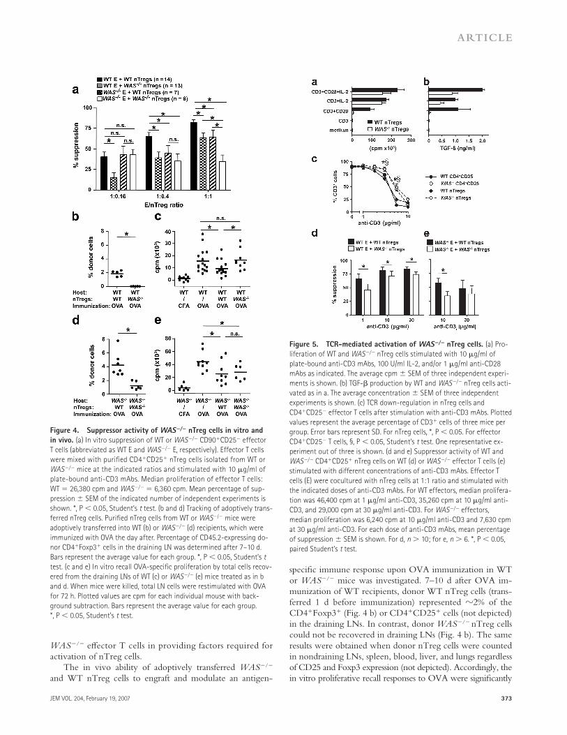
Figure 4. Suppressor activity of WAS−/− nTreg cells in vitro and
in vivo. (a) In vitro suppression of WT or WAS−/− CD90+CD25− effector
T cells (abbreviated as WT E and WAS−/− E, respectively). Effector T cells
were mixed with purifi ed CD4+CD25+ nTreg cells isolated from WT or
WAS−/− mice at the indicated ratios and stimulated with 10 μg/ml of
plate-bound anti-CD3 mAbs. Median proliferation of effector T cells:
WT = 26,380 cpm and WAS−/− = 6,360 cpm. Mean percentage of sup-
pression ± SEM of the indicated number of independent experiments is
shown. *, P < 0.05, Student’s t test. (b and d) Tracking of adoptively trans-
ferred nTreg cells. Purifi ed nTreg cells from WT or WAS−/− mice were
adoptively transferred into WT (b) or WAS−/− (d) recipients, which were
immunized with OVA the day after. Percentage of CD45.2-expressing do-
nor CD4+Foxp3+ cells in the draining LN was determined after 7–10 d.
Bars represent the average value for each group. *, P < 0.05, Student’s t
test. (c and e) In vitro recall OVA-specifi c proliferation by total cells recov-
ered from the draining LNs of WT (c) or WAS−/− (e) mice treated as in b
and d. When mice were killed, total LN cells were restimulated with OVA
for 72 h. Plotted values are cpm for each individual mouse with back-
ground subtraction. Bars represent the average value for each group.
*, P < 0.05, Student’s t test.
Figure 5. TCR-mediated activation of WAS−/− nTreg cells. (a) Pro-
liferation of WT and WAS−/− nTreg cells stimulated with 10 μg/ml of
plate-bound anti-CD3 mAbs, 100 U/ml IL-2, and/or 1 μg/ml anti-CD28
mAbs as indicated. The average cpm ± SEM of three independent experi-
ments is shown. (b) TGF-β production by WT and WAS−/− nTreg cells acti-
vated as in a. The average concentration ± SEM of three independent
experiments is shown. (c) TCR down-regulation in nTreg cells and
CD4+CD25− effector T cells after stimulation with anti-CD3 mAbs. Plotted
values represent the average percentage of CD3+ cells of three mice per
group. Error bars represent SD. For nTreg cells, *, P < 0.05. For effector
CD4+CD25− T cells, §, P < 0.05, Student’s t test. One representative ex-
periment out of three is shown. (d and e) Suppressor activity of WT and
WAS−/− CD4+CD25+ nTreg cells on WT (d) or WAS−/− effector T cells (e)
stimulated with different concentrations of anti-CD3 mAbs. Effector T
cells (E) were cocultured with nTreg cells at 1:1 ratio and stimulated with
the indicated doses of anti-CD3 mAbs. For WT effectors, median prolifera-
tion was 46,400 cpm at 1 μg/ml anti-CD3, 35,260 cpm at 10 μg/ml anti-
CD3, and 29,000 cpm at 30 μg/ml anti-CD3. For WAS−/− effectors,
median proliferation was 6,240 cpm at 10 μg/ml anti-CD3 and 7,630 cpm
at 30 μg/ml anti-CD3. For each dose of anti-CD3 mAbs, mean percentage
of suppression ± SEM is shown. For d, n > 10; for e, n > 6. *, P < 0.05,
paired Student’s t test.

374 ROLE OF WASP IN CD4+CD25+FOXP3+ NTREG CELLS | Marangoni et al.
suppressed after adoptive transfer of WT nTreg cells, but not of WAS−/− nTreg cells (Fig. 4 c). Signifi cantly lower percentages of CD4+Foxp3+ (Fig. 4 d) or CD4+CD25+ (not depicted) WAS−/− nTreg cells as compared with WT nTreg cells were also recovered in WAS−/− recipients (1 and 4%, respectively). However, both WT and WAS−/− nTreg cells modulated the OVA-specifi c immune response elicited in WAS−/− mice, as demonstrated by the in vitro recall response to OVA (Fig. 4 e). It should be noted that the overall prolif-eration of WAS−/− T cells to OVA was higher than that of WT T cells, even when the WAS−/− nTreg cells were trans-ferred in vivo. Collectively, our results indicate that WASP-defi cient nTreg cells fail to engraft in WT and displayed a reduced engraftment in WAS−/− hosts, suggesting that this defect may contribute to their dysfunction in vivo.
Impaired TCR/CD28 activation of WAS−/− nTreg cells
Activation through the TCR is required for the suppressive function of nTreg cells (8). Therefore, it is possible that defective suppressor activity of WAS−/− nTreg cells is due to a defect in their activation. To address this question, we mea-sured the proliferative response and the cytokine secretion profi le of WAS−/− nTreg cells after TCR stimulation. As expected, neither WT nor WAS−/− nTreg cells proliferated (Fig. 5 a) or produced TGF-β (Fig. 5 b) in response to anti-CD3 mAbs alone. Furthermore, in contrast to WT nTreg cells, WAS−/− nTreg cells failed to proliferate and secrete TGF-β after activation with anti-CD3 plus anti-CD28 mAbs (Fig. 5, a and b). The addition of exogenous IL-2 to anti-CD3 or anti-CD3 plus anti-CD28 mAbs stimulation induced comparable proliferation of WT and WAS−/− nTreg cells (Fig. 5 a) and promoted TGF-β production, although at
reduced levels in WAS−/− nTreg cells (Fig. 5 b). Neither WT nor WAS−/− nTreg cells produced IL-2, IL-4, IL-5, IL-10, IFN-γ, and TNF-α after stimulation through the TCR/CD28 (not depicted). These data show that WAS−/− nTreg cells have a major activation defect downstream of the TCR and the CD28 receptor, and that proliferation, but not TGF-β production, is fully restored by the addition of exog-enous IL-2. To determine whether WAS−/− nTreg cells have a higher threshold for TCR-mediated activation as compared with WT nTreg cells, we measured TCR down-regulation after stimulation with increasing doses of anti-CD3 mAbs. Both WT nTreg cells and eff ector T cells down-regulated CD3 in a comparable dose-dependent man-ner (Fig. 5 c). CD3 was also down-regulated in WAS−/− nTreg cells and eff ector T cells, but higher doses of anti-CD3 mAbs were required to obtain levels of down-regula-tion similar to those observed in WT cells (Fig. 5 c). We next investigated whether the impaired regulatory function of WAS−/− nTreg cells could depend on the strength of TCR stimulation. WT eff ector T cells were suppressed by WT nTreg cells in an anti-CD3 mAb dose-dependent man-ner (Fig. 5 d). On the other hand, WAS−/− nTreg cells had a reduced suppressive ability compared with WT nTreg cells at the dose of 1 μg/ml, but the suppressive capacity was enhanced when the anti-CD3 mAb dose was increased (Fig. 5 d). However, the suppressive activity of WAS−/− nTreg cells could never reach normal levels (Fig. 5 d). Subsequently, increasing doses of anti-CD3 mAbs were used to activate WAS−/− eff ector T cells to determine whether higher levels of activation render these cells more prone to nTreg cell–mediated suppression. WAS−/− eff ector T cells failed to proliferate after stimulation with 1 μg/ml anti-CD3 mAbs
Table I. WAS gene mutations and clinical status of WAS patients
Patient Age (yr)a Mutation
type
gDNA
mutation
Protein
change
Scoreb Thrombo-
cytopenia
T cell
lymphopenia
Eczema Infections Autoimmunity
WAS1 24 Splice
Intron 9
IVS9 +2 del
tgag
ND 4 Yes No Mild Severe HSV,
pneumonia
No
WAS4 23 Deletion
Exon 6
570del t fs stop
aa260
5 Yesc Yes Mild Various and
recurrent
infections
Vasculitis, arthritis,
IgA nephropathy
WAS13 13.8 Missense
Exon 1
150t>c L39P 2 Yesc Yes Mild No No
WAS14 1.8 Missense
Exon 4
431g>a E133K 5 Yes Yes Mild No Colitis, vasculitisd
WAS17 2.8 Splice
Intron 8
IVS8 +1 del
gtga
fs stop
aa246
4 Yes No Severe Recurrent otitis,
pneumonia
No
WAS21 18 Missense
Exon 7
741c>g A236G 2 Yesc Yes No Recurrent otitis No
WAS22 19 Missense
Exon 2
201c>t A56V 1 Yes No No No No
fs, frameshift; del, deletion; IVS, intervening sequence (intron); ND, not determined.aAge refers to the time of blood sampling.bDisease score is given according to the classifi cation reported previously (reference 34).cSplenectomized.dPatient undergoing therapy with prednisone.

JEM VOL. 204, February 19, 2007 375
ARTICLE
(not depicted). At the dose of 10 μg/ml anti-CD3 mAbs, WAS−/− nTreg cells suppressed WAS−/− eff ector T cells less effi ciently than WT nTreg cells (Fig. 5 e). An increase in the anti-CD3 mAbs dose (30 μg/ml) led to comparable levels of suppression of WAS−/− eff ector T cells by WT and WAS−/− nTreg cells (Fig. 5 e), although the suppressive activity was lower than that observed with WT eff ector T cells. Overall, these data indicate that the suppressive defect of WAS−/− nTreg cells can depend both on the strength of TCR stimu-lation and on the dysfunction of WAS−/− eff ector T cells, suggesting an important role of WASP in both eff ector and regulatory T cells.
Normal percentage of CD4+CD25+FOXP3+ nTreg cells
in the blood of WAS patients
To our knowledge, the role of WASP in the generation and function of human nTreg cells has never been studied. As observed in murine cells, levels of WASP expression in CD4+CD25+ and CD4+CD25− T cells from the blood of healthy donors (HDs) were comparable (not depicted). To investigate the role of WASP in the generation of human nTreg cells, we analyzed CD4+CD25+FOXP3+ T cells from
the peripheral blood of seven WAS patients of diff erent ages and diff erent clinical scores (Table I). Percentages of FOXP3+ cells among CD4+ T cells of WAS patients were either higher or comparable to those observed in HDs (Fig. 6 a). Overall analysis revealed a slight increase in FOXP3+ cells within
Figure 6. Immunophenotype of nTreg cells from human peripheral
blood. (a) CD4+FOXP3+ cells in the peripheral blood of WAS patients.
PBMCs from four representative HDs and seven WAS patients (WAS) were
stained with anti-CD4 and anti-FOXP3 mAbs. Results shown are gated on
the CD4+ T cells. Numbers indicate the percentages of FOXP3+ cells
among CD4+ T cells. (b) Percentage of FOXP3+ cells (left), CD25+FOXP3+
cells (middle), and CD25− FOXP3+ cells (right) among CD4+ T cells of HDs
(n = 16) and WAS patients (n = 7). Bars represent the median value for
each group.
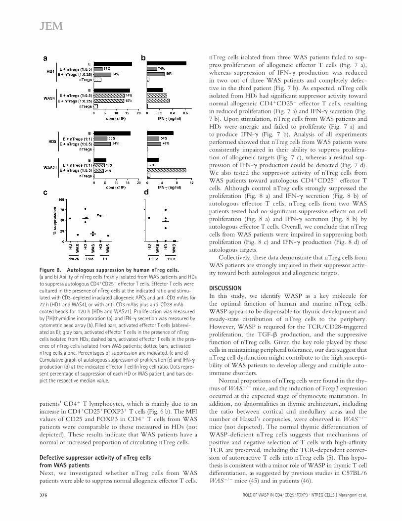
Figure 7. Allogeneic suppression by human nTreg cells. (a and b)
Ability of nTreg cells freshly isolated from WAS patients and HDs to sup-
press allogeneic CD4+CD25− effector T cells from HDs. Effector T cells
were cultured in the presence of nTreg cells at the indicated ratio and
stimulated with CD3-depleted irradiated allogeneic APCs and anti-CD3
mAbs for 72 h (HD1 and WAS4), anti-CD3 mAbs plus anti-CD28 mAb–
coated beads for 72 h (HD2 and WAS14), or anti-CD3 mAbs plus anti-
CD28 mAb–coated beads for 120 h (HD4 and WAS17). Proliferation was
measured by [3H]thymidine incorporation (a), and IFN-γ secretion was
measured by cytometric bead array (b). Filled bars, activated effector T
cells (abbreviated as E); gray bars, activated effector T cells in the presence
of nTreg cells isolated from HDs; dashed bars, activated effector T cells in
the presence of nTreg cells isolated from WAS patients; dotted bars, acti-
vated nTreg cells alone. Percentages of suppression are indicated. (c and d)
Cumulative graph of allogeneic suppression of proliferation (c) and
IFN-γ production (d) at the indicated effector T cell/nTreg cell ratio. Dots
represent percentage of suppression of each HD or WAS patient, and bars
depict the respective median value. *, P < 0.05, Mann-Whitney test.
376 ROLE OF WASP IN CD4+CD25+FOXP3+ NTREG CELLS | Marangoni et al.
patients’ CD4+ T lymphocytes, which is mainly due to an increase in CD4+CD25+FOXP3+ T cells (Fig. 6 b). The MFI values of CD25 and FOXP3 in CD4+ T cells from WAS patients were comparable to those measured in HDs (not depicted). These results indicate that WAS patients have a normal or increased proportion of circulating nTreg cells.
Defective suppressor activity of nTreg cells
from WAS patients
Next, we investigated whether nTreg cells from WAS patients were able to suppress normal allogeneic eff ector T cells.
nTreg cells isolated from three WAS patients failed to sup-press proliferation of allogeneic eff ector T cells (Fig. 7 a), whereas suppression of IFN-γ production was reduced in two out of three WAS patients and completely defec-tive in the third patient (Fig. 7 b). As expected, nTreg cells isolated from HDs had signifi cant suppressor activity toward normal allogeneic CD4+CD25− eff ector T cells, resulting in reduced proliferation (Fig. 7 a) and IFN-γ secretion (Fig. 7 b). Upon stimulation, nTreg cells from WAS patients and HDs were anergic and failed to proliferate (Fig. 7 a) and to produce IFN-γ (Fig. 7 b). Analysis of all experiments performed showed that nTreg cells from WAS patients were consistently impaired in their ability to suppress prolifera-tion of allogeneic targets (Fig. 7 c), whereas a residual sup-pression of IFN-γ production could be detected (Fig. 7 d). We also tested the suppressor activity of nTreg cells from WAS patients toward autologous CD4+CD25− eff ector T cells. Although control nTreg cells strongly suppressed the proliferation (Fig. 8 a) and IFN-γ secretion (Fig. 8 b) of autologous eff ector T cells, nTreg cells from two WAS patients tested had no signifi cant suppressive eff ects on cell proliferation (Fig. 8 a) and IFN-γ secretion (Fig. 8 b) by autologous eff ector T cells. Overall, we conclude that nTreg cells from WAS patients were impaired in suppressing both proliferation (Fig. 8 c) and IFN-γ production (Fig. 8 d) of autologous targets.
Collectively, these data demonstrate that nTreg cells from WAS patients are strongly impaired in their suppressor activ-ity toward both autologous and allogeneic targets.
DISCUSSION
In this study, we identify WASP as a key molecule for the optimal function of human and murine nTreg cells. WASP appears to be dispensable for thymic development and steady-state distribution of nTreg cells to the periphery. However, WASP is required for the TCR/CD28-triggered proliferation, the TGF-β production, and the suppressive function of nTreg cells. Given the key role played by these cells in maintaining peripheral tolerance, our data suggest that nTreg cell dysfunction might contribute to the high suscepti-bility of WAS patients to develop allergy and multiple auto-immune disorders.
Normal proportions of nTreg cells were found in the thy-mus of WAS−/− mice, and the induction of Foxp3 expression occurred at the expected stage of thymocyte maturation. In addition, no abnormalities in thymic architecture, including the ratio between cortical and medullary areas and the number of Hassal’s corpuscles, were observed in WAS−/− mice (not depicted). The normal thymic diff erentiation of WASP-defi cient nTreg cells suggests that mechanisms of positive and negative selection of T cells with high-affi nity TCR are preserved, including the TCR-dependent conver-sion of autoreactive T cells into nTreg cells (5). This hypo-thesis is consistent with a minor role of WASP in thymic T cell diff erentiation, as suggested by previous studies in C57BL/6 WAS−/− mice (45) and in patients (46).
Figure 8. Autologous suppression by human nTreg cells.
(a and b) Ability of nTreg cells freshly isolated from WAS patients and HDs
to suppress autologous CD4+CD25− effector T cells. Effector T cells were
cultured in the presence of nTreg cells at the indicated ratio and stimu-
lated with CD3-depleted irradiated allogeneic APCs and anti-CD3 mAbs for
72 h (HD1 and WAS4), or with anti-CD3 mAbs plus anti-CD28 mAb–
coated beads for 120 h (HD5 and WAS21). Proliferation was measured
by [3H]thymidine incorporation (a), and IFN-γ secretion was measured by
cytometric bead array (b). Filled bars, activated effector T cells (abbrevi-
ated as E); gray bars, activated effector T cells in the presence of nTreg
cells isolated from HDs; dashed bars, activated effector T cells in the pres-
ence of nTreg cells isolated from WAS patients; dotted bars, activated
nTreg cells alone. Percentages of suppression are indicated. (c and d)
Cumulative graph of autologous suppression of proliferation (c) and IFN-γ
production (d) at the indicated effector T cell/nTreg cell ratio. Dots repre-
sent percentage of suppression of each HD or WAS patient, and bars de-
pict the respective median value.

JEM VOL. 204, February 19, 2007 377
ARTICLE
nTreg cells generated in the thymus undergo a robust peripheral expansion that is mainly dependent on paracrine delivery of IL-2 by activated T cells and possibly DCs (47). The IL-2–IL-2R axis appears to be dispensable during thymic development, but it is required for the survival of nTreg cells in the periphery (14, 15). Although WAS is characterized by defective IL-2 production by T cells (41–44), no reduction in the relative numbers of nTreg cells in the spleens of WAS−/− mice was observed. These fi ndings suggest that low levels of IL-2 in WAS−/− mice are suffi cient to provide the appropri-ate survival signals to peripheral nTreg cells. Although nor-mal numbers of nTreg cells were found in the spleens of WAS−/− mice, adoptive transfer of WAS−/− nTreg cells into WT or WAS−/− hosts resulted in no or reduced, respec-tively, recovery of those cells in draining LNs after immuni-zation. This defective engraftment of WAS−/− nTreg cells could be due to impaired migration, survival, and/or prolif-eration, and it may be even more evident in the competitive environment represented by WT hosts. In a distinct WASP-defi cient murine strain, Maillard et al. (reference 48; p. 379 of this issue) found decreased numbers of nTreg cells in the spleen, suggesting that the degree of defects in nTreg cell homeostasis may depend on the genetic background of the WAS−/− mouse model.
We showed here that murine WASP-defi cient nTreg cells were impaired in their regulatory function. Indeed, in vitro experiments showed that WAS−/− nTreg cells had a signifi cantly reduced capacity to suppress WT eff ector T cell proliferation at every cell ratio tested. After strong TCR stimulation (30 μg/ml anti-CD3 mAbs), a residual suppressor activity of WAS−/− nTreg cells on WT eff ector T cells could be detected, suggesting that the in vitro dysfunction of WAS−/− nTreg cells is mainly due to an activation defect. Previous studies have shown that the suppressor activity of nTreg cells directly depends on their activation through the TCR (8). Indeed, we clearly demonstrated that WAS−/− nTreg cells have a profound defect in TCR/CD28-mediated activation. Upon stimulation with anti-CD3 and anti-CD28 mAbs, WAS−/− nTreg cell proliferation and TGF-β produc-tion were abolished. It remains to be determined whether the defect in TGF-β production contributes to the dysfunctional nTreg cell activity.
Given the master role of WASP in immunological syn-apse assembly, it is tempting to speculate that WASP contrib-utes to the assembly of an activatory synapse in nTreg cells and that the lack of this protein prevents optimal TCR acti-vation in nTreg cells. However, we showed that, in contrast to eff ector T cells, murine nTreg cells failed to polarize F-actin and concomitantly WASP to the site of contact with anti-CD3 mAb-loaded APCs or beads coated with anti-CD3 and anti-CD28 mAbs (not depicted). Therefore, a role of WASP cannot be demonstrated in this process. On the other hand, it cannot be excluded that nTreg cells assemble a structurally diff erent immunological synapse, or that its assembly follows diff erent kinetics. It is also possible that the activation and functional defects of WAS−/− nTreg cells are independent
from immunological synapse formation and are due to defects in signaling downstream of the TCR. The observation that defective suppressive function in WAS−/− nTreg cells was present at a dose of TCR stimulation leading to normal TCR internalization is consistent with the hypothesis that additional defects in late TCR-mediated signaling may play a role in the activation of these cells (49).
The activation state of the eff ector T cells may also play a role in the suppressor activity of nTreg cells. We showed that WT nTreg cells could normally suppress the proliferation of WAS−/− eff ector T cells at the eff ector T cell/nTreg cell ratio of 1:0.16, but at lower ratios, the suppressor activity was reduced as compared with that observed in cultures with WT eff ector T cells. These data suggest a lack of activatory signals by WAS−/− eff ector T cells. WAS−/− nTreg cells’ failure to suppress autologous WAS−/− eff ector T cells was even more profound than that of WT nTreg cells (ratio 1:1), supporting a greater dependence of the former on signals delivered by eff ector T cells that are required for their activation. There-fore, we propose that defective activation of WAS−/− eff ec-tor T cells, which leads to impaired production of IL-2, might further amplify the intrinsic defect of WAS−/− nTreg cells. In line with this hypothesis, a defective cytokine pro-duction by eff ector T cells isolated from patients with immune dysregulation, polyendocrinopathy, enteropathy, X-linked syndrome was recently proposed to contribute to the dys-function of nTreg cells in this disease (50). We found that WAS−/− nTreg cells were responsive to TCR/CD28 stimu-lation in the presence of exogenous IL-2. In a complementary study, administration of exogenous IL-2 to anti-CD3 mAb–stimulated WAS−/− nTreg cells could strongly enhance their suppressive ability in vitro (48). Collectively, these data suggest that in WAS−/− mice, conditions leading to non-optimal activation of eff ector T cells and defective IL-2 production might amplify the intrinsic activation defect of nTreg cells.
Recent data demonstrated that transfer of a non-antigen–specifi c WT nTreg cell population in normocytic mice was able to control antigen-specifi c responses (51). In our study, we compared the ability of adoptively transferred WT versus WAS−/− nTreg cells to modulate an in vivo immune response to OVA. WAS−/− nTreg cells transferred in WT recipients were not found in LNs draining the site of OVA immuni-zation, and subsequently, modulation of the OVA-specifi c immune response was not observed. Redistribution of WAS−/− nTreg cells transferred in autologous WAS−/− recipients was reduced, and in this condition, down-modulation of the OVA-specifi c immune response was still observed. It should be noted that in the immunization protocol used here, the proliferation of WAS−/− eff ector T cells upon in vitro restimulation with OVA was signifi cantly higher com-pared with that observed in WT mice. This could be related to a status of spontaneous immune activation in WAS−/− mice, as illustrated by the presence of increased numbers of CD4+CD25+Foxp3− splenocytes. These conditions of potent immune reaction might therefore be permissive for suppression

378 ROLE OF WASP IN CD4+CD25+FOXP3+ NTREG CELLS | Marangoni et al.
by WAS−/− nTreg cells, although the recall response of cells from WAS−/− mice treated with WAS−/− nTreg cells was still very high. These data do not exclude that functional defects of WAS−/− nTreg cells may arise in vivo with diff er-ent antigens and in diff erent settings, such as those presented in the complementary study by Maillard et al. (48) using a diff erent WAS−/− mouse strain. Indeed, WAS−/− nTreg cells displayed a defective in vivo suppressor activity toward colitis induced by transfer of WT CD45RBhigh cells. WAS−/− nTreg cell defects may therefore be exacerbated during the develop-ment of spontaneous infl ammatory bowel disease (42).
Collectively, our data suggest that in WASP defi ciency the impaired suppressor activity of nTreg cells may be due to multiple defects, including intrinsic defects in activation and TGF-β production, as well as defects in other cell types, which therefore determine the extent of the impairment in homeostasis and peripheral tolerance.
Importantly, the role of WASP in regulating nTreg cell function was confi rmed by our study in WAS patients. In seven WAS patients of diff erent ages and with diff erent dis-ease severity, we found that the percentages of circulating CD4+CD25+FOXP3+ nTreg cells were within or above normal range, excluding a selective defect in nTreg cell dif-ferentiation and production. Increased frequency of FOXP3+ cells in the blood of two patients was attributed to an increase in the CD4+CD25−FOXP3+ cell population, as seen in the thymuses and spleens of WAS−/− mice. This population has been proposed to be a pool of tissue-homing nTreg cells (9). Therefore, the increase in this cell subset might refl ect a reduced ability of WAS−/− nTreg cells to migrate to periph-eral nonlymphoid organs. The in vitro suppressor activity of WASP-defi cient nTreg cells on proliferation was clearly impaired, both toward allogeneic and autologous eff ector T cells. nTreg cells from WAS patients (two out of two) also failed to suppress IFN-γ production by autologous eff ectors, whereas a residual regulatory activity toward IFN-γ produc-tion by allogeneic responder cells (two out of three patients) was observed. This is in line with a previous report showing that suppression of proliferation and cytokine production by nTreg cells isolated from patients with rheumatoid arthritis could occur independently (52).
Based on the results presented in this study, we hypothe-size that one possible cause of autoimmunity and atopic dis-ease in WAS patients is a functional defect of nTreg cells. Recent studies reported a compromised function of nTreg cells in several autoimmune diseases, including immune dys-regulation, polyendocrinopathy, enteropathy, X-linked syn-drome (50), multiple sclerosis (53), autoimmune polyglandular syndrome type II (54), rheumatoid arthritis (52), myasthenia gravis (55), and type 1 diabetes (56).
The similarities between our murine and human studies (normal steady-state distribution of nTreg cells in the periph-ery with defective suppressor activity) validate the WAS−/− murine model for further investigation of the contribution of nTreg cell defects to autoimmunity. It will be particularly interesting to study in this model how microbial triggers may
act in concert with the functional defect in nTreg cells to lead to autoimmunity.
In conclusion, we show here that WASP is specifi cally required for the activation and suppressive function of CD4+CD25+FOXP3+ nTreg cells of both human and murine origin, but not for their diff erentiation. We propose that the functional failure of nTreg cells contributes to the develop-ment of autoimmunity and atopic disease in WAS.
MATERIALS AND METHODSMice. C57BL/6 WAS−/− mice were provided by K.A. Siminovitch (Mt.
Sinai Hospital, Toronto, Canada) (43). WAS−/−-CD45.1 mice were gener-
ated in our animal facility. No spontaneous colitis was observed in these
mouse models. WT C57BL/6 and WT C57BL/6-CD45.1 control mice
were purchased from Charles River Laboratories. All procedures were per-
formed according to protocols approved by the Animal Care and Use Com-
mittee of the San Raff aele Scientifi c Institute.
Patients and human cells. The detailed clinical status of the patients is
reported in Table I. Peripheral blood samples from WAS patients and age-
matched HDs were obtained following standard ethical procedures (includ-
ing informed consent) and with the approval of the San Raff aele Scientifi c
Institute Internal Review Board. PBMCs were purifi ed on Ficoll gradient
(Nycomed Pharma A/S).
Immunophenotype and immunofl uorescence analysis. The following
mAbs were used for surface staining of murine cells: anti-CD4 (RM4-5),
anti-CD8 (53–6.7), anti-CD24 (M1/69), anti-CD25 (PC61), anti-CD45.2
(104), anti-CD45RB (16A), anti-CD62L (MEL14), and anti-CD69
(H1.2F3), all from BD Biosciences. Intracytoplasmic staining was performed
using the following mAbs: anti-WASP (B-9; Santa Cruz Biotechnology,
Inc.), anti–CTLA-4 (UC10-4F10-11; BD Biosciences), and anti-GITR
(108619; R&D Systems). The staining with anti-Foxp3 mAbs (FJK-16s;
eBioscience) was performed according to the manufacturer’s instructions.
For immunofl uorescence staining, purifi ed T cells were incubated for 20
min with Orange-CMTMR–loaded CD90− splenocytes as APCs (at a 1:1
cell ratio) in absence or presence of anti-CD3 mAbs (2C11; BD Biosci-
ences). Cells were transferred onto Poly-l-lysine–coated glass slides, fi xed/
permeabilized, and stained with AlexaFluor633-conjugated Phalloidin (Invit-
rogen) and anti-WASP antibodies (H-250; Santa Cruz Biotechnology, Inc.),
followed by AlexaFluor488-conjugated goat anti–rabbit antibodies. The sam-
ples were examined on a Carl Zeiss LSM 510 confocal microscope with an
x63 Plan-Apochromat objective.
Human PBMCs were stained with anti-CD4 mAbs (SK3) and anti-
CD25 mAbs (2A3), all from BD Biosciences. FOXP3 expression was evalu-
ated using PHC101 mAbs from eBioscience.
nTreg cell activation assays. CD90+CD25− eff ector T cells and CD4+CD25+
nTreg cells were purifi ed from murine splenocytes by a combination of
immunomagnetic beads (Miltenyi Biotec) and FACS sorting. Purity of
CD4+CD25+ nTreg cells was ≥90% in all experiments. Cells were plated
at 105 cells/well in the presence of the indicated dose of plate-bound anti-
CD3 mAbs (17A2; BD Biosciences) either alone or in combination with
either 1 μg/ml anti-CD28 mAbs (37.51; BD Biosciences) or 100 U/ml
rhIL-2 (Chiron Corporation). Cell proliferation was measured after a 96-h
stimulation by liquid scintillation counting. TGF-β secretion was measured
by specifi c ELISA (R&D Systems) on supernatants conditioned for 96 h.
The Bio-Plex technology using the Mouse Th1/Th2 panel (all from Bio-
Rad Laboratories) was used to measure the levels of IL-10 and IL-5 (after a
96-h stimulation), and IL-2, IL-4, IFN-γ, and TNF-α (after a 48-h stimula-
tion) in conditioned supernatants. TCR down-regulation was evaluated by
FACS analysis on splenocytes previously stained with anti-CD4 and anti-
CD25 mAbs, and subsequently stimulated with the indicated dose of plate-
bound anti-CD3 mAbs for 5 h.

JEM VOL. 204, February 19, 2007 379
ARTICLE
In vitro suppression assays. Suppression assays using murine cells were
performed as follows: CD90+CD25− eff ector T cells were plated at 105 cells
per well and cocultured with 105 (1:1 eff ector T cell/nTreg cell ratio), 4 ×
104 (1:0.4 eff ector T cell/nTreg cell ratio), or 1.6 × 10⁴ (1:0.16 eff ector T
cell/nTreg cell ratio) CD4+CD25+ nTreg cells in the presence of the indi-
cated amount of plate-bound anti-CD3 mAbs. Cell proliferation was mea-
sured after a 96-h stimulation by liquid scintillation counting. Percentage of
suppression was calculated as compared with eff ector T cells.
For suppression assays using WAS patients’ cells, CD4+CD25+ nTreg
cells and CD4+CD25− eff ector T cells were isolated from PBMCs by FACS
sorting. Cells from HDs were used as control: CD4+ T cells were purifi ed
either by FACS sorting or, alternatively, by immunomagnetic beads. In both
cases, the purity of nTreg cells ranged from 80 to 95%, and the purity of
eff ector cells was ≥95%. Suppression assays with purifi ed human nTreg cells
were performed as follows: 5 × 10⁴ CD4+CD25− eff ector T cells were
stimulated by CD3-depleted APCs (irradiated at 6,000 rad) and 1 μg/ml of
soluble anti-CD3 mAbs (Orthoclone OKT3; Janssen-Cilag). Alternatively,
when very low numbers of nTreg cells were recovered, 104 eff ector T cells
were cocultured with nTreg cells and stimulated with beads coated with
anti-CD3 and anti-CD28 mAbs (Dynal). For each lot of beads used, the
optimal responder/bead ratio and the optimal culture time were carefully
determined. Suppressive activity of nTreg cells was assessed by coculture of
eff ector T cells with nTreg cells at diff erent ratios. Proliferation was evalu-
ated by liquid scintillation counting, and IFN-γ secretion was evaluated on
supernatants by the human Th1/Th2 cytokine cytometric bead array system
(BD Biosciences). Values of IFN-γ in the supernatants were normalized to a
number of 2 × 10⁵ cells.
In vivo modulation of OVA-specifi c immune response. nTreg cells
from either WT or WAS−/− mice (carrying the CD45.2 allele) were injected
i.v. (1–1.5 × 10⁶ per mouse) into WT or WAS−/− syngeneic mice. For
tracking experiments, WT-CD45.1 and WAS−/−-CD45.1 mice were used
as recipients. 1 d later, mice were immunized by footpad injection of 100 μg
OVA (grade V; Sigma-Aldrich) emulsifi ed in CFA (Sigma-Aldrich). 7–10 d
later, donor-derived cells were tracked in draining and nondraining LNs,
blood, spleen, lungs, and liver by staining with anti-CD45.2 mAbs. In
parallel, the in vitro recall response to OVA was tested as follows: total
lymphocytes isolated from draining LNs were plated at 5 × 10⁵ cells per
well and cultured for 72 h with 5 μg/ml OVA. Cells were then pulsed
with [3H]thymidine, and cell proliferation was measured by liquid scintilla-
tion counting.
Statistical analysis. Where data followed a normal distribution (checked
by Kolmogorov-Smirnov test), experimental groups were compared with a
two-tailed Student’s t test. Otherwise, a two-tailed Mann-Whitney test was
used. p-values of <0.05 were considered signifi cant.
We thank A. Annoni, S. Gregori, L. Passerini, E. Hauben, and S. Valitutti for helpful
discussion; A. Palini for expertise in FACS sorting; K.A. Siminovitch for providing the
WAS−/− mouse strain; and M. Ponzoni for expert analysis of histological sections.
We are grateful to Lucia D. Notarangelo and G. Lefranc for providing blood samples.
Finally, we thank S.B. Snapper, M. Maillard, and V. Cotta-de-Almeida for sharing
results before publication.
This work was supported by grants from the Italian Telethon and Fondo per gli
investimenti della Ricerca di Base (FIRB) (to M.-G. Roncarolo and L. Dupré).
The authors have no confl icting fi nancial interests.
Submitted: 22 June 2006
Accepted: 11 January 2007
R E F E R E N C E S 1. Sakaguchi, S., N. Sakaguchi, M. Asano, M. Itoh, and M. Toda. 1995.
Immunologic self-tolerance maintained by activated T cells expressing IL-2 receptor alpha-chains (CD25). Breakdown of a single mecha-nism of self-tolerance causes various autoimmune diseases. J. Immunol. 155:1151–1164.
2. Sakaguchi, S. 2005. Naturally arising Foxp3-expressing CD25+CD4+ regulatory T cells in immunological tolerance to self and non-self. Nat. Immunol. 6:345–352.
3. Kronenberg, M., and A. Rudensky. 2005. Regulation of immunity by self-reactive T cells. Nature. 435:598–604.
4. O’Garra, A., and P. Vieira. 2004. Regulatory T cells and mechanisms of immune system control. Nat. Med. 10:801–805.
5. Shevach, E.M. 2002. CD4+ CD25+ suppressor T cells: more ques-tions than answers. Nat. Rev. Immunol. 2:389–400.
6. Roncarolo, M.G., R. Bacchetta, C. Bordignon, S. Narula, and M.K. Levings. 2001. Type 1 T regulatory cells. Immunol. Rev. 182:68–79.
7. Levings, M.K., and M.G. Roncarolo. 2005. Phenotypic and functional diff erences between human CD4+CD25+ and type 1 regulatory T cells. Curr. Top. Microbiol. Immunol. 293:303–326.
8. Thornton, A.M., and E.M. Shevach. 2000. Suppressor eff ector func-tion of CD4+CD25+ immunoregulatory T cells is antigen nonspecifi c. J. Immunol. 164:183–190.
9. Fontenot, J.D., J.P. Rasmussen, L.M. Williams, J.L. Dooley, A.G. Farr, and A.Y. Rudensky. 2005. Regulatory T cell lineage specifi cation by the forkhead transcription factor foxp3. Immunity. 22:329–341.
10. Salomon, B., D.J. Lenschow, L. Rhee, N. Ashourian, B. Singh, A. Sharpe, and J.A. Bluestone. 2000. B7/CD28 costimulation is essential for the homeostasis of the CD4+CD25+ immunoregulatory T cells that control autoimmune diabetes. Immunity. 12:431–440.
11. Bour-Jordan, H., B.L. Salomon, H.L. Thompson, G.L. Szot, M.R. Bernhard, and J.A. Bluestone. 2004. Costimulation controls diabetes by altering the balance of pathogenic and regulatory T cells. J. Clin. Invest. 114:979–987.
12. Fontenot, J.D., M.A. Gavin, and A.Y. Rudensky. 2003. Foxp3 pro-grams the development and function of CD4+CD25+ regulatory T cells. Nat. Immunol. 4:330–336.
13. Allan, S.E., L. Passerini, R. Bacchetta, N. Crellin, M. Dai, P.C. Orban, S.F. Ziegler, M.G. Roncarolo, and M.K. Levings. 2005. The role of 2 FOXP3 isoforms in the generation of human CD4+ Tregs. J. Clin. Invest. 115:3276–3284.
14. Fontenot, J.D., J.P. Rasmussen, M.A. Gavin, and A.Y. Rudensky. 2005. A function for interleukin 2 in Foxp3-expressing regulatory T cells. Nat. Immunol. 6:1142–1151.
15. D’Cruz, L.M., and L. Klein. 2005. Development and function of ago-nist-induced CD25+Foxp3+ regulatory T cells in the absence of inter-leukin 2 signaling. Nat. Immunol. 6:1152–1159.
16. Hsieh, C.S., Y. Zheng, Y. Liang, J.D. Fontenot, and A.Y. Rudensky. 2006. An intersection between the self-reactive regulatory and non-regulatory T cell receptor repertoires. Nat. Immunol. 7:401–410.
17. Nishikawa, H., T. Kato, K. Tanida, A. Hiasa, I. Tawara, H. Ikeda, Y. Ikarashi, H. Wakasugi, M. Kronenberg, T. Nakayama, et al. 2003. CD4+ CD25+ T cells responding to serologically defi ned autoanti-gens suppress antitumor immune responses. Proc. Natl. Acad. Sci. USA. 100:10902–10906.
18. Nishimura, E., T. Sakihama, R. Setoguchi, K. Tanaka, and S. Sakaguchi. 2004. Induction of antigen-specifi c immunologic tol-erance by in vivo and in vitro antigen-specifi c expansion of natu-rally arising Foxp3+CD25+CD4+ regulatory T cells. Int. Immunol. 16:1189–1201.
19. Belkaid, Y., and B.T. Rouse. 2005. Natural regulatory T cells in infec-tious disease. Nat. Immunol. 6:353–360.
20. Watanabe, N., Y.H. Wang, H.K. Lee, T. Ito, W. Cao, and Y.J. Liu. 2005. Hassall’s corpuscles instruct dendritic cells to induce CD4+CD25+ regulatory T cells in human thymus. Nature. 436:1181–1185.
21. Thornton, A.M., E.E. Donovan, C.A. Piccirillo, and E.M. Shevach. 2004. Cutting edge: IL-2 is critically required for the in vitro activation of CD4+CD25+ T cell suppressor function. J. Immunol. 172:6519–6523.
22. Nakamura, K., A. Kitani, and W. Strober. 2001. Cell contact–depen-dent immunosuppression by CD4+CD25+ regulatory T cells is medi-ated by cell surface–bound transforming growth factor β. J. Exp. Med. 194:629–644.
23. Takahashi, T., T. Tagami, S. Yamazaki, T. Uede, J. Shimizu, N. Sakaguchi, T.W. Mak, and S. Sakaguchi. 2000. Immunologic self- tolerance maintained by CD25+CD4+ regulatory T cells constitutively

380 ROLE OF WASP IN CD4+CD25+FOXP3+ NTREG CELLS | Marangoni et al.
expressing cytotoxic T lymphocyte–associated antigen 4. J. Exp. Med. 192:303–310.
24. Piccirillo, C.A., J.J. Letterio, A.M. Thornton, R.S. McHugh, M. Mamura, H. Mizuhara, and E.M. Shevach. 2002. CD4+CD25+ regula-tory T cells can mediate suppressor function in the absence of trans-forming growth factor β1 production and responsiveness. J. Exp. Med. 196:237–246.
25. Thornton, A.M., C.A. Piccirillo, and E.M. Shevach. 2004. Activation requirements for the induction of CD4+CD25+ T cell suppressor function. Eur. J. Immunol. 34:366–376.
26. Grossman, W.J., J.W. Verbsky, W. Barchet, M. Colonna, J.P. Atkinson, and T.J. Ley. 2004. Human T regulatory cells can use the perforin path-way to cause autologous target cell death. Immunity. 21:589–601.
27. Gondek, D.C., L.F. Lu, S.A. Quezada, S. Sakaguchi, and R.J. Noelle. 2005. Cutting edge: contact-mediated suppression by CD4+CD25+ regulatory cells involves a granzyme B-dependent, perforin-indepen-dent mechanism. J. Immunol. 174:1783–1786.
28. Green, E.A., L. Gorelik, C.M. McGregor, E.H. Tran, and R.A. Flavell. 2003. CD4+CD25+ T regulatory cells control anti-islet CD8+ T cells through TGF-beta-TGF-beta receptor interactions in type 1 diabetes. Proc. Natl. Acad. Sci. USA. 100:10878–10883.
29. Chen, M.L., M.J. Pittet, L. Gorelik, R.A. Flavell, R. Weissleder, H. von Boehmer, and K. Khazaie. 2005. Regulatory T cells suppress tumor-specifi c CD8 T cell cytotoxicity through TGF-beta signals in vivo. Proc. Natl. Acad. Sci. USA. 102:419–424.
30. Asseman, C., S. Mauze, M.W. Leach, R.L. Coff man, and F. Powrie. 1999. An essential role for interleukin 10 in the function of regulatory T cells that inhibit intestinal infl ammation. J. Exp. Med. 190:995–1004.
31. Baecher-Allan, C., and D.A. Hafl er. 2006. Human regulatory T cells and their role in autoimmune disease. Immunol. Rev. 212:203–216.
32. Akdis, M., K. Blaser, and C.A. Akdis. 2005. T regulatory cells in allergy: novel concepts in the pathogenesis, prevention, and treatment of allergic diseases. J. Allergy Clin. Immunol. 116:961–968.
33. Ochs, H.D., and L.D. Notarangelo. 2005. Structure and function of the Wiskott-Aldrich syndrome protein. Curr. Opin. Hematol. 12:284–291.
34. Imai, K., T. Morio, Y. Zhu, Y. Jin, S. Itoh, M. Kajiwara, J. Yata, S. Mizutani, H.D. Ochs, and S. Nonoyama. 2004. Clinical course of patients with WASP gene mutations. Blood. 103:456–464.
35. Trifari, S., G. Sitia, A. Aiuti, S. Scaramuzza, F. Marangoni, L.G. Guidotti, S. Martino, P. Saracco, L.D. Notarangelo, M.G. Roncarolo, and L. Dupre. 2006. Defective Th1 cytokine gene transcription in CD4+ and CD8+ T cells from Wiskott-Aldrich syndrome patients. J. Immunol. 177:7451–7461.
36. Dupuis-Girod, S., J. Medioni, E. Haddad, P. Quartier, M. Cavazzana-Calvo, F. Le Deist, G. de Saint Basile, J. Delaunay, K. Schwarz, J. Casanova, et al. 2003. Autoimmunity in Wiskott-Aldrich syndrome: risk factors, clinical features, and outcome in a single-center cohort of 55 patients. Pediatrics. 111:e622–e627.
37. Higgs, H., and T. Pollard. 2000. Activation by Cdc42 and PIP2 of Wiskott-Aldrich syndrome protein (WASp) stimulates actin nucleation by Arp2/3 complex. J. Cell Biol. 150:1311–1320.
38. Dupre, L., A. Aiuti, S. Trifari, S. Martino, P. Saracco, C. Bordignon, and M.G. Roncarolo. 2002. Wiskott-Aldrich syndrome protein reg-ulates lipid raft dynamics during immunological synapse formation. Immunity. 17:157–166.
39. Snapper, S.B., P. Meelu, D. Nguyen, B.M. Stockton, P. Bozza, F.W. Alt, F.S. Rosen, U.H. von Andrian, and C. Klein. 2005. WASP defi -ciency leads to global defects of directed leukocyte migration in vitro and in vivo. J. Leukoc. Biol. 77:993–998.
40. Rawlings, S.L., G.M. Crooks, D. Bockstoce, L.W. Barsky, R. Parkman, and K.I. Weinberg. 1999. Spontaneous apoptosis in lymphocytes from
patients with Wiskott-Aldrich syndrome: correlation of accelerated cell death and attenuated bcl-2 expression. Blood. 94:3872–3882.
41. Molina, I.J., J. Sancho, C. Terhorst, F.S. Rosen, and E. Remold-O’Donnell. 1993. T cells of patients with the Wiskott-Aldrich syn-drome have a restricted defect in proliferative responses. J. Immunol. 151:4383–4390.
42. Snapper, S., F. Rosen, E. Mizoguchi, P. Cohen, W. Khan, C.-H. Liu, T. Hagemann, S.-P. Kwan, R. Ferrini, L. Davidson, et al. 1998. Wiskott-Aldrich syndrome protein-defi cient mice reveal a role for WASP in T but not B cell activation. Immunity. 9:81–91.
43. Zhang, J., A. Shehabeldin, L.A.G. da Cruz, J. Butler, A.-K. Somani, M. McGavin, I. Kozieradzki, A.O. dos Santos, A. Nagy, S. Grinstein, et al. 1999. Antigen receptor–induced activation and cytoskeletal rear-rangement are impaired in Wiskott-Aldrich syndrome protein–defi cient lymphocytes. J. Exp. Med. 190:1329–1341.
44. Dupre, L., S. Trifari, A. Follenzi, F. Marangoni, T. Lain de Lera, A. Bernad, S. Martino, S. Tsuchiya, C. Bordignon, L. Naldini, et al. 2004. Lentiviral vector-mediated gene transfer in T cells from Wiskott-Aldrich syndrome patients leads to functional correction. Mol. Ther. 10:903–915.
45. Zhang, J., F. Shi, K. Badour, Y. Deng, M.K. McGavin, and K.A. Siminovitch. 2002. WASp verprolin homology, cofi lin homology, and acidic region domain-mediated actin polymerization is required for T cell development. Proc. Natl. Acad. Sci. USA. 99:2240–2245.
46. Wada, T., S.H. Schurman, E.K. Garabedian, A. Yachie, and F. Candotti. 2005. Analysis of T-cell repertoire diversity in Wiskott-Aldrich syn-drome. Blood. 106:3895–3897.
47. Malek, T.R., and A.L. Bayer. 2004. Tolerance, not immunity, crucially depends on IL-2. Nat. Rev. Immunol. 4:665–674.
48. Maillard, M.H., V. Cotta-de-Almeida, F. Takeshima, D.D. Nguyen, P. Michetti, C. Nagler, A.K. Bhan, and S.B. Snapper. 2007. The Wiskott-Aldrich syndrome protein is required for the function of CD4+CD25+FoxP3+ regulatory T cells. J. Exp. Med. 204:379–389.
49. Crellin, N.K., R.V. Garcia, and M.K. Levings. 2006. Altered activation of AKT is required for the suppressive function of human CD4+CD25+ T regulatory cells. Blood. In press.
50. Bacchetta, R., L. Passerini, E. Gambineri, M. Dai, S.E. Allan, L. Perroni, F. Dagna-Bricarelli, C. Sartirana, S. Matthes-Martin, A. Lawitschka, et al. 2006. Defective regulatory and eff ector T cell functions in patients with FOXP3 mutations. J. Clin. Invest. 116:1713–1722.
51. Thorstenson, K.M., L. Herzovi, and A. Khoruts. 2005. A model of suppression of the antigen-specifi c CD4 T cell response by regulatory CD25+CD4 T cells in vivo. Int. Immunol. 17:335–342.
52. Ehrenstein, M.R., J.G. Evans, A. Singh, S. Moore, G. Warnes, D.A. Isenberg, and C. Mauri. 2004. Compromised function of regulatory T cells in rheumatoid arthritis and reversal by anti-TNFα therapy. J. Exp. Med. 200:277–285.
53. Viglietta, V., C. Baecher-Allan, H.L. Weiner, and D.A. Hafl er. 2004. Loss of functional suppression by CD4+CD25+ regulatory T cells in patients with multiple sclerosis. J. Exp. Med. 199:971–979.
54. Kriegel, M.A., T. Lohmann, C. Gabler, N. Blank, J.R. Kalden, and H.M. Lorenz. 2004. Defective suppressor function of human CD4+ CD25+ regulatory T cells in autoimmune polyglandular syndrome type II. J. Exp. Med. 199:1285–1291.
55. Balandina, A., S. Lecart, P. Dartevelle, A. Saoudi, and S. Berrih-Aknin. 2005. Functional defect of regulatory CD4(+)CD25+ T cells in the thymus of patients with autoimmune myasthenia gravis. Blood. 105:735–741.
56. Lindley, S., C.M. Dayan, A. Bishop, B.O. Roep, M. Peakman, and T.I. Tree. 2005. Defective suppressor function in CD4(+)CD25(+) T-cells from patients with type 1 diabetes. Diabetes. 54:92–99.
Copyright © 2022 FDOKUMEN