Regulatory T cell development in the absence of functional Foxp3
Upload
khangminh22Category
view
1download
0
Research ArticleThe Expansion of CD25highIL-10highFoxP3high B Regulatory CellsIs in Association with SLE Disease Activity
Zahava Vadasz,1 Regina Peri,1 Nasren Eiza,1 Gleb Slobodin,2
Alexandra Balbir-Gurman,3 and Elias Toubi1
1Division of Allergy & Clinical Immunology, Bnai Zion Medical Center, Faculty of Medicine, Technion, 4940 Haifa, Israel2Rheumatology Unit, Bnai Zion Medical Center, Faculty of Medicine, Technion, 4940 Haifa, Israel3B. Shine Rheumatology Unit, Rambam Health Care Campus, Faculty of Medicine, Technion, 4940 Haifa, Israel
Correspondence should be addressed to Elias Toubi; [email protected]
Received 28 July 2015; Accepted 8 September 2015
Academic Editor: Carlo Perricone
Copyright © 2015 Zahava Vadasz et al. This is an open access article distributed under the Creative Commons Attribution License,which permits unrestricted use, distribution, and reproduction in any medium, provided the original work is properly cited.
B regulatory cells (Bregs) belong to a subgroup of activated B cells tasked with maintaining self-tolerance and preventingautoimmunity. While sharing similar regulatory mechanisms such as IL-10 dependency, they also defer in exhibiting theirsuppressive effects by expressing Fas-Ligand, TGF-beta, and PDL-1. In this study we show, for the first time, the expansion ofCD25highFoxP3high Bregs in systemic lupus erythematosus (SLE) patients compared to healthy individuals (18.5 ± 3.052% versus11.0 ± 1.654%, 𝑝 < 0.001, resp.). This expansion was also shown to correlate with SLE disease activity (𝑟 = 0.75). In addition,CD25highFoxP3high Bregs were also IL-10high expressing and further expanded when stimulated with semaphorin 3A. In sum weshow that CD25highFoxP3high are an additional subtype of Bregs, involved in regulating SLE disease activity. Being IL-10 expressing,we may assume that they are one of the sources of increased serum IL-10 in SLE patients. Further studies are required in order toassess the relation between high serum IL-10 and CD25highFoxP3high Breg cells.
1. Introduction
Among the many immune mediated responses involved insystemic lupus erythematosus (SLE) is the imbalance betweenT-helper cells (Th) subsets, namely, Th1/Th2/Th17, and bothT and B regulatory (reg) cells [1]. Th1 proinflammatorycytokine levels such as IL-12, IL-6, and IFNs are usuallyincreased in association with SLE disease activity index(SLEDAI). Th17 related cytokines such as IL-17 and IL-21 are also reported to be enhanced and contribute toinflammatory processes in SLE and other rheumatic dis-eases such as rheumatoid arthritis (RA) and psoriasis. Th2related cytokines, that is, IL-4 and IL-10, are known fortheir ability in driving humoral immune responses, B celloveractivation, and the production of many specific autoan-tibodies [2–5]. Many studies during the last decade havereported on the failure of Treg cells tomaintain self-tolerance,allowing the development of many autoimmune diseases.The failure in suppressing effector Th cell proliferation is
mainly considered to be IL-10 dependent (lower expressionand/or production of IL-10) due to the altered expression ofFoxP3 and/or inhibitory molecules such as CTLA-4 in Tregcells [6]. Breg cells are involved in regulating/suppressingimmune mediated inflammation but act earlier than Tregcells. They use similar suppressive modalities, that is, IL-10, TGF-beta, and the expression of proapoptotic membranemolecules which vary across different Breg subtypes [7].Among these different subtypes, CD19+CD24highCD38high
and CD19+CD25highCD86highCD1dhigh were both describedas being involved in suppressing autoimmune processes,both in an IL-10 dependent way and with an alteredfunction in SLE [8, 9]. Breg cells have also been charac-terized as CD5high, FoxP3high, and Fas-Ligand expressingcells. CD19+CD5highFoxP3high Breg cells were reported tobe involved in non-IgE-mediated food allergies, namely,in maintaining tolerance to milk allergies [10]. In addi-tion to this subtype, Breg cells were defined as being
Hindawi Publishing CorporationJournal of Immunology ResearchVolume 2015, Article ID 254245, 6 pageshttp://dx.doi.org/10.1155/2015/254245

2 Journal of Immunology Research
CD19+CD5highFas-Lhigh, also called “killer B cells.” Numer-ous researchers have reported that these cells participatein the escape of viral infections from the efficient cyto-toxic T cell response [11]. The similarities and differencesbetween all the above-mentioned Breg cells are not suf-ficiently understood. Are they similar in their regulatoryeffects? Do they express/produce similar amounts of IL-10and TGF-beta? How do they react to various stimuli? (see[12]). In previous studies, we and others showed that Bregcell function was enhanced when stimulated by CpG andCD40L, increasing by this autologous Treg cell propertiesfollowing their coculture [9, 13]. When cocultured withsemaphorin 3A (sema3A), IL-10 and TGF-beta expressionwas enhanced in CD19+CD25high Breg cells, suggesting thatsema3A is a frontier factor in improving Breg cell function(unpublished data). Later, we reported on the ability ofsema3A in enhancing Breg cell properties by increasingCD72(a regulatory molecule) expression on B cells [14]. Expectingto find lower serum levels of IL-10 in some autoimmunediseases, namely, in SLE, the opposite was found. Paradoxi-cally, serum IL-10 is reported to be increased in associationwith increased SLEDAI and with high titers of anti-dsDNAantibodies. The source of increased serum IL-10 in SLE isyet undefined, suggested to be overproduced by Th2 and/orby one of the Breg subtypes. In addition, the association ofAtg5 rs573775 single nucleotide polymorphism (SNP) withSLE susceptibility and IL-10 serum levels was analyzed. Here,carriage of the rs573775 T allele was associated with IL-10upregulation and clinical features of SLE, concluding thatsuchmutated allele influenced both SLE susceptibility and IL-10 production [15]. In this study, we aim to evaluate the statusof CD19+CD25highFoxP3high Breg cells, namely, whether theyare IL-10 expressing. We will also assess the status of thesecells in SLE patients when compared to healthy individuals.We speculate on their possible contribution to increasedserum IL-10 in SLE patients. Finally, we will evaluate theresponse of this subtype of Breg cells to sema3A, to see if thiscoculture increases IL-10 expression as it does in other Bregcells.
2. Patients and Methods
2.1. Patients Population. This study examined 21 SLE patients(20 females and 1 male; age range 16–59 years; mean30.5 ± 9.2). All patients are routinely followed up by well-trained rheumatologists and all fulfill the ACR criteria forthe classification of SLE [16]. Clinical and serological data(skin involvement; arthritis; renal involvement; full cellblood count; serum complement levels; anti-dsDNA andother extractable nuclear autoantibodies) were all available,enabling the determination of SLEDAI.The serological work-up was performed at the Bnai Zion Medical Center by asingle experienced technician to insure uniformity of allanalyses, utilizing identical kits. Patients in whom SLEDAIwas between 4 and 6 points were treated with hydroxychloro-quine and in some patients prednisolone (2.5mg/daily) wasadded. When SLEDAI was above 7 points, azathioprine was
added, but only after analyzing specific serology and purify-ing B cells. When SLEDAI was above 12 points the additionof cyclophosphamide or MMF was considered again, onlyafter performing SLE serology and purifying B cells. Twentyhealthy controls, sex and age matched, were assessed andanalyzed for all above-mentioned parameters.This study wasapproved by both the local Helsinki Committee of the BnaiZion Medical Center and the RambamHealth Care Campus,Haifa, Israel.
2.2. B Cell Purification. B cells were purified from peripheralblood of healthy controls and SLE patients. To do so,peripheral blood mononuclear cells (PBMCs) were isolatedon Lymphoprep (Axis-Shield, Oslo, Norway), and B lympho-cytes were then twice purified by positive selection usingCD22 microbeads (20 𝜇L/107 cells; Miltenyi Biotec, Ber-gisch Gladbach, Germany) according to the manufacturer’sinstructions, achieving by this >99% purity.
2.3. FoxP3 and IL-10 Expression in CD19+CD25ℎ𝑖𝑔ℎ B Cells.The expression of FoxP3 and IL-10 in CD19+CD25high cells(considered as Breg cells) from healthy controls and SLEpatients was initially assessed by staining purified B cellsafter 48 hours of activation with ODN-CpG and CD40L.The staining was performed by using monoclonal antibodies,human anti-CD19-BUV737 (BDHorizon, Becton Dickinson,NJ, USA) and human anti-CD25 BUV395 (BD Horizon,Becton Dickinson, NJ, USA) as outer membrane antibod-ies, and FoxP3 PE\CF594 and IL-10 APC (BD Horizon,Becton Dickinson, NJ, USA) as intracellular staining, usinga “Fix and Perm” kit (Invitrogen, NY, USA) according tothe manufacturer’s instructions. The staining was evaluatedusing flow cytometry software (FC500 and CXP software,Beckman Coulter, Brea, CA, USA, and Becton Dickinson,NJ, USA). CD3 positive cells in the purified cell culture weredetermined by usingmonoclonal CD3 PerCP-Cy5.5 antibody(BD Pharmingen, Becton Dickinson, NJ, USA) and analyzedby Becton Dickinson FACS-Fortessa. The results are shownas % of CD19+CD25high Breg cells expressing FoxP3 or IL-10,taking into consideration that the absolute number of Bregcells in all groups was found to be comparable. Standarddeviation (STDEV) was used to quantify the amount ofvariation of a set of data values (e.g., percentage of Breg cellsexpressing FoxP3 among the patients in each indicated groupof disease or normal control).
2.4. Semaphorin 3A Enhances FoxP3 Expression. Aiming toevaluate the effect of sema3AonFoxP3 expression, condition-media from HEK293− cells, which were infected by NSPI-CMV-FLAG lentivirus with or without human sema3AcDNA, a kind gift from Professor Gera Neufeld and Dr.Ofra Kessler, Ruth and Bruce Rappaport Faculty ofMedicine,Technion, Israel, as previously described [17], were added tothe above-mentioned purified B cells activated by ODN-CpGand CD40L and incubated for 48 hours. After incubation,CD19+CD25high cells were analyzed for the possible changein FoxP3 expression using the above-mentioned specificmonoclonal antibodies and evaluated using an FC500 flow

Journal of Immunology Research 3
103
102
101
100
CD25
PC5
FoxP3 Pe
28%48%
100 101 102 103
(a)
100 102101 103 104 105
0
SSC-
A
Comp-PerCP-Cy5.5-A::CD3
250K
200K
150K
100K
50K
CD3+ 0.33%
(b)
Figure 1: (a) A representative FACS analysis of purified B cells (expressing CD25high FoxP3) following CpG-ODN and CD40L activation. Ofnote is that CD25dim-low B cells (upper left quadrant) do not express FoxP3. However, CD25high B cells coexpress significant amount of FoxP3(upper right quadrant). (b) A representative FACS analysis of purified activated B cells, showing that CD3+ T cell contamination (gated CD3+T cells) is less than 0.5%.
cytometer and Becton Dickinson FACS-Fortessa. The resultsare shown as % of Breg cells expressing FoxP3, taking intoconsideration that the absolute number of Breg cells in allgroups was found to be comparable.
2.5. Clinical Correlation and Statistical Analysis. Comparisonof FoxP3 expression in B cells from SLE patients and healthycontrols was done using the unpaired Student 𝑡-test. Thecorrelation coefficient (𝑟) of clinical correlation betweenSLEDAI score and % of Breg cells expressing FoxP3 wasdetermined using the Pearson correlation test. A two-tailed𝑝 value of 0.05 or less was considered to be statisticallysignificant.
3. Results
3.1. CD19+CD25ℎ𝑖𝑔ℎ Activated B Cells Are FoxP3ℎ𝑖𝑔ℎ. First, weexaminedwhether CD19+CD25high B regulatory cells are alsoFoxP3 expressing cells. Purified resting B cells (immediatelyfollowing purification) were FoxP3dim (weakly detectable)(data not shown). However, following their stimulation withCpG-ODN and CD40L for 48 h, CD19+CD25high B cellsturned to become FoxP3high (Figure 1(a)). As also seen, thereare less than 0.5% gated CD3 T cells and therefore B cellcontamination with CD3 is unlikely and FoxP3 expression inCD25high B cells is very prominent (Figure 1(b)).
3.2. Activated CD19+CD25ℎ𝑖𝑔ℎ FoxP3ℎ𝑖𝑔ℎ Are Also IL-10ℎ𝑖𝑔ℎ.Gating on activated CD25highFoxP3high one can see that most
Com
p-A
PC-A
::IL-
10
Comp-PE-CF594-A::FoxP3
Q16.84%
Q42.91%
Q34.06%
Q286.2%
100 102101 103 104 105
105
104
103
101
102
100
Figure 2: A demonstrative FACS analysis of purified B cells,showing that activated CD19+CD25highFoxP3high are also IL-10high.Of note is that CD25dim-low
\FoxP3dim-low B cells express very littleIL-10.
of these cells (>85% of these cells) are IL-10high (Figure 2) incontrast to B cells that are FoxP3dim being also IL-10dim.
3.3. CD19+CD25ℎ𝑖𝑔ℎFoxP3ℎ𝑖𝑔ℎ in SLE. Thepercentage of Bregcells (CD19+CD25high cells) in peripheral blood (highly
4 Journal of Immunology Research
0
5
10
15
20
25
30
Controls SLE
W/O sema3AWith sema3A
p < 0.002
p < 0.005
p < 0.001
Aver
age %
of C
D19+
CD25
high
expr
essin
g Fo
xP3
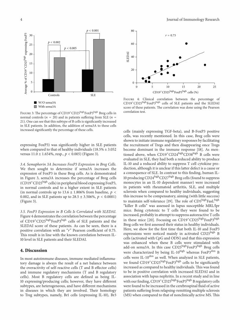
Figure 3: The percentage of CD19+CD25highFoxP3high Breg cells innormal controls (𝑛 = 20) and in patients suffering from SLE (𝑛 =21). One can see that this subtype of B cells is significantly increasedin SLE patients. In addition, the addition of sema3A to these cellsincreased significantly the percentage of these cells.
expressing FoxP3) was significantly higher in SLE patientswhen compared to that of healthy individuals (18.5% ± 3.052versus 11.0 ± 1.654%, resp., 𝑝 < 0.005) (Figure 3).
3.4. Semaphorin 3A Increases FoxP3 Expression in Breg Cells.We then sought to determine if sema3A increases theexpression of FoxP3 in these Breg cells. As is demonstratedin Figure 3, sema3A increases the percentage of Breg cells(CD19+CD25high cells) in peripheral blood expressing FoxP3,in normal controls and to a higher extent in SLE patients(in normal controls up to 13.6 ± 1.806% from baseline, 𝑝 <0.002, and in SLE patients up to 28.5 ± 3.506%, 𝑝 < 0.0001)(Figure 3).
3.5. FoxP3 Expression in B Cells Is Correlated with SLEDAI.Figure 4 demonstrates the correlation between the percentageof CD19+CD25highFoxP3high cells of SLE patients and theSLEDAI score of these patients. As can be seen, there is apositive correlation with an “𝑟” Pearson coefficient of 0.75.This result is in line with the known correlation between IL-10 level in SLE patients and their SLEDAI.
4. Discussion
In most autoimmune diseases, immune mediated inflamma-tory damage is always the result of a net balance betweenthe overactivity of self-reactive cells (T and B effector cells)and immune regulatory mechanisms (T and B regulatorycells). Most B regulatory cells are defined as being IL-10 expressing/producing cells; however, they have differentsubtypes, are heterogeneous, and have different mechanismsin diseases in which they are involved. Their homologyto Treg subtypes, namely, Br1 cells (expressing IL-10), Br3
0 5 10 15 20 250
20
40
60
80
CD19+CD25highFoxP3high cells (%)
r = 0.75
SLED
AI
Figure 4: Clinical correlation between the percentage ofCD19+CD25highFoxP3high cells of SLE patients and the SLEDAIscore of these patients. The correlation was done using the Pearsoncorrelation test.
cells (mainly expressing TGF-beta), and B-FoxP3 positivecells, was recently mentioned. In this case, Breg cells wereshown to initiate immune regulatory responses by facilitatingthe recruitment of Tregs and then disappearing once Tregsbecome dominant in the immune response [18]. As men-tioned above, when CD19+CD24highCD38high B cells wereevaluated in SLE, they had both a reduced ability to produceIL-10 and a reduced ability to suppress T cell cytokine pro-duction, although it is unclear if this latter defect is a cause ora consequence of SLE. In contrast to this finding, human IL-10 producing CD24highCD27high Breg cells (found to suppressmonocytes in an IL-10 dependent manner) were increasedin patients with rheumatoid arthritis, SLE, and multiplesclerosis when compared to healthy individuals, suggestingthis increase to be compensatory, aiming (with little success)to maintain self-tolerance [19]. The role of CD5highFasLhigh“killer B cells” was assessed in lupus susceptible MRL/lprmice. Being cytotoxic to T cells they were found to beincreased, probably in attempt to suppress autoreactive T cellsin these mice [20]. Focusing on CD19+CD25highFoxP3highBreg cells we first assessed their status in healthy individuals.Here, we show for the first time that both IL-10 and FoxP3expressions were noticed mainly in activated CD25high Bcells (activated with CpG and ODN) and that this expressionwas enhanced when these B cells were stimulated withadd-on sema3A. In this case CD25highFoxP3high Breg cellswere characterized by being IL-10high whereas FoxP3dim Bcells were IL-10dim as well. When analyzed in SLE patients,we found CD19+CD25highFoxP3high cells to be significantlyincreased as compared to healthy individuals.This was foundto be in positive correlation with increased SLEDAI and inassociation with lupus nephritis. In a recent study and in linewith our finding, CD19+CD25highFoxP3high B regulatory cellswere found to be increased in the cerebrospinal fluid of activepatients suffering from relapsing-remitting multiple sclerosis(MS) when compared to that of nonclinically active MS.This

Journal of Immunology Research 5
expansion of B regulatory cells was attributed to the compen-satory attempt of these cells to maintain immune regulatoryprocesses [21]. In contrast to this study, rheumatoid arthritispatients had significantly lower proportions of peripheralblood CD19+FoxP3+ B cells as compared to healthy controls,particularly in patients with interstitial lung disease. Thisfinding suggests that Breg phenotypes may have differentfunctions in the pathogenesis of different rheumatic diseases[22]. The fact that serum IL-10 is increased in SLE and inassociation with SLE disease activity has been established inmany previous studies. In one, increased IL-10 was shown toexhibit a modulatory effect by suppressing the differentiationand function of monocyte-derived dendritic cells [23]. Ina recent study, increased IL-10 in the sera of SLE patientswas capable of inducing Fas and FasL expression on CD4+T cell surfaces, promoting apoptosis of this cell subset, thuscontributing to many other mechanisms of self-tolerance[24]. However, we still need to explain the mechanisms bywhich serum IL-10 is increased in SLE. In this regard, theexpansion of IL-10 producing B cells was shown to be inpart the result of increased B cell activating factor (BAFF).Enhanced serum BAFF in SLE was described inmany studiesas being associated with increased expression of TLR-9 andother markers of B cell activation [25, 26]. This may explainour finding of increased IL-10highFoxP3high Breg cells as wellas increased serum IL-10 in SLE. Another significance ofFoxP3high B cells being increased in SLE is the possibilitythat by multiplying they also increase their IL-10 productionimproving by this their regulatory function. When B cellswere cocultured with sema3A they responded by increasingtheir FoxP3 expression. This raises the possibility that ifprovided with the proper stimulation Bregs may develophigher regulatory properties and that by increasing their IL-10production they may induce a better regulatory mechanismin SLE.
5. Conclusion
CD25highFoxP3high Bregs (highly expressing IL-10) are sig-nificantly increased in SLE, in correlation with SLEDAI.Semaphorin 3A increases FoxP3 expression in Breg cellsimproving by this their regulatory properties.We assume thatthe expansion of these cells is the attempt of our regulatoryimmune responses to maintain self-tolerance and to suppressas much as possible SLE disease activity. Further studies arerequired in order to better understand the role of this subsetof B cells in autoimmunity.
Conflict of Interests
The authors declare that there is no conflict of interestsregarding the publication of this paper.
References
[1] R. M. Talaat, S. F. Mohamed, I. H. Bassyouni, and A. A.Raouf, “Th1/Th2/Th17/Treg cytokine imbalance in systemiclupus erythematosus (SLE) patients: correlation with diseaseactivity,” Cytokine, vol. 72, no. 2, pp. 146–153, 2015 (Chinese).
[2] K. Shah, W.-W. Lee, S.-H. Lee et al., “Dysregulated balance ofTh17 and Th1 cells in systemic lupus erythematosus,” ArthritisResearch &Therapy, vol. 12, no. 2, article R53, 2010.
[3] D. Li, B. Guo, H.Wu, L. Tan, C. Chang, and Q. Lu, “Interleukin-17 in systemic lupus erythematosus: a comprehensive review,”Autoimmunity, vol. 48, no. 6, pp. 353–361, 2015.
[4] B. Terrier, N. Costedoat-Chalumeau, M. Garrido et al., “Inter-leukin 21 correlates with T cell and B cell subset alterations insystemic lupus erythematosus,” The Journal of Rheumatology,vol. 39, no. 9, pp. 1819–1828, 2012.
[5] S. Futatsugi-Yumikura, K. Matsushita, A. Fukuoka et al.,“Pathogenic Th2-type follicular helper T cells contribute tothe development of lupus in Fas-deficient mice,” InternationalImmunology, vol. 26, no. 4, pp. 221–231, 2014.
[6] B. Yan, S. Ye, G. Chen, M. Kuang, N. Shen, and S. Chen,“Dysfunctional CD4+,CD25+ regulatory T cells in untreatedactive systemic lupus erythematosus secondary to interferon-𝛼-producing antigen-presenting cells,” Arthritis and Rheumatism,vol. 58, no. 3, pp. 801–812, 2008.
[7] A. Ray, L. Wang, and B. N. Dittel, “IL-10-independent regu-latory B-cell subsets and mechanisms of action,” InternationalImmunology, 2015.
[8] P. A. Blair, L. Y. Norena, F. Flores-Borja et al.,“CD19+CD24hiCD38hi B cells exhibit regulatory capacityin healthy individuals but are functionally impaired in systemiclupus erythematosus,” Immunity, vol. 32, no. 1, pp. 129–140,2010.
[9] A. Kessel, T. Haj, R. Peri et al., “Human CD19+CD25high Bregulatory cells suppress proliferation of CD4+ T cells andenhance Foxp3 and CTLA-4 expression in T-regulatory cells,”Autoimmunity Reviews, vol. 11, no. 9, pp. 670–677, 2012.
[10] J. Noh and G. Noh, “Allergen specific responses of CD19highand CD19low B cells in non-IgE mediated food allergy of lateeczematous reactions in atopic dermatitis: presence of IL-17 andIL-32 producing regulatory B cells (Br17 & Br32),” Inflammation& Allergy-Drug Targets, vol. 11, no. 4, pp. 320–329, 2012.
[11] S. K. Lundy, “Killer B lymphocytes: the evidence and thepotential,” Inflammation Research, vol. 58, no. 7, pp. 345–357,2009.
[12] F. Mion, S. Topnon, B. Toffoletto, D. Cesselli, C. E. Pucillo, andG. Vitale, “IL-10 production by B cells is differentially regulatedby immune-mediated and infectious stimuli and requires p38activation,” Molecular Immunology, vol. 62, no. 2, pp. 266–276,2014.
[13] K. Yanaba, J.-D. Bouaziz, T. Matsushita, T. Tsubata, and T. F.Tedder, “The development and function of regulatory B cellsexpressing IL-10 (B10 cells) requires antigen receptor diversityand TLR signals,” Journal of Immunology, vol. 182, no. 12, pp.7459–7472, 2009.
[14] Z. Vadasz, T. Haj, A. Balbir et al., “A regulatory role forCD72 expression on B cells in systemic lupus erythematosus,”Seminars in Arthritis & Rheumatism, vol. 43, no. 6, pp. 767–771,2014.
[15] P. Lopez, E. Alonso-Perez, J. Rodrıguez-Carrio, and A. Suarez,“Influence of Atg5mutation in SLE depends on functional IL-10genotype,” PLoS ONE, vol. 8, no. 10, Article ID e78756, 2013.
[16] C. Bombardier, D. D. Gladman, M. B. Urovitz, D. Caron, and C.H. Chang, “Derivation of the SLEDAI. A disease activity indexfor lupus patients. The committee on prognosis studies in SLE,”Arthritis and Rheumatism, vol. 35, no. 6, pp. 630–640, 1992.

6 Journal of Immunology Research
[17] B. Kigel, A. Varshavsky, O. Kessler, and G. Neufeld, “Suc-cessful inhibition of tumor development by specific class-3 semaphorins is associated with expression of appropriatesemaphorin receptors by tumor cells,” PLoS ONE, vol. 3, no. 9,article e3287, 2008.
[18] J.-M. Berthelot, C. Jamin, K. Amrouche, B. Le Goff, Y. Maugars,and P. Youinou, “Regulatory B cells play a key role in immunesystem balance,” Joint Bone Spine, vol. 80, no. 1, pp. 18–22, 2013.
[19] Y. Iwata, T. Matsushita, M. Horikawa et al., “Characterizationof a rare IL-10-competent B-cell subset in humans that parallelsmouse regulatory B10 cells,” Blood, vol. 117, no. 2, pp. 530–541,2011.
[20] D. Bonardelle, K. Benihoud, N. Kiger, and P. Bobe, “B lym-phocytesmediate Fas-dependent cytotoxicity inMRL/lprmice,”Journal of Leukocyte Biology, vol. 78, no. 5, pp. 1052–1059, 2005.
[21] C. de Andres, M. Tejera-Alhambra, B. Alonso et al., “Newregulatory CD19+CD25+ B-cell subset in clinically isolatedsyndrome and multiple sclerosis relapse. Changes after gluco-corticoids,” Journal of Neuroimmunology, vol. 270, no. 1-2, pp.37–44, 2014.
[22] Y. Guo, X. Zhang,M. Qin, and X.Wang, “Changes in peripheralCD19+Foxp3+ and CD19+TGF-𝛽+ regulatory B cell populationsin rheumatoid arthritis patients with interstitial lung disease ,”Journal of Thoracic Disease, vol. 7, no. 3, pp. 471–477, 2015.
[23] Z. Sun, R. Zhang, H. Wang et al., “Serum IL-10 from systemiclupus erythematosus patients suppresses the differentiationand function of monocyte-derived dendritic cells,” Journal ofBiomedical Research, vol. 26, no. 6, pp. 456–466, 2012.
[24] X. Yang, B. Sun, H.Wang, C. Yin, X.Wang, and X. Ji, “Increasedserum IL-10 in lupus patients promotes apoptosis of T cellsubsets via the caspase 8 pathway initiated by Fas signaling,”Journal of Biomedical Research, vol. 29, no. 3, pp. 232–240, 2015.
[25] M. Yang, L. Sun, S. Wang et al., “Cutting edge: novel functionof B cell-activating factor in the induction of IL-10-producingregulatory B cells,” The Journal of Immunology, vol. 184, no. 7,pp. 3321–3325, 2010.
[26] D. Yehudai, A. Snir, R. Peri et al., “B cell-activating factorenhances interleukin-6 and interleukin-10 production byODN-activated human B cells,” Scandinavian Journal of Immunology,vol. 76, no. 4, pp. 371–377, 2012.

Submit your manuscripts athttp://www.hindawi.com
Stem CellsInternational
Hindawi Publishing Corporationhttp://www.hindawi.com Volume 2014
Hindawi Publishing Corporationhttp://www.hindawi.com Volume 2014
MEDIATORSINFLAMMATION
of
Hindawi Publishing Corporationhttp://www.hindawi.com Volume 2014
Behavioural Neurology
EndocrinologyInternational Journal of
Hindawi Publishing Corporationhttp://www.hindawi.com Volume 2014
Hindawi Publishing Corporationhttp://www.hindawi.com Volume 2014
Disease Markers
Hindawi Publishing Corporationhttp://www.hindawi.com Volume 2014
BioMed Research International
OncologyJournal of
Hindawi Publishing Corporationhttp://www.hindawi.com Volume 2014
Hindawi Publishing Corporationhttp://www.hindawi.com Volume 2014
Oxidative Medicine and Cellular Longevity
Hindawi Publishing Corporationhttp://www.hindawi.com Volume 2014
PPAR Research
The Scientific World JournalHindawi Publishing Corporation http://www.hindawi.com Volume 2014
Immunology ResearchHindawi Publishing Corporationhttp://www.hindawi.com Volume 2014
Journal of
ObesityJournal of
Hindawi Publishing Corporationhttp://www.hindawi.com Volume 2014
Hindawi Publishing Corporationhttp://www.hindawi.com Volume 2014
Computational and Mathematical Methods in Medicine
OphthalmologyJournal of
Hindawi Publishing Corporationhttp://www.hindawi.com Volume 2014
Diabetes ResearchJournal of
Hindawi Publishing Corporationhttp://www.hindawi.com Volume 2014
Hindawi Publishing Corporationhttp://www.hindawi.com Volume 2014
Research and TreatmentAIDS
Hindawi Publishing Corporationhttp://www.hindawi.com Volume 2014
Gastroenterology Research and Practice
Hindawi Publishing Corporationhttp://www.hindawi.com Volume 2014
Parkinson’s Disease
Evidence-Based Complementary and Alternative Medicine
Volume 2014Hindawi Publishing Corporationhttp://www.hindawi.com
Copyright © 2022 FDOKUMEN