Religion, Diaspora and the Politics of a Homing Desire in the ...
Upload
independentCategory
view
0download
0
Immunity
Article
Intestinal Tolerance Requires Gut Homingand Expansion of FoxP3+ Regulatory T Cellsin the Lamina PropriaUsriansyah Hadis,1 Benjamin Wahl,1 Olga Schulz,1 Matthias Hardtke-Wolenski,2 Angela Schippers,3 Norbert Wagner,3
Werner Muller,4 Tim Sparwasser,5 Reinhold Forster,1 and Oliver Pabst1,*1Institute of Immunology2Department of Gastroenterology, Hepatology and EndocrinologyHannover Medical School, 30625 Hannover, Germany3Department of Pediatrics, Medical Faculty, RWTH Aachen University, 52074 Aachen, Germany4Faculty of Life Sciences, University of Manchester, Manchester M13 9PT, UK5Institute of Infection Immunology, TWINCORE, Centre for Experimental and Clinical Infection Research, 30625 Hannover, Germany*Correspondence: [email protected]
DOI 10.1016/j.immuni.2011.01.016
SUMMARY
Tolerance to food antigen manifests in the absenceand/or suppression of antigen-specific immuneresponses locally in the gut but also systemically,a phenomenon known as oral tolerance. Oral toler-ance is thought to originate in the gut-draining lymphnodes, which support the generation of FoxP3+ regu-latory T (Treg) cells. Here we use several mousemodels to show that Treg cells, after their generationin lymph nodes, need to home to the gut to undergolocal expansion to install oral tolerance. Proliferationof Treg cells in the intestine and production of inter-leukin-10 by gut-resident macrophages was bluntedin mice deficient in the chemokine (C-X3-C motif)receptor 1 (CX3CR1). We propose a model ofstepwise oral tolerance induction comprising thegeneration of Treg cells in the gut-draining lymphnodes, followed by migration into the gut and subse-quent expansion of Treg cells driven by intestinalmacrophages.
INTRODUCTION
Understanding the immune system’s ability to balance between
tolerance and protective immunity remains one of the key chal-
lenges in immunology. A particularly intimate contact between
exogenous antigens and the immune system occurs in the intes-
tine. In humans the intestine receives an estimated amount of
130–190 g of protein per day and is colonized with a diverse
microflora outnumbering the body’s own cells. Yet the intestinal
immune system reacts with tolerance to these innocuous anti-
gens despite its ability to raise powerful immunity toward enter-
opathogens and other dangerous agents. The basic phenom-
enon of intestinal tolerance was demonstrated in 1946 by
Chase, who reported that feeding of an antigen results in an
active ‘‘immunologic nonresponsiveness’’ to a subsequent
systemic antigen challenge (Chase, 1946). Such active induction
of regulated immune responses to intestinal antigens is referred
to as oral tolerance (Faria and Weiner, 2005; Mayer and Shao,
2004; Strobel and Mowat, 2006).
Oral tolerance has been extensively investigated in rodents
and has also been demonstrated in humans (Husby et al.,
1994). The establishment of oral tolerance has body-wide impli-
cations governing systemic immune responses including de-
layed type hypersensitivity reactions, lymphocyte proliferation,
and antibody formation (Faria and Weiner, 2005; Mayer and
Shao, 2004; Strobel and Mowat, 2006). However, there is also
evidence that oral tolerance acts locally in the gut, thereby pre-
venting, for example, chronic inflammation resulting from exces-
sive immune reactivity against food proteins. Mechanistically,
T cell deletion or anergy as well as active regulatory processes
have been implicated in oral tolerance. Recently, plasmacytoid
dendritic cells (pDCs) have been shown to be involved in the
depletion of hapten-reactive CD8+ T cells (Goubier et al.,
2008). pDCs constitute a substantial fraction of antigen-present-
ing cells (APC) in the liver and, along with nonhematopoietic
endothelial cells (Limmer et al., 2000) and hepatic stellate cells
(Winau et al., 2007), might present intestinal antigens in the liver
to induce intestinal tolerance. Yet at present, the liver’s aston-
ishing tolerogenic capacity could not be directly traced to any
of these cell types and still remains enigmatic.
Presentation of regular food protein largely depends on
antigen presentation by classic dendritic cells (DCs) in the gut-
draining mesenteric lymph nodes (mLN) (Worbs et al., 2006).
DCs in the lamina propria (LP) take up intestinal antigen and in
a chemokine receptor (C-Cmotif) receptor 7-dependent process
migrate to the mLN to activate naive T cells (Jang et al., 2006;
Worbs et al., 2006). Intestinal DCs capable of intralymphatic
migration express the aE integrin CD103 (Schulz et al., 2009)
and have a high capacity to produce the vitamin A metabolite
retinoic acid (RA) (Coombes et al., 2007). Once settled in the
mLN, LP-derived CD103+ DCs cooperate with mLN stroma cells
to shape a unique environment that critically determines the
signature of T cell responses induced here (Hammerschmidt
et al., 2008; Molenaar et al., 2009). In particular, T cells activated
in the mLN express the gut-homing molecules integrin-a4b7 and
Immunity 34, 237–246, February 25, 2011 ª2011 Elsevier Inc. 237
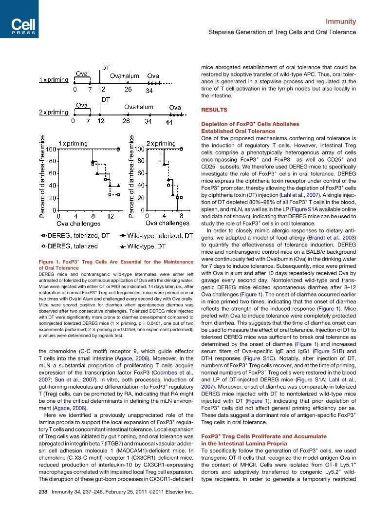
Figure 1. FoxP3+ Treg Cells Are Essential for the Maintenance
of Oral Tolerance
DEREG mice and nontransgenic wild-type littermates were either left
untreated or tolerized by continuous application of Ovawith the drinking water.
Mice were injected with either DT or PBS as indicated. 14 days later, i.e., after
restoration of normal FoxP3+ Treg cell frequencies, mice were primed one or
two times with Ova in Alum and challenged every second day with Ova orally.
Mice were scored positive for diarrhea when spontaneous diarrhea was
observed after two consecutive challenges. Tolerized DEREG mice injected
with DT were significantly more prone to diarrhea development compared to
noninjected tolerized DEREG mice (1 3 priming, p = 0.0401, one out of two
experiments performed; 2 3 priming p = 0.0259, one experiment performed);
p values were determined by logrank test.
Immunity
Stepwise Generation of Treg Cells and Oral Tolerance
the chemokine (C-C motif) receptor 9, which guide effector
T cells into the small intestine (Agace, 2006). Moreover, in the
mLN a substantial proportion of proliferating T cells acquire
expression of the transcription factor FoxP3 (Coombes et al.,
2007; Sun et al., 2007). In vitro, both processes, induction of
gut-homing molecules and differentiation into FoxP3+ regulatory
T (Treg) cells, can be promoted by RA, indicating that RA might
be one of the critical determinants in defining the mLN environ-
ment (Agace, 2006).
Here we identified a previously unappreciated role of the
lamina propria to support the local expansion of FoxP3+ regula-
tory T cells andconcomitant intestinal tolerance. Local expansion
of Treg cells was initiated by gut homing, and oral tolerance was
abrogated in integrin beta 7 (ITGB7) andmucosal vascular addre-
sin cell adhesion molecule 1 (MADCAM1)-deficient mice. In
chemokine (C-X3-C motif) receptor 1 (CX3CR1)-deficient mice,
reduced production of interleukin-10 by CX3CR1-expressing
macrophages correlated with impaired local Treg cell expansion.
The disruption of these gut-born processes in CX3CR1-deficient
238 Immunity 34, 237–246, February 25, 2011 ª2011 Elsevier Inc.
mice abrogated establishment of oral tolerance that could be
restored by adoptive transfer of wild-type APC. Thus, oral toler-
ance is generated in a stepwise process and regulated at the
time of T cell activation in the lymph nodes but also locally in
the intestine.
RESULTS
Depletion of FoxP3+ Cells AbolishesEstablished Oral ToleranceOne of the proposed mechanisms conferring oral tolerance is
the induction of regulatory T cells. However, intestinal Treg
cells comprise a phenotypically heterogenous array of cells
encompassing FoxP3+ and FoxP3� as well as CD25+ and
CD25� subsets. We therefore used DEREG mice to specifically
investigate the role of FoxP3+ cells in oral tolerance. DEREG
mice express the diphtheria toxin receptor under control of the
FoxP3+ promoter, thereby allowing the depletion of FoxP3+ cells
by diphtheria toxin (DT) injection (Lahl et al., 2007). A single injec-
tion of DT depleted 80%–98% of all FoxP3+ T cells in the blood,
spleen, andmLN, as well as in the LP (Figure S1A available online
and data not shown), indicating that DEREGmice can be used to
study the role of FoxP3+ cells in oral tolerance.
In order to closely mimic allergic responses to dietary anti-
gens, we adapted a model of food allergy (Brandt et al., 2003)
to quantify the effectiveness of tolerance induction. DEREG
mice and nontransgenic control mice on a BALB/c background
were continuously fed with Ovalbumin (Ova) in the drinking water
for 7 days to induce tolerance. Subsequently, mice were primed
with Ova in alum and after 10 days repeatedly received Ova by
gavage every second day. Nontolerized wild-type and trans-
genic DEREG mice elicited spontaneous diarrhea after 8–12
Ova challenges (Figure 1). The onset of diarrhea occurred earlier
in mice primed two times, indicating that the onset of diarrhea
reflects the strength of the induced response (Figure 1). Mice
prefed with Ova to induce tolerance were completely protected
from diarrhea. This suggests that the time of diarrhea onset can
be used to measure the effect of oral tolerance. Injection of DT to
tolerized DEREG mice was sufficient to break oral tolerance as
determined by the onset of diarrhea (Figure 1) and increased
serum titers of Ova-specific IgE and IgG1 (Figure S1B) and
DTH responses (Figure S1C). Notably, after injection of DT,
numbers of FoxP3+ Treg cells recover, and at the time of priming,
normal numbers of FoxP3+ Treg cells were restored in the blood
and LP of DT-injected DEREG mice (Figure S1A; Lahl et al.,
2007). Moreover, onset of diarrhea was comparable in tolerized
DEREG mice injected with DT to nontolerized wild-type mice
injected with DT (Figure 1), indicating that prior depletion of
FoxP3+ cells did not affect general priming efficiency per se.
These data suggest a dominant role of antigen-specific FoxP3+
Treg cells in oral tolerance.
FoxP3+ Treg Cells Proliferate and Accumulatein the Intestinal Lamina PropriaTo specifically follow the generation of FoxP3+ cells, we used
transgenic OT-II cells that recognize the model antigen Ova in
the context of MHCII. Cells were isolated from OT-II Ly5.1+
donors and adoptively transferred to congenic Ly5.2+ wild-
type recipients. In order to generate a temporarily restricted

Figure 2. Antigen-Specific FoxP3+ Treg Cells Accumulate in the Gut
OT-II T cells were adoptively transferred to wild-type recipients, which
received two oral doses of 50 mg Ova each.
(A) Mice were analyzed 5 and 12 days after adoptive transfer. Plots are gated
on Ly5.1+CD4+ cells. The frequency and total number of FoxP3+ Treg cells
generated out of adoptively transferred OT-II Ly5.1+CD4+Vb5.1+ cells was
determined in pLN, mLN, and LP. Circles represent individual mice pooled
from two independent experiments.
(B) Plots are gated on CD4+FoxP3+ and depict adoptively transferred Ly5.1+
and recipient Ly5.2+ cells at day 5 as indicated. Please note plots in (B) are
not gated on Vb5.1, and Ly5.1+Helios+ cells in the LP might represent non-
Ova-specific T cells.
Plots display 10% probability contours, numbers indicate percentage gated
cells.
Immunity
Stepwise Generation of Treg Cells and Oral Tolerance
cohort of activated T cells, mice received two doses of 50 mg
Ova orally 1 and 2 days after adoptive transfer (Figure 2) instead
of Ova application via the drinking water as described for
Figure 1. Five days after transfer, 8.17% ± 2.61% (mean ± SD,
n = 27, cross experiment analysis of results shown in Figures 2
and 5C and nondepicted experiments) of adoptively transferred
OT-II T cells in the mLN expressed FoxP3. Similar frequencies
were observed in the intestinal LP at day 5 as well as in the
peripheral lymph nodes (pLN, pool of subiliac and axillary lymph
nodes) (Figure 2). However, the mLN contained substantially
higher total numbers of antigen-specific FoxP3+ Treg cells
compared to pLN and LP at day 5 after transfer. As we have
shown earlier, with this experimental setup, T cell activation
does not occur in pLN (Worbs et al., 2006) and no Ly5.1+FoxP3+
Treg cells were detectable in recipients not receiving Ova (data
not shown). Thus, at day 5 after transfer, the frequency and
number of Ly5.1+FoxP3+ Treg cells observed in pLN reflected
the activation of antigen-specific T cells in the mLN and their
subsequent dissemination and homing to other sites.
In striking contrast to the situation on day 5, on day 12 more
than 50% of all Ly5.1+CD4+ OT-II T cells expressed FoxP3+ in
the LP. The frequency in the mLN and pLN did not substantially
change between day 5 and day 12 (Figure 2A). The increase in LP
FoxP3+ frequencies also affected total cell numbers. Whereas
the total number of Ly5.1+FoxP3+ Treg cells dropped between
day 5 and 12 after antigen feeding in pLN andmLN, their number
persisted or even increased in the LP (Figure 2A). Donor-derived
Ly5.1+FoxP3+ Treg cells in the mLN homogenously expressed
the Treg cell marker GITR (data not shown) and lacked expres-
sion of the transcription factor helios (Figure 2B), which has
been suggested to discriminate between helios+ natural Treg
cells and helios� induced Treg cells (Thornton et al., 2010). Simi-
larly, in the LP the majority of all Ly5.1+FoxP3+ cells lacked
expression of helios (Figure 2B). This indicates that conversion
of naive T cells to a FoxP3+ fate rather than expansion of low
numbers of natural Treg cells accounted for the accumulation
of FoxP3+ Treg cells in the LP. Virtually no adoptively transferred
Ly5.1+CD4+FoxP3+ OT-II T cells were found in the LP of mice
that did not receive Ova (data not shown). Interestingly,
FoxP3+ T cell frequencies still increased in the LP when antigen
presentation in the mLN had already ceased (Figure S2).
The increase in antigen-specific FoxP3+ Treg cell frequencies
in the LP might be due to selective proliferation, cell death, and/
or retention. We thus determined the time window of OT-II T cell
expansion after antigen feeding. Mice were adoptively trans-
ferred with OT-II T cells and fed doses of Ova and proliferating
cells were labeled by continuous application of BrdU with the
drinking water. A first group of mice continuously received
BrdU until sacrifice at day 5 (group I). In these mice a substantial
proportion of adoptively transferred Ly5.1+CD4+FoxP3+ OT-II
T cells in the mLN and almost all cells in the LP were BrdU+
(Figure 3A). This situation changed when mice subjected to
the identical treatment were analyzed at day 12 (group II).
Whereas the mLN had retained a substantial proportion of
Ly5.1+CD4+FoxP3+ BrdU+ OT-II T cells, the proportion in the
LP had dropped to about 10% (Figure 3A). This observation
suggests that OT-II Treg cells present in the mLN underwent
only a few divisions between 5 and 12 days after transfer,
whereas OT-II Treg cells present in the LP expanded and lost
the BrdU label. Moreover, BrdU application starting at day 5 until
sacrifice at day 12 (group III) yielded a high proportion of BrdU-
positive Ly5.1+CD4+FoxP3+ OT-II Treg cells in the LP, whereas
such cells were almost undetectable in mLN and spleen.
Because previous studies revealed a high rate of constitutive
FoxP3+ Treg cell proliferation (Tang et al., 2008a, 2008b), we
compared the proliferation of endogenous FoxP3+ Treg cells in
the LP to secondary lymphoid organs. 48 hr after continued
application of BrdU with the drinking water, more than 30% of
all LP FoxP3+ Treg cells were BrdU+ (Figure 3B). In contrast,
CD4+FoxP3� cells showed a substantially lower proportion of
BrdU+ cells and even less BrdU incorporation was observed in
lymph nodes and spleen (Figure 3B). Moreover, also a short
2 hr BrdU pulse revealed BrdU+FoxP3+ Treg cells in the small
intestinal LP (Figure 3B). This indicates that the LP intrinsically
Immunity 34, 237–246, February 25, 2011 ª2011 Elsevier Inc. 239
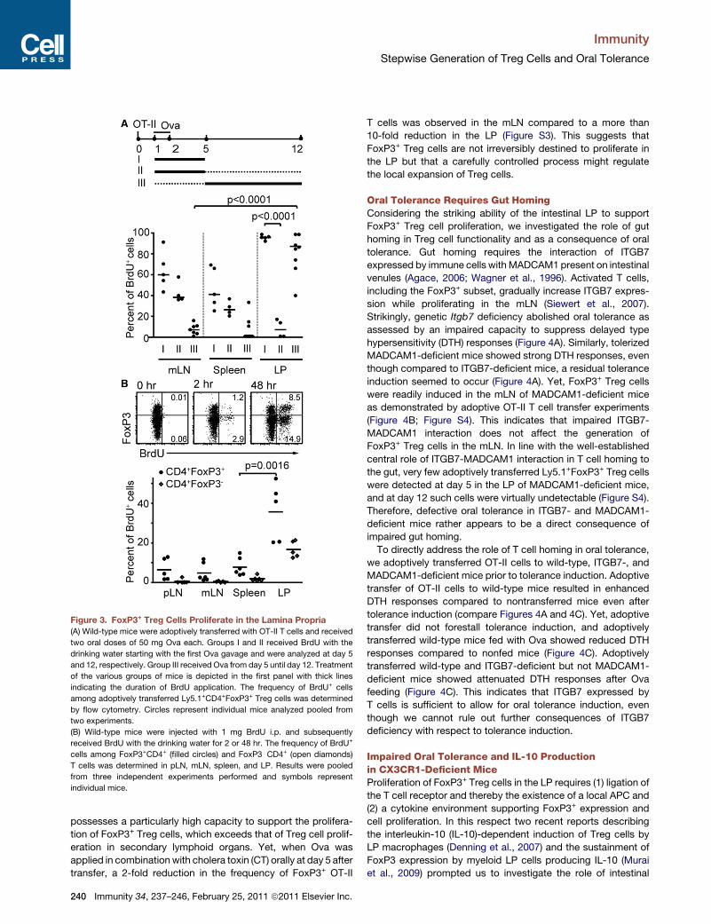
Figure 3. FoxP3+ Treg Cells Proliferate in the Lamina Propria
(A) Wild-type mice were adoptively transferred with OT-II T cells and received
two oral doses of 50 mg Ova each. Groups I and II received BrdU with the
drinking water starting with the first Ova gavage and were analyzed at day 5
and 12, respectively. Group III received Ova from day 5 until day 12. Treatment
of the various groups of mice is depicted in the first panel with thick lines
indicating the duration of BrdU application. The frequency of BrdU+ cells
among adoptively transferred Ly5.1+CD4+FoxP3+ Treg cells was determined
by flow cytometry. Circles represent individual mice analyzed pooled from
two experiments.
(B) Wild-type mice were injected with 1 mg BrdU i.p. and subsequently
received BrdU with the drinking water for 2 or 48 hr. The frequency of BrdU+
cells among FoxP3+CD4+ (filled circles) and FoxP3�CD4+ (open diamonds)
T cells was determined in pLN, mLN, spleen, and LP. Results were pooled
from three independent experiments performed and symbols represent
individual mice.
Immunity
Stepwise Generation of Treg Cells and Oral Tolerance
possesses a particularly high capacity to support the prolifera-
tion of FoxP3+ Treg cells, which exceeds that of Treg cell prolif-
eration in secondary lymphoid organs. Yet, when Ova was
applied in combination with cholera toxin (CT) orally at day 5 after
transfer, a 2-fold reduction in the frequency of FoxP3+ OT-II
240 Immunity 34, 237–246, February 25, 2011 ª2011 Elsevier Inc.
T cells was observed in the mLN compared to a more than
10-fold reduction in the LP (Figure S3). This suggests that
FoxP3+ Treg cells are not irreversibly destined to proliferate in
the LP but that a carefully controlled process might regulate
the local expansion of Treg cells.
Oral Tolerance Requires Gut HomingConsidering the striking ability of the intestinal LP to support
FoxP3+ Treg cell proliferation, we investigated the role of gut
homing in Treg cell functionality and as a consequence of oral
tolerance. Gut homing requires the interaction of ITGB7
expressed by immune cells with MADCAM1 present on intestinal
venules (Agace, 2006; Wagner et al., 1996). Activated T cells,
including the FoxP3+ subset, gradually increase ITGB7 expres-
sion while proliferating in the mLN (Siewert et al., 2007).
Strikingly, genetic Itgb7 deficiency abolished oral tolerance as
assessed by an impaired capacity to suppress delayed type
hypersensitivity (DTH) responses (Figure 4A). Similarly, tolerized
MADCAM1-deficient mice showed strong DTH responses, even
though compared to ITGB7-deficient mice, a residual tolerance
induction seemed to occur (Figure 4A). Yet, FoxP3+ Treg cells
were readily induced in the mLN of MADCAM1-deficient mice
as demonstrated by adoptive OT-II T cell transfer experiments
(Figure 4B; Figure S4). This indicates that impaired ITGB7-
MADCAM1 interaction does not affect the generation of
FoxP3+ Treg cells in the mLN. In line with the well-established
central role of ITGB7-MADCAM1 interaction in T cell homing to
the gut, very few adoptively transferred Ly5.1+FoxP3+ Treg cells
were detected at day 5 in the LP of MADCAM1-deficient mice,
and at day 12 such cells were virtually undetectable (Figure S4).
Therefore, defective oral tolerance in ITGB7- and MADCAM1-
deficient mice rather appears to be a direct consequence of
impaired gut homing.
To directly address the role of T cell homing in oral tolerance,
we adoptively transferred OT-II cells to wild-type, ITGB7-, and
MADCAM1-deficient mice prior to tolerance induction. Adoptive
transfer of OT-II cells to wild-type mice resulted in enhanced
DTH responses compared to nontransferred mice even after
tolerance induction (compare Figures 4A and 4C). Yet, adoptive
transfer did not forestall tolerance induction, and adoptively
transferred wild-type mice fed with Ova showed reduced DTH
responses compared to nonfed mice (Figure 4C). Adoptively
transferred wild-type and ITGB7-deficient but not MADCAM1-
deficient mice showed attenuated DTH responses after Ova
feeding (Figure 4C). This indicates that ITGB7 expressed by
T cells is sufficient to allow for oral tolerance induction, even
though we cannot rule out further consequences of ITGB7
deficiency with respect to tolerance induction.
Impaired Oral Tolerance and IL-10 Productionin CX3CR1-Deficient MiceProliferation of FoxP3+ Treg cells in the LP requires (1) ligation of
the T cell receptor and thereby the existence of a local APC and
(2) a cytokine environment supporting FoxP3+ expression and
cell proliferation. In this respect two recent reports describing
the interleukin-10 (IL-10)-dependent induction of Treg cells by
LP macrophages (Denning et al., 2007) and the sustainment of
FoxP3 expression by myeloid LP cells producing IL-10 (Murai
et al., 2009) prompted us to investigate the role of intestinal

Figure 4. Gut Homing Is Essential for Oral Tolerance
(A) Oral tolerance was assessed in wild-type, ITGB7-, and MADCAM1-
deficient mice. Circles represent individual mice pooled from two or more
experiments.
(B) FoxP3+ Treg cell generation was determined in MADCAM1-deficient mice
by adoptive transfer experiments as described for Figure 2. Circles represent
the frequency of FoxP3+ Treg cells generated from adoptively transferred OT-II
Ly5.1+CD4+Vb5.1+ cells in mLN of individual MADCAM1-deficient recipients.
(C) 5 3 106 cells isolated from lymph nodes of OT-II mice were adoptively
transferred to wild-type, ITGB7-, andMADCAM1-deficient mice and oral toler-
ance was assessed. Circles represent individual mice.
Results in (B) and (C) were pooled from two experiments.
Figure 5. Reduced Frequency of IL-10-Producing F4/80+CD11b+
MHCIIint Cells and Accumulation of FoxP3+ Treg Cells in the LP of
CX3CR1-Deficient Mice
(A) The number of F4/80+CD11b+ LP cells in wild-type and CX3CR1-deficient
mice was determined by flow cytometry. Gates were set on live (DAPI�) CD45+
LP cells or F4/80+CD11b+ cells as indicated. Circles represent individual mice
analyzed in two independent experiments.
(B) LP cells were isolated, stimulated in vitro for 4 hr with PMA + ionomycin,
and stained for intracellular IL-10. Representative FACS plots are shown.
The diagram depicts the percentage of IL-10+ cells among F4/80+CD11b+
LP cells. Circles represent individual mice analyzed in two independent exper-
iments.
(C) The frequency of FoxP3+ Treg cells among adoptively transferred OT-II
CD4+Ly5.1+Vb5.1+ T cells was determined 5 days after transfer in mLN and
LP and 12 days in pLN, mLN, and LP. Circles represent individual mice pooled
from two to three independent experiments.
Immunity
Stepwise Generation of Treg Cells and Oral Tolerance
macrophages in driving FoxP3+ Treg cell accumulation in
the gut. Intestinal macrophages capable of FoxP3+ Treg cell
induction are identical or at least largely overlap with LP cells
expressing high levels of CX3CR1 (Figure 5A). The designation
of CX3CR1+ LP cells in the literature is somewhat inconsistent
because these cells share properties of both DCs and macro-
phages. The vast majority of CX3CR1+ LP cells are CD11b+
F4/80+CD103� with intermediate expression of MHCII (Denning
et al., 2007; Schulz et al., 2009). Because these cells are gut
resident (Schulz et al., 2009) and differ from classical DCs
with respect to their developmental origin (Bogunovic et al.,
2009; Varol et al., 2009), in the following we will refer to
CX3CR1+CD11b+ LP cells as intestinal macrophages. Notably,
this denomination does not deny that CX3CR1+ intestinal macro-
phages differ from bona fide intestinal macrophages, e.g., in
terms of their capacity to extend transepithelial cell processes,
but merely is chosen to emphasize their nonmigratory pheno-
type. The chemokine (C-X3-C motif) ligand 1 (CX3CL1) has
been suggested to modulate the cytokine production by macro-
phages (Mizutani et al., 2007). Because we have previously
observed that uptake of Ova by CX3CR1+ intestinal macro-
phages is intact in CX3CR1-deficient mice (Schulz et al., 2009)
lacking transepithelial cell processes (Niess et al., 2005), we
tested whether CX3CR1 expression might affect IL-10 produc-
tion by these cells.
Strikingly, intracellular staining of IL-10 showed a substantially
reduced frequency of IL-10+ intestinal macrophages in CX3CR1-
deficient mice compared to wild-type controls (Figure 5B).
Immunity 34, 237–246, February 25, 2011 ª2011 Elsevier Inc. 241
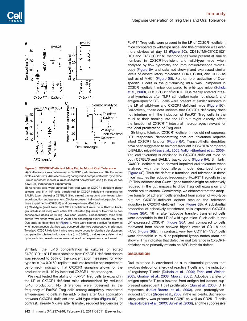
Figure 6. CX3CR1-Deficient Mice Fail to Mount Oral Tolerance
(A) Oral tolerance was determined in CX3CR1-deficient mice on BALB/c (open
circles) and C57BL/6 (closed circles) background compared towild-typemice.
Circles represent individual mice analyzed pooled from one (BALB/c) or two
(C57BL/6) independent experiments.
(B) Adherent cells were enriched from wild-type or CX3CR1-deficient donor
spleens and 5 3 106 cells transferred to CX3CR1-deficient recipients on
BALB/c (open circles) or C57BL/6 (filled circles) background prior to oral toler-
ance induction and assessment. Circles represent individual mice pooled from
three experiments (C57BL/6) and one experiment (BALB/c).
(C) Wild-type (solid lines) and CX3CR1-deficient mice on a BALB/c back-
ground (dashed lines) were either left untreated (squares) or tolerized by two
consecutive doses of 50 mg Ova each (circles). Subsequently, mice were
primed two times with Ova in Alum and challenged every second day with
Ova orally as described for Figure 1. Mice were scored positive for diarrhea
when spontaneous diarrhea was observed after two consecutive challenges.
Tolerized CX3CR1-deficient mice were more prone to diarrhea development
compared to tolerized wild-type mice (p = 0.0494), p values were determined
by logrank test; results are representative of two experiments performed.
Immunity
Stepwise Generation of Treg Cells and Oral Tolerance
Similarly, the IL-10 concentration in cultures of sorted
F4/80+CD11b+ LP cells obtained from CX3CR1-deficient donors
was reduced to 55% of the concentration measured for wild-
type cells (p = 0.0159, replicate cultures tested in one experiment
performed), indicating that CX3CR1 signaling allows for the
production of IL-10 by intestinal CX3CR1+ macrophages.
We next tested the ability of FoxP3+ Treg cells to expand in
the LP of CX3CR1-deficient mice characterized by reduced
IL-10 production. No differences were observed in the
frequency of FoxP3+ Treg cells among adoptively transferred
antigen-specific cells in the mLN 5 days after Ova application
between CX3CR1-deficient and wild-type mice (Figure 5C). In
contrast, already 5 days after transfer, reduced frequencies of
242 Immunity 34, 237–246, February 25, 2011 ª2011 Elsevier Inc.
FoxP3+ Treg cells were present in the LP of CX3CR1-deficient
mice compared to wild-type mice, and this difference was even
more obvious at day 12 (Figure 5C). CD11c+MHCII+CD103+
DCs and F4/80+CD11b+ macrophages were present at similar
numbers in CX3CR1-deficient and wild-type mice when
analyzed by flow cytometry and immunofluorescence micros-
copy (Figure 5A and data not shown) and expressed similar
levels of costimulatory molecules CD40, CD80, and CD86 as
well as of MHCII (Figure S5). Furthermore, activation of Ova-
specific T cells in the gut-draining mLN was unimpaired in
CX3CR1-deficient mice compared to wild-type mice (Schulz
et al., 2009), CD103+CD11c+MHCII+ DCs readily entered intes-
tinal lymphatics after TLR7 stimulation (data not shown), and
antigen-specific OT-II cells were present at similar numbers in
the LP of wild-type and CX3CR1-deficient mice (Figure 5C).
Collectively, these data indicate that CX3CR1 deficiency does
not interfere with the induction of FoxP3+ Treg cells in the
mLN or their homing into the LP but might directly affect
the function of CX3CR1+ intestinal macrophages relevant for
the local proliferation of Treg cells.
Strikingly, tolerized CX3CR1-deficient mice did not suppress
DTH responses, demonstrating that oral tolerance requires
intact CX3CR1 function (Figure 6A). Transepithelial dendrites
have been suggested to bemore frequent in C57BL/6 compared
to BALB/c mice (Niess et al., 2005; Vallon-Eberhard et al., 2006).
Yet, oral tolerance is abolished in CX3CR1-deficient mice on
both C57BL/6 and BALB/c background (Figure 6A). Similarly,
CX3CR1-deficient mice showed impaired oral tolerance when
analyzed with the food allergy model described before
(Figure 6C). Thus the defect in functional oral tolerance in these
mice matches the reduced frequency of FoxP3+ Treg cells in the
LP. This indicates thatCx3cr1 gene function might be selectively
required in the gut mucosa to drive Treg cell expansion and
enable oral tolerance. Consistently, we observed that the adop-
tive transfer of adherent cells enriched from spleen of wild-type
but not CX3CR1-deficient donors rescued the tolerance
induction in CX3CR1-deficient mice (Figure 6B). A substantial
proportion of adoptively transferred cells expressed CX3CR1
(Figure S6A). 16 hr after adoptive transfer, transferred cells
were detectable in the LP of wild-type mice. Such cells in the
LP expressed CX3CR1 (Figure S6A) and compared to cells
recovered from spleen showed higher levels of CD11b and
F4/80 (Figure S6B). In contrast, very few CD11b+F4/80+ cells
were detectable in mLN or peripheral lymph nodes (data not
shown). This indicates that defective oral tolerance in CX3CR1-
deficient mice primarily reflects an APC-intrinsic defect.
DISCUSSION
Oral tolerance is envisioned as a multifactorial process that
involves deletion or anergy of reactive T cells and the induction
of regulatory T cells (Dubois et al., 2009; Faria and Weiner,
2005; Goubier et al., 2008; Mowat, 2003). Adoptive transfer of
antigen-specific T cells isolated from antigen-fed donors sup-
pressed subsequent T cell proliferation (Sun et al., 2006), DTH
responses (Hauet-Broere et al., 2003), and proteoglycan-
induced arthritis (Broere et al., 2008) in the recipients. Such regu-
latory activity was present in CD25+ as well as CD25� T cells
(Hauet-Broere et al., 2003; Sun et al., 2006), and the suppressive
Immunity
Stepwise Generation of Treg Cells and Oral Tolerance
activity induced in the T cell compartment could not be pin-
pointed to a particular T cell subset. Here we used a genetic
mouse model that allows the depletion of FoxP3+ cells (Lahl
et al., 2007) to study the role of FoxP3+ cells in oral tolerance.
Depletion of FoxP3+ cells completely abolished oral tolerance
when monitored by enhanced allergic antigen-induced diarrhea,
Ova-specific immunoglobulin titers, and DTH responses. Thus,
FoxP3+ Treg cells rather than anergy and depletion confer func-
tional oral tolerance. At a first glance these results seem to
contradict other reports emphasizing the role of T cell depletion
in oral tolerance (Goubier et al., 2008). However, the respective
contribution of FoxP3+ Treg cell-mediated immune regulation
compared to T cell deletion or anergy might depend on the
particular experimental setup used. Hypersensitivity responses
to haptens are dominated by CD8+ T cells and their control might
primarily rely on depletion or anergy. In contrast, allergic
responses in the gut investigated here are characterized by the
production of Th2 cytokines, IgE antibodies, and degranulation
of mast cells (Brandt et al., 2003). We therefore suggest that
the contribution of T cell depletion and anergy as opposed to
the function of FoxP3+ Treg cells in oral tolerance might largely
depend on the type of antigen and read out system investigated.
Similar conclusions were drawn when Ova-specific tolerance
was investigated in a model of asthma, which indicated that
FoxP3+ Treg cells are critical for the suppression of IL-4 produc-
tion, whereas IL-5 production and eosinophil recruitment into the
lung do not require FoxP3+ T cells (Curotto de Lafaille et al.,
2008).
Conversion of naive CD4+ T cells to FoxP3+ Treg cells requires
TGF-b (Chen et al., 2003) and IL-2 and such Treg cells are
referred to as adaptive or induced FoxP3+ Treg cells. For the
purpose of this study, we did not seek to discriminate between
induced and natural Treg cells and in our model the expansion
of low numbers of endogenous FoxP3+ cells present among
the transferred OT-II cells would be indistinguishable from
bona fide induced Treg cells. Still, given the very low frequency
of FoxP3+ cells among the transferred Vb5.1+ OT-II T cells and
absence of helios expression in the donor-derived FoxP3+ cells,
it is likely that the vast majority of the FoxP3+ Treg cells looked at
arose from the conversion of naive T cells to a FoxP3+ fate.
A number of mechanisms have been proposed for the role of
DCs in the generation of tolerance. In particular, low expression
of costimulatory molecules by so-called semimature DCs (Ron-
carolo et al., 2001), the capacity to produce RA (Benson et al.,
2007; Coombes et al., 2007; Mucida et al., 2007; Sun et al.,
2007), and expression of the immunregulatory enzyme indole-
amine 2,3-dioxygenase (IDO) (Onodera et al., 2009) favor
FoxP3+ Treg cell generation. In vivo FoxP3+ Treg cell differentia-
tion is particularly effective in gut-associated lymphoid tissue
(Coombes et al., 2007; Mucida et al., 2005; Sun et al., 2007),
andDC-intrinsic propertiesmight act in concert with other hema-
topoietic and nonhematopoietic cells to generate a unique envi-
ronment supporting Treg cell generation (Hammerschmidt et al.,
2008; Molenaar et al., 2009).
Thus, most previous efforts to understand immune tolerance
have aimed at defining the requirements that allow for the induc-
tion of regulatory cells at the time of T cell activation in the
lymphoid organs. This critical function of DCs is not to be
disputed. Yet, we found that oral tolerance was abrogated in
mice with gut-homing defects, despite regular generation of
FoxP3-expressing cells in the lymph nodes. Moreover, adoptive
transfer of ITGB7-sufficient OT-II cells attenuated DTH
responses in ITGB7-deficient but not in MADCAM1-deficient
recipients. Collectively, these data indicate that oral tolerance
cannot be fully established at the time of T cell activation in the
lymph nodes but needs to be sustained locally in the intestinal
LP. In this respect the unique environment of the mLN might
serve a dual role in oral tolerance induction: The mLN supports
high rates of FoxP3+ Treg cell generation and T cells acquire
the capacity to home to the small intestine.
In the LP the frequency and number of FoxP3+ Treg cells still
increased more than 1 week after tolerance induction. The rate
of FoxP3+ Treg cell proliferation in the LP exceeded the generally
high proliferative capacity of FoxP3+ Treg cells compared to
naive T cells (Tang et al., 2008a), and Ova-specific Treg cells
showed sustained expansion in the LP after antigen presentation
in the mLN had ceased. Whereas a high overall proliferation in
the LP might at least in part reflect the omnipresence of nonself
antigen in the LP, the latter observation indicates a unique
capacity of the LP to retain antigen levels sufficient to sustain
FoxP3+ Treg cell proliferation and/or a special cytokine environ-
ment favoring FoxP3+ Treg cell proliferation in the steady state.
Expansion of FoxP3+ T cell frequencies did not occur when
Ova was coadministrated with CT, and CT administration pre-
vented oral tolerance (data not shown). Thus, FoxP3+ Treg cells
entering the LP are not irrevocably destined to expand. Instead,
expansion of Treg cells in the LP can be blunted in the course of
inflammatory processes, mimicked by CT application. Such
regulation may principally involve the overall cytokine milieu in
the tissue but could occur also at the level of LP-resident
APCs. Indeed, intestinal inflammation blunts the tolerogenic
properties of CD103-expressing DCs in the mLN (Laffont et al.,
2010). The severe reduction in FoxP3+ Treg cells in the LP in
the presence of CT, despite only mildly reduced numbers in
the mLN, suggests that intestinal tolerance might be controlled
at different hierarchical levels, i.e., at the time of immune activa-
tion in the lymphoid organs and later in situ in the intestine. In this
respect, gut-resident macrophages seem to play a central role
and expansion of FoxP3+ Treg cell frequency required functional
CX3CR1. CX3CR1+ LP cells do not migrate in intestinal lymph
and have poor capacity to prime naive T cells (Schulz et al.,
2009), indicating that they might act locally in the intestinal
mucosa. Transfer of adherence-enriched splenocytes yielded
a substantial population of CD11b+F4/80+ donor cells in the
recipient’s LP, indicating a striking capacity of the cells to enter
the intestine. Moreover, such adoptive transfer at least in part
rescued oral tolerance in CX3CR1-deficient mice (this study),
an observation that is reminiscent of previous reports demon-
strating rescue of oral tolerance by such transfers in CD11b-
and F4/80-deficient mice (Ehirchiou et al., 2007; Lin et al.,
2005). In the LP, FoxP3+ cells are located in close proximity to
CD11c+ cells (Guo et al., 2008), indicating that T cells physically
interact with myeloid cells in the LP. CD11b+F4/80+ intestinal
macrophages were shown to constitutively produce IL-10 and
drive FoxP3+ Treg cell differentiation in vitro (Denning et al.,
2007). Here we show that IL-10 production by these cells
was largely reduced in CX3CR1-deficient mice and CX3CR1-
deficient mice failed to induce oral tolerance. Also in another
Immunity 34, 237–246, February 25, 2011 ª2011 Elsevier Inc. 243

Immunity
Stepwise Generation of Treg Cells and Oral Tolerance
animal facility, impaired oral tolerance was observed in CX3CR1-
deficient mice, even though less completely compared to our
results (S. Jung, personal communication). This indicates that
differences in the microbiota might modulate the function of
LP-resident CX3CR1+ macrophages. CX3CR1 is required in LP
cells to contact the intestinal flora by transepithelial cell
processes (Niess et al., 2005) and thereby might initiate IL-
10-inducing signals. Besides, CX3CR1 signaling might directly
affect the signature of intestinal macrophages. In support of
the latter scenario, the CX3CR1 ligand CX3CL1 has been shown
to reduce the proinflammatory cytokine production by macro-
phages upon stimulation by modulating mitogen-activated
protein kinase 3 or 1 phosphorylation (Mizutani et al., 2007).
We therefore speculate that CX3CR1 signaling is directly
required to enable IL-10 production in intestinal macrophages.
IL-10 produced by CD11b+CD11c+F4/80+ LP cells has recently
been shown to be required for the suppressive activity of
FoxP3+ Treg cells in a transfer colitis model (Murai et al., 2009).
CX3CR1-expressing cells coexpressing CD11b are abundantly
present in the colon. Therefore, the mechanisms that sustain
FoxP3 expression might share interesting similarities in the small
intestine and colon.
Suppression of systemic immune responses such as DTH
performed in this study may depend on Treg cell function in
the lymphoid organs and/or in the tissue itself. Numerous studies
demonstrated the capacity of FoxP3+ Treg cells to suppress
T cell proliferation in vitro including LP-derived Treg cells (Guo
et al., 2008). In vivo, Treg cell-mediated suppression has been
shown at the level of the lymph node and within tissues (Car-
valho-Gaspar et al., 2008; Siegmund et al., 2005), and at present
we do not know whether FoxP3+ cells suppress responses
locally in the LP. A sequential migration pattern was described
for Treg cells in an islet allograft model (Zhang et al., 2009), which
showed that Treg cells gain full suppressive function in the tissue
to subsequently suppress immune responses in the draining
lymph nodes. Such sequential migration pattern is reminiscent
of our observations presented here, emphasizing the coopera-
tion of lymph nodes and LP in FoxP3 Treg cell generation. In
any case, gut homing will need to affect immune responses
outside the intestinal immune system. Indeed, we observed
that FoxP3+ Treg cells were present in intestinal lymph (data
not shown) and Treg cells have been shown to emigrate from
skin to enter draining lymph nodes (Tomura et al., 2010). Thus,
FoxP3+ Treg cells that exit from the small intestinal mucosa
may contribute to the FoxP3+ Treg cell pool in mLN and also in
other sites. Yet at present it remains unclear to what extent the
intestinal mucosa affects the pool of Treg cells in the periphery.
Based on the evidence presented here, we would like to
propose a model describing a stepwise generation of FoxP3+
Treg cells and the concomitant establishment of tolerance in
the intestinal immune system. The first step encompasses acti-
vation of antigen-specific naive T cells in the gut-draining mLN
to generate a founder pool of FoxP3+ Treg cells coined to seed
the intestine. Here we show that functional oral tolerance
requires consecutive tissue-bound steps. This second step
requires gut homing of activated T cells, thereby linking T cell
responses initiated in themLN to the intestinal mucosa.Whereas
the first step generates a latent tolerance, the second tissue-
bound step irreversibly installs tolerance. Such division of labor
244 Immunity 34, 237–246, February 25, 2011 ª2011 Elsevier Inc.
between lymph node- andmucosa-based processes might offer
an economic way of placing a regulatory pool of cells directly in
the tissue where it is most needed and might additionally serve
as a second checkpoint controlling the balance between immu-
nity and tolerance. Thus, two kinds of APCs are required to install
oral tolerance: amigratory DC initiating the response in the drain-
ing lymph nodes and a gut-resident APC sustaining local toler-
ance induction. In the intestine, macrophage-like DCs (and gut
epithelial cells) appear predestined to act as local APCs. By pre-
senting antigenic epitopes in a tolerogenic context, they deliver
the necessary signal to trigger FoxP3+ Treg cell proliferation
and regulate the size of the local Treg cell pool. A putative third
step in oral tolerance establishment might encompass the
dissemination of Treg cells throughout the body. Even though
present at low density, such Treg cells might represent a power-
ful tool to suppress adaptive immune responses quickly in its
initial stages—a scenario somewhat reminiscent of the time
advantage offered bymemory cells in adaptive immunity to path-
ogens. Evidence supporting such scenario is limited to induced
Treg cell generated in the intestinal immune system and capable
of gut homing. It will be interesting to learn whether a split
pathway for tolerance establishment might also contribute to
natural Treg cell function and is of relevance for other compart-
ments surveying large surface areas such as the lung and skin.
But even systemically induced tolerance may rely on the omni-
presence of macrophage-like APCs in extralymphatic tissue to
fine tune the balance between immunity and tolerance.
EXPERIMENTAL PROCEDURES
Mice
OT-II Ly5.1 mice, C57BL/6-Itgb7tm1Cgn/J (ITGB7-deficient mice), MADCAM1-
deficient mice (Schippers et al., 2009), B6.129P-Cx3cr1tm1Litt/J and BALB/
c.129P-Cx3cr1tm1Litt/J (CX3CR1-deficient mice on C57BL/6 and BALB/c
backgrounds, respectively), and DEREG mice expressing DTR receptor and
GFP under control of the FoxP3 promotor (Lahl et al., 2007) were bred at the
central animal facility of Hannover Medical School under specific-pathogen-
free conditions. C57BL/6 and BALB/c mice were additionally purchased
from Charles River Laboratory. All experiments have been approved by the
institutional review board and the ‘‘Niedersachsisches Landesamt fur Verbrau-
cherschutz und Lebensmittelsicherheit.’’
Adoptive Cell Transfer
Cells were isolated from OT-II Ly5.1 transgenic mice and 107 cells were intra-
venously transferred into recipients. Mice received 50 mg Ova by gavage on
2 consecutive days after transfer. To isolate adherent spleen cells, single-
cell suspensions from the spleens were layered on the petri dishes at the
concentration 5 3 106 cells/ml in HBSS with 10% FCS for 2 hr. Nonadherent
cells were washed away three times and adherent cells were harvested. 5 3
106 harvested cells were transferred to each recipient.
Induction and Measurement of DTH Responses
DTH experiments were performed as previously described (Worbs et al.,
2006). Mice were tolerized by intragastric administration of two doses of
50 mg Ova (Grade III, Sigma-Aldrich) each on 2 consecutive days or drinking
water was supplemented with 1 mg/ml Ova for 7 days. 4–7 days later, mice
were immunized by subcutaneous injection of 300 mg Ova (Grade VI, Sigma-
Aldrich) in 200 ml PBS/CFA emulsion (Sigma-Aldrich). Two weeks after immu-
nization, mice were challenged by subcutaneous injection of 50 mg ovalbumin
(Grade VI) in 20 ml PBS into the right ear pinnawhile 20 ml PBSwithout Ovawere
injected into the left ear pinna for control purposes. Ear thickness was
measured in a blinded fashion prior to and 48 hr after injection with
a custom-built spring-driven micrometer. Ova-specific ear swelling was
Immunity
Stepwise Generation of Treg Cells and Oral Tolerance
calculated as (right ear thickness � left ear thickness)48h � (right ear
thickness � left ear thickness)0h.
Diarrhea Model
Allergic diarrhea was induced as previously described (Brandt et al., 2003).
Mice were tolerized by giving Ova (Grade III, Sigma-Aldrich) in the drinking
water for 7 days or by intragastric administration of 50 mg Ova in PBS two
times. In some experiments mice were injected with 1 mg DT (Sigma) 5 days
later. Mice were sensitized once or twice, 2 weeks apart, by intraperitoneal
injection of 100 mg Ova (Grade VI, Sigma-Aldrich) in 200 ml PBS/Alum (1:1,
Sigma). 10 days later, mice were challenged every other day with 200 ml
PBS containing 50 mg Ova (Grade III) intragastrically. Diarrhea symptoms
were assessed 1 hr after the intragastric challenge. Mice were scored positive
the first time two consecutive challenges showed obvious diarrhea.
Flow Cytometry
Cell isolation and surface and intracellular stainings were carried out as
described in Supplemental Experimental Procedures.
BrdU Incorporation Assay
Proliferating cells were labeled in vivo by intraperitoneal injection of 3 mg BrdU
(Sigma) in 200 ml PBS and subsequent administration of drinking water con-
taining 0.8 mg/ml BrdU. Intracellular stainings for BrdU and Foxp3 were
done as described before (Stephens et al., 2007) with anti-BrdU-Alexa Fluor
488 (BD Biosciences).
Statistical Analysis
Statistical analysiswasperformedwithGraphPadPrismsoftware.Unlessother-
wise mentioned, all significant values were determined with nonpaired two-
tailed t test and horizontal lines depicted in scatter plot represent mean values.
SUPPLEMENTAL INFORMATION
Supplemental Information includes Supplemental Experimental Procedures
and six figures and can be found with this article online at doi:10.1016/j.
immuni.2011.01.016.
ACKNOWLEDGMENTS
This work was supported by Deutsche Forschungsgemeinschaft Grants
SFB621-A1 and SFB621-A11 to R.F. and O.P, SFB587-B15 to T.S., and WA
1127/2-1 to N.W. We thank I. Prinz, A. Krueger, G. Bernhardt, W.W. Agace,
A. Mowat, and S. Jung for discussion and comments on the manuscript. We
thank J. Farache and K. Kim for checking oral tolerance in CX3CR1-deficient
mice in another animal facility. The TWINCORE is a joint venture between the
Helmholtz Centre for Infection Research (HZI) Braunschweig and the Hann-
over Medical School (MHH). U.H. designed and performed experiments.
B.W. and O.S. performed experiments. A.S., W.M., N.W., and T.S. provided
transgenic mice. R.F. provided guidance and reagents and helped to prepare
the manuscript. O.P. designed the study and wrote the manuscript.
Received: March 25, 2010
Revised: August 10, 2010
Accepted: December 8, 2010
Published online: February 17, 2011
REFERENCES
Agace, W.W. (2006). Tissue-tropic effector T cells: Generation and targeting
opportunities. Nat. Rev. Immunol. 6, 682–692.
Benson, M.J., Pino-Lagos, K., Rosemblatt, M., and Noelle, R.J. (2007). All-
trans retinoic acid mediates enhanced T reg cell growth, differentiation, and
gut homing in the face of high levels of co-stimulation. J. Exp. Med. 204,
1765–1774.
Bogunovic, M., Ginhoux, F., Helft, J., Shang, L., Hashimoto, D., Greter, M., Liu,
K., Jakubzick, C., Ingersoll, M.A., Leboeuf, M., et al. (2009). Origin of the lamina
propria dendritic cell network. Immunity 31, 513–525.
Brandt, E.B., Strait, R.T., Hershko, D., Wang, Q., Muntel, E.E., Scribner, T.A.,
Zimmermann, N., Finkelman, F.D., andRothenberg,M.E. (2003). Mast cells are
required for experimental oral allergen-induced diarrhea. J. Clin. Invest. 112,
1666–1677.
Broere, F., Wieten, L., Klein Koerkamp, E.I., van Roon, J.A., Guichelaar, T.,
Lafeber, F.P., and van Eden, W. (2008). Oral or nasal antigen induces regula-
tory T cells that suppress arthritis and proliferation of arthritogenic T cells in
joint draining lymph nodes. J. Immunol. 181, 899–906.
Carvalho-Gaspar, M., Jones, N.D., Luo, S., Martin, L., Brook, M.O., andWood,
K.J. (2008). Location and time-dependent control of rejection by regulatory
T cells culminates in a failure to generate memory T cells. J. Immunol. 180,
6640–6648.
Chase, M.W. (1946). Inhibition of experimental drug allergy by prior feeding of
the sensitizing agent. Proc. Soc. Exp. Biol. Med. 61, 257–259.
Chen,W., Jin,W., Hardegen, N., Lei, K.J., Li, L., Marinos, N., McGrady, G., and
Wahl, S.M. (2003). Conversion of peripheral CD4+CD25- naive T cells to
CD4+CD25+ regulatory T cells by TGF-beta induction of transcription factor
Foxp3. J. Exp. Med. 198, 1875–1886.
Coombes, J.L., Siddiqui, K.R., Arancibia-Carcamo, C.V., Hall, J., Sun, C.M.,
Belkaid, Y., and Powrie, F. (2007). A functionally specialized population of
mucosal CD103+ DCs induces Foxp3+ regulatory T cells via a TGF-beta and
retinoic acid-dependent mechanism. J. Exp. Med. 204, 1757–1764.
Curotto de Lafaille, M.A., Kutchukhidze, N., Shen, S., Ding, Y., Yee, H., and
Lafaille, J.J. (2008). Adaptive Foxp3+ regulatory T cell-dependent and
-independent control of allergic inflammation. Immunity 29, 114–126.
Denning, T.L., Wang, Y.C., Patel, S.R., Williams, I.R., and Pulendran, B. (2007).
Lamina propria macrophages and dendritic cells differentially induce regula-
tory and interleukin 17-producing T cell responses. Nat. Immunol. 8, 1086–
1094.
Dubois, B., Joubert, G., Gomez de Aguero, M., Gouanvic, M., Goubier, A., and
Kaiserlian, D. (2009). Sequential role of plasmacytoid dendritic cells and regu-
latory T cells in oral tolerance. Gastroenterology 137, 1019–1028.
Ehirchiou, D., Xiong, Y., Xu, G., Chen, W., Shi, Y., and Zhang, L. (2007). CD11b
facilitates the development of peripheral tolerance by suppressing Th17 differ-
entiation. J. Exp. Med. 204, 1519–1524.
Faria, A.M., and Weiner, H.L. (2005). Oral tolerance. Immunol. Rev. 206,
232–259.
Goubier, A., Dubois, B., Gheit, H., Joubert, G., Villard-Truc, F., Asselin-Paturel,
C., Trinchieri, G., and Kaiserlian, D. (2008). Plasmacytoid dendritic cells
mediate oral tolerance. Immunity 29, 464–475.
Guo, Z., Jang, M.H., Otani, K., Bai, Z., Umemoto, E., Matsumoto, M.,
Nishiyama, M., Yamasaki, M., Ueha, S., Matsushima, K., et al. (2008).
CD4+CD25+ regulatory T cells in the small intestinal lamina propria show an
effector/memory phenotype. Int. Immunol. 20, 307–315.
Hammerschmidt, S.I., Ahrendt, M., Bode, U., Wahl, B., Kremmer, E., Forster,
R., and Pabst, O. (2008). Stromal mesenteric lymph node cells are essential for
the generation of gut-homing T cells in vivo. J. Exp. Med. 205, 2483–2490.
Hauet-Broere, F., Unger, W.W., Garssen, J., Hoijer, M.A., Kraal, G., and
Samsom, J.N. (2003). Functional CD25- and CD25+ mucosal regulatory
T cells are induced in gut-draining lymphoid tissuewithin 48 h after oral antigen
application. Eur. J. Immunol. 33, 2801–2810.
Husby, S., Mestecky, J., Moldoveanu, Z., Holland, S., and Elson, C.O. (1994).
Oral tolerance in humans. T cell but not B cell tolerance after antigen feeding.
J. Immunol. 152, 4663–4670.
Jang, M.H., Sougawa, N., Tanaka, T., Hirata, T., Hiroi, T., Tohya, K., Guo, Z.,
Umemoto, E., Ebisuno, Y., Yang, B.G., et al. (2006). CCR7 is critically impor-
tant for migration of dendritic cells in intestinal lamina propria to mesenteric
lymph nodes. J. Immunol. 176, 803–810.
Laffont, S., Siddiqui, K.R., and Powrie, F. (2010). Intestinal inflammation abro-
gates the tolerogenic properties of MLN CD103+ dendritic cells. Eur. J.
Immunol. 40, 1877–1883.
Lahl, K., Loddenkemper, C., Drouin, C., Freyer, J., Arnason, J., Eberl, G.,
Hamann, A., Wagner, H., Huehn, J., and Sparwasser, T. (2007). Selective
Immunity 34, 237–246, February 25, 2011 ª2011 Elsevier Inc. 245
Immunity
Stepwise Generation of Treg Cells and Oral Tolerance
depletion of Foxp3+ regulatory T cells induces a scurfy-like disease. J. Exp.
Med. 204, 57–63.
Limmer, A., Ohl, J., Kurts, C., Ljunggren, H.G., Reiss, Y., Groettrup, M.,
Momburg, F., Arnold, B., and Knolle, P.A. (2000). Efficient presentation of
exogenous antigen by liver endothelial cells to CD8+ T cells results in
antigen-specific T-cell tolerance. Nat. Med. 6, 1348–1354.
Lin, H.H., Faunce, D.E., Stacey, M., Terajewicz, A., Nakamura, T., Zhang-
Hoover, J., Kerley, M., Mucenski, M.L., Gordon, S., and Stein-Streilein, J.
(2005). The macrophage F4/80 receptor is required for the induction of
antigen-specific efferent regulatory T cells in peripheral tolerance. J. Exp.
Med. 201, 1615–1625.
Mayer, L., and Shao, L. (2004). Therapeutic potential of oral tolerance. Nat.
Rev. Immunol. 4, 407–419.
Mizutani, N., Sakurai, T., Shibata, T., Uchida, K., Fujita, J., Kawashima, R.,
Kawamura, Y.I., Toyama-Sorimachi, N., Imai, T., and Dohi, T. (2007).
Dose-dependent differential regulation of cytokine secretion from macro-
phages by fractalkine. J. Immunol. 179, 7478–7487.
Molenaar, R., Greuter, M., van der Marel, A.P., Roozendaal, R., Martin, S.F.,
Edele, F., Huehn, J., Forster, R., O’Toole, T., Jansen, W., et al. (2009).
Lymph node stromal cells support dendritic cell-induced gut-homing of
T cells. J. Immunol. 183, 6395–6402.
Mowat, A.M. (2003). Anatomical basis of tolerance and immunity to intestinal
antigens. Nat. Rev. Immunol. 3, 331–341.
Mucida, D., Kutchukhidze, N., Erazo, A., Russo, M., Lafaille, J.J., and Curotto
de Lafaille, M.A. (2005). Oral tolerance in the absence of naturally occurring
Tregs. J. Clin. Invest. 115, 1923–1933.
Mucida, D., Park, Y., Kim, G., Turovskaya, O., Scott, I., Kronenberg, M., and
Cheroutre, H. (2007). Reciprocal TH17 and regulatory T cell differentiation
mediated by retinoic acid. Science 317, 256–260.
Murai, M., Turovskaya, O., Kim, G., Madan, R., Karp, C.L., Cheroutre, H., and
Kronenberg, M. (2009). Interleukin 10 acts on regulatory T cells to maintain
expression of the transcription factor Foxp3 and suppressive function in
mice with colitis. Nat. Immunol. 10, 1178–1184.
Niess, J.H., Brand, S., Gu, X., Landsman, L., Jung, S., McCormick, B.A., Vyas,
J.M., Boes, M., Ploegh, H.L., Fox, J.G., et al. (2005). CX3CR1-mediated
dendritic cell access to the intestinal lumen and bacterial clearance. Science
307, 254–258.
Onodera, T., Jang, M.H., Guo, Z., Yamasaki, M., Hirata, T., Bai, Z., Tsuji, N.M.,
Nagakubo, D., Yoshie, O., Sakaguchi, S., et al. (2009). Constitutive expression
of IDO by dendritic cells of mesenteric lymph nodes: functional involvement of
the CTLA-4/B7 and CCL22/CCR4 interactions. J. Immunol. 183, 5608–5614.
Roncarolo, M.G., Levings, M.K., and Traversari, C. (2001). Differentiation of T
regulatory cells by immature dendritic cells. J. Exp. Med. 193, F5–F9.
Schippers, A., Leuker, C., Pabst, O., Kochut, A., Prochnow, B., Gruber, A.D.,
Leung, E., Krissansen, G.W., Wagner, N., and Muller, W. (2009). Mucosal
addressin cell-adhesion molecule-1 controls plasma-cell migration and func-
tion in the small intestine of mice. Gastroenterology 137, 924–933.
Schulz, O., Jaensson, E., Persson, E.K., Liu, X., Worbs, T., Agace, W.W., and
Pabst, O. (2009). Intestinal CD103+, but not CX3CR1+, antigen sampling cells
migrate in lymph and serve classical dendritic cell functions. J. Exp. Med. 206,
3101–3114.
Siegmund, K., Feuerer, M., Siewert, C., Ghani, S., Haubold, U., Dankof, A.,
Krenn, V., Schon, M.P., Scheffold, A., Lowe, J.B., et al. (2005). Migration
matters: Regulatory T-cell compartmentalization determines suppressive
activity in vivo. Blood 106, 3097–3104.
246 Immunity 34, 237–246, February 25, 2011 ª2011 Elsevier Inc.
Siewert, C., Menning, A., Dudda, J., Siegmund, K., Lauer, U., Floess, S.,
Campbell, D.J., Hamann, A., and Huehn, J. (2007). Induction of organ-
selective CD4+ regulatory T cell homing. Eur. J. Immunol. 37, 978–989.
Stephens, G.L., Andersson, J., and Shevach, E.M. (2007). Distinct subsets of
FoxP3+ regulatory T cells participate in the control of immune responses.
J. Immunol. 178, 6901–6911.
Strobel, S., and Mowat, A.M. (2006). Oral tolerance and allergic responses to
food proteins. Curr. Opin. Allergy Clin. Immunol. 6, 207–213.
Sun, J.B., Raghavan, S., Sjoling, A., Lundin, S., and Holmgren, J. (2006).
Oral tolerance induction with antigen conjugated to cholera toxin B subunit
generates both Foxp3+CD25+ and Foxp3-CD25- CD4+ regulatory T cells.
J. Immunol. 177, 7634–7644.
Sun, C.M., Hall, J.A., Blank, R.B., Bouladoux, N., Oukka, M., Mora, J.R., and
Belkaid, Y. (2007). Small intestine lamina propria dendritic cells promote de
novo generation of Foxp3 T reg cells via retinoic acid. J. Exp. Med. 204,
1775–1785.
Tang, A.L., Teijaro, J.R., Njau, M.N., Chandran, S.S., Azimzadeh, A., Nadler,
S.G., Rothstein, D.M., and Farber, D.L. (2008a). CTLA4 expression is an
indicator and regulator of steady-state CD4+ FoxP3+ T cell homeostasis.
J. Immunol. 181, 1806–1813.
Tang, Q., Adams, J.Y., Penaranda, C., Melli, K., Piaggio, E., Sgouroudis, E.,
Piccirillo, C.A., Salomon, B.L., and Bluestone, J.A. (2008b). Central role of
defective interleukin-2 production in the triggering of islet autoimmune
destruction. Immunity 28, 687–697.
Thornton, A.M., Korty, P.E., Tran, D.Q., Wohlfert, E.A., Murray, P.E., Belkaid,
Y., and Shevach, E.M. (2010). Expression of Helios, an Ikaros transcription
factor family member, differentiates thymic-derived from peripherally induced
Foxp3+ T regulatory cells. J. Immunol. 184, 3433–3441.
Tomura, M., Honda, T., Tanizaki, H., Otsuka, A., Egawa, G., Tokura, Y.,
Waldmann, H., Hori, S., Cyster, J.G., Watanabe, T., et al. (2010). Activated
regulatory T cells are the major T cell type emigrating from the skin during
a cutaneous immune response in mice. J. Clin. Invest. 120, 883–893.
Vallon-Eberhard, A., Landsman, L., Yogev, N., Verrier, B., and Jung, S. (2006).
Transepithelial pathogen uptake into the small intestinal lamina propria.
J. Immunol. 176, 2465–2469.
Varol, C., Vallon-Eberhard, A., Elinav, E., Aychek, T., Shapira, Y., Luche, H.,
Fehling, H.J., Hardt, W.D., Shakhar, G., and Jung, S. (2009). Intestinal lamina
propria dendritic cell subsets have different origin and functions. Immunity 31,
502–512.
Wagner, N., Lohler, J., Kunkel, E.J., Ley, K., Leung, E., Krissansen, G.,
Rajewsky, K., andMuller, W. (1996). Critical role for beta7 integrins in formation
of the gut-associated lymphoid tissue. Nature 382, 366–370.
Winau, F., Hegasy, G., Weiskirchen, R., Weber, S., Cassan, C., Sieling, P.A.,
Modlin, R.L., Liblau, R.S., Gressner, A.M., and Kaufmann, S.H. (2007). Ito cells
are liver-resident antigen-presenting cells for activating T cell responses.
Immunity 26, 117–129.
Worbs, T., Bode, U., Yan, S., Hoffmann, M.W., Hintzen, G., Bernhardt, G.,
Forster, R., and Pabst, O. (2006). Oral tolerance originates in the intestinal
immune system and relies on antigen carriage by dendritic cells. J. Exp.
Med. 203, 519–527.
Zhang, N., Schroppel, B., Lal, G., Jakubzick, C., Mao, X., Chen, D., Yin, N.,
Jessberger, R., Ochando, J.C., Ding, Y., and Bromberg, J.S. (2009).
Regulatory T cells sequentially migrate from inflamed tissues to draining lymph
nodes to suppress the alloimmune response. Immunity 30, 458–469.
Copyright © 2022 FDOKUMEN