Strategies to improve homing of mesenchymal stem cells for greater efficacy in stem cell therapy
12
Cell Biology International ISSN 1065-6995 doi: 10.1002/cbin.10378 REVIEW Strategies to improve homing of mesenchymal stem cells for greater efficacy in stem cell therapy Hojjat Naderi-Meshkin 1 , Ahmad Reza Bahrami 2,3 , Hamid Reza Bidkhori 1,3 , Mahdi Mirahmadi 1 and Naghmeh Ahmadiankia 4 * 1 Stem Cell and Regenerative Medicine Research Department, Iranian Academic Center for Education, Culture and Research (ACECR), Mashhad Branch, Mashhad, Iran 2 Cell and Molecular Biotechnology Research Group, Institute of Biotechnology, Ferdowsi University of Mashhad, Mashhad, Iran 3 Department of Biology, Ferdowsi University of Mashhad, Mashhad, Iran 4 Shahroud University of Medical Sciences, Shahroud, Iran Abstract Stem/progenitor cell-based therapeutic approach in clinical practice has been an elusive dream in medical sciences, and improvement of stem cell homing is one of major challenges in cell therapy programs. Stem/progenitor cells have a homing response to injured tissues/organs, mediated by interactions of chemokine receptors expressed on the cells and chemokines secreted by the injured tissue. For improvement of directed homing of the cells, many techniques have been developed either to engineer stem/progenitor cells with higher amount of chemokine receptors (stem cell-based strategies) or to modulate the target tissues to release higher level of the corresponding chemokines (target tissue-based strategies). This review discusses both of these strategies involved in the improvement of stem cell homing focusing on mesenchymal stem cells as most frequent studied model in cellular therapies. Keywords: chemokine receptors; homing; mesenchymal stem cells; preconditioning; therapy Introduction Although the healthcare sectors have made great advances in medical sciences during recent decades, there are millions of people living with incurable diseases around the world waiting for scientific breakthroughs to improve their health. Stem cell therapy, which may hold promise for patients in their normal life, also became a hope line for the community. As the field of stem cell biology progressed, it was hypothesized that MSCs have the potential to deliver this “promise”. Some criteria were proposed to define MSCs; first, MSCs must be plastic-adherent when maintained in standard culture conditions using tissue culture flasks; second, 95% of the MSC population must express CD105, CD73, and CD90; and these cells must lack expression ( 2% positive) of CD45, CD34, CD14 or CD11b, CD79a or CD19, and HLA class II; third, the cells must be able to differentiate into osteoblasts, adipocytes, and chondroblasts under standard in vitro differentiating conditions (Dominici et al., 2006). MSCs have been isolated from bone marrow (Edalatmanesh et al., 2011) and many other tissues, e.g. adipose tissue (Poloni et al., 2013), umbilical cord matrix (Simoes et al., 2013), synovium (Futami et al., 2012), hair follicle (Zhang et al., 2013) and olfactory bulbs (Huang et al., Corresponding author: e-mail: [email protected] Abbreviations: MSCs, mesenchymal stem cells; GCSF, granulocyte colony-stimulating factor; GPCRs, G protein-coupled receptors; VPA, valproate; Li, lithium; HDAC, histone deaceteylase; SDF, stromal cell derived factor; DFO, deferoxamine; HIF-1a, hypoxia-inducible factor 1a; hAd, human adipose tissue derived; HGF, hepatocyte growth factor; PDGF, platelet-derived growth factor; SCF, stem cell factor; IL, Interleukin; MLC, myosin light chain; BMMSC, bone-marrow-derived multipotent stromal cells; PPG, palmitated protein G; IBD, Inflammatory bowel disease; HCELL, hematopoietic cell E- selectin/L-selectin ligand; GPS, glycosyltransferase-programmed stereosubstitution; SLeX, sialyl Lewis X; UTMD, ultrasound-targeted microbubble destruction; CH-GP-HEC, chitosan-beta glycerophosphate-hydroxyethyl cellulose; PLGA, poly (lactic-co-glycolic acid); HDFs, hypo-dermal fibroblasts; ATCs, Achilles tendon cells; HA, hydroxyapatite; LhCG, living hyaline cartilage graft; ECM, extracellular matrix; MCP3, monocyte chemotactic protein-3; EF, electric fields; hiPSCs, human induced pluripotent stem cells; hESCs, human embryonic stem cells; PDGF-BB-R, platelet-derived growth factor BB receptor; TGFbR1, transforming growth factor-b1 receptor; IGF-1, insulin-like growth factor-1; MMP-2, matrix metalloproteinase-2; MT1-MMP, membrane type-1 matrix metalloproteinase 23 Cell Biol Int 39 (2015) 23–34 © 2014 International Federation for Cell Biology
Transcript of Strategies to improve homing of mesenchymal stem cells for greater efficacy in stem cell therapy
Cell Biology International ISSN 1065-6995doi: 10.1002/cbin.10378
REVIEW
Strategies to improve homing of mesenchymal stemcells for greater efficacy in stem cell therapyHojjat Naderi-Meshkin1, Ahmad Reza Bahrami2,3, Hamid Reza Bidkhori1,3,Mahdi Mirahmadi1 and Naghmeh Ahmadiankia4*
1 StemCell and RegenerativeMedicine Research Department, IranianAcademic Center for Education, Culture and Research (ACECR),Mashhad Branch,Mashhad, Iran2 Cell and Molecular Biotechnology Research Group, Institute of Biotechnology, Ferdowsi University of Mashhad, Mashhad, Iran3 Department of Biology, Ferdowsi University of Mashhad, Mashhad, Iran4 Shahroud University of Medical Sciences, Shahroud, Iran
Abstract
Stem/progenitor cell-based therapeutic approach in clinical practice has been an elusive dream in medical sciences, andimprovement of stem cell homing is one of major challenges in cell therapy programs. Stem/progenitor cells have a homingresponse to injured tissues/organs, mediated by interactions of chemokine receptors expressed on the cells and chemokinessecreted by the injured tissue. For improvement of directed homing of the cells, many techniques have been developed either toengineer stem/progenitor cells with higher amount of chemokine receptors (stem cell-based strategies) or to modulate thetarget tissues to release higher level of the corresponding chemokines (target tissue-based strategies). This review discussesboth of these strategies involved in the improvement of stem cell homing focusing onmesenchymal stem cells asmost frequentstudied model in cellular therapies.
Keywords: chemokine receptors; homing; mesenchymal stem cells; preconditioning; therapy
Introduction
Although the healthcare sectors havemade great advances inmedical sciences during recent decades, there are millions ofpeople living with incurable diseases around the worldwaiting for scientific breakthroughs to improve their health.Stem cell therapy, which may hold promise for patients intheir normal life, also became a hope line for the community.As the field of stem cell biology progressed, it washypothesized that MSCs have the potential to deliver this“promise”. Some criteria were proposed to define MSCs;first, MSCs must be plastic-adherent when maintained in
standard culture conditions using tissue culture flasks;second,� 95% of the MSC population must express CD105,CD73, and CD90; and these cells must lack expression (� 2%positive) of CD45, CD34, CD14 or CD11b, CD79a or CD19,and HLA class II; third, the cells must be able to differentiateinto osteoblasts, adipocytes, and chondroblasts understandard in vitro differentiating conditions (Dominiciet al., 2006). MSCs have been isolated from bone marrow(Edalatmanesh et al., 2011) and many other tissues, e.g.adipose tissue (Poloni et al., 2013), umbilical cord matrix(Simoes et al., 2013), synovium (Futami et al., 2012), hairfollicle (Zhang et al., 2013) and olfactory bulbs (Huang et al.,
�Corresponding author: e-mail: [email protected]:MSCs, mesenchymal stem cells; GCSF, granulocyte colony-stimulating factor; GPCRs, G protein-coupled receptors; VPA, valproate; Li,lithium; HDAC, histone deaceteylase; SDF, stromal cell derived factor; DFO, deferoxamine; HIF-1a, hypoxia-inducible factor 1a; hAd, human adiposetissue derived; HGF, hepatocyte growth factor; PDGF, platelet-derived growth factor; SCF, stem cell factor; IL, Interleukin; MLC, myosin light chain;BMMSC, bone-marrow-derived multipotent stromal cells; PPG, palmitated protein G; IBD, Inflammatory bowel disease; HCELL, hematopoietic cell E-selectin/L-selectin ligand; GPS, glycosyltransferase-programmed stereosubstitution; SLeX, sialyl Lewis X; UTMD, ultrasound-targeted microbubbledestruction; CH-GP-HEC, chitosan-beta glycerophosphate-hydroxyethyl cellulose; PLGA, poly (lactic-co-glycolic acid); HDFs, hypo-dermal fibroblasts;ATCs, Achilles tendon cells; HA, hydroxyapatite; LhCG, living hyaline cartilage graft; ECM, extracellular matrix; MCP3, monocyte chemotactic protein-3;EF, electric fields; hiPSCs, human induced pluripotent stem cells; hESCs, human embryonic stem cells; PDGF-BB-R, platelet-derived growth factor BBreceptor; TGFbR1, transforming growth factor-b1 receptor; IGF-1, insulin-like growth factor-1; MMP-2, matrix metalloproteinase-2; MT1-MMP,membrane type-1 matrix metalloproteinase
23Cell Biol Int 39 (2015) 23–34 © 2014 International Federation for Cell Biology
2013). MSCs might be beneficial tools for tissue repair, sincethey produce a variety of cytokines and paracrine factors,such as anti-inflammatory, neurotrophic, angiogenic,immunomodulatory, antifibrotic, antiapoptotic, and surviv-al factors (Caplan and Correa 2011; Larsen and Lewis, 2011;Mundra et al., 2013).
Endogenous MSCs as well as exogenously transplantedMSCs can migrate and participate in tissue repair. Based onthis hypothesis, several clinical trials have assessed the safetyand efficacy of MSCs for treatment of several diseases(Ohnishi and Nagaya 2007). GCSF and AMD3100 (aCXCR4-antagonist) can mobilize endogenous MSCs frombone marrow into the peripheral blood followed byintegration into injured tissues (Deng et al., 2011;Karimabad et al., 2011). However, efficacy of the cellrecruitment by the mobilizing factors in patients has had notherapeutic success in related clinical trials (Karimabad et al.,2011). As a consequence of the failure to reach a practicaland therapeutic method, scientists have considered usingexogenously expanded MSCs. This approach, however,seems to suffer from a major obstacle, as the transplantedcells fail to find their way to damaged tissue. They either diein circulation without leaving vessels after their intravenousinjection into the body (Karp and Leng Teo, 2009), or aretrapped in unwanted organs, e.g. liver, lungs, and spleen(Barbash et al., 2003; Makinen et al., 2006; Haddad-Mashadrizeh et al., 2013). About 1% of the delivered cellscan find their way to the target tissues (LaBarge and Blau,2002; Barbash et al., 2003; Zhang et al., 2007). It might behypothesized that increasing the number of injected cellscould compensate for the low density of the migrated stemcells, but injection of too many cells may be risky fordisturbance in blood flow causing worse problems (Walczaket al., 2008). To acquire a huge number of cells, they shouldbe cultured for a long period of time, whichmay change theirproperties and make them unsuitable for clinical applica-tions. The alternative approach to avoid cell loss would bedirect injection of the cells into the damaged tissue.However, the invasive procedures for cell delivery down-grade its validity in clinical level (Charwat et al., 2008;Wagner et al., 2009). Furthermore, most of the locallyinjected cells escape from the injury site (Dell’Accio et al.,2001; Huang et al., 2008). Thus, the focus should be given onthe development of appropriate strategies with standards ofsafety and efficacy acceptable for clinical practices.
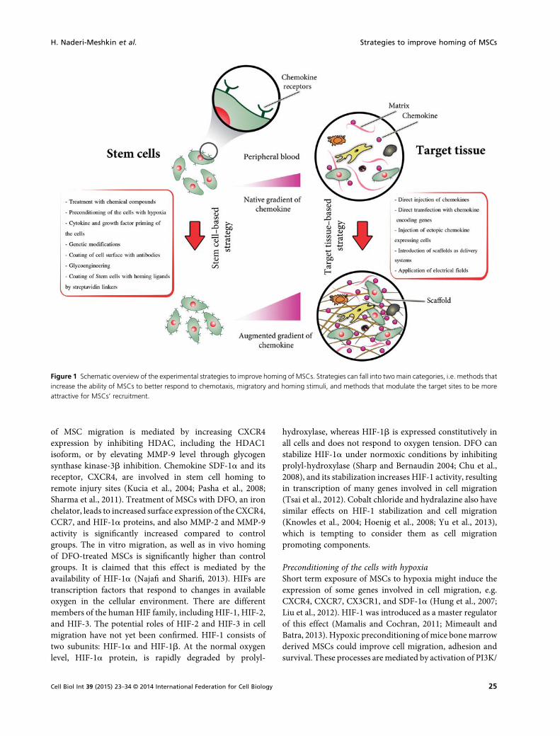
Based on the experimental observations that the effectivehoming of the exogenously transplanted cells greatlyimprove the efficacy of cells to integrate and function inthe target tissues, this review discusses the two main aspectsof subject: (1) factors that increase the ability of stem cells torespond to the migratory stimuli; and (2) methods formodulating the target sites to be more attractive for stem cellrecruitment (Figure 1).
Some prefer to use the terminology of mesenchymalstromal cells or mesenchymal stem/stromal cells instead ofMSCs because the cells used for research and therapy areoften heterogeneously cultured cells that are not strictly allstem cells. This may affect their homing and retention intissues. In this review, we have tried to keep terms used in thecited articles.
Strategies for improvement of stem cell homing
Increasing the ability of stem cells to respond tomigratorystimuli (stem cell-based strategies)
MSCs express a group of receptors (Sordi et al., 2005;Ahmadian Kia et al., 2011) that play a crucial role in cellchemotaxis and migration by interaction with appropriateligands (Wu and Zaho, 2012). Chemokines and theirreceptors have been identified as mediators of cell traffick-ing. Chemokines or chemoattractant cytokines are a largefamily of small secreted proteins that bind to GPCRs, andwhich can be categorized into four classes based on the basisof variations in a conserved cysteine motif of the matureproteins. The first group of chemokines is the CC family,composed of 28 members, and the second group is CXCfamily, possessing a single variable amino acid between thefirst two cysteines, and having 17 members. The CXCchemokines can be further classified into two subfamiliesbased on the presence or absence of specific motif, namelyglu-leu-arg (ELR). Other families are CX3C and XC, withonly one member in each (Lazennec and Richmond, 2010).There are 47 chemokines that bind to four classes ofchemokine receptors. Many of these chemokines bind tomultiple receptors and most of them, except for CX3CR1and CXCR4, also bind tomultiple chemokines. This suggeststhe possibility of functional redundancy, and their spatialand temporal control of expression. CXCR4/SDF-1 con-stitutes one of the most efficient chemokine/chemokinereceptor pairs regarding cell homing (Lazennec andRichmond 2010). Inadequate amounts of the crucialreceptors on the cell surface may be responsible forinefficient homing of the cells to their target tissues(Wynn et al., 2004; Komarova et al., 2010). Here, we discusssome strategies to overcome this pitfall.
Treatment with chemical compoundsMSC treatment with certain chemicals can trigger signalingpathways leading to expression of key mediators involved incell trafficking. Tsai et al. (2011) showed that treatment of ratMSCs with VPA and/or Li and then their transplantion intoa strokemodel of rat resulted in robustmigration and properhoming of the MSCs towards the ischemic site followed byfunctional recovery, increased angiogenesis, and a reducedinfracted zone in the brain. They proved that enhancement
Strategies to improve homing of MSCs H. Naderi-Meshkin et al.
24 Cell Biol Int 39 (2015) 23–34 © 2014 International Federation for Cell Biology
of MSC migration is mediated by increasing CXCR4expression by inhibiting HDAC, including the HDAC1isoform, or by elevating MMP-9 level through glycogensynthase kinase-3b inhibition. Chemokine SDF-1a and itsreceptor, CXCR4, are involved in stem cell homing toremote injury sites (Kucia et al., 2004; Pasha et al., 2008;Sharma et al., 2011). Treatment of MSCs with DFO, an ironchelator, leads to increased surface expression of the CXCR4,CCR7, and HIF-1a proteins, and also MMP-2 and MMP-9activity is significantly increased compared to controlgroups. The in vitro migration, as well as in vivo homingof DFO-treated MSCs is significantly higher than controlgroups. It is claimed that this effect is mediated by theavailability of HIF-1a (Najafi and Sharifi, 2013). HIFs aretranscription factors that respond to changes in availableoxygen in the cellular environment. There are differentmembers of the human HIF family, including HIF-1, HIF-2,and HIF-3. The potential roles of HIF-2 and HIF-3 in cellmigration have not yet been confirmed. HIF-1 consists oftwo subunits: HIF-1a and HIF-1b. At the normal oxygenlevel, HIF-1a protein, is rapidly degraded by prolyl-
hydroxylase, whereas HIF-1b is expressed constitutively inall cells and does not respond to oxygen tension. DFO canstabilize HIF-1a under normoxic conditions by inhibitingprolyl-hydroxylase (Sharp and Bernaudin 2004; Chu et al.,2008), and its stabilization increases HIF-1 activity, resultingin transcription of many genes involved in cell migration(Tsai et al., 2012). Cobalt chloride and hydralazine also havesimilar effects on HIF-1 stabilization and cell migration(Knowles et al., 2004; Hoenig et al., 2008; Yu et al., 2013),which is tempting to consider them as cell migrationpromoting components.
Preconditioning of the cells with hypoxiaShort term exposure of MSCs to hypoxia might induce theexpression of some genes involved in cell migration, e.g.CXCR4, CXCR7, CX3CR1, and SDF-1a (Hung et al., 2007;Liu et al., 2012). HIF-1 was introduced as a master regulatorof this effect (Mamalis and Cochran, 2011; Mimeault andBatra, 2013). Hypoxic preconditioning of mice bonemarrowderived MSCs could improve cell migration, adhesion andsurvival. These processes aremediated by activation of PI3K/
Figure 1 Schematic overview of the experimental strategies to improve homing of MSCs. Strategies can fall into twomain categories, i.e. methods thatincrease the ability of MSCs to better respond to chemotaxis, migratory and homing stimuli, and methods that modulate the target sites to be moreattractive for MSCs’ recruitment.
H. Naderi-Meshkin et al. Strategies to improve homing of MSCs
25Cell Biol Int 39 (2015) 23–34 © 2014 International Federation for Cell Biology
AKT-HIF-1a-CXCR4/CXCR7 pathway (Liu et al., 2010).Hu et al. (2011) demonstrated that hypoxic preconditioningincreasedMSCsmigration into the infarcted myocardium ofmice, and provided evidence that this effect was mediated byincreased expression of the Kv2.1 channel protein, leading toFAK phosphorylation/activation. Activated FAK binds toseveral proteins involved in regulation of cell adhesion andmigration.
Hypoxic preconditioning of MSCs has been introduced asa positive therapeutic approach for ischemic diseases (Weiet al., 2013; Yue et al., 2013). The oxygen tensions depend onthe type of cells, as these rates for the typical cell cultureconditions, normal bone marrow, and ischemic tissues are21, 5, and 1% or lower, respectively. In cell transplantationprograms, it is believed that the injected MSCs must rapidlyadapt themselves to the significantly lower oxygen tension inthe ischemic tissue. It seems that in vitro preconditioning ofthe cells with hypoxia would increase their survival rate invivo. Previous studies have indicated that the hypoxicpreconditioned human MSCs have shown much betterperformance than unconditioned control cells in motilitystatus and therapeutic potential (Rosova et al., 2008).Therefore, based on these data, short-term hypoxicpreconditioning has been introduced as another strategyfor increasing the injected MSCs population at an injuredsite because of their enhanced migration and survival rates.
Priming of the cells with cytokines and growth factorsTo see whether the migration activity of hAd-MSCs could beinfluenced by prior stimulation with chemokines or growthfactors, Baek et al. (2011) pretreated hAd-MSCs withRANTES, SDF-1a, HGF, TNF-a, PDGF-AB, or TGF-b1.Among these, TNF-a induced the highest level ofchemotaxis. Shi et al. (2007) showed that a cocktail of fivecytokine containing Flt-3 ligand, SCF, IL-6, HGF, and IL-3increased the homing ability of FLKþ MSCs derived fromhuman fetal bone marrow to SDF-1. They also proved thatthis effect is mediated by upregulation of CXCR4 protein inhAd-MSCs, and that the CXCR4/SDF1 axis is important inthe homing process.
The effect of CCL25 stimulation on chemotaxis of humanMSCs has also been investigated, indicating that the genescoding for proteins, known to be involved in cellularmovement, are highly regulated, and in accordance with it,the secretion of proteins and chemotaxis are also increased(Binger et al., 2009).
Genetic modificationsOverexpression of a4 subunit of the VLA-4 integrin inMSCs, using adenoviral vector, results in increased MSCshoming to bone marrow in a mouse model (Kumar andPonnazhagan 2007). VLA-4 integrin is composed of CD49d(a4) and CD29 (b1), whose heterodimerization and
interaction with VCAM1 results in firm adherence ofcirculating cells to the endothelium, followed by endothelialtransmigration (Springer 1994; Jacobsen et al., 1996;Schweitzer and Drager, 1996).
Bobis-Wozowicz et al. (2011) found that hAd-MSCs, withan overexpressed level of CXCR4, showed increasedmotility, invasiveness and homing to bone marrow ofNOD/SCID mice. Ryser et al. (2008) also showed thatoverexpression of the CXCR4 receptor at mRNA level allowsthe transient initiation of chemotaxis in MSCs. Transientover-expression of CXCR4 could lead to an increased in vivomobilization, and engraftment of the MSCs into theischemic areas promoted neomyoangiogenesis and alleviat-ed early signs of the left ventricular remodeling (Zhang et al.,2008).
CXCR4 and CXCR7 genes have been ectopically overex-pressed in mouse bone marrow derived MSCs (BM-MSCs).However, the results indicated that the migration of bothnative and genetically manipulated MSCs to the injuredkidney were the same at very low level, and that thetransplantation of the manipulated cells gave no signs oftissue recovery (Gheisari et al., 2012).
CXCR4 mRNA transcripts have been transfected into thehuman MSCs, without any significant improvement in theircell migration; and therefore, it was concluded that theremight be other factors responsible for MSC chemokinesis,independent from CXCR4/SDF-1a axis (Wiehe et al., 2012).Although, CXCR4/SDF-1a axis has emerged as an impor-tant regulator of cell mobilization and trafficking duringtissue regeneration, it seems that this might not be theprecise axis to enhance the MSCs homing. Hence, thechallenge remains to introduce the responsible playersbeside the previously reported CXCR4/SDF-1a axis in theprocess of cell homing.
Coating of cell surface with antibodiesMaking an antibody with double affinity has been anotherinnovative idea introduced for targeting and retaining stemcells in the damaged tissues. Gundlach et al. (2011)synthesized a bispecific antibody, including an anti-CD90recognizing MSCs and an anti-MLC. MLC increases aftercardiac infarction in ischemicmyocardium (Lyn et al., 2000).This construct induced murine BMMSC adhesion to theimmobilized MLC1 substrate (Gundlach et al., 2011), whilethe efficiency of this construct in clinical application hasbeen remained to be examined.
MSCs have been coated with antibodies that react againstVCAM-1 and MAdCAM-1; these endothelial addressinsdirect leukocyte migration to inflamed tissues by theircognate receptors, such asa4b1 integrin (VLA-4), that bindsVCAM-1 or fibronectin (Alon et al., 1995), and a4b7 thatbinds MAdCAM-1 (Takada et al., 2007). Conjugation of theantibodies to the surface of MSCs was done with
Strategies to improve homing of MSCs H. Naderi-Meshkin et al.
26 Cell Biol Int 39 (2015) 23–34 © 2014 International Federation for Cell Biology
intervention of PPG. This coating increased cell homing tothe inflamed colon after systemic delivery and elevated theefficacy of theMSCs to improve treatment of IBD. This effectwas mediated by MSCs immunosuppressive capabilities (Koet al., 2010).
MSCs coated with PPG followed by antibodies againstICAM-1 promoted MSCs attachment to the endothelialcells, which resulted in resistance to high flow conditions(Ko et al., 2009). However, whether rolling and transmigra-tion of the MSCs from endothelium is possible after thisthigh attachment, remains questionable.
GlycoengineeringCell migration involves a cascade of events initiated byshear-resistant adhesive interactions between flowing andendothelial cells at the target tissue. Interaction of E-selectinwhich is expressed on the endothelial cells, with its ligand onthe migrating cells has a key role in this step. E-selectin is alectin that binds to the specialized carbohydrate determi-nants, prototypically consisting of sialofucosylations con-taining an a-2,3-linked sialic acid substitution on galactoseand an a-1,3-linked fucose modification on N-acetylglucos-amine. Together they are displayed as the terminaltetrasaccharide sialyl Lewis X. This ligand is also namedHCELL, because it is expressed in the hematopoietic stemcells. MSCs do not express E-selectin ligands, whereasexpression of a CD44 glycoform bearing a-2,3-sialylmodifications by them is obvious. After application of ana-1,3-fucosyltransferase under specific enzymic conditions,the native CD44 glycoform onMSCs is converted to a ligandfor E-selectin without effects on cell viability or multi-potency. As E-selectin is mostly expressed in bone marrow,dermal microvascular endothelium and post-capillaryvenules at all sites of the injured tissues, HCELL in itsengineered form on the human MSCs therefore can serve asa homing receptor (Sackstein, 2009, 2011a,b). Injection ofthe HCELL-bearing human MSCs into mice led tosignificant homing of the cells into bone marrow (Sacksteinet al., 2008). A high level of HCELL directly induces VLA-4activation via a Rac1/Rap1 GTPase signaling pathway,resulting in transendothelial migration of human MSCs tothe stimulated human umbilical vein endothelial cellswithout chemokine input (Thankamony and Sackstein2011). GPS was named after the technology developed formodifying CD44 glycans to create HCELL on the surface ofliving cells (Sackstein, 2009, 2012a,b).
Coating of stem cells with homing ligands bystreptavidin linkersTethering, rolling, adhesion, extravasation, and engraft-ment are consecutive steps of homing processes. Enhance-ment of each step could lead to improvement of the cellhoming process. Some researchers have focused on
improvement of rolling step by introduction of SLeX tothe surface of MSCs (Sarkar et al., 2008, 2010, 2011a,b).SLeX with selectins, expressed by endothelial cells ofinflamed tissues, is required for promotion of cell rolling(Zhang et al., 2004; Luster et al., 2005; Simon and Green,2005). To achieve this goal, SLeX was conjugated to thesurface of primary human MSCs through a simpleprocedure (Sarkar et al., 2008, 2010, 2011a,b). They firstmodified the surface of MSCs by biotinylated lipid vesicles,followed by incubation with streptavidin to provide biotin-streptavidin bridges. Biotinylated SLeX was added to theculture to immobilize the homing ligand of SLeX on thecell surfaces. There was an increased rolling of the SLeX-MSCs on P-selectin coated substrate in vitro, and theirmigration towards the inflamed tissue was enhanced invivo when administered systemically. This strategy seemsto be applicable for different cell types and also fortargeting a number of tissues in cell therapies. For bettereffect, identification of the receptors, expressed specificallyon endothelium of the target tissues, is a very crucial step.This would pave the way for construction of chemicallyengineered cells with proposed ligands to improve theresults.
Although a number of strategies has been introduced toincrease the ability of stem cells to respond to migratorystimuli, ex vivo expansion and manipulation may altersome characteristics, such as proliferative capacity,differentiation potential, and genetic stability of cells.This may negatively affect their safety at clinical level.Therefore some prefer to modulate the target sites, anapproach for designing more attractive environments toenhance stem/progenitor cells recruitment. These strate-gies are conceived as being more effective for targetedhoming than stem cell-based strategies.
Modulating the target sites for attraction of stem cells
After tissue injury, SDF-1a expression increases in thedamaged cells leading to recruitment and retention ofprogenitor cells at the injury site via chemotactic attractiontowards a gradient of SDF-1a (Hu et al., 2007; Saxena et al.,2008). However, since the natural phenomenon of increasedSDF-1a does not seem sufficient for complete regenerationand repair of the lesions, more comprehensive approachesare being sought to enhance chemotactic attraction of thestem/progenitor cells to the injured tissue/organs, asdiscussed below.
Direct injection of chemokinesThe direct injection of the chemokines into target sites isunder investigation. Sasaki et al. (2007) found that directinjection of SDF-1a into the ischemicmyocardium inmousemodel decreased infarction by enhancing recruitment of the
H. Naderi-Meshkin et al. Strategies to improve homing of MSCs
27Cell Biol Int 39 (2015) 23–34 © 2014 International Federation for Cell Biology
bone marrow cells to the location, followed by angiogenesis.Similar results were reported by Yamaguchi et al. (2003) inlimb ischemia, from which it was suggested that functionalrecovery of an injured organ after direct delivery of SDF-1awas probably due to the enhanced recruitment of thecirculating cells, which is followed by neovascularization(Saxena et al., 2008). However, due to the short half-life ofSDF-1 protein and its degradation by proteolytic enzymes inthe injured tissues, a bioengineered protease-resistant SDF-1that retains its chemotactic potential was used (Segers et al.,2007, 2011).
Direct transfection of the target tissue with chemokineencoding genesUTMD may be a non-invasive and selective approach forgene delivery. In this approach, plasmids containing gene ofinterest are conjugated with lipid microbubbles, whichrelease plasmid DNA when exposed to ultrasound beam(Bekeredjian et al., 2005). In a study, therapeutic genes ofSCF and SDF-1awere delivered to infracted myocardium ofrat by UTMD method. The results indicated a significantincrease in SDF-1a at mRNA and protein levels, followed byan increased homing of CXCR4-positive cells to themyocardium (Fujii et al., 2011). In a clinical trial (phaseI), 17 patients were enrolled to receive JVS-100 byendomyocardial injection. JVS-100 is a DNA plasmidencoding SDF-1. After 12 months, improvement in qualityof life was reported (Penn et al., 2013).
Injection of cells expressing ectopic chemokineConsidering the increased natural level of SDF-1 after organinjury and its effect in recruitment of the endogenous stem/progenitor cells, it was hypothesized that transplantation ofstem cells with elevated level of SDF-1 might be anadvantage. Zhao et al. (2009) found that injection ofMSCs overexpressing SDF-1a to the ischemic heartssignificantly increased migration of bone marrow derivedprogenitor cells and greatly enhanced the cardiac regenera-tion. Others have confirmed functional recovery followingthe increasedmigration of the endogenous cells in the case ofischemic myocardium and diabetic wounds after applying ofsame protocol (Di Rocco et al., 2010; Blumenthal et al.,2011).
Application of scaffolds as delivery systems in target tissueControlled release of a chemokine from various biomaterialsenhances recruitment of MSCs towards them. Recently, ithas been proposed that injectable hydrogels, such as CH-GP-HEC, are good candidates for in situ cartilage tissueregeneration by recruitment of MSCs (Naderi-Meshkinet al., 2014). Schantz et al. (2007) achieved site-specifichoming of MSCs toward a cellular polycaprolactonescaffold, which was constantly releasing SDF-1 with micro
delivery device in vivo. Gelatin hydrogel (Kimura andTabata 2010), PLGA scaffolds (Thevenot et al., 2010), andpoly (lactide ethylene oxide fumarate) hydrogel (He et al.,2010) have also been used to achieve MSCs recruitment. Inparticular, Shen et al. (2010) developed a bioactive knittedsilk-collagen sponge scaffold by incorporation of exogenousSDF-1a. This strategy enabled selective migration andhoming of CXCR4-expressing fibroblast cells, HDFsand ATCs, and resulted in in situ tendon regenerationand decreased accumulation of inflammatory cells.
ch/g-PGA polyelectrolyte multilayer films (PEMS) havebeen used as scaffold and optimized as SDF-1 deliverysystem. SDF-1 retained its biological activity after incorpo-ration with proposed scaffold, and also sustained andcontrolled release of SDF-1 promoted migration of theMSCs in vitro (Goncalves et al., 2012).
Incorporation of some bone matrix molecules, such ascollagen I and HA, into scaffold would increase adsorptionand release of growth factors, followed by enhancedmigration and adhesion of the MSCs to the scaffold(Phipps et al., 2012). In these studies, scaffolds were usedas delivery systems for the chemokines. Others havepreferred to use the scaffolds as delivery systems forvectors carrying the genes, required for chemokinesecretion. Zhang et al. (2013) used a recombinantadenoviral vector, carrying SDF-1 transgene that wasconstructed and applied to transduce LhCG which is acartilage graft composed of living chondrocytes and theircartilaginous ECM. After implantation, SDF-1-LhCGreleased recombinant SDF-1 chemokine in an animalmodel and increased migration rate of the stem/progenitorcells via systemic circulation towards the implanted graftand also enhanced the cartilage regeneration of theproposed graft.
In some other studies, scaffolds were used as deliverysystems for modified cells. Shinohara et al. (2011) showedthat transplantation of a collagen scaffold, equipped withMSCs overexpressing SDF-1 orMCP3, adjacent to a fracturesite enhanced homing of systemic circulating osteogeniccells. This was support for the hypothesis claiming thepositive impact of SDF-1 andMCP3 for induction of homingin stem cells.
Thieme et al. (2009) reported that 3D porous bonesubstitute scaffolds, equipped with modified BM-MSCs, notonly serve as scaffold in large bone defects but also attractMSCs to the location. They showed that transient over-expression of the CXCR4 gene in human BM-MSCs,induced by mRNA transfection, enhances SDF-1a directedchemotactic capacity to internal compartments of theimplanted SDF-1a releasing scaffolds in vitro and in vivo.These techniques seem to be more targeted; localized andhigh efficiency, however, makes it difficult to optimize therelease profile of chemokines in vivo.
Strategies to improve homing of MSCs H. Naderi-Meshkin et al.
28 Cell Biol Int 39 (2015) 23–34 © 2014 International Federation for Cell Biology

Application of electrical fieldsEF induce rapid and directed migration of the neuralprecursor cells (Babona-Pilipos et al., 2011; Feng et al., 2012).A clinical trial in spinal cord injury showed considerablerecovery of the injured area after exposure to the weak EF(Borgens 1988; Tator 2005). Li et al. (2008) found thatphysiological EFs enhanced neural stem/progenitor cellmigration toward the cathode and suggested that theunderlying signal transduction pathway might be
NMDAR/Rac1/actin. It has been established that endoge-nous EF in embryo has a critical role in correct migration ofthe neural stem cells and development of the nervous system(Nuccitelli 2003; McCaig et al., 2005).
Zhang et al. (2011) investigated the application of EFs toguide migration of hiPSCs, and hESCs in 2D (matrigel-coated electrotactic chamber) and 3D (custom designed 3Delectrotactic chambers filled with polymerized matrigel)culture conditions. hiPSCs migrated directionally in the
Table 1 Advantage and disadvantage of suggested strategies to increase homing of stem cells to the target tissues.
Strategy Advantages Bottlenecks References
I) Strategies for improvement of stem cells respond to migratory stimuli (Stem cell-based)Treatment with chemical compounds Simple, Fast Safety problems, Probable changes in
gene expression, Insignificant effecton preventing cell distribution into
non-targeted organs
Knowles et al., 2004; Hoenig et al., 2008;Tsai et al., 2011; Tsai et al., 2012;Najafi and Sharifi 2013; Yu et al., 2013
Preconditioning of the cells withhypoxia
Simple, Fast Optimization problems, Insignificanteffect on preventing cell distribution
into non-targeted organs
Hung et al., 2007; Rosova et al., 2008; Liuet al., 2010; Hu et al., 2011; Liu et al.,2012; Wei et al., 2013; Yue et al.,2013
Cytokine and growth factor priming ofcells
Simple, Fast Safety problems, Probable changes inexpression of unwanted genes,Expensive, Insignificant effect on
preventing cell distribution into non-targeted organs
Shi et al., 2007; Binger et al., 2009; Baeket al. 2011
Genetic modifications More directed Safety problems, Difficult andexpensive, Risk of tumorigenicity,
Insignificant effect in preventing celldistribution into non-targeted organs
Kumar and Ponnazhagan, 2007; Ryseret al., 2008; Zhang et al., 2008;Bobis-Wozowicz et al., 2011; Gheisariet al., 2012; Wiehe et al., 2012
Coating of cell surface with antibodies More directed Difficult and expensive Ko et al., 2009, 2010; Gundlach, 2011Glycoengineering More directed Difficult and expensive Sackstein et al., 2008; Sackstein, 2009,
2011, 2012a,bCoating of stem cells with homing
ligands bystrept-avidin linkersMore directed Difficult and expensive Sarkar et al., 2008, 2010, 2011a,b
II) Modulating the target sites for being more attractive for stem cells recruitment (Target tissue/organ-based)Direct injection of chemokines Simple, Fast,
TargetedShort half-life of chemokines,
Diffusion to surrounding milieu,Degradation by resident proteases
Yamaguchi et al., 2003; Sasaki et al.,2007; Segers et al., 2007, 2011;Saxena et al., 2008
Direct transfection of target tissue withchemokine encoding genes
Targeted Immunogenicity, Retroviral-mediatedinsertional mutagenesis, Expensive,
Difficult
Fujii et al., 2011; Penn et al., 2013
Injection of ectopic chemokineexpressing cells
High efficiency Safety problems, Difficult andexpensive
Zhao et al., 2009; Di Rocco et al., 2010;Blumenthal et al., 2011
Application of scaffolds as deliverysystems into target tissues
More targeted,Localized,
Highly efficient
Difficulty of optimization for releaseprofile of chemokines, Expensive
Schantz et al., 2007; Thieme et al., 2009;He et al., 2010; Kimura and Tabata,2010; Shen et al., 2010; Thevenotet al., 2010; Shinohara et al., 2011;Goncalves et al., 2012; Phipps et al.,2012; Zhang et al., 2013; NaderiMeshkin et al., 2014
Application of electrical fields Easy to use,Cheap
Hard to optimize Borgens, 1988; Nuccitelli, 2003; McCaiget al., 2005; Tator, 2005; Li et al.,2008; Babona-Pilipos et al., 2011;Griffin et al., 2011; Zhang et al., 2011;Feng et al., 2012
H. Naderi-Meshkin et al. Strategies to improve homing of MSCs
29Cell Biol Int 39 (2015) 23–34 © 2014 International Federation for Cell Biology
presence of EF in a voltage dependent manner and EFexposure did not affect expression of stem cell markers.hiPSCs showed more sensitivity and directedness to the EFin comparison with hESCs. Accordingly, they suggested acritical role for Rho/ROCK signaling in galvanotaxis ofhiPSCs. Griffin et al. (2011) found that BM-MSCs treated byEF significantly overexpressed several migratory relatedgenes, including SDF-1/CXCR4, PDGF-BB-R, TGFbR1,IGF-1, and its receptor IGF-1 receptor (IGF-1R). EF treatedcells also showed higher cell invasion through the collagenbarrier which correlates with the higher expression ofMMP-2 and MT1-MMP.
Although optimization is hard in vivo, EFs seem to be safein human, thus they are used in cell therapy and can bringbetter clinical results by enhancing directed cell migration tothe damaged tissues.
Advantages and drawbacks
In this review, two kinds of strategies regarding chemokine/chemokine receptor interactions, and their potential impactson cell therapy to improve stem cell homing, have beendiscussed. Despite the promising literature, the notion stillsuffers from seveal drawbacks and there may be a gapbetween these experimental approaches and their applica-tion in clinic. Some of the advantages and drawbacks facedby stem/progenitor cells and target tissues/organs regardingapplication of the above mentioned strategies are summa-rized in Table 1.
Conclusions and future outlook
A number of stem/progenitor cells respond positively tochemokines with a direct or indirect correlation to thehoming of cells. Accordingly, considerable effort has beenmade to develop engineering techniques, targeting eithercells or target tissues/organs utilizing chemokine ligand/receptor axis to enhance stem cell homing to the injury site.In spite of preliminary success in the improvement of cellproperties (cell-based strategies) to yield higher homingefficiency, they are either expensive or difficult to accom-plish. These strategies also have safety problems fromdifferent points of view, including incomplete localization tothe injury site. Therefore, to address these concerns, manyhave focused on developing target tissue/organ-basedstrategies. Among different strategies involved in modulat-ing target sites, it seems that using equipped scaffolds for thedelivery of the chemokines are more attractive. SeveralSDF1-incorporated scaffolds have been designed to attractendogenous or transplanted stem/progenitor cells with highcontent of its specific receptor, CXCR4, to regeneratedamaged tissues in situ. In an innovative way, it is proposedcombining the two kinds of strategies to get greater
efficiency of homing and better subsequent outcomes, i.e.using both cell-based and target tissue-based methodstogether.
These extensive investigations have provided significantpotential for enhancing targeted stem/progenitor cellhoming. There are some limitations that make it difficultto apply these findings in clinics. To overcome theselimitations, we need to understand the molecular andcellular mechanisms underlying endogenous cell traffickingduring physiological and pathological events, e.g. embryo-genesis, inflammation, wound healing and, cancermetastasis.
Addressing these biological issues would lead to higherefficiency and efficacy of stem cell homing, and hopefullyclinical trials will be replaced by routine clinical applicationof cells in the case of incurable diseases.
Acknowledgments
The corresponding author wishes to particularly thank Dr.Parvaneh Afsharian, Muhammad Irfan-Maqsood andHabibRezanejad for kindly editing the manuscript.
References
Ahmadian Kia N, Bahrami AR, Ebrahimi M, Matin MM, NeshatiZ, Almohaddesin MR, Aghdami N, Bidkhori HR (2011)Comparative analysis of chemokine receptor’s expression inmesenchymal stem cells derived from human bone marrowand adipose tissue. J Mol Neurosci 44(3): 178–85.
Alon R, Kassner PD, Carr MW, Finger EB, Hemler ME, SpringerA (1995) The integrin VLA-4 supports tethering and rolling inflow on VCAM-1. J Cell Biol 128(6): 1243–53.
Babona-Pilipos R, Droujinine IA, Popovic MR, Morshead CM(2011) Adult subependymal neural precursors, but notdifferentiated cells, undergo rapid cathodal migration in thepresence of direct current electric fields. PLoS ONE 6(8):e23808.
Baek SJ, Kang SK, Ra C (2011) In vitro migration capacity ofhuman adipose tissue-derived mesenchymal stem cells reflectstheir expression of receptors for chemokines and growthfactors. Exp Mol Med 43(10): 596–603.
Barbash IM, Chouraqui P, Baron J, Feinberg MS, Etzion S,Tessone A, Miller L, Guetta E, Zipori D, Kedes LH, Kloner RA,Leor J (2003) Systemic delivery of bone marrow-derivedmesenchymal stem cells to the infarcted myocardium:feasibility, cell migration, and body distribution. Circulation108(7): 863–8.
Bekeredjian R, Grayburn PA, Shohet RV (2005) Use of ultrasoundcontrast agents for gene or drug delivery in cardiovascularmedicine. J Am Coll Cardiol 45(3): 329–35.
Binger T, Stich S, Andreas K, Kaps C, Sezer O, Notter M, SittingerM, Ringe J (2009) Migration potential and gene expressionprofile of human mesenchymal stem cells induced by CCL25.Exp Cell Res 315(8): 1468–79.
Strategies to improve homing of MSCs H. Naderi-Meshkin et al.
30 Cell Biol Int 39 (2015) 23–34 © 2014 International Federation for Cell Biology
Blumenthal B, Poppe A, Golsong P, Blanke P, Rylski B, BeyersdorfF, Schlensak C, Siepe M (2011) Functional regeneration ofischemic myocardium by transplanted cells overexpressingstromal cell-derived factor-1 (SDF-1): intramyocardial injec-tion versus scaffold-based application. Eur J Cardiothorac Surg40(4): e135–e141.
Bobis-Wozowicz S, Miekus K, Wybieralska E, Jarocha D,Zawisz A, Madeja Z, M (2011) Genetically modifiedadipose tissue-derived mesenchymal stem cells overexpressingCXCR4 display increasedmotility, invasiveness, and homing tobone marrow of NOD/SCIDmice. Exp Hematol 39(6): 686–96e684.
Borgens RB (1988) Stimulation of neuronal regeneration anddevelopment by steady electrical fields. Adv Neurol 47: 547–64.
Caplan AI, Correa D (2011) The MSC: an injury drugstore. CellStem Cell 9(1): 11–5.
Charwat S, Gyongyosi M, Lang I, Graf S, Beran G, HemetsbergerR, Nyolczas N, Sochor H, Glogar D (2008) Role of adult bonemarrow stem cells in the repair of ischemic myocardium:current state of the art. Exp Hematol 36(6): 672–80.
Chu K, Jung KH, Kim SJ, Lee ST, Kim J, Park HK, Song EC, KimSU, Kim M, Lee SK, Roh K (2008) Transplantation of humanneural stem cells protect against ischemia in a preventive modevia hypoxia-inducible factor-1alpha stabilization in the hostbrain. Brain Res 1207: 182–92.
Dell’Accio F, De Bari C, Luyten P (2001) Molecular markerspredictive of the capacity of expanded human articularchondrocytes to form stable cartilage. Arthritis Rheum 44(7):1608–19.
Deng J, Zou ZM, Zhou TL, Su YP, Ai GP, Wang JP, Xu H, DongW (2011) Bone marrow mesenchymal stem cells can bemobilized into peripheral blood by G-CSF in vivo and integrateinto traumatically injured cerebral tissue. Neurol Sci 32(4):641–51.
Di Rocco G, Gentile A, Antonini A, Ceradini F, Wu JC,Capogrossi MC, Toietta G (2010) Enhanced healing of diabeticwounds by topical administration of adipose tissue-derivedstromal cells overexpressing stromal-derived factor-1: biodis-tribution and engraftment analysis by bioluminescent imaging.Stem Cells Int 2011: 304562.
Dominici M, Le Blanc K, Mueller I, Slaper-Cortenbach I, MariniF, Krause D, Deans R, Keating A, Prockop D, Horwitz E (2006)Minimal criteria for defining multipotent mesenchymalstromal cells. The International Society for Cellular Therapyposition statement. Cytotherapy 8(4): 315–7.
Edalatmanesh MA, Bahrami AR, Hosseini E, Hosseini M,Khatamsaz S (2011) Bone marrow derived mesenchymalstem cell transplantation in cerebellar degeneration: abehavioral study. Behav Brain Res 225(1): 63–70.
Edalatmanesh MA, Bahrami AR, Hosseini E, Hosseini M,Khatamsaz S (2011) Neuroprotective effects of mesenchymalstem cell transplantation in animal model of cerebellardegeneration. Neurol Res 33(9): 913–20.
Feng JF, Zhang XZ, Zhang L, Jiang JY, Nolta J, Zhao M (2012)Guided migration of neural stem cells derived from human
embryonic stem cells by an electric field. Stem Cells 30(2):349–55.
Fujii H, Li SH,Wu J, Miyagi Y, Yau TM, Rakowski H, Egashira K,Guo J,Weisel RD, Li K (2011) Repeated and targeted transfer ofangiogenic plasmids into the infarcted rat heart via ultrasoundtargeted microbubble destruction enhances cardiac repair. EurHeart J 32(16): 2075–84. doi: 10.1093/eurheartj/ehq475.Epub2010 Dec 31.
Futami I, Ishijima M, Kaneko H, Tsuji K, Ichikawa-Tomikawa N,Sadatsuki R, Muneta T, Arikawa-Hirasawa E, Sekiya I, KanekoK (2012) Isolation and characterization of multipotentialmesenchymal cells from the mouse synovium. PLoS ONE 7(9):e45517.
Gheisari Y, Azadmanesh K, Ahmadbeigi N, Nassiri SM,Golestaneh AF, Naderi M, Vasei M, Arefian E, Mirab-SamieeS, Shafiee A, Soleimani M, Zeinali S (2012) Genetic modifica-tion of mesenchymal stem cells to overexpress CXCR4 andCXCR7 does not improve the homing and therapeuticpotentials of these cells in experimental acute kidney injury.Stem Cells Dev 21(16): 2969–80.
Goncalves RM (2012) Mesenchymal stem cell recruitment bystromal derived factor-1-delivery systems based on chitosan/poly(gamma-glutamic acid) polyelectrolyte complexes. EurCell Mater 23: 249–60; discussion 241–260.
Griffin M, Iqbal SA, Sebastian A (2011) Degenerate waveand capacitive coupling increase human MSC invasionand proliferation while reducing cytotoxicity in an in vitrowound healing model. J Colthurst and A Bayat 6(8):e23404.
Gundlach C,W, Caivano T,A, Cabreira-Hansen Mda G, Gahre-manpour A, Brown WS, Zheng Y, McIntyre BW, Willerson JT,Dixon RA, Perin EC, Woodside DG (2011) Synthesis andevaluation of an anti-MLC1 x anti-CD90 bispecific antibody fortargeting and retaining bone-marrow-derived multipotentstromal cells in infarcted myocardium. Bioconjug Chem 22(8): 1706–14.
Haddad-Mashadrizeh A, Bahrami AR, MatinMM, EdalatmaneshMA, Zomorodipour A, Fallah A, Gardaneh M, Ahmadian N,Sanjarmoosavi N (2013) Evidence for crossing the bloodbarrier of adult rat brain by human adipose-derivedmesenchymal stromal cells during a 6-month period of post-transplantation. Cytotherapy 15(8): 951–60.
He X, Ma J, Jabbari E (2010) Migration of marrow stromal cellsin response to sustained release of stromal-derived factor-1alpha from poly(lactide ethylene oxide fumarate) hydrogels.Int J Pharm 390(2): 107–16.
HoenigMR, Bianchi C, SellkeW (2008) Hypoxia inducible factor-1 alpha, endothelial progenitor cells, monocytes, cardiovascu-lar risk, wound healing, cobalt and hydralazine: a unifyinghypothesis. Curr Drug Targets 9(5): 422–35.
Hu X, Dai S,WuWJ, TanW, Zhu X,Mu J, Guo Y, Bolli R, RokoshG (2007) Stromal cell derived factor-1 alpha confers protectionagainst myocardial ischemia/reperfusion injury: role of thecardiac stromal cell derived factor-1 alpha CXCR4 axis.Circulation 116(6): 654–63.
H. Naderi-Meshkin et al. Strategies to improve homing of MSCs
31Cell Biol Int 39 (2015) 23–34 © 2014 International Federation for Cell Biology
Hu X, Wei L, Taylor TM, Wei J, Zhou X, Wang JA, Yu SP (2011)Hypoxic preconditioning enhances bone marrow mesenchy-mal stem cell migration via Kv2. 1 channel and FAK activation.Am J Physiol Cell Physiol 301(2): C362–C372.
Huang AH, Yeger-McKeever M, Stein A, Mauck L (2008) Tensileproperties of engineered cartilage formed from chondrocyte-and MSC-laden hydrogels. Osteoarthritis Cartilage 16(9):1074–82.
Huang YS, Li IH, Chueh SH, Hueng DY, Tai MC, Liang CM, LienSB, Sytwu HK, Ma H (2013) Mesenchymal stem cells from ratolfactory bulbs can differentiate into cells with cardiomyocytecharacteristics. J Tissue Eng Regen Med doi: 10.1002/term.1684.
Hung SC, Pochampally RR, Hsu SC, Sanchez C, Chen SC (2007)Short-term exposure ofmultipotent stromal cells to low oxygenincreases their expression of CX3CR1 and CXCR4 and theirengraftment in vivo. J Spees and D J Prockop 2(5): e416.
Jacobsen K, Kravitz J, Kincade PW, Osmond G (1996) Adhesionreceptors on bone marrow stromal cells: in vivo expression ofvascular cell adhesion molecule-1 by reticular cells andsinusoidal endothelium in normal and gamma-irradiatedmice. Blood 87(1): 73–82.
Karimabad HM, Shabestari M, Baharvand H, Vosough A,Gourabi H, Shahverdi A, Shamsian A, Abdolhoseini S,Moazzami K, Marjanimehr MM, Emami F, Bidkhori HR,Hamedanchi A, Talebi S, Farrokhi F, Jabbari-Azad F, FadaviM,Garivani U, Mahmoodi M, Aghdami N (2011) Lack ofbeneficial effects of granulocyte colony-stimulating factor inpatients with subacute myocardial infarction undergoing laterevascularization: a double-blind, randomized, placebo-con-trolled clinical trial. Acta Cardiol 66(2): 219–24.
Karp JM, Leng Teo S (2009) Mesenchymal stem cell homing: thedevil is in the details. Cell Stem Cell 4(3): 206–16.
Kimura Y, Tabata Y (2010) Controlled release of stromal-cell-derived factor-1 from gelatin hydrogels enhances angiogenesis.J Biomater Sci Polym Ed 21(1): 37–51.
Knowles HJ, Tian YM, Mole DR, Harris L (2004) Novelmechanism of action for hydralazine: induction of hypoxia-inducible factor-1alpha, vascular endothelial growth factor,and angiogenesis by inhibition of prolyl hydroxylases. Circ Res95(2): 162–9.
Ko IK, Kean TJ, Dennis E (2009) Targeting mesenchymal stemcells to activated endothelial cells. Biomaterials 30(22): 3702–10.
Ko IK, KimBG, Awadallah A,Mikulan J, Lin P, Letterio JJ, DennisJE (2010) Targeting improves MSC treatment of inflammatorybowel disease. Mol Ther 18(7): 1365–72.
Komarova S, Roth J, Alvarez R, Curiel DT, Pereboeva L (2010)Targeting of mesenchymal stem cells to ovarian tumors via anartificial receptor. J Ovarian Res 3: 12. doi: 10.1186/1757-2215-3-12.
Kucia M, Jankowski K, Reca R, Wysoczynski M, Bandura L,Allendorf DJ, Zhang J, Ratajczak J, Ratajczak MZ (2004)CXCR4-SDF-1 signalling, locomotion, chemotaxis and adhe-sion. J Mol Histol 35(3): 233–45.
Kumar S, Ponnazhagan S (2007) Bone homing of mesenchymalstem cells by ectopic alpha 4 integrin expression. Faseb J 21(14):3917–27.
LaBarge MA, Blau M (2002) Biological progression from adultbone marrow to mononucleate muscle stem cell to multinu-cleate muscle fiber in response to injury. Cell 111(4): 589–601.
Larsen S, Lewis D (2011) Potential therapeutic applications ofmesenchymal stromal cells. Pathology 43(6): 592–604.
Lazennec G, Richmond A (2010) Chemokines and chemokinereceptors: new insights into cancer-related inflammation.Trends Mol Med 16(3): 133–44.
Li L, El-Hayek YH, Liu B, Chen Y, Gomez E, Wu X, Ning K,Chang N, Zhang L, Wang Z, Hu X, Wan Q (2008) Direct-current electrical field guides neuronal stem/progenitor cellmigration. Stem Cells 26(8): 2193–200.
LiuH, Liu S, Li Y,Wang X, XueW,GeG, Luo X (2012) The role ofSDF-1-CXCR4/CXCR7 axis in the therapeutic effects ofhypoxia-preconditioned mesenchymal stem cells for renalischemia/reperfusion injury. PLoS One 7(4): e34608.
Liu H, XueW, Ge G, Luo X, Li Y, Xiang H, Ding X, Tian P, Tian X(2010) Hypoxic preconditioning advances CXCR4 and CXCR7expression by activating HIF-1alpha in MSCs. BiochemBiophys Res Commun 401(4): 509–15.
Luster AD, Alon R, von Andrian UH (2005) Immune cellmigration in inflammation: present and future therapeutictargets. Nat Immunol 6(12): 1182–90.
Lyn D, Liu X, Bennett NA, Emmett L (2000) Gene expressionprofile in mouse myocardium after ischemia. Physiol Geno-mics 2(3): 93–100.
Makinen S, Kekarainen T, Nystedt J, Liimatainen T, Huhtala T,NarvanenA, Laine J, Jolkkonen J (2006) Human umbilical cordblood cells do not improve sensorimotor or cognitive outcomefollowing transient middle cerebral artery occlusion in rats.Brain Res 1123(1): 207–15.
Mamalis AA, Cochran L (2011) The therapeutic potential ofoxygen tensionmanipulation via hypoxia inducible factors andmimicking agents in guided bone regeneration. A review ArchOral Biol 56(12): 1466–75.
McCaig CD, Rajnicek AM, Song B, Zhao M (2005) Controllingcell behavior electrically: current views and future potential.Physiol Rev 85(3): 943–78.
Mimeault M, Batra K (2013) Hypoxia-inducing factors as masterregulators of stemness properties and altered metabolism ofcancer- and metastasis-initiating cells. J Cell Mol Med 17(1):30–54.
Mundra V, Gerling IC, Mahato I (2013) Me senchymal stem cell-based therapy. Mol Pharm 10(1): 77–89.
Naderi-Meshkin H, Andreas K, Matin MM, Sittinger M, BidkhoriHR, Ahmadiankia N, Bahrami AR, Ringe J (2014) Chitosan-based injectable hydrogel as a promising in situ forming scaffoldfor cartilage tissue engineering. Cell Biol Int 38(1): 72–84.
Najafi R, Sharifi M (2013) Deferoxamine preconditioningpotentiates mesenchymal stem cell homing in vitro and instreptozotocin-diabetic rats. Expert Opin Biol Ther 13(7):959–72.
Strategies to improve homing of MSCs H. Naderi-Meshkin et al.
32 Cell Biol Int 39 (2015) 23–34 © 2014 International Federation for Cell Biology
Nuccitelli R (2003) Endogenous electric fields in embryos duringdevelopment, regeneration and wound healing. Radiat ProtDosimetry 106(4): 375–83.
Ohnishi S, Nagaya N (2007) Prepare cells to repair the heart:mesenchymal stem cells for the treatment of heart failure. Am JNephrol 27(3): 301–7.
Pasha Z, Wang Y, Sheikh R, Zhang D, Zhao T, Ashraf M (2008)Preconditioning enhances cell survival and differentiation ofstem cells during transplantation in infarcted myocardium.Cardiovasc Res 77(1): 134–42.
PennMS,MendelsohnFO,SchaerGL, ShermanW,FarrM,PastoreJ, Rouy D, Clemens R, Aras R, LosordoW (2013) An open-labeldose escalation study to evaluate the safety of administration ofnonviral stromal cell-derived factor-1 plasmid to treat symp-tomatic ischemic heart failure. Circ Res 112(5): 816–25.
Phipps MC, Xu Y, Bellis L (2012) Delivery of platelet-derivedgrowth factor as a chemotactic factor for mesenchymal stemcells by bone-mimetic electrospun scaffolds. PLoS One 7(7):e40831.
Poloni A, Maurizi G, Serrani F, Mancini S, Zingaretti MC,Frontini A, Cinti S, Olivieri A, Leoni P (2013) Molecular andfunctional characterization of human bonemarrow adipocytes.Exp Hematol 41(6): 558–66.
Rosova I, Dao M, Capoccia B, Link D, Nolta A (2008) Hypoxicpreconditioning results in increased motility and improvedtherapeutic potential of human mesenchymal stem cells. StemCells 26(8): 2173–82.
Ryser MF, Ugarte F, Thieme S, Bornhauser M, Roesen-Wolff A,Brenner S (2008) MRNA transfection of CXCR4-GFP fusion–simply generated by PCR-results in efficient migration ofprimary human mesenchymal stem cells. Tissue Eng Part CMethods 14(3): 179–84.
Sackstein R (2009) Glycosyltransferase-programmed stereosub-stitution (GPS) to create HCELL: engineering a roadmap forcell migration. Immunol Rev 230(1): 51–74.
Sackstein R (2011) The biology of CD44 and HCELL inhematopoiesis: the ‘step 2-bypass pathway’ and other emergingperspectives. Curr Opin Hematol 18(4): 239–48.
Sackstein R (2012) Engineering cellular trafficking via glycosyl-transferase-programmed stereosubstitution. Ann N YAcad Sci1253: 193–200.
Sackstein R (2012) Glycoengineering of HCELL, the human bonemarrow homing receptor: sweetly programming cell migra-tion. Ann Biomed Eng 40(4): 766–76.
Sackstein R, Merzaban JS, Cain DW, Dagia NM, Spencer JA, LinCP,Wohlgemuth R (2008) Ex vivo glycan engineering of CD44programs human multipotent mesenchymal stromal celltrafficking to bone. Nat Med 14(2): 181–7.
Sarkar D, Vemula PK, Teo GS, Spelke D, Karnik R, Wee Y, KarpM (2008) Chemical engineering of mesenchymal stem cells toinduce a cell rolling response. Bioconjug Chem 19(11): 2105–9.
Sarkar D, Vemula PK, Zhao W, Gupta A, Karnik R, Karp M(2010) Engineeredmesenchymal stem cells with self-assembledvesicles for systemic cell targeting. Biomaterials 31(19):5266–74.
Sarkar D, Zhao W, Gupta A, Loh WL, Karnik R, Karp M (2011a)Cell surface engineering of mesenchymal stem cells. MethodsMol Biol 698: 505–23.
Sarkar D, Spencer JA, Phillips JA, Zhao W, Schafer S, Spelke DP,Mortensen LJ, Ruiz JP, Vemula PK, Sridharan R, Kumar S,Karnik R, Lin CP, Karp M (2011b) Engineered cell homing.Blood; 15;118(25):e 184–91.
Sasaki T, Fukazawa R, Ogawa S, Kanno S, Nitta T, Ochi M,Shimizu K (2007) Stromal cell-derived factor-1alpha improvesinfarcted heart function through angiogenesis in mice. PediatrInt 49(6): 966–71.
Saxena A, Fish JE,WhiteMD, Yu S, Smyth JW, Shaw RM, DiMaioJM, Srivastava D (2008) Stromal cell-derived factor-1alpha iscardioprotective after myocardial infarction. Circulation 117(17): 2224–31.
Schantz JT, Chim H, Whiteman M (2007) Cell guidance in tissueengineering: SDF-1 mediates site-directed homing of mesen-chymal stem cells within three-dimensional polycaprolactonescaffolds. Tissue Eng 13(11): 2615–24.
Schweitzer KM, Drager AM, P. van der Valk, Thijsen SF,Zevenbergen A, Theijsmeijer AP, van der Schoot CE, Langen-huijsen MM (1996) Constitutive expression of E-selectin andvascular cell adhesion molecule-1 on endothelial cells ofhematopoietic tissues. Am J Pathol 148(1): 165–75.
Segers VF, Revin V,WuW, Qiu H, Yan Z, Lee RT, Sandrasagra A(2011) Protease-resistant stromal cell-derived factor-1 for thetreatment of experimental peripheral artery disease. Circula-tion 123(12): 1306–15.
Segers VF, Tokunou T, Higgins LJ, MacGillivray C, Gannon J, LeeRT (2007) Local delivery of protease-resistant stromal cellderived factor-1 for stem cell recruitment after myocardialinfarction. Circulation 116(15): 1683–92.
Sharma M, Afrin F, Satija N, Tripathi RP, Gangenahalli U (2011)Stromal-derived factor-1/CXCR4 signaling: indispensable rolein homing and engraftment of hematopoietic stem cells in bonemarrow. Stem Cells Dev 20(6): 933–46.
Sharp FR, Bernaudin M (2004) HIF1 and oxygen sensing in thebrain. Nat Rev Neurosci 5(6): 437–48.
Shen W, Chen X, Chen J, Yin Z, Heng BC, Chen W, Ouyang W(2010) The effect of incorporation of exogenous stromal cell-derived factor-1 alpha within a knitted silk-collagen spongescaffold on tendon regeneration. Biomaterials 31(28): 7239–49.
Shi M, Li J, Liao L, Chen B, Li B, Chen L, Jia H, Zhao C (2007)Regulation of CXCR4 expression in humanmesenchymal stemcells by cytokine treatment: role in homing efficiency in NOD/SCID mice. Haematologica 92(7): 897–904.
Shinohara K, Greenfield S, Pan H, Vasanji A, Kumagai K, MiduraRJ, Kiedrowski M, Penn MS, Muschler F (2011) Stromal cell-derived factor-1 and monocyte chemotactic protein-3 improverecruitment of osteogenic cells into sites of musculoskeletalrepair. J Orthop Res 29(7): 1064–9.
Simoes IN, Boura JS, Dos Santos F, Andrade PZ, Cardoso CM,Gimble JM, da Silva CL, Cabral M (2013) Human mesenchy-mal stem cells from the umbilical cord matrix: successfulisolation and ex-vivo expansion using serum-/xeno-freeculture media. Biotechnol J 8(4): 448–58.
H. Naderi-Meshkin et al. Strategies to improve homing of MSCs
33Cell Biol Int 39 (2015) 23–34 © 2014 International Federation for Cell Biology
Simon SI, Green E (2005) Molecular mechanics and dynamics ofleukocyte recruitment during inflammation. Annu RevBiomed Eng 7: 151–85.
Sordi V, Malosio ML, Marchesi F, Mercalli A, Melzi R, GiordanoT, Belmonte N, Ferrari G, Leone BE, Bertuzzi F, Zerbini G,Allavena P, Bonifacio E, Piemonti L (2005) Bone marrowmesenchymal stem cells express a restricted set of functionallyactive chemokine receptors capable of promoting migration topancreatic islets. Blood 106(2): 419–27.
Springer TA (1994) Traffic signals for lymphocyte recirculationand leukocyte emigration: the multistep paradigm. Cell 76(2):301–14.
Takada Y, Ye X, Simon S (2007) The integrins. Genome Biol 8(5):215.
Tator CH (2005) Phase 1 trial of oscillating field stimulation forcomplete spinal cord injury in humans. J Neurosurg Spine 2(1):1; discussion 1–2.
Thankamony SP, Sackstein R (2011) Enforced hematopoietic cellE- and L-selectin ligand (HCELL) expression primes trans-endothelial migration of human mesenchymal stem cells. ProcNatl Acad Sci U S A 108(6): 2258–63.
Thevenot PT, Nair AM, Shen J, Lotfi P, Ko CY, Tang L (2010) Theeffect of incorporation of SDF-1alpha into PLGA scaffolds onstem cell recruitment and the inflammatory response.Biomaterials 31(14): 3997–4008.
Thieme S, Ryser M, Gentsch M, Navratiel K, Brenner S, StiehlerM, Rolfing J, Gelinsky M, Rosen-Wolff A (2009) Stromal cell-derived factor-1alpha-directed chemoattraction of transientlyCXCR4-overexpressing bone marrow stromal cells intofunctionalized three-dimensional biomimetic scaffolds. TissueEng Part C Methods 15(4): 687–96.
Tsai CC, Yew TL, Yang DC, Huang WH, Hung C (2012) Benefitsof hypoxic culture on bone marrow multipotent stromal cells.Am J Blood Res 2(3): 148–59.
Tsai LK, Wang Z, Munasinghe J, Leng Y, Leeds P, Chuang DM(2011) Mesenchymal stem cells primed with valproate andlithium robustly migrate to infarcted regions and facilitaterecovery in a stroke model. Stroke 42(10): 2932–9.
Wagner J, Kean T, Young R, Dennis JE, Caplan AI (2009)Optimizing mesenchymal stem cell-based therapeutics. CurrOpin Biotechnol 20(5): 531–6.
Walczak P, Zhang J, Gilad AA, Kedziorek DA, Ruiz-Cabello J,Young RG, Pittenger MF, van Zijl PC, Huang J, Bulte JW(2008) Dual-modality monitoring of targeted intraarterialdelivery of mesenchymal stem cells after transient ischemia.Stroke 39(5): 1569–74.
Wei N, Yu SP, Gu X, Taylor TM, Song D, Liu XF, Wei L (2013)Delayed intranasal delivery of hypoxic-preconditioned bonemarrow mesenchymal stem cells enhanced cell homing andtherapeutic benefits after ischemic stroke in mice. CellTransplant 22(6): 977–91.
Wiehe JM, Kaya Z, Homann JM, Wohrle J, Vogt K, Nguyen T,Rottbauer W, Torzewski J, Fekete N, Rojewski M, Schrezen-meier H, Moepps B, Zimmermann O (2012) GMP-adapted
overexpression of CXCR4 in human mesenchymal stem cellsfor cardiac repair. Int J Cardiol 167(5): 2073–81.
Wu Y, Zaho C (2012) The role of chemokines in mesenchymalstem cell homing to myocardium. Stem Cell Rev 8(1): 243–50.
Wynn RF, Hart CA, Corradi-Perini C, O’Neill L, Evans CA,Wraith JE, Fairbairn LJ, Bellantuono I (2004) A smallproportion of mesenchymal stem cells strongly expressesfunctionally active CXCR4 receptor capable of promotingmigration to bone marrow. Blood 104(9): 2643–5.
Yamaguchi J, Kusano KF, Masuo O, Kawamoto A, Silver M,Murasawa S, Bosch-Marce M, Masuda H, Losordo DW, IsnerJM, Asahara T (2003) Stromal cell-derived factor-1 effects onex vivo expanded endothelial progenitor cell recruitment forischemic neovascularization. Circulation 107 (9): 1322–8.
Yu X, Lu C, LiuH, Rao S, Cai J, Liu S, Kriegel AJ, GreeneAS, LiangM, Ding X (2013) Hypoxic preconditioning with cobalt of bonemarrow mesenchymal stem cells improves cell migration andenhances therapy for treatment of ischemic acute kidneyinjury. PLoS One 8(5): e62703.
Yue Y, Zhang P, Liu D, Yang JF, Nie C, Yang D (2013) Hypoxiapreconditioning enhances the viability of ADSCs to increasethe survival rate of ischemic skin flaps in rats. Aesthetic PlastSurg 37(1): 159–70.
Zhang D, Fan GC, Zhou X, Zhao T, Pasha Z, XuM, Zhu Y, AshrafM, Wang Y (2008) Over-expression of CXCR4 on mesenchy-mal stem cells augments myoangiogenesis in the infarctedmyocardium. J Mol Cell Cardiol 44(2): 281–92.
Zhang F, Leong W, Su K, Fang Y, Wang A (2013) A TransducedLiving Hyaline Cartilage Graft Releasing Transgenic StromalCell-Derived Factor-1 Inducing Endogenous Stem CellHoming In Vivo. Tissue Eng Part A 19(9–10): 1091–9.
Zhang J, Calafiore M, Zeng Q, Zhang X, Huang Y, Li RA, DengW, Zhao M (2011) Electrically guiding migration of humaninduced pluripotent stem cells. Stem Cell Rev 7(4): 987–96.
Zhang M, Mal N, Kiedrowski M, Chacko M, Askari AT, PopovicZB, Koc ON, Penn S (2007) SDF-1 expression bymesenchymalstem cells results in trophic support of cardiac myocytes aftermyocardial infarction. FASEB J 21(12): 3197–207.
Zhang X, Bogorin DF, Moy T (2004) Molecular basis of thedynamic strength of the sialyl Lewis X–selectin interaction.Chemphyschem 5(2): 175–82.
Zhang X, Wang Y, Gao Y, Liu X, Bai T, Li M, Li L, Chi G, Xu H,Liu F, Liu JY, Li Y (2013)Maintenance of high proliferation andmultipotent potential of human hair follicle-derived mesen-chymal stem cells by growth factors. Int J Mol Med 31(4): 913–21.
Zhao T, Zhang D, Millard RW, Ashraf M, Wang Y (2009) Stemcell homing and angiomyogenesis in transplanted hearts areenhanced by combined intramyocardial SDF-1alpha deliveryand endogenous cytokine signaling. Am J Physiol Heart CircPhysiol 296(4): H976–986.
Received 6 April 2014; accepted 19 June 2014.
Final version published online 13 October 2014.
Strategies to improve homing of MSCs H. Naderi-Meshkin et al.
34 Cell Biol Int 39 (2015) 23–34 © 2014 International Federation for Cell Biology