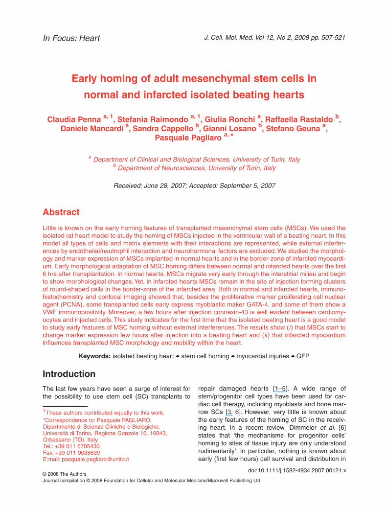
Early homing of adult mesenchymal stem cells in normal and infarcted isolated beating hearts
15
Introduction The last few years have seen a surge of interest for the possibility to use stem cell (SC) transplants to repair damaged hearts [1–5]. A wide range of stem/progenitor cell types have been used for car- diac cell therapy, including myoblasts and bone mar- row SCs [3, 6]. However, very little is known about the early features of the homing of SC in the receiv- ing heart. In a recent review, Dimmeler et al. [6] states that ‘the mechanisms for progenitor cells’ homing to sites of tissue injury are only understood rudimentarily’. In particular, nothing is known about early (first few hours) cell survival and distribution in Early homing of adult mesenchymal stem cells in normal and infarcted isolated beating hearts Claudia Penna a, 1 , Stefania Raimondo a, 1 , Giulia Ronchi a , Raffaella Rastaldo b , Daniele Mancardi a , Sandra Cappello b , Gianni Losano b , Stefano Geuna a , Pasquale Pagliaro a, * a Department of Clinical and Biological Sciences, University of Turin, Italy b Department of Neurosciences, University of Turin, Italy Received: June 28, 2007; Accepted: September 5, 2007 Abstract Little is known on the early homing features of transplanted mesenchymal stem cells (MSCs). We used the isolated rat heart model to study the homing of MSCs injected in the ventricular wall of a beating heart. In this model all types of cells and matrix elements with their interactions are represented, while external interfer- ences by endothelial/neutrophil interaction and neurohormonal factors are excluded. We studied the morphol- ogy and marker expression of MSCs implanted in normal hearts and in the border-zone of infarcted myocardi- um. Early morphological adaptation of MSC homing differs between normal and infarcted hearts over the first 6 hrs after transplantation. In normal hearts, MSCs migrate very early through the interstitial milieu and begin to show morphological changes.Yet, in infarcted hearts MSCs remain in the site of injection forming clusters of round-shaped cells in the border-zone of the infarcted area. Both in normal and infarcted hearts, immuno- histochemistry and confocal imaging showed that, besides the proliferative marker proliferating cell nuclear agent (PCNA), some transplanted cells early express myoblastic maker GATA-4, and some of them show a VWF immunopositivity. Moreover, a few hours after injection connexin-43 is well evident between cardiomy- ocytes and injected cells. This study indicates for the first time that the isolated beating heart is a good model to study early features of MSC homing without external interferences. The results show (i ) that MSCs start to change marker expression few hours after injection into a beating heart and (ii ) that infarcted myocardium influences transplanted MSC morphology and mobility within the heart. Keywords: isolated beating heart • stem cell homing • myocardial injuries • GFP J. Cell. Mol. Med. Vol 12, No 2, 2008 pp. 507-521 1 These authors contributed equally to this work. *Correspondence to: Pasquale PAGLIARO, Dipartimento di Scienze Cliniche e Biologiche, Università di Torino, Regione Gonzole 10, 10043, Orbassano (TO), Italy. Tel.: +39 011 6705430 Fax: +39 011 9038639 E:mail: [email protected] © 2008 The Authors Journal compilation © 2008 Foundation for Cellular and Molecular Medicine/Blackwell Publishing Ltd doi: 10.1111/j.1582-4934.2007.00121.x In Focus: Heart
-
Upload
independent -
Category
Documents
-
view
0 -
download
0
Transcript of Early homing of adult mesenchymal stem cells in normal and infarcted isolated beating hearts
Introduction
The last few years have seen a surge of interest forthe possibility to use stem cell (SC) transplants to
repair damaged hearts [1–5]. A wide range ofstem/progenitor cell types have been used for car-diac cell therapy, including myoblasts and bone mar-row SCs [3, 6]. However, very little is known about the early features of the homing of SC in the receiv-ing heart. In a recent review, Dimmeler et al. [6]states that ‘the mechanisms for progenitor cells’homing to sites of tissue injury are only understoodrudimentarily’. In particular, nothing is known aboutearly (first few hours) cell survival and distribution in
Early homing of adult mesenchymal stem cells in
normal and infarcted isolated beating hearts
Claudia Penna a, 1
, Stefania Raimondo a, 1
, Giulia Ronchi a, Raffaella Rastaldo
b,
Daniele Mancardia, Sandra Cappello
b, Gianni Losano
b, Stefano Geuna
a,
Pasquale Pagliaro a, *
a Department of Clinical and Biological Sciences, University of Turin, Italyb Department of Neurosciences, University of Turin, Italy
Received: June 28, 2007; Accepted: September 5, 2007
Abstract
Little is known on the early homing features of transplanted mesenchymal stem cells (MSCs). We used theisolated rat heart model to study the homing of MSCs injected in the ventricular wall of a beating heart. In thismodel all types of cells and matrix elements with their interactions are represented, while external interfer-ences by endothelial/neutrophil interaction and neurohormonal factors are excluded. We studied the morphol-ogy and marker expression of MSCs implanted in normal hearts and in the border-zone of infarcted myocardi-um. Early morphological adaptation of MSC homing differs between normal and infarcted hearts over the first6 hrs after transplantation. In normal hearts, MSCs migrate very early through the interstitial milieu and beginto show morphological changes. Yet, in infarcted hearts MSCs remain in the site of injection forming clustersof round-shaped cells in the border-zone of the infarcted area. Both in normal and infarcted hearts, immuno-histochemistry and confocal imaging showed that, besides the proliferative marker proliferating cell nuclearagent (PCNA), some transplanted cells early express myoblastic maker GATA-4, and some of them show aVWF immunopositivity. Moreover, a few hours after injection connexin-43 is well evident between cardiomy-ocytes and injected cells. This study indicates for the first time that the isolated beating heart is a good modelto study early features of MSC homing without external interferences. The results show (i ) that MSCs start tochange marker expression few hours after injection into a beating heart and (ii ) that infarcted myocardiuminfluences transplanted MSC morphology and mobility within the heart.
Keywords: isolated beating heart • stem cell homing • myocardial injuries • GFP
J. Cell. Mol. Med. Vol 12, No 2, 2008 pp. 507-521
1These authors contributed equally to this work.*Correspondence to: Pasquale PAGLIARO,Dipartimento di Scienze Cliniche e Biologiche,Università di Torino, Regione Gonzole 10, 10043,Orbassano (TO), Italy.Tel.: +39 011 6705430Fax: +39 011 9038639E:mail: [email protected]
© 2008 The AuthorsJournal compilation © 2008 Foundation for Cellular and Molecular Medicine/Blackwell Publishing Ltd
doi:10.1111/j.1582-4934.2007.00121.x
In Focus: Heart
508
the receiving beating heart. Thus, many issuesremain to be clarified on the process following mes-enchymal stem cells (MSCs) implantation into themyocardium [7].
We decided to use the isolated beating heartpreparation as a model to throw some light on theearly stages of MSC migration, survival and homingin normal and infarcted myocardium.
At present, different methods have been used forhoming/integration studies. The common methodsare based on experiments in vitro in which SCs areco-cultured with different cell types, and in vivowhere SCs are injected intravascularly or directly intothe studied organ [2, 3, 6, 8–11]. However, both tech-niques (in vitro and in vivo) have disadvantages. Forinstance, in vitro co-culture cannot include all typesof cells and matrix elements that interact in the heart.Moreover, usually myocardiocytes do not contractwhen in culture or co-culture. We recently reportedthat MSCs show a limited plasticity when co-culturedwith non-beating adult cardiomyocytes [12]. Otherresults with adult primary-cardiomyocytes and MSCswere controversial and some studies obtained betterresults if the cardiac cells co-cultured with MSCswere immature [8, 13–15].
In addition to the fact that in vivo the early cellularmechanisms involved in the interaction betweenMSCs and recepient’s myocardium have not yetbeen described it should be considered that the invivo scenario does not allow an adequate compari-son between non-ischaemic and post-ischaemichearts, since in the latter external ‘disturbing’ eventssuch as endothelial/neutrophil interaction and neuro-hormonal responses can interfere with differentia-tion, homing and integration of MSCs [16]. Forinstance, in myocardial infarction adrenergic dis-charge and angiotensin release are greatlyincreased. Therefore, it seems necessary to have anexperimental ‘clean’ condition, in which all types ofcells and matrix elements with their interactions arerepresented, while external ‘disturbing’ events areexcluded or, if necessary, included under a well-defined experimental control.
Since the isolated beating heart perfused with anoxygenated buffer solution instead of blood can pro-vide sufficient ‘clean’ conditions of investigation, weused this experimental model to study the expressionof differentiation/proliferation markers and the mor-phological aspects of the homing of bone marrowMSCs. We obtained these cells from green fluores-
cent protein (GFP) stably transfected adult rats. Thecells from these animals can be easily recognized inthe receiving organ because of their well evidentgreen autofluorescence. Using immunohistochem-istry, light and confocal microscopy we studied theearly changes induced by host myocardial environment on implanted MSC either in normal orinfarcted hearts.
We found that migration and integration occursearlier in a non-ischaemic heart, whereas MSCsform clusters around the infarcted areas of post-ischaemic hearts. Nevertheless, in both conditions(post-ischaemic and non-ischaemic hearts), fewhours after implantation MSCs express new markersof differentiation, which co-exist with the markersalready present in cultured cells.
Materials and methods
Isolation of stem cells and cell cultures
To allow grafted cells to be identified in the recipient tissue,donor cells were obtained from transgenic rats expressingthe enhanced-GFP under the control of the cytomegalovirusenhancer and the chicken �-actin promoter derived froman expression vector, pCAGGS [17, 18].
MSCs were harvested from femur bone marrow of GFPstably transfected adult rats (body-weight 450–550 g). Therats were housed in plastic cages and were allowed toaccess water and a standard rodent diet ad libitum. Theanimals received care in accordance with the Italian law(DL-116, Jan. 27, 1992), which complies with the Guide forthe Care and Use of Laboratory Animals by the USNational Research Council. In brief, they were sacrificed bydecapitation after anaesthesia with urethane (1 g/kg i.p.).MSC-GFP were extracted by inserting a 21-gauge needleinto the shaft of the bone and flushing it with 30 ml of com-plete �-modified Eagle’s medium (�-MEM) containing 20%foetal bovine serum (FBS), 2 mM L-glutamine, 100 U/mlpenicillin and 100 �g/ml streptomycin. The cells were fil-tered through a 70-�m nylon filter (Falcon; Franklin Lakes,NJ, USA). The cells from one rat were plated into one 75 cm2 flask. They were grown in complete �-MEM con-taining 10% FBS, 2 mM L-glutamine, 100 U/ml penicillinand 100 �g/ml streptomycin at 37°C and 5% CO2.
The medium was then replaced with fresh medium andthe adherent cells were grown to 90% confluence to obtainsamples here defined as passage zero (P0) cells. The P0MSCs were washed with phosphate-buffered saline (PBS)and detached by incubation with 0.25% trypsin and 0.1%
© 2008 The AuthorsJournal compilation © 2008 Foundation for Cellular and Molecular Medicine/Blackwell Publishing Ltd
J. Cell. Mol. Med. Vol 12, No 2, 2008
509
ethylenediaminetetraacetic acid (EDTA) (Sigma, St. Louis,MO, USA) for 5–10 min. at 37°C. Complete medium wasadded to inactivate the trypsin. The cells were centrifugedat 900 � g for 5 min., resuspended in 1–10 ml of completemedium, counted manually in duplicate using a Bürker’schamber, and plated as P1 on 58 cm2 plates (Falcon).Complete medium was replaced every 3–4 days over the18- to 24-day period of culture (P1–P6) [19, 20].
To verify that the cell population we used for transplan-tation was composed of MSCs, we showed that the cellswere CD90 positive and CD34 negative [21]. Moreover, adifferentiation experiment was carried out by adding to cul-ture medium 1�M Dextamethasone, 1.7 �M insulin, 0.5 mMisobutyl-methylxanthine for 5 days. Under these experi-mental conditions, it was shown that MSCs differentiatedinto adipocytes [21].
Isolated hearts
Male Wistar rats (n = 22; body-weight 450–550 g) wereheparinized (2.500 U. i.m.) and anaesthetized with urethane(1 g/kg i.p.) 10 min later.Then, the hearts were rapidly excisedand attached to a perfusion apparatus and retrogradelyperfused with oxygenated Krebs-Henseleit buffer contain-ing (in mM) 127 NaCl, 17.7 NaHCO3, 5.1 KCl, 1.5 CaCl2,1.26 MgCl2 and 11 D-glucose, supplemented with 5 �g/mllidocaine and gassed with 95% O2 and 5% CO2. To detectventricular and coronary perfusion pressure, the heartswere instrumented as previously described [22–24].
A constant flow was adjusted with a proper pump(Watson-Marlow 313, Falmouth, Cornwall, UK) to obtain atypical coronary perfusion pressure of 80–85 mm Hg dur-ing the initial part of stabilization. Thereafter, the same flow level (9 ± 1 ml/min/g) was maintained throughout theexperiment. Also temperature of perfusate and hearts wasstrictly controlled and kept constant (37°C) throughout theexperiments. Under these experimental conditions, rathearts beat spontaneously and regularly for about 6–7 hrsafter isolation. After then, heart function begins to be pro-gressively impaired and, for this reason, 6-hrs is the ultimateend-point of the isolated heart model.
In group 1 (normal hearts, n =11), after a period of sta-bilization (20 min.), MSC-GFP were injected in the anteriorpart of the free wall of the left ventricle in the area perfusedby the left descending coronary artery (LDCA).
In group 2 (infarcted hearts, n =11), after a period of sta-bilization (20 min.), the LDCA was occluded for 30 min. fol-lowed by 30 min. of reperfusion before the MSC-GFP wereinjected. The occlusion of the LDCA was obtained with aligature around the artery. The ligature was then loosenedfor full re-flow.
Hearts of both groups were removed from the apparatusfor histological fixation (see below) at four different
times after MSC injection (5 min., 2 hrs, 4 hrs, 6 hrs after injection).
All the buffer compounds were obtained from Sigma (St Louis, MO, USA). Heparin was obtained from Roche(Milan, Italy).
MSCs transplantation
The MSCs at P6 were washed with PBS and detached byincubation with 0.25% trypsin and 0.1% EDTA (Sigma, StLouis, MO, USA) for 5–10 minutes at 37°C. Complete medi-um was added to inactivate trypsin. The cells were cen-trifuged at 900� g for 5 min. and counted using a Bürker’schamber. Then 1 �106 cells were resuspended in 300 �lof PBS. In the heart of group 1, these cells were then inject-ed in the wall of the left ventricle with a 27.5-gauge needle.In the hearts of group 2, the injection was performed in thewall of the left ventricle close to the border zone of the pre-viously ischaemic area. In this model the ischaemic area iseasily recognizable as it turn pale-coloured in a few sec-onds after coronary occlusion. Ischaemia was confirmed bya drop in left ventricular developed pressure.
Light and confocal imaging
Tissues were fixed with 4% paraformaldehyde for 2 hrs andthen washed in a solution of 0.2% glycin in 0.1 M phos-phate buffer (pH 7.2) for 30 min. Then the tissues wereembedded in increasing solutions of sucrose (7.5% for 1 hr,15% for 1 hr, 30% over-night) in 0.1 M phosphate bufferand then in a solution 1:1 of PBS/sucrose 30% and OCTfor 30 min. Finally, the tissues were embedded in 100%optimal cutting temperature (OCT) and stored at –80°C.
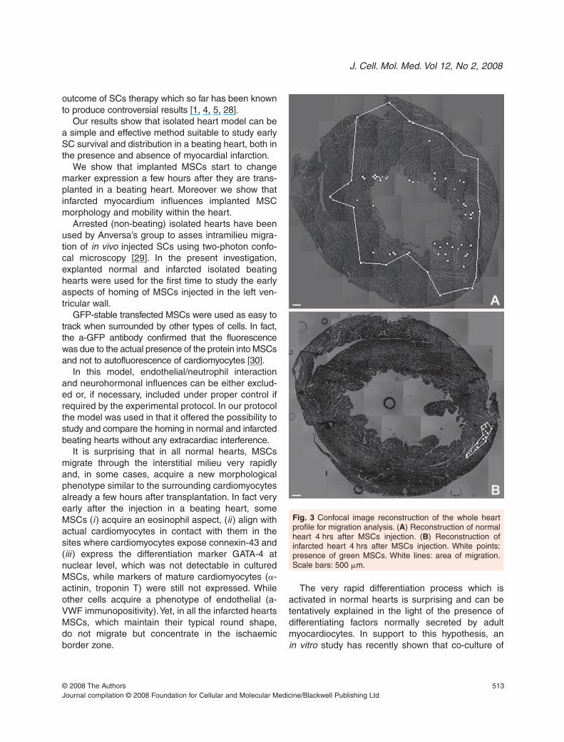
The whole heart was cut in 10 �m thick slices transver-sally to the axis and starting from the apex. All sectionswere analysed by confocal laser microscopy (see later onfor technical details) to detect the presence of GFP-positivetransplanted cells. On the sections where GFP-positivecells were found, either double immunofluorescence orhaematoxylin and eosin staining were carried out asdescribed below. Although the objective of this analysiswas to describe qualitatively the changes occurring totransplanted MSCs, in two hearts (one normal and oneinfarcted, at 4 hrs after injection) quantitative estimation ofthe volume of cell migration was also carried out by theCavalieri method [25]. In one section out of each 50 sec-tions, the area of migration was measured on reconstruct-ed pictures of the whole heart profile (Fig. 3). Then, themean area was finally multiplied by the number of sectionsobtaining thus an estimation of the entire myocardial vol-ume through which GFP-positive cells migrated.
© 2008 The AuthorsJournal compilation © 2008 Foundation for Cellular and Molecular Medicine/Blackwell Publishing Ltd
510
For double immunofluorescence, the sections wererinsed in PBS, blocked with normal goat serum (NGS, 1%)for 1 hr and then incubated overnight with primary antibod-ies against CD90 (Thy-1.1 mouse, monoclonal, 1:50, BDPharmingen), Connexin-43 (rabbit, polyclonal, 1:400,Sigma, St. Louis, MO, USA), GATA-4 (rabbit, polyclonal,1:50, Santa Cruz Biotechnology, Santa Cruz, CA), PCNA(mouse, monoclonal, 1:1000, Sigma, St. Louis, MO, USA),�-actinin (mouse, monoclonal, 1:800, Sigma, St. Louis,MO, USA), troponin T (mouse, monoclonal, 1:50, LabVision Corporation, Westinghouse, CA, USA), VonWillebrand Factor (VWF) (rabbit, policlonal, 1:200, Sigma,St. Louis, MO, USA).
After washing in PBS, sections were incubated withTRITC antimouse IgG (Dako, Milano, Italy) or CY3 anti-rab-bit IgG (Jackson Immuno Research, Cambridgeshire, UK)for 1 hr at room temperature. Sections were finally washedagain in PBS and mounted with a fluorescent mountingmedium (Dako, Milano, Italy).
The slides were observed with a LSM 510 confocal lasermicroscopy system (Zeiss, Jena, Germany), which incor-porates two lasers (Argon and HeNe) and is equipped withan inverted Axiovert 100M microscope. Confocal fluores-cence images were taken using a 20� Plan-NEOFLUARobjective with a numerical aperture (NA) of 0.50 and a 40�
Plan-NEOFLUAR objective with a NA of 0.75. An electron-ic zoom with a magnification ranging from 1� to 8� wasemployed to obtain the magnifications indicated in each fig-ures. To visualize GFP fluorescence, we used excitationfrom 488-nm Argon laser line and emission passingthrough a band-pass (BP) 505–530 filter passing wave-lengths 505 nm to 530 nm to the detector. Images createdwith the BP 505–530 filter were digitally coloured green. Tovisualize TRITC and CY3 red fluorescence we used excita-tion from 543-nm HeNe laser line and emission passingthrough a high-pass filter (LP 560) which passes wave-lengths superior to 560 nm to the detector. Images createdwith the BP 505–530 filter were digitally coloured green.Images created with the LP 560 filter were digitallycoloured red.
After confocal observation and digital image acquisition,the slides with evidence of GFP-positive cells were stainedwith haematoxylin and eosin, observed and photographedwith a DM4000B microscope equipped with a DFC320 dig-ital camera and a IM50 image manager system (LeicaMicrosystems, Wetzlar, Germany). Finally, the fluorescentand light microscope images were superimposed to allowan accurate morphological description of GFP-positive cells.
In addition, to verify that the observed fluorescence wasnot due to the tissue autofluorescence of the heart, someslides were immunostained with an anti-GFP antibody(rabbit, polyclonal, 1:100, Abcam, Cambridge, UK) andthen incubated with an anti-rabbit biotinylated secondaryantibody (Dako, Milan, Italy). Sections were then processedwith peroxidase-conjugated Vectastain ABC kit (Vector,
Burlingame, CA, USA), revealed with diaminobenzidine(Sigma, St Louis, MO, USA), and observed and pho-tographed with the DM4000B light microscope.
Assessment of antibody specificity
For specificity assessment, all antibodies were checked bysecondary antibody labelling with omission of the primaryantibody, a procedure that led to no immunopositivity. Inaddition, we have also carried out a ‘morphology-basedspecificity test’ [26], consisting of the same immunola-belling protocol to stain sections from other tissues or cellsin which antigens are known to be present with a clearlocalization easily detectable from a morphological view-point. Normal heart was used for specificity assessment ofconnexin-43, VWF, �-actinin and troponin T and normalsmall intestine for a-PCNA because of the presence of theeasily detectable pool of fast dividing crypt cells. Culturesof MSCs were used for specificity assessment of a-CD90and cultured myoblasts (H9C2 cell line, ECACC, UK) for a-GATA-4. All these tests supported the specificity of eachreagent by demonstrating that it specifically labels only thetissue elements where the immunogen is known to bepresent (data not shown).
Results
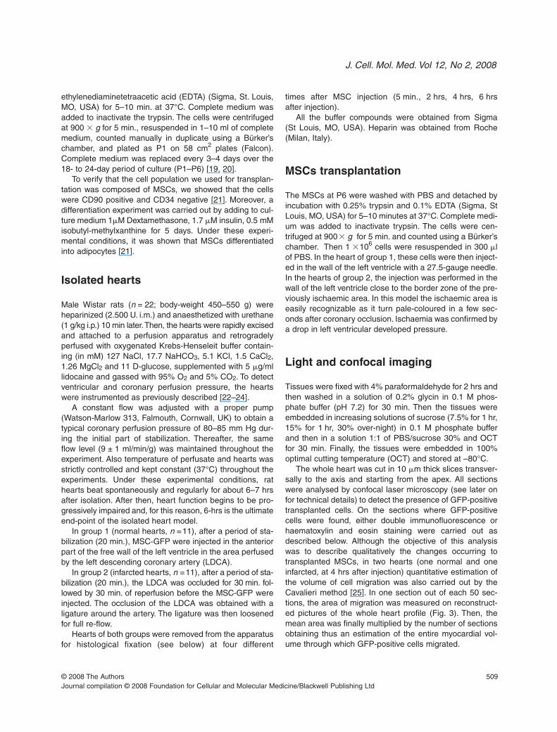
Figure 1 illustrates the appearance of suspendedGFP-positive MSCs before (Fig. 1A and B) and 5 min.after transplantation (Figs 1C–E) in a normal heart.The strong green autofluorescence makes trans-planted MSCs easily recognizable inside the injec-tion part of myocardium where they are concentrated(Fig. 1C). Moreover, in the same slide haematoxylinand eosin staining reveals the clear morphologicaldifference between MSCs (e.g. basophil cytoplasm)and the surrounding cardiomyocytes (e.g. eosinophilcytoplasm) (Fig. 1D). Finally, transplanted MSCswere also easily recognizable after anti-GFPimmunostaining (Fig. 1E).
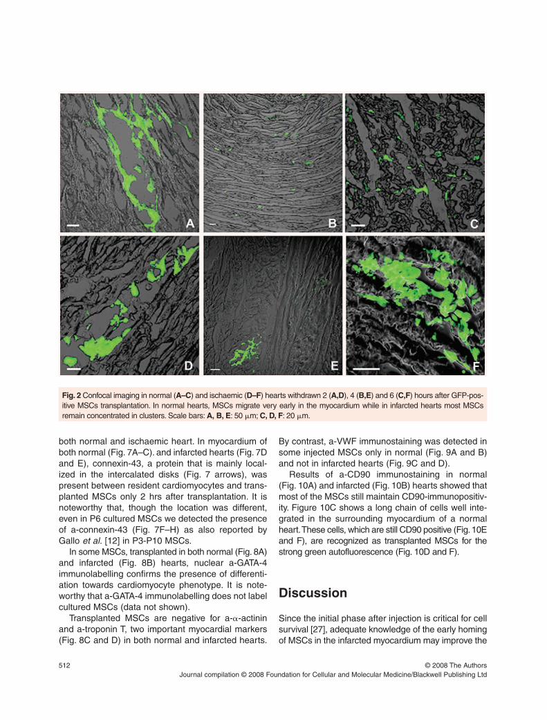
Figures 2, 3 and 4 illustrate the observationsmade on hearts removed 2, 4 and 6 hrs after MSCtransplantation.
The pattern of migration inside the host tissue(Figs 2 and 3) and the acquisition of a new morpholog-ical phenotype (Fig. 4) revealed the different influenceof normal and infarcted hearts on MSCs homing.
In regards to migration, our observations showedthat in normal hearts (Figs 2A–C and Fig. 3A) MSCs
© 2008 The AuthorsJournal compilation © 2008 Foundation for Cellular and Molecular Medicine/Blackwell Publishing Ltd
J. Cell. Mol. Med. Vol 12, No 2, 2008
511
migrated very early (2 hrs) in the myocardium (Fig. 2A)spreading throughout a large myocardial volume(1.22 ± 0.16 �1012
�m3) within 4–6 hrs (Fig. 2B andC, Fig. 3A). In contrast, in infarcted hearts mostMSCs remained concentrated in clusters locatednear the site of injection (Figs 2D–F, Fig. 3B) occupy-ing a myocardial volume 1000 times smaller than innormal hearts (1.01±0.26�109
�m3) (Fig. 3B).The acquisition of a new morphological phenotype
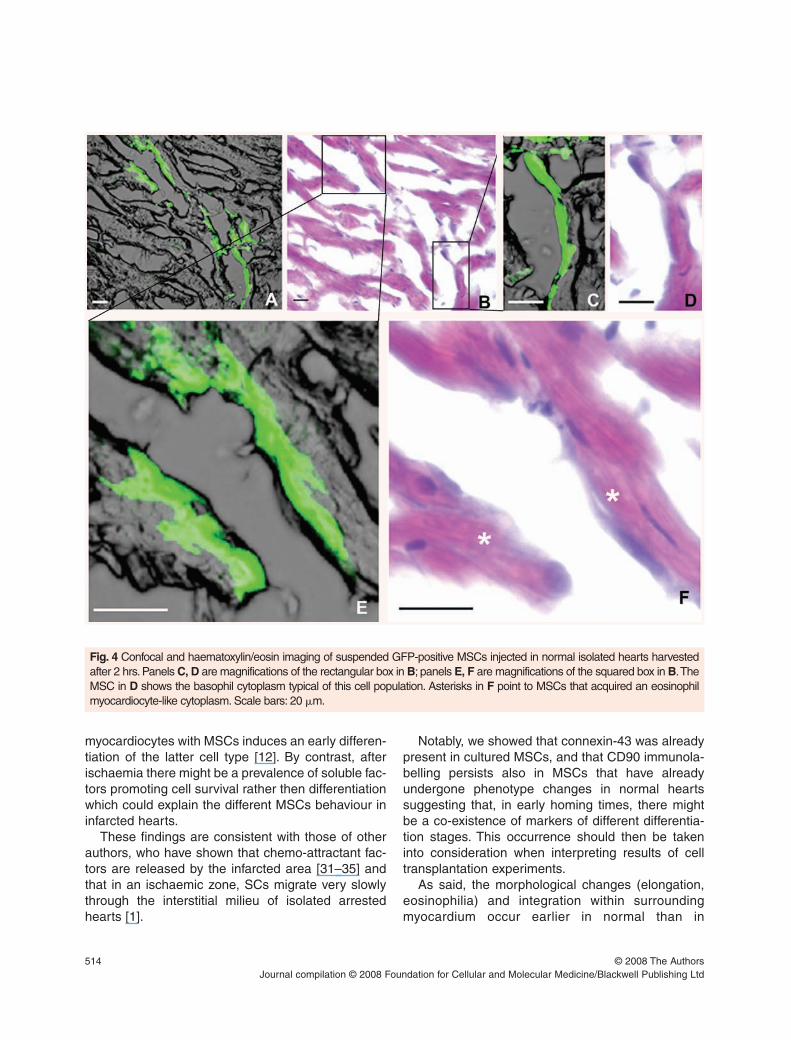
by the transplanted cells in non-ischaemic heart isshown in Figure 4. Starting 2 hrs after transplantation,some MSCs still maintained the original histologicalaspect (Figs 4A–D), while others began to acquirenew morphological features, in particular a plasmaeosinophilia similar to that of the surrounding car-
diomyocytes (Fig. 4E, F asterisks). This was not thecase of infarcted myocardium (data not shown).
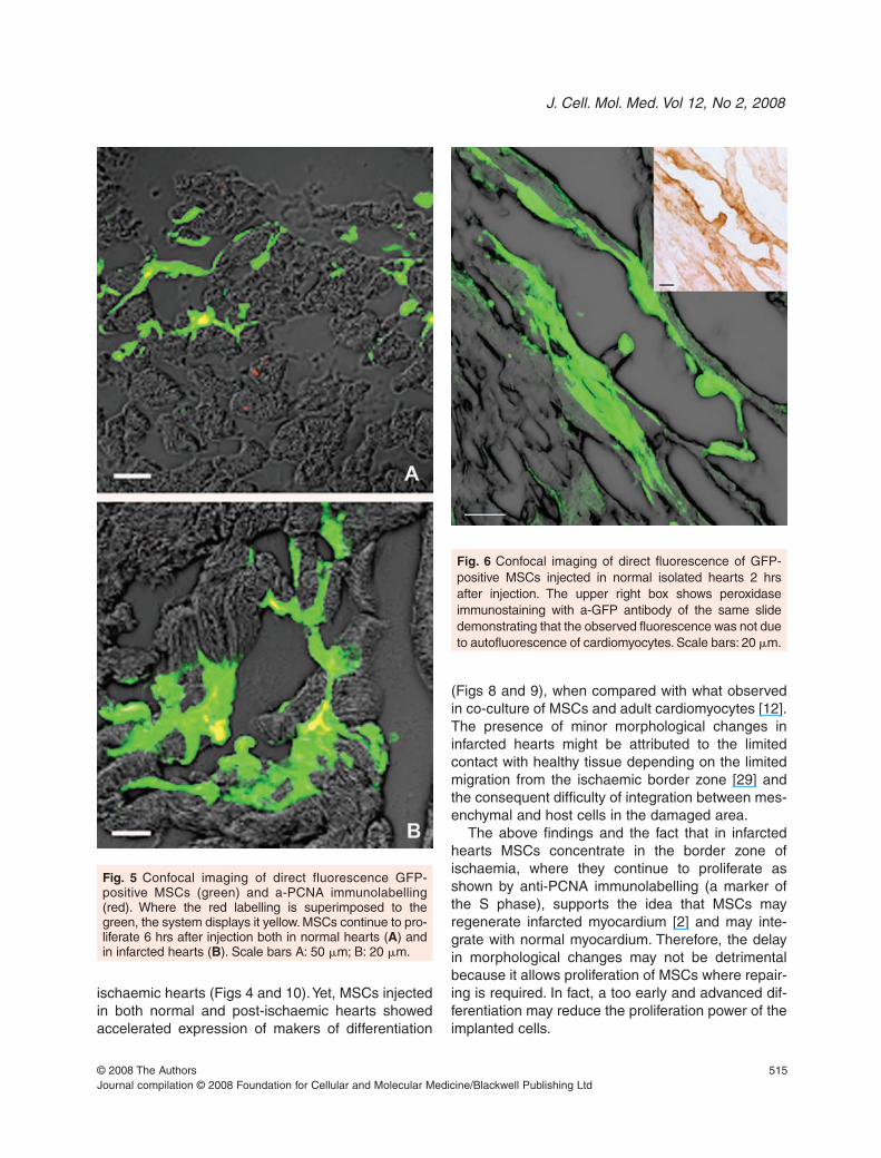
Anti-PCNA immunostaining showed that some ofthe MSCs continued to proliferate after 6 hrs of trans-plantation both in normal and in infarcted hearts (Fig. 5).
Sequential direct GFP confocal imaging followedby immunostaining with anti-GFP antibody (Fig. 6)shows that the observed fluorescence was not due toautofluorescence of cardiomyocytes.
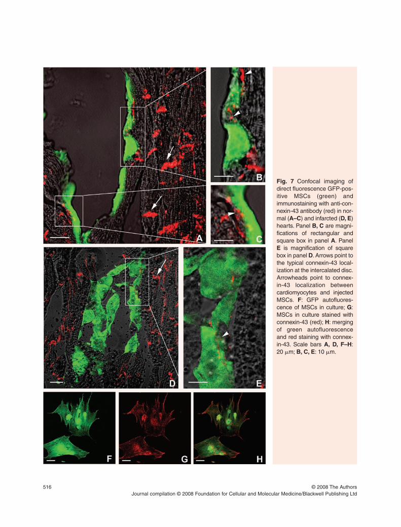
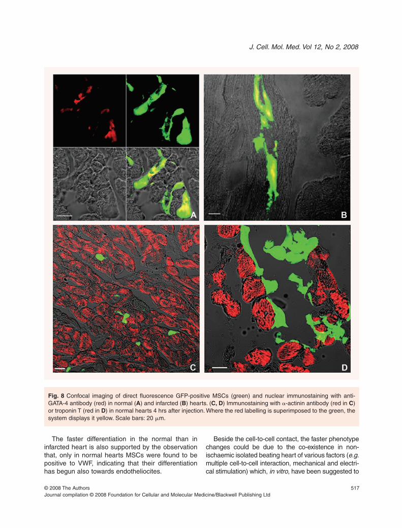
Despite the fact that changes in the morphologicalphenotype demonstrated by histological examinationwere observed in the normal hearts only (Fig. 4),confocal imaging after a-connexin-43 (Fig. 7) and a-GATA4 (Fig. 8A and B) immunolabelling showed sim-ilar signs of differentiation of transplanted MSCs in
© 2008 The AuthorsJournal compilation © 2008 Foundation for Cellular and Molecular Medicine/Blackwell Publishing Ltd
Fig. 1 Images of suspended green fluorescent protein (GFP)-positive mesenchymal stem cells (MSCs) before (A, B)and 5 min. after transplantation (C–E) in a normal beating heart. (A, C) Direct images of fluorescence merged with con-trast; (B, D) Haematoxylin/eosin staining; (E) Peroxidase of a-GFP antibody contrastained with haematoxylin. After haema-toxylin/eosin staining MSCs appear with a basophil cytoplasm while the surrounding cardiomyocytes have an eosinophilcytoplasm. Scale bars: 20 �m.
512
both normal and ischaemic heart. In myocardium ofboth normal (Fig. 7A–C). and infarcted hearts (Fig. 7Dand E), connexin-43, a protein that is mainly local-ized in the intercalated disks (Fig. 7 arrows), waspresent between resident cardiomyocytes and trans-planted MSCs only 2 hrs after transplantation. It isnoteworthy that, though the location was different,even in P6 cultured MSCs we detected the presenceof a-connexin-43 (Fig. 7F–H) as also reported byGallo et al. [12] in P3-P10 MSCs.
In some MSCs, transplanted in both normal (Fig. 8A)and infarcted (Fig. 8B) hearts, nuclear a-GATA-4immunolabelling confirms the presence of differenti-ation towards cardiomyocyte phenotype. It is note-worthy that a-GATA-4 immunolabelling does not labelcultured MSCs (data not shown).
Transplanted MSCs are negative for a-�-actininand a-troponin T, two important myocardial markers(Fig. 8C and D) in both normal and infarcted hearts.
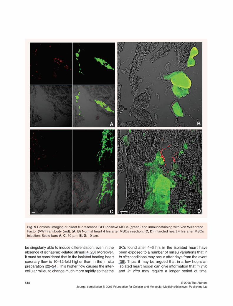
By contrast, a-VWF immunostaining was detected insome injected MSCs only in normal (Fig. 9A and B)and not in infarcted hearts (Fig. 9C and D).
Results of a-CD90 immunostaining in normal (Fig. 10A) and infarcted (Fig. 10B) hearts showed thatmost of the MSCs still maintain CD90-immunopositiv-ity. Figure 10C shows a long chain of cells well inte-grated in the surrounding myocardium of a normalheart.These cells, which are still CD90 positive (Fig.10Eand F), are recognized as transplanted MSCs for thestrong green autofluorescence (Fig. 10D and F).
Discussion
Since the initial phase after injection is critical for cellsurvival [27], adequate knowledge of the early homingof MSCs in the infarcted myocardium may improve the
© 2008 The AuthorsJournal compilation © 2008 Foundation for Cellular and Molecular Medicine/Blackwell Publishing Ltd
Fig. 2 Confocal imaging in normal (A–C) and ischaemic (D–F) hearts withdrawn 2 (A,D), 4 (B,E) and 6 (C,F) hours after GFP-pos-itive MSCs transplantation. In normal hearts, MSCs migrate very early in the myocardium while in infarcted hearts most MSCsremain concentrated in clusters. Scale bars: A, B, E: 50 �m; C, D, F: 20 �m.
J. Cell. Mol. Med. Vol 12, No 2, 2008
513
outcome of SCs therapy which so far has been knownto produce controversial results [1, 4, 5, 28].
Our results show that isolated heart model can bea simple and effective method suitable to study earlySC survival and distribution in a beating heart, both inthe presence and absence of myocardial infarction.
We show that implanted MSCs start to changemarker expression a few hours after they are trans-planted in a beating heart. Moreover we show thatinfarcted myocardium influences implanted MSCmorphology and mobility within the heart.
Arrested (non-beating) isolated hearts have beenused by Anversa’s group to asses intramilieu migra-tion of in vivo injected SCs using two-photon confo-cal microscopy [29]. In the present investigation,explanted normal and infarcted isolated beatinghearts were used for the first time to study the earlyaspects of homing of MSCs injected in the left ven-tricular wall.
GFP-stable transfected MSCs were used as easy totrack when surrounded by other types of cells. In fact,the a-GFP antibody confirmed that the fluorescencewas due to the actual presence of the protein into MSCsand not to autofluorescence of cardiomyocytes [30].
In this model, endothelial/neutrophil interactionand neurohormonal influences can be either exclud-ed or, if necessary, included under proper control ifrequired by the experimental protocol. In our protocolthe model was used in that it offered the possibility tostudy and compare the homing in normal and infarctedbeating hearts without any extracardiac interference.
It is surprising that in all normal hearts, MSCsmigrate through the interstitial milieu very rapidlyand, in some cases, acquire a new morphologicalphenotype similar to the surrounding cardiomyocytesalready a few hours after transplantation. In fact veryearly after the injection in a beating heart, someMSCs (i ) acquire an eosinophil aspect, (ii ) align withactual cardiomyocytes in contact with them in thesites where cardiomyocytes expose connexin-43 and(iii ) express the differentiation marker GATA-4 atnuclear level, which was not detectable in culturedMSCs, while markers of mature cardiomyocytes (�-actinin, troponin T) were still not expressed. Whileother cells acquire a phenotype of endothelial (a-VWF immunopositivity).Yet, in all the infarcted heartsMSCs, which maintain their typical round shape, do not migrate but concentrate in the ischaemic border zone.
The very rapid differentiation process which isactivated in normal hearts is surprising and can betentatively explained in the light of the presence ofdifferentiating factors normally secreted by adultmyocardiocytes. In support to this hypothesis, an in vitro study has recently shown that co-culture of
© 2008 The AuthorsJournal compilation © 2008 Foundation for Cellular and Molecular Medicine/Blackwell Publishing Ltd
Fig. 3 Confocal image reconstruction of the whole heartprofile for migration analysis. (A) Reconstruction of normalheart 4 hrs after MSCs injection. (B) Reconstruction ofinfarcted heart 4 hrs after MSCs injection. White points:presence of green MSCs. White lines: area of migration.Scale bars: 500 �m.
514
myocardiocytes with MSCs induces an early differen-tiation of the latter cell type [12]. By contrast, afterischaemia there might be a prevalence of soluble fac-tors promoting cell survival rather then differentiationwhich could explain the different MSCs behaviour ininfarcted hearts.
These findings are consistent with those of otherauthors, who have shown that chemo-attractant fac-tors are released by the infarcted area [31–35] andthat in an ischaemic zone, SCs migrate very slowlythrough the interstitial milieu of isolated arrestedhearts [1].
Notably, we showed that connexin-43 was alreadypresent in cultured MSCs, and that CD90 immunola-belling persists also in MSCs that have alreadyundergone phenotype changes in normal heartssuggesting that, in early homing times, there mightbe a co-existence of markers of different differentia-tion stages. This occurrence should then be takeninto consideration when interpreting results of celltransplantation experiments.
As said, the morphological changes (elongation,eosinophilia) and integration within surroundingmyocardium occur earlier in normal than in
© 2008 The AuthorsJournal compilation © 2008 Foundation for Cellular and Molecular Medicine/Blackwell Publishing Ltd
Fig. 4 Confocal and haematoxylin/eosin imaging of suspended GFP-positive MSCs injected in normal isolated hearts harvestedafter 2 hrs. Panels C, D are magnifications of the rectangular box in B; panels E, F are magnifications of the squared box in B.TheMSC in D shows the basophil cytoplasm typical of this cell population. Asterisks in F point to MSCs that acquired an eosinophilmyocardiocyte-like cytoplasm. Scale bars: 20 �m.
J. Cell. Mol. Med. Vol 12, No 2, 2008
515
ischaemic hearts (Figs 4 and 10).Yet, MSCs injectedin both normal and post-ischaemic hearts showedaccelerated expression of makers of differentiation
(Figs 8 and 9), when compared with what observedin co-culture of MSCs and adult cardiomyocytes [12].The presence of minor morphological changes ininfarcted hearts might be attributed to the limitedcontact with healthy tissue depending on the limitedmigration from the ischaemic border zone [29] andthe consequent difficulty of integration between mes-enchymal and host cells in the damaged area.
The above findings and the fact that in infarctedhearts MSCs concentrate in the border zone ofischaemia, where they continue to proliferate asshown by anti-PCNA immunolabelling (a marker ofthe S phase), supports the idea that MSCs mayregenerate infarcted myocardium [2] and may inte-grate with normal myocardium. Therefore, the delayin morphological changes may not be detrimentalbecause it allows proliferation of MSCs where repair-ing is required. In fact, a too early and advanced dif-ferentiation may reduce the proliferation power of theimplanted cells.
© 2008 The AuthorsJournal compilation © 2008 Foundation for Cellular and Molecular Medicine/Blackwell Publishing Ltd
Fig. 5 Confocal imaging of direct fluorescence GFP-positive MSCs (green) and a-PCNA immunolabelling(red). Where the red labelling is superimposed to thegreen, the system displays it yellow. MSCs continue to pro-liferate 6 hrs after injection both in normal hearts (A) andin infarcted hearts (B). Scale bars A: 50 �m; B: 20 �m.
Fig. 6 Confocal imaging of direct fluorescence of GFP-positive MSCs injected in normal isolated hearts 2 hrsafter injection. The upper right box shows peroxidaseimmunostaining with a-GFP antibody of the same slidedemonstrating that the observed fluorescence was not dueto autofluorescence of cardiomyocytes. Scale bars: 20 �m.
516 © 2008 The AuthorsJournal compilation © 2008 Foundation for Cellular and Molecular Medicine/Blackwell Publishing Ltd
Fig. 7 Confocal imaging ofdirect fluorescence GFP-pos-itive MSCs (green) andimmunostaining with anti-con-nexin-43 antibody (red) in nor-mal (A–C) and infarcted (D, E)hearts. Panel B, C are magni-fications of rectangular andsquare box in panel A. PanelE is magnification of squarebox in panel D. Arrows point tothe typical connexin-43 local-ization at the intercalated disc.Arrowheads point to connex-in-43 localization betweencardiomyocytes and injectedMSCs. F: GFP autofluores-cence of MSCs in culture; G:MSCs in culture stained withconnexin-43 (red); H: mergingof green autofluorescenceand red staining with connex-in-43. Scale bars A, D, F–H:20 �m; B, C, E: 10 �m.
J. Cell. Mol. Med. Vol 12, No 2, 2008
517
The faster differentiation in the normal than ininfarcted heart is also supported by the observationthat, only in normal hearts MSCs were found to bepositive to VWF, indicating that their differentiationhas begun also towards endotheliocites.
Beside the cell-to-cell contact, the faster phenotypechanges could be due to the co-existence in non-ischaemic isolated beating heart of various factors (e.g.multiple cell-to-cell interaction, mechanical and electri-cal stimulation) which, in vitro, have been suggested to
© 2008 The AuthorsJournal compilation © 2008 Foundation for Cellular and Molecular Medicine/Blackwell Publishing Ltd
Fig. 8 Confocal imaging of direct fluorescence GFP-positive MSCs (green) and nuclear immunostaining with anti-GATA-4 antibody (red) in normal (A) and infarcted (B) hearts. (C, D) Immunostaining with �-actinin antibody (red in C)or troponin T (red in D) in normal hearts 4 hrs after injection. Where the red labelling is superimposed to the green, thesystem displays it yellow. Scale bars: 20 �m.
518
be singularly able to induce differentiation, even in theabsence of ischaemic-related stimuli [4, 28]. Moreover,it must be considered that in the isolated beating heartcoronary flow is 10–12-fold higher than in the in situpreparation [22–24]. This higher flow causes the inter-cellular milieu to change much more rapidly so that the
SCs found after 4–6 hrs in the isolated heart havebeen exposed to a number of milieu variations that inin situ conditions may occur after days from the event[36]. Thus, it may be argued that in a few hours anisolated heart model can give information that in vivoand in vitro may require a longer period of time,
© 2008 The AuthorsJournal compilation © 2008 Foundation for Cellular and Molecular Medicine/Blackwell Publishing Ltd
Fig. 9 Confocal imaging of direct fluorescence GFP-positive MSCs (green) and immunostaining with Von WillebrandFactor (VWF) antibody (red). (A, B) Normal heart 4 hrs after MSCs injection; (C, D) Infarcted heart 4 hrs after MSCsinjection. Scale bars A, C: 50 �m; B, D: 10 �m.

J. Cell. Mol. Med. Vol 12, No 2, 2008
519
because rapid changes in the milieu, in the absence ofextra-cardiac interference, are likely to induce fasterresponse of MSCs. The prompt initiation of differentia-tion in normal hearts clearly indicates that this rapidresponse is possible.
Methodological considerations
Despite the rapidity of events, we recognize that ourisolated beating heart model is limited by the fact thatit is inadequate for studying the fate of transplantedcells over a long period of observation. The reason isthat, after 6–7 hrs of isolation, heart function beginsto be impaired. For the same reason, the model is notadequate for studying survival of transplanted cells.Our rough estimates of the total number of GFP�
cells detectable in isolated hearts up to six hoursafter transplantation suggest that more of 50% of theinjected amount of cells survive in these experimen-tal conditions. However, longer observation times are
needed to follow up survival rates and, to this end, invivo studies are required.
The absence of blood could be another limitation.Yet with buffer perfused hearts, it is possible to pre-vent the presence of potentially confounding externalfactors, such as inflammatory and redox eventswhich follow endothelial/neutrophil interaction. Ifrequired, the model can also be used to study therole of each external factor by administering them tothe isolated heart individually or in association. Thus,for instance, the heart can be perfused with whole orpartial blood elements.
Moreover, important determinants of myocardialresponse to ischaemia (organ temperature, heartrate, ventricular volume, coronary perfusion and col-lateral flow) are kept under the control of theresearcher, whereas in vivo they are extremely vari-able and can influence the results of cell implantation.
Although it was beyond the aims of the presentstudy, ideally the clean conditions of the isolatedheart models might also permit to carry out quantitative
© 2008 The AuthorsJournal compilation © 2008 Foundation for Cellular and Molecular Medicine/Blackwell Publishing Ltd
Fig. 10 Confocal imaging of direct fluorescence GFP-positive MSCs (green) and sarcolemmal immunostaining with anti-CD90 antibody (red) in normal (A) and infarcted (B) hearts. Panels C–F show pictures of serial sections (from a nor-mal heart withdrawn 6 hrs after transplantation) which after confocal imaging of GFP autoflourescence (green) werestained with haematoxylin and eosin (C) and a-CD90 immunolabelling (E, F, red). Where the red labelling is superim-posed to the green, the system displays it yellow (F). Scale bars A: 10 �m; B–F: 20 �m.

520
stereological assessment [37] of MSCs behaviourafter transplantation. However, it has been pointedout that stereological assessment is particularly diffi-cult when dealing with fluorescent labelling [38],because the data obtained with this procedure mayvary significantly depending on various factors thatmay generate unpredictable variability in the intensityof fluorescence at the time of quantitative analysis(e.g. fluorescence bleaching can be related to manydifferent variables and remains difficult to keep undercontrol in fluorescence microscopy). Our experiencehas shown that this limitation is particularly evident inthe in vivo heart in which GFP positive MSCs wereinjected 4 weeks before the post-mortem examina-tion because of the autofluorescence of the myocar-dial tissue (unpublished observations).
Conclusions
The isolated beating heart model provides a simpleand efficient method to study early SC homing in acardiac environment, in which the various intrinsicand extrinsic factors may be strictly controlled. Thismodel can be used to answer emerging questionsabout the early migration, proliferation and differenti-ation of SCs injected into the heart muscle. We haveshown that in early homing times, in the MSCs thereis a co-existence of markers (PCNA, CD90, connex-in-43, GATA-4, VWF) and histological signs of differ-ent degree of integration with the surroundingmyocardium. We have also observed that MSCs dif-ferently integrate, migrate and change shape,whether they are injected in normal or infarctedheart. Yet data and hypothesis presented here arejust few examples of scenarios in which this methodcan be used. Further hypotheses fertile for experi-mental interventions may arise in which the isolatedbeating heart experimental model may be suitable.
Acknowledgements
The authors wish to thank Jennifer Marie Lee for theEnglish language revision. This work was supported byCompagnia di S. Paolo, University of Turin, RegionePiemonte, MIUR (FIRB and PRIN funds), and INRC. Theauthors thank also Dr. Okabe for providing transgenic rats.
References
1. Dawn B, Bolli R. Adult bone marrow-derived cells:regenerative potential, plasticity, and tissue commit-ment. Basic Res Cardiol. 2005; 100: 494–503.
2. Orlic D, Kajstura J, Chimenti S, Jakoniuk I, Anderson
SM, Li B, Pickel J, McKay R, Nadal-Ginard B, Bodine
DM, Leri A, Anversa P. Bone marrow cells regenerateinfarcted myocardium. Nature. 2001; 410: 701–5.
3. Pittenger MF, Martin BJ. Mesenchymal stem cellsand their potential as cardiac therapeutics. Circ Res.2004; 95: 9–20.
4. Smits AM, van Vliet P, Hassink RJ, Goumans MJ,
Doevendans PA. The role of stem cells in cardiacregeneration. J Cell Mol Med. 2005; 9: 25–36.
5. Zimmet JM, Hare JM. Emerging role for bone marrowderived mesenchymal stem cells in myocardial regen-erative therapy. Basic Res Cardiol. 2005; 100: 471–81.
6. Dimmeler S, Zeiher AM, Schneider MD. Unchainmy heart: the scientific foundations of cardiac repair.J Clin Invest 2005; 115: 572–83.
7. Javazon EH, Beggs KJ, Flake AW. Mesenchymalstem cells: paradoxes of passaging. Exp Hematol.2004; 32: 414–25.
8. Bardoff C BR, Popp R, Rupp S, Urbich C, Aicher A
Fleming I, Busse R, Zeiher AM, Dimmiler S.
Transdifferentiation of blood-derived human adultendothelial progenitor cells into functionally activecardiomyocytes. Circulation. 2003; 107: 1024–32.
9. Goldenthal MJ, Marin-Garcia J. Stem cells and car-diac disorders: an appraisal. Cardiovasc Res. 2003;58: 369–77.
10. Stocum DL. Stem cells in CNS and cardiac regener-ation. Adv Biochem Eng Biotechnol. 2005; 93: 135–59.
11. Hristov M, Heussen N, Schober A, Weber C.
Intracoronary infusion of autologous bone marrowcells and left ventricular function after acute myocar-dial infarction: a meta-analysis. J Cell Mol Med. 2006;10: 727–33.
12. Gallo MP, Ramella R, Alloatti G, Penna C, Pagliaro P,
Marcantoni A, Bonafe F, Losano G, Levi R. Limitedplasticity of mesenchymal stem cells cocultured withadult cardiomyocytes.J Cell Biochem. 2007; 100: 86–99.
13. Valiunas V, Doronin S, Valiuniene L, Potapova I,
Zuckerman J, Walcott B, Robinson RB, Rosen
MR, Brink PR, Cohen IS. Human mesenchymalstem cells make cardiac connexins and form func-tional gap junctions. J Physiol. 2004; 555: 617–26.
14. Yoon J, Shim WJ, Ro YM, Lim DS. Transdifferentiationof mesenchymal stem cells into cardiomyocytes bydirect cell-to-cell contact with neonatal cardiomyocytebut not adult cardiomyocytes. Ann Hematol. 2005; 84:715–21.
© 2008 The AuthorsJournal compilation © 2008 Foundation for Cellular and Molecular Medicine/Blackwell Publishing Ltd

J. Cell. Mol. Med. Vol 12, No 2, 2008
521
15. Wang T, Xu Z, Jiang W, Ma A. Cell-to-cell contactinduces mesenchymal stem cell to differentiate intocardiomyocyte and smooth muscle cell. Int J Cardiol.2006; 109: 74–81.
16. Rodgers KE, Xiong S, Steer R, diZerega GS. Effectof angiotensin II on hematopoietic progenitor cell pro-liferation. Stem Cells. 2000; 18: 287–94.
17. Ito T, Suzuki A, Okabe M, Imai E, Hori M. Applicationof bone marrow-derived stem cells in experimentalnephrology. Exp Nephrol. 2001; 9: 444–50.
18. Okabe M, Ikawa M, Kominami K, Nakanishi T,
Nishimune Y. ‘Green mice’ as a source of ubiquitousgreen cells. FEBS Lett. 1997; 407: 313–9.
19. Javazon EH, Colter DC, Schwarz EJ, Prockop DJ.
Rat marrow stromal cells are more sensitive to plat-ing density and expand more rapidly from single-cell-derived colonies than human marrow stromal cells.Stem Cells. 2001; 19: 219–25.
20. Muscari C, Bonafe F, Stanic I, Flamigni F,
Stefanelli C, Farruggia G, Guarnieri C, Caldarera
CM. Polyamine depletion reduces TNFalpha/MG132-induced apoptosis in bone marrow stromal cells.Stem Cells. 2005; 23: 983–91.
21. Raimondo S, Penna C, Pagliaro P, Geuna S.
Morphological characterization of GFP stably trans-fected adult mesenchymal bone marrow stem cells. JAnat. 2006; 208: 3–12.
22. Penna C, Rastaldo R, Mancardi D, Raimondo S,
Cappello S, Gattullo D, Losano G, Pagliaro P. Post-conditioning induced cardioprotection requires signal-ing through a redox-sensitive mechanism, mitochon-drial ATP-sensitive K+ channel and protein kinase Cactivation. Basic Res Cardiol. 2006; 101: 180–9.
23. Penna C, Cappello S, Mancardi D, Raimondo S,
Rastaldo R, Gattullo D, Losano G, Pagliaro P.
Post-conditioning reduces infarct size in the isolatedrat heart: role of coronary flow and pressure and thenitric oxide/cGMP pathway. Basic Res Cardiol. 2006;101: 168–79.
24. Pagliaro P, Mancardi D, Rastaldo R, Penna C,
Gattullo D, Miranda KM, Feelisch M, Wink DA,
Kass DA, Paolocci N. Nitroxyl affords thiol-sensitivemyocardial protective effects akin to early precondi-tioning. Free Radic Biol Med. 2003; 34: 33–43.
25. Howard CV, Reed MG. Unbiased stereology: three-dimensional measurement in microscopy. New York:Springer-Verlag; 1998.
26. Raimondo S, Nicolino S, Tos P, Battiston B,
Giacobini-Robecchi MG, Perroteau I, Geuna S.
Schwann cell behavior after nerve repair by means oftissue-engineered muscle-vein combined guides. JComp Neurol. 2005; 489: 249–59.
27. Rubart M, Field LJ. Cardiac regeneration: repopulat-ing the heart. Annu Rev Physiol. 2006; 68: 29–49.
28. Haider H, Ashraf M. Bone marrow stem cell trans-plantation for cardiac repair. Am J Physiol Heart CircPhysiol. 2005; 288: H2557–67.
29. Dawn B, Stein AB, Urbanek K, Rota M, Whang B,
Rastaldo R, Torella D, Tang XL, Rezazadeh A,
Kajstura J, Leri A, Hunt G, Varma J, Prabhu SD,
Anversa P, Bolli R. Cardiac stem cells deliveredintravascularly traverse the vessel barrier, regenerateinfarcted myocardium, and improve cardiac function. Proc Natl Acad Sci USA. 2005; 102:3766–71.
30. Brazelton TR, Blau HM. Optimizing techniques fortracking transplanted stem cells in vivo. Stem Cells.2005; 23: 1251–65.
31. Abbott JD, Huang Y, Liu D, Hickey R, Krause DS,
Giordano FJ. Stromal cell-derived factor-1alphaplays a critical role in stem cell recruitment to theheart after myocardial infarction but is not sufficient toinduce homing in the absence of injury. Circulation.2004; 110: 3300–5.
32. Frangogiannis NG, Smith CW, Entman ML. Theinflammatory response in myocardial infarction.Cardiovasc Res. 2002; 53: 31–47.
33. Ma J, Ge J, Zhang S, Sun A, Shen J, Chen L, Wang
K, Zou Y. Time course of myocardial stromal cell-derived factor 1 expression and beneficial effects ofintravenously administered bone marrow stem cellsin rats with experimental myocardial infarction. BasicRes Cardiol. 2005; 100: 217–23.
34. Sauer H,Wartenberg M, Hescheler J. Reactive oxy-gen species as intracellular messengers during cellgrowth and differentiation. Cell Physiol Biochem.2001; 11: 173–86.
35. Wojakowski W, Tendera M, Michalowska A, Majka
M, Kucia M, Maslankiewicz K, Wyderka R, Ochala
A, Ratajczak MZ. Mobilization of CD34/CXCR4+,CD34/CD117+, c-met+ stem cells, and mononuclearcells expressing early cardiac, muscle, and endothe-lial markers into peripheral blood in patients withacute myocardial infarction. Circulation. 2004; 110:3213–20.
36. Yang XM, Philipp S, Downey JM, Cohen MV.
Postconditioning’s protection is not dependent on cir-culating blood factors or cells but involves adenosinereceptors and requires PI3-kinase and guanylylcyclase activation. Basic Res Cardiol. 2005; 100:57–63.
37. Geuna S. Appreciating the difference betweendesign-based and model-based sampling strategiesin quantitative morphology of the nervous system.J Comp Neurol. 2000; 427: 333–9.
38. Geuna S. The revolution of counting “tops”: twodecades of the disector principle in morphologicalresearch. Microsc Res Tech. 2005; 66: 270–4.
© 2008 The AuthorsJournal compilation © 2008 Foundation for Cellular and Molecular Medicine/Blackwell Publishing Ltd