The Presence of Foxp3 Expressing T Cells Within Grafts of Tolerant Human Liver Transplant Recipients
Upload
independentCategory
view
3download
0
Iran.J.Immunol. VOL.10 NO.3 September 2013 139
Arteether Exerts Antitumor Activity and
Reduces CD4+CD25+FOXP3+ T-reg Cells
in Vivo
Maryam Azimi Mohamadabadi1, Zuhair Mohammad Hassan1*, Ahmad Zavaran Hosseini1, Mehrdad Gholamzad1, Shekoofe Noori2, Mehdi Mahdavi3, Hamidreza Maroof4 1Department of Immunology, School of Medical Sciences, Tarbiat Modares University,
2Department of
Biochemistry, School of Medical Sciences, Shahid Beheshti University, 3Department of Virology, Pasteur
Institute of Iran, Tehran, 4Department of Microbiology, Zanjan Branch, Islamic Azad University, Zanjan,
Iran
ABSTRACT Background: Chemo-immunotherapy is one of the new achievements for treatment of cancer, by which the success of anti-cancer therapy can be increased. In vitro studies have been shown that Arteether (ARE) induces apoptosis in tumor cells, but not in normal cells. Objective: To investigate the cytotoxic and immunomodulatory properties of Arteether in-vivo and in-vitro. Methods: In this study, we used MTT assay for evaluation of cytotoxicity of Arteether on tumor cell line and Peripheral Blood Mononuclear Cells (PBMCs) from healthy individuals. Balb/c mice were subcutaneously transplanted with tumor tissue taken from Spontaneous Mouse Mammary Tumor (SMMT) bearing female mice. Arteether was administered to breast tumor-bearing Balb/c mice at a dose of 6 mg/kg/day intraperitoneally. Tumor sizes, lymphocyte proliferation, cytokines production, and the percentage of splenic T-reg cells were measured. Results: We observed that ARE could reduce the cell growth of 4T1 cell line in a dose-dependent manner but it had no cytotoxic effect on the growth of peripheral blood lymphocytes. ARE administered intraperitoneally to tumor-bearing Balb/c mice could reduce the tumor growth rate and splenic T-reg cells. No difference in the IFN-γ, IL-10 and IL-4 production was observed between tumor antigen-stimulated splenocytes of mice treated with ARE and control mice. Conclusion: These results underscore antitumor properties of Arteether that may aid in development of more effective antitumor agents. Azimi Mohamadabadi M, et al. Iran J Immunol. 2013; 10(3):139-49
Keywords: Arteether, Breast Cancer, Immunotherapy -------------------------------------------------------------------------------------------------------------------------------------------------- *Corresponding author: Dr. Zuhair Mohammad Hassan, Department of Immunology, School of Medical Sciences, Tarbiat Modares University, Tehran, Iran, Tel: (+) 98 21 82883565, Fax: (+) 98 21 82884555, e-mail: [email protected]
Anti-tumor properties of Arteether
Iran.J.Immunol. VOL.10 NO.3 September 2013 140
INTRODUCTION Among all types of cancer, breast cancer is one of the most important causes of women’s death (1); the increasing speed of cancer research has failed to keep pace with breast cancer growth, leaving many people suffering from it. Hence, cancer control is a major health care priority. There are several cancer therapy methods like surgery, chemotherapy, and radiotherapy, to name a few, but each of them has a specific shortcoming; for example, metastasis impedes surgical success. In addition, chemotherapy not only has a non-specific mode of killing dividing cells, but also is toxic to normal cells (2). It is therefore important to develop new methods for cancer therapy that targets tumor cells in a specific manner. Artemisia annua in Chinese traditional medicine is used for extracting Artemisinin (ART), which its anti malarial activity was published in early 1980s. Due to its molecular structure, Artemisinin has low solubility in water and oil, and much research has focused in modifying its structure in order to enhance its solubility. Many structural derivatives of Artemisinin have been synthesized since 1980. Most acknowledged derivatives including Arteether are very active against acute malaria and possess very low side effects. Arteether (ARE) is an ethyl ether derivative of dihydroartemisinin, a metabolite of Artemisinin, which is characterized by a rapid oral absorption due to its high hydrophobic properties (3-6). Alcohol soluble derivatives of Artemisinin also show effects against malignant cells. Although their mode of action in these cells is not clear; recent studies have been focusing on the antitumor activity of Artemisinin's derivatives, especially Arteether; which show an extreme anticancer activity in vitro and in vivo. ART and ARE are sesquiterpene lactones with a highly active endoperoxide bridge. They can produce great amounts of reactive oxygen species in the presence of iron (7-9), which is distinct from other anti-malarial drugs, a property most interesting to cancer therapists. To facilitate the rapid growth, tumor cells need to increase the influx of iron; these cells have higher amounts of transferrin receptors and intracellular iron, hence increasing sensitivity of tumor cells to Arteether's killing mechanism as opposed to normal cells (9). In general, tumor cells utilize the regulatory mechanism of T-reg cells to evade the immune system. That is why many studies have focused on methods that can reduce the number of T-reg cells in the tumor (10). Artemisins and its derivatives also posses immunomodulatory effects in a dose- and time-dependent manner; although their stimulatory effect on T-lymphocyte-mediated immune responses has also been reported in a study by Yang et al. (11-15). In this study, we investigated the cytotoxic and immunomodulatory properties of Arteether in vivo. MATERIALS AND METHODS Mice. A group of inbred female BALB/c mice, at 4-6 weeks of age, were purchased from Pasteur Institute of Iran. Animal care and treatment were conducted in compliance with the guideline of Animal Care and Research Committee of Tarbiat Modares University, which is in conformity with the Guide for the Care and Use of Laboratory Animals (DHEW Publication No. (NIH) 85-23, Revised 1985, Office of Science and Health Reports, DRR/NIH, Bethesda, MD 20205) THE.
Azimi Mohamadabadi M, et al
Iran.J.Immunol. VOL.10 NO.3 September 2013 141
Arteether. Arteether powder was purchased from Exim-Pharm International Co., India. It was freshly prepared for each administration by dissolving the powder in absolute ethanol (Merck Co.), and further dilution in PBS (40:60). The solution was checked for endotoxin and mycoplasma contamination in our lab and Pasteur Institute of Iran, respectively. Solubilized Arteether was then filter sterilized to avoid any contamination. Cell culture. Mice breast cancer cell line (4T1) was purchased from Pasteur Institute of Iran. The cells were cultured in RPMI 1640 medium (Gibco), supplemented with 10% FCS, 100 µg/ml streptomycin, and 100 IU/ml penicillin. All cells were grown in a humidified atmosphere containing 5% CO2 at 37C. Normal lymphocytes were collected from healthy individuals in the Medical School clinic. Cell Viability Analysis. Cell viability was evaluated by MTT viability assay. Briefly, growing cells were re-cultured (10000 cells/well) in 96-well tissue culture microplates (Nunc, Denmark). Three doses of ARE was added to the respective wells. After 48 and 72 hours, 20 µl MTT (Sigma-Aldrich) was added to each well with a final concentration of 5 mg/ml. After 4 h incubation, the medium containing MTT was discarded and 100 μl DMSO (Sigma-Aldrich) was added. MTT crystals were completely solubilized with vibration for 10 min. Absorbance was measured at 450 nm using ELISA reader and viability was expressed as stimulation index (SI), using the following formula: SI = OD of ARE+ cells/OD of ARE- cells In Vivo Breast Cancer Model. Spontaneous mouse mammary tumor (SMMT) is an invasive ductal carcinoma, which develops spontaneously in female BALB/c mouse after transplantation with tumor cells. SMMT is regarded to be similar to the invasive ductal carcinoma observed in human samples (16). In our study, mice were subcutaneously transplanted with 0.5 mm3 of tumor tissue taken from SMMT-bearing BALB/c mice. Tumors were allowed to reach an approximate size of 500 mm3. Ten tumor-bearing mice were randomly divided into two groups, each consisting of five mice. The first group was treated with 0.1 ml of Arteether at a dose of 6 mg/kg/day via the intraperitoneal (IP) route. The controls received ARE diluents in the same volume and via the same route. The treatments were applied for thirteen consecutive days. The tumor volumes were measured every other day using digital vernier callipers (Mitutoyo, Japan). The volumes were calculated with the following formula (17):
V=1/2 (LW2) Where V: volume, L: length and W: width.
Antigen Preparation. A volume of approximately 3000 mm3 tumor tissue was extracted from the breast cancer-bearing BALB/c mouse. The tumor suspension was prepared using five rounds of freezing/thawing and was passed through a 150 µm stainless steel mesh. The suspension was sonicated in a power of 4W for 30 seconds, followed by a 20-second pause for five times. To inactivate serine proteases, 1 mM of phenyl methyl sulfonyl fluoride (PMSF) was added to the cell lysate. The extract was filtered through a 0.22 µm filter and its concentration was determined using the Bradford method. Finally, it was stored at -20°C until use. Separation of Splenic Mononuclear cells (MNC). The mice were sacrificed on day 13; spleens were resected under sterile conditions and were suspended in PBS. The splenic cell suspension was RBC-lysed with a solution of 0.75% NH4Cl and Tris buffer (0.02%) (pH=7.4). The cells were washed and the single-cell suspension was prepared
Anti-tumor properties of Arteether
Iran.J.Immunol. VOL.10 NO.3 September 2013 142
in RPMI 1640 containing stable glutamine (Cytogen) and 10% heat-inactivated fetal calf serum (Gibco, UK). To define the viability and density of cells in the suspension, Trypan blue dye exclusion method was used. The cells were counted using a hemocytometer by light microscopy method. The viability of splenocytes was generally above 95%. After additional washing, the suspension was adjusted to 4×10⁶ cells per milliliters in RPMI 1640 supplemented with 10% FCS, 100 µg/ml streptomycin, and 100 IU/ml penicillin (complete RPMI), and kept at 4°C. Lymphocyte Proliferation Assay. Splenic MNCs were cultured in RPMI 1640 (Gibco, UK) (adjusted to 2×10⁶ cells/ml in RPMI supplement). Briefly, 100 μl of cell suspensions were transferred to 96-well flat-bottom microplates (Nunc, Denmark). 6 wells were considered for each sample. 20 μl of tumor antigen was added to three wells of each sample. Plates were incubated at 37°C (separate wells were cultured with untreated normal splenocytes and incubated with PHA as positive control). Proliferation assay was done using Roche Cell Proliferation ELISA, BrdU kit according to the manufacture's protocols. The absorbance of the samples was measured at 450 nm using ELISA reader. Results are presented as stimulation index (SI), using the following formula: SI = OD of tumor Ag+ splenocytes/OD of tumor Ag- splenocytes
Splenocyte Cytokine Production. The isolated spleen MNCs were cultured in 24-well plates (Nunc, Denmark) in a final concentration of 2×10⁶ cells/ml. To stimulate the cell, 20 μl of purified tumor antigen was added to each well, the supernatants were collected after 72 h incubation at 37°C and 5% CO2 (11), and were kept frozen at -70°C until used. IFN-γ, IL-4 and IL-10 concentrations were measured using R&D DuoSet ELISA Development kit according to the manufacturers’ protocol. Each sample was analyzed in triplicate. Three-Color Immunostaining and Flow Cytometry Analysis. The MNCs, purified from mice spleens, were immunostained with FITC anti-mouse CD4, PE anti-mouse CD25, and subsequently PE-Cy5 anti-mouse Foxp3, according to the instructions given in the eBioscience mouse regulatory T cell staining kit. The samples were analyzed by FACS Calibur flow cytometer (BD, USA) and the results were analyzed using WinMDI software. Statistical Analysis. In this study, each experiment was performed in duplicate or triplicate, using either one-way analysis of variance (ANOVA) or the Mann-Whitney non-parametric test, to determine the statistical significance (p<0.05) between the test and control groups. The data were analyzed using SPSS software version 16. The results are expressed as the mean ± standard error (mean ± SE). RESULTS Effect of Arteether on Cell Growth. To evaluated the effect of ARE on the growth of tumor cells and peripheral blood lymphocytes, three doses of ARE (10-20 μM) was applied in vitro. The result showed that ARE could reduced the cell growth of 4T1 cell line in a dose-dependent manner but it has no cytotoxic effects on the growth of peripheral blood lymphocytes (Figure 1).

Azimi Mohamadabadi M, et al
Iran.J.Immunol. VOL.10 NO.3 September 2013 143
Figure 1. Cytotoxity effect of arteether against 4T1 cell line. Cells were incubated with increasing concentration (10-20 μM) for 48 and 72h. Graph shows the significant diffrence between ARE-treated 4T1 cell line and ARE- treated normal cells (p<0.05).
Effect of Arteether on Lymphocyte Proliferation Assay. In order to assess the effect of ARE on specific Lymphocyte proliferation, ten mice were used and the protocol in figure 2 was applied. Our results indicated a slightly increase in SI at the dose of 6 mg/kg but no significant difference was observed (p>0.05). Figure 2. The results of lymphocyte proliferation assay: ARE-treated group showed no significant differences in comparison with control (p>0.05).positive group (PHA) showed a significant difference (p<0.05). Tip=arteether-treated group (Test, IntraPeriteonally), Cip=control group(Control IntraPeritoneally).
Anti-tumor properties of Arteether
Iran.J.Immunol. VOL.10 NO.3 September 2013 144
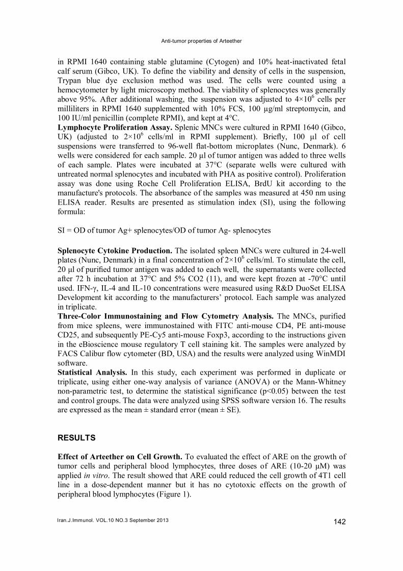
The results of proliferation assay were evaluated in triplicate after stimulation with a specific tumor antigen. Effect of Arteether on Lymphpcyte Cytokine Production. The concentration of IFN-γ and IL-4, typical cytokines produced by Th1 and Th2 cells, in both treated and untreated tumor-bearing mice were evaluated by ELISA technique. The result have shown no significant difference (p>0.05) between the two groups. Figure 3. The results of cytokine ELISA assay: the levels of IFN-γ and IL-4 cytokines produced from stimulated splenocytes by specific tumor antigen are shown. Result showed no statistically significant difference between the ARE-treated group and untreated group (p>0.05).
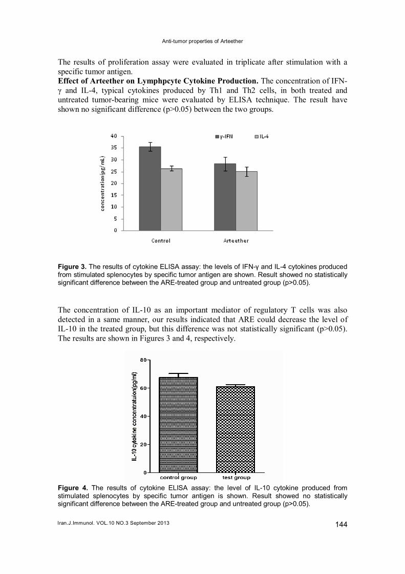
The concentration of IL-10 as an important mediator of regulatory T cells was also detected in a same manner, our results indicated that ARE could decrease the level of IL-10 in the treated group, but this difference was not statistically significant (p>0.05). The results are shown in Figures 3 and 4, respectively. Figure 4. The results of cytokine ELISA assay: the level of IL-10 cytokine produced from stimulated splenocytes by specific tumor antigen is shown. Result showed no statistically significant difference between the ARE-treated group and untreated group (p>0.05).
Azimi Mohamadabadi M, et al
Iran.J.Immunol. VOL.10 NO.3 September 2013 145
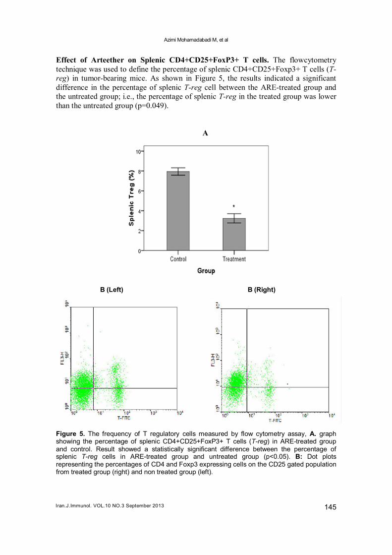
Effect of Arteether on Splenic CD4+CD25+FoxP3+ T cells. The flowcytometry technique was used to define the percentage of splenic CD4+CD25+Foxp3+ T cells (T-reg) in tumor-bearing mice. As shown in Figure 5, the results indicated a significant difference in the percentage of splenic T-reg cell between the ARE-treated group and the untreated group; i.e., the percentage of splenic T-reg in the treated group was lower than the untreated group (p=0.049). A B (Left) B (Right)
Figure 5. The frequency of T regulatory cells measured by flow cytometry assay, A. graph showing the percentage of splenic CD4+CD25+FoxP3+ T cells (T-reg) in ARE-treated group and control. Result showed a statistically significant difference between the percentage of splenic T-reg cells in ARE-treated group and untreated group (p<0.05). B: Dot plots representing the percentages of CD4 and Foxp3 expressing cells on the CD25 gated population from treated group (right) and non treated group (left).
Anti-tumor properties of Arteether
Iran.J.Immunol. VOL.10 NO.3 September 2013 146
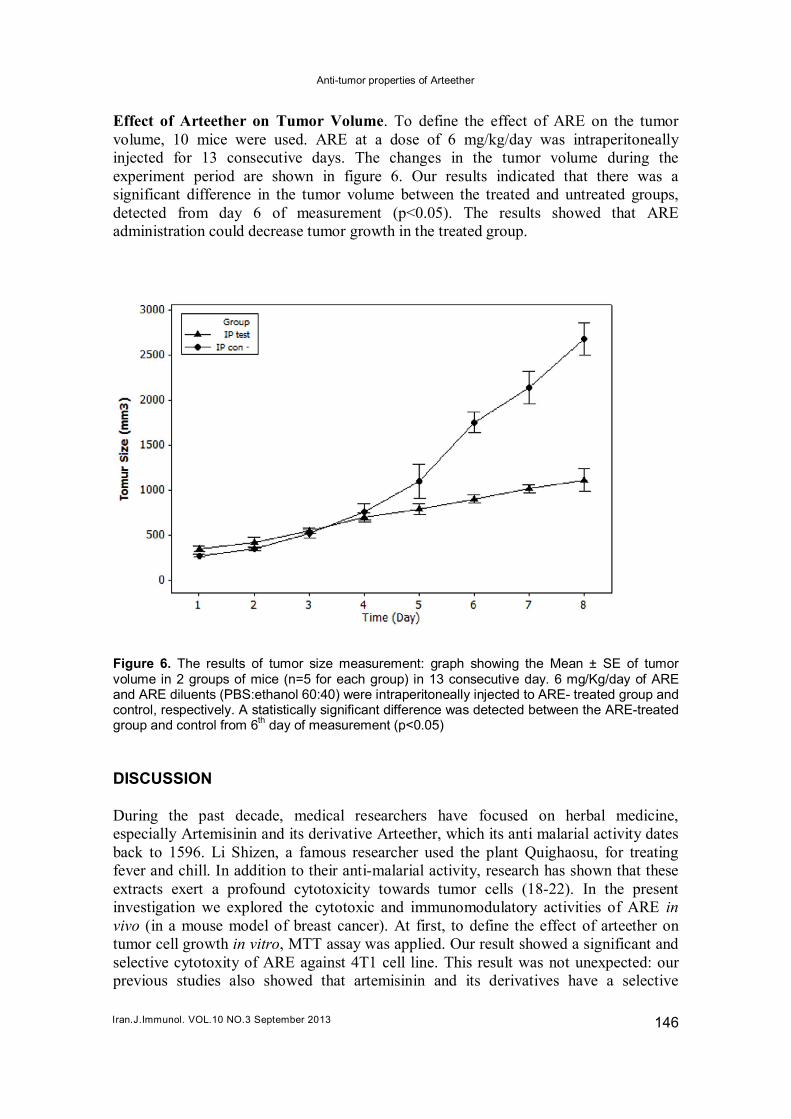
Effect of Arteether on Tumor Volume. To define the effect of ARE on the tumor volume, 10 mice were used. ARE at a dose of 6 mg/kg/day was intraperitoneally injected for 13 consecutive days. The changes in the tumor volume during the experiment period are shown in figure 6. Our results indicated that there was a significant difference in the tumor volume between the treated and untreated groups, detected from day 6 of measurement (p<0.05). The results showed that ARE administration could decrease tumor growth in the treated group.
Figure 6. The results of tumor size measurement: graph showing the Mean ± SE of tumor volume in 2 groups of mice (n=5 for each group) in 13 consecutive day. 6 mg/Kg/day of ARE and ARE diluents (PBS:ethanol 60:40) were intraperitoneally injected to ARE- treated group and control, respectively. A statistically significant difference was detected between the ARE-treated group and control from 6
th day of measurement (p<0.05)
DISCUSSION During the past decade, medical researchers have focused on herbal medicine, especially Artemisinin and its derivative Arteether, which its anti malarial activity dates back to 1596. Li Shizen, a famous researcher used the plant Quighaosu, for treating fever and chill. In addition to their anti-malarial activity, research has shown that these extracts exert a profound cytotoxicity towards tumor cells (18-22). In the present investigation we explored the cytotoxic and immunomodulatory activities of ARE in vivo (in a mouse model of breast cancer). At first, to define the effect of arteether on tumor cell growth in vitro, MTT assay was applied. Our result showed a significant and selective cytotoxity of ARE against 4T1 cell line. This result was not unexpected: our previous studies also showed that artemisinin and its derivatives have a selective
Azimi Mohamadabadi M, et al
Iran.J.Immunol. VOL.10 NO.3 September 2013 147
cytotoxity toward tumor cells owing to their novel killing mechanism. Based on our previous findings and other published studies (9,23-25), the dose of 6 mg/Kg was identified as the optimal immunostimulatory dose, and was used for further tests. The recent studies show that ART and its derivatives have a different dose-dependent pattern on the production of hemagglutination antibody (14,15), which was confirmed by our previous results. The same result has also been established for other antitumor agents (26). Lymphocyte proliferation assay showed no statistically significant difference in the ARE-treated group in comparison with the untreated group; a similar result has previously been reported in a study of Artemisinin and its derivatives, suggesting that they had no effect on lymphocyte proliferation (11). As shown in Figure 5and Figure 6, the levels of IFN-γ, IL-4 and IL-10 cytokines were not statistically significant, while we indicated a significant increase in the level of IFN-γ after the treatment with Artemisinin. It may be that the dose-dependent manner of ARE and other derivatives or increasing in hydrophobicity can reduce the intracellular communication of ARE. In another study, we noticed that an Artemisinin derivative, Artemetheter, had no effect on the pattern of cytokine production (27), while other investigations indicated that these derivatives have different immunomodulatory or immunosuppressive effects on the immune system based in their structure and dose (13,14). Nevertheless, further investigation is needed in order to determine an effective and immunostimulator dose of ARE. We indicated that ARE can decrease the tumor growth rate in tumor transplanted mice. This result is not unexpected; our previous studies also showed that the dose-dependent use of ART derivatives is more effective in reducing the tumor mass than ART (11,27). We also showed that ARE could decrease the total number of splenic T-reg cells in the treated group in comparison with the untreated group. Although the mechanism by which ARE reduces T-reg cells has yet to be understood, the decrease in T-reg cells may be because of the tumor mass reduction. However, other studies reported that ART derivatives can reduce the number of T-reg cells through inhibiting the production of NO, a inducer metabolite for T-reg expansion, from macrophage cells (28,29). In conclusion, our results showed that ARE is more effective in slowing down the tumor growth than to be an immunomodulator. Decrease in the level of IFN-γ, IL-4, IL-10 and T-reg cell expansion were also noted; and these abilities might be ascribed to the hydrophobic properties of ARE. Our findings in this study are consistent with our previous findings about Artemether, a methyl ether derivative of ART (27). ACKNOWLEDGEMENTS In present study financial support was provided by Tarbiat Modares University and we also thank Dr. Ladan Langroudi for technical assistance. REFERENCES
1 Porter P. Global trends in breast cancer incidence and mortality. salud pública de Méx. 2009; 51:141-6.
2 Sertel S, Eichhorn T, Sieber S, Sauer A, Weiss J, Plinkert PK, et al. Factors determining sensitivity or resistance of tumor cell lines towards artesunate. Chem Biol Interact. 2010; 185:42-52.
Anti-tumor properties of Arteether
Iran.J.Immunol. VOL.10 NO.3 September 2013 148
3 Klayman DL. Qinghaosu (artemisinin) :an antimalarial drug from China. Science. 1985; 228:1049-55.
4 Dhingra V, Vishweshwar Rao K, Lakshmi Narasu M. Current status of artemisinin and its derivatives as antimalarial drugs. Life Sci. 2000; 66:279-300.
5 Robert A, Benoit-Vical F, Meunier B. The key role of heme to trigger the antimalarial activity of trioxanes. Coord Chem Rev. 2005; 249:1927-36.
6 Brossi A, Venugopalan B, Dominguez Gerpe L, Yeh HJ, Flippen-Anderson JL, Buchs P, et al. Arteether, a new antimalarial drug: synthesis and antimalarial properties. J Med Chem. 1988; 31:645-50.
7 Krishna S, Uhlemann, AC,Haynes RK. Artemisinins: mechanisms of action and potential for resistance. Drug Resist Updat. 2004; 7:233-44.
8 Efferth T, Benakis A, Romero M, Tomicic M, Rauh R, Steinbach D, et al. Enhancement of cytotoxicity of artemisinins toward cancer cells by ferrous iron. Free Radic Biol Med. 2004; 37:998-1009.
9 Singh NP, Lai H. Selective toxicity of dihydroartemisinin and holotransferrin toward human breast cancer cells. Life Sci. 2001;70:49-56.
10 Curiel TJ, Coukos G, Zou L, Alvarez X, Cheng P, Mottram P, et al. Specific recruitment of regulatory T cells in ovarian carcinoma fosters immune privilege and predicts reduced survival. Nat Med. 2004; 10:942-9.
11 Langroudi L, Hassan ZM, Ebtekar M, Mahdavi M, Pakravan N, Noori S. A comparison of low-dose cyclophosphamide treatment with artemisinin treatment in reducing the number of regulatory T cells in murine breast cancer model. Int Immunopharmacol. 2010; 10:1055-61.
12 Yang SX, Xie SS, Gao HL, Long ZZ. Artemisinin and Its Derivatives Enhance T Lymphocyte-Mediated Immune Responses in Normal Mice and Accelerate Immunoreconstitution of Mice with Syngeneic Bone Marrow Transplantation. Clinical immunology and immunopathology. 1993;69:143-8.
13 Shakir L, Hussain M, Javeed A, Ashraf M, Riaz A. Artemisinins and immune system. Eur J Pharmacol. 2011; 668:6-14.
14 Zhang YX, Sun HX. Immunosuppressive effect of ethanol extract of Artemisia annua on specific antibody and cellular responses of mice against ovalbumin. Immunopharmacol Immunotoxicol. 2009; 31:625-30.
15 Tawfik AF, Bishop SJ, Ayalp A, el-Feraly FS. Effects of artemisinin, dihydroartemisinin and arteether on immune responses of normal mice. Int J Immunopharmacol. 1990; 12:385-9.
16 Hassan ZM, Yaraee R, Zare N, GhazanfariT, SarrafNejad AH, Nazori B. Immunomodulatory effect of R10 fraction of garlic extract on natural killer activity. Int Immunopharmacol. 2003; 3:1483-9.
17 Noori S, Taghikhani M, Hassan ZM,Allameha A, Mostafaei A. Tehranolide molecule modulates the immune response, reduce regulatory T cell and inhibits tumor growth in vivo. Mol Immunol. 2010; 47:1579-84.
18 Efferth T, Dunstan H, Sauerbrey A, Miyachi H, Chitambar CR. The anti-malarial artesunate is also active against cancer. Int J Oncol. 2001; 18:767-73.
19 Lu YY, Chen TS,Qu, JL, Pan WL, Sun L,Wei XB. Dihydroartemisinin (DHA) induces caspase-3-dependent apoptosis in human lung adenocarcinoma ASTC-a-1 cells. J Biomed Sci. 2009;16:16.
20 Chen HH, Zhou HJ, Wang WQ, Wu GD. Antimalarial dihydroartemisinin also inhibits angiogenesis. Cancer Chemother Pharmacol. 2004; 53:423-32.
21 Chen H, Sun B, Pan SH, Jiang HC, Sun XY. Dihydroartemisinin inhibits growth of pancreatic cancer cells in vitro and in vivo. Anti-Cancer Drugs. 2009; 20:131-40.
22 Chen HH, Zhou HJ, Fang X. Inhibition of human cancer cell line growth and humanu mbilical vein endothelial cell angiogenesis by artemisinin derivatives in vitro. Pharmacol Res. 2003; 48:231-6.
23 Li QG, Mog SR, Si YZ, Gettayacamin M, Mihous WK. Neurotoxicity and efficacy of arteether related to its exposure times and exposure levels in rodents. Am J Trop Med Hyg. 2002; 66:516-25.
24 Genovese RF, Newman DB, Li Q, Peggins JO, Brewer TG. Dose-dependent brainstem neuropathology following repeated arteether administration in rats. Brain Res Bull. 1998; 45:199-202.
Azimi Mohamadabadi M, et al
Iran.J.Immunol. VOL.10 NO.3 September 2013 149
25 Genovese RF, Newman DB, Brewer TG. Behavioral and neural toxicity of the artemisinin antimalarial, arteether, but not artesunate and artelinate, in rats. Pharmacol Biochem Behav. 2000; 67:37-44.
26 Askenase PW, Hayden BJ, Gershon RK. Augmentation of delayed-type hypersensitivity by doses of cyclophosphamide which do not affect antibody responses. J Exp Med. 1975; 141:697-702.
27 Farsam V, Hassan ZM, Zavaran-Hosseini A, Noori S, Mahdavi M, Ranjbar M. Antitumor and immunomodulatory properties of artemether and its ability to reduce CD4(+) CD25(+) FoxP3(+) T reg cells in vivo. Int Immunopharmacol. 2011; 11:1802-8.
28 Konkimalla VB, Blunder M, Korn B, Soomro SA, Jansen H, Chang W, et al. Effect of artemisinins and other endoperoxides on nitric oxide-related signaling pathway in RAW264.7 mouse macrophage cells. Nitric Oxide. 2008;19:184-91.
29 Niedbala W, Cai B, Liu H, Pitman N, Chang L, Liew FY. Nitric oxide induces CD4 CD25 Foxp3 regulatory T cells from CD4 CD25 T cells via p53, IL-2, and OX40. Proc Natl Acad Sci U S A. 2007; 104:15478-83.
Copyright © 2022 FDOKUMEN