TGFβ1 deficiency does not affect the generation and maintenance of CD4 +CD25 +FOXP3 + putative T...
14
TGFβ1 deficiency does not affect the generation and maintenance of CD4 + CD25 + FOXP3 + putative T reg cells, but causes their numerical inadequacy and loss of regulatory function Ramireddy Bommireddy a,b , George F. Babcock c,d , Ram R. Singh e,f,g , and Thomas Doetschman a,h,i,* a BIO5 Institute, University of Arizona, PO Box 245217, Tucson, AZ 85724-5217, USA b Department of Immunobiology, University of Arizona, PO Box 245217, Tucson, AZ 85724-5217, USA c Department of Surgery, University of Cincinnati College of Medicine, Cincinnati, OH 45267, USA d Shriners Hospital for Children, Cincinnati, OH 45229, USA e Department of Medicine/Rheumatology, David Geffen School of Medicine, University of California at Los Angeles (UCLA), Los Angeles, CA 90095, USA f Department of Pathology and Laboratory Medicine, David Geffen School of Medicine, University of California at Los Angeles (UCLA), Los Angeles, CA 90095, USA g Jonsson Comprehensive Cancer Center, David Geffen School of Medicine, University of California at Los Angeles (UCLA), Los Angeles, CA 90095, USA h Department of Cell Biology and Anatomy, University of Arizona, PO Box 245217, Tucson, AZ 85724-5217, USA i Cancer Center, University of Arizona, PO Box 245217, Tucson, AZ 85724-5217, USA Abstract TGFβ1 is considered to be required for peripheral maintenance of CD4 + CD25 + FOXP3 + T reg cells. However, we demonstrate no reduction in the percentage of such Tcells in the spleens and thymi of Tgfb1 −/− mice. Although putative T reg cells, characterized as CD4 + CD25 + FOXP3 + CD62L + Tcells, are increased in Tgfb1 −/− mice, they may be inadequate to control activated Tcells since the ratio of activated T cells:putative T reg cells is several-fold higher in Tgfb1 −/− mice than in control mice. We further show that whereas Tgfb1 −/− mice that express a chicken OVA-specific TCR transgene (DO11.10) have an increase in putative T reg cells, there are no detectable CD4 + CD25 + T cells in the spleens of DO11.10 Rag1 −/− mice suggesting that T reg -cell generation is self-antigen dependent regardless of whether they express Tgfb1. Finally, we demonstrate that Tgfb1 −/− Tcells remain responsive to the suppressive effect of TGFβ1 in vitro. These data suggest that TGFβ1 is required for the regulatory function of T reg cells to prevent activation of Tcells and autoimmunity. Keywords Autoimmunity; DO11.10; FOXP3; Inflammation; Knockout mice; TGFβ1; Tolerance; T reg cells * Corresponding author. BIO5 Institute, The University of Arizona, Tucson, AZ 85724, USA. Fax: +1 520 626 7600. E-mail address: [email protected] (T. Doetschman). NIH Public Access Author Manuscript Clin Immunol. Author manuscript; available in PMC 2009 May 1. Published in final edited form as: Clin Immunol. 2008 May ; 127(2): 206–213. doi:10.1016/j.clim.2007.12.008. NIH-PA Author Manuscript NIH-PA Author Manuscript NIH-PA Author Manuscript
Transcript of TGFβ1 deficiency does not affect the generation and maintenance of CD4 +CD25 +FOXP3 + putative T...

TGFβ1 deficiency does not affect the generation and maintenanceof CD4+CD25+FOXP3+ putative Treg cells, but causes theirnumerical inadequacy and loss of regulatory function
Ramireddy Bommireddya,b, George F. Babcockc,d, Ram R. Singhe,f,g, and ThomasDoetschmana,h,i,*
a BIO5 Institute, University of Arizona, PO Box 245217, Tucson, AZ 85724-5217, USA
b Department of Immunobiology, University of Arizona, PO Box 245217, Tucson, AZ 85724-5217, USA
c Department of Surgery, University of Cincinnati College of Medicine, Cincinnati, OH 45267, USA
d Shriners Hospital for Children, Cincinnati, OH 45229, USA
e Department of Medicine/Rheumatology, David Geffen School of Medicine, University of California at LosAngeles (UCLA), Los Angeles, CA 90095, USA
f Department of Pathology and Laboratory Medicine, David Geffen School of Medicine, University ofCalifornia at Los Angeles (UCLA), Los Angeles, CA 90095, USA
g Jonsson Comprehensive Cancer Center, David Geffen School of Medicine, University of California at LosAngeles (UCLA), Los Angeles, CA 90095, USA
h Department of Cell Biology and Anatomy, University of Arizona, PO Box 245217, Tucson, AZ 85724-5217,USA
i Cancer Center, University of Arizona, PO Box 245217, Tucson, AZ 85724-5217, USA
AbstractTGFβ1 is considered to be required for peripheral maintenance of CD4+CD25+FOXP3+ Treg cells.However, we demonstrate no reduction in the percentage of such Tcells in the spleens and thymi ofTgfb1−/− mice. Although putative Treg cells, characterized as CD4+CD25+FOXP3+CD62L+ Tcells,are increased in Tgfb1−/− mice, they may be inadequate to control activated Tcells since the ratio ofactivated T cells:putative Treg cells is several-fold higher in Tgfb1−/− mice than in control mice. Wefurther show that whereas Tgfb1−/− mice that express a chicken OVA-specific TCR transgene(DO11.10) have an increase in putative Treg cells, there are no detectable CD4+CD25+ T cells in thespleens of DO11.10 Rag1−/− mice suggesting that Treg-cell generation is self-antigen dependentregardless of whether they express Tgfb1. Finally, we demonstrate that Tgfb1−/− Tcells remainresponsive to the suppressive effect of TGFβ1 in vitro. These data suggest that TGFβ1 is requiredfor the regulatory function of Treg cells to prevent activation of Tcells and autoimmunity.
KeywordsAutoimmunity; DO11.10; FOXP3; Inflammation; Knockout mice; TGFβ1; Tolerance; Treg cells
* Corresponding author. BIO5 Institute, The University of Arizona, Tucson, AZ 85724, USA. Fax: +1 520 626 7600. E-mail address:[email protected] (T. Doetschman).
NIH Public AccessAuthor ManuscriptClin Immunol. Author manuscript; available in PMC 2009 May 1.
Published in final edited form as:Clin Immunol. 2008 May ; 127(2): 206–213. doi:10.1016/j.clim.2007.12.008.
NIH
-PA Author Manuscript
NIH
-PA Author Manuscript
NIH
-PA Author Manuscript

IntroductionMulti-organ inflammation is a feature of many systemic immune-mediated inflammatorydiseases such as systemic lupus erythematosus and Sjogren's syndrome [1]. Systemicinflammation in these diseases is associated with activation of self-reactive T cells and loss ofself-tolerance. Peripheral blood cells from patients and splenic Tcells from animals with thesediseases also produce lower levels of TGFβ1 than do control subjects [2–4]. That TGFβ1 maybe involved in the maintenance of self-tolerance and pathogenesis of systemic inflammatorydiseases is indicated in studies which show the development of multi-organ inflammation inTgfb1−/− mice [5,6]. We have shown that inflammation in Tgfb1−/− mice is dependent on Tcells,which undergo massive activation [7,8]. Furthermore, T-cell activation in Tgfb1−/− micerequires self-antigen presentation, again indicating loss of self T-cell tolerance as a cause ofinflammation [9]. What causes loss of self-tolerance in Tgfb1−/− mice remains to be determined.
There are at least three major mechanisms of the induction and maintenance of self-tolerance:negative selection by clonal deletion, clonal anergy, and suppression by CD4+CD25+ T cellsreferred to as regulatory T (Treg) cells [10]. Several lines of evidence implicate TGFβ1 in thegeneration and functioning of such Treg cells [11]. For example, CD4+CD25+ Treg cells produceTGFβ1, and blocking it with neutralizing antibody prevents Treg- cell activity [12,13].TGFβ1 also induces expression of Foxp3, a signature molecule of Treg cells, in CD4+CD25−T cells in vitro [14]. Other studies have found a decrease in Foxp3-expressing T cells in theperiphery of Tgfb1−/− mice, but no difference in CD4+CD25+ Tcells [15], and a significantdecrease in CD4+CD25+ Tcells in Tgfb1−/− spleens, but not in thymi [11]. These studies arecomplicated by the fact that TGFβ1-deficient animals have massive inflammation; therefore,some cellular changes likely result from the secondary effects of inflammation, rather thanfrom the TGFβ1 deficiency. In fact, the proinflammatory cytokine IL-6 that is a primarymediator of T-cell-mediated inflammation in Tgfb1−/− mice (our unpublished observation)inhibits FOXP3 expression and Treg cells [16].
To obviate the confounding roles of inflammation and proinflammatory cytokines in thegeneration and maintenance of Treg cells, we recently generated TCR transgenic Tgfb1−/− micewhich live longer and develop milder and delayed inflammation [9]. In this article, we tookadvantage of those animals to address the role of TGFβ1 in the generation and maintenance ofTreg cells. We also analyzed CD4+CD25+ T cells in animals that have no endogenous T cells(Rag1−/− DO.11 Tg mice), which allowed us to show that self-antigen recognition is requiredfor the generation and maintenance of CD4+CD25+ Tcells. Finally, we asked whetherTGFβ1-deficient T cells remain amenable to suppression by TGFβ1, and showed that TGFβ1is not required for the generation and maintenance of CD4+CD25+FOXP3+ T cells, but likelyconfers suppressive function to CD4+CD25+ T cells.
Materials and methodsMice
Tgfb1+/− (BALB/c, N7) and DO11.10 mice (gifts from James D. Gorham, Dartmouth MedicalSchool and J. Gabriel Michael, Cincinnati, respectively) were genetically combined withRag1−/− mice. All animal experiments were performed following approved institutionalguidelines.
Flow cytometryPhenotype analysis of splenocytes and thymocytes was determined by flow cytometry asdescribed [9]. FITC-, PE-or APC-KJ1-26 (clonotypic anti-TCR Ab against DO11.10 TCR)and FOXP3 staining kit (clone FJK-16s) were from CALTAG (Burlingame, CA) and
Bommireddy et al. Page 2
Clin Immunol. Author manuscript; available in PMC 2009 May 1.
NIH
-PA Author Manuscript
NIH
-PA Author Manuscript
NIH
-PA Author Manuscript

eBioscience (San Diego, CA), respectively. All other antibodies were either from BDBiosciences or eBioscience. Surface-stained cells were fixed and permeabilized using the Fix/Perm buffer overnight at 4 °C before staining for FOXP3.
Assessment of proliferation by tritiated thymidine incorporation2×105 splenocytes were cultured in 200 μl Aim-V serum-free medium (Gibco/Invitrogen) inthe presence of mitogens with and without TGFβ1 in triplicates in flat bottom 96-well tissueculture plates. Cell proliferation was measured using tritiated thymidine, as describedpreviously [8]. Data are represented as the percent of control from triplicate cultures±standarddeviation. Background incorporation of unstimulated cultures was always <3000 dpm. Theoptimum concentration of PMA is 1 nM and of ionomycin is 250 nM in Aim-V serum-freemedium supplemented with L-glutamine, streptomycin sulfate, gentamicin sulfate and humanserum albumin.
ResultsCD4+CD25+FOXP3+ T cells are not reduced in the spleen of Tgfb1−/− mice
To investigate the effect of TGFβ1 deficiency on the maintenance of CD4+CD25+ T cells, weanalyzed the frequency of these cells in the spleen of Tgfb1−/− mice and their wild-typelittermates. We found that Tgfb1−/− mice have a significant CD4+CD25+ T-cell population(Fig. 1A). In fact, Tgfb1−/− DO11.10 mice that develop milder inflammation and survive longerhave a significant increase in CD4+CD25+ T cells in their spleen at 4–6 weeks of age (p<0.01;Fig. 1B and C). The increase in CD4+CD25+ T cells in Tgfb1−/− DO11.10 mice becomes lessprofound as the animals age and develop inflammation (p=NS at 7–8 weeks of age; Fig. 1C).Thus, the minor decrease in CD4+CD25+ T cells in some non-transgenic Tgfb1−/− mice (Fig.1A, 2 week Ntg) could be due to the inflammatory environment rather than to a TGFβ1deficiency.
The increase in CD25-expressing T cells in Tgfb1−/− DO11.10 mice (Fig. 1C) could simplyreflect the presence of activated T cells since CD25 is expressed both on Treg cells as well asactivated T cells. Hence, we analyzed the expression of the Treg-cell signature marker FOXP3[17,18]. In Tgfb1−/− DO11.10 mice we observed a 2-fold increase in the proportion of CD4+
T cells that express FOXP3 (Fig. 1D). Further analysis shows a 2-fold increase in the FOXP3-expressing CD4+ TCR transgenic (KJ1-26+) as well as endogenous TCR-expressing(KJ1-26−) T cells (Fig. 1D). The proportion of FOXP3-expressing CD4+ T cells that recognizeself-Ag (KJ1-26− T cells) (Fig. 1D) is higher than that of FOXP3+CD4+ T cells that expresstransgenic TCR (KJ1-26+) in both WT and KO mice (Fig. 1D), suggesting that Treg-cellgeneration is dependent upon self-Ag recognition and that decreasing the self-reactive TCRrepertoire decreases Treg-cell generation. We have recently shown that transgenic KJ1-26+
Tcells harbor the hybrid TCR (transgenic as well as endogenous) [9], which may confer tothese T cells (KJ1-26+) the ability to express Foxp3 and become Treg cells.
CD4+CD25+FOXP3+ Treg cells induce suppression of effector CD4+ Tcells both in a cell–cellcontact and cytokine-mediated manner. Among the CD25+ population CD25high cells are moreeffective in their regulatory function. In fact, in NOD mice only CD25high but notCD25+CD4+ Tcells were shown to exhibit regulatory function [12]. The most likelyexplanation for this effect could be the ability of CD25high cells to sequester IL-2 from thevicinity of effector T cells and cause cytokine deprivation [13]. Hence, we further analyzedthe frequency of CD25high cells on gated CD4+ T cells (Fig. 1E). Results show thatCD4+CD25high cells were also not reduced in Tgfb1−/− mice as compared to control animals.
Bommireddy et al. Page 3
Clin Immunol. Author manuscript; available in PMC 2009 May 1.
NIH
-PA Author Manuscript
NIH
-PA Author Manuscript
NIH
-PA Author Manuscript

The spleen is generally smaller and the splenocyte number is lower in older Tgfb1−/− mice thanin wild-type littermates (2–3 months; data not shown), probably due to activation-induced celldeath (AICD) and egress of these cells to other organs [8,9]. Hence, to determine the effect ofTGFβ1 deficiency on absolute cell numbers, we analyzed CD4+CD25+/hi T cells in youngerTgfb1−/− DO11.10 animals and their wild-type littermates that had comparable numbers ofCD4+ T cells in the spleen (Fig. 1F). Results show that CD4+CD25+/hi T cells were higher inTgfb1−/− DO11.10 mice than in control mice, while the total numbers of CD4+ T cells werecomparable between the two groups.
Characterization of activation markers CD69 and CD62L on CD4+CD25+ T cells in Tgfb1−/−
miceNext, we characterized the phenotype of CD4+CD25+ Tcells in TGFβ1-deficient mice to detectif CD4+CD25+ T cells express other activation markers. We found that CD4+CD25+CD69−and CD4+FOXP3+CD69− T cells are lower in Tgfb1−/− (TCR non-transgenic) mice than intheir wild-type littermate controls; whereas, CD69+-expressing CD4+CD25+ andCD4+FOXP3+ cells were higher in Tgfb1−/− mice than in controls (Fig. 2A, B). These datasuggest that CD4+CD25+ cells in Tgfb1−/− mice are either activated Treg cells or they are simplyactivated effector T cells which cause early lethality. To test this further, we analyzed CD62Lexpression in splenic CD4+ cells in TGFβ1-deficient mice using 6-week-old Tgfb1−/− DO11.10mice that exhibit a substantial increase in CD4+CD25+ cells. We found a two-fold increase inboth CD62L+ and CD62L−CD4+CD25+ cells in Tgfb1−/− DO11.10 mice as compared toTgfb1+/+ DO11.10 mice (Fig. 2D, E). Similar data were obtained in another set of 7-week-oldTgfb1−/− DO11.10 mice, which exhibit increases in both CD69+ and CD69−CD4+CD25+ cellsas compared to control mice (Fig. 2G). Taken together, CD4+CD25+ cells in TGFβ1-deficientmice are not simply activated T cells; they likely represent phenotypic CD4+CD25+ T cells.
CD4+CD25+ putative Treg cells are numerically inadequate vis-à-vis activated Tcells inTgfb1−/− mice
We have previously characterized the activated state of Tgfb1−/− T cells in mice by showingan increase in CD69+ T cells [8]. Here we analyze CD69 expression on CD4+CD25+ Tcells inthese mice. Despite increases in CD4+CD25+ putative Treg cells, Tgfb1−/− mice developinflammatory disease and exhibit an increase in activated Tcells, as shown by markedlyincreased frequencies of CD69+CD25−FOXP3−CD4+ T cells as compared to control animals(Fig. 2A, B). Thus, the ratio of activated T cells (CD4+CD69+) to Treg cells(CD4+CD25+CD69− or CD4+FOXP3+CD69−) is markedly higher in TGFβ1-deficient micethan in control animals (Fig. 2C). These data suggest that CD4+CD25+ T cells might be simplyoutnumbered and overwhelmed by activated T cells in TGFβ1-deficient mice as compared tonormal animals. Such numerical inadequacy of CD4+CD25+ T cells was less obvious inTgfb1−/− DO11.10 mice (Fig. 2F) which develop milder inflammation and T-cell activationthan TCR non-transgenic Tgfb1−/− mice [9]. In fact, in one set of Tgfb1−/− DO11.10 mice thatexhibit minimal T-cell activation (Fig. 2G), the ratio of activated T cells (CD4+CD69+) toCD4+CD25+ T cells (CD4+CD25+CD69−) was almost similar between Tgfb1−/− andTgfb1+/+ control mice. Thus, numerical inadequacy of CD4+CD25+ T cells in TGFβ1-deficientmice appears to correlate with the level of T-cell activation and inflammation. This is consistentwith a recent study in EAE (a mouse model for MS) where it was found that effector Th17 cellsoutnumber the Treg cells during the active phase of disease in the target organs [19].
CD4+CD25+ T cells are not reduced in the thymus of Tgfb1−/− miceTo test whether the impaired CD4+CD25+ T-cell phenotype is due to thymic abnormality thatis usually observed in Tgfb1−/− mice due to inflammation we have analyzed thymocyte profilesand found that thymocyte profiles are nearly normal in Tgfb1−/− DO11.10 mice (Fig. 3A).
Bommireddy et al. Page 4
Clin Immunol. Author manuscript; available in PMC 2009 May 1.
NIH
-PA Author Manuscript
NIH
-PA Author Manuscript
NIH
-PA Author Manuscript
Similar to the increase in CD4+CD25+ T cells in the spleen, FOXP3 and CD25-expressingCD4+CD8− thymocytes were several-fold higher in Tgfb1−/− DO11.10 mice than in Tgfb1+/+
DO11.10 mice. This suggests an increased generation of thymic CD4+CD25+ T cells in theabsence of TGFβ1 (Fig. 3B).
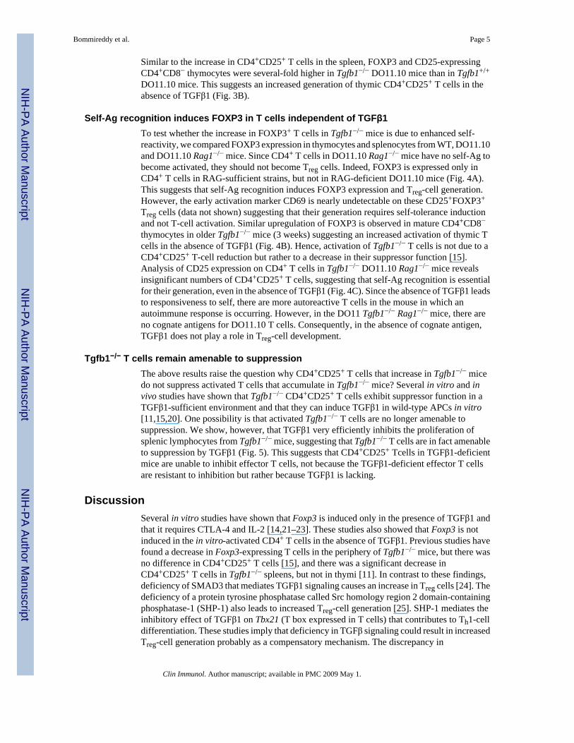
Self-Ag recognition induces FOXP3 in T cells independent of TGFβ1To test whether the increase in FOXP3+ T cells in Tgfb1−/− mice is due to enhanced self-reactivity, we compared FOXP3 expression in thymocytes and splenocytes from WT, DO11.10and DO11.10 Rag1−/− mice. Since CD4+ T cells in DO11.10 Rag1−/− mice have no self-Ag tobecome activated, they should not become Treg cells. Indeed, FOXP3 is expressed only inCD4+ T cells in RAG-sufficient strains, but not in RAG-deficient DO11.10 mice (Fig. 4A).This suggests that self-Ag recognition induces FOXP3 expression and Treg-cell generation.However, the early activation marker CD69 is nearly undetectable on these CD25+FOXP3+
Treg cells (data not shown) suggesting that their generation requires self-tolerance inductionand not T-cell activation. Similar upregulation of FOXP3 is observed in mature CD4+CD8−thymocytes in older Tgfb1−/− mice (3 weeks) suggesting an increased activation of thymic Tcells in the absence of TGFβ1 (Fig. 4B). Hence, activation of Tgfb1−/− T cells is not due to aCD4+CD25+ T-cell reduction but rather to a decrease in their suppressor function [15].Analysis of CD25 expression on CD4+ T cells in Tgfb1−/− DO11.10 Rag1−/− mice revealsinsignificant numbers of CD4+CD25+ T cells, suggesting that self-Ag recognition is essentialfor their generation, even in the absence of TGFβ1 (Fig. 4C). Since the absence of TGFβ1 leadsto responsiveness to self, there are more autoreactive T cells in the mouse in which anautoimmune response is occurring. However, in the DO11 Tgfb1−/− Rag1−/− mice, there areno cognate antigens for DO11.10 T cells. Consequently, in the absence of cognate antigen,TGFβ1 does not play a role in Treg-cell development.
Tgfb1−/− T cells remain amenable to suppressionThe above results raise the question why CD4+CD25+ T cells that increase in Tgfb1−/− micedo not suppress activated T cells that accumulate in Tgfb1−/− mice? Several in vitro and invivo studies have shown that Tgfb1−/− CD4+CD25+ T cells exhibit suppressor function in aTGFβ1-sufficient environment and that they can induce TGFβ1 in wild-type APCs in vitro[11,15,20]. One possibility is that activated Tgfb1−/− T cells are no longer amenable tosuppression. We show, however, that TGFβ1 very efficiently inhibits the proliferation ofsplenic lymphocytes from Tgfb1−/− mice, suggesting that Tgfb1−/− T cells are in fact amenableto suppression by TGFβ1 (Fig. 5). This suggests that CD4+CD25+ Tcells in TGFβ1-deficientmice are unable to inhibit effector T cells, not because the TGFβ1-deficient effector T cellsare resistant to inhibition but rather because TGFβ1 is lacking.
DiscussionSeveral in vitro studies have shown that Foxp3 is induced only in the presence of TGFβ1 andthat it requires CTLA-4 and IL-2 [14,21–23]. These studies also showed that Foxp3 is notinduced in the in vitro-activated CD4+ T cells in the absence of TGFβ1. Previous studies havefound a decrease in Foxp3-expressing T cells in the periphery of Tgfb1−/− mice, but there wasno difference in CD4+CD25+ T cells [15], and there was a significant decrease inCD4+CD25+ T cells in Tgfb1−/− spleens, but not in thymi [11]. In contrast to these findings,deficiency of SMAD3 that mediates TGFβ1 signaling causes an increase in Treg cells [24]. Thedeficiency of a protein tyrosine phosphatase called Src homology region 2 domain-containingphosphatase-1 (SHP-1) also leads to increased Treg-cell generation [25]. SHP-1 mediates theinhibitory effect of TGFβ1 on Tbx21 (T box expressed in T cells) that contributes to Th1-celldifferentiation. These studies imply that deficiency in TGFβ signaling could result in increasedTreg-cell generation probably as a compensatory mechanism. The discrepancy in
Bommireddy et al. Page 5
Clin Immunol. Author manuscript; available in PMC 2009 May 1.
NIH
-PA Author Manuscript
NIH
-PA Author Manuscript
NIH
-PA Author Manuscript

CD4+CD25+ T-cell frequency between Tgfb1−/− mice and Smad3−/− or Shp1−/− mice could berelated to differences in the extent of inflammation in these strains. We have resolved theseapparent discrepancies by generating Tgfb1−/− DO11.10 TCR transgenic mice that havereduced inflammation and enhanced survival. In this strain, TGFβ1 deficiency is associatedwith increased frequency of CD4+CD25+ T cells in the spleen and thymi.
In the present study we show that FOXP3 is expressed in activated T cells (CD69+) inTgfb1−/− mice, further confirming that FOXP3 expression is modulated by activation signalsand not directly induced by TGFβ1 in vivo. Recently Li et al. and Marie et al. found that FOXP3expression decreases in TGFβ receptor type II-deficient T cells as the Tbr2 conditionalknockout mice progressively develop inflammation. Since there was no defect in the generationof Treg cells in the thymus of these conditional knockout mice, it was argued that TGFβsignaling in Treg cells is important for their maintenance in the periphery [26,27]. Since wefound that the generation or maintenance of CD4+CD25+ Tcells is unaffected in the absenceof TGFβ1, it is possible that other ligands such as TGFβ2 which signal through the samereceptor (Tbr2) may be protecting the Treg cells from apoptosis in Tgfb1−/− mice.
The activation of Tgfb1−/− Tcells is not due to a reduction in CD4+CD25+ T-cell numbers, butrather due to a TGFβ1 deficiency which causes a decrease in their suppressor function [15].Our results also suggest that FOXP3 is expressed in activated Tcells in vivo in Tgfb1−/− mice,that its expression depends on the quality of signal strength, and that TGFβ1 plays an indirectrole in Foxp3 induction [28]. We have shown that CD4+ T cells undergo activation inTgfb1−/− DO11.10 mice, even in the presence of increased numbers of CD4+CD25+ Treg cellsin vivo, indicating that the Treg cells do depend on TGFβ1 for their function (rather thandevelopment) in vivo [9]. Recent studies from two groups independently confirmed theTGFβ-signaling-dependent suppressor functions of Treg cells by showing that wild-type Tregcells fail to prevent autoimmune responses of TGFβ receptor-deficient T cells in vivo, therebyconfirming a role for TGFβ signaling in effector T cells for inhibition by Treg cells [26,27].
It is possible that Treg cells deliver several suppressor signals and that TGFβ1 is only one ofthem. Treg cells suppress effector T cells via multiple mechanisms, including: i) cell–cellcontact, ii) secretion of suppressor cytokines such as IL-10 and TGFβ1, and iii) cytokinedeprivation such as sequestration of IL-2 due to constitutive expression of CD25 (IL-2 receptorα chain) on Treg cells. Hence, Tgfb1−/− Treg cells could function as suppressor cells but are notas potent as Tgfb1+/+ Treg cells [15]. Using CD4+CD25+ Treg cells from Tgfb1−/− mice andCD4+ T cells from dnTBR2 tg and Smad3−/− mice, Piccirillo et al. have suggested thatTGFβ1 is not essential for Treg-cell suppressor function in vitro [20]. However, that in vitrostudy used irradiated splenocytes from wild-type mice as APC that can produce TGFβ1. Thus,it is possible that extraneous TGFβ1 produced by wild-type APCs or that is present in FCS incultures might render a suppression that is being attributed to CD4+CD25+ Treg cells fromTGFβ1-deficient mice. Moreover, others, using both in vitro [11] and adoptive transfer studies[15,29], have shown that Treg cells need not produce TGFβ1, but they can acquire it if they arein a TGFβ1-sufficient environment. We have addressed the situation for a TGFβ1-deficientenvironment, and found that TGFβ1 is important for Treg-cell function in vivo because in acompletely TGFβ1-deficient environment, CD4+ T cells undergo activation in DO11.10 mice,even in the presence of increased numbers of CD4+CD25+ T cells. Since TGFβ1 is a negativeregulator of T-cell activation, the increased CD4+CD25+ T-cell generation in Tgfb1−/−
DO11.10 mice could be due to increased signal strength generated during the interaction ofTCR or hybrid TCR with self-Ags [30]. The co-existence of activated T cells and FOXP3+ Tcells in lymphoid organs of TGFβ1-deficient mice suggests that TGFβ1 is required for theregulatory function of Treg cells to prevent activation of T cells and autoimmunity. SinceTgfb1−/− T cells respond to TGFβ1 in vitro, it is the absence of TGFβ1 production by Treg cellsthat leads to their loss of regulatory function. Taken together, these data indicate a role of
Bommireddy et al. Page 6
Clin Immunol. Author manuscript; available in PMC 2009 May 1.
NIH
-PA Author Manuscript
NIH
-PA Author Manuscript
NIH
-PA Author Manuscript

TGFβ1 in the functioning, but not in the generation and maintenance, of CD4+CD25+ Tregcells.
AcknowledgementsThe authors thank Dr. David A. Hildeman (Cincinnati Children's Hospital) for his critical comments. The authors alsothank Sandy Schwemberger for her expert assistance in flow cytometry.
This study was supported by NIH HD26471, ES06096 and CA84291 to TD, AR50797 and AR47322 to RRS, and bya grant from Shriners of North America to GFB.
References1. Singh RR. SLE: translating lessons from model systems to human disease. Trends Immunol
2005;26:572–579. [PubMed: 16153890]2. Ohtsuka K, Gray JD, Stimmler MM, Toro B, Horwitz DA. Decreased production of TGF-beta by
lymphocytes from patients with systemic lupus erythematosus. J Immunol 1998;160:2539–2545.[PubMed: 9498800]
3. Singh RR, Ebling FM, Albuquerque DA, Saxena V, Kumar V, Giannini EH, Marion TN, FinkelmanFD, Hahn BH. Induction of autoantibody production is limited in nonautoimmune mice. J Immunol2002;169:587–594. [PubMed: 12077292]
4. Saxena V, Lienesch DW, Zhou M, Bommireddy R, Azhar M, Doetschman T, Singh RR. Dual rolesof immunoregulatory cytokine transforming growth factor beta (TGFβ in the pathogenesis ofautoimmunity-mediated) organ damage. J Immunol. In Press
5. Shull MM, Ormsby I, Kier AB, Pawlowski S, Diebold RJ, Yin M, Allen R, Sidman C, Proetzel G,Calvin D, Doetschman T. Targeted disruption of the mouse transforming growth factor-beta 1 generesults in multifocal inflammatory disease. Nature 1992;359:693–699. [PubMed: 1436033]
6. Kulkarni AB, Huh CG, Becker D, Geiser A, Lyght M, Flanders KC, Roberts AB, Sporn MB, WardJM, Karlsson S. Transforming growth factor beta 1 null mutation in mice causes excessiveinflammatory response and early death. Proc Natl Acad Sci U S A 1993;90:770–774. [PubMed:8421714]
7. Bommireddy R, Ormsby I, Yin M, Boivin GP, Babcock GF, Doetschman T. TGFbeta1 inhibits Ca2+-Calcineurin-mediated activation in thymocytes. J Immunol 2003;170:3645–3652. [PubMed:12646629]
8. Bommireddy R, Saxena V, Ormsby I, Yin M, Boivin GP, Babcock GF, Singh RR, Doetschman T.TGF-beta1 regulates lymphocyte homeostasis by preventing activation and subsequent apoptosis ofperipheral lymphocytes. J Immunol 2003;170:4612–4622. [PubMed: 12707339]
9. Bommireddy R, Pathak LJ, Martin J, Ormsby I, Engle SJ, Boivin GP, Babcock GF, Eriksson AU,Singh RR, Doetschman T. Self-antigen recognition by TGFbeta1-deficient T cells causes theiractivation and systemic inflammation. Lab Invest 2006;86:1008–1019. [PubMed: 16865088]
10. Fontenot JD, Rudensky AY. A well adapted regulatory contrivance: regulatory T cell developmentand the forkhead family transcription factor Foxp3. Nat Immunol 2005;6:331–337. [PubMed:15785758]
11. Marie JC, Letterio JJ, Gavin M, Rudensky AY. TGF-{beta}1 maintains suppressor function andFoxp3 expression in CD4 +CD25+ regulatory T cells. J Exp Med 2005;201:1061–1067. [PubMed:15809351]
12. Belghith M, Bluestone JA, Barriot S, Megret J, Bach JF, Chatenoud L. TGF-beta-dependentmechanisms mediate restoration of self-tolerance induced by antibodies to CD3 in overt autoimmunediabetes. Nat Med 2003;9:1202–1208. [PubMed: 12937416]
13. Bommireddy R, Doetschman T. TGF-beta, T-cell tolerance and anti-CD3 therapy. Trends Mol Med2004;10:3–9. [PubMed: 14720578]
14. Chen W, Jin W, Hardegen N, Lei KJ, Li L, Marinos N, McGrady G, Wahl SM. Conversion ofperipheral CD4+CD25− naive Tcells to CD4+CD25+ regulatory Tcells by TGF-beta induction oftranscription factor Foxp3. J Exp Med 2003;198:1875–1886. [PubMed: 14676299]
Bommireddy et al. Page 7
Clin Immunol. Author manuscript; available in PMC 2009 May 1.
NIH
-PA Author Manuscript
NIH
-PA Author Manuscript
NIH
-PA Author Manuscript

15. Mamura M, Lee W, Sullivan TJ, Felici A, Sowers AL, Allison JP, Letterio JJ. CD28 disruptionexacerbates inflammation in Tgf-beta1−/− mice: in vivo suppression by CD4+CD25+ regulatory Tcells independent of autocrine TGF-beta1. Blood 2004;103:4594–4601. [PubMed: 15016653]
16. Bettelli E, Carrier Y, Gao W, Korn T, Strom TB, Oukka M, Weiner HL, Kuchroo VK. Reciprocaldevelopmental pathways for the generation of pathogenic effector TH17 and regulatory T cells.Nature 2006;441:235–238. [PubMed: 16648838]
17. Fontenot JD, Gavin MA, Rudensky AY. Foxp3 programs the development and function of CD4+CD25+ regulatory T cells. Nat Immunol 2003;4:330–336. [PubMed: 12612578]
18. Hori S, Nomura T, Sakaguchi S. Control of regulatory T cell development by the transcription factorFoxp3. Science 2003;299:1057–1061. [PubMed: 12522256]
19. Korn T, Reddy J, Gao W, Bettelli E, Awasthi A, Petersen TR, Backstrom BT, Sobel RA,Wucherpfennig KW, Strom TB, Oukka M, Kuchroo VK. Myelin-specific regulatory T cellsaccumulate in the CNS but fail to control autoimmune inflammation. Nat Med 2007;13:423–431.[PubMed: 17384649]
20. Piccirillo CA, Letterio JJ, Thornton AM, McHugh RS, Mamura M, Mizuhara H, Shevach EM. CD4(+)CD25(+) regulatory T cells can mediate suppressor function in the absence of transforming growthfactor beta1 production and responsiveness. J Exp Med 2002;196:237–246. [PubMed: 12119348]
21. Fantini MC, Becker C, Monteleone G, Pallone F, Galle PR, Neurath MF. Cutting edge: TGF-betainduces a regulatory phenotype in CD4+CD25− T cells through Foxp3 induction and down-regulation of Smad7. J Immunol 2004;172:5149–5153. [PubMed: 15100250]
22. Zheng SG, Wang JH, Stohl W, Kim KS, Gray JD, Horwitz DA. TGF-beta requires CTLA-4 earlyafter T cell activation to induce FoxP3 and generate adaptive CD4+CD25+ regulatory cells. JImmunol 2006;176:3321–3329. [PubMed: 16517699]
23. Zheng SG, Wang J, Wang P, Gray JD, Horwitz DA. IL-2 is essential for TGF-beta to convert naiveCD4+CD25− cells to CD25+Foxp3+ regulatory T cells and for expansion of these cells. J Immunol2007;178:2018–2027. [PubMed: 17277105]
24. Wang ZB, Cui YF, Liu YQ, Jin W, Xu H, Jiang ZJ, Lu YX, Zhang Y, Liu XL, Dong B. Increase ofCD4(+)CD25(+) T cells in Smad3(−/−) mice, World. J Gastroenterol 2006;12:2455–2458.
25. Carter JD, Calabrese GM, Naganuma M, Lorenz U. Deficiency of the Src homology region 2 domain-containing phosphatase 1 (SHP-1) causes enrichment of CD4+CD25+ regulatory T cells. J Immunol2005;174:6627–6638. [PubMed: 15905501]
26. Li MO, Sanjabi S, Flavell RA. Transforming growth factor-beta controls development homeostasis,and tolerance of Tcells by regulatory T cell-dependent and -independent mechanisms. Immunity2006;25:455–471. [PubMed: 16973386]
27. Marie JC, Liggitt D, Rudensky AY. Cellular mechanisms of fatal early-onset autoimmunity in micewith the T cell-specific targeting of transforming growth factor-beta receptor. Immunity2006;25:441–454. [PubMed: 16973387]
28. Gavin MA, Torgerson TR, Houston E, Deroos P, Ho WY, Stray-Pedersen A, Ocheltree EL, GreenbergPD, Ochs HD, Rudensky AY. Single-cell analysis of normal and FOXP3-mutant human T cells:FOXP3 expression without regulatory T cell development. Proc Natl Acad Sci U S A 2006;103:6659–6664. [PubMed: 16617117]
29. Fahlen L, Read S, Gorelik L, Hurst SD, Coffman RL, Flavell RA, Powrie F. T cells that cannot respondto TGF-beta escape control by CD4(+)CD25(+) regulatory Tcells. J Exp Med 2005;201:737–746.[PubMed: 15753207]
30. Graca L, Chen TC, Le Moine A, Cobbold SP, Howie D, Waldmann H. Dominant tolerance: activationthresholds for peripheral generation of regulatory T cells. Trends Immunol 2005;26:130–135.[PubMed: 15745854]
Bommireddy et al. Page 8
Clin Immunol. Author manuscript; available in PMC 2009 May 1.
NIH
-PA Author Manuscript
NIH
-PA Author Manuscript
NIH
-PA Author Manuscript

Figure 1.TGFβ1 deficiency results in increased CD4+CD25+ Treg cells in the periphery. Splenocytesfrom 2-week-old Tgfb1−/− (TCR non-transgenic [Ntg]) or 2–12-week-old Tgfb1−/− DO11.10(Tg) and littermate control mice were surface-stained for CD4 and CD25. (A and B)Representative dot plots show percentages of CD4+CD25+ cells in the spleens of 2-week-oldTgfb1−/− (Ntg), 2-week-old Tgfb1−/− DO11.10 (Tg), and age-matched littermate controls. (C)CD4+CD25+ Tcells are shown as the percentages of CD4+ cells in Tgfb1−/− DO11.10 and theirlittermate control mice at different age groups. Data from individual animals are shown ascircles. The means of the groups are shown as horizontal grey lines. *p<0.05 (Student's t-test).(D) Dot plots with percentages of populations expressing FOXP3 and CD4 (top panels) andCD4+-gated splenocytes expressing FOXP3 and CD25, and TCR (KJ1-26) (middle and bottompanels) in 7-week-old Tgfb1−/− DO11.10 mice. Dot plots with percentages of populations ineach quadrant are shown. (E) Gated CD3+CD4+ T cells from 8-week-old Tgfb1−/− mice andtheir controls were analyzed for CD25high Tcells. Data shown are representative of similar datain three mice in each group. (F) Total numbers of CD4+ Tcells (left panel) and
Bommireddy et al. Page 9
Clin Immunol. Author manuscript; available in PMC 2009 May 1.
NIH
-PA Author Manuscript
NIH
-PA Author Manuscript
NIH
-PA Author Manuscript

CD4+CD25+/high Tcells (right panel) in the spleen of Tgfb1−/− mice and their wild-typelittermates. Data are mean±SD of three mice for each group (4–7 weeks). Results fromTgfb1−/− and wild-type littermates that have comparable numbers of CD4+ T cells are shown.Tgfb1−/− mice, as compared to wild-type littermates, have higher numbers ofCD4+CD25+/high T cells in the spleen, although the differences were not statisticallysignificant.
Bommireddy et al. Page 10
Clin Immunol. Author manuscript; available in PMC 2009 May 1.
NIH
-PA Author Manuscript
NIH
-PA Author Manuscript
NIH
-PA Author Manuscript

Figure 2.Phenotypes of CD4+CD25+ Treg cells in Tgfb1−/− mice: increased activation markers andincreased ratio of activated Tcells: Treg cells. (A–C) Splenocytes from Tgfb1−/− (TCR non-transgenic) and control mice were stained for CD4, CD25, CD69 and intracellular FOXP3. (A)Percentages of CD4+ T cells expressing these markers are shown on representative dot plots.(B) Subsets of CD4+ T cells based on the expression of CD25 and CD69 or of FOXP3 andCD69 in WTand KO mice. (C) Ratio of activated (CD69+):Treg (CD25+CD69− orFOXP3+CD69−) CD4+ Tcells determined from data in (B). (D–G) Splenocytes from 6-week-old (D–F) or 7-week-old (G) Tgfb1−/− DO11.10 and littermate control mice were analyzed forCD4, CD25, CD69 and CD62L. (D) Percentages of CD4+ cells expressing CD25 and CD62Lare shown on representative dot plots. (E) Subsets of CD4+ T cells based on the expression ofCD25 and CD62L in WT and KO mice are shown. Ratio of activated (CD62L−):Treg(CD25+CD62L+) CD4+ Tcells is shown in (F). (G) CD25 and CD69 expression was analyzedon KJ1-26+ versus KJ1-26− CD4+ splenocytes. Results shown represent two experiments.
Bommireddy et al. Page 11
Clin Immunol. Author manuscript; available in PMC 2009 May 1.
NIH
-PA Author Manuscript
NIH
-PA Author Manuscript
NIH
-PA Author Manuscript
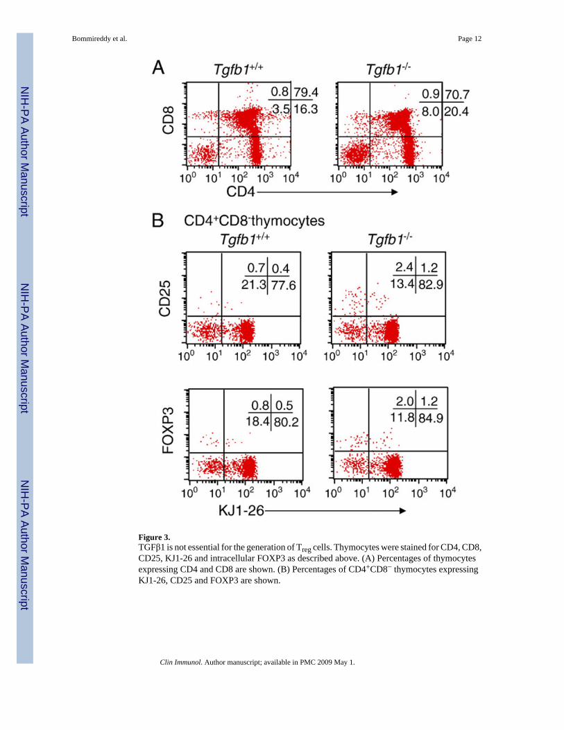
Figure 3.TGFβ1 is not essential for the generation of Treg cells. Thymocytes were stained for CD4, CD8,CD25, KJ1-26 and intracellular FOXP3 as described above. (A) Percentages of thymocytesexpressing CD4 and CD8 are shown. (B) Percentages of CD4+CD8− thymocytes expressingKJ1-26, CD25 and FOXP3 are shown.
Bommireddy et al. Page 12
Clin Immunol. Author manuscript; available in PMC 2009 May 1.
NIH
-PA Author Manuscript
NIH
-PA Author Manuscript
NIH
-PA Author Manuscript

Figure 4.FOXP3 expression and Treg-cell generation is self-Ag specific. (A) Splenocytes andthymocytes from 4-week-old mice were stained for CD4, CD25 and intracellular FOXP3.FOXP3 expression in the TCR NTg control (green) and DO11.10 (red) CD4+CD8− SPthymocytes (left panel) and CD4+ splenocytes (right panel) but not in DO11.10 Rag1−/− mice(blue) because of elimination of endogenous TCR. (B) Analysis of intracellular FOXP3expression in Tgfb1−/− (non-transgenic) thymocytes. (C) CD25 expression on CD4+ T cells inTgfb1−/− DO11.10 Rag1−/− and littermate control mice.
Bommireddy et al. Page 13
Clin Immunol. Author manuscript; available in PMC 2009 May 1.
NIH
-PA Author Manuscript
NIH
-PA Author Manuscript
NIH
-PA Author Manuscript

Figure 5.TGFβ1 inhibits the proliferation of TGFβ1-deficient T cells in vitro. Splenocytes fromTgfb1−/− and littermate control mice were stimulated with 1 nM PMA (P) and 250 nMionomycin (I), with or without TGFβ1, for 3 days. Cells were pulsed with tritiated thymidinefor the last 14 h of the culture. Cells were harvested and radioactivity was counted. Results areshown as the percent inhibition by TGFβ1 in reference to control cultures without anyexogenous TGFβ1. Results from 17-day-old animals are shown.
Bommireddy et al. Page 14
Clin Immunol. Author manuscript; available in PMC 2009 May 1.
NIH
-PA Author Manuscript
NIH
-PA Author Manuscript
NIH
-PA Author Manuscript