Preferential replication of FIV in activated CD4 +CD25 +T cells independent of cellular...
16
Preferential replication of FIV in activated CD4 + CD25 + T cells independent of cellular proliferation Anjali Joshi, Thomas W. Vahlenkamp, Himanshu Garg, Wayne A.F. Tompkins, and Mary B. Tompkins * Immunology Program, College of Veterinary Medicine, North Carolina State University, Raleigh, NC 27606, USA Received 11 August 2003; returned to author for revision 12 December 2003; accepted 7 January 2004 Abstract Studies attempting to identify reservoirs of HIV-1 latency have documented that the virus persists as both a latent and productive infection in subsets of CD4 + cells. Reports regarding establishment of a stable HIV-1 infection in quiescent T cells in vitro, however, are controversial. In the present study, we investigated the susceptibility of naive and activated CD4 + cell subsets (distinguished by differential expression of CD25) to feline immunodeficiency virus (FIV) infection, their ability to replicate the virus, and potentially act as a reservoir for virus persistence in infected animals. While both CD4 + CD25 + and CD4 + CD25 cells are susceptible to FIV infection in vitro and in vivo, only CD4 + CD25 + cells produce infectious virions when cultured with interleukin-2 (IL-2). Latently infected CD4 + CD25 cells produce infectious virions following ConcanvalinA (ConA) stimulation, which correlates with upregulated surface expression of CD25. In contrast to CD4 + CD25 cells, CD4 + CD25 + cells remain unresponsive to mitogen stimulation and are relatively resistant to apoptosis whether or not infected with FIV. The ability of CD4 + CD25 + cells to replicate FIV efficiently in the presence of IL-2 but remain anergic and unresponsive to apoptotic signaling suggests that these cells may provide a reservoir of productive FIV infection. On the contrary, CD4 + CD25 cells seem to establish as latent viral reservoirs capable of being reactivated after stimulation. D 2004 Elsevier Inc. All rights reserved. Keywords: FIV; HIV; AIDS; Latency; Stimulation; Anergy; Apoptosis; Reservoir Introduction Feline immunodeficiency virus (FIV) is a lentivirus of cats with a pathogenesis and disease pattern that parallels HIV-1-induced AIDS in humans (Bendinelli et al., 1995; English et al., 1994). Infected cats develop an acute infec- tion syndrome, including low-grade fever and transient generalized lymphadenopathy, followed by a long asymp- tomatic period in which the CD4/CD8 ratio declines due to an early increase in CD8 + and a progressive decline in CD4 + cell numbers. This asymptomatic period is followed by the development of a variety of disorders, many of which mimic HIV infection in humans (English et al., 1994). HIV and FIV have been shown to infect a variety of cell types in their respective hosts including T cells, macro- phages, and CNS glial cells (Bendinelli et al., 1995). While it is well established that HIV-1 displays a particular tropism for CD4 + lymphocytes, which may lead to their gradual depletion, there are conflicting reports regarding HIV infection of resting T cells. While some studies have shown that HIV can readily enter quiescent T cells in culture and complete the infectious cycle with transcription of early mRNA (Stevenson et al., 1990), others have shown that naive or resting T cells are resistant to de novo HIV infection (Chou et al., 1997). Whether resting T cells are infected de novo or are derived from activated infected cells, which subsequently revert to a resting phenotype, remains to be elucidated. Although there is extensive documentation of FIV infecting CD4 + cells in vivo and in vitro (Bendinelli et al., 1995; Dean et al., 1996; English et al., 1994), there are no studies addressing differential in vitro susceptibility of resting or activated CD4 + cells. With the advent of highly active anti-retroviral therapy (HAART), additional information has been gained on HIV 0042-6822/$ - see front matter D 2004 Elsevier Inc. All rights reserved. doi:10.1016/j.virol.2004.01.014 * Corresponding author. Immunology Program, College of Veterinary Medicine, North Carolina State University, 4700 Hillsborough Street, Raleigh, NC 27606. Fax: +1-919-513-6464. E-mail address: Mary _ [email protected] (M.B. Tompkins). www.elsevier.com/locate/yviro Virology 321 (2004) 307– 322
-
Upload
independent -
Category
Documents
-
view
3 -
download
0
Transcript of Preferential replication of FIV in activated CD4 +CD25 +T cells independent of cellular...

www.elsevier.com/locate/yviro
Virology 321 (2004) 307–322
Preferential replication of FIV in activated CD4+CD25+T cells
independent of cellular proliferation
Anjali Joshi, Thomas W. Vahlenkamp, Himanshu Garg,Wayne A.F. Tompkins, and Mary B. Tompkins*
Immunology Program, College of Veterinary Medicine, North Carolina State University, Raleigh, NC 27606, USA
Received 11 August 2003; returned to author for revision 12 December 2003; accepted 7 January 2004
Abstract
Studies attempting to identify reservoirs of HIV-1 latency have documented that the virus persists as both a latent and productive infection
in subsets of CD4+ cells. Reports regarding establishment of a stable HIV-1 infection in quiescent T cells in vitro, however, are controversial.
In the present study, we investigated the susceptibility of naive and activated CD4+ cell subsets (distinguished by differential expression of
CD25) to feline immunodeficiency virus (FIV) infection, their ability to replicate the virus, and potentially act as a reservoir for virus
persistence in infected animals. While both CD4+CD25+ and CD4+CD25� cells are susceptible to FIV infection in vitro and in vivo, only
CD4+CD25+ cells produce infectious virions when cultured with interleukin-2 (IL-2). Latently infected CD4+CD25� cells produce infectious
virions following ConcanvalinA (ConA) stimulation, which correlates with upregulated surface expression of CD25. In contrast to
CD4+CD25� cells, CD4+CD25+ cells remain unresponsive to mitogen stimulation and are relatively resistant to apoptosis whether or not
infected with FIV. The ability of CD4+CD25+ cells to replicate FIVefficiently in the presence of IL-2 but remain anergic and unresponsive to
apoptotic signaling suggests that these cells may provide a reservoir of productive FIV infection. On the contrary, CD4+CD25� cells seem to
establish as latent viral reservoirs capable of being reactivated after stimulation.
D 2004 Elsevier Inc. All rights reserved.
Keywords: FIV; HIV; AIDS; Latency; Stimulation; Anergy; Apoptosis; Reservoir
Introduction phages, and CNS glial cells (Bendinelli et al., 1995). While
Feline immunodeficiency virus (FIV) is a lentivirus of
cats with a pathogenesis and disease pattern that parallels
HIV-1-induced AIDS in humans (Bendinelli et al., 1995;
English et al., 1994). Infected cats develop an acute infec-
tion syndrome, including low-grade fever and transient
generalized lymphadenopathy, followed by a long asymp-
tomatic period in which the CD4/CD8 ratio declines due to
an early increase in CD8+ and a progressive decline in CD4+
cell numbers. This asymptomatic period is followed by the
development of a variety of disorders, many of which mimic
HIV infection in humans (English et al., 1994).
HIV and FIV have been shown to infect a variety of cell
types in their respective hosts including T cells, macro-
0042-6822/$ - see front matter D 2004 Elsevier Inc. All rights reserved.
doi:10.1016/j.virol.2004.01.014
* Corresponding author. Immunology Program, College of Veterinary
Medicine, North Carolina State University, 4700 Hillsborough Street,
Raleigh, NC 27606. Fax: +1-919-513-6464.
E-mail address: [email protected] (M.B. Tompkins).
it is well established that HIV-1 displays a particular
tropism for CD4+ lymphocytes, which may lead to their
gradual depletion, there are conflicting reports regarding
HIV infection of resting T cells. While some studies have
shown that HIV can readily enter quiescent T cells in
culture and complete the infectious cycle with transcription
of early mRNA (Stevenson et al., 1990), others have
shown that naive or resting T cells are resistant to de novo
HIV infection (Chou et al., 1997). Whether resting T cells
are infected de novo or are derived from activated infected
cells, which subsequently revert to a resting phenotype,
remains to be elucidated. Although there is extensive
documentation of FIV infecting CD4+ cells in vivo
and in vitro (Bendinelli et al., 1995; Dean et al., 1996;
English et al., 1994), there are no studies addressing
differential in vitro susceptibility of resting or activated
CD4+ cells.
With the advent of highly active anti-retroviral therapy
(HAART), additional information has been gained on HIV

A. Joshi et al. / Virology 321 (2004) 307–322308
infection of CD4+ subsets in vivo. Treatment of asymp-
tomatic HIV-1-infected individuals with HAART decreases
the amount of virus in plasma to levels below the limit of
detection by standard clinical assays (Perelson et al., 1996).
However, a small but detectable reservoir of latently
infected, resting CD4+ cells harboring replication compe-
tent HIV-1 has been shown to persist in essentially all
patients receiving therapy (Dornadula et al., 1999; Finzi et
al., 1997; Wong et al., 1997). Recently, Chun et al. (2003)
reported that resting CD4+ cells from the majority of
viremic HIV-1-positive patients are capable of producing
cell-free HIV-1 spontaneously ex vivo. No extracellular
virus was produced by resting CD4+ cells from aviremic
patients despite the presence of HIV-1 mRNA. Interesting-
ly, DNA microarray analysis revealed that several genes
involved in mRNA and protein synthesis and processing
were significantly upregulated in resting CD4+ cells from
HIV viremic patients compared to aviremic patients, sug-
gesting that virus replication impacted the physiological
state of resting CD4+ cells, or alternatively, that partial
activation of CD4+ cells is a prerequisite for virus replica-
tion. This latter speculation is of interest in that earlier
studies have demonstrated that productive infection with
HIV-1 in vitro was associated with a CD4+ cell subset
characterized by a partial activation phenotype as indicated
by cell surface expression of the CD25 receptor (IL-2
receptor a chain).
Borvak et al. (1995) and Ramilo et al. (1993) used
immunotoxin-labeled anti-CD25 to show that PBMCs de-
Fig. 1. Schematic representation of linear proviral DNA, one LTR circle junctions,
the study. (A) Double-stranded linear reverse-transcribed unintegrated proviral DN
circle junction products showing the position of the Env-sense (Env-s) and Ga
productively infected cells. (C) Linear double-stranded proviral DNA integrated in
Gag-as primers depicted in the figure can be used to detect either linear unintegrate
are specifically detected by Env-s and Gag-as primer pair.
pleted of CD25+ cells were markedly diminished in their
ability to replicate HIV when infected in vitro. Further, they
also showed that CD4+CD25+ cells could be productively
infected with HIV in vitro, whereas highly purified resting
CD4+CD25� cells were resistant to HIV infection (Chou et
al., 1997). These observations are of interest inasmuch as
the CD4+CD25+ cell phenotype possesses unique immuno-
logical characteristics that could make a particularly favor-
able target for sustained HIV infection. Studies in several
experimental models have firmly established the existence
of a naturally occurring CD4+CD25+T regulatory (Treg)
cell population that is important in maintaining peripheral
self-tolerance (Maloy and Powrie, 2001). These CD4+CD25+
cells also play a major role in regulating immune responses
to microbial infections (Sakaguchi, 2003). The most useful
marker to date for identification of Treg cells is the CD25
antigen that has been demonstrated on 5–10% of CD4+ cells
in the circulation of normal rodents and humans (Jonuleit et
al., 2001). While Treg cells have a partial activation pheno-
type, they are anergic in that they proliferate poorly upon
TCR stimulation in vitro and their growth is dependent on
exogenous IL-2 (Thornton and Shevach, 1998). Importantly,
CD4+CD25+ cells appear to be resistant to clonal deletion
and apoptosis when stimulated with anti-CD3 (Taams et al.,
2001) or super antigen (Banz et al., 2002), suggesting they
could be long-lived in lymphoid tissues. While CD4+CD25+
cells reported to support HIV replication were not identified
as Treg cells, it will be important to define some of the
functional characteristics of these virus-infected cells to
and two LTR circle junctions depicting the position of primer pairs used in
A showing 5Vand 3VLTRs and major viral genes. (B) One LTR and two LTR
g-antisense (Gag-as) primer pair used to amplify these circle junctions in
to the host genome. The LTR-sense (LTR-s), LTR-antisense (LTR-as), and
d or linear integrated proviral DNA forms but not the circle junctions, which

Fig. 2. Correlation between appearance of circle junctions by PCR and
detectable gag-p24 antigen in culture supernatants. FCD4E cells were
infected in vitro with FIV-NCSU1 at an moi of 0.1 and plated in culture
medium supplemented with 100 U/ml IL-2. Cells and culture supernatant
were harvested at 0 h, 7 h, 24 h, 48 h, 6 days, and 8 days postinfection for
(A) DNA isolation and PCR using primer pairs described in Fig. 1 and (B)
p24 antigen detection by antigen capture ELISA. The p24 values at each
time point represent mean F standard error of triplicate cultures. The
experiment was repeated twice with similar results. Negative control for
PCR comprised of water plus reagents and did not result in amplification of
any bands (data not shown).
Fig. 3. Representative two-color flow cytometric dot-plot analysis of
CD4+CD25+ cells in the PBMC (A) and LN (B) of normal cats. PBMC and
LN cells were derived from SPF cats, stained with FITC-conjugated anti-
CD25 and PE-conjugated anti-CD4 antibodies, and analyzed on a
FACSCalibur flow cytometer. Numbers represent the percent CD4+CD25+
cells of the total CD4+ population.
A. Joshi et al. / Virology 321 (2004) 307–322 309
help ascertain their potential importance as a reservoir of
productive infection.
In this study, we have examined FIV entry and repli-
cation in activated and resting CD4+ cell subsets distin-
guished by differential expression of CD25. Our studies
demonstrate that although both CD4+CD25+ and
CD4+CD25� cells are susceptible to FIV infection in vitro
and in vivo, only CD4+CD25+ cells replicate the virus in
the absence of mitogenic stimulation. In contrast to
CD4+CD25� cells, CD4+CD25+ cells, whether or not
infected with FIV, do not proliferate in response to
ConcanvalinA (ConA) stimulation and are relatively resis-
tant to activation-induced programmed cell death, suggest-
ing that they could represent a long-lived reservoir of
productive FIV infection.
Results
Correlation between appearance of circle junctions and FIV
p24 antigen production in acutely infected Feline CD4E
(FCD4E) cells
Retroviral infection of T cells is characterized by the
formation of linear reverse-transcribed double-stranded
DNA flanked by the 5V and 3V long terminal repeats
(Fig. 1A) (Farnet and Haseltine, 1990; Tang et al., 1999).
In addition to the linear products of reverse transcription,
two circular forms of viral DNA have also been found in the
nucleus of infected cells (Brown et al., 1987; Frey et al.,
2001; Sharkey and Stevenson, 2001) and contain either two
LTRs formed by circularization of linear viral DNA or one
LTR formed by homologous recombination between the
LTRs of linear DNA precursors (Fig. 1B) (Brown et al.,
1987). These episomal DNA intermediates, also called
circle junctions, are characteristics of cells productively
infected with HIV and are generally not present in latently
infected cells (Teo et al., 1997). By selecting certain primer
A. Joshi et al. / Virology 321 (2004) 307–322310
pairs, we detected by polymerase chain reaction (PCR) early
stages of reverse transcription (LTR-sense and LTR-anti-
sense primers; Fig. 1C), intermediate stages (LTR-sense and
Gag-antisense primers; Fig. 1C), and circle junction prod-
ucts (Env-sense and Gag-antisense primers; Fig. 1B) of
virus replication. Consistent with other studies in HIV and
FIV, we were unable to detect two LTR circle junction
products (Farnet and Haseltine, 1990; Frey et al., 2001).
This is likely due to the many fold higher abundance of one
LTR circles in the nucleus of infected cells compared to two
LTR circles, resulting in preferential amplification of one
LTR circles (Farnet and Haseltine, 1990).
To validate the FIV primers and determine the time
points at which different forms of reverse transcription
appeared in infected cells, FCD4E cells were infected in
vitro with the FIV-NCSU1 isolate and analyzed for reverse
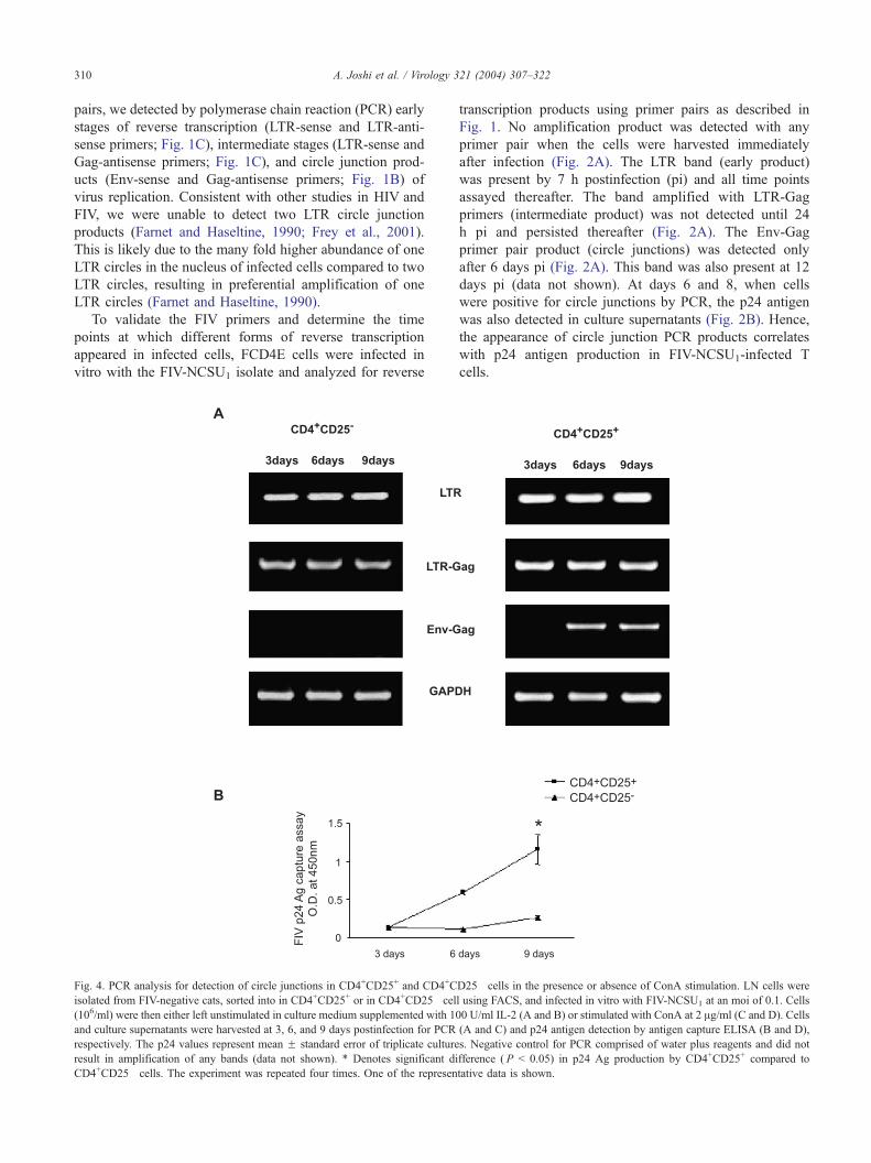
Fig. 4. PCR analysis for detection of circle junctions in CD4+CD25+ and CD4+C
isolated from FIV-negative cats, sorted into in CD4+CD25+ or in CD4+CD25� cel
(106/ml) were then either left unstimulated in culture medium supplemented with 10
and culture supernatants were harvested at 3, 6, and 9 days postinfection for PCR
respectively. The p24 values represent mean F standard error of triplicate culture
result in amplification of any bands (data not shown). * Denotes significant di
CD4+CD25� cells. The experiment was repeated four times. One of the represen
transcription products using primer pairs as described in
Fig. 1. No amplification product was detected with any
primer pair when the cells were harvested immediately
after infection (Fig. 2A). The LTR band (early product)
was present by 7 h postinfection (pi) and all time points
assayed thereafter. The band amplified with LTR-Gag
primers (intermediate product) was not detected until 24
h pi and persisted thereafter (Fig. 2A). The Env-Gag
primer pair product (circle junctions) was detected only
after 6 days pi (Fig. 2A). This band was also present at 12
days pi (data not shown). At days 6 and 8, when cells
were positive for circle junctions by PCR, the p24 antigen
was also detected in culture supernatants (Fig. 2B). Hence,
the appearance of circle junction PCR products correlates
with p24 antigen production in FIV-NCSU1-infected T
cells.
D25� cells in the presence or absence of ConA stimulation. LN cells were
l using FACS, and infected in vitro with FIV-NCSU1 at an moi of 0.1. Cells
0 U/ml IL-2 (A and B) or stimulated with ConA at 2 Ag/ml (C and D). Cells
(A and C) and p24 antigen detection by antigen capture ELISA (B and D),
s. Negative control for PCR comprised of water plus reagents and did not
fference ( P < 0.05) in p24 Ag production by CD4+CD25+ compared to
tative data is shown.
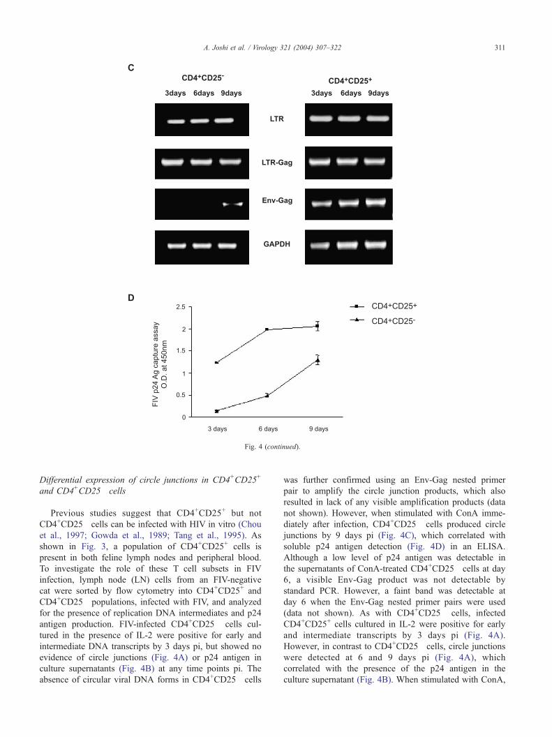
Fig. 4 (continued).
A. Joshi et al. / Virology 321 (2004) 307–322 311
Differential expression of circle junctions in CD4+CD25+
and CD4+CD25� cells
Previous studies suggest that CD4+CD25+ but not
CD4+CD25� cells can be infected with HIV in vitro (Chou
et al., 1997; Gowda et al., 1989; Tang et al., 1995). As
shown in Fig. 3, a population of CD4+CD25+ cells is
present in both feline lymph nodes and peripheral blood.
To investigate the role of these T cell subsets in FIV
infection, lymph node (LN) cells from an FIV-negative
cat were sorted by flow cytometry into CD4+CD25+ and
CD4+CD25� populations, infected with FIV, and analyzed
for the presence of replication DNA intermediates and p24
antigen production. FIV-infected CD4+CD25� cells cul-
tured in the presence of IL-2 were positive for early and
intermediate DNA transcripts by 3 days pi, but showed no
evidence of circle junctions (Fig. 4A) or p24 antigen in
culture supernatants (Fig. 4B) at any time points pi. The
absence of circular viral DNA forms in CD4+CD25� cells
was further confirmed using an Env-Gag nested primer
pair to amplify the circle junction products, which also
resulted in lack of any visible amplification products (data
not shown). However, when stimulated with ConA imme-
diately after infection, CD4+CD25� cells produced circle
junctions by 9 days pi (Fig. 4C), which correlated with
soluble p24 antigen detection (Fig. 4D) in an ELISA.
Although a low level of p24 antigen was detectable in
the supernatants of ConA-treated CD4+CD25� cells at day
6, a visible Env-Gag product was not detectable by
standard PCR. However, a faint band was detectable at
day 6 when the Env-Gag nested primer pairs were used
(data not shown). As with CD4+CD25� cells, infected
CD4+CD25+ cells cultured in IL-2 were positive for early
and intermediate transcripts by 3 days pi (Fig. 4A).
However, in contrast to CD4+CD25� cells, circle junctions
were detected at 6 and 9 days pi (Fig. 4A), which
correlated with the presence of the p24 antigen in the
culture supernatant (Fig. 4B). When stimulated with ConA,

Fig. 5. Rescue of productive infection from CD4+CD25� cells 6 days after
infection. Magnetic bead-purified CD4+CD25�PBMCs from a negative cat
were infected in vitro with FIV (moi of 0.1) and cultured in medium in the
presence of 100 U/ml IL-2. After 6 days of culture, (A) a portion of the cells
was harvested for PCR analysis using LTR-sense and LTR-antisense (LTR)
primer pair (lane 1), LTR-sense and Gag-antisense (LTR-Gag) primers (lane
2), and Env-sense and Gag-antisense (Env-Gag) primers (lane 3). M is a
100-bp molecular weight ladder. (B) The remaining cells were cultured in
the presence of ConA (2 Ag/ml) and supernatants harvested every 3 days for
p24 antigen detection by antigen capture ELISA. Controls consisted of
CD4+CD25� cells cultured in the presence of ConA immediately after
infection and in the presence of IL-2 only. The p24 values at each time point
represent mean F standard error of duplicate cultures. The experiment was
repeated twice with similar results.
A. Joshi et al. / Virology 321 (2004) 307–322312
CD4+CD25+ cells produced all three transcripts (early,
intermediate and circle junctions) (Fig. 4C) and soluble
p24 (Fig. 4D) as early as 3 days pi. In the absence of IL-2
or ConA, both cell populations produced the early and
intermediate transcripts, but not circle junctions or p24
(data not shown). Thus, while CD4+CD25+ cells were
productively infected with FIV, activation by ConA is
required for CD4+CD25� cells to produce the virus.
CD4+CD25� cells harbor a latent FIV infection that can be
activated by ConA
Infected CD4+CD25� cells contain specific proviral
DNA without detectable p24 antigen production in the
absence of cellular activation. Hence, we asked if this
proviral DNA form was biologically stable and whether
ConA stimulation several days after infection would lead to
reactivation of virus from these cells. PBMC-derived
CD4+CD25� cells from an FIV-negative cat were infected
in vitro with FIV and cultured in the presence of IL-2 for 6
days before stimulation with ConA. PCR of infected cells
just before addition of ConA revealed the presence of early
(Fig. 5A, lane 1) and intermediate (Fig. 5A, lane 2) stages of
proviral reverse transcription, but no detectable circle junc-
tions (Fig. 5A, lane 3) or p24 antigen in culture supernatant
(Fig. 5B). Following addition of ConA, the p24 antigen was
detectable at 12 days poststimulation and reached maximum
levels by 15 days (Fig. 5B). The kinetics of virus production
in cells stimulated immediately after infection was, how-
ever, more rapid compared to cells stimulated after 6 days of
infection (Fig. 5B). These data suggest that CD4+CD25�
cells develop a latent infection which can be reactivated
upon mitogenic stimulation.
Productive infection of CD4+CD25� cells on ConA
stimulation correlates with surface upregulation of CD25
As CD4+CD25� cells become productively infected when
stimulated with ConA but not IL-2, we asked whether there is
a differential activation response of CD4+CD25� cells to
these two stimuli. PBMCs were depleted of CD25+ cells by
Ab-coated magnetic beads and shown by flow cytometry to
contain less than 1% CD4+CD25+ cells (Fig. 6A). The
resulting CD25� cells were cultured in the presence or
absence of either ConA or IL-2 and analyzed for surface
expression of CD25 by flow cytometry. By 3 days of culture,
ConA-stimulated CD25� cells upregulated CD25 on their
surface such that more than 80% of the CD4+ cells expressed
surface-associated CD25 (Figs. 6B and C). In contrast, in the
presence of IL-2 or culture medium alone, CD4+CD25� cells
did not show an upregulation of CD25 on their surface
(Fig. 6C). These data, in conjunction with the previous
experiment, demonstrate that ConA, but not IL-2, pheno-
typically activates CD4+CD25� cells, and activated
CD4+CD25� cells are capable of supporting a productive
FIV infection.
Proliferation of CD4+CD25+ cells inversely correlates with
virus production
Recent literature emphasizes that naturally occurring
cells bearing the CD4+CD25+ phenotype are a thymus-
derived lineage distinct from CD4+CD25� T helper cells
(Maloy and Powrie, 2001). These cells are unique in that
they are anergic and relatively resistant to apoptosis (Banz
et al., 2002; Taams et al., 2001). As CD4+CD25+ cells

Fig. 6. Flow cytometric analysis of CD25 expression on cultured CD4+CD25� cells in the presence or absence of stimulation. PBMCs were isolated from FIV-
negative cats and CD25+ cells depleted using anti-CD25-coated magnetic beads. The CD25-depleted cells were cultured either in (1) growth medium alone, (2)
growth medium supplemented with 100 U/ml IL-2, or (3) growth medium containing 2 Ag/ml ConA. Cells were harvested, stained, and analyzed by flow
cytometry for the expression of CD4 and CD25 after 3, 6, and 9 days of different treatments. The dot plots demonstrate the percent of cells that (A) express
CD25 and CD4 immediately after depletion or (B) after 9 days in culture with ConA. The numbers represent the percentage of cells within each quadrant. (C)
The percentage of CD25+ cells within the CD4+ population (CD4+CD25+ cells/total CD4+ cells � 100) after different treatments is plotted as a function of
time. Data at each time point represent mean F standard error of triplicate cultures. * Denotes significant increase ( P < 0.01) in CD25 expression in
CD4+CD25� cells in the presence of ConA compared to treatment with IL-2 or culture media alone. The experiment was repeated three independent times. One
of the representative results is depicted.
A. Joshi et al. / Virology 321 (2004) 307–322 313
replicated FIV in vitro, we assessed these functional
characteristics of the feline CD4+CD25+ cell subset and
the effect FIV infection may have on their function. To
address this question, LN-derived uninfected or in vitro
FIV-infected CD4+CD25+ and CD4+CD25� cells were
stimulated with IL-2 or ConA and assayed for proliferative
responses (tritiated thymidine [3HTdR uptake]) and virus
production (p24 antigen capture ELISA). In the absence of
exogenous IL-2 or ConA, neither CD4+CD25+ nor
CD4+CD25� cells showed any significant proliferative
response or p24 production (Figs. 7A and B). Stimulation
of CD4+CD25� cells with either IL-2 or ConA resulted in
significant (P < 0.01) levels of cell proliferation (Fig. 7B)
compared to CD4+CD25+ cells. However, virus production
by CD4+CD25� cells was detected only in the presence of
ConA (Fig. 7A). In contrast, CD4+CD25+ cells produced
virus when stimulated with either ConA or IL-2 (Fig. 7A),
yet did not proliferate (Fig. 7B). Thus, while FIV repli-
cated efficiently in CD4+CD25+ cells in the presence of
IL-2 and ConA, these cells themselves remained unrespon-
sive to these mitogenic stimuli. Experiments with PBMC-
derived CD4+CD25+ and CD4+CD25� cells gave similar
results.
To obviate the possibility that lack of detection of
proliferation in 3HTdR uptake assay was because early or
late proliferative events were missed in our analysis, the total
number of cell divisions in CD4+CD25+ and CD4+CD25�
cells was also determined by CFSE labeling. CFSE is

A. Joshi et al. / Virology 321 (2004) 307–322314
transferred equally to daughter cells resulting in a reduc-
tion of mean fluorescence intensity (MFI), an indicator of
cell division. FIV-infected CD4+CD25+ and CD4+CD25�
cells were labeled with CFSE and cultured with IL-2,
ConA, or medium alone as in Fig. 7B. When stimulated
with ConA for 6 days, CD4+CD25� cells showed a
marked dilution in the CFSE dye content compared to
CD4+CD25� cells that were analyzed immediately after
labeling (Fig. 7C). CD4+CD25� cells in the presence of
IL-2 also showed a dilution in CFSE content although not
as marked as in the presence of ConA. In contrast,
CD4+CD25+ cells when cultured with any of the above
treatments did not show a significant shift in the mean
fluorescence intensities compared to 0 day controls indi-
cating that they did not divide (Fig. 7C). These data further
confirm our results from 3HTdR uptake assay suggesting
that while CD4+CD25� cells underwent cell division both
Fig. 7. FIV replication and cellular proliferation in CD4+CD25+ or in CD4+
CD4+CD25+ and CD4+CD25� cells from LN of FIV-negative cats were either i
uninfected CD4+CD25+ and CD4+CD25� cells were cultured in the presence of eit
6 culture, supernatants were harvested from infected cells and assayed for p24 a
production by CD4+CD25+ cells compared to CD4+CD25� cells cultured in the pre
at 1 ACi/well at 37 jC. After 18 h, cells were harvested and 3HTdR uptake deter
readings for each treatment represent mean F standard error of triplicates and five3HTdR by CD4+CD25� cells in the presence of IL-2 or ConA compared to CD4
depicted here. (C) CFSE labeling studies of infected CD4+CD25+ and CD4+CD25�
of FIV-negative cats were infected with FIV-NCSU1 as above and labeled with CFD
6 days in the presence of IL-2, ConA, or medium alone. At day 6 culture, supernat
for dilution of CFDA-SE dye using flow cytometry. Analyses were performed in d
data from infected cells is depicted here. Similar assays were performed with unin
(data not shown). In 0-day data, open histograms represent unlabeled cells and fi
in the presence of IL-2 and ConA, CD4+CD25+ cells did
not proliferate under similar conditions.
CD4+CD25+ cells show a relative resistance to apoptosis
Failure of CD4+CD25+ cells to proliferate in response to
mitogenic stimulation could possibly be due to their acti-
vated phenotype, which on further stimulation might result
in apoptosis in a large number of these cells. To address this
possibility, uninfected or in vitro-infected LN-derived
CD4+CD25+ and CD4+CD25� cells were stained for sur-
face expression of apoptotic markers under different culture
conditions. When cultured in the presence or absence of IL-
2, CD4+CD25� cells showed significantly (P < 0.01, P <
0.05, respectively) higher percent of apoptotic cells than
CD4+CD25+ cells (Fig. 8). Both infected and uninfected
CD4+CD25+ and CD4+CD25� cells showed a high percent-
CD25� cells under different culture conditions. Magnetic bead-purified
nfected with FIV-NCSU1 at an moi of 0.1 or left uninfected. Infected and
her 100 U/ml IL-2 or 2 Ag/ml ConA or left untreated for 6 days. (A) On day
ntigen in an ELISA. * Denotes significant increase ( P < 0.01) in p24 Ag
sence of IL-2. (B) On the same day, all the cultures were pulsed with 3HTdR
mined using a liquid scintillation beta counter. The p24 values and 3HTdR
wells, respectively. * Denotes significant increase ( P < 0.01) in uptake of+CD25+ cells. Results from one of the three representative experiments are
cells. Magnetic bead-purified CD4+CD25+and CD4+CD25� cells from LN
A-SE. Cells were either analyzed immediately after labeling or cultured for
ants were assayed as in A above (data not shown). Cells were also analyzed
uplicates and the results were repeated four times. One of the representative
fected CD4+CD25+ and CD4+CD25� cells and yielded comparable results
lled histograms represent cells analyzed immediately after labeling.
Fig. 7 (continued).
A. Joshi et al. / Virology 321 (2004) 307–322 315

Fig. 8. Determination of percent apoptosis in infected or uninfected CD4+CD25+ and CD4+CD25� cells. LN-derived cells were sorted into CD4+CD25+ and
CD4+CD25� populations by FACS. In vitro-infected or uninfected CD4+CD25+ and CD4+CD25� cells were either left untreated or treated with IL-2 or ConA
for 6 days as in Fig. 6. On day 6 FIV p24 antigen was determined in supernatants of infected cells by ELISA. On day 7 infected as well as uninfected
CD4+CD25+ and CD4+CD25� cells were stained with Annexin V and analyzed by flow cytometry. Dead cells were differentiated from apoptotic cells by dual
staining with PI. Percent apoptosis in CD4+CD25+ and CD4+CD25� cells is represented as mean F standard error of triplicates. * Denotes significant increase
( P < 0.01) in percent apoptosis in CD4+CD25� cells compared to CD4+CD25+ cells. ** Denotes significant increase ( P < 0.05) in percent apoptosis in
infected CD4+CD25� cells compared to infected CD4+CD25+ cells in the absence of any treatment. The experiment was repeated three times with similar
results.
A. Joshi et al. / Virology 321 (2004) 307–322316
age (approximately 40%) of apoptotic cells when stimulated
with ConA. Interestingly, there was no significant difference
in the percentage of apoptotic cells between the infected and
uninfected groups (both CD4+CD25+ and CD4+CD25�).
Thus, it can be concluded that in both uninfected and
infected cell populations, CD4+CD25+ cells show similar
(in the case of ConA stimulation) or less apoptosis (in the case
of IL2 or no stimulation) when compared to the CD4+CD25�
cells. Thus, the lack of proliferation seen in CD4+CD25+ cells
cannot be attributed to the higher percentage of apoptosis-
prone cells in this population. Experiments with PBMC-
derived CD4+CD25+ and CD4+CD25� cells gave similar
results.
Detection of circle junction products and rescue of
replication competent virus from CD4+CD25+ cells from
FIV-infected cats
The in vitro studies conducted thus far indicate that in
vitro-infected CD4+CD25+ cells efficiently replicate FIV,
are unresponsive to exogenous mitogenic stimuli, and do
not exhibit higher levels of apoptosis when compared to
their CD4+CD25� counterparts. To corroborate these find-
ings in vivo, we studied CD4+CD25+ and CD4+CD25�
cells purified by flow cytometric sorting (>99.5% purity)
from FIV-NCSU1-infected cats. Fig. 9A illustrates data from
a representative asymptomatic FIV+ cat demonstrating the
presence of LTR and LTR-Gag products, but no detectable
Env-gag products, in CD4+CD25� cells indicating the
absence of circle junction forms and hence a nonproductive
latent infection in these cells (Fig. 9A). The absence of Env-
Gag products was confirmed using nested primer pairs (Fig.
9A). In contrast, analysis of CD4+CD25+ cells from the
same cat revealed circle junction products indicative of a
productive infection. The circle junction products were
detected in CD4+CD25+ cells but not CD4+CD25� cells
in four of eight FIV-infected cats analyzed (data not shown).
All the infected cats were positive when DNA from PBMCs
was screened using a nested primer pair for the Gag gene
(data not shown). The above results suggest that while the
LTR products were always detected and LTR-Gag products
were mostly detected in CD4+CD25+ cells, as well as
CD4+CD25� cells from FIV-positive cats, detection of
circle junction products was variable and restricted to
CD4+CD25+ cells. In an attempt to rescue replication-
competent virus, CD4+CD25+ and CD4+CD25� cells from
FIV-positive cats were cocultured with uninfected FCD4E
cells. In the presence of IL-2, CD4+CD25+-FCD4E cocul-
tures yielded a significantly strong (P < 0.01) p24 signal,
whereas CD4+CD25� cells only gave a low p24 reading
when cocultured with FCD4E cells (Fig. 9B). However,
treatment of CD4+CD25� cells with ConA for 48 h and
subsequent co-culture with uninfected FCD4E cells yielded
significantly higher (P < 0.01) p24 readings than when the

Fig. 9. Analysis of virus replication in CD4+CD25+ and CD4+CD25� cells from cats infected with the NCSU1 isolate of FIV. (A) Detection of circle junction
forms in CD4+CD25+ cells from a FIV-positive cat. DNA was isolated from FACS-purified CD4+CD25+ and CD4+CD25�PBMCs from FIV-infected cats.
PCR reactions were set up using LTR-sense and LTR-antisense (LTR) primer pair (lane 1), LTR-sense and Gag-antisense (LTR-Gag) primers (lane 2), and Env-
sense and Gag-antisense (Env-Gag) primers (lane 3). For detection of circle junctions from FIV-positive cats, the Env-Gag PCR product was subjected to a
nested reaction. M is a 100-bp molecular weight ladder. Equal amount of DNAwas used for each PCR reaction as determined by intensity of GAPDH bands
(data not shown). Negative control comprised of water plus reagents and did not result in amplification of any bands (data not shown). One of the PCR gels out
of the eight cats analyzed is shown. (B) Rescue of replication competent virus from FIV-positive cats. FACS-purified CD4+CD25+ and CD4+CD25� cells from
LN of FIV-positive cats were co-cultured with an equal number of uninfected FCD4E cells in the presence of IL-2 at 100 U/ml. Culture supernatants were
harvested after 9 days of co-culture and p24 antigen determined using an antigen capture ELISA. The p24 values for each cell type represent mean F standard
error triplicates. * Denotes significant increase in p24 Ag production by CD4+CD25+ cells in the presence of IL-2 compared to CD4+CD25+cells in the absence
of IL-2 or to CD4+CD25� cells in the presence or absence of IL-2. ** Denotes significant increase (P < 0.05) in p24 production by CD4+CD25+ cells compared
to total PBMC in the presence of IL-2. The experiment was repeated twice with similar results.
A. Joshi et al. / Virology 321 (2004) 307–322 317
cells were stimulated with IL-2 alone (data not shown).
Unsorted PBMCs when co-cultured with FCD4E cells gave
p24 readings slightly higher than CD4+CD25� cells but
significantly lower (P < 0.05) than co-cultured CD4+CD25+
cells (Fig. 9B). Thus, CD4+CD25+ cells from FIV-positive
cats showed IL-2-dependent amplification of circle junc-
tions and production of p24 in culture supernatants, which
was not seen in CD4+CD25� cells.
Discussion
While it is well established that CD4+ cells are prime
targets for HIV infection in vivo, reports of HIV infecting
naive CD4+ cells in vitro have been conflicting. Gowda et
al. (1989) were unable to detect the presence of HIV DNA
in fresh unstimulated CD4+ cells that had been exposed to
the virus for prolonged periods of time. Similarly, Chou et

A. Joshi et al. / Virology 321 (2004) 307–322318
al. (1997) reported that naive CD4+ cells were refractory to
HIV infection in vitro as evidenced by failure to detect viral
DNA by PCR. In contrast, Stevenson et al. (1990) reported
the presence of relatively stable full-length unintegrated
DNA forms in latently infected cells. Binding of HIV to
resting CD4+ cells followed by synthesis of partially re-
verse-transcribed proviral DNA intermediates has also been
reported (Chou et al., 1997; Zack et al., 1992). These
conflicting reports on the ability of HIV to infect naive
CD4+ cells may be attributed to the presence of CD4+
subsets with different degrees of susceptibility to infection
(Douek et al., 2002). In this regard, there have been several
reports indicating that CD4+ cells expressing CD25, the
IL-2 receptor a chain, could be productively infected with
HIV-1, whereas CD4+CD25� supports neither latent nor
productive infections (Borvak et al., 1995; Chou et al.,
1997; Ramilo et al., 1993). Under normal physiological
conditions in the blood, majority of CD4+T cells are
represented by a naive CD4+CD25� T helper phenotype
(90–95%) and a smaller, activated CD4+CD25+ phenotype
(5–10%) of distinct lineage (Jonuleit et al., 2001). In this
study, we addressed the question of FIV infection of resting
and activated feline CD4+T cells in vitro and in vivo as a
relevant animal model for HIV-AIDS in humans. We
focused our research on CD4+ cell subsets differing in
expression of the IL-2 R a chain (CD25) as potential targets
of latent and productive FIV infection.
Recently, it has been demonstrated that, in addition to
linear products of HIV reverse transcription, circular DNA
intermediates or circle junctions are formed in the nucleus.
Detection of these circular DNA intermediates is a hallmark
of productively infected cells as they are generally not found
in resting CD4+ cells. Frey et al. (2001) showed that
production of circle junctions in cells infected in vitro or in
vivo with FIV correlated with the presence of p24 antigen,
indicative of a productive infection. In this study, an Env-
sense and Gag-antisense primer pair was utilized to detect
LTR-circular DNA intermediates in feline CD4+CD25+ and
CD4+CD25� cell subsets infected in vitro and in vivo with
FIV. Circular DNA intermediates were detected as one
LTR circles in FIV-infected CD4+CD25+ cells, but not
CD4+CD25� naive cells whether infected in vitro or in vivo.
The kinetics of detection of DNA circular intermediates in
FIV-infected CD4+CD25+ cells correlated with detection of
the p24 antigen in the culture supernatants, and both were
dependent on the presence of IL-2 in culture medium. PCR
analysis of CD4+CD25� naive T cells did reveal evidence of
early and intermediate stages of FIV reverse transcription,
and ConA stimulation resulted in formation of circular DNA
intermediates and production of p24, indicating that the
CD4+CD25� cells harbored full-length latent replication
competent FIV genome. It is unlikely that the inability to
productively infect CD4+CD25� naive T cells is due to the
low expression of the FIV co-receptor CXCR4, as, in
contrast to human T cells, this molecule does not appear to
be expressed on feline T cells (Willett et al., 2003).
Our findings of productive FIV infection of CD4+CD25+
cells in vitro agree with Borvak et al. (1995) and Ramilo et
al. (1993), who used anti-CD25-ricin A chain immunotoxin
to demonstrate that elimination of activated CD25+ cells
from normal PBMC before in vitro HIV infection results in
a marked reduction in p24 antigen production. However, in
contrast to our observation, Chou et al. (1997) reported that
purified CD4+CD25� cells were not susceptible to HIV
infection in vitro as indicated by failure to detect viral
DNA by PCR. It is unlikely that FIV infection of the
CD4+CD25�-enriched population observed in our studies
could be attributed to contaminating CD4+CD25+ cells, as
flow cytometric-based sorting yielded a highly purified
(>99.5%) CD4+CD25� population. Others have reported
that HIV is able to establish latent, replication-competent
infection in naive CD4+T cells in vitro (Zack et al., 1990,
1992).
It is of interest that while IL-2 is necessary and sufficient
to promote FIV replication in cultured CD4+CD25+ cells, a
strong mitogenic stimulus (ConA) is required to rescue
infectious virus from latently infected CD4+CD25� cells.
Active virus replication in ConA-stimulated CD4+CD25�
cells is preceded by surface upregulation of the CD25
molecule on this cell population, which does not occur
when CD4+CD25� cells are incubated with IL-2 alone. It
is possible that the inability of FIV to replicate in IL-2-
stimulated CD4+CD25� cells is due to the absence of the
IL-2-R a chain in these cells and hence failure to form the
high affinity IL-2 receptor. However, it has been reported
that the IL-2 receptor beta and gamma chains are responsive
to IL-2 signaling in the absence of the a chain (Ellery and
Nicholls, 2002). It is equally possible that CD4+CD25+
cells, but not CD4+CD25� cells, have in place an IL-2-
responsive intracellular signaling pathway necessary for
FIV replication. In this regard, Chun et al. (2003) recently
reported that CD4+ cells harboring productive HIV-1 infec-
tion upregulated several genes associated with mRNA and
protein synthesis and processing, suggestive of a partial
activation genotype. In agreement with our observations,
these authors speculated that a certain level of metabolic
activity or cellular activation (e.g., IL-2 stimulation) is
necessary to support a productive infection in CD4+ cells.
In contrast to our studies, the productively HIV-infected
CD4+ cells described by Chun et al. (2003) showed no
evidence of phenotypic activation and did not express the
CD25 antigen on their surface. Whatever the mechanisms,
our data suggest that the partially activated CD4+CD25+
cells but not the naive CD4+CD25� cells have in place the
IL-2-responsive extracellular or intracellular signaling ele-
ments necessary for active FIV replication. It will be of
much interest to further define these CD4+ reservoirs of
infection and determine if this partial activation phenotype
is the cause or effect of productive FIV infection.
A naturally occurring CD4+CD25+ phenotype has been
described in rodents and humans that differs from naive
CD4+CD25� T helper cells in that it is partially activated,
A. Joshi et al. / Virology 321 (2004) 307–322 319
yet anergic, and does not proliferate in response to antigenic
or mitogenic stimulation (Maloy and Powrie, 2001). Our
evidence suggests that the CD4+CD25+ phenotype harbor-
ing a productive FIV infection may also be functionally
anergic. While CD4+CD25+ cells produced significant
levels of p24 antigen in the presence of IL-2 or ConA, they
were unresponsive to proliferation signals (ConA stimula-
tion) when compared to their CD4+CD25� counterparts.
Lack of proliferation in CD4+CD25+ cells was not due to
higher levels of cell death in the CD4+CD25+ population, as
Annexin V staining demonstrated that whether infected or
uninfected, CD4+CD25+ cells always exhibited similar or
less apoptosis than CD4+CD25� cells.
The functional characteristics of the CD4+CD25+ cell
populations harboring a productive FIV infection described
herein are reminiscent of CD4+ Treg cells. CD4+CD25+
Treg cells perform an important function of suppressing
autoreactive T cells (Maloy and Powrie, 2001) and regu-
lating immune responses to microbial pathogens (Sakagu-
chi, 2003). Similar to the CD4+CD25+ cells described
herein, they are partially activated and responsive to IL-
2, and yet are arrested in a G0/G1 anergic state and
relatively resistant to apoptosis (Banz et al., 2002; Taams
et al., 2001). Phenotypically, CD4+CD25+ Treg cells are
CD45RA�, CD45RO+, and express the co-stimulatory
molecules B7.1, B7.2, and CTLA4 (Caramalho et al.,
2003; Jonuleit et al., 2001). The defining feature of
CD4+CD25+ Treg cells is their ability to inhibit in a
contact-dependent manner the proliferation of other acti-
vated T cells in vitro (Jonuleit et al., 2001). Although
reagents are not available to distinguish the different iso-
forms of feline CD45, we have recently documented the
expression of the co-stimulatory molecules on feline
CD4+CD25+ cells (Vahlenkamp et al., in press). In addi-
tion, we have been able to demonstrate that CD4+CD25+
cells from FIV-infected cats were able to suppress the
proliferative response of ConA-stimulated autologous
CD4+CD25� cells (Vahlenkamp et al., in press). The fact
that CD4+CD25+ cells appear to be anergic and relatively
resistant to clonal deletion could favor a stable, long-lived
reservoir of FIV infection in lymphoid tissues. This pos-
sibility was not addressed in the present study as we have
not determined the frequency of FIV-infected CD4+CD25+
cells in the blood and lymph node and whether
CD4+CD25+ cells support a chronic or lytic FIV infection.
Our PCR analysis suggests that a relatively small fraction
of CD4+CD25+ cells are productively infected with FIV in
vivo in asymptomatic-infected cats. However, limiting-
dilution co-culture revealed that >50% of CD4+CD25+
cells can be productively infected with FIV in vitro (Joshi,
unpublished), suggesting that most, if not all, freshly
isolated CD4+CD25+’s are susceptible to ex vivo infection.
Moreover, these long-term cultures show no evidence of
increased cell death despite high virus burden, suggesting
that the cells may be supporting a non-lytic chronic
infection (Joshi, unpublished).
Studies have shown that latent infection of resting CD4+
cells provides a mechanism for lifelong persistence of
HIV even in patients on effective HAART therapy. More
recently, a reservoir of productive HIV-infection has been
reported in essentially all HIV+ patients on HAART (Finzi et
al., 1997; Sharkey et al., 2000; Wong et al., 1997). Thus, by
virtue of their unique anergic characteristic, CD4+CD25+
cells may provide an important sanctuary for HIV survival
even in the face of potent HAART. How CD4+CD25+
cells produce infectious virions yet remain in a partially
activated, non-proliferating state resisting activation-
induced cell death is an important question for future
investigations.
Materials and methods
Cats
Specific pathogen-free (SPF) cats were obtained from
Liberty Labs (Liberty Corners, NJ, USA) or Cedar River
Laboratory (IA) and housed at the Laboratory Animal
Resource Facility at the College of Veterinary Medicine,
North Carolina State University. FIV-infected cats (32) were
inoculated with the NCSU1 isolate of FIV as described
previously (Tompkins et al., 2002). All infected cats were
positive for FIV infection as confirmed by immunoblot
analysis using antibody to FIV antigens and provirus pos-
itive as detected by polymerase chain reaction (PCR) using
primers for the gag-p24 gene sequence. At the time samples
were taken, cats had been infected with FIV for more than 5
years, were asymptomatic, and had inverted CD4+/CD8+
ratios in the blood. Uninfected age-matched control cats
(16) ranged in age between 5 and 6 years and were housed
separately from FIV-infected cats.
Blood and lymph node cell collection
Lymphocytes were obtained from either whole blood or
lymph nodes. Due to a limited number of available lymph
nodes for biopsy, PBMCs were used whenever possible.
However, if an experiment required a large number of
cells, lymph node cells were collected. Preliminary studies
using cells from both sources gave equivalent results.
Whole blood was collected by jugular venipuncture into
EDTA vacutainer tubes. PBMCs were isolated by Percoll
(Sigma, St. Louis, MO) density gradient centrifugation as
described by Tompkins et al. (1987). LN cells were
obtained by peripheral LN biopsies as described previously
(Tompkins et al., 2002). Briefly, cats were anesthetized
with intravenous ketamine and diazepam and maintained
with inhalant isoflurane. One popliteal LN was excised and
butorphanol tartrate administered to control postoperative
discomfort. Single-cell suspensions of LN cells were
prepared by gently passing the tissue through a steel mesh
screen.

logy 321 (2004) 307–322
Purification of T cells
To investigate single lymphocyte subsets, CD4+CD25+
and CD4+CD25� cells were sorted with a Cytomation
MoFlo fluorescence activated cell sorter (FACS) in the flow
cytometry facility at University of North Carolina-Chapel
Hill. The purity of FACS-sorted cell populations was
>99.5%. For some experiments, CD4+ subsets were en-
riched by negative selection using goat anti-mouse IgG-
coated magnetic beads (Dynabeads M-450, Dynal, Oslo,
Norway). PBMCs were depleted of B cells with anti-CD21
monoclonal antibody (mAb), monocytes and macrophages
with anti-CD14 mAb (Dako, Carpentria, CA) followed by
plastic adherence, and CD8+cells with mAb 3.357 (Tomp-
kins et al., 1990). The CD25-expressing CD4+ cells were
then enriched by positive selection using anti-CD25 mAb
(Ohno et al., 1992) coated beads. The purity of enriched
CD4+CD25+ or CD4+CD25� cell population was verified
by flow cytometric analysis and determined to be greater
than 98% for the CD4+ cell population and greater than
99.5% for CD25+ and CD25� cells.
Cell culture and stimulation
PBMCs, LN cells, or purified T cell subsets were
cultured at 106 cells/ml in growth medium (RPMI 1640
containing 10% heat-inactivated fetal bovine serum, 1%
penicillin-streptomycin, 1% sodium bicarbonate, 1%
sodium pyruvate, 1% L-glutamine, and 1 mM HEPES
buffer) in the presence or absence of 2 Ag/ml ConA or
100 U/ml recombinant human IL-2 (IL-2) kindly provided
by the NIH AIDS Research and Reagent Program. FCD4E
cells were established through long-term culture of PBMC
from a SPF cat in the presence of IL-2 and cultured in RPMI
1640 medium (English et al., 1993). These cells are 100%
positive for the feline pan T cell marker 1.572 and 60–65%
positive for the feline CD4 homolog (English et al.,
1993).
FIV-NCSU1 virus stock generation and in vitro infection
PBMCs were isolated from FIV-NCSU1-positive cats
and stimulated with ConA (2 Ag/ml) for 24 h. Cells were
washed twice with culture medium and co-cultured with an
equal number of FCD4E cells in the presence of 200 U/ml
IL-2. Culture supernatants were harvested when the cells
formed large syncytia and gag-p24 antigen production was
strongly positive in an ELISA. The virus stock was titrated
in FCD4E cells and had a TCID50 of 106.5. In all the infec-
tion assays, total PBMCs, CD4+CD25+, or CD4+CD25�
cells were infected with FIV-NCSU1 using a multiplicity
of infection (moi) of 0.1. No viral-associated DNA was
present in the viral stock as analyzed by PCR. Before the
infections the virus stock was treated with DNAseI (300
U/ml) for 30 min at room temperature. Cells were exposed
to the virus for 2 h at 37 jC. Following virus adsorption,
A. Joshi et al. / Viro320
the cells were washed three times with culture medium
and plated.
FIV p24 antigen capture ELISA
Detection of FIV gag-p24 antigen in culture supernatants
was done using an antigen capture ELISA. Immunolon 2HB
plates (Dynex) were coated overnight with mAb p24Cr1
(Custom Monoclonals, Sacramento, CA) at 4 jC and
blocked. Antigen was prepared by treating culture super-
natants with 1% Triton X-100. Samples were added to the
plate and incubated for 1.5 h at 37 jC. This was followed bythe addition of biotin-conjugated anti-gag antibody PAK3-
2C1 (Custom Monoclonals) and subsequently extravidin-
peroxidase (Sigma), each allowed to incubate for 1 h at 37
jC. The plate was developed using TMB peroxidase sub-
strate (KPL Labs, Maryland). Color reaction was stopped
using 100 Al 2 M H2SO4 and the optical density (OD)
measured at 450 nm (reference filter 405 nm).
Flow cytometry analysis
PBMCs or purified cell populations were stained for
surface expression of various markers using fluorescein
isothiocyanate (FITC)-conjugated anti-CD4 (Tompkins
et al., 1990), phycoerythrin (PE)-conjugated anti-CD8
(Tompkins et al., 1990), and allophycocyanin (APC)-con-
jugated anti-CD25 antibodies (Ohno et al., 1992). Samples
were analyzed using a FACSCalibur Flow Cytometer (Bec-
ton Dickinson, Los Angeles, CA). At least 5 � 105 cells
were used for staining and 15000 cells acquired using
Becton Dickinson Cell Quest software.
Polymerase chain reaction
PCR studies were conducted as described earlier (Frey et
al., 2001) with minor modifications. All the primers used in
the study were based on the FIV-NCSU1 sequence. The
episomal DNA intermediates, characteristics of cells pro-
ductively infected with HIV (see Fig. 1), were detected
using Env-sense (5V-GGC AAT GTG GCATGT CTG AAA
AAG AGG AGG AAT GAT-3V) and Gag-antisense primers
(5V-CGC CCC TGT CCA TTC CCC ATG TTG CTG TAG
AAT CTC-3V). In some cases, a nested PCR reaction was
used for detection of circle junctions. The nested primers
were Env-sense-2 (5V-GAG GAG GAA TGA TGA AGT
ATC TCA GAC-3V) and Gag-antisense-2 (5V-CCC ATG
TTG CTG TAG AAT CTC TCC TAC-3V). The intermediate
stages of reverse transcription were detected either by LTR-
sense (5V-GCG CTA GCA GCT GCC TAA CCG CAA
AAC CAC-3V) and Gag-antisense primers or by FIV-gag-
specific nested primer pair. The early products of reverse
transcription were detected using primers LTR-sense and
LTR-antisense (5V-GTA TCT GTG GGA GCC TCA AGG
GAG AAC TC-3V) (see Fig. 1). Sensitivity of the LTR and
LTR-Gag primer pairs was determined using serial 10-fold

A. Joshi et al. / Virology 321 (2004) 307–322 321
dilutions of a known copy number of the FIV-NCSU1
plasmid in a PCR reaction with the primers. The resulting
sensitivity was 10 copies for the LTR-Gag and 100 copies
for the LTR primer pairs. Due to a lack of a positive control
for circle junctions, the sensitivity of the Env-Gag primer
pair could not be determined. However, as sequences of this
pair are the same as those described by Frey et al. (2001),
we believe that the sensitivity should be similar and are 10
copies. DNA was isolated from equal numbers of infected
cells using QIAamp DNA Blood Mini Kit (Qiagen, Ger-
many) and 200 ng DNA was used as template. GAPDH
sense (5 V- CCT TCATTG ACC TCA ACTACAT-3V) andGAPDH antisense (5 V- CCA AAG TTG TCA TGG ATG
ACC-3V) primer pair was used as control to ensure the use
of equal amounts of DNA template in identical experiments.
PCR amplification was performed after denaturation for
3 min at 95 jC followed by 40 cycles of 45 s at 94 jC, 45 s at55 jC, 1 min at 72 jC, and a final incubation of 10 min at
72 jC.
Cell proliferation assay
Cell proliferation was determined by measuring the
uptake of 3HTdR following mitogenic stimulation. Cells
(105/well) were cultured in a round bottom 96-well plate in
the presence or absence of 2 Ag/ml ConA or 100 U/ml IL-2.
On day 6, cultures were pulsed with 1 ACi of 3HTdR for 18
h. The cells were harvested using a Filtermake Harvester
(Packard Bioscience) and 3HTdR uptake determined using a
Top Count NXT Microplate scintillation counter (Packard
Bioscience). Lack of proliferation in CD4+CD25+ cells was
also confirmed by CFSE labeling studies. Cells (5 � 105)
were labeled with Vybrant CFDA-SE cell tracer kit (Mo-
lecular Probes) immediately after isolation using a 1 AM dye
concentration following the manufacturer’s protocol. Fol-
lowing labeling, the cells were cultured as described above.
At day 6, labeled cells were analyzed using the FACSCa-
libur flow cytometer. Cell divisions were monitored by
dilution of CFSE dye measured as a reduction in MFI in
the FITC channel.
Measurement of apoptosis
Cells undergoing apoptosis were differentiated from non-
apoptotic cells by staining for the presence of phosphatidyl-
serine on their surface using the Annexin V Staining Kit
(Roche Laboratories) following the manufacturer’s instruc-
tions. Dead cells were differentiated from apoptotic cells by
dual staining with PI and analyzed by two color-flow
cytometric analysis using a FACSCalibur flow cytometer.
Statistical analysis
The Student’s t test was used to compare differences in
p24 antigen production between infected CD4+CD25+ and
CD4+CD25� cells. Differences in apoptosis, cellular prolif-
eration, and cell surface markers between CD4+CD25+ and
CD4+CD25� cells were determined using the Mann–Whit-
ney’s test (t test-like for non-parametric data). Differences
were considered significant at P < 0.05.
Acknowledgments
We thank Janet Dow and Debra Anderson for their
excellent technical assistance. We thank Dr. Koishi Ohno for
providing the anti-CD25 antibody. The present study was
supported in part by National Institute of Health grants
AI38177 and AI43858.
References
Banz, A., Pontoux, C., Papiernik, M., 2002. Modulation of Fas-dependent
apoptosis: a dynamic process controlling both the persistence and
death of CD4 regulatory T cells and effector T cells. J. Immunol.
169, 750–757.
Bendinelli, M., Pistello, M., Lombardi, S., Poli, A., Garzelli, C., Matteucci,
D., Ceccherini-Nelli, L., Malvaldi, G., Tozzini, F., 1995. Feline im-
munodeficiency virus: an interesting model for AIDS studies and an
important cat pathogen. Clin. Microbiol. Rev. 8, 87–112.
Borvak, J., Chou, C.S., Bell, K., Van Dyke, G., Zola, H., Ramilo, O.,
Vitetta, E.S., 1995. Expression of CD25 defines peripheral blood mono-
nuclear cells with productive versus latent HIV infection. J. Immunol.
155, 3196–3204.
Brown, P.O., Bowerman, B., Varmus, H.E., Bishop, J.M., 1987. Correct
integration of retroviral DNA in vitro. Cell 49, 347–356.
Caramalho, I., Lopes-Carvalho, T., Ostler, D., Zelenay, S., Haury, M.,
Demengeot, J., 2003. Regulatory T cells selectively express toll-like
receptors and are activated by lipopolysaccharide. J. Exp. Med. 197,
403–411.
Chou, C.S., Ramilo, O., Vitetta, E.S., 1997. Highly purified CD25-resting
T cells cannot be infected de novo with HIV-1. Proc. Natl. Acad. Sci.
U.S.A. 94, 1361–1365.
Chun, T.W., Justement, J.S., Lempicki, R.A., Yang, J., Dennis Jr., G.,
Hallahan, C.W., Sanford, C., Pandya, P., Liu, S., McLaughlin, M.,
Ehler, L.A., Moir, S., Fauci, A.S., 2003. Gene expression and viral
production in latently infected, resting CD4+T cells in viremic versus
aviremic HIV-infected individuals. Proc. Natl. Acad. Sci. U.S.A. 100,
1908–1913.
Dean, G.A., Reubel, G.H., Moore, P.F., Pedersen, N.C., 1996. Proviral
burden and infection kinetics of feline immunodeficiency virus in lym-
phocyte subsets of blood and lymph node. J. Virol. 70, 5165–5169.
Dornadula, G., Zhang, H., VanUitert, B., Stern, J., Livornese Jr., L., Inger-
man, M.J., Witek, J., Kedanis, R.J., Natkin, J., DeSimone, J., Pomer-
antz, R.J., 1999. Residual HIV-1 RNA in blood plasma of patients
taking suppressive highly active antiretroviral therapy. JAMA 282,
1627–1632.
Douek, D.C., Brenchley, J.M., Betts, M.R., Ambrozak, D.R., Hill, B.J.,
Okamoto, Y., Casazza, J.P., Kuruppu, J., Kunstman, K., Wolinsky, S.,
Grossman, Z., Dybul, M., Oxenius, A., Price, D.A., Connors, M., Koup,
R.A., 2002. HIV preferentially infects HIV-specific CD4+T cells. Na-
ture 417, 95–98.
Ellery, J.M., Nicholls, P.J., 2002. Alternate signaling pathways from the
interleukin-2 receptor. Cytokine Growth Factor Rev. 13, 27–40.
English, R.V., Johnson, C.M., Gebhard, D.H., Tompkins, M.B., 1993. In
vivo lymphocyte tropism of feline immunodeficiency virus. J. Virol. 67,
5175–5186.
English, R.V., Nelson, P., Johnson, C.M., Nasisse, M., Tompkins, W.A.,
Tompkins, M.B., 1994. Development of clinical disease in cats exper-

A. Joshi et al. / Virology 321 (2004) 307–322322
imentally infected with feline immunodeficiency virus. J. Infect. Dis.
170, 543–552.
Farnet, C.M., Haseltine, W.A., 1990. Integration of human immunodefi-
ciency virus type 1 DNA in vitro. Proc. Natl. Acad. Sci. U.S.A. 87,
4164–4168.
Finzi, D., Hermankova, M., Pierson, T., Carruth, L.M., Buck, C., Chaisson,
R.E., Quinn, T.C., Chadwick, K., Margolick, J., Brookmeyer, R.,
Gallant, J., Markowitz, M., Ho, D.D., Richman, D.D., Siliciano,
R.F., 1997. Identification of a reservoir for HIV-1 in patients on highly
active antiretroviral therapy. Science 278, 1295–1300.
Frey, S.C., Hoover, E.A., Mullins, J.I., 2001. Feline immunodeficiency
virus cell entry. J. Virol. 75, 5433–54340.
Gowda, S.D., Stein, B.S., Mohagheghpour, N., Benike, C.J., Engleman,
E.G., 1989. Evidence that T cell activation is required for HIV-1 entry in
CD4+ lymphocytes. J. Immunol. 142, 773–780 (Feb.).
Jonuleit, H., Schmitt, E., Stassen, M., Tuettenberg, A., Knop, J., Enk,
A.H., 2001. Identification and functional characterization of human
CD4(+)CD25(+) T cells with regulatory properties isolated from pe-
ripheral blood. J. Exp. Med. 193, 1285–1294.
Maloy, K.J., Powrie, F., 2001. Regulatory T cells in the control of immune
pathology. Nat. Immunol. 2, 816–822.
Ohno, K., Watari, T., Goitsuka, R., Tsujimoto, H., Hasegawa, A., 1992.
Altered surface antigen expression on peripheral blood mononuclear
cells in cats infected with feline immunodeficiency virus. J. Vet. Med.
Sci. 54, 517–522.
Perelson, A.S., Neumann, A.U., Markowitz, M., Leonard, J.M., Ho, D.D.,
1996. HIV-1 dynamics in vivo: virion clearance rate, infected cell life-
span, and viral generation time. Science 271, 1582–1586.
Ramilo, O., Bell, K.D., Uhr, J.W., Vitetta, E.S., 1993. Role of CD25+
and CD25-T cells in acute HIV infection in vitro. J. Immunol. 150,
5202–5208.
Sakaguchi, M., 2003. Regulatory T cells: mediating compromises between
host and parasite. Nat. Immunol. 4, 10–11.
Sharkey, M.E., Stevenson, M., 2001. Two long terminal repeat circles and
persistent HIV-1 replication. Curr. Opin. Infect. Dis. 14, 5–11.
Sharkey, M.E., Teo, I., Greenough, T., Sharova, N., Luzuriaga, K., Sulli-
van, J.L., Bucy, R.P., Kostrikis, L.G., Haase, A., Veryard, C., Davaro,
R.E., Cheeseman, S.H., Daly, J.S., Bova, C., Ellison III, R.T., Mady,
B., Lai, K.K., Moyle, G., Nelson, M., Gazzard, B., Shaunak, S., Ste-
venson, M., 2000. Persistence of episomal HIV-1 infection intermedi-
ates in patients on highly active anti-retroviral therapy. Nat. Med. 6,
76–81.
Stevenson, M., Stanwick, T.L., Dempsey, M.P., Lamonica, C.A., 1990.
HIV-1 replication is controlled at the level of T cell activation and
proviral integration. EMBO J. 9, 1551–1560.
Taams, L.S., Smith, J., Rustin, M.H., Salmon, M., Poulter, L.W., Akbar,
A.N., 2001. Human anergic/suppressive CD4(+)CD25(+) T cells: a
highly differentiated and apoptosis-prone population. Eur. J. Immu-
nol. 31, 1122–1131.
Tang, S., Patterson, B., Levy, J.A., 1995. Highly purified quiescent hu-
man peripheral blood CD4+T cells are infectible by human immuno-
deficiency virus but do not release virus after activation. J. Virol. 69,
5659–5665.
Tang, H., Kuhen, K.L., Wong-Staal, F., 1999. Lentivirus replication and
regulation. Annu. Rev. Genet. 33, 133–170.
Teo, I., Veryard, C., Barnes, H., An, S.F., Jones, M., Lantos, P.L., Luthert,
P., Shaunak, S., 1997. Circular forms of unintegrated human immuno-
deficiency virus type 1 DNA and high levels of viral protein expression:
association with dementia and multinucleated giant cells in the brains of
patients with AIDS. J. Virol. 71, 2928–2933.
Thornton, A.M., Shevach, E.M., 1998. CD4+CD25+ immunoregulatory T
cells suppress polyclonal T cell activation in vitro by inhibiting inter-
leukin 2 production. J. Exp. Med. 188, 287–296.
Tompkins, M.B., Ogilvie, G.K., Franklin, R.A., Kelley, K.W., Tompkins,
W.A., 1987. Induction of IL-2 and lymphokine activated killer cells in
the cat. Vet. Immunol. Immunopathol. 16, 1–10.
Tompkins, M.B., Gebhard, D.H., Bingham, H.R., Hamilton, M.J., Davis,
W.C., Tompkins, W.A., 1990. Characterization of monoclonal anti-
bodies to feline T lymphocytes and their use in the analysis of lym-
phocyte tissue distribution in the cat. Vet. Immunol. Immunopathol. 4,
305–317.
Tompkins, M.B., Bull, M.E., Dow, J.L., Ball, J.M., Collisson, E.W.,
Winslow, B.J., Phadke, A.P., Vahlenkamp, T.W., Tompkins, W.A.,
2002. Feline immunodeficiency virus infection is characterized by
B7+CTLA4+T cell apoptosis. J. Infect. Dis. 185, 1077–1093.
Vahlenkamp, T., Tompkins, M., Tompkins, W., 2004. Feline immunodefi-
ciency virus (FIV) infection phenotypically and functionally activates
immunosuppressive CD4+CD25+ T regulatory (Treg) cells. J. Immu-
nol. (in press).
Willett, B.J., Cannon, C.A., Hosie, M.J., 2003. Expression of CXCR4 on
feline peripheral blood mononuclear cells: effect of feline immunode-
ficiency virus infection. J. Virol. 77, 709–712.
Wong, J.K., Hezareh, M., Gunthard, H.F., Havlir, D.V., Ignacio, C.C.,
Spina, C.A., Richman, D.D., 1997. Recovery of replication-competent
HIV despite prolonged suppression of plasma viremia. Science 278,
1291–1295.
Zack, J.A., Arrigo, S.J., Weitsman, S.R., Go, A.S., Haislip, A., Chen,
I.S., 1990. HIV-1 entry into quiescent primary lymphocytes: mo-
lecular analysis reveals a labile, latent viral structure. Cell 61,
213–222.
Zack, J.A., Haislip, A.M., Krogstad, P., Chen, I.S., 1992. Incompletely
reverse-transcribed human immunodeficiency virus type 1 genomes in
quiescent cells can function as intermediates in the retroviral life cycle.
J. Virol. 66, 1717–1725.