Contact-dependent delivery of IL-2 by dendritic cells to CD4 T ...
Upload
independentCategory
view
1download
0
CD4+ T-cell memory: generation and multi-faceted roles for CD4+
T cells in protective immunity to influenza
Susan L. Swain1, Javed N. Agrewala1, Deborah M. Brown1, Dawn M. Jelley-Gibbs1, SusanneGolech2, Gail Huston1, Stephen C. Jones1, Cris Kamperschroer1, Won-Ha Lee2, K. KaiMcKinstry1, Eulogia Román1, Tara Strutt1, and Nan-ping Weng2
1Trudeau Institute, Saranac Lake, NY, USA
2Laboratory of Immunology, National Institute on Aging, National Institutes of Health, Baltimore, MD 21224
SummaryWe have outlined the carefully orchestrated process of CD4+ T-cell differentiation from naïve toeffector and from effector to memory cells with a focus on how these processes can be studied invivo in responses to pathogen infection. We emphasize that the regulatory factors that determine thequality and quantity of the effector and memory cells generated include (i) the antigen dose duringthe initial T-cell interaction with antigen-presenting cells; (ii) the dose and duration of repeatedinteractions; and (iii) the milieu of inflammatory and growth cytokines that responding CD4+ T cellsencounter. We suggest that heterogeneity in these regulatory factors leads to the generation of aspectrum of effectors with different functional attributes. Furthermore, we suggest that it is thepresence of effectors at different stages along a pathway of progressive linear differentiation thatleads to a related spectrum of memory cells. Our studies particularly highlight the multi-faceted rolesof CD4+ effector and memory T cells in protective responses to influenza infection and support theconcept that efficient priming of CD4+ T cells that react to shared influenza proteins could contributegreatly to vaccine strategies for influenza.
Overview and historyOver the past decade, others and we have concluded that naïve precursor T cells must undergomany steps of division and differentiation before they acquire the effector functions necessaryfor their many regulatory activities (1). One of these activities is ‘help’ for B cells, whichpromotes B-cell isotype switching, somatic mutation, and differentiation in germinal centersto plasma cells and memory cells (2–4). Another key regulatory activity carried out by CD4+
T cells involves help for naïve CD8+ T cells to promote their optimum differentiation intocytotoxic effectors and memory cells and to support their maintenance (5–7). In addition, thereare a host of other regulatory effects of CD4+ effectors on macrophages as well as other antigen-presenting cells (APCs). These CD4+ T-cell functions are mediated by surface coreceptors onthe effector cells, including CD40L, CD28, cytotoxic T-lymphocyte antigen-4, etc., thatinteract with receptors on B cells, dendritic cells, macrophages, or other APCs, and by potentcytokines secreted by the CD4+ effectors upon recognition of antigen on APCs.
CD4+ T-cell effectors represent a collection of distinct subsets characterized in part by theirabilities to produce different patterns of cytokines. The two best characterized subsets aredesignated T-helper 1 (Th1), producing interferon-γ (IFN-γ), and Th2, producing interleukin-4(IL-4), IL-5, and IL-13 as ‘signature’ cytokines. Recently, evidence has accumulated for a third
Correspondence to: Susan L. Swain, Trudeau Institute, 154 Algonquin Avenue, Saranac Lake, NY 12983, USA, Tel.: +1 518 891 3080,Fax: +1 518 891 5126, E-mail: [email protected].
NIH Public AccessAuthor ManuscriptImmunol Rev. Author manuscript; available in PMC 2008 March 12.
Published in final edited form as:Immunol Rev. 2006 June ; 211: 8–22.
NIH
-PA Author Manuscript
NIH
-PA Author Manuscript
NIH
-PA Author Manuscript

subset that produces IL-17 (Th-17) (8). The development of the distinct subsets is itselfdetermined to a great extent by cytokines. Development of Th1-polarized cells is promoted byIL-12 and IFN-γ and inhibited by IL-4 (9,10), and the development of Th2-polarized T cellsis promoted by IL-4 and inhibited by IFN-γ. Thus, as subsets become polarized, their autocrinesecretion of cytokines intensifies the further development of their Th1 or Th2 pattern. Thedevelopment of Th-17 is dependent on IL-23 and inhibited by either IL-4 or IFN-γ (8). Mostprobably the APCs that stimulate the naïve CD4+ T cells are also the initial source of cytokinesthat imprint these subsets in situ (11). It is also increasingly accepted that the polarizingcytokines secreted by the APCs are dictated by the context of the antigen, be it from a pathogenor some alternate source. Alternately, CD4+ T cells can become regulatory T cells, which actto down-modulate CD4+ T-cell responses. These cells have been extensively reviewed in recentpublications (12,13) and will not be included here.
The different cytokine-defined CD4+ T-cell subsets have very different functions relateddirectly to the spectrum of cytokines they produce. Th1 effectors are most important inregulating responses to intracellular pathogens, while Th2 effectors seem most essential forclearance of certain parasites (14). The Th-17 cells have been implicated in autoimmunepathogenesis (15,16), but they also would be expected to have some role in control of somedisease, although this role is not yet clearly identified.
The effectors, once generated, carry out their multifaceted activities upon re-encounter withpeptide antigen/APC (17). They also have the potential to develop into long-lived memoryCD4+ T cells that can respond more quickly and vigorously when they again recognize antigen(18). This secondary or ‘primed’ response is one of the key factors in protection againstpathogens after either primary infection or vaccination.
In this review, we focus on the regulation of the transition of CD4+ effectors to CD4+ memoryT cells. In particular, we focus on the issue of whether different effectors at different stages intheir development are able to progress to memory and whether this progression is induced byspecific positive signals or is stochastic, and whether the transition is inherently rapid or moregradual.
Throughout this review, we have used a model of influenza infection to evaluate the generationof CD4+ T-cell effector subsets and their progression to memory cells (19). Many of theconclusions we have reached in the more simplified and reductionist model antigen systemshave been found to apply to this more complex setting of viral infection. Moreover, the infectionmodel has started to reveal additional aspects of the CD4+ T-cell response that we did notanticipate. The growing threat of the development of a new pandemic strain of influenza fromthe circulating avian influenza strains, due to mutation or to reassortment between the avianand human strains, reinforces our appreciation of the importance of developing vaccineapproaches that promote the best possible immunity to the broadest range of influenza strains.The ability of T cells immune to heterosubtypic determinants to participate in protectiveresponses to influenza is well-established, and we believe that vaccines based on strategies tooptimize CD4+ T-cell immunity will have substantial potential to provide cross-speciesprotection from the lethal effects of emerging strains of influenza.
Rules governing effector generationMany factors that regulate the generation of CD4+ effector T cells from naïve T cells have beenidentified over the last two decades. The ability to generate CD4+ effectors in vitro (10,20,21) facilitated the identification of necessary components, which at the very least includeantigen presented by class II positive APCs and costimulatory ligands coexpressed on theAPCs, such as the combination of B7 (B7.1, B7.2; CD80, CD86) and intercellular adhesionmolecule-1 (CD54) (22,23). These peptide-expressing APCs need to be present for at least 2–
Swain et al. Page 2
Immunol Rev. Author manuscript; available in PMC 2008 March 12.
NIH
-PA Author Manuscript
NIH
-PA Author Manuscript
NIH
-PA Author Manuscript

3 days for optimum effector development (24,25). The antigen must be present at sufficientdensity, and IL-2, either autocrine, produced by the responding naïve cells, or addedexogenously (26), is necessary for sustained division (25) and for the sustained differentiationof the responding cells into highly differentiated effectors (25,27). The importance of IL-2 isunderscored by the defects seen in naïve CD4+ T cells from aged animals. The most obviousdefect in the aged cells is a reduced ability to make IL-2, which results in diminished effectorgeneration and production of poorly functional memory (27–29). The defects in effectorgeneration are overcome to a large extent by addition of exogenous IL-2 to the ‘aged’ cells(27).
Many laboratories reported long ago that cytokines now classified as ‘proinflammatory’ (PI),including IL-1, IL-6, and tumor necrosis factor-α (TNF-α), also dramatically augmentedeffector cell generation (30–32). Over the years, these PI cytokine pathways have received lessattention than the costimulatory receptor : co-receptor interactions such as CD28 : CD80/86and others. However, we are beginning to see evidence that they may be obligatory players inmodels of infection. Infectious agents express ligands for Toll-like receptors (TLRs) expressedon APC precursors. Triggering of TLRs plays a central role in determining the quantity andquality of the CD4+ T-cell response generated (33). On one hand, TLR triggering induces APCsto make PI cytokines. On the other, TLR triggering causes APCs to become activated andfurther differentiate and to upregulate costimulatory ligands (33). This finding suggests thatthe naïve T cell interacting with a pathogen-activated APC may receive T-cell receptor (TCR)signals and both PI cytokines and costimulatory receptor-mediated signals (Fig. 1). We haveshown recently that PI cytokines acting directly on naïve CD4+ T cells can augment not onlythe response of naïve CD4+ T cells from young mice but also the otherwise defective responseof naïve CD4+ T cells from aged mice (28). Thus, we have added PI cytokines to our depictionof the components necessary for efficient effector generation. Fig. 1 shows the progressivedifferentiation of naïve CD4+ T cells to effectors and memory cells and also indicates importantfunctional characteristics of the cells at each stage.
The effectors produced under optimum conditions of antigen, costimulation, and PI cytokinesin vitro develop benchmark characteristics that endow them with their functions. They arepolarized toward either Th1 or Th2 cytokine production (26,27) and are poised to respondrapidly by producing high levels of cytokines upon restimulation (34). Such fully differentiatedeffectors can be efficiently restimulated by a wide range of APCs, including those bearing verylow levels of antigen and no costimulatory ligands (35) (Fig. 1, functional characteristics). Theprocess of differentiating to a full effector state takes 4–5 days in vitro (26,35) and a day or solonger in vivo after introduction of either protein in adjuvant (36) or to infection with influenzavirus (19).
In vitro cytokine polarization can be achieved efficiently by addition of IFN-γ or IL-12 andblocking of IL-4 to generate effectors with a Th1 profile and addition of IL-4 and blocking ofIFN-γ to generate a Th2 profile (9,10,20). These polarizing conditions produce Th1 effectors,which secrete IFN-γ and other canonical Th1 cytokines [TNF-β, lymphotoxin-α (Ltα)] but noIL-2 or IL-4, and Th2 effectors, which produce IL-4, IL-5, IL-10, and IL-13 but little or noIFN-γ. This state of extensive polarization is stable in that it is not easily reversed, because itis maintained by epigenetic changes at the level of chromatin (37,38). Indeed, we showed overa decade ago that in vitro-generated Th1 and Th2 effectors would develop into comparablypolarized memory cells after adoptive transfer, and even a year later when we recoveredCD4+ memory cells, they responded by producing the appropriate pattern of cytokines (39).
In collaborative studies, we have characterized the gene expression profile of both Th1 andTh2 effectors compared with the naïve CD4+ T cells that gave rise to them (Fig. 2). We observedthat effectors had a dramatically altered profile compared with naïve CD4+ T cells. We used
Swain et al. Page 3
Immunol Rev. Author manuscript; available in PMC 2008 March 12.
NIH
-PA Author Manuscript
NIH
-PA Author Manuscript
NIH
-PA Author Manuscript

custom-made gene filters that contained over 4000 distinct mouse lymphocyte-expressed genesobtained by screening over 15 000 unique cDNA clones. The differentially expressed genesexpressed by each subset were identified based on statistical analysis and confirmed using realtime reverse transcriptase polymerase chain reaction. Of 4000 selected lymphocyte-expressedgenes analyzed, 362 (nearly 10%), which were common to Th1 and Th2 effectors, were alteredgreater than twofold as naïve cells progressed to effectors in vitro. Some 258 genes wereupregulated, and 104 were downregulated. As expected, the genes that were highly expressedin effector compared with naïve CD4+ T cells included a large number of induced genes relatedto (i) cell cycle and proliferation (cyclin B2, cyclin H, and others); (ii) apoptosis (caspase-3,modulator of apoptosis 1, and others); (iii) chromatin and transcription alteration (high mobilitygroup box 1, high mobility group box 2, and others); and (iv) elevated effector and immunefunctions (granzyme A, granzyme B, etc.). This global change in the expression of genesinvolved in function is accompanied by the large size of effectors and by important functionalchanges indicated below. Together, this dramatic change, in a broad pattern, expressed genessuggests that the ‘effector’ phenotype constitutes a unique stage of differentiation.
In contrast to the many changes noted between naïve and effectors that were common to bothTh1 and Th2 effectors, the differences in gene expression were confined to a few genes (Fig.2). Only 41 genes of the repertoire studied showed distinct expression (greater than twofoldrelative difference) between Th1 and Th2 at the effector stage. This finding supports theconcept that the differentiation of naïve cells to the effector stage involves a large coordinatedchange in the behavior of the cell that is shared by both Th1 and Th2 effectors. This contraststo the cytokine polarization process that leads to developmentally related subsets that differ ina relatively few expressed genes, at least before restimulation of the cells by antigen.
In addition to the lower requirements for restimulation and the more rapid response, a keyfeature of effectors, one critically involved in their function, is their ability to migrate in largenumbers to nonlymphoid tissues and also to appropriate locations within lymphoid organs(40). This relocation of effector cells is necessary so they can interact with infected cells andbe stimulated in those sites to carry out their functional programs. In vitro-generated effectorsexpress high levels of adhesion molecules including integrins, such as leukocyte function-associated antigen-1 (CD11a/CD18), very late antigen-4 (CD49), and others, such as CD44.These effectors have altered expression of selectins with the loss of L-selectin (CD62L), whichis needed for lymph node homing, and increased expression of P-selectin ligand (CD162),which is needed for nonlymphoid homing. Effectors produce high levels of chemokines andexpress shifts in chemokine receptors including loss of CCR7 (19,40–42), involved inlymphoid tissue location, and expression of CCR5 and other receptors, which interact withchemokines induced by inflammation in nonlymphoid sites. While resting naïve cellspreferentially home to lymph node and spleen (34), in vitro-generated Th1 and Th2 effectorsare able to enter all organs examined, including the lungs, peritoneum, and fat pads (42). Theshift is dramatically visualized in situ, where the widespread dispersion has been widely noted(43,44).
We have visualized effector generation in vivo by transferring naïve CD4+ T cells from TCRtransgenic (Tg) mice (Thy1.1, HNT) specific for a determinant in influenza hemagglutinin(HA) to normal BALB/c mice. The donor cells were carboxyfluoresein succinimidyl ester(CFSE)-labeled, and response of the transferred cohort of cells to influenza infection can betracked in the host by staining for the Thy1.1 congenic marker. Division is indicated by theirloss of CFSE (19). The Tg T cells respond first in the draining lymph nodes (DLNs), whereeffectors divide at least eight times before they are found in the lung. There is also a vigorousresponse in the spleen, commencing 1–2 days after that in the DLNs. As effectors reach peaknumbers and are most divided, a unique cohort with properties similar to the highlydifferentiated in vitro-generated Th1 effectors accumulate in the infected lung, where large
Swain et al. Page 4
Immunol Rev. Author manuscript; available in PMC 2008 March 12.
NIH
-PA Author Manuscript
NIH
-PA Author Manuscript
NIH
-PA Author Manuscript

numbers of CD4+ (and CD8+) effectors are found between 6 and 8 days after infection (19).In agreement with the profile of ‘optimum’ in vitro-derived effectors, the in vivo effectors thatare recovered from lung are CFSE negative (highly divided) and have the phenotype of invitro-generated effectors. They are also CD43 high and CD27 low. Like the most advancedeffectors generated in vitro (26), they secrete IFN-γ without IL-2, and in the lung whereinfluenza antigens are concentrated, they do so without restimulation ex vivo. This findingsuggests that the effectors in the lung have been restimulated in situ (19).
One of the striking observations from this model is that the effectors found in secondarylymphoid organs are heterogeneous (19) (Fig. 3). This heterogeneity is easily visualized in thetransfer model when large numbers of naïve cells specific for influenza are added at theinitiation of infection. At this high frequency of donor cells, the rate of division is slower,making the differences that occur with progressive differentiation more readily seen when acombination of CFSE staining and cell surface staining is used to visualize the donor cells (45,Roman, Jelley-Gibbs, and Swain, unpublished data) (Fig. 3). The phenotypic heterogeneity inexpression of CD62L, CD49d, CCR7, and other markers linked to migration, as well asfunctional heterogeneity in cytokine production is still pronounced when two logs fewer naïvecells are present, although in this case, all the naïve precursors undergo extensive division (46,Roman et al., unpublished data). Therefore, when smaller numbers of donor naïve CD4+ Tcells are transferred, the visualization of the subsets, which is facilitated by the distinct CFSEprofiles, is less clear. Thus, this model affords us the opportunity to examine in situ the potentialfor the progression of a heterogeneous range of effectors to memory cells, which is discussedbelow.
The generation of heterogeneous effectors as a consequence of progressive differentiation isillustrated in the model presented in Fig. 4 (upper portion). This model also illustrates theprogressive changes in phenotypic and functional characteristics that we have observed insitu.
The progressive differentiation model of effector generation is useful for interpreting ourrecently published in vivo studies, in which we either transferred naïve CD4+ T cells at day 0,at the time of influenza virus inoculation, or added them a week or more after infection (47).In the first case, the naïve T cells were exposed to very high levels of virus for a prolongedperiod of time, and they were present during the time of the maximum viral inducedinflammatory response (Fig. 5). Alternatively, when naïve CD4+ T cells are added 1 or 2 weeksafter infection, they are exposed to much lower levels of or no live virus, resulting in fewerantigens and very little inflammation and presumably resulting in lower levels of costimulationand PI cytokines. The impact on effector generation of these different protocols is revealing.When naïve T cells are introduced coincident with viral infection, they divide extensively andexpand into a large number of effectors that are highly differentiated, as indicated by phenotype(CD62Llo, CD49dhi, CD11/CD18hi, CCR7lo, etc.), by cytokine production (maximum IFN-γwithout IL-2), and by their recruitment to the lung, where large numbers accumulate and makeIFN-γ (19,47) (Fig. 5). In contrast, naïve CD4+ T cells introduced 1 or 2 weeks after infectionalso divided extensively, but they expanded progressively less, displayed a somewhat less-differentiated phenotype, and were not found in appreciable numbers in the lung (47). Thisfinding suggests that only the fully differentiated effectors go to the lung and that theserepresent the cohort that has developed under conditions of maximal stimulation (19). Thisstudy identifies variables such as the level and duration of exposure to antigen andinflammation that appear to dictate how far along the total differentiation pathway a cell willprogress.
We conclude from the effector generation studies that there are multiple stages of effectordifferentiation that cells progress along, as they receive signals first during direct interaction
Swain et al. Page 5
Immunol Rev. Author manuscript; available in PMC 2008 March 12.
NIH
-PA Author Manuscript
NIH
-PA Author Manuscript
NIH
-PA Author Manuscript

with APCs and subsequently from cytokines (25) (Fig. 4). We postulate that the extent ofdifferentiation of responding naïve CD4+ T cells is determined by their experience of antigendose and duration of stimulation, by the APCs with which they interact that differ in extent ofactivation, expression of costimulatory ligands, and by their exposure to PI cytokines from theAPCs or other sources. Their extent of differentiation is also determined by the availability ofgrowth and differentiation cytokines such as IL-2. In earlier in vitro studies, the higher levelsof IL-2 supported the most differentiation (26). We also suggest that individual naïve CD4+ Tcells can continue to be recruited throughout the course of the primary response and that thoserecruited late in the response will, because of limitations on stimulation outlined above, becomeprogressively less differentiated with later times of recruitment (47) (Fig. 4). We also postulatethat the heterogeneity so achieved contributes to the plasticity and multifaceted functionalityof the CD4+ T-cell-driven components of the immune response. For instance, as we discussbelow, the most highly differentiated CD4+ effectors generated in response to influenza arestrongly Th1 polarized, and many migrate to the lung, where they participate in influenzaclearance (19, Brown, Dilzer, Meents, and Swain, manuscript submitted) (Fig. 6,Table 1).When they re-encounter antigen in the lung, they are likely to undergo activation-induced celldeath (48,49) (Fig. 1). However, the generation of isotype-switched, somatically mutatedantibody responses is probably best achieved by CD4+ helper T cells present in secondarylymphoid organs and especially by those that can access germinal centers, as suggested byButcher and colleagues (50). Perhaps some of the somewhat less differentiated CD4+ T cellsthat remain in lymph node and spleen provide the most of the help, as is suggested in our model(Fig. 4), or perhaps the migration to the lung once CD4+ T cells are well differentiated is astochastic process, and the location of the cells and what cells present antigen to them dictatestheir expressed function.
Role of CD4+ effectors in influenza protectionMany of the large number of influenza-specific CD4+ effector T cells that accumulate in thelung just prior to viral clearance are secreting IFN-γ (19), suggesting that CD4+ effectors areplaying one or more critical roles in the resolution of influenza. Induction of CD4+ T-cellimmunity could be an important component of vaccine-induced protection against influenza,because CD4+ T cells recognize epitopes in internal influenza proteins, such as nucleoproteinand polymerase subunit PA, that are likely to be shared among strains (51). The main weaknessof current vaccine strategies for influenza is that the vaccines induce antibody that is specificfor exterior proteins that change each year. Influenza viruses mutate both because they areRNA viruses that lack proofreading mechanisms and because they have a segmented genome.With the eight major genes each on a separate segment, the segments readily recombine withthose of other influenza viruses when coinfection occurs. A comparison of the evolutionarychanges in influenza virus genes circulating in the last 100 years confirms that the HA andneuraminidase (NA) subtypes have varied extensively both because of reassortment (antigenicshift) and because of nonsilent mutations (antigenic drift). Variability in the internal proteinshas been much less extensive (52–54).
Immunity induced by current inactivated subunit vaccines is mediated mostly by circulatingneutralizing antibody. The antibody generated is directed primarily to the external surface HAand to a lesser extent to the NA. These antibodies are strain specific and not very effective incombating yearly variants that represent mutations within a subtype, and they are not at alleffective against new subtypes. Heterosubtypic T-cell immunity does not by itself provide‘sterilizing immunity’ like that provided by neutralizing antibody (55). Antibody, especiallysecretory immunoglobulin A (IgA), presents at the site and time of viral exposure and canprevent significant infection (56). Because T cells will only recognize antigen peptidesgenerated following viral infection of cells, replication, and uptake by bystander APCs, viruswill infect and replicate before T cells respond. Once primed, resting memory T cells can very
Swain et al. Page 6
Immunol Rev. Author manuscript; available in PMC 2008 March 12.
NIH
-PA Author Manuscript
NIH
-PA Author Manuscript
NIH
-PA Author Manuscript

rapidly secrete cytokines following restimulation (19), but our studies suggest that CD4+
memory cells take 2–3 days after that to become effectors that efficiently migrate tononlymphoid sites such as the lung (42, Agrewala, Brown, and Swain, unpublished data) (Fig.1, memory effectors). Thus, T-cell immunity mediated by resting or ‘central memory’ will bedelayed compared with that mediated by memory cells already in the lung that are ‘effectormemory’ (57). Nonetheless, even a response delayed by a few days could conceivably protectfrom the lethal effects of influenza infection if it were sufficiently robust.
We wanted to identify to what extent specific CD4+ T-cell immunity could protect againstlethality due to influenza infection and what CD4+ T-cell-mediated mechanisms were able tomediate protection. We transferred influenza-specific, in vitro-generated, TCR Tg Th1-polarized effectors into normal syngeneic hosts (19) that were then challenged with a lethaldose of PR8, a relatively pathogenic strain of influenza. Introducing CD4+ effector cellsabrogated the weight loss otherwise observed after the first week of infection (Brown et al.,manuscript submitted) (Fig. 7, Table 1). Most dramatically, the transferred CD4+ T cellssupported survival at a range of viral doses. Mice that did not receive primed CD4+ T cells orwhich received naïve influenza-specific cells were not protected from weight loss, and theysuccumbed to lethal doses of virus (Fig. 7).
Several facets of this response reveal hints about the mechanism of the protection afforded bythe primed CD4+ T cells. First, the in vitro CD4+ effectors were as effective in T-cell-deficientnu/nu hosts, and they did not depend on production of IFN-γ, because they were equallyprotective when they were derived form IFN-γ knockout donor (Brown et al., manuscriptsubmitted) (Table 1). The in vitro-generated effectors were able to specifically kill peptide-pulsed targets by a mechanism dependent on perforin, and in turn, perforin-deficient effectorswere less efficient in reversing weight loss and promoting survival (Table 1). This findingsuggests that some component of the primed CD4+ T-cell efficacy is due to cytotoxic attack,presumably on virally infected host epithelial cells.
A major clue to other aspects of the mechanism of action of CD4+ effector T cells came fromthe fact that CD4+ effectors failed to protect when they were introduced into B-cell-deficientJhD mice that were then challenged with influenza. The addition of effectors did have atransient effect, slowing weight loss in the first week of infection; however, in a few days, theB-cell-deficient mice again started to lose weight and eventually died (Table 1, Fig. 6). Oneinterpretation of the failure of protection in B-cell-deficient mice is that the donor CD4+ T cellsact in large part indirectly, by inducing an antibody response, which is then directly responsiblefor protection. Indeed, the antibody titers in adoptive hosts receiving transferred CD4+ T cellsrose much more rapidly and dramatically in response to influenza infection (Brown et al.,manuscript submitted). To further evaluate this hypothesis, we introduced small amounts ofantibody-containing serum 1 week after the transfer of CD4+ effector T cells and lethal virusinfection. The influenza-specific serum, but not control serum, protected the B-cell-deficienthosts reconstituted with CD4+ T cells but not those without T cells (Table 1). Mice receivingserum alone did not survive. These observations support the concept that there are multipleroles for CD4+ T cells in protection against lethal influenza infection. They suggest thatCD4+ T cells can provide both killer and helper type activities. Furthermore, these resultsprovide proof of principle that primed CD4+ T cells in consort with host B cells can provideeffective protection against influenza infection.
Although a monoclonal population of in vitro-generated CD4+ effectors could promotesurvival to lethal influenza, we wondered to what extent broadly specific CD4+ T-cell immunitygenerated by sublethal infection would protect against lethal influenza. Such in situ-generatedcells are most likely to include ones that can react against new emerging influenza strains aswell as historical ones, which will not share HA or NA determinants but will share
Swain et al. Page 7
Immunol Rev. Author manuscript; available in PMC 2008 March 12.
NIH
-PA Author Manuscript
NIH
-PA Author Manuscript
NIH
-PA Author Manuscript

‘heterosubtypic’ ones. Mice were primed by a sublethal (500 EIU) dose of influenza PR8 andisolated from both nonlymphoid (lung) and lymphoid (spleen DLN) tissues. Most impressivewas the ability of in vivo-primed CD4+ effectors from all organs tested to provide protectionupon adoptive transfer (Table 1). IFN-γ knockout mice did not generate protective CD4+
effectors. This outcome could be a consequence of a requirement for IFN-γ at any point ineffector generation or action. It is tempting to suggest that IFN-γ produced by in vivo effectorsalso can contribute to protection. Further studies of the mechanisms of protection used by thein vivo-generated effectors are in progress.
Our data strongly support the concept that CD4+ T cells have multiple functions that contributeto their ability to protect from lethal influenza infection. As discussed above, the mechanismsused by effector cells to mediate help for B cells may be fundamentally distinct from those thatmediate other CD4+ T-cell functions, such as the cytotoxic activity we detect among Th1-polarized in vitro-generated effectors.
Recently, we have been examining the impact of deficiency in SLAM-associated protein (SAP)on helper T-cell function. In response to influenza infection, SAP-deficient CD4+ T cellsexpand normally, develop into Th1 effector cells, and are maintained long-term in normalnumbers after infection with influenza; yet, they are defective in their ability to drive B-cellresponses (Kamperschroer, manuscript submitted). SAP-deficient mice develop a nearlynormal anti-influenza IgM antibody response, but primary expansion of B cells and plasmacells of all other isotypes are dramatically reduced, leaving the mice with approximately 100-fold less circulating antiviral IgG. Interestingly, IgA responses are more independent of SAPexpression. Mice with defective SAP can control a ‘sublethal’ influenza infection. However,when mice primed by such sublethal infection receive a secondary high dose challenge, thewildtype but not the SAP-deficient mice control infection. Additionally, ‘immune’ serum fromwild-type mice transferred into naïve mice allows them to survive a dose of influenza that islethal for unprimed mice, whereas immune serum from SAP-deficient mice does not transferprotection from lethal influenza infection (Kamperschroer, unpublished observations).Although SAP-deficient CD4+ T cells have a defect in production of Th2 cytokines, this defectdoes not seem to be responsible for their inability to promote B-cell responses becausepolarization of SAP-deficient CD4+ T cells toward Th2 in vitro restores Th2 cytokineproduction but does not restore B-cell help upon adoptive transfer into mice. These studiessupport the concept that B-cell help for generation of mature antibody responses is mediatedby SAP-dependent CD4+ functions that are distinct from other CD4+ functions that are SAPindependent. This supports the hypothesis, discussed above, that CD4+ effectors that help Bcells use different mechanisms than those that act directly in the lung to clear virus. They maybe either distinct cells or cells at different stages in development (as depicted in Fig. 3), or theymay be the same cells that carry out a spectrum of functions dictated by the particular signalsthey receive when they are restimulated by an APC.
Effector to memory transition: effector to rested effector to memoryIn contrast to the many steps involved and factors required in the generation of highlydifferentiated effector T cells, the transition from effector to memory cells seems a simple one.When effectors generated in vitro (or in vivo) are transferred to adoptive hosts, they developinto memory cells without any intentional stimulation (39, 58, 59, Roman and Swain,unpublished data) (Fig. 8).
Many of the key functional attributes of effector cells are maintained by the memory cells towhich they give rise. These attributes include the following: (i) rapid production of cytokinesafter restimulation (26,60); (ii) polarization of cytokine production (39); (iii) responsivenessto relatively lower doses of antigen (61–64); and (iv) independence from most costimulatory
Swain et al. Page 8
Immunol Rev. Author manuscript; available in PMC 2008 March 12.
NIH
-PA Author Manuscript
NIH
-PA Author Manuscript
NIH
-PA Author Manuscript

requirements (64) (Fig. 1, functional characteristics). Memory cells also continue to expresshigher levels of some adhesion molecules, including CD44, CD49d, and CD11a/CD18, whichmay enable memory cells to interact more efficiently with APCs and stromal cells that providesurvival factors (34,61).
Memory cells also have a set of phenotypic and functional activities that represent a re-acquisition of features of naïve cells. They are small resting cells that are resistant to activation-induced cell death (62,65), and they express lower levels than effectors of many adhesionmolecules and chemokine receptors (19). This loss of activation-associated adhesion propertiesis presumably responsible for their loss of ready access to nonlymphoid sites (Agrewala etal., unpublished data) (Fig. 1, functional characteristics). In addition to their ability to producepolarized cytokines, memory CD4+ T cells also re-acquire the ability to produce IL-2 (26,Roman, McKinstry, and Swain, unpublished data), as depicted in Fig. 1.
Gene expression profile analysis also suggests that the majority of the changes associated withthe transition from CD4+ effector to memory T cells occur coincident with the return of thecells to a resting state. Three days after washing and reculture in media without cytokines orantigen, cells return to a resting state (59). These ‘rested effectors’ display a pattern of geneexpression that closely resembles that of resting memory cells. The pattern seen in gene arrayanalysis depicted in Fig. 2 indicates that effector to rested effector transition in vitro involveschanges in expression of a relatively large number of genes, 116 of the 4000 lymphocyte-expressed genes analyzed. This can be compared with the changes that occur over the remainderof the transition to memory. When the rested effectors are compared with effectors transferredto class II knockout hosts and left for 8 or more weeks to become memory cells, we observedchanges in expression between them of only 29 of the 4000 genes. If effectors are rested invivo, instead of in vitro, by transfer of effectors to class II negative hosts, the transition fromeffector to memory pattern of expression is even more pronounced (Fig. 9). When we analyzedin vivo-rested effectors, 3 days after transfer by re-isolating them from the hosts, the progressionwas even more complete. Fig. 9 shows the profile of selected genes that were highly expressedin either effectors (left) or memory cells (right). This analysis shows the comparison of invitro-rested effectors (RE1) and in vivo-rested effectors (RE2). Of the 82 genes that declinefrom the effector to memory stage, all have changed to near memory levels after the in vivorest. Of memory highly expressed genes, 48 of 65 were well on their way to memory levels inrested effectors. For only 17 of the 65 genes were the levels in effector and rested effectorscomparable. This analysis of the 147 most differentially expressed genes between effector andmemory thus indicates that 89% of the changes in expression occurred during the transitionfrom effector to rested effector.
Genes highly expressed in memory cells include those involved in (i) cell survival and growth(Il7r, Ltb, etc.); (ii) signaling and immune function (Igtp, Tgtp, etc.), and chromatinmodification and transcription (Dnmt3l, Jun, etc.). Both the naïve and the memory CD4+ Tcells we examined in our studies are small resting lymphocytes, and the majority of genes theyexpress in their resting state are shared. We detected significantly different expression of only26 of the 4000 lymphocyte-expressed genes, when we compared the memory cells to theoriginal naïve cells (Fig. 2). Thus, we suspect the changes in gene expression detected in themicroarray analyses are to a large extent a reflection of the activation state of the cells. Incontrast, we believe that epigenetic changes in chromatin structure that affect the potential foraltered gene expression following restimulation are not usually detected in this analysis,because they are not expressed. Such epigenetic remodeling may be involved in facilitating arapid memory response in the event of subsequent re-encounter of the same antigen. Analysesof stimulation-induced gene expression changes of both naïve and memory cells are currentlyunderway and, combined with the results without restimulation, should point the way to genes
Swain et al. Page 9
Immunol Rev. Author manuscript; available in PMC 2008 March 12.
NIH
-PA Author Manuscript
NIH
-PA Author Manuscript
NIH
-PA Author Manuscript

whose expression explains the distinct function of memory cells compared with that of naïvecells.
On the basis of available data, we suggest that the naïve to effector transition could beconceptualized as a bipartite change, including, on one hand, the acquisition of permanentepigenetic changes that will be carried forward to memory, and on the other, activation-associated changes, which will be lost as effectors become resting memory cells. This conceptis illustrated in Fig. 9.
For CD4+ T cells, the effector to memory transition can be defined by several importantbenchmark features that others and we have identified over the last decade. These featuresinclude the following: (i) reversion to a resting state; (ii) downregulation of expression ofIL-2Rα and upregulation of IL-7Rα (Li and McKinstry, unpublished data); (iii) re-acquisitionof the ability to produce IL-2 (Roman, McKinstry, and Swain, unpublished data); (iv) loss ofsusceptibility to activation-induced cell death (62); and (v) loss of the ability to migrate tononlymphoid sites resulting in a concentration in secondary lymphoid organs (Agrewala etal., unpublished data). By those criteria, rested CD4+ effector cells are almost indistinguishablefrom memory cells. Effectors generated and rested in vitro are small cells in G0 of cell cycle.They are also IL-2Rα low, and unlike effectors, they do not divide in response to IL-2 or othercommon γ-chain cytokines (59). Resting causes effectors to re-express the IL-7Rα (CD127)(66, Roman et al., unpublished data). CD127 expression is highest on resting naïve and memorycells and is downregulated during the generation of effectors in vitro (66). Interestingly, restingeffectors no longer migrate efficiently to nonlymphoid sites such as the lung and peritoneum,and they have partially downregulated a spectrum of adhesion receptors and ligands that arelikely to be involved in that migration (Agrewala et al., unpublished data). Rested effectorsalso have regained the ability to produce IL-2 (Roman et al., unpublished data). We arecurrently evaluating whether and how quickly susceptibility to activation-induced cell deathchanges as effectors return to a resting state. However, the overall pattern that emergesexamining these key functions is that the transition of effectors to memory cells seems to belargely completed by the time effectors return to a resting state.
Further support for the conclusion that CD4+ memory T cells are different from rested effectorcells only in a few ways comes from two additional kinds of evidence. First, other thaninteraction with survival signals, we have found no signals required for the effector to memorytransition. The transition in vitro and in vivo occurs quickly, once antigen and cytokines areremoved experimentally (59). Transfer of effectors to class II knockout hosts results in theirtransition to resting non-dividing cells within a few days. The population of donor cells remainsstable, and the cells display memory functions thereafter (59). This finding indicates thatneither antigen nor class II recognition is required for the effector to memory transition.Moreover, the Rajewsky laboratory (67) has shown that survival of memory CD4+ T cells doesnot require TCR expression. We also found that the effector to memory transition that occursin the adoptive transfer model can occur without division when rested effectors, rather thanactivated effectors, are transferred (59). In these analyses, the recovery of memory cells wassufficiently high as to suggest that most of the rested effector cells become memory cells. Theonly positive signals that have been identified are those that function by mediating survival.We found that IL-7, which at physiologic levels is a survival factor but does not supportdivision, was necessary for the in vivo transition from effector to memory stages (66).
If, indeed, the resting effector to memory transition involves no ‘differentiation’ or division-dependent epigenetic events, one might predict that a functionally and phenotypicallyheterogeneous population of effectors should give rise in vivo to a comparably heterogeneouspopulation of memory cells, once antigen is gone. This outcome would be particularly expectedfor those functions and markers that are shared between effectors and memory and that are not
Swain et al. Page 10
Immunol Rev. Author manuscript; available in PMC 2008 March 12.
NIH
-PA Author Manuscript
NIH
-PA Author Manuscript
NIH
-PA Author Manuscript

associated with the resting or activated state of the cells. We have started to evaluate whetherthis case applies in vivo in the response to influenza. When naïve CFSE-labeled cells aretransferred and the response to influenza is visualized, heterogeneous effector populations areevident in peripheral lymphoid organs by the peak of response on days 6 and 7. After this time,virus titers decline quickly, and virus is cleared by day 10. We can compare the range ofpopulations present just at the peak of the effector stage and a number of weeks later at thememory stage (Fig. 3, comparing effector and memory).
In the spleen and DLN cells, where a broad spectrum of division and expression of CD44,CD62L, and CD49 are seen at the peak of response, the pattern 5–8 weeks later is surprisinglysimilar. In the lung, where a much more homogeneous cohort is seen at the effector level, thememory cells recovered are also homogeneous (CD44hi, CD62Llo, and CD49d+). Thus, thepattern in each site at the peak of response is maintained during the memory phase.
Not only do these results suggest a default transition of effectors to memory, they also indicatethat effectors at the various stages (preceding the most highly differentiated) can each give riseto memory. This hypothesis is implicit in the model in Fig. 4, which shows the progressivechanges giving rise to a spectrum of effectors, which at any stage along the progression canbecome memory cells. Other experiments indicate that re-challenge with antigen in situ candrive these partially differentiated or ‘intermediate’ effectors to a more highly differentiatedstate (Roman and Swain, unpublished data).
We hypothesize this model differs from some similar progressive models from others thatsuggest a progressive loss of memory potential as effectors differentiate (68,69). We suggestinstead that cells at many of the stages of differentiation can become memory and that only themost (or least) differentiated lose their abilities to become memory. Our viewpoint is depictedin Fig. 10.
The model also has implications for evaluating the efficiency of vaccines to induce CD4+
memory subsets. Our results suggest that the spectrum of effectors present during a CD4+
effector response to a vaccine should be predictive of the spectrum of the memory that willdevelop later, at least under the circumstances where there is not continuing stimulation or ahigh level of terminal differentiation.
Vaccine relevanceBecause effector CD4+ T cells can use multiple mechanisms to combat influenza and becausememory CD4+ T cells seem to differ little from the rested effector from which they are derived,we predict that memory CD4+ T cells, present in sufficient numbers, may also be able to provideprotection against influenza. This concept is supported by Woodland's studies (70) and ourown studies of memory CD4+ T-cell transfer (Roman and Swain, unpublished data) (Fig. 3).Many CD4+ memory functions are rapidly induced upon restimulation, but re-expression ofthe full effector stage requires 2–3 days (Fig. 1). Therefore, it is essential that we investigateto what extent CD4+ memory cells can be protective. These studies are now underway in thelaboratory.
As proof of principle to support the general concept that CD4+ T-cell immunity could indeedbe broadly specific, we evaluated whether CD4+ effector cells generated by strains ofheterosubtypic influenza would react to epitopes of internal proteins. We examined whether Tcells generated in vivo to several heterosubtypic strains of influenza will recognize epitopesidentified in our PR8 virus. Cold-adapted viruses are used in the live-attenuated influenzavaccine. They replicate in the nose and upper respiratory track after intranasal administrationbut cannot replicate in the lower track or lung, because it is too warm (71). Thus, they mightgive a less vigorous response than wildtype virus. We used viruses that have been constructed
Swain et al. Page 11
Immunol Rev. Author manuscript; available in PMC 2008 March 12.
NIH
-PA Author Manuscript
NIH
-PA Author Manuscript
NIH
-PA Author Manuscript

with different HA and NA on the background of the cold-adapted Ann Arbor strain (kind giftsof Brian Murphy). We have mapped the class II determinants of PR8 virus in both B6 (70) andBALB/c mice (Brown, unpublished data). Indeed, the cold-adapted viruses primed T cells thatreacted to both class I (presumably CD8) and class II-restricted peptides (Strutt, Hollenbaugh,Woodland, Dutton, Swain, unpublished data) (Fig. 11), as measured by IL-2 or IFN-γ enzyme-linked immunospot assay. Heterosubtypic protection is well established (73–78). Our recentstudies support the concept that attenuated live viruses of one strain can induce quite impressiveresponses that cross-react with a different virulent strain (in this case mouse-adapted PR8) withdifferent external proteins. The results warrant further exploration of the ability of cold-adaptedviruses to prime CD4+ T-cell memory that could be harnessed to participate in protectionagainst a new strain of influenza.
Acknowledgements
This work has been supported by grants from the National Institutes of Health: HL63925, AI021600, AI46530,AG025805, and Trudeau Institute, Inc.
The authors are most grateful to Dr Richard W. Dutton for his helpful comments during the editing process.
References1. Swain SL. Lymphocyte effector functions – lymphocyte heterogeneity – is it limitless? Curr Opin
Immunol 2003;15:332–335.2. Finkelman FD, et al. Lymphokine control of in vivo immunoglobulin isotype selection. Annu Rev
Immunol 1990;8:303–333. [PubMed: 1693082]3. McHeyzer-Williams LJ, McHeyzer-Williams MG. Antigen-specific memory B cell development.
Annu Rev Immunol 2005;23:487–513. [PubMed: 15771579]4. Garside P, Ingulli E, Merica RR, Johnson JG, Noelle RJ, Jenkins MK. Visualization of specific B and
T lymphocyte interactions in the lymph node. Science 1998;281:96–99. [PubMed: 9651253]5. Bevan MJ. Helping the CD8 (+) T-cell response. Nat Rev Immunol 2004;4:595–602. [PubMed:
15286726]6. Northrop JK, Shen H. CD8+ T-cell memory: only the good ones last. Curr Opin Immunol 2004;16:451–
455. [PubMed: 15245738]7. Janssen EM, Lemmens EE, Wolfe T, Christen U, von Herrath MG, Schoenberger SP. CD4+ T cells
are required for secondary expansion and memory in CD8+ T lymphocytes. Nature 2003;421:852–856. [PubMed: 12594515]
8. Wynn TA. T(H)-17: a giant step from T(H)1 and T(H)2. Nat Immunol 2005;6:1069–1070. [PubMed:16239919]
9. Swain SL, et al. Helper T-cell subsets: phenotype, function and the role of lymphokines in regulatingtheir development. Immunol Rev 1991;123:115–144. [PubMed: 1684776]
10. Seder RA, Paul WE. Acquisition of lymphokine-producing phenotype by CD4+ T cells. Annu RevImmunol 1994;12:635–673. [PubMed: 7912089]
11. Ottenhoff TH, Bevan MJ. Host–pathogen interactions. Curr Opin Immunol 2004;16:439–442.[PubMed: 15245736]
12. Sakaguchi S. Naturally arising Foxp3-expressing CD25+CD4+ regulatory T cells in immunologicaltolerance to self and non-self. Nat Immunol 2005;6:345–352. [PubMed: 15785760]
13. Belkaid Y, Rouse BT. Natural regulatory T cells in infectious disease. Nat Immunol 2005;6:353–360.[PubMed: 15785761]
14. Mosmann TR, Coffman RL. TH1 and TH2 cells: different patterns of lymphokine secretion lead todifferent functional properties. Annu Rev Immunol 1989;7:145–173. [PubMed: 2523712]
15. Park H, et al. A distinct lineage of CD4 T cells regulates tissue inflammation by producing interleukin17. Nat Immunol 2005;6:1133–1141. [PubMed: 16200068]
16. Langrish CL, et al. IL-23 drives a pathogenic T cell population that induces autoimmuneinflammation. J Exp Med 2005;201:233–240. [PubMed: 15657292]
Swain et al. Page 12
Immunol Rev. Author manuscript; available in PMC 2008 March 12.
NIH
-PA Author Manuscript
NIH
-PA Author Manuscript
NIH
-PA Author Manuscript

17. Dubey C, Croft M, Swain SL. Naive and effector CD4 T cells differ in their requirements for T cellreceptor versus costimulatory signals. J Immunol 1996;157:3280–3289. [PubMed: 8871622]
18. Dutton RW, Swain SL, Bradley LM. The generation and maintenance of memory T and B cells.Immunol Today 1999;20:291–293. [PubMed: 10475676]
19. Roman E, et al. CD4 effector T cell subsets in the response to influenza: heterogeneity, migration,and function. J Exp Med 2002;196:957–968. [PubMed: 12370257]
20. Swain SL, Weinberg AD, English M, Huston G. IL-4 directs the development of Th2-like helpereffectors. J Immunol 1990;145:3796–3806. [PubMed: 2147202]
21. Swain SL, Weinberg AD, English M. CD4+ T cell subsets. Lymphokine secretion of memory cellsand of effector cells that develop from precursors in vitro. J Immunol 1990;144:1788–1799.[PubMed: 1968490]
22. Croft M, Duncan DD, Swain SL. Response of naive antigen-specific CD4+ T cells in vitro:characteristics and antigen-presenting cell requirements. J Exp Med 1992;176:1431–1437. [PubMed:1357074]
23. Dubey C, Croft M, Swain SL. Costimulatory requirements of naive CD4+ T cells. ICAM-1 or B7-1can costimulate naive CD4 T cell activation but both are required for optimum response. J Immunol1995;155:45–57. [PubMed: 7541426]
24. Lanzavecchia A, Lezzi G, Viola A. From TCR engagement to T cell activation: a kinetic view of Tcell behavior. Cell 1999;96:1–4. [PubMed: 9989490]
25. Jelley-Gibbs DM, Lepak NM, Yen M, Swain SL. Two distinct stages in the transition from naiveCD4 T cells to effectors, early antigen-dependent and late cytokine-driven expansion anddifferentiation. J Immunol 2000;165:5017–5026. [PubMed: 11046030]
26. Rogers PR, Huston G, Swain SL. High antigen density and IL-2 are required for generation of CD4effectors secreting Th1 rather than Th0 cytokines. J Immunol 1998;161:3844–3852. [PubMed:9780149]
27. Haynes L, Linton PJ, Eaton SM, Tonkonogy SL, Swain SL. Interleukin 2, but not other commongamma chain-binding cytokines, can reverse the defect in generation of CD4 effector T cells fromnaive T cells of aged mice. J Exp Med 1999;190:1013–1024. [PubMed: 10510091]
28. Haynes L, Eaton SM, Burns EM, Rincon M, Swain SL. Inflammatory cytokines overcome age-relateddefects in CD4 T cell responses in vivo. J Immunol 2004;172:5194–5199. [PubMed: 15100256]
29. Haynes L, Eaton SM, Burns EM, Randall TD, Swain SL. CD4 T cell memory derived from youngnaive cells functions well into old age, but memory generated from aged naive cells functions poorly.Proc Natl Acad Sci USA 2003;100:15053–15058. [PubMed: 14657384]
30. Sepulveda H, Cerwenka A, Morgan T, Dutton RW. CD28, IL-2-independent costimulatory pathwaysfor CD8 T lymphocyte activation. J Immunol 1999;163:1133–1142. [PubMed: 10415007]
31. Croft M. Co-stimulatory members of the TNFR family: keys to effective T-cell immunity? Nat RevImmunol 2003;3:609–620. [PubMed: 12974476]
32. Banerjee D, Liou HC, Sen R. c-Rel dependent priming of naïve T cells by inflammatory cytokines.Immunity 2005;23:445–458. [PubMed: 16226509]
33. Kawai T, Akira S. Pathogen recognition with Toll-like receptors. Curr Opin Immunol 2005;17:338–344. [PubMed: 15950447]
34. Rogers PR, Dubey C, Zhang X, Huston G, Lepak N, Swain S. Qualitative changes accompany memoryT cell generation: faster, more effective responses at lower doses of antigen. J Immunol2000;164:2338–2346. [PubMed: 10679068]
35. Swain SL, et al. From naive to memory T cells. Immunol Rev 1996;150:143–167. [PubMed: 8782706]36. Bradley LM, Duncan DD, Tonkonogy S, Swain SL. Characterization of antigen-specific CD4+
effector T cells in vivo. immunization results in a transient population of MEL-14-, CD45RB- helpercells that secretes interleukin 2 (IL-2), IL-3, IL-4, and interferon gamma. J Exp Med 1991;174:547–559. [PubMed: 1678774]
37. Ansel KM, Lee DU, Rao A. An epigenetic view of helper T cell differentiation. Nat Immunol2003;4:616–623. [PubMed: 12830136]
38. Fitzpatrick DR, Wilson CB. Methylation and demethylation in the regulation of genes, cells, andresponses in the immune system. Clin Immunol 2003;109:37–45. [PubMed: 14585274]
Swain et al. Page 13
Immunol Rev. Author manuscript; available in PMC 2008 March 12.
NIH
-PA Author Manuscript
NIH
-PA Author Manuscript
NIH
-PA Author Manuscript

39. Swain SL. Generation and in vivo persistence of polarized Th1 and Th2 memory cells. Immunity1994;1:543–552. [PubMed: 7600283]
40. Campbell DJ, Kim CH, Butcher EC. Chemokines in the systemic organization of immunity. ImmunolRev 2003;195:58–71. [PubMed: 12969310]
41. Brown DM, Roman E, Swain SL. CD4 T cell responses to influenza infection. Semin Immunol2004;16:171–177. [PubMed: 15130501]
42. Swain SL, Agrewala JN, Brown DM, Roman E. Regulation of memory CD4 T cells: generation,localization and persistence. Adv Exp Med Biol 2002;512:113–120. [PubMed: 12405194]
43. Marshall DR, et al. Measuring the diaspora for virus-specific CD8+ T cells. Proc Natl Acad Sci USA2001;98:6313–6318. [PubMed: 11344265]
44. Masopust D, Vezys V, Marzo AL, Lefrancois L. Preferential localization of effector memory cellsin nonlymphoid tissue. Science 2001;291:2413–2417. [PubMed: 11264538]
45. Marzo AL, Klonowski KD, Le Bon A, Borrow P, Tough DF, Lefrancois L. Initial T cell frequencydictates memory CD8+ T cell lineage commitment. Nat Immunol 2005;6:793–799. [PubMed:16025119]
46. Powell TJ, et al. CD8+ T cells responding to influenza infection reach and persist at higher numbersthan CD4+ T cells independently of precursor frequency. Clin Immunol 2004;113:89–100. [PubMed:15380534]
47. Jelley-Gibbs DM, Brown DM, Dibble JP, Haynes L, Eaton SM, Swain SL. Unexpected prolongedpresentation of influenza antigens promotes CD4 T cell memory generation. J Exp Med2005;202:697–706. [PubMed: 16147980]
48. Zhang X, et al. Unequal death in T helper cell (Th)1 and Th2 effectors: Th1, but not Th2, effectorsundergo rapid Fas/FasL-mediated apoptosis. J Exp Med 1997;185:1837–1849. [PubMed: 9151709]
49. Zhang X, Giangreco L, Broome HE, Dargan CM, Swain SL. Control of CD4 effector fate:transforming growth factor beta 1 and interleukin 2 synergize to prevent apoptosis and promoteeffector expansion. J Exp Med 1995;182:699–709. [PubMed: 7650478]
50. Campbell DJ, Kim CH, Butcher EC. Separable effector T cell populations specialized for B cell helpor tissue inflammation. Nat Immunol 2001;2:876–881. [PubMed: 11526405]
51. Lamb JR, Eckels DD, Phelan M, Lake P, Woody JN. Antigen-specific human T lymphocyte clones:viral antigen specificity of influenza virus-immune clones. J Immunol 1982;128:1428–1432.[PubMed: 6173437]
52. Webster RG, Bean WJ, Gorman OT, Chambers TM, Kawaoka Y. Evolution and ecology of influenzaA viruses. Microbiol Rev 1992;56:152–179. [PubMed: 1579108]
53. Webster RG, Laver WG, Air GM, Schild GC. Molecular mechanisms of variation in influenza viruses.Nature 1982;296:115–121. [PubMed: 6174870]
54. Gorman OT, Bean WJ, Kawaoka Y, Donatelli I, Guo YJ, Webster RG. Evolution of influenza A virusnucleoprotein genes: implications for the origins of H1N1 human and classical swine viruses. J Virol1991;65:3704–3714. [PubMed: 2041090]
55. Ulmer JB, et al. Heterologous protection against influenza by injection of DNA encoding a viralprotein. Science 1993;259:1745–1749. [PubMed: 8456302]
56. Treanor J, et al. Intranasal administration of a proteosome-influenza vaccine is well-tolerated andinduces serum and nasal secretion influenza antibodies in healthy human subjects. Vaccine2005;24:254–262. [PubMed: 16129526]
57. Sallusto F, Geginat J, Lanzavecchia A. Central memory and effector memory T cell subsets: function,generation, and maintenance. Annu Rev Immunol 2004;22:745–763. [PubMed: 15032595]
58. Swain SL, Hu H, Huston G. Class II-independent generation of CD4 memory T cells from effectors.Science 1999;286:1381–1383. [PubMed: 10558997]
59. Hu H, Huston G, Duso D, Lepak N, Roman E, Swain SL. CD4(+) T cell effectors can become memorycells with high efficiency and without further division. Nat Immunol 2001;2:705–710. [PubMed:11477406]
60. Stockinger B, Kassiotis G, Bourgeois C. CD4 T-cell memory. Semin Immunol 2004;16:295–303.[PubMed: 15528074]
Swain et al. Page 14
Immunol Rev. Author manuscript; available in PMC 2008 March 12.
NIH
-PA Author Manuscript
NIH
-PA Author Manuscript
NIH
-PA Author Manuscript

61. Swain SL. Regulation of the generation and maintenance of T-cell memory: a direct, default pathwayfrom effectors to memory cells. Microbes Infect 2003;5:213–219. [PubMed: 12681410]
62. Carter LL, Zhang X, Dubey C, Rogers P, Tsui L, Swain SL. Regulation of T cell subsets from naiveto memory. J Immunother 1998;21:181–187. [PubMed: 9610909]
63. Carter LL, Swain SL. From naive to memory. Development and regulation of CD4+ T cell responses.Immunol Res 1998;18:1–13. [PubMed: 9724845]
64. Croft M, Dubey C. Accessory molecule and costimulation requirements for CD4 T cell response. CritRev Immunol 1997;17:89–118. [PubMed: 9034725]
65. Grayson JM, Harrington LE, Lanier JG, Wherry EJ, Ahmed R. Differential sensitivity of naive andmemory CD8+ T cells to apoptosis in vivo. J Immunol 2002;169:3760–3770. [PubMed: 12244170]
66. Li J, Huston G, Swain SL. IL-7 promotes the transition of CD4 effectors to persistent memory cells.J Exp Med 2003;198:1807–1815. [PubMed: 14676295]
67. Polic B, Kunkel D, Scheffold A, Rajewsky K. How alpha beta T cells deal with induced TCR alphaablation. Proc Natl Acad Sci USA 2001;98:8744–8749. [PubMed: 11447257]
68. Lanzavecchia A, Sallusto F. Progressive differentiation and selection of the fittest in the immuneresponse. Nat Rev Immunol 2002;2:982–987. [PubMed: 12461571]
69. Gourley TS, Wherry EJ, Masopust D, Ahmed R. Generation and maintenance of immunologicalmemory. Semin Immunol 2004;16:323–333. [PubMed: 15528077]
70. Woodland DL, Hogan RJ, Zhong W. Cellular immunity and memory to respiratory virus infections.Immunol Res 2001;24:53–67. [PubMed: 11485209]
71. Murphy BR, Coelingh K. Principles underlying the development and use of live attenuated cold-adapted influenza A and B virus vaccines. Viral Immunol 2002;15:295–323. [PubMed: 12081014]
72. Crowe SR, Miller SC, Woodland DL. Identification of protective and non-protective T cell epitopesin influenza. Vaccine 2006;24:452–456. [PubMed: 16140438]
73. Effros RB, Doherty PC, Gerhard W, Bennink J. Generation of both cross-reactive and virus-specificT-cell populations after immunization with serologically distinct influenza A viruses. J Exp Med1977;145:557–568. [PubMed: 233901]
74. Fiers W, De Filette M, Birkett A, Neirynck S, Min Jou W. A ‘universal’ human influenza A vaccine.Virus Res 2004;103:173–176. [PubMed: 15163506]
75. Liang S, Mozdzanowska K, Palladino G, Gerhard W. Heterosubtypic immunity to influenza type Avirus in mice. Effector mechanisms and their longevity. J Immunol 1994;152:1653–1661. [PubMed:8120375]
76. Slepushkin VA, Katz JM, Black RA, Gamble WC, Rota PA, Cox NJ. Protection of mice againstinfluenza A virus challenge by vaccination with baculovirus-expressed M2 protein. Vaccine1995;13:1399–1402. [PubMed: 8578816]
77. Epstein SL, et al. Mechanisms of heterosubtypic immunity to lethal influenza A virus infection infully immunocompetent, T cell-depleted, beta2-microglobulin-deficient, and J chain-deficient mice.J Immunol 1997;158:1222–1230. [PubMed: 9013963]
78. Tumpey TM, Renshaw M, Clements JD, Katz JM. Mucosal delivery of inactivated influenza vaccineinduces B-cell-dependent heterosubtypic cross-protection against lethal influenza A H5N1 virusinfection. J Virol 2001;75:5141–5150. [PubMed: 11333895]
Swain et al. Page 15
Immunol Rev. Author manuscript; available in PMC 2008 March 12.
NIH
-PA Author Manuscript
NIH
-PA Author Manuscript
NIH
-PA Author Manuscript

Fig. 1. Stages of CD4+ T-cell differentiationThis figure depicts the stages of CD4+ T-cell differentiation from naïve to memory, as we havefound in our studies. Noted are the factors driving naïve expansion and differentiation,including Ag (antigen) and PI (proinflammatory) cytokines. Also shown under functionalcharacteristics are benchmark features that distinguish the different stages.
Swain et al. Page 16
Immunol Rev. Author manuscript; available in PMC 2008 March 12.
NIH
-PA Author Manuscript
NIH
-PA Author Manuscript
NIH
-PA Author Manuscript
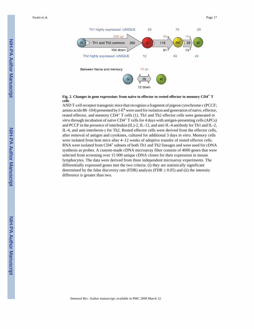
Fig. 2. Changes in gene expression: from naïve to effector to rested effector to memory CD4+ TcellsAND T-cell receptor transgenic mice that recognize a fragment of pigeon cytochrome c (PCCF;amino acids 88–104) presented by I-Ek were used for isolation and generation of naïve, effector,rested effector, and memory CD4+ T cells (1). Th1 and Th2 effector cells were generated invitro through incubation of naïve CD4+ T cells for 4 days with antigen-presenting cells (APCs)and PCCF in the presence of interleukin (IL)-2, IL-12, and anti-IL-4 antibody for Th1 and IL-2,IL-4, and anti-interferon-γ for Th2. Rested effector cells were derived from the effector cells,after removal of antigen and cytokines, cultured for additional 3 days in vitro. Memory cellswere isolated from host mice after 4–12 weeks of adoptive transfer of rested effector cells.RNA were isolated from CD4+ subsets of both Th1 and Th2 lineages and were used for cDNAsynthesis as probes. A custom-made cDNA microarray filter consists of 4000 genes that wereselected from screening over 15 000 unique cDNA clones for their expression in mouselymphocytes. The data were derived from three independent microarray experiments. Thedifferentially expressed genes met the two criteria: (i) they are statistically significantdetermined by the false discovery rate (FDR) analysis (FDR ≤ 0.05) and (ii) the intensitydifference is greater than two.
Swain et al. Page 17
Immunol Rev. Author manuscript; available in PMC 2008 March 12.
NIH
-PA Author Manuscript
NIH
-PA Author Manuscript
NIH
-PA Author Manuscript

Fig. 3. Maintenance of phenotypic heterogeneity: effector to memoryNaïve carboxyfluoresein succinimidyl ester (CFSE)-labeled CD4+ T cells from HNT.TCRTg.Thy1.1 mice (5 × 10⁶) were transferred into intact BALB/c hosts. Recipient mice wereinfected intranasally with 0.5 LD50 of influenza A virus (A/PR8/34) a day later (19). After day7 and 6 weeks postinfluenza infection, cell suspensions from lymphoid organs of individualmice (spleen, peripheral LN, draining LN) and nonlymphoid tissues (lung and bronchoalveolarlavage) were stained with anti-Thy1.1-biotin followed by streptavidin- allophycocyanin andanti-CD4-cychrome to identify donor T cells. The dot plots (one representative from lymphoidand nonlymphoid tissues) show the expression of CD44 and CD62L against residual CFSE ongated donor cells at effector and memory stages. The dot plots representing lymphoid tissuesshow that heterogeneity, in terms of subpopulations at different stages of cell division andexpression of CD44 and CD62L, were maintained into memory cells. Donor cells fromnonlymphoid tissues remained CFSEloCD44hiCD62Llo. The maintenance of phenotypicheterogeneity was similar for the expression of many other markers analyzed (CD49d, CD11a,CCR7). These data are a representation of three individual experiments using three mice pergroup.
Swain et al. Page 18
Immunol Rev. Author manuscript; available in PMC 2008 March 12.
NIH
-PA Author Manuscript
NIH
-PA Author Manuscript
NIH
-PA Author Manuscript

Fig. 4. Generating effector and memory heterogeneityA model depicting the postulated progressive differentiation of CD4+ T cells as they becomefully differentiated effector cells. The model indicates phenotypic and functional changes thatoccur. The expression of helper function in the distinct subsets has not yet been tested, asindicated by the question mark. Arrows indicate the direction of progression. It is suggestedthat most effector subsets can become small resting memory cells, except for the naïveprecursors and terminally differentiated effectors, which undergo activation-induced orprogrammed cell death.
Swain et al. Page 19
Immunol Rev. Author manuscript; available in PMC 2008 March 12.
NIH
-PA Author Manuscript
NIH
-PA Author Manuscript
NIH
-PA Author Manuscript

Fig. 5. Effect of antigen dose and duration and inflammation on effector differentiation and memoryThis figure depicts a summary of the results and conclusions in Jelley-Gibbs et al. (47). Whennaïve CD4+ influenza-specific indicator cells are introduced at day 0, they encounter highlevels of virus (pink lines) and inflammation (orange line). They expand (blue lines), becomingeffectors capable of high levels of interferon-γ (IFN-γ) production and nonlymphoid migrationto the lung. They also lose ability to produce interleukin (IL)-2. Naïve cells introduced atsuccessively later times (1 and 2 weeks are shown) expand less and have a less pronouncedeffector phenotype and function, as indicated by the triangles. Memory generation, in terms ofrecovery of memory cells at 4 or more weeks, is equivalent.
Swain et al. Page 20
Immunol Rev. Author manuscript; available in PMC 2008 March 12.
NIH
-PA Author Manuscript
NIH
-PA Author Manuscript
NIH
-PA Author Manuscript

Fig. 6. Mechanism of protection by Th1 effectors: a 1–2 punchThis illustration depicts the major mechanisms of protection revealed by the responses seen inwildtype and B-cell-deficient JhD mice. In both strains, transferred donor CD4+ Th1 cellsappear in the lung soon after transfer and then decay after a few days (red line). Antibodydevelops early (relative to a primary encounter) in the wildtype mouse (magenta solid line),but of course not in the JhD (magenta dotted line). Weight loss plateaus in both groups, butthe wildtype mice go on to gain weight (solid blue line) and recover, while the JhD mice againlose weight and die (dotted blue line). Antibody from convalescent wildtype mice, added insmall amounts (10 μL), rescues the JhD hosts (not shown, see Table 1). Thus, we suggest theCD4+ effector T cells work in two stages. First, they help in the clearance of infected cells(days 3–8) using cytotoxic and perhaps other mechanisms. Second, they help host B cells tomake antibody responses, which mediate viral clearance after 8 days (Brown, Dilzer, Meents,and Swain, manuscript submitted).
Swain et al. Page 21
Immunol Rev. Author manuscript; available in PMC 2008 March 12.
NIH
-PA Author Manuscript
NIH
-PA Author Manuscript
NIH
-PA Author Manuscript

Fig. 7. Protection due to CD4+ effector T-cell transferInfluenza-specific CD4+ Th1-polarized effectors were generated in vitro. Effectors weretransferred to adoptive hosts (dotted lines). Controls received no cells (solid lines). All micewere infected intranasally with 5 LD50 PR8 influenza virus. Depicted are levels of virus(log10 live viral titers) in blue, donor CD4+ T cells (log number in lung) in red, IgG antibodyto influenza in serum in green, and percentage weight loss.
Swain et al. Page 22
Immunol Rev. Author manuscript; available in PMC 2008 March 12.
NIH
-PA Author Manuscript
NIH
-PA Author Manuscript
NIH
-PA Author Manuscript
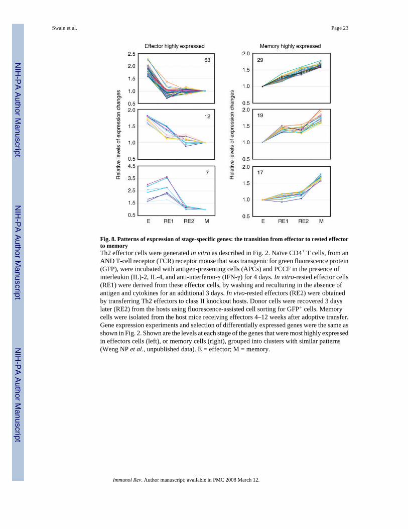
Fig. 8. Patterns of expression of stage-specific genes: the transition from effector to rested effectorto memoryTh2 effector cells were generated in vitro as described in Fig. 2. Naïve CD4+ T cells, from anAND T-cell receptor (TCR) receptor mouse that was transgenic for green fluorescence protein(GFP), were incubated with antigen-presenting cells (APCs) and PCCF in the presence ofinterleukin (IL)-2, IL-4, and anti-interferon-γ (IFN-γ) for 4 days. In vitro-rested effector cells(RE1) were derived from these effector cells, by washing and reculturing in the absence ofantigen and cytokines for an additional 3 days. In vivo-rested effectors (RE2) were obtainedby transferring Th2 effectors to class II knockout hosts. Donor cells were recovered 3 dayslater (RE2) from the hosts using fluorescence-assisted cell sorting for GFP+ cells. Memorycells were isolated from the host mice receiving effectors 4–12 weeks after adoptive transfer.Gene expression experiments and selection of differentially expressed genes were the same asshown in Fig. 2. Shown are the levels at each stage of the genes that were most highly expressedin effectors cells (left), or memory cells (right), grouped into clusters with similar patterns(Weng NP et al., unpublished data). E = effector; M = memory.
Swain et al. Page 23
Immunol Rev. Author manuscript; available in PMC 2008 March 12.
NIH
-PA Author Manuscript
NIH
-PA Author Manuscript
NIH
-PA Author Manuscript

Fig. 9. Stages of CD4+ T-cell responses: receptors and functionsDepicted are changes in known functions and related receptors and markers as naïve cellsbecome effector and then memory cells. We have suggested a bipartite separation that occursduring the effector to memory transition. Many functions and surface receptors associated withactivation state of the T cells go either up or down with effector development (blue, up; green,down), and these return to naïve levels when the cells become resting memory. Others, whichare associated with differentiation and the improved response potential of memory cells, stayat effector levels (orange, up; magenta, down).
Swain et al. Page 24
Immunol Rev. Author manuscript; available in PMC 2008 March 12.
NIH
-PA Author Manuscript
NIH
-PA Author Manuscript
NIH
-PA Author Manuscript

Fig. 10. Which effectors become memory cells?This figure represents the model we have developed based on our studies. As naïve CD4+ Tcells respond to infection, they progressively differentiate with successive rounds of divisionand exposure to growth and inflammatory cytokines, eventually becoming terminallydifferentiated effectors (as depicted also in Fig. 4). We suggest that all activated cells canbecome memory cells, except for those naïve cells that have not responded to antigen, and theend-stage effectors that either are destined to die or have progressed to a nonresponsive state(79).
Swain et al. Page 25
Immunol Rev. Author manuscript; available in PMC 2008 March 12.
NIH
-PA Author Manuscript
NIH
-PA Author Manuscript
NIH
-PA Author Manuscript

Fig. 11. Heterosubtypic CD4+ and CD8+ T-cell responsesB6 mice were uninfected (black) or infected with either PR8 (blue), cold-adapted Alaska (red),or cold-adapted Hong Kong (yellow) influenza virus to test whether heterosubtypic responsesto internal proteins (NP, PA) would be generated. Spleen cells from sublethally infected micewere tested for interferon-γ (IFN-γ)- and interleukin (IL)-2-producing enzyme-linkedimmunospots (ELISPOTs) following stimulation with influenza peptide-pulsed antigen-presenting cells (Strutt, Hollenbough, Dutton, Roberts, Woodland, and Swain, unpublisheddata). Peptides were identified using screening overlapping 15-mers, as described in our earlierreport (71).
Swain et al. Page 26
Immunol Rev. Author manuscript; available in PMC 2008 March 12.
NIH
-PA Author Manuscript
NIH
-PA Author Manuscript
NIH
-PA Author Manuscript

NIH
-PA Author Manuscript
NIH
-PA Author Manuscript
NIH
-PA Author Manuscript
Swain et al. Page 27
Table 1Protection from otherwise lethal influenza infection
Donor Host Degree of protection
None Intact, WT −Th1 (in vitro) Intact, WT ++++
T-cell deficient (Nu/Nu) ++++IFN-γ KO ++++B-cell deficient (JhD) ±JhD + flu antiserum (day 7) +++
Th1. IFN-γ KO Intact, WT ++++T-cell deficient (Nu/Nu) ++++IFN-γ KO ++++B-cell deficient (JhD) ±JhD + flu antiserum (day 7) +++
Th1. Perforin KO Intact ++Th2 (in vitro) Intact ++Th2. IFN-γ KO Intact ±Polyclonal (in vivo)Lung CD4+ +++DLN CD4+ +++Spleen CD4+ +++IFN-γ−/− Lung total +++IFN-γ−/− Lung CD4+ −IFN-γ−/− DLN CD4+ −IFN-γ−/− Spleen CD4+ −
In all cases, hosts were mice on a BALB/c background infected with a lethal dose of PR8 influenza. Donor CD4+ Th1 or Th2 effectors were generatedin vitro from HNT.T-cell receptor (TCR) transgenic (Tg) mice (19) that were otherwise normal, or were interferon (IFN)-γ KO, or perforin KO. Polyclonaleffectors were isolated from indicated organs at days 7–9 of an in vivo response of BALB/c mice to influenza. (Brown, Dilzer, Meents, and Swain,manuscript submitted). Degree of protection is a reflection of the rate of ‘survival’ of the mice.
Immunol Rev. Author manuscript; available in PMC 2008 March 12.
Copyright © 2022 FDOKUMEN