Heat-stable Antigen Is a Costimulatory Molecule for CD4 T ...
9
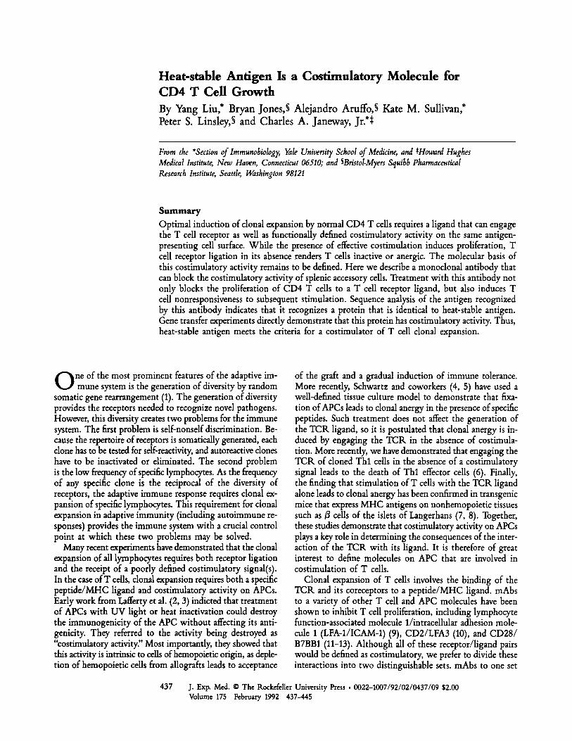
Heat-stable Antigen Is a Costimulatory Molecule for CD4 T Cell Growth By Yang Liu,* Bryan Jones,$ Alejandro Aruffo, S Kate M. Sullivan,* Peter S. Linsley,$ and Charles A. Janeway, Jr.*~ From the "Section of Immunobiology, Yale University School of Medicine, and SHowardHughes Medical Institute, New Haven, Connecticut 06510; and SBristol-Myers Squibb Pharmaceutical Research Institute, Seattle, Washington 98121 Summary Optimal induction of clonal expansion by normal CD4 T cells requires a ligand that can engage the T cell receptor as well as functionally defined costimulatory activity on the same antigen- presenting cell surface. While the presence of effective costimulation induces proliferation, T cell receptor ligation in its absence renders T cells inactive or anergic. The molecular basis of this costimulatory activity remains to be defined. Here we describe a monoclonal antibody that can block the costimulatory activity of splenic accessory cells. Treatment with this antibody not only blocks the proliferation of CD4 T cells to a T cell receptor ligand, but also induces T cell nonresponsiveness to subsequent stimulation. Sequence analysis of the antigen recognized by this antibody indicates that it recognizes a protein that is identical to heat-stable antigen. Gene transfer experiments directly demonstrate that this protein has costimulatory activity. Thus, heat-stable antigen meets the criteria for a costimulator of T cell clonal expansion. O ne of the most prominent features of the adaptive im- mune system is the generation of diversity by random somatic gene rearrangement (1). The generation of diversity provides the receptors needed to recognize novel pathogens. However, this diversity creates two problems for the immune system. The first problem is self-nonself discrimination. Be- cause the repertoire of receptors is somatically generated, each clone has to be tested for self-reactivity, and autoreactive clones have to be inactivated or eliminated. The second problem is the low frequency of specific lymphocytes. As the frequency of any specific clone is the reciprocal of the diversity of receptors, the adaptive immune response requires clonal ex- pansion of specific lymphocytes. This requirement for clonal expansion in adaptive immunity (including autoimmune re- sponses) provides the immune system with a crucial control point at which these two problems may be solved. Many recent experiments have demonstrated that the clonal expansion of all lymphocytes requires both receptor ligation and the receipt of a poorly defined costimulatory signal(s). In the case ofT cells, clonal expansion requires both a specific peptide/MHC ligand and costimulatory activity on APCs. Early work from Lafferty et al. (2, 3) indicted that treatment of APCs with UV light or heat inactivation could destroy the immunogenicity of the APC without affecting its anti- genicity. They referred to the activity being destroyed as "costimulatory activity" Most importantly, they showed that this activity is intrinsic to cells of hemopoietic origin, as deple- tion of hemopoietic cells from allografts leads to acceptance of the graft and a gradual induction of immune tolerance. More recently, Schwartz and coworkers (4, 5) have used a well-defined tissue culture model to demonstrate that fixa- tion of APCs leads to clonal anergy in the presence of specific peptides. Such treatment does not affect the generation of the TCR ligand, so it is postulated that clonal anergy is in- duced by engaging the TCK in the absence of costimula- tion. More recently, we have demonstrated that engaging the TCR of cloned Thl cells in the absence of a costimulatory signal leads to the death of Thl effector cells (6). Finally, the finding that stimulation of T cells with the TCR ligand alone leads to clonal anergy has been confirmed in transgenic mice that express MHC antigens on nonhemopoietic tissues such as B cells of the islets of Langerhans (7, 8). Together, these studies demonstrate that costimulatory activity on APCs plays a key role in determining the consequences of the inter- action of the TCR with its ligand. It is therefore of great interest to define molecules on APC that are involved in costimulation of T cells. Clonal expansion of T cells involves the binding of the TCR and its coreceptors to a peptide/MHC ligand, mAbs to a variety of other T cell and APC molecules have been shown to inhibit T cell proliferation, including lymphocyte function-associated molecule 1/intracellular adhesion mole- cule 1 (LFA-1/ICAM-1) (9), CD2/LFA3 (10), and CD28/ B7BB1 (11-13). Although all of these receptor/ligand pairs would be defined as costimulatory, we prefer to divide these interactions into two distinguishable sets. mAbs to one set 437 J. Exp. Med. The Rockefeller University Press 0022-1007/92/02/0437/09 $2.00 Volume 175 February 1992 437--445
-
Upload
khangminh22 -
Category
Documents
-
view
0 -
download
0
Transcript of Heat-stable Antigen Is a Costimulatory Molecule for CD4 T ...
Heat-stable Antigen Is a Costimulatory Molecule for CD4 T Cell Growth By Yang Liu,* Bryan Jones,$ Alejandro Aruffo, S Kate M. Sullivan,* Peter S. Linsley,$ and Charles A. Janeway, Jr.*~
From the "Section of Immunobiology, Yale University School of Medicine, and SHoward Hughes Medical Institute, New Haven, Connecticut 06510; and SBristol-Myers Squibb Pharmaceutical Research Institute, Seattle, Washington 98121
Summary Optimal induction of clonal expansion by normal CD4 T cells requires a ligand that can engage the T cell receptor as well as functionally defined costimulatory activity on the same antigen- presenting cell surface. While the presence of effective costimulation induces proliferation, T cell receptor ligation in its absence renders T cells inactive or anergic. The molecular basis of this costimulatory activity remains to be defined. Here we describe a monoclonal antibody that can block the costimulatory activity of splenic accessory cells. Treatment with this antibody not only blocks the proliferation of CD4 T cells to a T cell receptor ligand, but also induces T cell nonresponsiveness to subsequent stimulation. Sequence analysis of the antigen recognized by this antibody indicates that it recognizes a protein that is identical to heat-stable antigen. Gene transfer experiments directly demonstrate that this protein has costimulatory activity. Thus, heat-stable antigen meets the criteria for a costimulator of T cell clonal expansion.
O ne of the most prominent features of the adaptive im- mune system is the generation of diversity by random
somatic gene rearrangement (1). The generation of diversity provides the receptors needed to recognize novel pathogens. However, this diversity creates two problems for the immune system. The first problem is self-nonself discrimination. Be- cause the repertoire of receptors is somatically generated, each clone has to be tested for self-reactivity, and autoreactive clones have to be inactivated or eliminated. The second problem is the low frequency of specific lymphocytes. As the frequency of any specific clone is the reciprocal of the diversity of receptors, the adaptive immune response requires clonal ex- pansion of specific lymphocytes. This requirement for clonal expansion in adaptive immunity (including autoimmune re- sponses) provides the immune system with a crucial control point at which these two problems may be solved.
Many recent experiments have demonstrated that the clonal expansion of all lymphocytes requires both receptor ligation and the receipt of a poorly defined costimulatory signal(s). In the case ofT cells, clonal expansion requires both a specific peptide/MHC ligand and costimulatory activity on APCs. Early work from Lafferty et al. (2, 3) indicted that treatment of APCs with UV light or heat inactivation could destroy the immunogenicity of the APC without affecting its anti- genicity. They referred to the activity being destroyed as "costimulatory activity" Most importantly, they showed that this activity is intrinsic to cells of hemopoietic origin, as deple- tion of hemopoietic cells from allografts leads to acceptance
of the graft and a gradual induction of immune tolerance. More recently, Schwartz and coworkers (4, 5) have used a well-defined tissue culture model to demonstrate that fixa- tion of APCs leads to clonal anergy in the presence of specific peptides. Such treatment does not affect the generation of the TCR ligand, so it is postulated that clonal anergy is in- duced by engaging the TCK in the absence of costimula- tion. More recently, we have demonstrated that engaging the TCR of cloned Thl cells in the absence of a costimulatory signal leads to the death of Thl effector cells (6). Finally, the finding that stimulation of T cells with the TCR ligand alone leads to clonal anergy has been confirmed in transgenic mice that express MHC antigens on nonhemopoietic tissues such as B cells of the islets of Langerhans (7, 8). Together, these studies demonstrate that costimulatory activity on APCs plays a key role in determining the consequences of the inter- action of the TCR with its ligand. It is therefore of great interest to define molecules on APC that are involved in costimulation of T cells.
Clonal expansion of T cells involves the binding of the TCR and its coreceptors to a peptide/MHC ligand, mAbs to a variety of other T cell and APC molecules have been shown to inhibit T cell proliferation, including lymphocyte function-associated molecule 1/intracellular adhesion mole- cule 1 (LFA-1/ICAM-1) (9), CD2/LFA3 (10), and CD28/ B7BB1 (11-13). Although all of these receptor/ligand pairs would be defined as costimulatory, we prefer to divide these interactions into two distinguishable sets. mAbs to one set
437 J. Exp. Med. �9 The Rockefeller University Press �9 0022-1007/92/02/0437/09 $2.00 Volume 175 February 1992 437--445

of molecules disrupts T cell responses even in situations where clonal expansion is not critical, as in target cell recognition by killer T cells. Molecules of this class are involved in an- tigen recognition by the T cell. mAbs to the other set affect only clonal expansion. These recognize what we would call costimulators, as they regulate T cell behavior after an an- tigen is recognized. In the presence ofmAbs to such costimula- tory molecules, not only would clonal expansion be blocked, but also one would expect a state of T cell inactivation or anergy to be induced. By contrast, mAbs that prevent TCR signaling should prevent the induction of anergy. This pro- vides a test for distinguishing between the two sets of mAbs that prevent T cell clonal expansion.
In this report, we describe a mAb, 20C9, that blocks clonal expansion of normal CD4 T cells. Engagement of the TCR in the presence of this mAh leads to functional inactivation of T cells. Thus, this mAb meets the criteria for inhibition of the delivery of costimulatory activity by APCs. Expres- sion cloning and cDNA sequencing reveal that this mAb recog- nizes the heat-stable antigen. Gene transfer experiments confirm that heat-stable antigen has a direct costimulatory activity for clonal expansion of normal CD4 T cells.
Materials and Methods
Production ofmAbs. BALB/c ByJ and CBA/CaJ mice, used at 8-10 wk old, were purchased from The Jackson Laboratory (Bar Harbor, ME). Armenian hamsters (Cytogen Research and Devel- opment, West Roxbury, MA) were immunized by four consecu- tive intraperitoneal injections each of 107 LPS-activated B cells from CBA/CaJ mice. The spleen cells were fused to Ag8.653 and hybrids selected in HAT medium. The supernatants were screened for their ability to inhibit the proliferation of CD4 T cells purified from BALB/cByJ mice to the anti-CD3 mAb YCD3-1 (14) and irradiated LPS-activated syngeneic B cells as described (15). Posi- tive clones were subcloned three times, mAbs were purified from hybridoma supernatants on a protein G--Sepharose column.
Expression Cloning of 20C9 cDNA and Generation of CHO Trans- fectants. 20C9 cDNA was cloned as previously described (16). Briefly, a cDNA library was prepared from mKNA isolated from LPS- and IL-4-activated BALB/c spleen ceils and inserted into the pCDM8 expression vector. The cDNA library was transfected into COS cells by the DEAE-dextran method (16), COS cells were in- cubated with 20C9 mAb, and the cells expressing 20C9 protein were isolated by panning onto plates coated with goat anti-ham- ster IgG (Caltag Laboratories, San Francisco, CA). Episomal DNA was recovered from the adherent cells, amplified in Escherichia coli, and reintroduced into COS cells. After three rounds of transfec- tion and panning, plasmid DNA was prepared from individual bac- terial colonies and transfected into COS cells. The cell surface expression of 20C9 protein was determined by indirect immuno- fluorescence.
cDNA from clone 20C9C7 was transfected into CHO cells that had previously been transfected with the gene encoding FcKIIB2 (kind gift of Dr. Ira Mellman, Yale University; 17), according to previously described procedures (11). Briefly, a mixture of 10 #g of 20C9C7 DNA and 1 #g of EBOpLPP vector carrying a hygromycin-resistance gene was used to transfect COS cells by lipofection. These ceils were then selected with DMEM containing 0.4 mg/ml of hygromycin and 5% FCS. Positive cells were sorted 2 wk later in a FACStar Plus | cell sorter (Becton Dickinson & Co.), and individual clones were obtained by limiting dilution.
438
Assays for Proliferation of CD4 T Cells. Both normal CD4 T cells and cloned Thl ceils were used in this study. Normal CD4 T cells were prepared from CBA/CaJ and BALB/cByJ mice as de- scribed (15). The Thl clone 5.9 has also been described fully (18). B ceUs were prepared from spleen nonadherent ceils after two rounds of anti-Thy-1 mAb (HO 13.4.9; reference 19) and complement lysis, and were activated with 10 ~tg/ml of LPS (15). These B cells were either irradiated (2,000 rad) or fixed with 1% paraformaldehyde (15) before being used as accessory cells. In some experiments, acti- vated B ceUs (107/ml) were incubated with 100 #g/ml of mAb for i h at 4~ before fixation. Transfectants of CHO cells were treated with mitomycin C (100 #g/ml) before use as accessory cells. Un- less specified in the text, CD4 T cells were purified as previously described and incubated with accessory cells and a 1:40 dilution of YCD3-1. Proliferation was determined after 42 h by pulsing the culture for 6 h with 1 #Ci/well of [~H]TdR. Results of dupli- cate/triplicate cultures in which the variation was g20% are reported.
Cleavage of Glffosyl-Phosphatidylinositol (GPI)' Linkage by Phosphoinositol-s~cific Phospholipase C (PI-PLC) and Flow Cytometry. A20 cells (10Vml) in serum-free Click's EHAA medium were in- cubated with PI-PLC (152354; ICN Biochemicals, Cleveland, OH), at a 1:200 dilution at 37~ in a water bath for 1 h. The enzyme was washed away with PBS, and the treated and untreated cells were stained with 20C9 mAb undiluted hybridoma supernatant followed by FITC-labeled goat anti-hamster IgG (mouse and rat Ig adsorbed; Cahag Laboratories). Anti-Mac-1 mAb M1/70 (20), anti-FcR mAb 2.4G2 (21), and anti-heat-stable antigen mAbs Jlld (22) and M1/69 (23) were also used in flow cytometry and/ or proliferation assays, which was performed as described pre- viously (15).
Results A mAb that Inhibits T Cell Clonal Expansion by Blocking
the Delivery of Costimulatory Activity by APC. To study the clonal expansion of normal CD4 T cells, we have used anti- CD3 mAb-induced proliferation of CD4 T cells as a model system. Our previous work has demonstrated that B cells are the major APC for this response in murine spleen (15). Activation with LPS induces costimulatory activity in B cells that is resistant to aldehyde fixation. To generate mAbs that can block the delivery of this costimulatory signal for T cell activation, we immunized hamsters with LPS-activated B cells and made hybridomas. Supematants were screened for inhibi- tory effects on the proliferation of normal CD4 T cells to anti-CD3 mAb and irradiated, LPS-activated B cells as acces- sory cells. The hybridomas that secreted inhibitory mAbs were subcloned three times. One of these inhibitory rnAbs, 20C9, is characterized here. 20C9 significantly inhibits the prolifer- ation of CD4 T calls induced by anti-CD3 (Fig. 1 a) or by allogeneic LPS-activated B calls (Fig. 1 b). When a cloned T cell is used as a responder, 20C9 blocks proliferation of this cloned line to its antigen, OVA (Fig. 1 c). 20C9 also inhibits proliferation of total spleen cells to anti-CD3 (see Fig. 3 a), indicating that the antibody has a broad spectrum
1 Abbreviations used in this paper: GPI, glycosyl-phosphatidylinositol; PI- PLC, phosphoinosltol-specific phospholipase C.
Heat-stable Antigen Is a Costimulator of T Cell Clonal Expansion
E D,. 0
20C9
103 . . . . . . , ....... . ....... 10 0 101 10 2 10 3
10 s
1 / D i l u t i o n
80000'
60000 '
40000
20000
0' 1
80000"
60000-
40OOO
~ , 20000-
"% 0 4 1 0 s 1 0 e
Accessory cells
Med 20C9
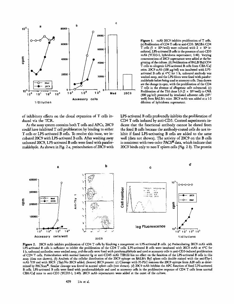
Figure 1. mAb 20C9 inhibits proliferation of T cells. (a) Proliferation of CD4 T cells to anti-CD3. BALB/c CD4 T cells (5 x 10⁴/we11) were cultured with 2 x 10⁴ ir- radiated, LPS-activated B cells in the presence of anti-CD3 mAb (YCD3-1, hybridoma supernatant; 1:40). Varying concentrations of 20C9 supernatant were added at the be- ginning of the culture. (b) Proliferation ofBALB/ByJ CD4 T cells to allogenic LPS-actiwted B cells from CBA/CaJ mice. 20C9 mAb (100 #g/ml) was incubated with LPS- activated B cells at 4~ for 1 h, unbound antibody was washed away, and the LPS-blasts were fixed with parafor- maldehyde before being used as accessory cells. Data shown are the change in cpm, with the proliferation of the CD4 T cells in the absence of allogeneic cells subtracted. (c) Proliferation of the Thl clone 5.9 (2 x 10⁴/weU) to OVA (100 #g/ml) presented by irradiated adherent cells (105/ well) from BALB/c mice. 20C9 mAb was added at a 1:2 dilution of hybridoma supernatant.
of inhibitory effects on the clonal expansion of T cells in- duced via the TCR.
As the assay system contains both T cells and APCs, 20C9 could have inhibited T cell proliferation by binding to either T cells or LPS-activated B cells. To resolve this issue, we in- cubated 20C9 with LPS-activated B cells. After washing away unbound 20C9, LPS-activated B cells were fixed with parafor- maldehyde. As shown in Fig. 2 a, preincubation of 20C9 with
LPS-activated B cells profoundly inhibits the proliferation of CD4 T cells induced by anti-CD3. Control experiments in- dicate that the functional antibody cannot be eluted from the fixed B cells because the antibody-coated cells do not in- hibit if fixed LPS-activating B cells are added to the same well (data not shown). The activity of 20C9 on the B cells is consistent with two-color FACS | data, which indicate that 20C9 binds only to non-T spleen cells (Fig. 2 b). The protein
a
E Q. 0
400000-
300000 �9
200000"
100000
I 20C9
03 1 04 1 0 s 1 0 s
A c c e s s o r y cel ls /wel l
k-
4 ~:"~ ~ .
i ' i?--"
�9 .~L ,%',~ .
~:; ~-y*~
.~ 7reatmert 0erecting mad nil1 20~g PI -~LC n~
----Pl-~LC 20C9
L N
log Fluorescence
10 s o--o--o--o--o
I - - 2 0 C 9
~ Med
E ~ 10 3
0 ~ 1 01 1 0 2 1 0 3
20C9 1 / d i l u t i o n s
Figure 2. 20C9 mAb inhibits proliferation of CD4 T cells by blocking a component on LPS-activated B cells. (a) Preincubating 20C9 mAb with LPS-activated B cells is sufficient to inhibit the proliferation of the CD4 T cells. LPS-activated B cells were incubated with 20C9 mAb at 4~ for 1 h, unbound antibodies were washed away, and the cells were fixed with parafotmaldehyde and used as accessory cells in anti-CD3-induced proliferation of CD4 T cells. Preincubation with normal hamster Ig or anti-CD4S mAb TIB122 has no effect on the function of the LPS-activated B cells in this assay (data not shown). (b) Analysis of the cellular distribution of the 20(29 epitope on BALB/c ByJ spleen cells double stained with the anti-Thy-1 mAb Y19 and with 20C9. (~p) No 20C9 added; (bottom) 20C9 present. (c) Cleavage with PI-PLC removes the 20C9 epitope from A20 cells as deter- mined by FACScan | Similar cleavage was found in normal spleen cells (not shown). (d) 20C9 mAb inhibits the APC function of fixed LPS-activated B cells. LPS-activated B cells were fixed with paraformaldehyde and used as accessory cells in the proliferative response of CD4 T cells from normal CBA/CaJ mice to anti-CD3 (YCD3-1; 1:40). 20C9 mAb supernatants were added at the onset of the culture.
439 Liu et al.

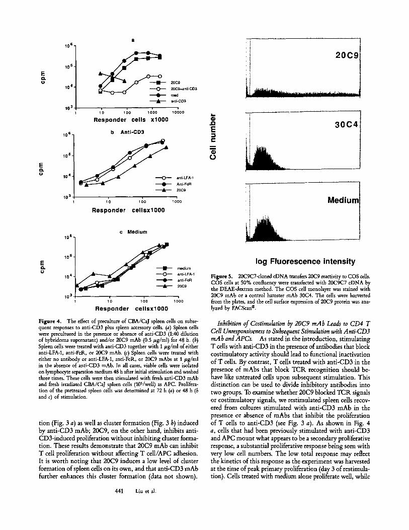
Figure 3. Inhibition of anti-CD3 mAb-induced proliferation and cluster formation of spleen cells by various mAbs. Spleen cells (lOS/weU) from CBA/CaJ mice were incubated with a 1:40 dilution of YCD3-1 together with either anti-LFA-1 mAb M17/5.2, anti-FcR mAb 2.4G2, or 20C9 at various concentrations. Proliferation of the spleen cells (a) was determined at 48 h of culture. Clusters (b) were photographed at 16 h of culture in the presence of anti-CD3 alone or with 1 /zg/ml of the inhibitory mAbs.
that this antibody recognizes is anchored to the cell mem- brane by a GPI linkage, as PI-PLC can specifically cleave the 20C9 epitope from B cells (Fig. 2 c). Furthermore, this anti- body strongly inhibits the accessory cell function of fixed B cells (Fig. 2 d). As paraformaldehyde-faxed B cells are meta- bolically inert, it is most likely that 20C9 mAb binds to and inhibits the action of a component expressed on LPS-activated B cells, which is necessary for proliferation of CD4 T cells. 20C9 mAb does not bind to the FcR, as it binds to FcR+ A20 cells and an FcR- variant equally well (data not shown). In addition, 20C9 mAb does not affect the function of the FcR. on LPS-activated B cells in anti-CD3-induced ac-
tivation of cloned T cells (Liu, Y., and C. A. Janeway, manu- script submitted for publication). Thus, 20C9 mAb inhibits T cell proliferation by a mechanism other than affecting the function of the FcR.
Many mAbs specific for accessory molecules required for T cell activation inhibit the formation of T cell APC clusters. 20C9 inhibits anti-CD3-induced proliferation of spleen cells without inhibiting cluster formation induced by anti-CD3 mAb. This contrasts with the effects of anti-LFA-1 mAb M17/5.2 (20) and anti-FcR mAb 2.4G2 (21), which inhibit cluster formation as well as .proliferation. Anti-LFA-1 mAb M17.5.2 and anti-FcR mAb 2.4G2 inhibit spleen prolifera-
440 Heat-stable Antigen Is a Costimulator of T Cell Clonal Expansion
E es o
E r O
10 6,
10 s . ~ ~ ~
20C9 10 4. -- . ,- ,On 20C9+anti-CD3
r r~d J- anti-CD3
103 , , 1'0 100 1000 10000
Responder cells x l 0 0 0
10 s] b Anti-CD3
1 0 s ]
104 1 anti-LFA-1 Anti-FoR
' ' 20C9
10311 1000
Responder c e l l s x l O 0 0
z.- 4)
,,,Q
E r,,
(3
.I �9 ' I
20C9
30C4
Medium
E es r
c Med ium 10 6
10 s
medium
104 anti-LFA- I anti-FcR 20C9
10 3 l i i 1 1 0 1 00 1000
Responder cel lsxl000
log Fluorescence intensity
Figure 5. 20C9C7-cloned cDNA transfers 20C9 reactivity to COS cells. COS cells at 50% confluency were transfected with 20C9C7 cDNA by the DEAE-dextran method. The COS cell monolayer was stained with 20C9 mAb or a control hamster mAb 30C4. The cells were harvested from the plates, and the cell surface expression of 20C9 protein was ana- lyzed by FACScan |
Figure 4. The effect of preculture of CBA/CaJ spleen cells on subse- quent responses to anti-CD3 plus spleen accessory cells. (a) Spleen cells were precuhured in the presence or absence of anti-CD3 (1:40 dilution of hybridoma supernatant) and/or 20C9 mAb (0.5/zg/ml) for 48 h. (b) Spleen calls were treated with anti-CD3 together with 1 #g/ml of either anti-LFA-1, anti-FcR, or 20C9 mAb. (c) Spleen cells were treated with either no antibody or anti-LFA-1, anti-FcR, or 20C9 mAbs at 1/zg/ml in the absence of anti-CD3 mAb. In all cases, viable cells were isolated on lymphocyte separation medium 48 h after initial stimulation and washed three times. These calls were then stimulated with fresh anti-CD3 mAb and fresh irradiated CBA/CaJ spleen cells (105/we11) as APC. Prolifera- tion of the pretreated spleen cells was determined at 72 h (a) or 48 h (b and c) of stimulation.
tion (Fig. 3 a) as well as cluster formation (Fig. 3 b) induced by anti-CD3 mAb; 20C9, on the other hand, inhibits anti- CD3-induced proliferation without inhibiting duster forma- tion. These results demonstrate that 20C9 mAb can inhibit T cell proliferation without affecting T cell/APC adhesion. It is worth noting that 20C9 induces a low level of duster formation of spleen cells on its own, and that anti-CD3 mAb further enhances this cluster formation (data not shown).
Inhibition of Costimulation by 20C9 mAb Leads to CD4 T Cell Unresponsiveness to Subsequent Stimulation with Anti-CD3 mAb and APCs. As stated in the introduction, stimulating T cells with anti-CD3 in the presence of antibodies that block costimulatory activity should lead to functional inactivation of T ceils. By contrast, T cells treated with anti-CD3 in the presence of mAbs that block TCR recognition should be- have like untreated ceUs upon subsequent stimulation. This distinction can be used to divide inhibitory antibodies into two groups. To examine whether 20C9 blocked TCK signals or costimulatory signals, we restimulated spleen cells recov- ered from cultures stimulated with anti-CD3 mAb in the presence or absence of mAbs that inhibit the proliferation of T cells to anti-CD3 (see Fig. 3 a). As shown in Fig. 4 a, ceils that had been previously stimulated with anti-CD3 and APC mount what appears to be a secondary proliferative response, a substantial proliferative response being seen with very low cell numbers. The low total response may reflect the kinetics of this response as the experiment was harvested at the time of peak primary proliferation (day 3 of restimula- tion). Cells treated with medium alone proliferate wdl, while
441 Liu et al.
cells that were pretreated with anti-CD3 in the presence of 20C9 mAb make only a marginal proliferative response re- quiring very high cell numbers, indicating that the pretreat- ment induces functional inactivation of the T cells. The reduc- tion in response observed is -o20-fold relative to untreated cells. This inactivation requires exposure to anti-CD3 in the first culture and was not due to the carry-over of the 20C9 mAb, because treatment of the cells with 20C9 mAb in the absence of anti-CD3 mAb allows a proliferative response in the second culture similar to that of untreated cells. These data suggest that engaging the TCR in the absence of 20C9 protein leads to functional inactivation of the T cells.
As seen in Fig. 3 a, mAbs directed at several different cell surface molecules can prevent clonal expansion of CD4 T cells induced by anti-CD3 and APCs. To test whether treatment with mAbs that interfere with cell adhesion (anti-LFA-1) or TCR ligation (anti-FcR) also results in functional inactiva- tion of T cells exposed to anti-CD3, we treated spleen cells with anti-LFA-1, anti-FcR, and 20C9 mAbs in the presence or absence of anti-CD3 mAb. 2 d later, the antibodies were washed away and the viable cells were restimulated with anti- CD3 in the presence of competent APC. As shown in Fig. 4 b, cells treated with anti-LFA-1 and anti-FcR mAbs plus anti-CD3 generate a vigorous proliferative response upon re- stimulation, comparable with that of untreated spleen cells (Fig. 4 c), while those pretreated with 20C9 plus anti-CD3
give a significantly reduced response. As the dose of all the mAbs used (1 #g/ml) can almost totally inhibit the primary proliferative response of T cells to anti-CD3 (Fig. 3 a), these results indicate that anti-CD3 combined with anti-LFA-1 or anti-FcR mAb does not induce functional inactivation of the T ceils, in contrast to anti-CD3 plus 20C9 mAb, nor do we observe the augmented response induced by anti-CD3 alone, as seen in Fig. 4 a, in any of these cultures. The inhibition observed with 20C9 mAb was not due to carry-over of the mAb in the culture, as spleen cells treated with 20C9 in the absence of the anti-CD3 mAb mount a normal proliferative response in secondary stimulation (Fig. 4 c). Thus, the inac- tivation observed requires both anti-CD3 and the presence of blocking concentrations of 20C9.
Taken together, the above data indicate that 20C9 anti- body blocks clonal expansion of T cells induced by anti-CD3, allogeneic LPS-activated B ceils, and antigen presented by syn- geneic spleen cells. As 20C9 does not affect ligand recogni- tion by the TCR or cell adhesion, it is likely to block a costimulatory signal. This conclusion is supported by the finding that exposure of T cells to anti-CD3 in the presence of 20C9 leads to functional inactivation of T cells.
Expression Cloning of 20C9 Antigen Reveals Its Identity with Heat-stable Antigen, To clone the gene encoding the 20C9 antigen, COS cells were transfected with a pCDM8-cDNA library prepared from LPS-activated spleen B cells. Positive
q) .Io E (..
m atom
o
oh a ;~ ~ J11d
. . . . . . _
. . . . . . . . . . . . M1/69
NO B lock ing Ab
IB B 1~ I 1~i2 103 V I , 2
IO
b 2oc9 . . . . . . 20C9 , ~
NO B lock ing A b ~ I f.., J q / "~
~- ; ,
, ' , % . , , , , . . , , , , 1 . ,
rL2
Ceil Abs
CliO-FoR 0 . . . . . . . CliO-FoR 20C9 . . . . . . . . . . . . . CHO-FcR-20C9 0 . . . . . CHO-FcR-2OC9 20C9
�9 j . / , t '~
.... .... ........
F'L[
. . d A c.. , ,
l: ! _ _ CHO-FcR J l l d { ! . . . . . . CHO-FcR M1/69
" ~ i ....... CHO-FcR-20C9 J l l d ~. f [ . . . . CHO-FcR-20C9 M1/69
�9 . / i r ~
- / / i. " - J J i
V l _ I
log Fluorescence
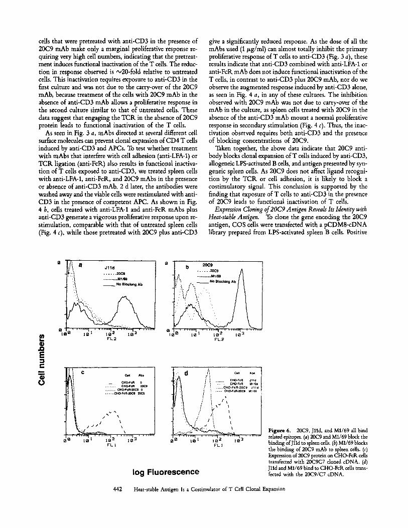
Figure 6. 20C9, Jlld, and M1/69 all bind related epitopes. (a) 20(29 and M1/69 block the binding of Jlld to spleen cells. (b) M1/69 blocks the binding of 20(29 mAb to spleen cells. (c) Expression of 20C9 protein on CHO-FcR cells transfected with 20C9C7 cloned cDNA. (d) Jlld and M1/69 bind to CHO-FcR cells trans- fected with the 20C9/C7 cDNA.
442 Heat-stable Antigen Is a Costimulator of T Cell Clonal Expansion
clones were enriched by four cycles of transfection followed by panning for 20C9-positive COS cells. Plasmid DNA pre- pared from the fourth cycle of panning was found to transfer 20C9 reactivity to COS cells. The plasmid DNA was cloned, and of 55 clones tested, four transferred 20C9 reactivity to COS cells. As shown in Fig. 5, COS cells transfected with cDNA from one of those dones (20C9C7) bound 20C9 mAb but not 30C4, a control hamster mAb. The sequence of this cDNA clone demonstrated that it was the same as that of the heat-stable antigen (24). Furthermore, Jlld binding to spleen cells was inhibited by 20C9 and M1/69, while 20C9 binding to spleen cells was inhibited by M1/69 (Fig. 6, a and b). The identity of these antigens was further confirmed by finding that CHO cells transfected with 20C9C7 cDNA gain reactivity with 20C9, Jlld, and M1/69 (Fig. 6, c and d). However, mAbs J l ld and M1/69 were not effective in inhibiting T cell proliferation to anti-CD3 mAb (data not shown), suggesting that these mAbs differ in either their binding sites or affinity for heat-stable antigen.
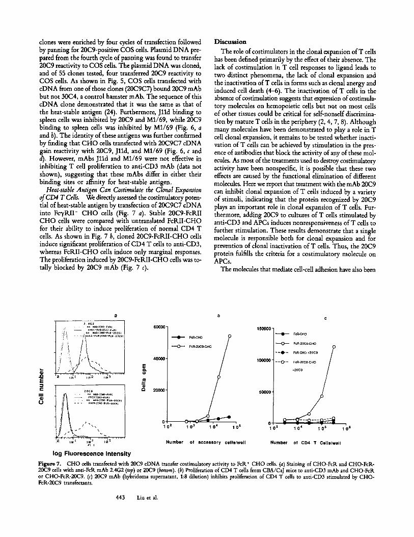
Heat-stable Antigen Can Costimulate the Clonal Expansion ofCD4 T Cells. We directly assessed the costimulatory poten- tial of heat-stable antigen by transfection of 20C9C7 cDNA into FcyRII + CHO cells (Fig. 7 a). Stable 20C9-FcRII CHO cells were compared with untranslated FcRII-CHO for their ability to induce proliferation of normal CD4 T cells. As shown in Fig. 7 b, cloned 20C9-FcRII-CHO cells induce significant proliferation of CD4 T ceils to anti-CD3, whereas FcRII-CHO cells induce only marginal responses. The proliferation induced by 20C9-FcRII-CHO cells was to- tally blocked by 20C9 mAb (Fig. 7 c).
Discussion
The role of costimulators in the clonal expansion of T cells has been defined primarily by the effect of their absence. The lack of costimulation in T cell responses to ligand leads to two distinct phenomena, the lack of clona] expansion and the inactivation of T cells in forms such as clonal anergy and induced cell death (4-6). The inactivation of T cells in the absence of costimulation suggests that expression of costimula- tory molecules on hemopoietic cells but not on most cells of other tissues could be critical for self-nonself discrimina- tion by mature T cells in the periphery (2, 4, 7, 8). Although many molecules have been demonstrated to play a role in T cell clonal expansion, it remains to be tested whether inacti- vation of T cells can be achieved by stimulation in the pres- ence of antibodies that block the activity of any of these mol- ecules. As most of the treatments used to destroy costimulatory activity have been nonspecific, it is possible that these two effects are caused by the functional elimination of different molecules. Here we report that treatment with the mAb 20C9 can inhibit clonal expansion of T cells induced by a variety of stimuli, indicating that the protein recognized by 20C9 plays an important role in clonal expansion of T cells. Fur- thermore, adding 20C9 to cultures of T cells stimulated by anti-CD3 and APCs induces nonresponsiveness of T cells to further stimulation. These results demonstrate that a single molecule is responsible both for clonal expansion and for prevention of clonal inactivation of T cells. Thus, the 20C9 protein fulfills the criteria for a costimulatory molecule on APCs.
The molecules that mediate cell-cell adhesion have also been
2 . 4G2 �9 . n o = l L b i C a o - r e , i )
.'t!! - - .o~:L-,'oRC:~O-,'~ = . . . . . n o m i b ( c l i o - r ~ l - 2 � 9
! .~ - _ ~ / ~ a n t ~ - 1 e K f C R 0 - / c e - 2 0 C l
e ~ e- Q
20C9 �9 �9 . . o = u ~ ( c J o - r e a )
- - 2 0 e l ( C H o - r = R ) . . . . . n o ~ b ( r
- - . 2 o c l ( c a o - r ~ A . 2 o c g l
log Fluorescence intensity
60000'
40000"
20000-
0 1 0 2
F c R - C H O
i
1 03 1 04 1 ()s
150000"
100000 -
50000 -
FcR-CHO
F c R - 2 0 c g - c H o
- ~ l " - F c R ~ I - I O + 2 0 C 9
/ "-<>-:;2:: ~176 /
0 1 - v - ";" 0 3 1 0 4 1 0 s
N u m b e r of a c c e s s o r y c e l l s / w e l l N u m b e r o f C D 4 T Ce l l s /we l l
Figure 7. CHO cells transfeeted with 20C9 cDNA transfer costimulatory activity to FcR* CHO cells. (a) Staining of CHO-FcR and CHO-FcR- 20C9 cells with anti-FcR mAb 2.4G2 (top) or 20C9 (bottom). (b) Proliferation of CD4 T cells from CBA/CaJ mice to anti-CD3 mAb and CHO-FcR or CHO-FcR-20cg. (c) 20C9 mAb (hybridoma supernatant, 1:8 dilution) inhibits proliferation of CD4 T cells to anti-CD3 stimulated by CHO- FcR-20C9 transfectants.
443 Liu et al.
regarded as costimulatory molecules because antibodies that block their function can block T cell proliferation, while trans- fection of these molecules into fibroblasts can enhance the T cell response to ligand. As T cells normally respond to peptide/MHC on the surface of APCs, cell-cell contact is a prerequisite for TCK engagement. We suggest that it is appropriate to differentiate those molecules that are required for ligand recognition, that is, delivery of signal one, from costimulatory molecules that deliver signal two. Despite their similar effects on T cell proliferation, the distinctive effects of anti-LFA-1 and 20C9 on cluster formation and on the in- duction of unresponsiveness clearly discriminates these two classes of molecules.
Molecular cloning and partial DNA sequencing reveals that the gene encoding the 20C9 protein is identical to the previ- ously cloned gene encoding heat-stable antigen. Transfection with cDNA encoding heat-stable antigen is sufficient to transfer costimulatory activity to CHO cells, demonstrating directly that heat-stable antigen costimulates T cells. It has been documented that expression of the heat-stable antigen is tightly controlled in immature T cells and in B cells at different functional stages (25). The function of this mole- cule has remained elusive. We show here that heat-stable an- tigen cDNA from activated B cells encodes a costimulatory molecule that can participate in clonal expansion of CD4 T cells.
The heat-stable antigen contains a very small peptide core with a large number of potential N-linked and O-linked glycosylation sites. The structural basis of its costimulatory activity is at present not understood. It is worth noting that the level of 20C9 binding activity does not correlate directly with the costimulatory activity of a cell. Normal B cells do not have constitutive costimulatory activity (15), yet they bind 20C9 significantly. This discrepanc.y can be explained at least
in two ways. First, normal B cells may express an inhibitor for the costimulatory activity of the heat-stable antigen, and this inhibitor is inactivated during B cell activation. Second, the heat-stable antigen may be modified during B cell activa- tion. Since different cell types seem to have different glycosy- lation patterns of the heat-stable antigen (24), one possibility would be that glycosylation regulates the costimulatory ac- tivity of the heat-stable antigen. This notion is consistent with the earlier findings by Frohman and Cowing (26), who showed that treatment with neuraminidase can enhance T cell stimulation by B cells.
Adaptive immune responses by ceUs having clonally dis- tributed receptors must discriminate self from nonsdf. Many experiments have demonstrated that donal deletion mediated by the interaction of immature thymocytes with antigen on APCs is a major mechanism for removing from the mature repertoire those T cells that recognize ligands borne by APCs (27, 28). This mechanism, however, does not apply to an- tigens that are expressed in tissues but are not present on APCs in the thymus (7, 8). Thus, there must be mechanisms to ensure self-nonsdf discrimination by mature T ceils in the periphery. TCK engagement on mature T cells leads to a number of different consequences depending on the costimula- tory activity of the cells that present antigens (4-6). Self an- tigens presented by tissue cells induce immune tolerance by inactivating specific T cells (7, 8), whereas antigens borne by microbes that induce costimulatory activity on APCs in- duce potent adaptive immune responses (29, 30). Although much data are consistent with the two-signal theory (30-32), this hypothesis has not been subjected to a stringent ex- perimental test because the nature of signal two on APCs has been obscure. Hopefully, with the identification of co- stimulatory molecules such as B7 (11-13) and heat-stable an- tigen, the two-signal theory itself can now be directly tested.
We thank Barry Jones for A20 and its FcK variant, Ira MeUman for CHO-FcKIIB2 transfectant, Anne Brancheau for preparation of the manuscript, and Jane Dunn for assistance in hybridoma production.
This work was supported by National Institutes of Health grant AI-26810 (to C. A. Janeway, Jr.), and by the Bristol Myers Squibb Company. Y. Liu is a recipient of an Irvington Institute post-doctoral fellowship.
Address correspondence to Y. Liu, Division of Immunology, Department of Pathology, New York University Medical Center, 550 First Avenue, New York, NY 10016.
Received.for publication 23 September 1991.
R~fl~rences
1. Davis, M.M., and P.J. Bjorkman. 1988. T cell antigen receptor genes and T cell recognition. Nature (Lond.). 334:395.
2. Lafferty, K.J., S.J. prowse, and C.J. Simeonovich. 1983. Im- munobiology of tissue transplantation: a return to passenger leukocyte concept. Annu. Rev. Imraunol. 1:143.
3. Lafferty, K.J., L. Andrus, and S.J. Prowse. 1980. Role oflym- phokine and antigen in the control of T cell responses. Ira-
munol. Rev. 51:279. 4. MueUer, D.L., M.K. Jenkins, and K.H. Schwartz. 1989. Clonal
expansion vs. functional clonal inactivation: a costimulatory signalling pathway determines the outcome of T cell antigen receptor occupancy. Annu. Rev. lraraunol. 7:445.
5. Jenkins, M.K., D.M. Pardoll, J. Mizugnchi, H. Quill, and K.H. Schwartz. 1987. T cell unresponsiveness in vivo and in vitro:
444 Heat-stable Antigen Is a Costimulator of T Call Clonal Expansion
fine specificity in induction and molecular characterization of the unresponsive state. ImmunoL Rev. 95:113.
6. Liu, Y., and C.A. Janeway, Jr. 1990. Interferon 7' plays a crit- ical role in induced cell death of effector T cells: a possible third mechanism of self-tolerance. J. Extx Med. 172:1735.
7. Morahan, G., J. Allison, and J.F.A.P. Miller. 1989. Tolerance of class I histocompatibility antigens expressed extrathymically. Nature (Lond.). 339:622.
8. Burkly, L., D. Lo, O. Kanagawa, R.L. Brinster, and R.A. Flavell. 1989. Clonal anergy of I-E tolerant T cells in trans- genic mice with pancreatic expression of MHC class II I-E. Nature (Lond.). 342:562.
9. Springer, T.A. 1990. Adhesion receptors of the immune system. Nature (Lond.). 346:425.
10. Bierer, B.E., B.P. Sleckman, S.E. Ratnofsky, and S.J. Burakoff. 1989. Biological role of CD2, CD4, and CD8 in T-cell activa- tion. Annu. Rev. Immunol. 7:579.
11. Linsley, P.S., E.A. Clark, andJ.A. Ledbetter. 1990. The T cell antigen, CD28, mediates adhesion with B cells by interacting with the activation antigen B7/BB1. Proc Natl. Acad. Sci. USA. 87:5031.
12. Linsley, P.S., W. Brady, L. Grosmaire, A. Aruffo, N.K. Damle, and J.A. Ledbetter. 1991. Binding of the B cell activation an- tigen B7 to CD28 costimulates T cell proliferation and inter- leukin 2 mRNA accumulation. J. Extz Med. 173:721.
13. Koulova, L., E.A. Clark, G. Shu, and B. Dupont. 1991. The CD28 ligand B7/BB1 provides costimulatory signal for alloac- tivation of CD4 T cells. J. Extx Ailed. 173:759.
14. Portoles, P., J. Rojo, A. Golby, M. Bonneville, S.H. Grom- kowski, L. Greenbaum, C.A. Janeway, Jr., D.B. Murphy, and K. Bottomly. 1989. Monoclonal antibodies to murine CD3e define new epitopes, one of which may interact with CD4 during T cell activation. J. Immunol. 142:4169.
15. Liu, Y., and C.A. Janeway, Jr. 1991. Microbial induction of costimulatory activity for CD4 T cell growth. Int. lmmunol. 3:323.
16. Seed, B., and A. Aruffo. 1987. Molecular cloning of the CD2 antigen, the T cell erythrocyte receptor by rapid immunoselec- tion procedure. Proc. Natl. A_cad. Sci. USA. 84:3365.
17. Miettinen, H., J.K. Rose, and I. Mellman. 1989. Fc receptor isoforms exhibit distinct abilities for coated pit localization as a result of cytoplasmic domain heterogeneity. Cell. 58:317.
18. Conrad, P., and C.A. Janeway, Jr. 1984. The expression of the I-E d molecule in F1 hybrid mice detected with antigen- specific, I-E d restricted cloned T cell lines. Immunogenetics.
20:311. 19. Marshak-Rothstein, A., P. Fink, T. Gridley, D.H. Raulet, M.J.
Bevan, and M.L. Gefter. 1981. Properties and application of monoclonal antibodies directed against determinants of the Thyl locus. J. lmmunol. 122:2491.
20. Sanchez-Madrid, F., P. Simon, S. Thompson, and T.A. Springer. 1983. Mapping of antigenic and functional epitopes on the oe and ~ subunits of the two related mouse glycoproteins involved in cell interaction: LFA-1 and Mac-1. J. Exl~ Med. 158:586.
21. Unkeless, J.C. 1979. Characterization of a monoclonal anti- body directed against macrophage-lymphocyte Fc receptor. J. Exl~ Med. 150:1152.
22. Bruce, J., F.W. Symington, T.J. McKearn, andJ. Sprent. 1981. A monoclonal antibody discriminating between strands of T and B cells. J. Immunol. 127:2496.
23. Springer, T., G. Galfre, D.S. Secher, and C. Milstein. 1978. Monoclonal xenogeneic antibodies to murine cell surface an- tigen: identification of novel differentiation antigens. Eur. J. Immunol. 8:539.
24. Kay, R., F. Takei, and R.K. Humphries. 1990. Expression cloning of a cDNA encoding M1/69-J11d heat-stable antigens. J. Immunol. 145:1952.
25. Crispe, I.N., and M.J. Bevan. 1987. Expression and functional significance of the J11d marker on mouse thymocytes. J. Im- munol. 138:2013.
26. Frohman, M., and C. Cowing. 1985. Presentation of antigen by B cells: functional dependence of radiation dose, interleukins, cellular activation and differential glycosilation. J. lmmunol. 134:2269.
27. Kiesielow, P., H. Bluthmann, U.D. Staerz, M. Steinmetz, and H. yon Boehmer. 1988. Tolerance in transgenic mice involves deletion of nonmature CD4+CD8 + thymocytes. Nature (Lond.). 333:742.
28. Kappler, J.W., T. Wade, J. White, E. Kushnir, M. Blackman, J. Bill, N. Roehm, and P. Marrack. 1987. Tolerance by clonal elimination in the thymus. Cell. 49:273.
29. Janeway, C.A., Jr. 1989. Immunogenicity: signal 1, 2, 3, and O. Immunol. Today. 10:283.
30. Janeway, C.A., Jr. 1989. Approaching the asymptote? Evolu- tion and revolution in immunology. Cold Spring Harbor Syml~ Quant. Biol. 54:1.
31. Bretscher, P.A., and M. Cohn. 1970. The theory of self non- self discrimination. Science (Wash. DC). 169:1042.
32. Schwartz, R.H. 1989. Acquisition of immunological self- tolerance. Cell. 57:1073.
445 Liu et al.