The fate of the duplicated androgen receptor in fishes: a late neofunctionalization event?
Upload
independentCategory
view
0download
0
Immunity
Article
Roquin Differentiates the SpecializedFunctions of Duplicated T Cell CostimulatoryReceptor Genes Cd28 and IcosMichelle A. Linterman,1 Robert J. Rigby,1 Raphael Wong,1 Diego Silva,1 David Withers,2 Graham Anderson,2
Naresh K. Verma,3 Robert Brink,4 Andreas Hutloff,5 Chris C. Goodnow,1,6 and Carola G. Vinuesa1,*1Division of Immunology and Genetics, John Curtin School of Medical Research, Australian National University, Canberra 2601, Australia2MRC Centre for Immune Regulation, University of Birmingham, Birmingham B15 2TT, UK3School of Biochemistry and Molecular Biology, Australian National University, Canberra 2601, Australia4Garvan Institute of Medical Research, Sydney, NSW 2010, Australia5Robert Koch Institute, Nordufer 20, Berlin 13353, Germany6Australian Phenomics Facility, Canberra 2601, Australia*Correspondence: [email protected]
DOI 10.1016/j.immuni.2008.12.015
SUMMARY
During evolutionary adaptation in the immunesystem, host defense is traded off against autoreac-tivity. Signals through the costimulatory receptorCD28 enable T cells to respond specifically topathogens, whereas those through the related costi-mulatory receptor, ICOS, which arose by gene dupli-cation, are critical for affinity maturation and memoryantibody responses. ICOS ligand, unlike the path-ogen-inducible CD28 ligands, is widely and constitu-tively expressed in the immune system. Here, weshow that crosstalk between these two pathwaysprovides a mechanism for obviating the normalT cell dependence on CD28. Several CD28-mediatedresponses—generation of follicular helper T cells,germinal center formation, T helper 1 cell-dependentextrafollicular antibody responses to Salmonella andbacterial clearance, and regulatory T cell homeo-stasis—became independent of CD28 and depen-dent on ICOS when the E3 ubiquitin ligase Roquinwas mutated. Mechanisms to functionally compart-mentalize ICOS and CD28 signals are thus criticalfor two-signal control of normal immune reactions.
INTRODUCTION
The appearance of high-affinity memory antibody responses
through B cell selection in germinal centers in phylogeny—in
birds and mammals—coincides with the appearance of the
inducible T cell costimulator (Icos) gene (Bernard et al., 2007).
ICOS is critical in man and mouse for this specialization of the
antibody response (Bossaller et al., 2006; Dong et al., 2001;
Grimbacher et al., 2003; McAdam et al., 2000; Tafuri et al.,
2001), and it is most highly expressed on a specialized subset
of effector CD4+ T cells within the germinal center, T follicular
helper (Tfh) cells (Hutloff et al., 1999). Phylogeny, sequence
homology, and genomic localization indicate that Icos emerged
228 Immunity 30, 228–241, February 20, 2009 ª2009 Elsevier Inc.
by duplication of the adjacent and evolutionarily more ancient
gene, Cd28, encoding a general T cell costimulatory receptor
(Bernard et al., 2007). Genomic analysis has indicated that
gene duplication provides an important substrate for evolution
(Ohno et al., 1968), but little is known about how duplicated
paralogous genes resolve adaptive conflicts while acquiring
specialized functions that confer a selective advantage. This
general question of conflict between paralogs is particularly
acute in the case of Cd28 and Icos because substitution by
ICOS for the role normally played by CD28 has the potential to
violate the two-signal mechanism of discrimination between
pathogens and self.
Little is known about the extent to which ICOS can substitute
for CD28 to promote in vivo immune responses. This question
becomes particularly important in a context in which the CD28
system is not active: a response against self. Biochemically,
both Cd28 and Icos encode T cell costimulatory receptors
sharing 39% amino acid identity and, in transformed cell lines,
largely overlapping capacity to activate the PI3K intracellular
signaling pathway and enhance T cell receptor (TCR)-induced
gene expression, cytokine synthesis, and cytokine proliferation
(Rudd and Schneider, 2003). Some biochemical specialization
exists: CD28 is the main inducer of IL-2, whereas ICOS is
a poor IL-2 inducer because of its inability to recruit the signaling
adaptor Grb2 (Harada et al., 2003; Watanabe et al., 2006). Also,
ICOS lacks a unique motif present within the polyproline cyto-
plasmic domain of CD28; such a motif is specific for binding the
protein tyrosine kinase Lck and important for CD28-dependent
Treg cell differentiation in the thymus (Tai et al., 2005). In addition
to this biochemical specialization, CD28 and ICOS exhibit highly
specialized patterns of expression. CD28 is constitutively ex-
pressed on most T cells, whereas ICOS is expressed at very
low amounts on naive T cells, is upregulated in all effector and
memory T cell subsets, including Treg cells, and is most highly ex-
pressed on the specialized Tfh cells located within germinal
centers (Beier et al., 2000; Gross et al., 1992; Hutloff et al., 1999).
CD28 has a well-understood role in T cell discrimination of self
and microbial antigens because microbial antigens are accom-
panied by ligands for pathogen-recognition receptors such as
those of the Toll-like receptor family, which induce expression
Immunity
Roquin Compartmentalizes Cd28 and Icos
of the CD28 ligands, CD80 and CD86 (Greenwald et al., 2005;
Sharpe and Freeman, 2002). Self-antigen peptides presented
in the absence of CD80 and CD86 signaling engage the TCR,
but not CD28, inducing tolerance responses such as anergy or
apoptosis (Harding et al., 1992; Matzinger, 1994). Simultaneous
delivery of two signals—engagement of the TCR by antigenic
peptide-major histocompatibility complex (MHC) complexes
and engagement of CD28 by CD80 and CD86—delivers a quali-
tatively and quantitatively enhanced intracellular signal to induce
T cell immune responses selectively in cells that recognize
microbial antigens (Janeway and Bottomly, 1994; Lafferty
et al., 1980; Matzinger, 1994). By contrast with CD80 and
CD86, ICOS ligand (ICOSL) is widely and constitutively ex-
pressed in the absence of microbial stimuli on antigen-present-
ing cells and several nonhemopoietic tissues, including endothe-
lium (Brodie et al., 2000; Ling et al., 2000; Swallow et al., 1999).
Should ICOS retain overlapping costimulatory function sufficient
to substitute for CD28, the constitutive presence of ICOSL could
bypass the need for microbial induction of costimulatory ligands.
Although this might be a benefit in the case of processed micro-
bial antigens that are retained in an inert state for months or years
within germinal centers, it would potentially undo a key self-path-
ogen discrimination mechanism and lead to autoimmunity.
Analysis of mice and humans lacking ICOS, CD28, or both has
emphasized the specialization of these two costimulatory recep-
tors. T cell-dependent immunity is dramatically depressed in
Cd28-deficient mice (Shahinian et al., 1993), which show
a complete absence of T cell-dependent germinal centers and
Tfh cells, impaired extrafollicular antibody responses to Salmo-
nella coupled with reduced pathogen clearance (McSorley and
Jenkins, 2000; Mittrucker et al., 1999), and a dramatic (>80%)
reduction in Treg cells (Ferguson et al., 1996; Salomon et al.,
2000; Tai et al., 2005; Tang et al., 2003; Walker et al., 1999).
By contrast, Icos�/� mice remain competent to form germinal
center B cells, Tfh cells, and Treg cells, albeit in reduced
numbers (Akiba et al., 2005; Bossaller et al., 2006; Burmeister
et al., 2008; Dong et al., 2001; McAdam et al., 2000; Tafuri
et al., 2001). Once germinal centers are established, CD28 is
dispensable for Tfh cell and germinal center survival and selec-
tion of somatically mutated B cells into the memory pool (Walker
et al., 2003), but these processes are profoundly impaired in
mice and humans that lack ICOS or ICOSL, who have a near-
total absence of memory B cells (Bossaller et al., 2006; Dong
et al., 2001; Grimbacher et al., 2003; McAdam et al., 2000; Tafuri
et al., 2001). Although combined deficiency of CD28 and ICOS
leads to a more severe defect in T cell-dependent antibody
response than deficiency of either alone (Suh et al., 2004), this
is consistent with sequential specialized roles and does not
resolve the question of whether ICOS might substitute for
CD28 in either a beneficial or harmful way.
The mechanisms that control expression and activity of CD28,
ICOS, and their ligands are only beginning to be revealed. Here,
we address the question of specialization versus overlap
between ICOS and CD28 and the consequences of changes in
their control mechanisms by analyzing an autoimmune mouse
mutant, sanroque, with a defect in the Rc3h1 gene encoding
the E3 ubiquitin ligase Roquin, that does not affect the sequence
of Cd28 or Icos but alters the stability of Icos messenger RNA
(mRNA) (Li et al., 2007; Vinuesa et al., 2005a; Yu et al., 2007).
We show that ICOS can indeed substitute for CD28 in the induc-
tion of in vivo immune responses, including primary antibody
responses, germinal center B cell formation, and homeostasis
of Foxp3+ Treg cells. These overlapping functions of the ICOS
costimulatory receptor are normally compartmentalized by Ro-
quin’s control on Icos mRNA, so that the important role of
CD28 in self-pathogen discrimination is not normally overturned
by ICOS. Pathological breakdown of this compartmentalization
may be important in autoimmune disease, whereas controlled
breakdown in Tfh cells may be a physiological adaptation to
assist selection of high-affinity antibodies in germinal centers.
RESULTS
Rc3h1san/san T Cells Overexpress ICOS in the Absenceof CD28Homozygosity for the M199R ‘‘Rc3h1san/san’’ mutation in Roquin
causes aberrant overexpression of ICOS on the surface of both
naive T cells and memory and effector T cells (Vinuesa et al.,
2005a). Signaling through CD28 during T cell priming has previ-
ously been shown to be essential for optimal upregulation of
ICOS (McAdam et al., 2000). To examine whether signaling
through CD28 is necessary for the overexpression of ICOS by
Rc3h1san/san T cells, we generated Rc3h1san/san Cd28�/� mice
and used flow cytometry to assess ICOS expression on periph-
eral blood CD44lo (naive) and CD44hi (effector and memory)
CD4+ T cells. CD28 deficiency only exerted a 30% correction
of ICOS expression on Rc3h1san/san T cells. The expression of
ICOS on naive CD4+ T cells and effector and memory CD4+
T cells was still more than 2-fold higher in Rc3h1san/san Cd28�/�
mice compared with Rc3h1+/+ mice (Figures 1A and 1B), indi-
cating that the M199R substitution in Roquin uncouples ICOS
expression from CD28-driven T cell activation.
As shown previously, ICOS is hyperinduced in activated
CD44hi cells in Rc3h1san/san mice (Figure 1B; ICOS mean fluores-
cence intensity [MFI] in naive CD44lo cells = 35.90 ± 9.50; ICOS
MFI in activated cells = 131.00 ± 13.00). This hyperinduction is
also seen after activation of Rc3h1san/san Cd28�/� mice
(Figure 1B): Indeed, a comparable �4-fold increase in ICOS
expression is seen in Rc3h1san/san Cd28�/� activated cells
(ICOS MFI = 91.15 ± 12.45) compared with naive cells from the
same mice (ICOS MFI = 21.80 ± 1.80). These data indicate that
ICOS is still overexpressed on Rc3h1san/san CD4+ T cells lacking
CD28.
Mutant Roquin Rescues Germinal Center Formationin CD28-Deficient MiceGiven the ability of Rc3h1san/san CD4+ T cells to express ICOS
independently of CD28 and the potential for ICOS to transmit
comparable costimulatory signals to CD28, we asked whether
the Rc3h1san/san mutation would restore CD28-dependent anti-
body responses in the absence of CD28. Signaling through
CD28 has been shown to be essential for the initiation of T cell-
dependent antibody responses and formation of germinal
centers (Ferguson et al., 1996; Walker et al., 2003). Previously,
it has been shown that Rc3h1san/san mice form spontaneous
germinal centers but mount normal antibody responses to foreign
antigens (Vinuesa et al., 2005a) (Figure 2A). We therefore exam-
ined the CD28 dependence of the antibody response to a model
Immunity 30, 228–241, February 20, 2009 ª2009 Elsevier Inc. 229

Immunity
Roquin Compartmentalizes Cd28 and Icos
protein antigen—chicken gammaglobulin (CGG). Whereas anti-
CGG IgG1 was undetectable in Rc3h1+/+ Cd28�/� mice 14
days after immunization, Rc3h1san/san Cd28�/� mice mounted
a robust antibody response, albeit lower than that seen in mice
expressing CD28 (Figure 2A). Thus, the Rc3h1san/san mutation
relieves the dependence on CD28 for this antibody response.
To analyze the cellular basis for the Rc3h1san/san effect on
CD28 dependence, we next examined the formation of germinal
centers 8 days after intraperitoneal immunization with sheep red
blood cells (SRBCs). Consistent with previous reports, CD28-
deficient mice failed to form GL-7+ CD95+ B220+ cells after
immunization (Figure 2C), and we were unable to observe PNA
staining in the IgD+ B cell follicles (Figure 2D). The absence of
CD28 eliminated the formation of spontaneous germinal centers
in unimmunized Rc3h1san/san mice (Figure 2B), but remarkably,
Rc3h1san/san Cd28�/� mice mounted robust germinal center
responses upon SRBC immunization that were greater than
those of immunized wild-type (Rc3h1+/+ Cd28+/+) mice
(Figure 2E).
The Rc3h1san/san mutation could relieve the CD28 dependence
of germinal center responses either by acting within the B cells or
in helper T cells or other cells needed to support the B cell
expansion. To distinguish between these alternatives, we
made mixed bone marrow chimeras in which sublethally irradi-
ated C57BL/6 Ly5a recipients were reconstituted with a 1:1
mix of Rc3h1san/san.Ly5b and Rc3h1+/+.Ly5a bone marrow or
a control Rc3h1+/+.Ly5b and Rc3h1+/+.Ly5a mix. Analysis of
these chimeras after SRBC immunization revealed that the
increase in germinal center B cells observed in recipients of
Rc3h1san/san.Ly5b: Rc3h1+/+.Ly5a mixed bone marrow con-
sisted of nearly equal cell numbers of Roquin wild-type (Ly5a)
and Roquin mutant (Ly5b) origin. This indicates that most of
Figure 1. Increased ICOS Expression on
Naive and Effector-Memory Rc3h1san/san
Cd28�/� CD4+ T Cells
(A) Representative flow-cytometric histograms
showing ICOS MFI of CD4+CD44lo (top panel) or
CD4+CD44hi (bottom panel) on peripheral blood
T cells from mice with the indicated genotypes.
Specificity of ICOS staining is determined by the
use of Icos�/� mice as a control.
(B) Bar graphs show ICOS MFI for the mice shown
in (A). Each circle represents one mouse, and bars
represent the median values for each group. ICOS
MFI is different between all groups in the CD44hi
analysis, with a statistical significance of p <
0.0001. Within the CD44lo comparison, all groups
also have a statistically significant difference of
p < 0.0001, with the exception of Rc3h1+/+
Cd28+/+ versus Rc3h1+/+ Cd28�/�, in which signif-
icance reached p = 0.0011. Data are representa-
tive of four experiments.
the increase in germinal center response
caused by the Rc3h1san/san mutation is
B cell extrinsic (Figure 2F).
In order to conclusively confirm that the
Rc3h1san/san mutation rescued selection
of antigen-specific germinal center B
cells in Cd28�/� mice, and that this is not due to B cell intrinsic
factors, we used the ‘‘SWHEL’’ B cell transfer system (Paus
et al., 2006; Phan et al., 2003). C57BL/6 B cells carrying
a ‘‘knocked-in’’ B cell receptor specific for hen egg lysozyme
(HEL) were injected into the tail vein of recipient mice together
with SRBCs conjugated to HEL2x, and the HEL-specific germinal
center response by the donor cells was assessed 5 days after
immunization (Figure 3A). In recipients with normal Roquin, the
absence of CD28 diminished the germinal center response by
SWHEL B cells to �2% of that in CD28-sufficient hosts, confirm-
ing that this process is normally CD28 dependent. By contrast,
CD28-deficient recipients with mutant Roquin were capable of
supporting the formation of immunogen-specific germinal center
B cells equally to Rc3h1+/+Cd28+/+, Rc3h1san/sanCd28+/+, and
Rc3h1san/san mice (Figure 3B). Taken together, these results indi-
cate that Rc3h1san/san can rescue the germinal center response
in CD28-deficient animals through a B cell-extrinsic mechanism.
The Sanroque Mutation in Roquin Restores the Numberof Tfh Cells in CD28-Deficient MiceThe absence of germinal centers in Cd28�/� mice is linked with
deficient T cell priming and the absence of differentiated helper
CD4+ T cells, including Tfh cells (Walker et al., 1999). Tfh cells
provide selection signals to somatically mutated germinal center
B cells that have increased their affinity for the immunizing
antigen, signals that cause them to differentiate into long-lived
antibody-secreting plasma cells or memory B cells. Tfh cells
are also required for maintaining germinal center reactions,
possibly through enabling the recycling of selected germinal
center B cells to undergo further rounds of division and somatic
hypermutation (Breitfeld et al., 2000; Kim et al., 2001; Schaerli
et al., 2000; Vinuesa et al., 2005b).
230 Immunity 30, 228–241, February 20, 2009 ª2009 Elsevier Inc.

Immunity
Roquin Compartmentalizes Cd28 and Icos
We assessed Tfh numbers by enumerating CXCR5+ CD4+
cells that also expressed the inhibitory receptor PD-1. Human
Tfh cells are known to express high amounts of PD-1 mRNA
(Chtanova et al., 2004). Several groups, including our own,
have identified Tfh cells in mice in the past, only on the basis
of CXCR5 expression on CD4+ T cells. Nevertheless, discrimina-
tion of Tfh cells on the basis of CXCR5 staining alone is
unsatisfactory because of the relatively low expression of this
chemokine receptor on T cells and the nonselective induction
of CXCR5 in vitro in all primed T cells. Analysis of follicular
T cells by immunohistochemistry revealed that PD-1 is in fact
the best marker when used alone for specifically identifying
germinal center T cells, with minimal PD-1 staining observed
Figure 2. Rc3h1san/san Rescues Germinal
Center Responses in Cd28�/� Mice
(A) Anti-CGG IgG1 antibody titers 0 and 14 days
after immunization measured by ELISA in Cd28+/+
or Cd28�/� mice that are either Rc3h1+/+ (white
bars) or Rc3h1san/san (black bars).
(B) Germinal center B cells (B220+ GL-7+ CD95+)
expressed as a percentage of total B220+ cells
from unimmunized Cd28+/+ or Cd28�/� mice that
are either Rc3h1+/+(white bars) or Rc3h1san/san
(black bars).
(C) Flow-cytometric contour plots showing GL-7
versus CD95 staining gated on B220+ cells from
mice with the indicated genotypes 8 days after
SRBC immunization. Oval gates show the
germinal center B cell subset (GL-7+ CD95+).
(D) Photomicrographs of frozen spleen sections
stained with the germinal center marker PNA
(blue) and IgD (brown) in Rc3h1+/+ Cd28�/� (top)
and Rc3h1san/san Cd28�/� (bottom) 8 days after
SRBC immunization.
(E) Bar graph showing the percentage of germinal
center B cells gated with the gates shown in (C) in
mice with the indicated genotypes 8 days after
SRBC immunization.
(F) Germinal center B cell percentages from 1:1
bone marrow chimeras 8 days after SRBC immu-
nization. The left panel shows results for three
recipients reconstituted 12 weeks earlier with
a 1:1 mix of Rc3h1+/+.Ly5a and Rc3h1+/+.Ly5b
bone marrow; the right panel shows results for
three recipients reconstituted 12 weeks earlier with
a 1:1 mix of Rc3h1+/+.Ly5a and Rc3h1san/san.Ly5b
bone marrow. In all bar graphs, each circle repre-
sents one mouse, and bars represent the median
values for each group. Data are representative of
two (A) or four (B–F) experiments.
anywhere else in the spleen (Figure 4A)
(Haynes et al., 2007). In fact, most
TCRb+ T cells in germinal centers are
PD1+. Also, combining CXCR5 with PD-1
provides better discrimination of this
subset by flow cytometry than using
CXCR5 alone (Figure 4B).
Unimmunized Rc3h1san/san mice accu-
mulate Tfh cells; this is partially corrected
in CD28-deficient Rc3h1san/san mice,
which have background Tfh cell levels comparable to those of
wild-type mice, although these are still 4-fold higher than those
seen in Cd28�/� mice (Figure 4C). This subset expands even
further after immunization in both Rc3h1san/san mice and control
littermates (Figure 4D). To determine whether the Rc3h1san/san
mutation could correct the deficiency in Tfh cells observed in
Cd28�/� mice, we assessed the number and percentage of
splenic Tfh cells (CD4+CXCR5+PD-1+) by flow cytometry 8
days after SRBC immunization. Both the percentage and abso-
lute number of Tfh cells were restored in Rc3h1san/san Cd28�/�
mice to levels comparable to those of wild-type mice (Figures
4D and 4E). These data show that Rc3h1san/san can restore Tfh
cell formation in CD28-deficient animals.
Immunity 30, 228–241, February 20, 2009 ª2009 Elsevier Inc. 231

Immunity
Roquin Compartmentalizes Cd28 and Icos
Rc3h1san/san Breaks Compartmentalization betweenCD28L and ICOSL in Germinal Center ResponsesWe speculated that Roquin ensures the compartmentalization
between CD28 ligands and ICOS ligands. Nevertheless, an alter-
native second hypothesis explaining the observed results could
be that Rc3h1san/san dysregulates signaling downstream from
CD28; in other words, Rc3h1san/san may allow for signaling
through the CD28 pathway in the absence of CD28. If this
were the case, removing ICOSL would not have an effect. To
test this hypothesis, we generated Rc3h1san/san Cd28�/�
Icosl�/� mice. The SRBC-induced germinal center response in
these triple-loci-altered animals was 75% reduced compared
to that in Rc3h1san/san Cd28�/� mice, indicating that the rescue
of the germinal center response is largely dependent on intact
signaling through ICOS-ICOSL and that a response that is nor-
mally ICOSL independent has become ICOSL dependent in
Rc3h1san/san Cd28�/� mice. Rc3h1san/san Cd28�/� Icosl�/�
mice were still capable of forming a small number of GL-7+
CD95+ B220+ cells (Figure 4F), suggesting that ICOS- and
CD28-independent pathways also contribute to the
Rc3h1san/san-mediated rescue of the germinal center response
in CD28-deficient mice. We next assessed the contribution of
signaling through ICOS to the correction of Tfh cell numbers in
Rc3h1san/san Cd28�/� mice. Similar to the germinal center B cell
response, the percentage and number of Tfh cells was 4-fold
lower in Rc3h1san/san Cd28�/� Icosl�/� mice 8 days after SRBC
immunization (Figure 4G). These findings support the idea that
Rc3h1san/san breaks down the compartmentalization of signals
through CD28 and ICOS and excludes the hypothesis that
Rc3h1san/sa allows signaling through the CD28 pathway in the
absence of ICOS.
We also considered the possibility that a defect in cytotoxic T
lymphocyte antigen-4 (CTLA-4) signaling may contribute to the
observed enhancement of germinal center reactions. Mice
transgenic for a soluble CTLA-4-immunoglobin (Ig) fusion protein
Figure 3. Rc3h1san/san Restoration of the
Germinal Center Response in Cd28�/�
Mice Is B Cell Extrinsic
(A) Gating strategy used for identifying the HEL-
specific germinal center response from trans-
ferred SWHEL B cells. Cells in the top panel have
been gated on a live lymphocyte gate and on
B220.
(B) Bar graphs showing HEL-specific donor-
derived germinal center B cells as a proportion
of B220+ cells (top panel) or the total number per
spleen (bottom panel) 5.5 days after immunization
with HEL2x SRBCs in mice with the indicated
genotypes. Each symbol represents one mouse.
Bars represent the median values in each group.
Data are representative of two experiments.
have been shown to form germinal
centers when immunized in conjunction
with a CD28 agonist. However, these
germinal centers do not appear to invo-
lute, with the response still being present
40 days after immunization, indicating
that CTLA-4 signaling helps terminate
germinal center reactions (Walker et al., 2003). We investigated
whether Rc3h1san/san Cd28�/� mice show prolonged germinal
center reactions typical of defective CTLA-4 signaling.
Rc3h1san/san Cd28�/� germinal centers peaked at approximately
day 8 after SRBC immunization and, like controls, were already
reduced at day 14, and only background numbers of cells
were present 30 days after immunization (data not shown). This
suggests that CTLA-4 is functioning normally in Rc3h1san/san
Cd28�/� mice to induce the involution of germinal center reac-
tions. CTLA-4 expression was also slightly elevated in activated
Roquinsan/san CD4+ T cells and in Rc3h1san/san Treg cells
(Figure S1 available online). Taken together, these data indicate
that the Rc3h1san/san-dependent rescue of germinal center and
Tfh cell formation in CD28-deficient mice relies on signaling
through the ICOS-ICOSL pathway.
Rc3h1san/san Breaks Down CD28L-ICOSL Discriminationto Salmonella InfectionTo assess whether Rc3h1san/san can also break the compart-
mentalization of CD28 and ICOS in responses to infectious
agents, we immunized mice with live attenuated Salmonella en-
terica serovar Dublin strain SL5631. The response to Salmonella
has been shown to be critically dependent on CD4+ T cells (Hess
et al., 1996; Yrlid and Wick, 2000). Furthermore, CD28 costimu-
lation appears to be essential for T helper 1 (Th1) cell-mediated
bacteria clearance and intact IgG2ab production (McSorley
and Jenkins, 2000; Mittrucker et al., 1999). Although antibody
production does not contribute to the resolution of a primary
infection, it has been shown to be essential to protect against
Salmonella reinfection (McSorley and Jenkins, 2000). An
attractive feature of this model is that the IgG2ab plasma cell
response during the first weeks after the infection is exclusively
extrafollicular, and germinal centers do not form until after
5 weeks postinfection (Cunningham et al., 2007). This also
allows us to test whether Roquin plays a role in ensuring the
232 Immunity 30, 228–241, February 20, 2009 ª2009 Elsevier Inc.

Immunity
Roquin Compartmentalizes Cd28 and Icos
compartmentalization between CD28 ligands and ICOS ligands
during extrafollicular antibody responses.
Twelve days after Salmonella infection, CD28-deficient mice
had more than ten times higher bacterial counts in the liver
(Figure 5A), and despite having intact anti-Salmonella IgM
production (Figure 5B), they were unable to produce any
Salmonella-specific IgG2ab (Figure 5C) antibodies. Immunohis-
tochemistry confirms previous work showing that this response
is occurring outside the B cell follicle and that the number of
CD138+ cells is greatly reduced in CD28-deficient mice
Figure 4. Restoration of Tfh Cell Formation
and Germinal Centers in Rc3h1san/san
Cd28�/� Mice Requires Signaling through
the ICOS-ICOSL Pathway
(A) Photomicrographs of frozen spleen sections
stained with IgD (brown), PD-1 (blue, top panel),
TCRb (blue, middle panel), and PNA (blue, bottom
panel).
(B) Gating strategy for CD4+B220�CXCR5+PD1+
Tfh cells.
(C) Bar graphs show CD4+CXCR5+PD-1+ (Tfh)
cells as a percentage of splenic CD4+ T cells as-
sessed by flow cytometry from unimmunized
mice with the indicated genotypes.
(D) Representative flow-cytometric contour plots
showing PD-1 versus CXCR5 staining of mice
with the indicated genotypes 8 days after immuni-
zation with SRBCs. Gates have been drawn
around the CXCR5hi PD1hi population represent-
ing Tfh cells. A stringent cell-gating strategy
allowed exclusion of dead cells, nonlymphoid
cells, B cells, B:T cell duplets, and autofluorescent
cells (Figure 1B).
(E) Bar graphs show CD4+CXCR5+PD-1+ (Tfh)
cells gated as shown in (B) as a percentage of
splenic CD4+ T cells from mice with the indicated
genotypes 8 days after SRBC immunization.
(F) Percentage of germinal center B cells in mice
with the indicated genotypes 8 days after SRBC
immunization. Observed differences between all
the genotype groups reached statistical signifi-
cance of p = 0.05.
(G) Percentage of Tfh cells in mice with the indi-
cated genotypes 8 days after SRBC immunization.
Observed differences between all the genotype
groups reached statistical significance of p <
0.01, with the exceptions of Rc3h1+/+ Cd28�/�
versus Rc3h1san/san Cd28�/�and Rc3h1+/+ Cd28�/�
versus Rc3h1san/san Cd28�/� Icosl�/�, in which
significance reached p < 0.001, and of Roquin+/+
Cd28+/+ versus Rc3h1san/san Cd28�/�, in which no
significant difference was observed. Data are repre-
sentative of four experiments.
(Figure 5D). These defective responses
were completely rescued in Cd28�/�
mice homozygous for the san allele of
Rc3h1. Again, this rescue was shown
to be dependent on ICOSL substituting
for CD28 ligands because bacterial
counts and Salmonella-specific IgG2ab
titers in Rc3h1san/san Cd28�/� Icosl�/�
mice were comparable to those of Rc3h1+/+ Cd28�/� Icosl+/+
mice (Figures 5A–5D).
A third alternative hypothesis that could explain the observed
rescue is that even in the presence of intact CD28L signaling,
there might be a role for ICOSL in the response. If this were the
case, ICOSL deficiency would confer a phenotype in CD28+/+
mice. To investigate this possibility, we also assessed the
response in ICOSL-deficient mice. Unlike the defective
responses observed in CD28�/� mice, these responses were
mostly intact in ICOSL�/� mice, confirming that ICOSL does
Immunity 30, 228–241, February 20, 2009 ª2009 Elsevier Inc. 233

Immunity
Roquin Compartmentalizes Cd28 and Icos
not play a major role in the extrafollicular IgG2ab response and
the T cell response that mediates bacterial clearance in CD28+/+
animals. Together, these results show that Roquin also ensures
the compartmentalization between CD28 ligands and ICOS
ligands during T cell-mediated responses to pathogens,
including extrafollicular antibody responses.
The Rc3h1san/san Allele Restores Homeostasisof Peripheral Treg Cells in Cd28�/� MiceHaving established that the critical function of CD28 in the estab-
lishment of adaptive immunity can become CD28 independent in
the presence of Roquinsan/san, we investigated whether this also
applied to the other major role of CD28: the support of Treg cell
development and homeostasis. Mice deficient in CD28 have
a >80% decrease in the numbers of thymic and peripheral
Figure 5. Rc3h1san/san Corrects the Path-
ogen-Clearance Defect and the IgG2ab
Response in CD28-Deficient Mice Inocu-
lated with Salmonella via Signaling through
ICOSL
(A) Number of colony-forming units in the liver of
mice of the indicated genotypes on day 12 after
inoculation. Each symbol represents one mouse
(ns = not significant; *p < 0.01, **p < 0.001, and
***p = 0.0001).
(B) Anti-Salmonella IgM titer as determined by
ELISA at day 0 and day 12 after infection. Each
symbol represents one mouse.
(C) Anti-Salmonella IgG2ab titer as determined by
ELISA on day 0 and day 12 after inoculation.
Each symbol represents one mouse.
(D) Photomicrographs of frozen spleen sections
stained with IgD (brown) and CD138 (blue) from
mice of the genotypes indicated on day 12 after
infection. Images are representative of five mice
per group.
CD4+CD25+FoxP3+ Treg cells (Salomon
et al., 2000; Tai et al., 2005; Tang et al.,
2003), whereas ICOS deficiency has
only a minor impact on peripheral
steady-state Treg cell numbers (Burmeis-
ter et al., 2008), and this impact appears
to be age related: By 9 weeks of age,
we did not observe any difference in the
percentage of circulating Treg cells in
mice lacking ICOS expression (Figure 6A).
By 18 weeks of age, ICOS-deficient mice
had a �20% reduction in spleen and
peripheral blood Treg cells (Figure S2).
As reported for the other CD28-depen-
dent responses (germinal center and Tfh
cell formation), Rc3h1san/san mice also
show a peripheral expansion of Treg
cells, both as a proportion CD4+ spleno-
cytes and in total numbers (Vinuesa
et al., 2005a) (Figure 6B). To determine
whether homozygosity for the san allele
of Rc3h1 can restore Treg cell numbers
in the absence of CD28, we compared the percentage and total
number of CD4+CD25+Foxp3+ Treg cells in the spleens of
Rc3h1san/san Cd28�/� mice with those found in CD28-sufficient
mice. Both the percentage and number of splenic Treg cells in
Rc3h1san/san Cd28�/� and wild-type mice were comparable
and more than four times greater than those found in mice
lacking CD28 (Figures 6D and 6D).
To determine whether the restoration of peripheral Treg
numbers in Cd28�/� mice is dependent on signaling through
the ICOS-ICOSL pathway, we analyzed the percentage and
number of splenic Treg cells in Rc3h1san/san Cd28�/� Icosl�/�
mice. In these mice, both the percentage and total number of
splenic Treg cells were less than half those of ICOSL-sufficient
mice, although they were still higher than those of CD28-defi-
cient mice (Figure 6E).
234 Immunity 30, 228–241, February 20, 2009 ª2009 Elsevier Inc.
Immunity
Roquin Compartmentalizes Cd28 and Icos
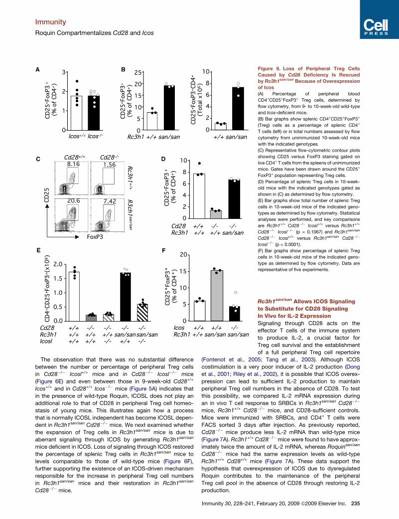
The observation that there was no substantial difference
between the number or percentage of peripheral Treg cells
in Cd28�/� Icosl+/+ mice and in Cd28�/� Icosl�/� mice
(Figure 6E) and even between those in 9-week-old Cd28+/+
Icos+/+ and in Cd28+/+ Icos�/� mice (Figure 5A) indicates that
in the presence of wild-type Roquin, ICOSL does not play an
additional role to that of CD28 in peripheral Treg cell homeo-
stasis of young mice. This illustrates again how a process
that is normally ICOSL independent has become ICOSL depen-
dent in Rc3h1san/san Cd28�/� mice. We next examined whether
the expansion of Treg cells in Rc3h1san/san mice is due to
aberrant signaling through ICOS by generating Rc3h1san/san
mice deficient in ICOS. Loss of signaling through ICOS restored
the percentage of splenic Treg cells in Rc3h1san/san mice to
levels comparable to those of wild-type mice (Figure 6F),
further supporting the existence of an ICOS-driven mechanism
responsible for the increase in peripheral Treg cell numbers
in Rc3h1san/san mice and their restoration in Rc3h1san/san
Cd28�/� mice.
Figure 6. Loss of Peripheral Treg Cells
Caused by Cd28 Deficiency Is Rescued
by Rc3h1san/san Because of Overexpression
of Icos
(A) Percentage of peripheral blood
CD4+CD25+FoxP3+ Treg cells, determined by
flow cytometry, from 9- to 10-week-old wild-type
and Icos-deficient mice.
(B) Bar graphs show splenic CD4+CD25+FoxP3+
(Treg) cells as a percentage of splenic CD4+
T cells (left) or in total numbers assessed by flow
cytometry from unimmunized 10-week-old mice
with the indicated genotypes.
(C) Representative flow-cytometric contour plots
showing CD25 versus FoxP3 staining gated on
live CD4+ T cells from the spleens of unimmunized
mice. Gates have been drawn around the CD25+
FoxP3+ population representing Treg cells.
(D) Percentage of splenic Treg cells in 10-week-
old mice with the indicated genotypes gated as
shown in (C) as determined by flow cytometry.
(E) Bar graphs show total number of splenic Treg
cells in 10-week-old mice of the indicated geno-
types as determined by flow cytometry. Statistical
analyses were performed, and key comparisons
are Rc3h1+/+ Cd28�/� Icosl+/+ versus Rc3h1+/+
Cd28�/� Icosl�/� (p = 0.1067) and Rc3h1san/san
Cd28�/� Icosl+/+ versus Rc3h1san/san Cd28�/�
Icosl�/� (p < 0.0001).
(F) Bar graphs show percentage of splenic Treg
cells in 10-week-old mice of the indicated geno-
type as determined by flow cytometry. Data are
representative of five experiments.
Rc3h1san/san Allows ICOS Signalingto Substitute for CD28 SignalingIn Vivo for IL-2 ExpressionSignaling through CD28 acts on the
effector T cells of the immune system
to produce IL-2, a crucial factor for
Treg cell survival and the establishment
of a full peripheral Treg cell repertoire
(Fontenot et al., 2005; Tang et al., 2003). Although ICOS
costimulation is a very poor inducer of IL-2 production (Dong
et al., 2001; Riley et al., 2002), it is possible that ICOS overex-
pression can lead to sufficient IL-2 production to maintain
peripheral Treg cell numbers in the absence of CD28. To test
this possibility, we compared IL-2 mRNA expression during
an in vivo T cell response to SRBCs in Rc3h1san/san Cd28�/�
mice, Rc3h1+/+ Cd28�/� mice, and CD28-sufficient controls.
Mice were immunized with SRBCs, and CD4+ T cells were
FACS sorted 3 days after injection. As previously reported,
Cd28�/� mice produce less IL-2 mRNA than wild-type mice
(Figure 7A). Rc3h1+/+ Cd28�/�mice were found to have approx-
imately twice the amount of IL-2 mRNA, whereas Roquinsan/san
Cd28�/� mice had the same expression levels as wild-type
Rc3h1+/+ Cd28+/+ mice (Figure 7A). These data support the
hypothesis that overexpression of ICOS due to dysregulated
Roquin contributes to the maintenance of the peripheral
Treg cell pool in the absence of CD28 through restoring IL-2
production.
Immunity 30, 228–241, February 20, 2009 ª2009 Elsevier Inc. 235

Immunity
Roquin Compartmentalizes Cd28 and Icos
Rc3h1san/san Cannot Rescue Thymic Treg CellDevelopment in Mice Deficient in CD28CD28 has been shown to be essential for thymic Treg cell devel-
opment through the induction of IL-2 and other IL-2-independent
signaling events (Tai et al., 2005; Tang et al., 2003). To examine
whether the rescue of peripheral Treg cells in Rc3h1san/san
Cd28�/� mice is a consequence of restoring Treg cell develop-
ment in the thymus, we assessed the number and percentage
of CD4 single-postive (SP) FoxP3+-expressing thymocytes in
4-week-old Rc3h1san/san Cd28�/�mice. As shown for peripheral
Treg cells, Rc3h1san/san mice have twice the number of Treg cells
within the CD4 SP thymic compartment (Figure 7B). Neverthe-
less, unlike the effect of Rc3h1san/san on peripheral Treg cells,
mutant Roquin could not restore thymic Treg cells in mice defi-
cient in CD28 (Figure 7C), although a small increase was
observed in Rc3h1san/san Cd28�/� mice as compared with
Rc3h1+/+ Cd28�/� mice. This small increase in thymic Treg cell
numbers, due to mutant Roquin, was independent of ICOS
signaling (Figure 7D), indicating a non-ICOS-related effect.
Figure 7. Rc3h1san/san Does Not Rescue the
Defective Thymic Treg Cell Development in
Cd28�/� Mice
(A) Fold change of IL-2 mRNA expression relative
to that in wild-type C57BL/6 mice 3 days after
SRBC immunization.
(B) Percentage and total number of thymic Treg
cells (CD4 SP FoxP3+) in 4-week-old unimmunized
Rc3h1+/+ (white bars) or Rc3h1san/san (black bars)
mice.
(C) Percentage of CD4 SP thymocytes that
express FoxP3 in mice with the indicated geno-
types.
(D) Number of Treg cells in 8-week-old unimmu-
nized mice with the indicated genotypes.
(E) Expression of CD11c (red) and ICOSL (green)
on 8-week-old C57BL/6 thymi.
(F) MFI of ICOS on the surface of CD4 SP thymo-
cytes in 8-week-old unimmunized mice with the
indicated genotypes. In all graphs, each symbol
represents one mouse, and bars are drawn
through the median values in each group. Data
are representative of three experiments.
To investigate the cause for the failure of
Rc3h1sansan to rescue thymic Treg cell
numbers in Cd28�/�mice, we first tested
whether ICOSL was expressed in the
thymus by confocal microscopy. Indeed,
ICOSL is strongly expressed in the thymic
medulla, with the majority of the expres-
sion colocalizing with CD11c+ cells
(Figure 7E). The next obvious question is
whether Rc3h1san/san Cd28�/� thymo-
cytes overexpress ICOS. We had previ-
ously observed that Rc3h1san/san CD4 SP
thymocytes (with intact CD28 signaling)
expressed slightly higher levels of ICOS
(Vinuesa et al., 2005a). When thymocyte
ICOS expression was assessed by flow
cytometry, we found that the levels of
ICOS on Rc3h1san/san Cd28�/� thymocytes were comparable to
those in wild-type mice (Figure 7F). This may explain why thymic
Treg cells are not restored in Rc3h1san/san mice lacking CD28.
Taken together, these results show that mutant Roquin, in the
absence of CD28, does not cause ICOS overexpression in CD4
SP thymocytes or restore thymic Treg cells.
DISCUSSION
The question addressed by these studies was whether the evolu-
tion of two biochemically paralogous costimulatory receptors
posed a potential for functional crosstalk and misregulation,
given that one of their ligands is constitutively expressed in the
absence of pathogen-associated molecular patterns. The results
demonstrate that functional compartmentalization of CD28 and
ICOS is readily broken down and is normally critical to ensure
that the initiation of immune responses depends on a path-
ogen-induced ‘‘signal 2’’ through CD28. Rc3h1san/san Cd28�/�
mice form normal primary antibody responses, Tfh cells, large
236 Immunity 30, 228–241, February 20, 2009 ª2009 Elsevier Inc.

Immunity
Roquin Compartmentalizes Cd28 and Icos
germinal centers, intact extrafollicular plasma cell responses,
and normal numbers of peripheral Treg cells—functions known
to be critically dependent on intact signaling through CD28.
Given that relatively little is understood about mechanisms
controlling ICOS and ICOSL expression, there are likely to be
a range of pathological and physiological circumstances in
which this crosstalk comes into play.
Three alternative hypotheses could explain the rescue of
responses in Rc3h1san/san Cd28�/� animals: First, Rc3h1san/san
allows ICOSL to substitute for CD28 ligands, whereas in
Rc3h1+/+ T cells, these two potential costimulatory sources are
distinguished and functionally compartmentalized so that ICOSL
cannot substitute for CD28L. Second, Roquin may allow for
signaling through the CD28 pathway in the absence of CD28. If
this were the case, removing ICOSL would not have an effect,
but we show profound abrogation of Rc3h1san/san-mediated
rescue in Rc3h1san/san mice doubly deficient in CD28 in all
responses tested. Third, even in the presence of intact CD28L
signaling, there might be a role for ICOSL in the response. If
this were the case, ICOSL deficiency would confer a phenotype
in Cd28+/+ mice. We have shown two examples in which ICOSL
does not play a role in the response of Cd28+/+ animals: First,
whereas Cd28�/� mice cannot mount a T cell-dependent anti-
body response to Salmonella or clear bacteria—both of these
responses are dependent on Th1 cells—these responses are
intact in ICOSL�/� mice. Second, peripheral Treg cell numbers
are 80% lower in 9-week-old Cd28�/�mice, but they are normal
in age-matched Icos�/� mice. Having excluded the latter two,
only the first hypothesis is consistent with the results of the
Rc3h1san/san Cd28�/� Icosl�/� triple-mutant studies.
We have shown that this compartmentalization acts during
immune responses to foreign antigens such as sheep red blood
cells and, importantly, to the infectious agent Salmonella. The
physiological counterpart of a challenge with antigen in the ab-
sence of CD28 ligands would be a situation in which the antigen
is a self-antigen. The most obvious evolutionary advantage of
functional compartmentalization of signals through CD28 and
ICOS is the retaining of the need for microbial induction of
costimulatory ligands at the time of T cell priming, because the
constitutive and broad expression of ICOSL could bypass the
need for danger signals to provide full costimulation. Our
previous work has provided evidence that this sort of dysregula-
tion can pose a real threat for autoimmunity: ICOS overexpres-
sion on naive T cells contributes to lupus manifestations in
Rc3h1san/san mice (Yu et al., 2007).
A ‘‘controlled’’ breakdown of this compartmentalization, or
what could also be seen as the ‘‘reverse’’ type of compartmen-
talization, i.e., making T cell costimulation in germinal centers
exclusively dependent on ICOS, and not on CD28, may be
equally important for physiological humoral immune responses.
Processed microbial antigens can be trapped in an inert state in
the form of immune complexes on the surface of follicular
dendritic cells for months or years within germinal centers, and
this source of antigen is probably important for the maintenance
of germinal center reactions and ongoing affinity maturation and
for the reactivation of memory B cells that may help maintain
long-lived protective humoral immunity. Also, the switch from
CD28 dependence to ICOS dependence that maintains Tfh cell
help for germinal center B cells and Tfh cell survival is likely to
also be critically important to prevent the emergence of high-
affinity autoantibodies from germinal centers. It would be
potentially dangerous to make this Tfh cell costimulation and
subsequent selection of mutated germinal center B cells depen-
dent on the presence of Toll-like receptor (TLR) ligands or other
danger signals; it is well known that signals delivered by TLR
ligands in conjunction with B cell receptor signals can lead to
T cell-independent self-reactive B cell differentiation (Vos et al.,
2000). The need for danger signals is even less desirable if we
take into account the fact that the germinal center milieu is
extremely rich in apoptotic cells that expose self-nuclear anti-
gens on their surface and that a proportion of somatically
mutated cells will have randomly acquired specificity against
these self-antigens (Diamond et al., 1992).
Gene duplication followed by positive selection is not only an
effective way to resolve adaptive conflicts through subfunction-
alization as described above. It also drives the acquisition of
unique functions by individual genomes: This process is termed
‘‘neofunctionalization,’’ and there is evidence to suggest that
neofunctionalization has also driven the selection of the adaptive
changes of Icos. In the context of evolution, the first time Cd28,
Ctla4, and Icos are present as an immunological cluster in the
same species is in chickens (Bernard et al., 2007), and this coin-
cides with the appearance of germinal centers, which occur only
in homeothermic vertebrates (birds and mammals). Although
somatic hypermutation during immune responses is seen in
ectotherms, which lack germinal centers, it is not accompanied
by efficient improvement in overall antibody affinity, suggesting
that affinity maturation depends on germinal centers for selec-
tion of mutated B cells by antigen-specific follicular T cells
(Hsu, 1998). It seems, therefore, that the acquisition of novel
functions by the Cd28 paralog Icos, specifically its new role in
the survival and B cell helper function of Tfh cells, opened up
the possibility for affinity maturation and effective immunological
memory to occur.
Although most of the rescue of responses normally dependent
on CD28, caused by Rc3h1san/san, requires signaling through
ICOS, there appear to be small non-ICOS mediated contribu-
tions to Tfh cell development and germinal center formation in
Rc3h1san/san mice lacking CD28. Our adoptive cell transfer of
wild-type B cells and bone marrow chimera experiments have
excluded a B cell-autonomous action of Roquin as an explana-
tion the rescued germinal center response. We have shown
previously that besides dysregulating ICOS expression
Rc3h1san/san CD4+ cells have high mRNA expression of other
key Tfh cell molecules, such as CD200, CXCR5, PD-1, CD84,
Bcl6, and the cytokine IL-21 (Vinuesa et al., 2005a). It is possible
that one or more of these factors are driving additional Tfh cell
development in Rc3h1san/san Cd28�/� T cells and/or directly
promoting germinal center responses, as has been shown in
mice overexpressing IL-21 (Ozaki et al., 2004).
The germinal centers observed in Rc3h1san/san Cd28�/� mice
are larger than those seen in wild-type mice, and this is reminis-
cent of the germinal centers induced in Ctla4-Ig transgenic mice
treated transiently with CD28 for allowing initiation of T cell-
dependent responses (Walker et al., 2003). In these mice,
germinal center reactions do not appear to involute normally,
and they display abundant Tfh cells expressing high amounts
of ICOS that persist long after the CD28 mAb has been cleared,
Immunity 30, 228–241, February 20, 2009 ª2009 Elsevier Inc. 237

Immunity
Roquin Compartmentalizes Cd28 and Icos
suggesting that CTLA-4 negatively regulates germinal center
longevity and Tfh cell numbers. Furthermore, a link between
signaling through CTLA-4 and Icos mRNA repression has been
suggested (Riley et al., 2001). Nevertheless, our results showing
that germinal center longevity is not prolonged in Rc3h1san/san
Cd28+/+ nor Rc3h1san/san Cd28�/� mice argues against the
possibility that CTLA-4 exerts its repressive effect through
Roquin.
Signals through CD28 also support generation of Treg cells
within the thymus and peripheral Treg cell homeostasis (Tai
et al., 2005; Tang et al., 2003). Although thymic Treg cell
numbers are increased in Rc3h1san/san mice, we show that this
increase is not dependent on ICOS signaling, which is not
surprising given the finding that ICOS is not overexpressed on
Rc3h1san/san Cd28�/� CD4SP thymocytes and that CD28 defi-
ciency alone partially corrects the excess of thymic Treg cells
seen in Rc3h1san/san mice. Furthermore, the most likely reason
why ICOSL cannot substitute for CD28L signals for Treg cell
generation in the thymus in the presence of the san allele of
Rc3h1 is that the latter depends on a critical Lck-binding motif
within the polyproline domain of CD28 that is absent in ICOS
(Tai et al., 2005). In contrast with the thymic Treg cell deficiency,
our data show that peripheral Treg cell numbers are restored in
Rc3h1san/san Cd28�/� mice in an ICOS-dependent manner.
This suggests that the smaller thymic Treg cell pool may be
able to expand to fill the peripheral niches.
Our data showing that in the presence of defective Roquin,
IL-2 production occurred normally in the absence of CD28
signaling suggests this is a consequence of ICOS overexpres-
sion and could contribute to the maintenance of peripheral
Treg cells in the absence of the major costimulator. Expansion
of naturally occurring thymic-derived Treg cells might not be
the only mechanism that fills the peripheral Treg cell niches in
Rc3h1san/san Cd28�/� mice. A non-mutually exclusive explana-
tion may lie in a possible increased generation of ‘‘adaptive’’
Treg cells (aTreg cells) in the periphery, which could also be
driven by excess ICOS signaling. Naive CD4+ CD25� T cells
stimulated with CD28 and TGFb can become regulatory cells
phenotypically identical to thymic-derived Treg cells (Liang
et al., 2005).
In general, there is still little experimental evidence of the
mechanisms by which gene duplicates are compartmentalized
and regulated for enabling genetic novelty. Elegant work has
dissected the molecular basis of the evolution of a bifunctional
ancestor in the yeast galactose pathway into a pair of duplicate
genes, GAL1 and GAL3, that acquired critical coinducer and gal-
actokinase subfunctions, respectively (Hittinger and Carroll,
2007). In this particular ‘‘genetic switch,’’ adaptive changes in
the promoter region that would have compromised one of the
functions allowed a differentially regulated transcriptional
response. Our data parallels these findings by identifying a mole-
cule, Roquin, that can regulate the functional switch between
two gene duplicates.
This regulation, unlike the transcriptionally regulated, inducible
nature of ICOS (Tan et al., 2006), appears to hinge on evolu-
tionary changes in the 30UTR of Icos that allow a differentially
regulated posttranscriptional induction of mRNA decay through
Roquin-controlled microRNAs (Yu et al., 2007). Roquin appears
to be constitutively expressed and constantly regulating ICOS in
238 Immunity 30, 228–241, February 20, 2009 ª2009 Elsevier Inc.
both naive and activated T cells, although its microRNA cofactor
(miR-101) is downregulated in Tfh cells (Yu et al., 2007). Indeed,
we show that ICOS is induced to the same extent (�4 fold) in
both Rc3h1san/san effector and memory CD4+ T cells and
controls.
In conclusion, our data reconciles how two related and func-
tionally overlapping receptor-ligand pairs can coordinately
initiate immune responses to protein antigens and select somat-
ically derived high-affinity mutants without jeopardizing the
maintenance of peripheral tolerance. This is achieved through
Roquin’s tight regulation of ICOS expression, which prevents
ICOS from taking over the critical CD28 functions of initiating
T cell help in the context of danger signals and determining the
size of the peripheral Treg cell pool, but allows its prominent
role in the selection of high-affinity germinal center B cell mutants
independently of CD28 and danger signals and in the regulation
of the size and longevity of germinal centers.
EXPERIMENTAL PROCEDURES
Mice and Immunizations
Rc3h1san/san C57BL/6 mice, crosses to Icos�/�, Icosl�/�, and Cd28�/� mice,
and SWHEL mice were housed in specific pathogen-free conditions at the
Australian Phenomics Facility. SWHEL mice have a VDJ region introduced by
homologous recombination in the Ig H chain locus that, in combination with
a transgene-encoded light chain, binds HEL with high affinity (Phan et al.,
2003). All animal procedures were approved by the Australian National Univer-
sity Animal Ethics and Experimentation Committee.
For generating thymus-dependent responses for assessing specific IgG
titers, mice were immunized intraperitoneally (i.p.) with 50 mg of alum-precipi-
tated chicken gammaglobulin (CGG) plus 1 3 108 heat killed B. pertussis.
Where indicated, 8- to 12-week-old mice were immunized i.p. with 2 3 109
SRBCs. For experiments involving SWHEL mice, 1 3 104 SWHEL B cells
were transferred into C57BL/6 recipients, which were immunized intrave-
nously with 2 3 108 SRBCs conjugated with mutant HEL, HEL2x (Paus et al.,
2006).
Bacteria and Inoculation
Salmonella enterica serovar Dublin strain SL5631 (Segall and Lindberg,
1991) was grown in Luria-Bertani medium overnight. Mice were inoculated
with 5 3 105 colony-forming units from a log-phase culture administered i.p.
in phosphate-buffered saline (PBS). Bacterial load was measured in the
liver of all mice at day 12 after infection by homogenizing organs, plating
serial dilutions in PBS onto Luria-Bertani agar, and incubating at 37�C
overnight.
Antibodies
Antibodies and strepatavidin conjugates for flow cytometry were from BD
PharMingen except where otherwise indicated: anti-mouse B220-PerCP,
CD4-PerCP, ICOS-PE (eBioscience), FoxP3 (eBioscience), GL-7-FITC,
CD95-PE, CXCR5-biotin, PD-1-PE, CTLA-4-PE, CD25-APC, CD8-APC, and
streptavidin-PerCP Cy5.5. Background ICOS staining was determined with
stained cells from ICOS-deficient mice. For immunohistochemistry, the
primary antibodies and reagents used were as follows: sheep anti-mouse
IgD (The Binding Site), biotinylated anti-mouse TCRbeta (PharMingen), rat
anti-mouse PD-1 (Biolegend), PNA-biotin (Vector Laboratories), and rat anti-
mouse CD138 (BD PharMingen). Rabbit anti-rat horseradish peroxidase
(HRP) (Dako) was used as a secondary antibody. For immunofluorescence,
monoclonal Abs used were rat anti-mouse Icosl (clone MIL-666, generated
by immunization of Lewis rats with an Icosl transfectant) and hamster anti-
mouse CD11c-biotin (eBioscience). Anti-mouse ICOSL was detected with
goat anti-rat FITC (Southern Biotechnology Associates), then rabbit anti-
FITC (Sigma-Aldrich), then goat anti-rabbit-FITC (Southern Biotechnology
Associates).

Immunity
Roquin Compartmentalizes Cd28 and Icos
Cell Isolation, Culture, and Stimulation
For flow cytometry, single-cell suspensions were prepared from spleens and/
or pooled lymph nodes (inguinal, axillary, subcapsular, cervical, mesenteric,
and para-aortic) of unimmunized and/or immunized mice.
Flow Cytometry
Spleen and or lymph node cell suspensions were prepared by sieving and
gentle pipetting. For surface staining, cells were maintained in the dark at
4�C throughout. Cells were washed twice in ice-cold FACS buffer (2% fetal
calf serum, 0.1% NaN3 in PBS), then incubated with each antibody and conju-
gate layer for 30 min and washed thoroughly with FACS buffer between each
layer. Intracellular staining used Cytofix/Cytoperm Kit (BD Biosciences)
following the manufacturer’s instructions. For detection of HEL-binding
B cells, HEL was conjugated to Alexa 647 (Molecular Probes). A FACSCalibur
(Becton Dickinson) was used for the acquisition of flow-cytometric data, and
Flowjo software was used for analysis.
Enzyme-Linked Immunosorbent Assay
Anti-Salmonella IgM, IgG1, IgG2ab, IgG2b, and IgG3 (Southern Biotechnology
Associates) were detected in plasma from blood taken at day 7 and day 12 by
enzyme-linked immunosorbent assay (ELISA). 96-well ELISA plates (Nunc)
were coated with SL5631 cell lysate. The lysate was prepared from an over-
night culture with a French press. Protein concentration in the lysate was
determined by Bradford assay, and each well was coated with 12.5 mg protein.
For the CGG immunization experiment, sera from mice were collected 14 days
after immunization with CGG and analyzed for aCGG-IgG1 antibodies with
CGG (1.5 mg/ml)-coated 96-well plates. Serial serum dilutions were applied,
immunoglobulin concentration was determined with HRP-conjugated goat
anti-mouse IgG1 (Southern Biotechnology Associates), and the enzyme bound
to plates was developed with Phosphatase Substrate tablets (Sigma S0942).
Plates were read at 405 nm with a Thermomax Microplate Reader (Molecular
Devices). The titers for serum samples were calculated as the log serum
concentration required to achieve 50% maximum optical density.
Immunohistochemistry
5 mm acetone-fixed frozen sections of spleen were air dried and washed in
0.1 M Tris-buffered saline (TBS) (pH 7.6), and then primary antibodies were
added in TBS and incubated for 45 min. After a further wash in TBS, secondary
reagents that had been previously absorbed in 10% normal mouse serum
were added to the sections for 45 min. When biotin-conjugated primary or
secondary reagents were used, streptavidin alkaline phosphatase (Vector
Laboratories) was added after a further wash in TBS and incubated for
20 min. HRP activity was detected with diaminobenzidine tetrahydrochloride
solution (Sigma) and hydrogen peroxide. Alkaline phosphatase activity was
detected with the AP-Substrate Kit III (SK-5300, Vector laboratories). Sections
were viewed under an Olympus IX71 Microscope (Olympus).
Immunofluorescence
Tissue samples for immunofluorescence were embedded in Tissue-Tek
OCT compound (Bayer Healthcare), and 6 mm thick sections were cut and
fixed in acetone. Antibodies were added in PBS and incubated in the dark
for 30 min. Biotinylated antibodies were detected with streptavidin-Alexa
Fluor 555 (Invitrogen). Sections were mounted with Vectashield mounting
medium (Vector Laboratories). Confocal images were obtained at room
temperature on a LSM 510 Meta microscope (Zeiss) equipped with either a
103 or a 403 1.4 N.A. water lens and 488 and 543 lasers with Zeiss LSM
software.
Real-Time PCR
CD4+ splenocytes from mice of the indicated genotypes 3 days after SRBC
immunization were sorted, frozen in Trizol, RNA extracted, and cDNA made.
Expression of IL-2 was determined with quantitative RT-PCR comparing
IL-2 to two housekeeping genes, b-actin and b2M, with four biological repli-
cates per strain and five technical replicates per sample and gene. Technical
replicates were averaged, and data were analyzed by both randomly pairing
mice in the control and experimental groups and averaging the cT of biological
replicates. The same pattern of differential expression was observed with both
analysis methods and with both housekeeping genes.
Statistical Analysis
Data were analyzed with a two-tailed Student’s t test with Prism software.
SUPPLEMENTAL DATA
Supplemental Data include three figures and can be found with this article
online at http://www.immunity.com/supplemental/S1074-7613(09)00068-5.
ACKNOWLEDGMENTS
This work was funded by a Viertel Senior Medical Research Fellowship,
National Health and Medical Research Council grants 316956 and 427620 to
C.G.V. and C.C.G., and Juvenile Diabetes Research Foundation grant
1-2006-96 to C.G.V. and D.S. We thank M. Cook and A. Liston for critical
review of the manuscript and helpful discussions.
Received: February 24, 2008
Revised: May 12, 2008
Accepted: December 18, 2008
Published online: February 12, 2009
REFERENCES
Akiba, H., Takeda, K., Kojima, Y., Usui, Y., Harada, N., Yamazaki, T., Ma, J.,
Tezuka, K., Yagita, H., and Okumura, K. (2005). The role of ICOS in the
CXCR5+ follicular B helper T cell maintenance in vivo. J. Immunol. 175,
2340–2348.
Beier, K.C., Hutloff, A., Dittrich, A.M., Heuck, C., Rauch, A., Buchner, K., Lude-
wig, B., Ochs, H.D., Mages, H.W., and Kroczek, R.A. (2000). Induction, binding
specificity and function of human ICOS. Eur. J. Immunol. 30, 3707–3717.
Bernard, D., Hansen, J.D., Du Pasquier, L., Lefranc, M.P., Benmansour, A.,
and Boudinot, P. (2007). Costimulatory receptors in jawed vertebrates:
Conserved CD28, odd CTLA4 and multiple BTLAs. Dev. Comp. Immunol.
31, 255–271.
Bossaller, L., Burger, J., Draeger, R., Grimbacher, B., Knoth, R., Plebani, A.,
Durandy, A., Baumann, U., Schlesier, M., Welcher, A.A., et al. (2006). ICOS
deficiency is associated with a severe reduction of CXCR5+CD4 germinal
center Th cells. J. Immunol. 177, 4927–4932.
Breitfeld, D., Ohl, L., Kremmer, E., Ellwart, J., Sallusto, F., Lipp, M., and
Forster, R. (2000). Follicular B helper T cells express CXC chemokine receptor
5, localize to B cell follicles, and support immunoglobulin production. J. Exp.
Med. 192, 1545–1552.
Brodie, D., Collins, A.V., Iaboni, A., Fennelly, J.A., Sparks, L.M., Xu, X.N., van
der Merwe, P.A., and Davis, S.J. (2000). LICOS, a primordial costimulatory
ligand? Curr. Biol. 10, 333–336.
Burmeister, Y., Lischke, T., Dahler, A.C., Mages, H.W., Lam, K.P., Coyle, A.J.,
Kroczek, R.A., and Hutloff, A. (2008). ICOS controls the pool size of effector-
memory and regulatory T cells. J. Immunol. 180, 774–782.
Chtanova, T., Tangye, S.G., Newton, R., Frank, N., Hodge, M.R., Rolph, M.S.,
and Mackay, C.R. (2004). T follicular helper cells express a distinctive tran-
scriptional profile, reflecting their role as non-Th1/Th2 effector cells that
provide help for B cells. J. Immunol. 173, 68–78.
Cunningham, A.F., Gaspal, F., Serre, K., Mohr, E., Henderson, I.R., Scott-
Tucker, A., Kenny, S.M., Khan, M., Toellner, K.M., Lane, P.J., and MacLennan,
I.C. (2007). Salmonella induces a switched antibody response without
germinal centers that impedes the extracellular spread of infection. J. Immu-
nol. 178, 6200–6207.
Diamond, B., Katz, J.B., Paul, E., Aranow, C., Lustgarten, D., and Scharff, M.D.
(1992). The role of somatic mutation in the pathogenic anti-DNA response.
Annu. Rev. Immunol. 10, 731–757.
Dong, C., Juedes, A.E., Temann, U.A., Shresta, S., Allison, J.P., Ruddle, N.H.,
and Flavell, R.A. (2001). ICOS co-stimulatory receptor is essential for T-cell
activation and function. Nature 409, 97–101.
Ferguson, S.E., Han, S., Kelsoe, G., and Thompson, C.B. (1996). CD28 is
required for germinal center formation. J. Immunol. 156, 4576–4581.
Immunity 30, 228–241, February 20, 2009 ª2009 Elsevier Inc. 239

Immunity
Roquin Compartmentalizes Cd28 and Icos
Fontenot, J.D., Rasmussen, J.P., Gavin, M.A., and Rudensky, A.Y. (2005). A
function for interleukin 2 in Foxp3-expressing regulatory T cells. Nat. Immunol.
6, 1142–1151.
Greenwald, R.J., Freeman, G.J., and Sharpe, A.H. (2005). The B7 family revis-
ited. Annu. Rev. Immunol. 23, 515–548.
Grimbacher, B., Hutloff, A., Schlesier, M., Glocker, E., Warnatz, K., Drager, R.,
Eibel, H., Fischer, B., Schaffer, A.A., Mages, H.W., et al. (2003). Homozygous
loss of ICOS is associated with adult-onset common variable immunodefi-
ciency. Nat. Immunol. 4, 261–268.
Gross, J.A., Callas, E., and Allison, J.P. (1992). Identification and distribution of
the costimulatory receptor CD28 in the mouse. J. Immunol. 149, 380–388.
Harada, Y., Ohgai, D., Watanabe, R., Okano, K., Koiwai, O., Tanabe, K., Toma,
H., Altman, A., and Abe, R. (2003). A single amino acid alteration in cytoplasmic
domain determines IL-2 promoter activation by ligation of CD28 but not induc-
ible costimulator (ICOS). J. Exp. Med. 197, 257–262.
Harding, F.A., McArthur, J.G., Gross, J.A., Raulet, D.H., and Allison, J.P.
(1992). CD28-mediated signalling co-stimulates murine T cells and prevents
induction of anergy in T-cell clones. Nature 356, 607–609.
Haynes, N.M., Allen, C.D., Lesley, R., Ansel, K.M., Killeen, N., and Cyster, J.G.
(2007). Role of CXCR5 and CCR7 in follicular Th cell positioning and appear-
ance of a programmed cell death gene-1high germinal center-associated
subpopulation. J. Immunol. 179, 5099–5108.
Hess, J., Ladel, C., Miko, D., and Kaufmann, S.H. (1996). Salmonella typhimu-
rium aroA- infection in gene-targeted immunodeficient mice: Major role of
CD4+ TCR-alpha beta cells and IFN-gamma in bacterial clearance indepen-
dent of intracellular location. J. Immunol. 156, 3321–3326.
Hittinger, C.T., and Carroll, S.B. (2007). Gene duplication and the adaptive
evolution of a classic genetic switch. Nature 449, 677–681.
Hsu, E. (1998). Mutation, selection, and memory in B lymphocytes of
exothermic vertebrates. Immunol. Rev. 162, 25–36.
Hutloff, A., Dittrich, A.M., Beier, K.C., Eljaschewitsch, B., Kraft, R., Anagnosto-
poulos, I., and Kroczek, R.A. (1999). ICOS is an inducible T-cell co-stimulator
structurally and functionally related to CD28. Nature 397, 263–266.
Janeway, C.A., Jr., and Bottomly, K. (1994). Signals and signs for lymphocyte
responses. Cell 76, 275–285.
Kim, C.H., Rott, L.S., Clark-Lewis, I., Campbell, D.J., Wu, L., and Butcher, E.C.
(2001). Subspecialization of CXCR5+ T cells: B helper activity is focused in
a germinal center-localized subset of CXCR5+ T cells. J. Exp. Med. 193,
1373–1381.
Lafferty, K.J., Andrus, L., and Prowse, S.J. (1980). Role of lymphokine and
antigen in the control of specific T cell responses. Immunol. Rev. 51, 279–314.
Li, W., Gao, B., Lee, S.M., Bennett, K., and Fang, D. (2007). RLE-1, an E3 ubiq-
uitin ligase, regulates C. elegans aging by catalyzing DAF-16 polyubiquitina-
tion. Dev. Cell 12, 235–246.
Liang, S., Alard, P., Zhao, Y., Parnell, S., Clark, S.L., and Kosiewicz, M.M.
(2005). Conversion of CD4+ CD25- cells into CD4+ CD25+ regulatory T cells
in vivo requires B7 costimulation, but not the thymus. J. Exp. Med. 201,
127–137.
Ling, V., Wu, P.W., Finnerty, H.F., Bean, K.M., Spaulding, V., Fouser, L.A., Leo-
nard, J.P., Hunter, S.E., Zollner, R., Thomas, J.L., et al. (2000). Cutting edge:
Identification of GL50, a novel B7-like protein that functionally binds to ICOS
receptor. J. Immunol. 164, 1653–1657.
Matzinger, P. (1994). Tolerance, danger, and the extended family. Annu. Rev.
Immunol. 12, 991–1045.
McAdam, A.J., Chang, T.T., Lumelsky, A.E., Greenfield, E.A., Boussiotis, V.A.,
Duke-Cohan, J.S., Chernova, T., Malenkovich, N., Jabs, C., Kuchroo, V.K.,
et al. (2000). Mouse inducible costimulatory molecule (ICOS) expression is
enhanced by CD28 costimulation and regulates differentiation of CD4+
T cells. J. Immunol. 165, 5035–5040.
McSorley, S.J., and Jenkins, M.K. (2000). Antibody is required for protection
against virulent but not attenuated Salmonella enterica serovar typhimurium.
Infect. Immun. 68, 3344–3348.
240 Immunity 30, 228–241, February 20, 2009 ª2009 Elsevier Inc.
Mittrucker, H.W., Kohler, A., Mak, T.W., and Kaufmann, S.H. (1999). Critical
role of CD28 in protective immunity against Salmonella typhimurium. J. Immu-
nol. 163, 6769–6776.
Ohno, S., Wolf, U., and Atkin, N.B. (1968). Evolution from fish to mammals by
gene duplication. Hereditas 59, 169–187.
Ozaki, K., Spolski, R., Ettinger, R., Kim, H.P., Wang, G., Qi, C.F., Hwu, P.,
Shaffer, D.J., Akilesh, S., Roopenian, D.C., et al. (2004). Regulation of B cell
differentiation and plasma cell generation by IL-21, a novel inducer of
Blimp-1 and Bcl-6. J. Immunol. 173, 5361–5371.
Paus, D., Phan, T.G., Chan, T.D., Gardam, S., Basten, A., and Brink, R. (2006).
Antigen recognition strength regulates the choice between extrafollicular
plasma cell and germinal center B cell differentiation. J. Exp. Med. 203,
1081–1091.
Phan, T.G., Amesbury, M., Gardam, S., Crosbie, J., Hasbold, J., Hodgkin,
P.D., Basten, A., and Brink, R. (2003). B cell receptor-independent stimuli
trigger immunoglobulin (Ig) class switch recombination and production of
IgG autoantibodies by anergic self-reactive B cells. J. Exp. Med. 197, 845–860.
Riley, J.L., Blair, P.J., Musser, J.T., Abe, R., Tezuka, K., Tsuji, T., and June,
C.H. (2001). ICOS costimulation requires IL-2 and can be prevented by
CTLA-4 engagement. J. Immunol. 166, 4943–4948.
Riley, J.L., Mao, M., Kobayashi, S., Biery, M., Burchard, J., Cavet, G., Greg-
son, B.P., June, C.H., and Linsley, P.S. (2002). Modulation of TCR-induced
transcriptional profiles by ligation of CD28, ICOS, and CTLA-4 receptors.
Proc. Natl. Acad. Sci. USA 99, 11790–11795.
Rudd, C.E., and Schneider, H. (2003). Unifying concepts in CD28, ICOS and
CTLA4 co-receptor signalling. Nat. Rev. Immunol. 3, 544–556.
Salomon, B., Lenschow, D.J., Rhee, L., Ashourian, N., Singh, B., Sharpe, A.,
and Bluestone, J.A. (2000). B7/CD28 costimulation is essential for the homeo-
stasis of the CD4+CD25+ immunoregulatory T cells that control autoimmune
diabetes. Immunity 12, 431–440.
Schaerli, P., Willimann, K., Lang, A.B., Lipp, M., Loetscher, P., and Moser, B.
(2000). CXC chemokine receptor 5 expression defines follicular homing T cells
with B cell helper function. J. Exp. Med. 192, 1553–1562.
Segall, T., and Lindberg, A.A. (1991). Salmonella dublin experimental infection
in calves: Protection after oral immunization with an auxotrophic aroA live
vaccine. Zentralbl. Veterinarmed. B. 38, 142–160.
Shahinian, A., Pfeffer, K., Lee, K.P., Kundig, T.M., Kishihara, K., Wakeham, A.,
Kawai, K., Ohashi, P.S., Thompson, C.B., and Mak, T.W. (1993). Differential
T cell costimulatory requirements in CD28-deficient mice. Science 261,
609–612.
Sharpe, A.H., and Freeman, G.J. (2002). The B7–CD28 superfamily. Nat. Rev.
Immunol. 2, 116–126.
Suh, W.K., Tafuri, A., Berg-Brown, N.N., Shahinian, A., Plyte, S., Duncan, G.S.,
Okada, H., Wakeham, A., Odermatt, B., Ohashi, P.S., and Mak, T.W. (2004).
The inducible costimulator plays the major costimulatory role in humoral
immune responses in the absence of CD28. J. Immunol. 172, 5917–5923.
Swallow, M.M., Wallin, J.J., and Sha, W.C. (1999). B7h, a novel costimulatory
homolog of B7.1 and B7.2, is induced by TNFalpha. Immunity 11, 423–432.
Tafuri, A., Shahinian, A., Bladt, F., Yoshinaga, S.K., Jordana, M., Wakeham, A.,
Boucher, L.M., Bouchard, D., Chan, V.S., Duncan, G., et al. (2001). ICOS is
essential for effective T-helper-cell responses. Nature 409, 105–109.
Tai, X., Cowan, M., Feigenbaum, L., and Singer, A. (2005). CD28 costimulation
of developing thymocytes induces Foxp3 expression and regulatory T cell
differentiation independently of interleukin 2. Nat. Immunol. 6, 152–162.
Tan, A.H., Wong, S.C., and Lam, K.P. (2006). Regulation of mouse inducible
costimulator (ICOS) expression by Fyn-NFATc2 and ERK signaling in T cells.
J. Biol. Chem. 281, 28666–28678.
Tang, Q., Henriksen, K.J., Boden, E.K., Tooley, A.J., Ye, J., Subudhi, S.K.,
Zheng, X.X., Strom, T.B., and Bluestone, J.A. (2003). Cutting edge: CD28
controls peripheral homeostasis of CD4+CD25+ regulatory T cells. J. Immu-
nol. 171, 3348–3352.
Vinuesa, C.G., Cook, M.C., Angelucci, C., Athanasopoulos,V., Rui, L., Hill, K.M.,
Yu, D., Domaschenz, H., Whittle, B., Lambe, T., et al. (2005a). A RING-type

Immunity
Roquin Compartmentalizes Cd28 and Icos
ubiquitin ligase family member required to repress follicular helper T cells and
autoimmunity. Nature 435, 452–458.
Vinuesa, C.G., Tangye, S.G., Moser, B., and Mackay, C.R. (2005b). Follicular B
helper T cells in antibody responses and autoimmunity. Nat. Rev. Immunol. 5,
853–865.
Vos, Q., Lees, A., Wu, Z.Q., Snapper, C.M., and Mond, J.J. (2000). B-cell acti-
vation by T-cell-independent type 2 antigens as an integral part of the humoral
immune response to pathogenic microorganisms. Immunol. Rev. 176,
154–170.
Walker, L.S., Gulbranson-Judge, A., Flynn, S., Brocker, T., Raykundalia, C.,
Goodall, M., Forster, R., Lipp, M., and Lane, P. (1999). Compromised OX40
function in CD28-deficient mice is linked with failure to develop CXC chemo-
kine receptor 5-positive CD4 cells and germinal centers. J. Exp. Med. 190,
1115–1122.
Walker, L.S., Wiggett, H.E., Gaspal, F.M., Raykundalia, C.R., Goodall, M.D.,
Toellner, K.M., and Lane, P.J. (2003). Established T cell-driven germinal center
B cell proliferation is independent of CD28 signaling but is tightly regulated
through CTLA-4. J. Immunol. 170, 91–98.
Watanabe, R., Harada, Y., Takeda, K., Takahashi, J., Ohnuki, K., Ogawa, S.,
Ohgai, D., Kaibara, N., Koiwai, O., Tanabe, K., et al. (2006). Grb2 and Gads
exhibit different interactions with CD28 and play distinct roles in CD28-medi-
ated costimulation. J. Immunol. 177, 1085–1091.
Yrlid, U., and Wick, M.J. (2000). Salmonella-induced apoptosis of infected
macrophages results in presentation of a bacteria-encoded antigen after
uptake by bystander dendritic cells. J. Exp. Med. 191, 613–624.
Yu, D., Tan, A.H., Hu, X., Athanasopoulos, V., Simpson, N., Silva, D.G., Hutloff,
A., Giles, K.M., Leedman, P.J., Lam, K.P., et al. (2007). Roquin represses auto-
immunity by limiting inducible T-cell co-stimulator messenger RNA. Nature
450, 299–303.
Note Added in Proof
In the version of this paper published online on February 12, the legend of
Figure 3 contained descriptions for panels A, B, and C, although Figure 3
only had panels A and B. This legend now has been corrected online and in
print.
Immunity 30, 228–241, February 20, 2009 ª2009 Elsevier Inc. 241
Copyright © 2022 FDOKUMEN