LNCaP prostate cancer cells with autocrine interleukin-6 expression are resistant to IL-6-induced...
11
The Prostate LNCaP Prostate Cancer Cells With Autocrine Interleukin- 6 Expression are Resistant to IL- 6 -Induced Neuroendocrine Differentiation Due to Increased Expression of Suppressors of Cytokine Signaling Dongxia Ge, 1,2 Allen C. Gao, 3 Qiuyang Zhang, 1,2 Sen Liu, 1,2 Yun Xue, 1,2 and Zongbing You, MD, PhD 1,2 * 1 Department of Structural & Cellular Biology,Tulane Cancer Center, Louisiana Cancer Research Consortium, Tulane Center for Aging,Tulane Center for Stem Cell Research and Regenerative Medicine, Tulane University School of Medicine, New Orleans, Louisiana 70112 2 Department of Orthopaedic Surgery,Tulane Cancer Center, Louisiana Cancer Research Consortium, Tulane Center for Aging,Tulane Center for Stem Cell Research and Regenerative Medicine, Tulane University School of Medicine, New Orleans, Louisiana 70112 3 Department of Urology,University of California at Davis Medical Center, Sacramento,California 95817 BACKGROUND. Neuroendocrine differentiation (NED) is one of the mechanisms underly- ing development of castration-resistant prostate cancer (CRPC). In this study, we investigated IL-6-induced NED in two LNCaP sublines. METHODS. LNCaP-S17, an LNCaP subline that secretes IL-6, and LNCaP-C3, a control sub- line that does not express IL-6, were analyzed for IL-6-induced NED, activation of JAK2 and STAT3 pathways, and expression of IL-6/IL-6R signaling proteins and downstream target genes. RESULTS. IL-6 did not induce NED in LNCaP-S17 cells, even though IL-6 induced NED in LNCaP-C3 cells. IL-6 activated JAK2 and STAT3 pathways in LNCaP-C3 cells but not in LNCaP-S17 cells. IL-6 did not activate ERK1/2, AKT, or NF-kB pathways in either cell line. Both LNCaP-C3 and LNCaP-S17 cell lines expressed IL-6R, gp130, and TYK2 at almost the same levels and did not express JAK1 or JAK3. The basal level of JAK2 expression was slight- ly higher in LNCaP-C3 cells than in LNCaP-S17 cells. Two suppressors of cytokine signaling, SOCS7 and cytokine-inducible SH2 protein (CIS), were expressed constitutively at higher levels in LNCaP-S17 cells than in LNCaP-C3 cells, while SOCS1 to SOCS6 were expressed at approximately the same levels. Using siRNA to knockdown SOCS7 and CIS expression in LNCaP-S17 cells led to increased phosphorylation of STAT3 upon IL-6 stimulation. Abbreviations: AR, androgen receptor; CIS, cytokine-inducible SH2 protein; CRPC, castration-resistant prostate cancer; GRP, gastrin-releasing peptide; MDK, midkine; MRA, humanized anti-human IL-6R monoclonal antibodies; MTT, 3-(4,5-Dimethylthiazol-2-yl)-2,5-diphenyltetrazo- lium bromide; NE, neuroendocrine; NED, neuroendocrine differentiation; NSE, neuron-specific enolase; NTS, neurotensin; siRNA, short inter- fering RNA; SOCS, suppressor of cytokine signaling; SYT1, synaptotagmin I. Grant sponsor: National Institutes of Health’s Centers of Biomedical Research Excellence (COBRE); Grant number: 2P20RR020152-06; Grant sponsor: Department of Defense; Grant numbers: W81XWH-05-1-0567; W81XWH-10-1-0937; Grant sponsor: The developmental funds of the Tulane Cancer Center, Louisiana Cancer Research Consortium, and Tulane Framework for Global Health Seed Grant. *Correspondence to: Zongbing You, MD, PhD, Department of Structural & Cellular Biology, Tulane University School of Medicine, 1430 Tulane Ave SL 49, New Orleans, LA 70112. E-mail: zyou@tulane.edu Received 28 July 2011; Accepted 2 December 2011 DOI 10.1002/pros.22479 Published online in Wiley Online Library (wileyonlinelibrary.com). ß 2011Wiley Periodicals, Inc.
Transcript of LNCaP prostate cancer cells with autocrine interleukin-6 expression are resistant to IL-6-induced...
TheProstate
LNCaPProstateCancerCellsWithAutocrine Interleukin-6Expression areResistantto IL-6-InducedNeuroendocrineDifferentiationDue to Increased Expressionof Suppressors
of Cytokine Signaling
Dongxia Ge,1,2 Allen C. Gao,3 Qiuyang Zhang,1,2 Sen Liu,1,2 Yun Xue,1,2
and Zongbing You, MD, PhD1,2*
1Departmentof Structural &Cellular Biology,TulaneCancer Center, LouisianaCancer ResearchConsortium,TulaneCenter for Aging,TulaneCenter for StemCell ResearchandRegenerativeMedicine,
TulaneUniversity SchoolofMedicine,NewOrleans, Louisiana 701122DepartmentofOrthopaedic Surgery,TulaneCancer Center, LouisianaCancer ResearchConsortium,
TulaneCenter for Aging,TulaneCenter for StemCell ResearchandRegenerativeMedicine,TulaneUniversity SchoolofMedicine,NewOrleans, Louisiana 70112
3DepartmentofUrology,Universityof CaliforniaatDavisMedical Center, Sacramento,California 95817
BACKGROUND. Neuroendocrine differentiation (NED) is one of the mechanisms underly-ing development of castration-resistant prostate cancer (CRPC). In this study, we investigatedIL-6-induced NED in two LNCaP sublines.METHODS. LNCaP-S17, an LNCaP subline that secretes IL-6, and LNCaP-C3, a control sub-line that does not express IL-6, were analyzed for IL-6-induced NED, activation of JAK2 andSTAT3 pathways, and expression of IL-6/IL-6R signaling proteins and downstream targetgenes.RESULTS. IL-6 did not induce NED in LNCaP-S17 cells, even though IL-6 induced NED inLNCaP-C3 cells. IL-6 activated JAK2 and STAT3 pathways in LNCaP-C3 cells but not inLNCaP-S17 cells. IL-6 did not activate ERK1/2, AKT, or NF-kB pathways in either cell line.Both LNCaP-C3 and LNCaP-S17 cell lines expressed IL-6R, gp130, and TYK2 at almost thesame levels and did not express JAK1 or JAK3. The basal level of JAK2 expression was slight-ly higher in LNCaP-C3 cells than in LNCaP-S17 cells. Two suppressors of cytokine signaling,SOCS7 and cytokine-inducible SH2 protein (CIS), were expressed constitutively at higherlevels in LNCaP-S17 cells than in LNCaP-C3 cells, while SOCS1 to SOCS6 were expressed atapproximately the same levels. Using siRNA to knockdown SOCS7 and CIS expression inLNCaP-S17 cells led to increased phosphorylation of STAT3 upon IL-6 stimulation.
Abbreviations: AR, androgen receptor; CIS, cytokine-inducible SH2 protein; CRPC, castration-resistant prostate cancer; GRP, gastrin-releasingpeptide; MDK, midkine; MRA, humanized anti-human IL-6R monoclonal antibodies; MTT, 3-(4,5-Dimethylthiazol-2-yl)-2,5-diphenyltetrazo-lium bromide; NE, neuroendocrine; NED, neuroendocrine differentiation; NSE, neuron-specific enolase; NTS, neurotensin; siRNA, short inter-fering RNA; SOCS, suppressor of cytokine signaling; SYT1, synaptotagmin I.
Grant sponsor: National Institutes of Health’s Centers of Biomedical Research Excellence (COBRE); Grant number: 2P20RR020152-06; Grantsponsor: Department of Defense; Grant numbers: W81XWH-05-1-0567; W81XWH-10-1-0937; Grant sponsor: The developmental funds of theTulane Cancer Center, Louisiana Cancer Research Consortium, and Tulane Framework for Global Health Seed Grant.
*Correspondence to: Zongbing You, MD, PhD, Department of Structural & Cellular Biology, Tulane University School of Medicine, 1430Tulane Ave SL 49, New Orleans, LA 70112. E-mail: [email protected] 28 July 2011; Accepted 2 December 2011DOI 10.1002/pros.22479Published online in Wiley Online Library (wileyonlinelibrary.com).
� 2011WileyPeriodicals,Inc.
CONCLUSIONS. LNCaP-S17 cells are resistant to exogenous IL-6-induced NED dueto increased levels of CIS/SOCS7 that block activation of JAK2-STAT3 pathways. Prostate #
2011 Wiley Periodicals, Inc.
KEY WORDS: IL-6; neuroendocrine; prostate cancer
INTRODUCTION
Prostate cancer is the most common malignant dis-ease in men in the developed countries. Initially, mostprostate cancers respond to androgen ablative (or cas-tration) therapy. However, even under the castratelevels of testosterone, prostate cancer eventually re-grows and becomes known as castration-resistantprostate cancer (CRPC), a lethal form of cancer [1,2].The molecular mechanisms underlying the progres-sion from a hormone sensitive cancer to CRPC in-clude alterations of androgen receptor (AR) signalingpathways, alterations of apoptosis-related genes suchas up-regulation of bcl-2 [3] and clusterin [4,5], andneuroendocrine differentiation (NED) of prostatecancer [6]. AR gene amplification [7,8], increase in ARexpression [9], and AR gene mutations [10,11] are im-portant factors in development of CRPC. AR can alsobe activated by intraprostatic tissue androgens (socalled intracrine androgens) [12,13]. Ligand-indepen-dent AR variants derived from splicing of crypticexons have been associtated with CRPC [14]. Somegrowth factors can activate AR signaling pathways inthe absence of androgen [15]. Activation of HER-2/neu [16] and Ras/mitogen-activated protein kinase(MAPK) pathways can activate AR signaling path-ways in androgen-independent manner [17]. Tran-scriptional coactivators are also involved in theligand-independent activation of AR signaling path-ways [18,19].
NED of prostate cancer has not been studied as ex-tensively as AR signaling. Neuroendocrine (NE) cells,part of the amine precursor uptake and decarboxyl-ation system, are specialized endocrine cells of thenervous system that produce neurohormones. In theprostate, NE cells are present in the developingand mature gland, forming the prostatic epitheliumwith the epithelial and basal cells [20]. NE cells arecharacterized morphologically by the presence ofcytoplasmic dense core granules and irregular den-drite-like processes extending underneath and be-tween adjacent epithelial cells [21]. The prostatic NEcells express neurotensin (NTS), synaptotagmin I(SYT1), gastrin-releasing peptide (GRP), neuron-spe-cific enolase (NSE), midkine (MDK), and other neuro-hormones [6,22]. NE cells are generally non-mitotic/growth-arrested [23,24], yet proliferating carcinomacells have been identified in close proximity to them[24,25], indicating a tumor-promoting role of NE cells
through secretion of neurohormones [26–29]. NE cellsare found in primary and metastatic prostate cancersand may have arisen from NED [30–32]. Focal NED isa common feature of prostate cancer, occurring inabout 30–100% of tumors studied [21,33,34]. Recentstudies suggest that NED is enhanced after long-termandrogen ablative therapy and may promote the de-velopment of CRPC [35–38]. NED is typically exem-plified by a human prostate cancer cell line, LNCaP,under conditions of androgen deprivation, increasedintracellular cAMP, activated MAPK pathway, andIL-6 treatment [23,39–49]. IL-6-induced NED is clini-cally relevant because elevated serum IL-6 levels areassociated with CRPC [50–54].
Studies in IL-6 and prostate cancer have revealedcomplex effects of IL-6 on proliferation of prostatecancer cells (see a recent review by Culig and Puhr[55]). Giri et al. [56] reported that exogenous IL-6treatment increased cell proliferation in LNCaP cells.In contrast, several groups of investigators indepen-dently found that exogenous IL-6 caused growth ar-rest in LNCaP cells, which was associated with NED[44,47,57–59]. The causes of the discrepancy are notknown. Nevertheless, the molecular mechanisms ofIL-6’s actions have been identified. IL-6 cytokine firstbinds to a non-signaling receptor, IL-6R, and thenforms a hexameric complex with gp130 [60]. Gp130 isa signal-transducing receptor shared among the IL-6family of cytokines including IL-6, IL-11, leukemia in-hibitory factor, ciliary neurotrophic factor, oncostatin-M, and others [61,62]. IL-6 leads to dimerization ofgp130, resulting in phosphorylation and activation ofJanus kinases (JAK1, JAK2, JAK3, and TYK2). Subse-quently, the cytoplasmic tail of gp130 is phosphory-lated, opening docking sites for STATs (STAT1 andSTAT3). STATs also become phosphorylated, formdimers and enter the nucleus, where they regulate tar-get gene expression [63,64].
LNCaP cell line does not express IL-6 but doesexpress its receptors IL-6R and gp130 [65,66]. Previ-ously, we established an LNCaP subline stablyexpressing IL-6 by introduction of a full-length IL-6cDNA into LNCaP cells [66]. We reported that IL-6-expressing LNCaP cells (i.e., LNCaP cells with auto-crine IL-6 expression) acquire the capacity to grow inthe absence of androgen in vitro and in vivo [67]. Inthe current study, we found that IL-6-expressingLNCaP cells were resistant to exogenous IL-6-inducedNED because the cells constitutively expressed high
2 Ge et al.
The Prostate
levels of negative feedback inhibitors, suppressor ofcytokine signaling 7 (SOCS7) and cytokine-inducibleSH2 protein (CIS). The results provide new insightsinto the molecular mechanisms of NED in prostatecancer.
MATERIALSANDMETHODS
CellCulture and InVitroCellGrowth
IL-6-expressing LNCaP subline (LNCaP-S17,expressing IL-6 at 2,743 pg/ml/million cells) andcontrol subline LNCaP-C3 (transfected with emptyvector, not expressing IL-6) were described previous-ly [66]. The cell lines were cultured in T-medium(Invitrogen) with 5% FBS in a 378C, 5% CO2 humidi-fied incubator. To analyze cell growth under exoge-nous IL-6 treatment, LNCaP-C3 and LNCaP-S17 cellswere cultured in serum-free medium with 50 ng/mlIL-6 for 4 days; then the cell number was determinedusing the 3-(4,5-Dimethylthiazol-2-yl)-2,5-diphenylte-trazolium bromide (MTT) assay (Sigma) according tothe manufacturer’s instructions [67]. To confirm ifIL-6’s effects were indeed mediated through IL-6receptor, humanized anti-human IL-6R monoclonalantibodies (MRA, a gift from Chugai PharmaceuticalCo., Ltd, Tokyo, Japan) were added to block IL-6’saction.
InductionofNEDandReverseTranscription-QuantitativeReal-TimePCR
LNCaP-C3 and LNCaP-S17 cells were culturedseparately in serum-free medium with 50 ng/ml IL-6for 4 days to induce NED. For co-culture of LNCaP-C3 and LNCaP-S17 cells, 100-mm dishes were sepa-rated into two halves by a dam made with a 1-cmstrip of sterilized paper pad; the two cell lines wereplated in each half of the dish side-by-side; after the
cells were attached overnight, the dam was removed;serum-free medium was added to allow free ex-change of secreted factors between LNCaP-C3 andLNCaP-S17 cells for 4 days. The cells were photo-graphed using a digital camera. RNA was isolatedfrom the cells using an RNeasy kit (QIAGEN); reversetranscription was performed using SuperscriptTM
First-Strand Synthesis System (Invitrogen); real-timePCR was performed on the iQ5 PCR instrument withSYBR Green reagents (Bio-Rad). PCR primer sequen-ces are shown in Table I. The mRNA levels were firstnormalized by that of GAPDH and then the relativemRNA fold change was calculated as 2DDCt comparedto the basal levels (either without IL-6 treatment or atDay 0).
WesternBlotAnalysis and Immunoprecipitation
The cells were washed with ice-cold phosphate-buffered saline and collected. Whole cell lysates wereprepared with RIPA buffer containing 1% NonidetP-40, 5% sodium deoxycholate, 1 mM phenylmethyl-sulfonyl fluoride, 100 mM sodium orthovanadate,and protease inhibitors mixture (Sigma). The proteinconcentrations were determined using the Bradfordprotein assay (Bio-Rad). Equal amounts of proteinextracts were subjected to 10% SDS–polyacrylamidegel electrophoresis and transferred to a PVDF mem-brane by electroblotting (Bio-Rad). The membraneswere blocked with 5% non-fat dry milk in 1� TBST(25 mM Tris-HCl, 125 mM NaCl, 0.1% Tween 20) for2 hr, probed with primary antibodies overnight at48C, and then with fluorphores-conjugated secondaryantibodies (LI-COR) for 1 hr. The results were visual-ized using an Odyssey Infrared Imaging System (LI-COR). For internal control, the blots were strippedand re-probed with anti-GAPDH or anti-b-tubulinantibodies. For immunoprecipitation of p-JAK2, 1 mg
TABLE I. PrimersUsedinReal-TimePCRAnalysis
Gene name Sequence (50 to 30)
NTS Sense GAGGAGCTTGTTGCAAGAAGGAAnti-sense GCCCTGCTGTGACAGATTTTGT
SYT1 Sense AACATGGGGTTGGCTGTTTAnti-sense CGGCAGACGGTTATTTTCCT
GRP Sense AGTCTCTGCTCTTCCAGCCAnti-sense CCGATGGACAACCAATCTAAG
NSE Sense TGTCTGCTGCTCAAGGTCAAAnti-sense CGATGACTCACCATGACCC
MDK Sense CGACTGCAAGTACAAGTTTGAGAACAnti-sense CTTGGTGACGCGGATGGT
GAPDH Sense TAAAAGCAGCCCTGGTGACCAnti-sense CCACATCGCTCAGACACCAT
Autocrine IL-6DesensitizesLNCaPCells 3
The Prostate
of anti-p-JAK2 antibodies were incubated with equalamounts of protein extracts for 30 min, followed byincubation with 10 ml of protein A- Sepharose beadsovernight at 48C; after washing four times, the precip-itated protein was analyzed using the proceduresdescribed above. For siRNA knockdown assays,LNCaP-S17 cells were plated at 70% confluence in 60-mm dishes; 100 nM of SOCS7 siRNA (siGENOMESMARTpool D-027197-04), CIS siRNA (siGENOMESMARTpool D-017381-04), or non-targeting scram-bled control siRNA were transfected into the cellsusing DharmaFECT12 reagents according to the man-ufacturer’s instructions (all reagents including siR-NAs were obtained from Thermo Fisher ScientificDharmacon Products, Lafayette, CO); 4 days later, thecells were treated with 50 ng/ml of IL-6 in serum-free medium for 5 min; and the whole cell lysateswere analyzed for expression of p-STAT3, SOCS7,and CIS, respectively, by Western blot. The primaryantibodies were purchased from the following com-mercial sources: Antibodies against STAT3, p-STAT3(Y705), JAK1, JAK2, JAK3, TYK2, p-JAK2, AKT,p-AKT, ERK1/2, IkBa, and p-IkBa were obtainedfrom Cell Signaling Technology; antibodies againstIL-6R, gp130, GAPDH, and b-tubulin were obtainedfrom R&D Systems; and antibodies against p-ERK1/2, CIS, and SOCS1 to SOCS7 were obtained fromSanta Cruz Biotechnology.
StatisticalAnalysis
All experiments were repeated three times. Photo-micrographs and Western blot results are presentedfrom a representative experiment. Student’s t test(two-tailed) was used to determine the significancebetween the control and treatment groups of LNCaP-C3 and LNCaP-S17 cells in the cell growth analysis,and P < 0.05 was considered statistically significant.
RESULTS
LNCaP-S17CellsWereResistanttoIL-6 -InducedNED
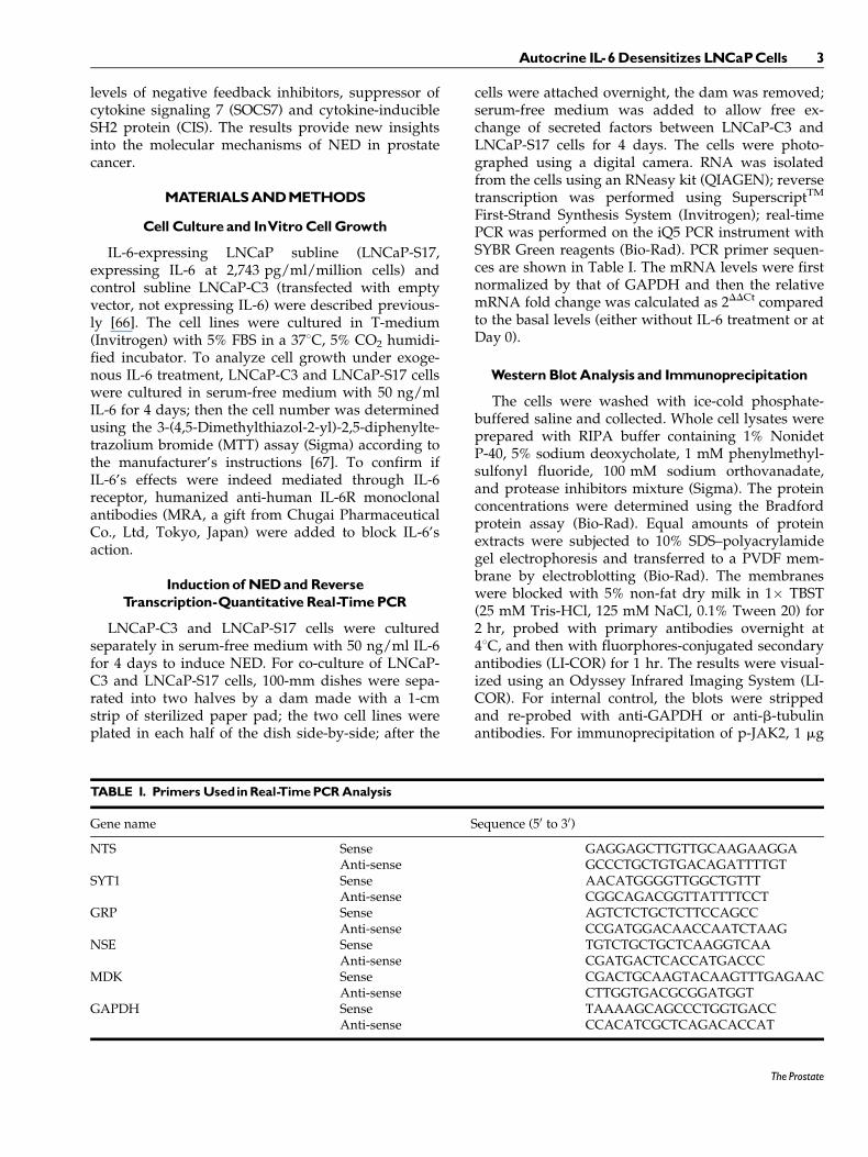
We previously showed that LNCaP-S17 cells couldgrow in the absence of androgen [67]. To test if thecells were still able to undergo NED, we treatedLNCaP-C3 and LNCaP-S17 cells with exogenous IL-6for 4 days. We found that LNCaP-C3 cells showedirregular dendrite-like processes typical of NE cells(Fig. 1B, compared to Fig. 1A). In contrast, theLNCaP-S17 cells did not show any obvious change incell morphology under the same exogenous IL-6 treat-ment (Fig. 1E, compared to Fig. 1D). To confirmthat LNCaP-S17 cells secreted IL-6, we co-culturedLNCaP-C3 and LNCaP-S17 cells in a system such thatIL-6 secreted by LNCaP-S17 cells could move freely toLNCaP-C3 cells but the two cell lines did not mix
Fig. 1. IL-6 induced formation of dentrite-like processes in LNCaP-C3 but not in LNCaP-S17 cells.A: LNCaP-C3 cells without treatment(Day 0).B: LNCaP-C3 cells treatedwith 50 ng/ml IL-6 for 4 days in serum-free culturemedium.C: LNCaP-C3 cells co-culturedwith LNCaP-S17cells for4days.D:LNCaP-S17cellswithouttreatment(Day0).E:LNCaP-S17cells treatedwith50 ng/ml IL-6 for4daysinserum-freeculturemedium.F: LNCaP-S17 cells co-culturedwith LNCaP-C3 cells for 4 days. Arrows indicate LNCaP-C3 cellswith dentrite-likeprocesses. Scalebars,10 mm.
4 Ge et al.
The Prostate
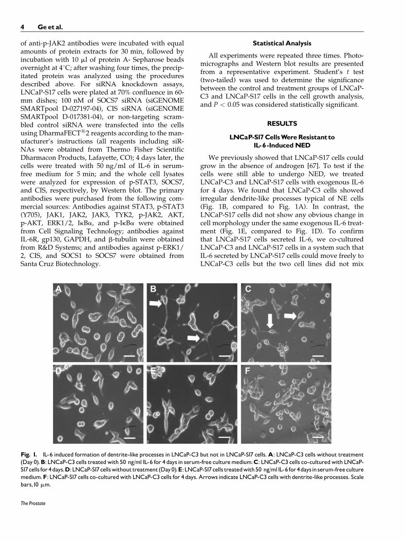
together. Indeed, we found that the co-culturedLNCaP-C3 cells extended dendrite-like processes(Fig. 1C), whereas LNCaP-S17 cells did not show anyprocesses (Fig. 1F). Because NE cells are generallynon-mitotic/growth-arrested [23,24], we examined ifIL-6 treatment induced growth arrest in the twocell lines. We found that IL-6 induced approximately50% reduction in the number of LNCaP-C3 cells(Fig. 2A, comparing group 2 versus group 1; P ¼0.007), whereas the number of LNCaP-S17 cells wassimilar to the untreated control group (Fig. 2A, com-paring group 2 versus group 1). The cell growth arrestobserved in LNCaP-C3 cells was specifically inducedby IL-6, as the anti-IL-6R antibody MRA completelyblocked IL-6’s function and rescued cell growth inLNCaP-C3 cells (Fig. 2A, comparing group 4 versusgroup 2). To further confirm that exogenous IL-6 in-duced NED in LNCaP-C3 cells but not in LNCaP-S17cells, we examined five markers of NED. As shown inFigure 2B, exogenous IL-6 dramatically induced
mRNA expression of NTS, SYT1, GRP, NSE, andMDK in LNCaP-C3 cells, particularly NTS mRNAthat was increased by approximately 68-fold. In con-trast, exogenous IL-6 minimally induced the expres-sion of these markers in LNCaP-S17 cells, e.g., only2.6-fold increase in NTS mRNA (Fig. 2B). Similarly,when the two cell lines were co-cultured for 4 days,IL-6 secreted by LNCaP-S17 cells considerably in-duced NTS and SYT1 mRNA expression in LNCaP-C3 cells but only minimally in LNCaP-S17 cells(Fig. 2C). In addition, we found that induction of NTSand NSE expression occurred mainly on the 3rd and4th day of exogenous IL-6 treatment (Fig. 1D).
Activationof STAT3Pathwaywas InhibitedinLNCaP-S17Cells
As we had noticed the differences in IL-6-inducedNED between LNCaP-C3 and LNCaP-S17 cells, weinvestigated the underlying molecular mechanisms.Because IL-6 has been shown to activate JAK-STAT3and/or PI3K-Etk-STAT3 pathways for induction ofNED [44,47,57–59], we examined phosphorylationof STAT3, ERK1/2, AKT, and IkBa. As shown inFigure 3A, exogenous IL-6 induced remarkable phos-phorylation of STAT3 as early as 5 min upon IL-6
Fig. 2. IL-6 induced growth arrest and expression of NEDmarkers in LNCaP-C3 but not in LNCaP-S17 cells. A: LNCaP-C3andLNCaP-S17cellswereculturedin12-wellplateswith serum-freemedium,with or without 50 ng/ml IL-6 and/or100 mg/mlMRA for4 days.The cell growthwas determined by MTTassay and normal-ized to the control group. Asterisk indicates P ¼ 0.007, comparingbetweengroup1andgroup 2 of LNCaP-C3 cells.B: LNCaP-C3 andLNCaP-S17 cells were treated with or without 50 ng/ml IL-6 inserum-freemedium for 4 days.ThemRNA levels were determinedwithquantitativereal-timePCR.C:LNCaP-C3andLNCaP-S17cellswere co-cultured in a custom-made system for 4 days.The two celllineswere separatelyharvestedandmRNAlevelsweredeterminedwith quantitative real-time PCR.D: LNCaP-C3 (C3) and LNCaP-S17 (S17) cellswere treatedwithoutorwith50 ng/ml IL-6 for thein-dicated time.ThemRNA levelswere determinedwith quantitativereal-time PCR. All experiments were performed in triplicate andthe results represented the means and standard deviations (errorbars)of threeindependentexperiments.
Fig. 3. IL-6 inducedphosphorylation of STAT3 in LNCaP-C3 butnot in LNCaP-S17 cells. A: LNCaP-C3 and LNCaP-S17 cells weretreated without or with 50 ng/ml IL-6 in serum-free medium forthe indicated time.B: LNCaP-S17 cells were cultured in serum-freemedium for 24 hr. The medium was harvested and centrifuged at4,000 rpmfor10 minand the supernatantwas collectedasLNCaP-S17 conditionedmedium (S17CM),whichwasused to treat the cellsfor the indicated time. Western blot analysis was performed asdescribedunder ‘‘Materials andMethods section’’.
Autocrine IL-6DesensitizesLNCaPCells 5
The Prostate
treatment in LNCaP-C3 cells, whereas there wasbarely any induction of p-STAT3 in LNCaP-S17 cells.The basal levels of p-AKT were very high in both celllines due to lack of PTEN expression in LNCaP cells,thus there was no obvious induction of p-AKT ineither cell line. The basal levels of p-ERK1/2 andp-IkBa were higher in LNCaP-C3 cells than inLNCaP-S17 cells, but no apparent induction wasfound in either cell line (Fig. 3A). To confirm thatLNCaP-S17 cells secreted IL-6 as previously mea-sured [66], we collected the conditioned mediumfrom the cultured LNCaP-S17 cells. Addition of theconditioned medium induced p-STAT3 in LNCaP-C3cells but barely in LNCaP-S17 cells (Fig. 3B).
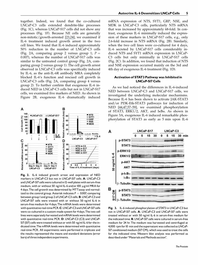
To demonstrate that induction of p-STAT3 inLNCaP-C3 cells was specifically through IL-6/IL-6Rsignaling pathway, we examined the dose-depen-dence and effects of specific inhibitors. We found thatIL-6 induced p-STAT3 at a dosage as low as 1 ng/ml,but the induction was maximal at a dosage of 50 ng/ml (Fig. 4A). The anti-IL-6R antibody MRA dose-de-pendently inhibited IL-6-induced p-STAT3 (Fig. 4B).As expected, a single dose of 100 mg/ml MRAblocked IL-6’s action over the 4-day treatment period(Fig. 4C).
LNCaP-S17CellsConstitutivelyExpressedHighLevelsof SOCS7 andCIS
To reveal the mechanisms that caused the differen-ces in STAT3 activation between LNCaP-C3 andLNCaP-S17 cells, we systematically examined the
IL-6/IL-6R signaling pathways. First, we determinedthe protein levels of IL-6 receptors. As shown inFigure 5A, the 80-kDa and 55-kDa forms of IL-6R aswell as gp130 were expressed at similar levels inLNCaP-C3 and LNCaP-S17 cells. Downstream to theIL-6 receptors are the JAK kinase family members.We found that JAK1 and JAK3 were not expressed ineither cell line. TYK2 was expressed at the same levelsin both cell lines. JAK2 expression was slightly higherin LNCaP-C3 cells than in LNCaP-S17 cells (Fig. 5A).Upon exogenous IL-6 treatment, phosphorylationof JAK2 was clearly induced in LNCaP-C3 cells butnot in LNCaP-S17 cells (Fig. 5B). To further confirmour findings, we did immunoprecipitation to enrichp-JAK2. Indeed, we found that exogenous IL-6 in-duced noticeable p-JAK2 accompanied by remarkabledegradation of JAK2 in LNCaP-C3 cells but not inLNCaP-S17 cells (Fig. 5C). To identify what factorswere inhibiting JAK2 phosphorylation, we examinedthe well-known negative feedback regulators CIS/SOCS family. This family of inhibitors has eight mem-bers including CIS and SOCS1 to SOCS7. As shown inFigure 6A, the basal levels of SOCS1 to SOCS6 werealmost the same between LNCaP-C3 and LNCaP-S17cells. There was not any obvious difference upon IL-6treatment. However, the basal levels of SOCS7 andCIS were clearly higher in LNCaP-S17 cells than inLNCaP-C3 cells (Fig. 6A). IL-6 treatment within the
Fig. 4. IL-6 inducedphosphorylationofSTAT3 inLNCaP-C3cellsvia IL-6R.A: LNCaP-C3 cells were treatedwithoutor with1, 5,10,and 50 ng/ml IL-6 for 5 min.B: LNCaP-C3 cellswere treatedwith-outor with 50 ng/ml IL-6 in serum-freemedium or in combinationwith different dosages of anti-IL-6R mAb (MRA) for 5 min. C:LNCaP-C3 cells were treatedwithout or with 50 ng/ml IL-6 in se-rum-freemediumorincombinationwith100 mg/mlMRAfor thein-dicated days. Western blot analysis was performed as describedunder ‘‘Materials andMethods section’’.
Fig. 5. IL-6 induced phosphorylation of JAK2 in LNCaP-C3 butnotinLNCaP-S17cells.A:Expressionof IL-6R,gp130, and JAKfami-ly in LNCaP-C3 (C3) and LNCaP-S17 (S17) cells in regular culturemedium without any treatment. B: Phosphorylation of JAK2 inLNCaP-C3 (C3) and LNCaP-S17 (S17) cells treated without orwith 50 ng/ml IL-6 in serum-free medium for the indicated time.C:LNCaP-C3 (C3) andLNCaP-S17 (S17) cellswere treatedwithoutor with 50 ng/ml IL-6 in serum-free medium for 5 min. Equalamounts of totalproteinswereprecipitatedwith anti-p-JAK2 anti-bodies and protein A-sepharose beads.Western blot analysis wasperformedasdescribedunder ‘‘Materials andMethods section’’.
6 Ge et al.
The Prostate
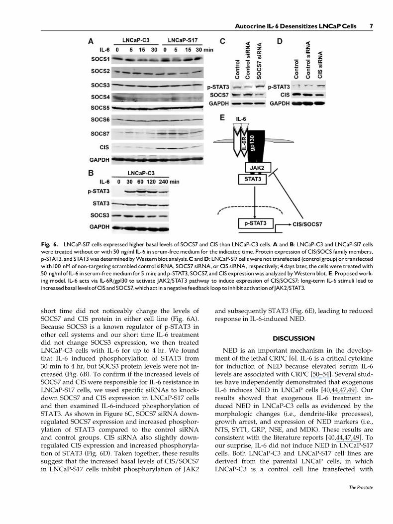
short time did not noticeably change the levels ofSOCS7 and CIS protein in either cell line (Fig. 6A).Because SOCS3 is a known regulator of p-STAT3 inother cell systems and our short time IL-6 treatmentdid not change SOCS3 expression, we then treatedLNCaP-C3 cells with IL-6 for up to 4 hr. We foundthat IL-6 induced phosphorylation of STAT3 from30 min to 4 hr, but SOCS3 protein levels were not in-creased (Fig. 6B). To confirm if the increased levels ofSOCS7 and CIS were responsible for IL-6 resistance inLNCaP-S17 cells, we used specific siRNAs to knock-down SOCS7 and CIS expression in LNCaP-S17 cellsand then examined IL-6-induced phosphorylation ofSTAT3. As shown in Figure 6C, SOCS7 siRNA down-regulated SOCS7 expression and increased phosphor-ylation of STAT3 compared to the control siRNAand control groups. CIS siRNA also slightly down-regulated CIS expression and increased phosphoryla-tion of STAT3 (Fig. 6D). Taken together, these resultssuggest that the increased basal levels of CIS/SOCS7in LNCaP-S17 cells inhibit phosphorylation of JAK2
and subsequently STAT3 (Fig. 6E), leading to reducedresponse in IL-6-induced NED.
DISCUSSION
NED is an important mechanism in the develop-ment of the lethal CRPC [6]. IL-6 is a critical cytokinefor induction of NED because elevated serum IL-6levels are associated with CRPC [50–54]. Several stud-ies have independently demonstrated that exogenousIL-6 induces NED in LNCaP cells [40,44,47,49]. Ourresults showed that exogenous IL-6 treatment in-duced NED in LNCaP-C3 cells as evidenced by themorphologic changes (i.e., dendrite-like processes),growth arrest, and expression of NED markers (i.e.,NTS, SYT1, GRP, NSE, and MDK). These results areconsistent with the literature reports [40,44,47,49]. Toour surprise, IL-6 did not induce NED in LNCaP-S17cells. Both LNCaP-C3 and LNCaP-S17 cell lines arederived from the parental LNCaP cells, in whichLNCaP-C3 is a control cell line transfected with
Fig. 6. LNCaP-S17 cells expressed higher basal levels of SOCS7 and CIS than LNCaP-C3 cells.A and B: LNCaP-C3 and LNCaP-S17 cellswere treatedwithout or with 50 ng/ml IL-6 in serum-freemedium for the indicated time.Protein expression of CIS/SOCS familymembers,p-STAT3, andSTAT3wasdeterminedbyWesternblot analysis.C andD:LNCaP-S17 cellswerenot transfected (controlgroup) or transfectedwith100 nMof non-targeting scrambled control siRNA, SOCS7 siRNA, or CIS siRNA, respectively; 4 days later, the cells were treatedwith50 ng/mlof IL-6 in serum-freemedium for 5 min; andp-STAT3, SOCS7, andCIS expressionwas analyzedbyWesternblot.E: Proposedwork-ing model. IL-6 acts via IL-6R/gp130 to activate JAK2/STAT3 pathway to induce expression of CIS/SOCS7; long-term IL-6 stimuli lead toincreasedbasal levelsofCISandSOCS7,whichactinanegative feedbackloop toinhibitactivationof JAK2/STAT3.
Autocrine IL-6DesensitizesLNCaPCells 7
The Prostate
empty vector, and LNCaP-S17 is transfected with full-length IL-6 cDNA [66]. Thus, the difference is thatLNCaP-C3 cells do not express IL-6 but LNCaP-S17cells secrete IL-6 in an autocrine manner.
To reveal the molecular basis underlying the differ-ences in IL-6-induced NED between LNCaP-C3 andLNCaP-S17 cell lines, we first examined activation ofSTAT3 by IL-6 because it has been well establishedthat IL-6 activates JAK-STAT3 and/or PI3K-Etk-STAT3 pathways to induce NED in LNCaP cells[44,47,57–59]. We identified that a remarkable differ-ence between LNCaP-C3 and LNCaP-S17 cell lines isin their response to exogenous IL-6 in terms of phos-phorylation of STAT3. IL-6 induced p-STAT3 in adose-dependent manner for a period of 4 days after asingle dose treatment in LNCaP-C3 cells, which wasdependent on IL-6R as anti-IL-6R antibodies couldabolish the induction completely. In contrast, IL-6failed to induce any noticeable p-STAT3 in LNCaP-S17 cells. IL-6 did not activate ERK1/2, AKT or NF-kBpathways in either LNCaP-C3 or LNCaP-S17 cell line,suggesting that IL-6 specifically activates the STAT3pathway to induce NED.
It is known that IL-6 acts through IL-6R and gp130[60]. Gp130 then activates JAKs, followed by activa-tion of STAT3 [63,64]. Our results suggest thatLNCaP-C3 and LNCaP-S17 cell lines express similarlevels of IL-6R and gp130. In addition, JAK familymember TYK2 is expressed at the same levels in thetwo cell lines while JAK1 and JAK3 are not expressedin either cell line. Thus, IL-6R, gp130, JAK1, JAK3,and TYK2 do not contribute to the differentialresponses observed in the two cell lines. The basallevel of JAK2 expression was slightly higher inLNCaP-C3 cell line than in LNCaP-S17 cell line. IL-6induced phosphorylation of JAK2 in LNCaP-C3 cellline but not in LNCaP-S17 cell line. This cannot becompletely attributed to the increased basal level ofJAK2 in LNCaP-C3 cell line because LNCaP-S17 cellline also expresses a decent level of JAK2. Thus, it ismore likely that activation of JAK2 is inhibited bysome factors. Given that there are eight well-knownnegative regulators of cytokine signaling includingCIS and SOCS1-7 [68], we examined the expressionof all of them in LNCaP-C3 and LNCaP-S17 cell lines.Our results suggest that SOCS1-6 are equallyexpressed in the two cell lines, thus SOCS1-6 do notcontribute to the differential inhibition of JAK2/STAT3 signaling. However, SOCS7 and CIS levelswere constitutively higher in LNCaP-S17 cell linethan in LNCaP-C3 cell line, suggesting that SOCS7and CIS are responsible for the observed differencesin IL-6-induced NED between the two cell lines. It hasbeen shown that CIS and SOCS proteins can directlybind to JAKs and reduce their tyrosine-kinase
activities and activation of STATs [69]. IL-6 can in-duce expression of CIS and SOCS7 [70,71], thus CISand SOCS7 act in a negative feedback loop to inhibitIL-6-induced activation of JAK2/STAT3 pathway.Furthermore, down-regulation of SOCS7 and CISexpression by siRNA transfection increased IL-6-induced phosphorylation of STAT3, which confirmsthat SOCS7 and CIS are responsible for the IL-6 resis-tance in LNCaP-S17 cells.
The results presented here significantly extendedour previous studies [66,67,72]. We demonstrated thatLNCaP-S17 cells constitutively express high levels ofCIS and SOCS7, which in turn inhibit activation ofJAK2 and STAT3, leading to a failure to respond toIL-6-induced NED. We speculate that the high levelsof CIS and SOCS7 expression are caused by the chron-ic stimuli of IL-6 secreted by LNCaP-S17 cells becauseCIS and SOCS7 are downstream target genes inducedby IL-6. We and other investigators have shown thatshort-term exogenous IL-6 treatment induces NED,whereas long-term exogenous IL-6 stimuli not onlycause LNCaP cells to express endogenous IL-6 butalso lead to increased cell proliferation [72,73]. Themechanisms are reportedly due to a decrease in IL-6binding capacity and/or an increase in androgenbinding capacity and AR signaling after long-termIL-6 treatment [72,73]. The present study providesa new mechanism that CIS and SOCS7 levels areincreased in LNCaP cells exposed to long-term auto-crine IL-6. Thus, the increased levels of CIS andSOCS7 expression desensitize LNCaP cells’ responseto IL-6-induced NED.
In conclusion, we demonstrated in this study thatLNCaP cells with autocrine IL-6 expression are resis-tant to exogenous IL-6-induced NED due to increasedlevels of CIS/SOCS7 that block activation of JAK2-STAT3 pathways.
ACKNOWLEDGMENTS
The authors thank Dr. Prescott L. Deininger (Direc-tor of Tulane Cancer Center, PI of the COBRE grant2P20RR020152-06) and Tulane Cancer Center Core Fa-cilities for research support. They also thank ChugaiPharmaceutical Co., Ltd, Tokyo, Japan, for the gift ofMRA.
REFERENCES
1. Chodak GW, Vogelzang NJ, Caplan RJ, Soloway M, Smith JA.Independent prognostic factors in patients with metastatic(stage D2) prostate cancer. The Zoladex Study Group. Jama1991;265(5):618–621.
2. Roudier MP, True LD, Higano CS, Vesselle H, Ellis W, LangeP, Vessella RL. Phenotypic heterogeneity of end-stage prostatecarcinoma metastatic to bone. Hum Pathol 2003;34(7):646–653.
8 Ge et al.
The Prostate
3. McDonnell TJ, Troncoso P, Brisbay SM, Logothetis C, ChungLW, Hsieh JT, Tu SM, Campbell ML. Expression of the proto-oncogene bcl-2 in the prostate and its association with emer-gence of androgen-independent prostate cancer. Cancer Res1992;52(24):6940–6944.
4. July LV, Akbari M, Zellweger T, Jones EC, Goldenberg SL,Gleave ME. Clusterin expression is significantly enhanced inprostate cancer cells following androgen withdrawal therapy.Prostate 2002;50(3):179–188.
5. Gleave M, Jansen B. Clusterin and IGFBPs as antisense targetsin prostate cancer. Ann N Y Acad Sci 2003;1002:95–104.
6. Abrahamsson PA. Neuroendocrine cells in tumour growth ofthe prostate. Endocr Relat Cancer 1999;6(4):503–519.
7. Visakorpi T, Hyytinen E, Koivisto P, Tanner M, Keinanen R,Palmberg C, Palotie A, Tammela T, Isola J, Kallioniemi OP.In vivo amplification of the androgen receptor gene and pro-gression of human prostate cancer. Nat Genet 1995;9(4):401–406.
8. Koivisto P, Kononen J, Palmberg C, Tammela T, Hyytinen E,Isola J, Trapman J, Cleutjens K, Noordzij A, Visakorpi T,Kallioniemi OP. Androgen receptor gene amplification: Apossible molecular mechanism for androgen deprivationtherapy failure in prostate cancer. Cancer Res 1997;57(2):314–319.
9. Chen CD, Welsbie DS, Tran C, Baek SH, Chen R, Vessella R,Rosenfeld MG, Sawyers CL. Molecular determinants of resis-tance to antiandrogen therapy. Nat Med 2004;10(1):33–39.
10. Newmark JR, Hardy DO, Tonb DC, Carter BS, Epstein JI, IsaacsWB, Brown TR, Barrack ER. Androgen receptor gene muta-tions in human prostate cancer. Proc Natl Acad Sci USA 1992;89(14):6319–6323.
11. Taplin ME, Bubley GJ, Shuster TD, Frantz ME, Spooner AE,Ogata GK, Keer HN, Balk SP. Mutation of the androgen-recep-tor gene in metastatic androgen-independent prostate cancer.N Engl J Med 1995;332(21):1393–1398.
12. Mohler JL, Gregory CW, Ford OH 3rd, Kim D, Weaver CM,Petrusz P, Wilson EM, French FS. The androgen axis in recur-rent prostate cancer. Clin Cancer Res 2004;10(2):440–448.
13. Stanbrough M, Bubley GJ, Ross K, Golub TR, Rubin MA, Pen-ning TM, Febbo PG, Balk SP. Increased expression of genesconverting adrenal androgens to testosterone in androgen-independent prostate cancer. Cancer Res 2006;66(5):2815–2825.
14. Hu R, Dunn TA, Wei S, Isharwal S, Veltri RW, Humphreys E,Han M, Partin AW, Vessella RL, Isaacs WB, Bova GS, Luo J.Ligand-independent androgen receptor variants derived fromsplicing of cryptic exons signify hormone-refractory prostatecancer. Cancer Res 2009;69(1):16–22.
15. Culig Z, Hobisch A, Cronauer MV, Radmayr C, Trapman J,Hittmair A, Bartsch G, Klocker H. Androgen receptor activa-tion in prostatic tumor cell lines by insulin-like growth factor-I,keratinocyte growth factor, and epidermal growth factor. Can-cer Res 1994;54(20):5474–5478.
16. Mellon K, Thompson S, Charlton RG, Marsh C, Robinson M,Lane DP, Harris AL, Horne CH, Neal DE. p53, c-erbB-2 andthe epidermal growth factor receptor in the benign and malig-nant prostate. J Urol 1992;147(2):496–499.
17. Craft N, Shostak Y, Carey M, Sawyers CL. A mechanism forhormone-independent prostate cancer through modulationof androgen receptor signaling by the HER-2/neu tyrosinekinase. Nat Med 1999;5(3):280–285.
18. Unni E, Sun S, Nan B, McPhaul MJ, Cheskis B, Mancini MA,Marcelli M . Changes in androgen receptor nongenotropic
signaling correlate with transition of LNCaP cells to androgenindependence. Cancer Res 2004;64(19):7156–7168.
19. Debes JD, Schmidt LJ, Huang H, Tindall DJ. p300 mediatesandrogen-independent transactivation of the androgen recep-tor by interleukin 6. Cancer Res 2002;62(20):5632–5636.
20. Abrahamsson PA. Neuroendocrine differentiation in prostaticcarcinoma. Prostate 1999;39(2):135–148.
21. di Sant’ Agnese PA. Neuroendocrine differentiation in carcino-ma of the prostate. Diagnostic, prognostic, and therapeuticimplications. Cancer 1992;70(1 Suppl):254–268.
22. Vashchenko N, Abrahamsson PA. Neuroendocrine differentia-tion in prostate cancer: Implications for new treatment modali-ties. Eur Urol 2005;47(2):147–155.
23. Bang YJ, Pirnia F, Fang WG, Kang WK, Sartor O, Whitesell L,Ha MJ, Tsokos M, Sheahan MD, Nguyen P, Niklinski WT,Myers CE, Trepel JB. Terminal neuroendocrine differentiationof human prostate carcinoma cells in response to increased in-tracellular cyclic AMP. Proc Natl Acad Sci USA 1994;91(12):5330–5334.
24. Bonkhoff H, Stein U, Remberger K. Endocrine-paracrine celltypes in the prostate and prostatic adenocarcinoma are postmi-totic cells. Hum Pathol 1995;26(2):167–170.
25. Bonkhoff H, Wernert N, Dhom G, Remberger K. Relation of en-docrine-paracrine cells to cell proliferation in normal, hyper-plastic, and neoplastic human prostate. Prostate 1991;19(2):91–98.
26. Dizeyi N, Bjartell A, Nilsson E, Hansson J, Gadaleanu V, CrossN, Abrahamsson PA. Expression of serotonin receptors androle of serotonin in human prostate cancer tissue and cell lines.Prostate 2004;59(3):328–336.
27. Levine L, Lucci JA 3rd, Pazdrak B, Cheng JZ, Guo YS,Townsend CM Jr, Hellmich MR. Bombesin stimulates nuclearfactor kappa B activation and expression of proangiogenicfactors in prostate cancer cells. Cancer Res 2003;63(13):3495–3502.
28. Iwamura M, di Sant’Agnese PA, Wu G, Benning CM, CockettAT, Deftos LJ, Abrahamsson PA. Immunohistochemical locali-zation of parathyroid hormone-related protein in human pros-tate cancer. Cancer Res 1993;53(8):1724–1726.
29. Jin RJ, Wang Y, Masumori N, Ishii K, Tsukamoto T, ShappellSB, Hayward SW, Kasper S, Matusik RJ. NE-10 neuroendocrinecancer promotes the LNCaP xenograft growth in castratedmice. Cancer Res 2004;64(15):5489–5495.
30. Di Sant’Agnese PA, Cockett AT. The prostatic endocrine-para-crine (neuroendocrine) regulatory system and neuroendocrinedifferentiation in prostatic carcinoma: A review and futuredirections in basic research. J Urol 1994;152(5 Pt 2):1927–1931.
31. di Sant’Agnese PA, de Mesy Jensen KL. Neuroendocrinedifferentiation in prostatic carcinoma. Hum Pathol 1987;18(8):849–856.
32. Bonkhoff H, Stein U, Remberger K. Multidirectional differenti-ation in the normal, hyperplastic, and neoplastic human pros-tate: Simultaneous demonstration of cell-specific epithelialmarkers. Hum Pathol 1994;25(1):42–46.
33. Abrahamsson PA, di Sant’Agnese PA. Neuroendocrine cells inthe human prostate gland. J Androl 1993;14(5):307–309.
34. Angelsen A, Syversen U, Haugen OA, Stridsberg M, MjolnerodOK, Waldum HL. Neuroendocrine differentiation in carcino-mas of the prostate: Do neuroendocrine serum markers reflectimmunohistochemical findings? Prostate 1997;30(1):1–6.
35. Ismail AH, Landry F, Aprikian AG, Chevalier S. Androgenablation promotes neuroendocrine cell differentiation in dogand human prostate. Prostate 2002;51(2):117–125.
Autocrine IL-6DesensitizesLNCaPCells 9
The Prostate
36. Sciarra A, Di Silverio F. Effect of nonsteroidal antiandrogenmonotherapy versus castration therapy on neuroendocrinedifferentiation in prostate carcinoma. Urology 2004;63(3):523–527.
37. Hirano D, Okada Y, Minei S, Takimoto Y, Nemoto N. Neuroen-docrine differentiation in hormone refractory prostate cancerfollowing androgen deprivation therapy. Eur Urol 2004;45(5):586–592 discussion 592.
38. Huss WJ, Gregory CW, Smith GJ. Neuroendocrine cell differ-entiation in the CWR22 human prostate cancer xenograft: As-sociation with tumor cell proliferation prior to recurrence.Prostate 2004;60(2):91–97.
39. Cox ME, Deeble PD, Lakhani S, Parsons SJ. Acquisition of neu-roendocrine characteristics by prostate tumor cells is revers-ible: Implications for prostate cancer progression. Cancer Res1999;59(15):3821–3830.
40. Deeble PD, Murphy DJ, Parsons SJ, Cox ME. Interleukin-6- andcyclic AMP-mediated signaling potentiates neuroendocrinedifferentiation of LNCaP prostate tumor cells. Mol Cell Biol2001;21(24):8471–8482.
41. Kim J, Adam RM, Freeman MR. Activation of the Erk mitogen-activated protein kinase pathway stimulates neuroendocrinedifferentiation in LNCaP cells independently of cell cyclewithdrawal and STAT3 phosphorylation. Cancer Res 2002;62(5):1549–1554.
42. Cox ME, Deeble PD, Bissonette EA, Parsons SJ. Activated3’,5’-cyclic AMP-dependent protein kinase is sufficient toinduce neuroendocrine-like differentiation of the LNCaPprostate tumor cell line. J Biol Chem 2000;275(18):13812–13818.
43. Juarranz MG, Bolanos O, Gutierrez-Canas I, Lerner EA, Rob-berecht P, Carmena MJ, Prieto JC, Rodriguez-Henche N. Neu-roendocrine differentiation of the LNCaP prostate cancer cellline maintains the expression and function of VIP and PACAPreceptors. Cell Signal 2001;13(12):887–894.
44. Spiotto MT, Chung TD. STAT3 mediates IL-6-induced neuro-endocrine differentiation in prostate cancer cells. Prostate 2000;42(3):186–195.
45. Shi XB, Ma AH, Tepper CG, Xia L, Gregg JP, Gandour-Edwards R, Mack PC, Kung HJ, deVere White RW. Molecularalterations associated with LNCaP cell progression to andro-gen independence. Prostate 2004;60(3):257–271.
46. Burchardt T, Burchardt M, Chen MW, Cao Y, de la Taille A,Shabsigh A, Hayek O, Dorai T, Buttyan R. Transdifferentiationof prostate cancer cells to a neuroendocrine cell phenotype invitro and in vivo. J Urol 1999;162(5):1800–1805.
47. Qiu Y, Robinson D, Pretlow TG, Kung HJ. Etk/Bmx, a tyrosinekinase with a pleckstrin-homology domain, is an effector ofphosphatidylinositol 3’-kinase and is involved in interleukin6-induced neuroendocrine differentiation of prostate cancercells. Proc Natl Acad Sci USA 1998;95(7):3644–3649.
48. Adam RM, Kim J, Lin J, Orsola A, Zhuang L, Rice DC, FreemanMR. Heparin-binding epidermal growth factor-like growthfactor stimulates androgen-independent prostate tumorgrowth and antagonizes androgen receptor function. Endocri-nology 2002;143(12):4599–4608.
49. Wang Q, Horiatis D, Pinski J. Interleukin-6 inhibits the growthof prostate cancer xenografts in mice by the process of neuro-endocrine differentiation. Int J Cancer 2004;111(4):508–513.
50. Michalaki V, Syrigos K, Charles P, Waxman J. Serum levels ofIL-6 and TNF-alpha correlate with clinicopathological featuresand patient survival in patients with prostate cancer. Br J Can-cer 2004;90(12):2312–2316.
51. Corcoran NM, Costello AJ. Interleukin-6: Minor player or star-ring role in the development of hormone-refractory prostatecancer? BJU Int 2003;91(6):545–553.
52. Adler HL, McCurdy MA, Kattan MW, Timme TL, Scardino PT,Thompson TC. Elevated levels of circulating interleukin-6 andtransforming growth factor-beta1 in patients with metastaticprostatic carcinoma. J Urol 1999;161(1):182–187.
53. Drachenberg DE, Elgamal AA, Rowbotham R, Peterson M,Murphy GP. Circulating levels of interleukin-6 in patients withhormone refractory prostate cancer. Prostate 1999;41(2):127–133.
54. Nakashima J, Tachibana M, Horiguchi Y, Oya M, Ohigashi T,Asakura H, Murai M. Serum interleukin 6 as a prognosticfactor in patients with prostate cancer. Clin Cancer Res 2000;6(7):2702–2706.
55. Culig Z, Puhr M. Interleukin-6: A multifunctional targetablecytokine in human prostate cancer. Mol Cell Endocrinol 2011.In Press; PMID: 21664423; Epub ahead of print, doi:10.1016/j.mce.2011.05.033.
56. Giri D, Ozen M, Ittmann M. Interleukin-6 is an autocrinegrowth factor in human prostate cancer. Am J Pathol 2001;159(6):2159–2165.
57. Tsai YT, Su YH, Fang SS, Huang TN, Qiu Y, Jou YS, Shih HM,Kung HJ, Chen RH. Etk, a Btk family tyrosine kinase, mediatescellular transformation by linking Src to STAT3 activation. MolCell Biol 2000;20(6):2043–2054.
58. Grossmann ME, Huang H, Tindall DJ. Androgen receptor sig-naling in androgen-refractory prostate cancer. J Natl CancerInst 2001;93(22):1687–1697.
59. Kim J, Adam RM, Solomon KR, Freeman MR. Involvement ofcholesterol-rich lipid rafts in interleukin-6-induced neuroendo-crine differentiation of LNCaP prostate cancer cells. Endocri-nology 2004;145(2):613–619.
60. Boulanger MJ, Chow DC, Brevnova EE, Garcia KC. Hexamericstructure and assembly of the interleukin-6/IL-6 alpha-receptor/gp130 complex. Science 2003;300(5628):2101–2104.
61. Murakami M, Hibi M, Nakagawa N, Nakagawa T, YasukawaK, Yamanishi K, Taga T, Kishimoto T. IL-6-induced homodi-merization of gp130 and associated activation of a tyrosine ki-nase. Science 1993;260(5115):1808–1810.
62. Benigni F, Fantuzzi G, Sacco S, Sironi M, Pozzi P, DinarelloCA, Sipe JD, Poli V, Cappelletti M, Paonessa G, Pennica D,Panayotatos N, Ghezzi P. Six different cytokines that shareGP130 as a receptor subunit, induce serum amyloid A and po-tentiate the induction of interleukin-6 and the activation of thehypothalamus-pituitary-adrenal axis by interleukin-1. Blood1996;87(5):1851–1854.
63. Ihle JN. Cytokine receptor signalling. Nature 1995;377(6550):591–594.
64. Heinrich PC, Behrmann I, Muller-Newen G, Schaper F, GraeveL. Interleukin-6-type cytokine signalling through the gp130/Jak/STAT pathway. Biochem J 1998;334(Pt 2):297–314.
65. Chung TD, Yu JJ, Spiotto MT, Bartkowski M, Simons JW. Char-acterization of the role of IL-6 in the progression of prostatecancer. Prostate 1999;38(3):199–207.
66. Lou W, Ni Z, Dyer K, Tweardy DJ, Gao AC. Interleukin-6 indu-ces prostate cancer cell growth accompanied by activation ofstat3 signaling pathway. Prostate 2000;42(3):239–242.
67. Lee SO, Lou W, Hou M, de Miguel F, Gerber L, Gao AC. Inter-leukin-6 promotes androgen-independent growth in LNCaPhuman prostate cancer cells. Clin Cancer Res 2003;9(1):370–376.
10 Ge et al.
The Prostate
68. Nicholson SE, Hilton DJ. The SOCS proteins: A new family ofnegative regulators of signal transduction. J Leukoc Biol1998;63(6):665–668.
69. Endo TA, Masuhara M, Yokouchi M, Suzuki R, Sakamoto H,Mitsui K, Matsumoto A, Tanimura S, Ohtsubo M, Misawa H,Miyazaki T, Leonor N, Taniguchi T, Fujita T, Kanakura Y,Komiya S, Yoshimura A. A new protein containing an SH2domain that inhibits JAK kinases. Nature 1997;387(6636):921–924.
70. Campbell JS, Prichard L, Schaper F, Schmitz J,Stephenson-Famy A, Rosenfeld ME, Argast GM, HeinrichPC, Fausto N. Expression of suppressors of cytokine signalingduring liver regeneration. J Clin Invest 2001;107(10):1285–1292.
71. Dogusan Z, Hooghe-Peters EL, Berus D, Velkeniers B, HoogheR. Expression of SOCS genes in normal and leukemic humanleukocytes stimulated by prolactin, growth hormone and cyto-kines. J Neuroimmunol 2000;109(1):34–39.
72. Lee SO, Chun JY, Nadiminty N, Lou W, Gao AC. Interleukin-6undergoes transition from growth inhibitor associated withneuroendocrine differentiation to stimulator accompanied byandrogen receptor activation during LNCaP prostate cancercell progression. Prostate 2007;67(7):764–773.
73. Hobisch A, Ramoner R, Fuchs D, Godoy-Tundidor S, BartschG, Klocker H, Culig Z. Prostate cancer cells (LNCaP) generatedafter long-term interleukin 6 (IL-6) treatment express IL-6 andacquire an IL-6 partially resistant phenotype. Clin Cancer Res2001;7(9):2941–2948.
Autocrine IL-6DesensitizesLNCaPCells 11
The Prostate


![Preparation of [5 + 6]-, [6 + 6]-, and [6 + 7]Bicyclic Guanidines fromC,C'Bis(iminophosphoranes](https://static.fdokumen.com/doc/165x107/631f2da3d10f1687490fada7/preparation-of-5-6-6-6-and-6-7bicyclic-guanidines-fromccbisiminophosphoranes.jpg)