The role of Stat5 transcription factors as tumor suppressors or oncogenes
11
Review The role of Stat5 transcription factors as tumor suppressors or oncogenes G. Ferbeyre a, ⁎, R. Moriggl b, ⁎ a Département de Biochimie, Université de Montréal, Montréal, Québec H3C 3J7, Canada b Ludwig Boltzmann Institute for Cancer Research, Vienna, Austria abstract article info Article history: Received 9 September 2010 Received in revised form 8 October 2010 Accepted 8 October 2010 Available online 20 October 2010 Keywords: Stat5 Jak–Stat signaling Senescence Mouse models Stat5 is constitutively activated in many human cancers affecting the expression of cell proliferation and cell survival controlling genes. These oncogenic functions of Stat5 have been elegantly reproduced in mouse models. Aberrant Stat5 activity induces also mitochondrial dysfunction and reactive oxygen species leading to DNA damage. Although DNA damage can stimulate tumorigenesis, it can also prevent it. Stat5 can inhibit tumor progression like in the liver and it is a tumor suppressor in fibroblasts. Stat5 proteins are able to regulate cell differentiation and senescence activating the tumor suppressors SOCS1, p53 and PML. Understanding the context dependent regulation of tumorigenesis through Stat5 function will be central to understand proliferation, survival, differentiation or senescence of cancer cells. © 2010 Elsevier B.V. All rights reserved. Contents 1. Introduction . . . . . . . . . . . . . . . . . . . . . . . . . . . . . . . . . . . . . . . . . . . . . . . . . . . . . . . . . . . . . . 105 2. Activation of Stat5 proteins and insights from different cancers . . . . . . . . . . . . . . . . . . . . . . . . . . . . . . . . . . . . . . 105 2.1. The role of Stat5 protein activation in hematopoietic neoplasms . . . . . . . . . . . . . . . . . . . . . . . . . . . . . . . . . . 105 2.2. Activation or inactivation of Stat5 proteins by viruses: HTLV-I and HIV . . . . . . . . . . . . . . . . . . . . . . . . . . . . . . . 106 2.3. Breast cancer . . . . . . . . . . . . . . . . . . . . . . . . . . . . . . . . . . . . . . . . . . . . . . . . . . . . . . . . . . 107 2.4. Prostate cancer . . . . . . . . . . . . . . . . . . . . . . . . . . . . . . . . . . . . . . . . . . . . . . . . . . . . . . . . . 107 2.5. Lung, head and neck cancers . . . . . . . . . . . . . . . . . . . . . . . . . . . . . . . . . . . . . . . . . . . . . . . . . . . 107 2.6. Liver cancer . . . . . . . . . . . . . . . . . . . . . . . . . . . . . . . . . . . . . . . . . . . . . . . . . . . . . . . . . . . 107 2.7. Melanoma . . . . . . . . . . . . . . . . . . . . . . . . . . . . . . . . . . . . . . . . . . . . . . . . . . . . . . . . . . . . 107 2.8. Other malignancies . . . . . . . . . . . . . . . . . . . . . . . . . . . . . . . . . . . . . . . . . . . . . . . . . . . . . . . 107 3. Stat5-regulated transcription: A matter of protein–protein interaction . . . . . . . . . . . . . . . . . . . . . . . . . . . . . . . . . . . 108 4. Mechanisms of oncogenic activity of Stat5 proteins and target gene regulation . . . . . . . . . . . . . . . . . . . . . . . . . . . . . . . 108 4.1. Anti-apoptotic functions of Stat5 proteins . . . . . . . . . . . . . . . . . . . . . . . . . . . . . . . . . . . . . . . . . . . . . 108 4.2. Cell proliferation and control through IGF-1 signaling . . . . . . . . . . . . . . . . . . . . . . . . . . . . . . . . . . . . . . . 108 4.3. DNA damage response . . . . . . . . . . . . . . . . . . . . . . . . . . . . . . . . . . . . . . . . . . . . . . . . . . . . . . 108 4.4. Invasion, metastasis and epithelial to mesenchymal transition (EMT) . . . . . . . . . . . . . . . . . . . . . . . . . . . . . . . . 109 5. Can Stat5 proteins also function as tumor suppressor proteins? . . . . . . . . . . . . . . . . . . . . . . . . . . . . . . . . . . . . . . 109 5.1. Mouse models of Stat5 inactivation . . . . . . . . . . . . . . . . . . . . . . . . . . . . . . . . . . . . . . . . . . . . . . . . 109 5.2. Cell differentiation . . . . . . . . . . . . . . . . . . . . . . . . . . . . . . . . . . . . . . . . . . . . . . . . . . . . . . . . 110 5.3. Cell senescence . . . . . . . . . . . . . . . . . . . . . . . . . . . . . . . . . . . . . . . . . . . . . . . . . . . . . . . . . 110 6. Concluding remarks. . . . . . . . . . . . . . . . . . . . . . . . . . . . . . . . . . . . . . . . . . . . . . . . . . . . . . . . . . . 110 Acknowledgements . . . . . . . . . . . . . . . . . . . . . . . . . . . . . . . . . . . . . . . . . . . . . . . . . . . . . . . . . . . . . 111 References . . . . . . . . . . . . . . . . . . . . . . . . . . . . . . . . . . . . . . . . . . . . . . . . . . . . . . . . . . . . . . . . . 111 Biochimica et Biophysica Acta 1815 (2011) 104–114 ⁎ Corresponding authors. G. Ferbeyre is to be contacted at Université de Montréal, Département de Biochimie, E-515, C.P. 6128, Succ. Centre-Ville, Montréal, Qc H3C 3J7. Tel.: +1 514 343 7571; fax: +1 514 343 2210. R. Moriggl, Ludwig Boltzmann Institute for Cancer Research, 1090 Vienna, Austria. Tel.: +43 1427764111; fax: +43 142779641. E-mail addresses: [email protected] (G. Ferbeyre), [email protected] (R. Moriggl). 0304-419X/$ – see front matter © 2010 Elsevier B.V. All rights reserved. doi:10.1016/j.bbcan.2010.10.004 Contents lists available at ScienceDirect Biochimica et Biophysica Acta journal homepage: www.elsevier.com/locate/bbacan
Transcript of The role of Stat5 transcription factors as tumor suppressors or oncogenes

Biochimica et Biophysica Acta 1815 (2011) 104–114
Contents lists available at ScienceDirect
Biochimica et Biophysica Acta
j ourna l homepage: www.e lsev ie r.com/ locate /bbacan
Review
The role of Stat5 transcription factors as tumor suppressors or oncogenes
G. Ferbeyre a,⁎, R. Moriggl b,⁎a Département de Biochimie, Université de Montréal, Montréal, Québec H3C 3J7, Canadab Ludwig Boltzmann Institute for Cancer Research, Vienna, Austria
⁎ Corresponding authors. G. Ferbeyre is to be contacte514 343 7571; fax: +1 514 343 2210. R. Moriggl, Ludw
E-mail addresses: [email protected] (G. Ferbe
0304-419X/$ – see front matter © 2010 Elsevier B.V. Adoi:10.1016/j.bbcan.2010.10.004
a b s t r a c t
a r t i c l e i n f oArticle history:Received 9 September 2010Received in revised form 8 October 2010Accepted 8 October 2010Available online 20 October 2010
Keywords:Stat5Jak–Stat signalingSenescenceMouse models
Stat5 is constitutively activated in many human cancers affecting the expression of cell proliferation and cellsurvival controlling genes. These oncogenic functions of Stat5 have been elegantly reproduced in mousemodels. Aberrant Stat5 activity induces also mitochondrial dysfunction and reactive oxygen species leading toDNA damage. Although DNA damage can stimulate tumorigenesis, it can also prevent it. Stat5 can inhibittumor progression like in the liver and it is a tumor suppressor in fibroblasts. Stat5 proteins are able toregulate cell differentiation and senescence activating the tumor suppressors SOCS1, p53 and PML.Understanding the context dependent regulation of tumorigenesis through Stat5 function will be central tounderstand proliferation, survival, differentiation or senescence of cancer cells.
d at Université de Montréal, Département de Biochimie,ig Boltzmann Institute for Cancer Research, 1090 Viennayre), [email protected] (R. Moriggl).
ll rights reserved.
© 2010 Elsevier B.V. All rights reserved.
Contents
1. Introduction . . . . . . . . . . . . . . . . . . . . . . . . . . . . . . . . . . . . . . . . . . . . . . . . . . . . . . . . . . . . . . 1052. Activation of Stat5 proteins and insights from different cancers . . . . . . . . . . . . . . . . . . . . . . . . . . . . . . . . . . . . . . 105
2.1. The role of Stat5 protein activation in hematopoietic neoplasms . . . . . . . . . . . . . . . . . . . . . . . . . . . . . . . . . . 1052.2. Activation or inactivation of Stat5 proteins by viruses: HTLV-I and HIV . . . . . . . . . . . . . . . . . . . . . . . . . . . . . . . 1062.3. Breast cancer . . . . . . . . . . . . . . . . . . . . . . . . . . . . . . . . . . . . . . . . . . . . . . . . . . . . . . . . . . 1072.4. Prostate cancer . . . . . . . . . . . . . . . . . . . . . . . . . . . . . . . . . . . . . . . . . . . . . . . . . . . . . . . . . 1072.5. Lung, head and neck cancers . . . . . . . . . . . . . . . . . . . . . . . . . . . . . . . . . . . . . . . . . . . . . . . . . . . 1072.6. Liver cancer . . . . . . . . . . . . . . . . . . . . . . . . . . . . . . . . . . . . . . . . . . . . . . . . . . . . . . . . . . . 1072.7. Melanoma . . . . . . . . . . . . . . . . . . . . . . . . . . . . . . . . . . . . . . . . . . . . . . . . . . . . . . . . . . . . 1072.8. Other malignancies . . . . . . . . . . . . . . . . . . . . . . . . . . . . . . . . . . . . . . . . . . . . . . . . . . . . . . . 107
3. Stat5-regulated transcription: A matter of protein–protein interaction . . . . . . . . . . . . . . . . . . . . . . . . . . . . . . . . . . . 1084. Mechanisms of oncogenic activity of Stat5 proteins and target gene regulation . . . . . . . . . . . . . . . . . . . . . . . . . . . . . . . 108
4.1. Anti-apoptotic functions of Stat5 proteins . . . . . . . . . . . . . . . . . . . . . . . . . . . . . . . . . . . . . . . . . . . . . 1084.2. Cell proliferation and control through IGF-1 signaling . . . . . . . . . . . . . . . . . . . . . . . . . . . . . . . . . . . . . . . 1084.3. DNA damage response . . . . . . . . . . . . . . . . . . . . . . . . . . . . . . . . . . . . . . . . . . . . . . . . . . . . . . 1084.4. Invasion, metastasis and epithelial to mesenchymal transition (EMT) . . . . . . . . . . . . . . . . . . . . . . . . . . . . . . . . 109
5. Can Stat5 proteins also function as tumor suppressor proteins? . . . . . . . . . . . . . . . . . . . . . . . . . . . . . . . . . . . . . . 1095.1. Mouse models of Stat5 inactivation . . . . . . . . . . . . . . . . . . . . . . . . . . . . . . . . . . . . . . . . . . . . . . . . 1095.2. Cell differentiation . . . . . . . . . . . . . . . . . . . . . . . . . . . . . . . . . . . . . . . . . . . . . . . . . . . . . . . . 1105.3. Cell senescence . . . . . . . . . . . . . . . . . . . . . . . . . . . . . . . . . . . . . . . . . . . . . . . . . . . . . . . . . 110
6. Concluding remarks. . . . . . . . . . . . . . . . . . . . . . . . . . . . . . . . . . . . . . . . . . . . . . . . . . . . . . . . . . . 110Acknowledgements . . . . . . . . . . . . . . . . . . . . . . . . . . . . . . . . . . . . . . . . . . . . . . . . . . . . . . . . . . . . . 111References . . . . . . . . . . . . . . . . . . . . . . . . . . . . . . . . . . . . . . . . . . . . . . . . . . . . . . . . . . . . . . . . . 111
E-515, C.P. 6128, Succ. Centre-Ville, Montréal, Qc H3C 3J7. Tel.: +1, Austria. Tel.: +43 1427764111; fax: +43 142779641.
105G. Ferbeyre, R. Moriggl / Biochimica et Biophysica Acta 1815 (2011) 104–114
1. Introduction
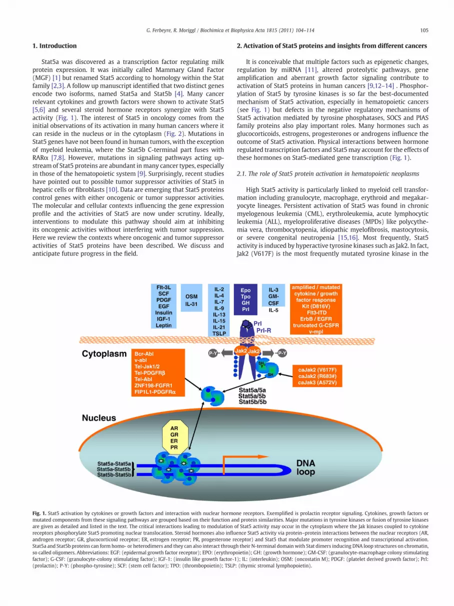
Stat5a was discovered as a transcription factor regulating milkprotein expression. It was initially called Mammary Gland Factor(MGF) [1] but renamed Stat5 according to homology within the Statfamily [2,3]. A follow up manuscript identified that two distinct genesencode two isoforms, named Stat5a and Stat5b [4]. Many cancerrelevant cytokines and growth factors were shown to activate Stat5[5,6] and several steroid hormone receptors synergize with Stat5activity (Fig. 1). The interest of Stat5 in oncology comes from theinitial observations of its activation in many human cancers where itcan reside in the nucleus or in the cytoplasm (Fig. 2). Mutations inStat5 genes have not been found in human tumors, with the exceptionof myeloid leukemia, where the Stat5b C-terminal part fuses withRARα [7,8]. However, mutations in signaling pathways acting up-stream of Stat5 proteins are abundant inmany cancer types, especiallyin those of the hematopoietic system [9]. Surprisingly, recent studieshave pointed out to possible tumor suppressor activities of Stat5 inhepatic cells or fibroblasts [10]. Data are emerging that Stat5 proteinscontrol genes with either oncogenic or tumor suppressor activities.The molecular and cellular contexts influencing the gene expressionprofile and the activities of Stat5 are now under scrutiny. Ideally,interventions to modulate this pathway should aim at inhibitingits oncogenic activities without interfering with tumor suppression.Here we review the contexts where oncogenic and tumor suppressoractivities of Stat5 proteins have been described. We discuss andanticipate future progress in the field.
Fig. 1. Stat5 activation by cytokines or growth factors and interaction with nuclear hormomutated components from these signaling pathways are grouped based on their function anare given as detailed and listed in the text. The critical interactions leading to modulation oreceptors phosphorylate Stat5 promoting nuclear translocation. Steroid hormones also influandrogen receptor; GR, glucocorticoid receptor; ER, estrogen receptor; PR, progesterone reStat5a and Stat5b proteins can form homo- or heterodimers and they can also interact througso called oligomers. Abbreviations: EGF: (epidermal growth factor receptor); EPO: (erythropfactor); G-CSF: (granulocyte-colony stimulating factor); IGF-1: (insulin like growth factor-1(prolactin); P-Y: (phospho-tyrosine); SCF: (stem cell factor); TPO: (thrombopoietin); TSLP
2. Activation of Stat5 proteins and insights from different cancers
It is conceivable that multiple factors such as epigenetic changes,regulation by miRNA [11], altered proteolytic pathways, geneamplification and aberrant growth factor signaling contribute toactivation of Stat5 proteins in human cancers [9,12–14] . Phosphor-ylation of Stat5 by tyrosine kinases is so far the best-documentedmechanism of Stat5 activation, especially in hematopoietic cancers(see Fig. 1) but defects in the negative regulatory mechanisms ofStat5 activation mediated by tyrosine phosphatases, SOCS and PIASfamily proteins also play important roles. Many hormones such asglucocorticoids, estrogens, progesterones or androgens influence theoutcome of Stat5 activation. Physical interactions between hormoneregulated transcription factors and Stat5may account for the effects ofthese hormones on Stat5-mediated gene transcription (Fig. 1).
2.1. The role of Stat5 protein activation in hematopoietic neoplasms
High Stat5 activity is particularly linked to myeloid cell transfor-mation including granulocyte, macrophage, erythroid and megakar-yocyte lineages. Persistent activation of Stat5 was found in chronicmyelogenous leukemia (CML), erythroleukemia, acute lymphocyticleukemia (ALL), myeloproliferative diseases (MPDs) like polycythe-mia vera, thrombocytopenia, idiopathic myelofibrosis, mastocytosis,or severe congenital neutropenia [15,16]. Most frequently, Stat5activity is induced by hyperactive tyrosine kinases such as Jak2. In fact,Jak2 (V617F) is the most frequently mutated tyrosine kinase in the
ne receptors. Exemplified is prolactin receptor signaling. Cytokines, growth factors ord protein similarities. Major mutations in tyrosine kinases or fusion of tyrosine kinasesf Stat5 activity may occur in the cytoplasm where the Jak kinases coupled to cytokineence Stat5 activity via protein–protein interactions between the nuclear receptors (AR,ceptor) and Stat5 that modulate promoter recognition and transcriptional activation.h their N-terminal domainwith Stat dimers inducing DNA loop structures on chromatin,oietin); GH: (growth hormone); GM-CSF: (granulocyte-macrophage colony stimulating); IL: (interleukin); OSM: (oncostatin M); PDGF: (platelet derived growth factor); Prl:: (thymic stromal lymphopoietin).
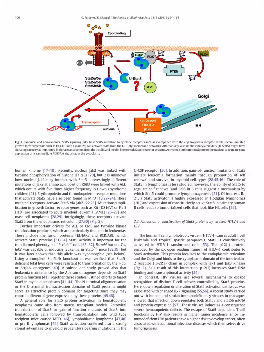
Fig. 2. Canonical and non-canonical Stat5 signaling. Jak2 links Stat5 activation to cytokine receptors such as exemplified with the erythropoietin receptor, while several mutatedgrowth factor receptors such as Flt3-ITD or Kit (D816V) can activate Stat5 from the ER-Golgi membrane networks. Alternatively, also unphosphorylated Stat5 (U-Stat5) might havesignaling capacity as implicated in signal transduction from the insulin and insulin like growth factor receptor systems. Activated Stat5 can translocate to the nucleus to regulate geneexpression or it can mediate PI3K/Akt signaling in the cytoplasm.
106 G. Ferbeyre, R. Moriggl / Biochimica et Biophysica Acta 1815 (2011) 104–114
human kinome [17–19]. Recently, nuclear Jak2 was linked withtyrosine phosphorylation of histone H3 tails [20], but it is unknownhow nuclear Jak2 may interact with Stat5. Interestingly, differentmutations of Jak2 at amino acid position R683 were linked with ALL,which occurs with five times higher frequency in Down's syndromechildren [21]. Erythropoietin and thrombopoietin receptor mutationsthat activate Stat5 have also been found in MPD [13,22–24]. Thesemutated receptors activate Stat5 via Jak2 [22,23]. Mutations/ampli-fication in growth factor receptor genes such as Kit (D816V) or Flt-3(ITD) are associated to acute myeloid leukemia (AML) [25–27] andmast cell neoplasms [28,29]. Intriguingly, these receptors activateStat5 from the endoplasmic reticulum [27,30] (Fig. 2).
Further important drivers for ALL or CML are tyrosine kinasetranslocation products, which are particularly frequent in leukemias.These include the fusion proteins TEL/JAK2 and BCR/ABL, whichactivate Stat5 proteins [31–34]. Stat5 activity is important for thetransformed phenotype of bcr/abl+ cells [35–37]. Bcr/abl but not Tel/Jak2 was capable of inducing leukemia in Stat5ΔN mice [38,39] butit was later shown that this allele was hypomorphic (see below).Using a complete Stat5a/b knockout it was verified that Stat5-deficient fetal liver cells were resistant to transformation by the v-ablor bcr/abl oncogenes [40]. A subsequent study proved also thatleukemia maintenance by the Abelson oncogenes depends on Stat5protein function [41]. Together these studies justified efforts to targetStat5 in myeloid neoplasms [41–44]. The N-terminal oligomerisationor the C-terminal transactivation domains of Stat5 proteins mightserve as attractive protein domains for targeting since they maycontrol differential gene expression by these proteins [45,46].
A general role for Stat5 protein activation in hematopoieticneoplasms came also from mouse transplant models. Retroviraltransduction of Stat5 or gain-of-function mutants of Stat5 intohematopoietic cells followed by transplantation into wild typerecipient mice caused MPD [46], lymphoblastic lymphoma [47,48]or pre-B lymphomas [49]. Stat5 activation conferred also a strongclonal advantage to myeloid progenitors bearing mutations in the
G-CSF receptor [50]. In addition, gain-of-function mutants of Stat5initiate leukemia formation mainly through promotion of selfrenewal and survival in myeloid cell types [28,45,46]. The role ofStat5 in lymphomas is less studied, however, the ability of Stat5 toregulate self renewal and Bcl6 in B cells suggest a mechanism bywhich Stat5 could promote lymphomagenesis [51]. Of interest, IL-21, a Stat5 activator is highly expressed in Hodgkin lymphomas(HL) and expression of constitutively active Stat5 in primary humanB cells leads to immortalized cells that look like HL cells [52].
2.2. Activation or inactivation of Stat5 proteins by viruses: HTLV-I andHIV
The human T cell lymphotropic virus-I (HTLV-I) causes adult T cellleukemia and tropical spastic paraparesis. Stat5 is constitutivelyactivated in HTLV-I-transformed cells [53]. The p12(I) protein,encoded by the pX open reading frame I of HTLV-1 contributes toStat5 activation. This protein localizes to the endoplasmic reticulumand the Golgi and binds to the cytoplasmic domain of the interleukin-2 receptor (IL-2R)β chain in complex with Jak1 and Jak3 kinases(Fig. 2). As a result of this interaction, p12(I) increases Stat5 DNAbinding and transcriptional activity [54].
In contrast, HIV viruses use several mechanisms to escaperecognition of distinct T cell subsets controlled by Stat5 proteins.Here, down regulation or alteration of Stat5 activation pathways wasassociated with changed IL-7 signaling [55,56]. A recent study carriedout with human and simian immunodeficiency viruses in macaquesshowed that infection down regulates both Stat5a and Stat5b mRNAand protein expression [57]. These viruses induce as a consequencesevere hematopoietic defects. The escape of Stat5-dependent T cellfunctions by HIV also results in higher tumor incidence, since im-munodeficient HIV patients have a higher risk to develop cancer, oftenassociated with additional infectious diseases which themselves drivetumorigenesis.

107G. Ferbeyre, R. Moriggl / Biochimica et Biophysica Acta 1815 (2011) 104–114
2.3. Breast cancer
Stat5 proteins are important for the development and functionsof the mammary gland [58,59]. Autocrine loops of growth hormone(GH) and prolactin (Prl) were described in breast cancers [60–62],explaining pYStat5 activity in those tumors. There is also experimen-tal data showing that increased GH expression correlates withmalignant behavior [63–65]. Stat5 can also be activated by theepidermal growth factor receptor (EGFR/ErbB) pathway [66]. Overall,Stat5 was suggested to have a dual role in breast cancer. On the onehand Stat5 promotes malignant transformation in mammary epithe-lial cells [67] and it is important for cancer initiation. For example, adominant-negative Stat5a was able to induce apoptosis in an estrogenpositive breast cancer cell line [68] and ablation of one Stat5a allelein mice engineered to express T antigen in mammary epithelial cellswas sufficient to reduce tumor incidence [69]. On the other hand,Stat5 activity is related to a better prognosis for patient survival sinceit indicates mammary epithelial cell differentiation [58,59,70] anddelayed metastatic progression. Interestingly, a recent study withbreast epithelial cells links Stat5 activationwith increased cell survivalupon Prl action through direct transcriptional activation of the Akt1kinase [71].
2.4. Prostate cancer
A large fraction (~95%) of hormone refractory prostate cancerdisplays persistent Stat5 activity [72], often in association with GHand Prl expression [73,74]. A mouse model of Stat5 activation by Prlrecapitulates prostate tumorigenesis from precancerous lesions toinvasive carcinoma [75]. High pYStat5 correlates with high histolog-ical Gleason grade of human prostate cancer and bad prognosis [74].Experimental cell line models suggest that high pYStat5 levels main-tain the malignant phenotype. Importantly, Stat5 synergizes with theandrogen receptor (AR; Fig. 1) increasing nuclear localization andtranscriptional activity of Stat5 [72]. Adenoviral gene delivery of adominant-negative Stat5a mutant induced cell death and inhibitedgrowth or invasive properties of human prostate cancer cells [76,77].
TGF- β /SMADsignaling:
Stat5-TGF- βinteraction
Stat5 actiontranscriptional
G1/S transitioncontrol:cyclin D1, D2, D3c-myc
Survival:miRNA15/16bcl-2mcl-1bcl-xL
Core cancer pathways controlled by
Core cancer pathways controlled by S
Stat5 action viaprotein intera
Fig. 3. Core cancer pathways (definition according to Vogelstein and Kinzler [127]) like TGF-β, Pby Stat5 action. The TGF-β and PI3K/PTEN signaling pathway are directly influenced by Stat5 propYStat5 can bind toGab2, a scaffoldmolecule activating PI3K/AKT signaling. The HIF1α signalingvia gene induction (black arrow up) or repression (black arrow down). Key Stat5 target genes
2.5. Lung, head and neck cancers
The Jak–Stat pathway is frequently activated in lung and headand neck cancer [78]. In the lung little is known about a role for Stat5in tumorigenesis. However, studies by Grandis and colleaguesdemonstrated that Stat5 contributes to growth and the formationof squamous cell carcinoma of the head and neck (SCCHN) andresistance to EGFR inhibition [79–81]. The mechanism of increasedStat5 activity in these solid tumors is not defined, but activation ofStat5 via Src and the EGFR in SCCHN cell lines was proposed [79,80].
2.6. Liver cancer
Stat5b activation was found in hepatocellular carcinoma (HCC)clinical samples in association with advanced tumor stages. Stat5benhances HCC aggressiveness through induction of epithelial–mes-enchymal transition [82]. In apparent contrast, loss of Stat5 in micecaused steatosis, liver fibrosis and promoted chemically induced livercancer [83]. This was partly explained by a compensatory pYStat1/pYStat3 and TGF-β axis, where a new role for the Stat5 N-terminuswas described in binding directly TGF-β [83,84] (Fig. 3). Alternativelyand provoking, it might also hint to a lack of tumor suppressorfunctions by Stat5 (see Section 5).
2.7. Melanoma
The study of hereditary malignant melanoma in the fish Xipho-phorus established a link between tyrosine kinase receptors and Stat5in melanomas [85]. pYStat5 was found in 62% of melanoma patientsanalyzed [86] and this activation could contribute to resistance to theanti-proliferative activity of interferons [87] and cell survival [88].
2.8. Other malignancies
Furthermore, activated Stat5 was found in intraductal papillarymucinous neoplasms but not in normal pancreatic benign adenomas[89]. Cucurbitacin B, an experimental drug for pancreatic cancercaused dose- and time-dependent G(2)-M-phase arrest and apoptosis
PI3K/PTENsignaling:
Stat5-Gab2-p85 interaction
as a regulator
HIF1αsignaling:vegf
DNA damage control:rad51
Stat5-mediated gene regulation
tat5 protein-protein interaction
protein-ction
I3K/PTEN, HIF1α signaling, survival, G1/S transition or DNA damage control are controlledtein–protein interaction (above). TGF-β can be bound to the Stat5 N-terminus or activated, survival, G1/S transition orDNA damage control pathways are directly regulated by Stat5for core cancer pathways are indicated by italic names.

108 G. Ferbeyre, R. Moriggl / Biochimica et Biophysica Acta 1815 (2011) 104–114
of pancreatic cancer cells in association with inhibition of activatedJak2, Stat3, and Stat5 [90]. Stat5 activity was further linked toglioblastoma and colon carcinoma progression or invasion [91,92].Stat5b was found up regulated in cervical cancers in association withHPV infection while Stat5a was down-regulated [93]. Finally, inovarian cancer, a link was proposed between Stat5 activation andangiogenesis regulation [94] (Fig. 3).
3. Stat5-regulated transcription: A matter of protein–proteininteraction
Stat5 proteins are considered transcriptional activators whentyrosine phosphorylated and bound to DNA. However, they have lessstrong transactivation domains compared to other Stat familymembers [95]. On the other hand, Stat5 proteins boost transcriptionin concerted action with other transcriptional regulators, e.g. theglucocorticoid and androgen receptors (GR;AR; Fig. 1) [13,72,96–100].Some Stat5 target genes have clusters of Stat5 binding sites(TTCNNNGAA) that may also increase Stat5-transcriptional activity[101–104]. A key factor in target recognition is the interplay betweenStat5 dimers and oligomers bound to DNA since the latter canrecognize non-consensus elements [105,106].
Stat5 proteins do repress genes [107,108] and microarray studiesof cells with gene deletion of Stat5 transcription factors revealed notonly significantly down-regulated genes, but also many up regulatedgenes [104,109,110]. One example is ovarian Stat5b, which represses20α-hydroxysteroid dehydrogenase [111] or lymphoid Stat5 thatrepresses Igκ recombination [112]. Stat5 proteins were shown to actrepressive for primitive and transactivating for definitive erythropoi-esis [113]. Interestingly, Stat5 was also shown to repressmiRNA15/16,which causes up regulated bcl-2 and bcl-xL mRNA expression [103].The underlying mechanism is less studied and it might depend on theconcerted action with corepressor molecules or other transcriptionfactors.
Stat5a and Stat5b proteins exert not only overlapping but alsodistinct functions. This originates from individual cell-specificdifferences in mRNA levels [80,114–116], slightly different DNAbinding specificity [117], altered half life of pYStat5 isoforms, nucleo-cytoplasmic shuttling [118,119] or differential activation by serinephosphorylation [120,121]. The cell type specific differences in Stat5aor Stat5b protein expression have also consequences for cancerinitiation or progression. Mammary-directed expression of wild typeStat5a resulted in mammary tumors [122] and both isoforms havedifferential activities in association with the ERα or ERβ isoforms[117]. In contrast, other tumors like HCC or glioblastoma rely onStat5b activation [82,91]. These studies indicate that factors controlledby distinct Stat5 isoformsmay also modulate organ specific oncogenicfunctions. One such factor is Bcl6, a well known transcriptionalregulator protein in lymphoid, mammary epithelial or liver cells.Stat5b was shown to directly cause Bcl6 up regulation controlling theself renewal of memory B cells [51]. In contrast, Stat5a repressed Bcl6expression in breast cancer cell lines [123]. Bcl6 recognizes identicalDNA sequences as Stat5 and can repress Stat5 actions as shown formammary gland cell differentiation [123–125]. In turn, Stat5a canrepress Bcl6 expression at the level of transcriptional elongation[125,126].
4. Mechanisms of oncogenic activity of Stat5 proteins and targetgene regulation
Vogelstein and Kinzler proposed in a landmark paper of 2004 thatthe majority of cancer mutations affect 12 core pathways [127], Jak–Stat signaling being one of them (Fig. 3). Stat5-target genes [104,128]can drive several other core oncogenic pathways in tumors whereStat5 is activated either through protein–protein interaction orthrough transcriptional regulation (Fig. 3). It is not understood,
what combination of tumor suppressor or oncogene mutations mustcooperate to promote and to rely on Stat5 protein function for growthand survival.
4.1. Anti-apoptotic functions of Stat5 proteins
Several Stat5 regulated genes have been implicated in conferring ananti-apoptotic or pro-survival phenotype. These include bcl-2, bcl-xL,pim-1, A1, serine protease inhibitors Spi2.1 and Spi2.2 and Mcl-1[103,104,110,128–132]. Interestingly, the pro-apoptotic miRNAs,miR15/16, which repress bcl-2 and bcl-xL are repressed by Stat5 by amechanism that required the N-terminal protein–protein interactionwith an unknown corepressor [103]. The bcl-2 or bcl-xL gene locicontain multiple functional Stat5 binding sites, some in the promoterothers in the exon–intron region [102,103]. Moreover, Stat5 wasshown in mammary epithelial cells to confer survival upon Prl actionvia direct regulation of the Akt gene locus promoting enhanced Akt1isoform expression [71].
Pim-1 to -3 belong to a family of serine/threonine kinases involvedin the control of cell growth, differentiation and apoptosis [133]. Itwas proposed that the major expression of Pim-1 is mediated throughactivation of the Jak–Stat pathway [134]. Pim-2 was induced in cellswith Flt3 mutations, but there is no direct proof that pim-2 is a directStat5 target gene. Pim kinases might act in a redundant function.Triple deletion of Pim-1 to -3 caused several remarkable similarphenotypes to deletion of Stat5 genes in lymphocytes, myeloid or liverepithelial cells [135].
A role of survival gene induction for oncogenic Stat5 action isbeyond doubt. However, Stat5 proteins have also important roles inthe cytoplasm to activate the PI3K/Akt kinase cascade through Gab2adaptor protein interaction (Figs. 2 and 3) [46,136–138]. In coloncancer, cytoplasmic Stat5 controls cell cycle progression, invasion andmigration, while in normal colon epithelial cells Stat5 is confined tothe nucleus [92].
4.2. Cell proliferation and control through IGF-1 signaling
Activated Stat5 proteins induce genes that accelerate cell cycleprogression such as cyclin D2 and c-Myc [40,41,131,139–141] (Figs. 1and 3). Stat5 plays also an essential role in mediating the growth andcell proliferation effects of GH [97,142]. Hepatic deletion of Stat5genes caused a defect in GH-induced target genes essential forpostnatal body growth, sexual maturation or RNA biosynthesis,similar to hepatic deletion of the GR gene [97,142]. While Stat5acontrols the PRL-induced proliferation, differentiation, and functionof mammary secretory epithelium, Stat5b is the main actor forhepatocytes and biliary epithelial cells to modulate cellular metabo-lism and bile regulation to prevent liver fibrosis upon chronic hepaticdamage [83,84,143]. An important growth regulatory gene of hepaticStat5b action is IGF-1 [101,144–147]. Bioactive IGF-1 (in complexwith the proteins ALS and IGFBP-3, both well known GH–GHR–Jak2–Stat5b targets) is the key regulator for postnatal body growth and ageneral growth factor for most cell types [97,148]. GH also inhibits theexpression of IGFBP-1 through Stat5b action, which impairs FoxO1expression [149]. Bioactive IGF-1 is a predisposing factor for manycancer types such as prostate cancer [150], and it may play a role inthe blast crisis of CML patients [151]. IGF-1 was also implicated inMPD or AML, since these cells are hypersensitive to IGF-1 stimulation[151–153]. However, it is questionable whether pYStat5 induces IGF-1in myeloid cells. On the other hand, IGF-1 plays a protective rolefor chronic liver damage [97,143].
4.3. DNA damage response
DNA damage and the DNA damage response (DDR) represent anearly barrier for tumor formation [154–159] and it is one core cancer

109G. Ferbeyre, R. Moriggl / Biochimica et Biophysica Acta 1815 (2011) 104–114
pathway (Fig. 3). Hence, modulation of this response is essentialfor carcinogenesis. Although the DNA damage response preventscancer formation upon oncogene expression early in the carcinogen-esis process, once other mutations inactivate tumor suppressionresponses, the DNA damage induced by oncogenes can acceleratetumor progression [157,158]. The mechanism by which Stat5 inducesDNA damage is not well understood, but as reported for oncogenic ras[160], Stat5 can induce changes in mitochondrial functions leading tothe production of reactive oxygen species (ROS) and DNA damage[158,161]. The eventual progression of cells with DNA damage alongmalignant transformation depends not only on the status of the tumorsuppressors activated by DNA damage but also on their ability torepair their DNA. Hence, the oncogenic functions of Stat5 may dependon connections to DNA repair genes.
So far, the only well characterized DNA repair gene downstreamof Stat5 is RAD51. RAD51 is one of six human homologous proteinsof the E. coli RecA protein that play a central role in homologousrecombination and repair of DNA double-strand breaks (DSBs). DSBsare increased upon high ROS production or upon altered peroxidation,both processes are involved in DNA damage response of manydifferent cancer types and associated with histone H2aX phosphor-ylation and sensing with ATM/ATR checkpoint control. The expres-sion of RAD51 and several RAD51-paralogs is regulated in a Stat5-dependent manner in BCR/ABL or ZNF198-FGFR1 transformed cells[162,163].
Intriguingly, the Stat5 target genes bcl-2 and bcl-xL suppressRad51-dependent homologous recombination [164], mismatch repair[165], and double-strand break repair [166]. The outcome of DNArepair inhibition for tumorigenesis must be context dependent.Suppression of DNA repair pathways can increase the frequency ofmutations to promote tumorigenesis. However, during early carcino-genesis, inhibition of repair may reinforce the senescence pathway(see discussion later) that protects normal cells from transformation.Moreover, it is clear that DSBs are induced by chemotherapy treat-ment, but whether Stat5 activation can change the fate of chemo-therapy of cancer cells is not clear.
4.4. Invasion, metastasis and epithelial tomesenchymal transition (EMT)
Active Stat5 promotes invasion and metastasis in prostate cancer.High expression of Stat5 in prostate cancer cells is correlated with lowE-cadherin expression and heterotypic adhesion of tumor to endo-thelial cells [167]. Intriguingly, the opposite was described in breastcancer where induction of Stat5a by Prl inhibited the ability of tumorcells to invade normal tissues and stimulated the expression of thetumor suppressor E-cadherin [168], which enabled epithelial polarity.Moreover, the motility and anchorage independent growth of breastcancer cells was shown to be dependent on protein–proteininteractions with the ER in isoform specific way [117].
Stat proteins regulate also the metalloproteinase family. The moremotile and invasive phenotype of epithelial cancer cells correlateswith epithelial to mesenchymal transition (EMT). Several reportsimply Stat5 protein function in EMT [81,82,169], but a role as directEMT inducer or blocker is still controversial [170].
5. Can Stat5 proteins also function as tumor suppressor proteins?
The Jak–Stat pathway exerts important tumor suppressing func-tions that are mainly attributed to interferon signaling. In particular,Stat1 is considered a potent tumor suppressor [171], with the excep-tion of leukemia where Stat1 can partly promote leukemogenesis[172]. Hints for a tumor suppressor role for Stat5 proteins were firstobtained in breast cancer patients, where activated Stat5 proteinsare predictors of good prognosis [173]. Stat5a induced E-cadherin andthe association of beta-catenin to the cell surface with homotypiccell clustering through E-Cadherin mediated junctions [168]. Stat5b
has hepatoprotective functions in chronic liver damage [83,143]. Thisis in line with a potential tumor suppressor function. However, itapparently contradicts a report indicating that Stat5b activationcontributes to EMT upon hepatitis infection [82]. A recent studyhas shown that loss of Stat5 in mouse embryonic fibroblasts andhepatocytes leads to enhanced cell cycle progression linked to a lowerexpression of the CDK inhibitors p15INK4b and p21CIP [10]. This is againconsistent with a tumor suppressor role of Stat5 in the liver or infibroblasts [10].
5.1. Mouse models of Stat5 inactivation
Mouse models of Stat5 inactivation have been described. Singleknockout animals are viable but display distinct phenotypes due toexpression pattern differences between both isoforms [58,174]. Micedeficient for Stat5a show a defective differentiation of the mammarygland [58]. In contrast, the absence of Stat5b led to dwarfism in males,sexual gene conversion, altered growth hormone (GH) regulated liverenzyme regulation [174] and defects in NK cell activity [175]. Theoriginal double knockout, targeting both Stat5a and Stat5b (nowreferred to as Stat5ΔN) led to incomplete deletion of the coding exons(for overview see [176]). Surprisingly, Stat5ΔN mice were viable, butthey displayed severe defects in the immune system, a block inpostnatal body growth in male and females, or a defective femalereproductive tract [177]. Today, Stat5ΔN mice are consideredhypomorphic because they express significant levels of N-terminallytruncated Stat5a and Stat5b molecules able to enter the nucleus.
A complete double knockout of both Stat5 genes (Stat5Null)resulted in no Stat5a/b proteins and diminished hematopoiesis. Theanimals die on a pure C57Bl/6 or Balb/c genetic background, mostlikely due to defects in erythropoiesis and iron metabolism [178,179].Surprisingly, some Stat5Null mice can survive up to eight weeks on amixed Sv129xC57Bl/6 background. These mice display strong Stat3tyrosine phosphorylation and it has been proposed that Stat3 maycompensate in some tissues for Stat5 loss [40,180]. Irrespective,Stat5Null survivor mice suffer from lymphopenia, develop autoim-mune infiltrates or display neutrophil infiltration. They lack CD8+ T,CD25+Foxp3+ T suppressor, differentiated B or NK cells, to name afew prominent lineage defects (reviewed in [176]).
Whereas mice lacking Stat1 are tumor prone, reports of increasedtumor frequency in mouse models of loss of Stat5a or Stat5b functionare missing [171]. Stat1 tumor suppressor functions are linked to theinduction of growth inhibitory genes such as IRF-1, p21 and caspase 3.In regard to Stat5, mammary-directed expression of a carboxyl-terminally truncated dominant-negative Stat5a form resulted inmammary tumors [122]. It is questionable if single knockouts ofStat5a or Stat5b were analyzed in detail for spontaneous late stagetumor spectra, a theme usually done in ageing models. Certainly,Stat5 is behaving in most tumor types rather oncogenic than anti-tumorigenic, but one must not forget that tumor cells have areorganization of their gene expression pattern and signaling path-ways. This occurs due to mutations and epigenetic silencing of genesthat control the cell cycle and cell proliferation. Stat5 can be pro-tumorigenic in the context of the aberrant genetic alterationsassociated to tumorigenesis while it could suppress tumor formationin normal cells (Fig. 4). How a normal cell overcomes these barriersupon cytokine action might remain a central theme of Stat5 functionin cancer cells for the future.
Undoubtedly, long term tumor studies with conditional tumormouse models are needed to finally elucidate a potential tumorsuppressing role for Stat5. Not the least since Stat5Null mice are notviable for longer times and suffer from autoimmune disease andhematopoietic failure. It is also plausible that Stat5 may gain tumorsuppressor activities upon its constitutive activation. Therefore,those activities cannot be observed in mouse models with disabledStat5 protein function as described for senescence induction (see

Fig. 4. Two models help to explain context dependent tumor suppression and oncogenic activities of Stat5 proteins. Stat5 activity supports normal proliferation and developmentunder healthy conditions, where it acts mainly tumor suppressive upon acute oncogenic threats (left). However, upon the chronic genetic and epigenetic alterations thatcharacterize tumorigenesis (right), Stat5 activities gain context dependent pro-tumorigenic functions. Key proteins involved in the regulation of proliferation, differentiation, cellcycle arrest or survival are highlighted since they are either directly regulated or influenced by Stat5 activity.
110 G. Ferbeyre, R. Moriggl / Biochimica et Biophysica Acta 1815 (2011) 104–114
Section 5.3). Moreover, in some tissues loss of Stat5 protein functioncaused higher Stat1 protein expression. Similarly, loss of Stat5 causedin certain cell types elevated pYStat3 activation levels [98]. However,due to the important role of Stat5 proteins in innate and acquiredimmunity it is clear that loss of Stat5 protein function in immune cellswill have consequences for tumor formation as discussed above withHIV infection.
5.2. Cell differentiation
Constitutively active Stat5a induces on the one hand celldifferentiation in mouse transplant models [45,136], but on theother hand it promotes MPD and leukemia progression. Theseopposing effects suggest cooperating hits which might occur inhematopoietic cancer stem cells which then block largely thedifferentiation capacity of cytokine induced Stat5 clonally (Fig. 4).Similarly, persistently active Stat5a induces differentiation in mouseleukemia M1 cells through autocrine production of IL-6 [181].Treatment with Hexamethylenebisacetamide (HMBA) induces celldifferentiation and causes tyrosine phosphorylation of Jak2 and Stat5ain murine erythroleukemia cell lines. Other chemical inducers likeDMSO and butyrate also induce a sustained activation of Jak2–Stat5proteins. These results suggest that persistent activation of the Stat5signaling facilitates differentiation [182]. High Stat5 induction wasalso shown to promote erythroid differentiation in a GATA-1-dependent context [183], whereas low Stat5a induction was consis-tent with self renewal of hematopoietic progenitor cells in humanCD34+ cells [184].
Stat5 plays also an important role for B and T cell differentiation[40,112], most likely in response to IL-7R signaling, which promotescell survival and expansion. IL-7-induced Stat5 represses Igκ recom-bination in pro-B cells. It was shown that it cooperates subsequentlywith the pre-B cell receptor to facilitate expansion [112]. Moreover,the good prognostic value of Stat5 activity in established breast cancermirrors Stat5 functions in mammary epithelial cell differentiation.
5.3. Cell senescence
Persistent Stat5a activation can trigger a permanent cell cyclearrest with all the characteristics of cell senescence such as activationof p53, suppression of E2F target gene expression by the retinoblas-toma family, activation of the PML-tumor suppressor pathway and aconstitutive activation of the DNA damage response [159,185]. Thisprogram can be viewed as the response of a normal cell to an activated
oncogene but is supported by the direct action of Stat5 on target genessuch as SOCS1 [104] and PML [161]. SOCS1 is shown to mediate p53activation and senescence in response to Stat5 [186]. In addition,SOCS1 was sufficient to induce senescence via p53 [186]. PML is also aregulator of cell senescence downstream of Jak–Stat signaling and likeSOCS1, PML is also capable of activating the p53 pathway [187]. Theseresults suggest that Stat5 is part of a signaling pathway that inducesPML, SOCS1 and/or p53 in response to cytokines or other oncogenesthat may cause senescence.
Many benign tumors show evidence of accumulation of senescentcells leading to the concept that cell senescence keeps benignneoplasms in check [188–195]. In a subset of patients with benignbreast tumors it has been found that they express a mutanthyperactive PrlR [196]. It is very plausible that those tumors representthe in vivo counterpart of the persistent Stat5a-senescent mechanismwe described in primary fibroblasts and primary mammary epithelialcells [159,185]. Differentiation of hematopoietic cells in context ofStat5 activation might mimic partly the senescence phenotype inadherent cells.
6. Concluding remarks
Stat5 protein activation can promote transformation, cell differ-entiation or senescence. This duality of action is not a particularityof Stat5 signaling since they have been observed for many otheroncogenes [197]. We need to understand how mutations in cancercells overtake tumor suppressive functions of Stat5 enhancing theironcogenic functions. Treatments should focus on restoring thisbalance rather than achieving a complete inhibition of a particularsignaling pathway, which is required in normal cells.
Current biological research has profited from reductionism tounderstand molecular mechanisms, but cancer is a very heteroge-neous and complex disease. We need to study the full spectrum ofStat5 target genes, interacting proteins and post-translational mod-ifications in in vivo models of Stat5 activation and suppression. Sofar, most studies we cited relied on a single cytokine response andconsequences for Stat5 gene induction in certain immortalized celllines at one given time point in tissue culture models. It is time tounravel the molecular basics of more complex actions since cytokines,growth factors and steroid action signal in parallel when measured incancers. We call for a definition of Stat5-dependent tumor suppres-sion or cancer cell stimulation as subsets of interactions, target genesor post-translational modifications. The definition of those subsetsmay offer better therapeutic targets than a global alteration of Stat5
111G. Ferbeyre, R. Moriggl / Biochimica et Biophysica Acta 1815 (2011) 104–114
functions, whichwill be probably very toxic. Such challengeswill keepStat5 researchers occupied.
Acknowledgements
We thank Veronika Sexl and Antonis Koromilas for critical reading.This work was supported by grants SFB-F28 from the Austrian BasicResearch Funds (FWF) to RM and CIHR 82887 to GF.
References
[1] M. Schmitt-Ney, B. Happ, R.K. Ball, B. Groner, Developmental and environmentalregulation of a mammary gland-specific nuclear factor essential for transcriptionof the gene encoding beta-casein, Proc. Natl Acad. Sci. USA 89 (1992) 3130–3134.
[2] H. Wakao, F. Gouilleux, B. Groner, Mammary gland factor (MGF) is a novelmember of the cytokine regulated transcription factor gene family and confersthe prolactin response, EMBO J. 13 (1994) 2182–2191.
[3] F. Gouilleux, H. Wakao, M. Mundt, B. Groner, Prolactin induces phosphorylationof Tyr694 of Stat5 (MGF), a prerequisite for DNA binding and induction oftranscription, EMBO J. 13 (1994) 4361–4369.
[4] X. Liu, G.W. Robinson, F. Gouilleux, B. Groner, L. Hennighausen, Cloning andexpression of Stat5 and an additional homologue (Stat5b) involved in prolactinsignal transduction in mouse mammary tissue, Proc. Natl Acad. Sci. USA 92(1995) 8831–8835.
[5] A.L. Mui, H. Wakao, A.M. O'Farrell, N. Harada, A. Miyajima, Interleukin-3,granulocyte-macrophage colony stimulating factor and interleukin-5 transducesignals through two STAT5 homologs, EMBO J. 14 (1995) 1166–1175.
[6] H. Wakao, N. Harada, T. Kitamura, A.L. Mui, A. Miyajima, Interleukin 2 anderythropoietin activate STAT5/MGF via distinct pathways, EMBO J. 14 (1995)2527–2535.
[7] C. Arnould, C. Philippe, V. Bourdon, M.J. Grégoire, R. Berger, P. Jonveaux, Thesignal transducer and activator of transcription STAT5b gene is a new partner ofretinoic acid receptor alpha in acute promyelocytic-like leukaemia, Hum. Mol.Genet. 8 (1999) 1741–1749.
[8] S. Dong, D.J. Tweardy, Interactions of STAT5b-RARalpha, a novel acutepromyelocytic leukemia fusion protein, with retinoic acid receptor and STAT3signaling pathways, Blood 99 (2002) 2637–2646.
[9] S.N. Constantinescu, M. Girardot, C. Pecquet, Mining for JAK–STAT mutations incancer, Trends Biochem. Sci. 33 (2008) 122–131.
[10] J.H. Yu, B.M. Zhu, M. Wickre, G. Riedlinger, W. Chen, A. Hosui, G.W. Robinson, L.Hennighausen, The transcription factors STAT5A and STAT5Bnegatively regulate cellproliferation through the activation of Cdkn2b and Cdkn1a expression, Hepatology(2010) (Accepted manuscript online: 5 AUG 2010), 52 (2010)1808–1818.
[11] M. Girardot, C. Pecquet, S. Boukour, L. Knoops, A. Ferrant, W. Vainchenker, S.Giraudier, S.N. Constantinescu, miR-28 is a thrombopoietin receptor targetingmicroRNAdetected in a fraction ofmyeloproliferative neoplasmpatient platelets,Blood 116 (2010) 437–445.
[12] Y. Wang, D. Cai, C. Brendel, C. Barett, P. Erben, P.W. Manley, A. Hochhaus, A.Neubauer, A. Burchert, Adaptive secretion of granulocyte-macrophage colony-stimulating factor (GM-CSF) mediates imatinib and nilotinib resistance in BCR/ABL+progenitors via JAK-2/STAT-5 pathway activation, Blood 109 (2007)2147–2155.
[13] C. Pecquet, J. Staerk, R. Chaligne, V. Goss, K.A. Lee, X. Zhang, J. Rush, J. Van Hees, H.A. Poirel, J.M. Scheiff, W. Vainchenker, S. Giraudier, R.D. Polakiewicz, S.N.Constantinescu, Induction of myeloproliferative disorder and myelofibrosis bythrombopoietin receptorW515mutants is mediated by cytosolic tyrosine 112 ofthe receptor, Blood 115 (2010) 1037–1048.
[14] O. Kabbarah, L. Chin, Revealing the genomic heterogeneity of melanoma, CancerCell 8 (2005) 439–441.
[15] N. Kotecha, N.J. Flores, J.M. Irish, E.F. Simonds, D.S. Sakai, S. Archambeault, E.Diaz-Flores, M. Coram, K.M. Shannon, G.P. Nolan, M.L. Loh, Single-cell profilingidentifies aberrant STAT5 activation in myeloid malignancies with specificclinical and biologic correlates, Cancer Cell 14 (2008) 335–343.
[16] A. Ecker, O. Simma, A. Hoelbl, L. Kenner, H. Beug, R. Moriggl, V. Sexl, The dark andthe bright side of Stat3: proto-oncogene and tumor-suppressor, Front. Biosci. 14(2009) 2944–2958.
[17] R. Kralovics, F. Passamonti, A.S. Buser, S.S. Teo, R. Tiedt, J.R. Passweg, A. Tichelli,M. Cazzola, R.C. Skoda, A gain-of-functionmutation of JAK2 inmyeloproliferativedisorders, N Engl J. Med. 352 (2005) 1779–1790.
[18] R. Zhao, S. Xing, Z. Li, X. Fu, Q. Li, S.B. Krantz, Z.J. Zhao, Identification of an acquiredJAK2 mutation in polycythemia vera, J. Biol. Chem. 280 (2005) 22788–22792.
[19] S. Aboudola, G. Murugesan, H. Szpurka, G. Ramsingh, X. Zhao, N. Prescott, R.R.Tubbs, J.P. Maciejewski, E.D. Hsi, Bone marrow phospho-STAT5 expression innon-CML chronic myeloproliferative disorders correlates with JAK2 V617Fmutation and provides evidence of in vivo JAK2 activation, Am. J. Surg. Pathol. 31(2007) 233–239.
[20] M.A. Dawson, A.J. Bannister, B. Gottgens, S.D. Foster, T. Bartke, A.R. Green, T.Kouzarides, JAK2 phosphorylates histone H3Y41 and excludes HP1alpha fromchromatin, Nature 461 (2009) 819–822.
[21] D. Bercovich, I. Ganmore, L.M. Scott, G. Wainreb, Y. Birger, A. Elimelech, C.Shochat, G. Cazzaniga, A. Biondi, G. Basso, G. Cario, M. Schrappe, M. Stanulla, S.Strehl, O.A. Haas, G. Mann, V. Binder, A. Borkhardt, H. Kempski, J. Trka, B. Bielorei,
S. Avigad, B. Stark, O. Smith, N. Dastugue, J.P. Bourquin, N.B. Tal, A.R. Green, S.Izraeli, Mutations of JAK2 in acute lymphoblastic leukaemias associated withDown's syndrome, Lancet 372 (2008) 1484–1492.
[22] S.S. Watowich, X. Xie, U. Klingmuller, J. Kere, M. Lindlof, S. Berglund, A. de laChapelle, Erythropoietin receptor mutations associated with familial erythrocy-tosis cause hypersensitivity to erythropoietin in the heterozygous state, Blood 94(1999) 2530–2532.
[23] A. de la Chapelle, A.L. Traskelin, E. Juvonen, Truncated erythropoietin receptorcauses dominantly inherited benign human erythrocytosis, Proc. Natl Acad. Sci.USA 90 (1993) 4495–4499.
[24] R. Kralovics, K. Indrak, T. Stopka, B.W. Berman, J.F. Prchal, J.T. Prchal, Two newEPO receptor mutations: truncated EPO receptors are most frequently associatedwith primary familial and congenital polycythemias, Blood 90 (1997)2057–2061.
[25] R. Zheng, M. Levis, O. Piloto, P. Brown, B.R. Baldwin, N.C. Gorin, M. Beran, Z. Zhu,D. Ludwig, D. Hicklin, L. Witte, Y. Li, D. Small, FLT3 ligand causes autocrinesignaling in acute myeloid leukemia cells, Blood 103 (2004) 267–274.
[26] D.G. Gilliland, J.D. Griffin, The roles of FLT3 in hematopoiesis and leukemia, Blood100 (2002) 1532–1542.
[27] F. Hayakawa, M. Towatari, H. Kiyoi, M. Tanimoto, T. Kitamura, H. Saito, T. Naoe,Tandem-duplicated Flt3 constitutively activates STAT5 and MAP kinase andintroduces autonomous cell growth in IL-3-dependent cell lines, Oncogene 19(2000) 624–631.
[28] N. Harir, C. Boudot, K. Friedbichler, K. Sonneck, R. Kondo, S. Martin-Lanneree, L.Kenner, M. Kerenyi, S. Yahiaoui, V. Gouilleux-Gruart, J. Gondry, L. Benit, I.Dusanter-Fourt, K. Lassoued, P. Valent, R. Moriggl, F. Gouilleux, Oncogenic Kitcontrols neoplastic mast cell growth through a Stat5/PI3-kinase signalingcascade, Blood 112 (2008) 2463–2473.
[29] C. Baumgartner, S. Cerny-Reiterer, K. Sonneck, M. Mayerhofer, K.V. Gleixner, R.Fritz, M. Kerenyi, C. Boudot, F. Gouilleux, J.W. Kornfeld, C. Sillaber, R. Moriggl, P.Valent, Expression of activated STAT5 in neoplastic mast cells in systemicmastocytosis: subcellular distribution and role of the transforming oncoproteinKIT D816V, Am. J. Pathol. 175 (2009) 2416–2429.
[30] K.U. Birkenkamp, M. Geugien, H.H. Lemmink, W. Kruijer, E. Vellenga, Regulationof constitutive STAT5 phosphorylation in acute myeloid leukemia blasts,Leukemia 15 (2001) 1923–1931.
[31] K. Spiekermann, M. Pau, R. Schwab, K. Schmieja, S. Franzrahe, W. Hiddemann,Constitutive activation of STAT3 and STAT5 is induced by leukemic fusionproteins with protein tyrosine kinase activity and is sufficient for transformationof hematopoietic precursor cells, Exp. Hematol. 30 (2002) 262–271.
[32] N. Carlesso, D.A. Frank, J.D. Griffin, Tyrosyl phosphorylation and DNA bindingactivity of signal transducers and activators of transcription (STAT) proteins inhematopoietic cell lines transformed by Bcr/Abl, J. Exp. Med. 183 (1996) 811–820.
[33] A. Klejman, S.J. Schreiner, M. Nieborowska-Skorska, A. Slupianek, M. Wilson, T.E.Smithgall, T. Skorski, The Src family kinase Hck couples BCR/ABL to STAT5activation in myeloid leukemia cells, EMBO J. 21 (2002) 5766–5774.
[34] J.M. Ho, B.K. Beattie, J.A. Squire, D.A. Frank, D.L. Barber, Fusion of the etstranscription factor TEL to Jak2 results in constitutive Jak–Stat signaling, Blood93 (1999) 4354–4364.
[35] C. Sillaber, F. Gesbert, D.A. Frank, M. Sattler, J.D. Griffin, STAT5 activationcontributes to growth and viability in Bcr/Abl-transformed cells, Blood 95(2000) 2118–2125.
[36] R.P. de Groot, J.A. Raaijmakers, J.W. Lammers, R. Jove, L. Koenderman, STAT5activation by BCR-Abl contributes to transformation of K562 leukemia cells,Blood 94 (1999) 1108–1112.
[37] M. Nieborowska-Skorska, M.A. Wasik, A. Slupianek, P. Salomoni, T. Kitamura, B.Calabretta, T. Skorski, Signal transducer and activator of transcription (STAT)5activation by BCR/ABL is dependent on intact Src homology (SH)3 and SH2domains of BCR/ABL and is required for leukemogenesis, J. Exp. Med. 189 (1999)1229–1242.
[38] V. Sexl, R. Piekorz, R. Moriggl, J. Rohrer, M.P. Brown, K.D. Bunting, K. Rothammer,M.F. Roussel, J.N. Ihle, Stat5a/b contribute to interleukin 7-induced B-cellprecursor expansion, but abl- and bcr/abl-induced transformation are indepen-dent of stat5, Blood 96 (2000) 2277–2283.
[39] J. Schwaller, E. Parganas, D. Wang, D. Cain, J.C. Aster, I.R. Williams, C.K. Lee, R.Gerthner, T. Kitamura, J. Frantsve, E. Anastasiadou, M.L. Loh, D.E. Levy, J.N. Ihle, D.G. Gilliland, Stat5 is essential for the myelo- and lymphoproliferative diseaseinduced by TEL/JAK2, Mol. Cell 6 (2000) 693–704.
[40] A. Hoelbl, B. Kovacic, M.A. Kerenyi, O. Simma, W. Warsch, Y. Cui, H. Beug, L.Hennighausen, R. Moriggl, V. Sexl, Clarifying the role of Stat5 in lymphoiddevelopment and Abelson-induced transformation, Blood 107 (2006) 4898–4906.
[41] A. Hoelbl, C. Schuster, B. Kovacic, B. Zhu, M. Wickre, M.A. Hoelzl, S. Fajmann, F.Grebien, W. Warsch, G. Stengl, L. Hennighausen, V. Poli, H. Beug, R. Moriggl, V.Sexl, Stat5 is indispensable for the maintenance of bcr/abl-positive leukaemia,EMBO Mol. Med. 2 (2010) 98–110.
[42] H. Schepers, D. van Gosliga, A.T. Wierenga, B.J. Eggen, J.J. Schuringa, E. Vellenga,STAT5 is required for long-term maintenance of normal and leukemic humanstem/progenitor cells, Blood 110 (2007) 2880–2888.
[43] I.B. Weinstein, Cancer. Addiction to oncogenes—the Achilles heel of cancer,Science 297 (2002) 63–64.
[44] D. Ye, N. Wolff, L. Li, S. Zhang, R.L. Ilaria Jr., STAT5 signaling is required for theefficient induction andmaintenance of CML inmice, Blood 107 (2006) 4917–4925.
[45] R. Moriggl, V. Sexl, L. Kenner, C. Duntsch, K. Stangl, S. Gingras, A. Hoffmeyer, A.Bauer, R. Piekorz, D. Wang, K.D. Bunting, E.F. Wagner, K. Sonneck, P. Valent, J.N.Ihle, H. Beug, Stat5 tetramer formation is associated with leukemogenesis,Cancer Cell 7 (2005) 87–99.

112 G. Ferbeyre, R. Moriggl / Biochimica et Biophysica Acta 1815 (2011) 104–114
[46] G. Li, K.L. Miskimen, Z. Wang, X.Y. Xie, W. Tse, F. Gouilleux, R. Moriggl, K.D.Bunting, Effective targeting of STAT5-mediated survival in myeloproliferativeneoplasms using ABT-737 combined with rapamycin, Leukemia 24 (2010)1397–1405.
[47] K. Bessette, M.L. Lang, R.A. Fava, M. Grundy, J. Heinen, L. Horne, R. Spolski, A. Al-Shami, H.C. Morse III, W.J. Leonard, J.A. Kelly, A Stat5b transgene is capable ofinducing CD8+ lymphoblastic lymphoma in the absence of normal TCR/MHCsignaling, Blood 111 (2008) 344–350.
[48] J.A. Kelly, R. Spolski, P.E. Kovanen, T. Suzuki, J. Bollenbacher, C.A. Pise-Masison,M.F. Radonovich, S. Lee, N.A. Jenkins, N.G. Copeland, H.C. Morse III, W.J. Leonard,Stat5 synergizes with T cell receptor/antigen stimulation in the development oflymphoblastic lymphoma, J. Exp. Med. 198 (2003) 79–89.
[49] T. Tsuruyama, T. Nakamura, G. Jin, M. Ozeki, Y. Yamada, H. Hiai, Constitutiveactivation of Stat5a by retrovirus integration in early pre-B lymphomas of SL/Khstrain mice, Proc. Natl Acad. Sci. USA 99 (2002) 8253–8258.
[50] F. Liu, G. Kunter, M.M. Krem, W.C. Eades, J.A. Cain, M.H. Tomasson, L.Hennighausen, D.C. Link, Csf3r mutations in mice confer a strong clonal HSCadvantage via activation of Stat5, J. Clin. Invest. 118 (2008) 946–955.
[51] F.A. Scheeren, M. Naspetti, S. Diehl, R. Schotte, M. Nagasawa, E. Wijnands, R.Gimeno, F.A. Vyth-Dreese, B. Blom, H. Spits, STAT5 regulates the self-renewalcapacity and differentiation of human memory B cells and controls Bcl-6expression, Nat. Immunol. 6 (2005) 303–313.
[52] F.A. Scheeren, S.A. Diehl, L.A. Smit, T. Beaumont, M. Naspetti, R.J. Bende, B. Blom,K. Karube, K. Ohshima, C.J. van Noesel, H. Spits, IL-21 is expressed in Hodgkinlymphoma and activates STAT5: evidence that activated STAT5 is required forHodgkin lymphomagenesis, Blood 111 (2008) 4706–4715.
[53] T.S. Migone, J.X. Lin, A. Cereseto, J.C. Mulloy, J.J. O'Shea, G. Franchini, W.J. Leonard,Constitutively activated Jak–STAT pathway in T cells transformed with HTLV-I,Science 269 (1995) 79–81.
[54] C. Nicot, J.C. Mulloy, M.G. Ferrari, J.M. Johnson, K. Fu, R. Fukumoto, R. Trovato, J.Fullen, W.J. Leonard, G. Franchini, HTLV-1 p12(I) protein enhances STAT5activation and decreases the interleukin-2 requirement for proliferation ofprimary human peripheral blood mononuclear cells, Blood 98 (2001) 823–829.
[55] A.M. Crawley, S. Faucher, J.B. Angel, Soluble IL-7Ralpha (sCD127) inhibits IL-7activity and is increased in HIV infection, J. Immunol. 184 (2010) 4679–4687.
[56] O. Juffroy, F. Bugault, O. Lambotte, I. Landires, J.P. Viard, L. Niel, A. Fontanet, J.F.Delfraissy, J. Theze, L.A. Chakrabarti, Dual mechanism of impairment ofinterleukin-7 (IL-7) responses in human immunodeficiency virus infection:decreased IL-7 binding and abnormal activation of the JAK/STAT5 pathway, J.Virol. 84 (2010) 96–108.
[57] S. Prost, M. Le Dantec, S. Auge, R. Le Grand, S. Derdouch, G. Auregan, N. Deglon, F.Relouzat, A.M. Aubertin, B. Maillere, I. Dusanter-Fourt, M. Kirszenbaum, Humanand simian immunodeficiency viruses deregulate early hematopoiesis through aNef/PPARgamma/STAT5 signaling pathway in macaques, J. Clin. Invest. 118(2008) 1765–1775.
[58] X. Liu, G.W. Robinson, K.U. Wagner, L. Garrett, A. Wynshaw-Boris, L.Hennighausen, Stat5a is mandatory for adult mammary gland developmentand lactogenesis, Genes Dev. 11 (1997) 179–186.
[59] K. Miyoshi, J.M. Shillingford, G.H. Smith, S.L. Grimm, K.U. Wagner, T. Oka, J.M.Rosen, G.W. Robinson, L. Hennighausen, Signal transducer and activator oftranscription (Stat) 5 controls the proliferation and differentiation of mammaryalveolar epithelium, J. Cell Biol. 155 (2001) 531–542.
[60] M. Raccurt, P.E. Lobie, E. Moudilou, T. Garcia-Caballero, L. Frappart, G. Morel, H.C.Mertani, High stromal and epithelial human gh gene expression is associatedwithproliferative disorders of themammary gland, J. Endocrinol. 175 (2002) 307–318.
[61] B.K. Vonderhaar, Prolactin involvement in breast cancer, Endocr Relat Cancer 6(1999) 389–404.
[62] H.C. Mertani, T. Zhu, E.L. Goh, K.O. Lee, G. Morel, P.E. Lobie, Autocrine humangrowth hormone (hGH) regulation of human mammary carcinoma cell geneexpression. Identification of CHOP as a mediator of hGH-stimulated humanmammary carcinoma cell survival, J. Biol. Chem. 276 (2001) 21464–21475.
[63] K.K. Kaulsay, H.C. Mertani, J. Tornell, G. Morel, K.O. Lee, P.E. Lobie, Autocrinestimulation of human mammary carcinoma cell proliferation by human growthhormone, Exp. Cell Res. 250 (1999) 35–50.
[64] S. Mukhina, H.C. Mertani, K. Guo, K.O. Lee, P.D. Gluckman, P.E. Lobie, Phenotypicconversion of human mammary carcinoma cells by autocrine human growthhormone, Proc. Natl Acad. Sci. USA 101 (2004) 15166–15171.
[65] T. Zhu, B. Starling-Emerald, X. Zhang, K.O. Lee, P.D. Gluckman, H.C. Mertani, P.E.Lobie, Oncogenic transformation of human mammary epithelial cells byautocrine human growth hormone, Cancer Res. 65 (2005) 317–324.
[66] M.T. Kloth, A.D. Catling, C.M. Silva, Novel activation of STAT5b in response toepidermal growth factor, J. Biol. Chem. 277 (2002) 8693–8701.
[67] V. Vafaizadeh, P. Klemmt, C. Brendel, K. Weber, C. Doebele, K. Britt, M. Grez, B.Fehse, S. Desrivieres, B. Groner, Mammary epithelial reconstitution with gene-modified stem cells assigns roles to Stat5 in luminal alveolar cell fate decisions,differentiation, involution, and mammary tumor formation, Stem Cells 28(2010) 928–938.
[68] H. Yamashita, H. Iwase, The role of stat5 in estrogen receptor-positive breastcancer, Breast Cancer 9 (2002) 312–318.
[69] S. Ren, H.R. Cai, M. Li, P.A. Furth, Loss of Stat5a delays mammary cancerprogression in a mouse model, Oncogene 21 (2002) 4335–4339.
[70] K.U. Wagner, H. Rui, Jak2/Stat5 signaling in mammogenesis, breast cancerinitiation and progression, J. Mammary Gland Biol. Neoplasia 13 (2008) 93–103.
[71] B.A. Creamer, K. Sakamoto, J.W. Schmidt, A.A. Triplett, R. Moriggl, K.U. Wagner,Stat5 promotes survival of mammary epithelial cells through transcriptionalactivation of a distinct promoter in Akt1, Mol. Cell. Biol. 30 (2010) 2957–2970.
[72] S.H. Tan, A. Dagvadorj, F. Shen, L. Gu, Z. Liao, J. Abdulghani, Y. Zhang, E.P.Gelmann, T. Zellweger, Z. Culig, T. Visakorpi, L. Bubendorf, R.A. Kirken, J. Karras,M.T. Nevalainen, Transcription factor Stat5 synergizes with androgen receptor inprostate cancer cells, Cancer Res. 68 (2008) 236–248.
[73] L.K. Chopin, T.L. Veveris-Lowe, A.F. Philipps, A.C. Herington, Co-expression of GHand GHR isoforms in prostate cancer cell lines, Growth Horm. IGF Res. 12 (2002)126–136.
[74] H. Li, T.J. Ahonen, K. Alanen, J. Xie, M.J. LeBaron, T.G. Pretlow, E.L. Ealley, Y. Zhang,M. Nurmi, B. Singh, P.M. Martikainen, M.T. Nevalainen, Activation of signaltransducer and activator of transcription 5 in human prostate cancer isassociated with high histological grade, Cancer Res. 64 (2004) 4774–4782.
[75] V. Rouet, R.L. Bogorad, C. Kayser, K. Kessal, C. Genestie, A. Bardier, D.R. Grattan, B.Kelder, J.J. Kopchick, P.A. Kelly, V. Goffin, Local prolactin is a target to preventexpansion of basal/stem cells in prostate tumors, Proc. Natl Acad. Sci. USA 107(2010) 15199–15204.
[76] T.J. Ahonen, J. Xie, M.J. LeBaron, J. Zhu, M. Nurmi, K. Alanen, H. Rui, M.T.Nevalainen, Inhibition of transcription factor Stat5 induces cell death of humanprostate cancer cells, J. Biol. Chem. 278 (2003) 27287–27292.
[77] A.V. Kazansky, D.M. Spencer, N.M. Greenberg, Activation of signal transducer andactivator of transcription 5 is required for progression of autochthonous prostatecancer: evidence from the transgenic adenocarcinoma of the mouse prostatesystem, Cancer Res. 63 (2003) 8757–8762.
[78] S.Y. Lai, F.M. Johnson, Defining the role of the JAK–STAT pathway in head andneck and thoracic malignancies: implications for future therapeutic approaches,Drug Resist. Updat. 13 (2010) 67–78.
[79] S. Xi, Q. Zhang, W.E. Gooding, T.E. Smithgall, J.R. Grandis, Constitutive activationof Stat5b contributes to carcinogenesis in vivo, Cancer Res. 63 (2003) 6763–6771.
[80] P.L. Leong, S. Xi, S.D. Drenning, K.F. Dyer, A.L. Wentzel, E.C. Lerner, T.E. Smithgall,J.R. Grandis, Differential function of STAT5 isoforms in head and neck cancergrowth control, Oncogene 21 (2002) 2846–2853.
[81] P. Koppikar, V.W. Lui, D. Man, S. Xi, R.L. Chai, E. Nelson, A.B. Tobey, J.R. Grandis,Constitutive activation of signal transducer and activator of transcription 5contributes to tumor growth, epithelial–mesenchymal transition, and resistanceto epidermal growth factor receptor targeting, Clin. Cancer Res. 14 (2008)7682–7690.
[82] T.K. Lee, K. Man, R.T. Poon, C.M. Lo, A.P. Yuen, I.O. Ng, K.T. Ng,W. Leonard, S.T. Fan,Signal transducers and activators of transcription 5b activation enhanceshepatocellular carcinoma aggressiveness through induction of epithelial–mesenchymal transition, Cancer Res. 66 (2006) 9948–9956.
[83] A. Hosui, A. Kimura, D. Yamaji, B.M. Zhu, R. Na, L. Hennighausen, Loss of STAT5causes liver fibrosis and cancer development through increased TGF-{beta} andSTAT3 activation, J. Exp. Med. 206 (2009) 819–831.
[84] Y. Cui, A. Hosui, R. Sun, K. Shen, O. Gavrilova, W. Chen, M.C. Cam, B. Gao, G.W.Robinson, L. Hennighausen, Loss of signal transducer and activator oftranscription 5 leads to hepatosteatosis and impaired liver regeneration,Hepatology 46 (2007) 504–513.
[85] C. Wellbrock, E. Geissinger, A. Gomez, P. Fischer, K. Friedrich, M. Schartl,Signalling by the oncogenic receptor tyrosine kinase Xmrk leads to activation ofSTAT5 in Xiphophorus melanoma, Oncogene 16 (1998) 3047–3056.
[86] A. Mirmohammadsadegh, M. Hassan, W. Bardenheuer, A. Marini, A. Gustrau, S.Nambiar, A. Tannapfel, H. Bojar, T. Ruzicka, U.R. Hengge, STAT5 phosphorylationin malignant melanoma is important for survival and is mediated through SRCand JAK1 kinases, J. Invest. Dermatol. 126 (2006) 2272–2280.
[87] C. Wellbrock, C. Weisser, J.C. Hassel, P. Fischer, J. Becker, C.S. Vetter, I. Behrmann,M. Kortylewski, P.C. Heinrich, M. Schartl, STAT5 contributes to interferonresistance of melanoma cells, Curr. Biol. 15 (2005) 1629–1639.
[88] J.C. Hassel, D. Winnemoller, M. Schartl, C. Wellbrock, STAT5 contributes toantiapoptosis in melanoma, Melanoma Res. 18 (2008) 378–385.
[89] T.R. Kataoka, T. Ioka, Y. Tsukamoto, M. Matsumura, S. Ishiguro, Y. Nishizawa,Nuclear expression of STAT5 in intraductal papillary mucinous neoplasms of thepancreas, Int. J. Surg. Pathol. 15 (2007) 277–281.
[90] N.H. Thoennissen, G.B. Iwanski, N.B. Doan, R. Okamoto, P. Lin, S. Abbassi, J.H.Song, D. Yin, M. Toh, W.D. Xie, J.W. Said, H.P. Koeffler, Cucurbitacin B inducesapoptosis by inhibition of the JAK–STAT pathway and potentiates antiprolifera-tive effects of gemcitabine on pancreatic cancer cells, Cancer Res. 69 (2009)5876–5884.
[91] Q.C. Liang, H. Xiong, Z.W. Zhao, D. Jia, W.X. Li, H.Z. Qin, J.P. Deng, L. Gao, H. Zhang,G.D. Gao, Inhibition of transcription factor STAT5b suppresses proliferation,induces G1 cell cycle arrest and reduces tumor cell invasion in humanglioblastoma multiforme cells, Cancer Lett. 273 (2009) 164–171.
[92] H. Xiong, W.Y. Su, Q.C. Liang, Z.G. Zhang, H.M. Chen, W. Du, Y.X. Chen, J.Y. Fang,Inhibition of STAT5 induces G1 cell cycle arrest and reduces tumor cell invasionin human colorectal cancer cells, Lab. Invest. 89 (2009) 717–725.
[93] R.C. Sobti, N. Singh, S. Hussain, V. Suri, M. Bharadwaj, B.C. Das, Deregulation ofSTAT-5 isoforms in the development of HPV-mediated cervical carcinogenesis,J. Recept. Signal Transduct. Res. 30 (2010) 178–188.
[94] H. Chen, D. Ye, X. Xie, B. Chen, W. Lu, VEGF, VEGFRs expressions and activatedSTATs in ovarian epithelial carcinoma, Gynecol. Oncol. 94 (2004) 630–635.
[95] R. Moriggl, S. Berchtold, K. Friedrich, G.J. Standke, W. Kammer, M. Heim, M.Wissler, E. Stocklin, F. Gouilleux, B. Groner, Comparison of the transactivationdomains of Stat5 and Stat6 in lymphoid cells and mammary epithelial cells, Mol.Cell. Biol. 17 (1997) 3663–3678.
[96] R. Moriggl, V. Gouilleux-Gruart, R. Jahne, S. Berchtold, C. Gartmann, X. Liu, L.Hennighausen, A. Sotiropoulos, B. Groner, F. Gouilleux, Deletion of the carboxyl-terminal transactivation domain of MGF-Stat5 results in sustained DNA bindingand a dominant negative phenotype, Mol. Cell. Biol. 16 (1996) 5691–5700.

113G. Ferbeyre, R. Moriggl / Biochimica et Biophysica Acta 1815 (2011) 104–114
[97] D. Engblom, J.W. Kornfeld, L. Schwake, F. Tronche, A. Reimann, H. Beug, L.Hennighausen, R. Moriggl, G. Schutz, Direct glucocorticoid receptor-Stat5interaction in hepatocytes controls body size and maturation-related geneexpression, Genes Dev. 21 (2007) 1157–1162.
[98] H. Dolznig, F. Grebien, E.M. Deiner, K. Stangl, A. Kolbus, B. Habermann, M.A.Kerenyi, M. Kieslinger, R. Moriggl, H. Beug, E.W. Mullner, Erythroid progenitorrenewal versus differentiation: genetic evidence for cell autonomous, essentialfunctions of EpoR, Stat5 and the GR, Oncogene 25 (2006) 2890–2900.
[99] E. Stoecklin, M. Wissler, R. Moriggl, B. Groner, Specific DNA binding of Stat5, butnot of glucocorticoid receptor, is required for their functional cooperation in theregulation of gene transcription, Mol. Cell. Biol. 17 (1997) 6708–6716.
[100] E. Stocklin, M. Wissler, F. Gouilleux, B. Groner, Functional interactions betweenStat5 and the glucocorticoid receptor, Nature 383 (1996) 726–728.
[101] D.J. Chia, B. Varco-Merth, P. Rotwein, Dispersed chromosomal Stat5b-bindingelements mediate growth hormone-activated insulin-like growth factor-I genetranscription, J. Biol. Chem. 285 (2010) 17636–17647.
[102] S. Dumon, S.C. Santos, F. Debierre-Grockiego, V. Gouilleux-Gruart, L. Cocault, C.Boucheron, P. Mollat, S. Gisselbrecht, F. Gouilleux, IL-3 dependent regulation ofBcl-xL gene expression by STAT5 in a bone marrow derived cell line, Oncogene18 (1999) 4191–4199.
[103] G. Li, K.L. Miskimen, Z. Wang, X.Y. Xie, J. Brenzovich, J.J. Ryan, W. Tse, R. Moriggl,K.D. Bunting, STAT5 requires the N-domain for suppression of miR15/16,induction of bcl-2, and survival signaling in myeloproliferative disease, Blood115 (2010) 1416–1424.
[104] B. Basham, M. Sathe, J. Grein, T. McClanahan, A. D'Andrea, E. Lees, A. Rascle, Invivo identification of novel STAT5 target genes, Nucleic Acids Res. 36 (2008)3802–3818.
[105] E. Soldaini, S. John, S. Moro, J. Bollenbacher, U. Schindler, W.J. Leonard, DNAbinding site selection of dimeric and tetrameric Stat5 proteins reveals a largerepertoire of divergent tetrameric Stat5a binding sites, Mol. Cell. Biol. 20 (2000)389–401.
[106] S. John, U. Vinkemeier, E. Soldaini, J.E. Darnell Jr., W.J. Leonard, The significanceof tetramerization in promoter recruitment by Stat5, Mol. Cell. Biol. 19 (1999)1910–1918.
[107] G. Luo, L. Yu-Lee, Transcriptional inhibition by Stat5. Differential activities atgrowth-related versus differentiation-specific promoters, J. Biol. Chem. 272(1997) 26841–26849.
[108] A.B. Maurer, C.Wichmann, A. Gross, H. Kunkel, T. Heinzel, M. Ruthardt, B. Groner,M. Grez, The Stat5-RARalpha fusion protein represses transcription anddifferentiation through interaction with a corepressor complex, Blood 99(2002) 2647–2652.
[109] K.H. Clodfelter, M.G. Holloway, P. Hodor, S.H. Park, W.J. Ray, D.J. Waxman, Sex-dependent liver gene expression is extensive and largely dependent upon signaltransducer and activator of transcription 5b (STAT5b): STAT5b-dependentactivation of male genes and repression of female genes revealed by microarrayanalysis, Mol. Endocrinol. 20 (2006) 1333–1351.
[110] M. Gatzka, R. Piekorz, R. Moriggl, J. Rawlings, J.N. Ihle, A role for STAT5A/B inprotection of peripheral T-lymphocytes from postactivation apoptosis: insightsfrom gene expression profiling, Cytokine 34 (2006) 143–154.
[111] R.P. Piekorz, S. Gingras, A. Hoffmeyer, J.N. Ihle, Y. Weinstein, Regulation ofprogesterone levels during pregnancy and parturition by signal transducer andactivator of transcription 5 and 20alpha-hydroxysteroid dehydrogenase, Mol.Endocrinol. 19 (2005) 431–440.
[112] S. Malin, S. McManus, C. Cobaleda, M. Novatchkova, A. Delogu, P. Bouillet, A.Strasser, M. Busslinger, Role of STAT5 in controlling cell survival andimmunoglobulin gene recombination during pro-B cell development, Nat.Immunol. 11 (2010) 171–179.
[113] M. Schmerer, I. Torregroza, A. Pascal, M. Umbhauer, T. Evans, STAT5 acts as arepressor to regulate early embryonic erythropoiesis, Blood 108 (2006)2989–2997.
[114] P.M. Grimley, F. Dong, H. Rui, Stat5a and Stat5b: fraternal twins of signaltransduction and transcriptional activation, Cytokine Growth Factor Rev. 10(1999) 131–157.
[115] L. Hennighausen, G.W. Robinson, Interpretation of cytokine signaling throughthe transcription factors STAT5A and STAT5B, Genes Dev. 22 (2008) 711–721.
[116] J.Z. Tang, Z.H. Zuo, X.J. Kong, M. Steiner, Z. Yin, J.K. Perry, T. Zhu, D.X. Liu, P.E.Lobie, Signal transducer and activator of transcription (STAT)-5A and STAT5Bdifferentially regulate human mammary carcinoma cell behavior, Endocrinology151 (2010) 43–55.
[117] J. Frasor, K. Park, M. Byers, C. Telleria, T. Kitamura, L.Y. Yu-Lee, J. Djiane, O.K. Park-Sarge, G. Gibori, Differential roles for signal transducers and activators oftranscription 5a and 5b in PRL stimulation of ERalpha and ERbeta transcription,Mol. Endocrinol. 15 (2001) 2172–2181.
[118] R. Zeng, Y. Aoki, M. Yoshida, K. Arai, S. Watanabe, Stat5B shuttles betweencytoplasm and nucleus in a cytokine-dependent and -independent manner, J.Immunol. 168 (2002) 4567–4575.
[119] J. Iyer, N.C. Reich, Constitutive nuclear import of latent and activated STAT5a byits coiled coil domain, FASEB J. 22 (2008) 391–400.
[120] H. Yamashita, J. Xu, R.A. Erwin, W.L. Farrar, R.A. Kirken, H. Rui, Differentialcontrol of the phosphorylation state of proline-juxtaposed serine residuesSer725 of Stat5a and Ser730 of Stat5b in prolactin-sensitive cells, J. Biol. Chem.273 (1998) 30218–30224.
[121] K. Friedbichler, M.A. Kerenyi, B. Kovacic, G. Li, A. Hoelbl, S. Yahiaoui, V. Sexl, E.W.Mullner, S. Fajmann, S. Cerny-Reiterer, P. Valent, H. Beug, F. Gouilleux, K.D.Bunting, R. Moriggl, Stat5a serine 725 and 779 phosphorylation is a prerequisitefor hematopoietic transformation, Blood 116 (2010) 1548–1558.
[122] E. Iavnilovitch, R.D. Cardiff, B. Groner, I. Barash, Deregulation of Stat5 expressionand activation causes mammary tumors in transgenic mice, Int. J. Cancer 112(2004) 607–619.
[123] T.H. Tran, F.E. Utama, J. Lin, N. Yang, A.B. Sjolund, A. Ryder, K.J. Johnson, L.M.Neilson, C. Liu, K.L. Brill, A.L. Rosenberg, A.K. Witkiewicz, H. Rui, Prolactin inhibitsBCL6 expression in breast cancer through a Stat5a-dependent mechanism,Cancer Res. 70 (2010) 1711–1721.
[124] S. Logarajah, P. Hunter, M. Kraman, D. Steele, S. Lakhani, L. Bobrow, A.Venkitaraman, S. Wagner, BCL-6 is expressed in breast cancer and preventsmammary epithelial differentiation, Oncogene 22 (2003) 5572–5578.
[125] S.R. Walker, E.A. Nelson, D.A. Frank, STAT5 represses BCL6 expression by bindingto a regulatory region frequently mutated in lymphomas, Oncogene 26 (2007)224–233.
[126] R.D. Meyer, E.V. Laz, T. Su, D.J. Waxman, Male-specific hepatic Bcl6: growthhormone-induced block of transcription elongation in females and binding totarget genes inversely coordinated with STAT5, Mol. Endocrinol. 23 (2009)1914–1926.
[127] B. Vogelstein, K.W. Kinzler, Cancer genes and the pathways they control, Nat.Med. 10 (2004) 789–799.
[128] E.A. Nelson, S.R. Walker, W. Li, X.S. Liu, D.A. Frank, Identification of humanSTAT5-dependent gene regulatory elements based on interspecies homology,J. Biol. Chem. 281 (2006) 26216–26224.
[129] F. Gesbert, J.D. Griffin, Bcr/Abl activates transcription of the Bcl-X gene throughSTAT5, Blood 96 (2000) 2269–2276.
[130] M. Nieborowska-Skorska, G. Hoser, P. Kossev, M.A. Wasik, T. Skorski, Comple-mentary functions of the antiapoptotic protein A1 and serine/threonine kinasepim-1 in the BCR/ABL-mediated leukemogenesis, Blood 99 (2002) 4531–4539.
[131] M. Huang, J.F. Dorsey, P.K. Epling-Burnette, R. Nimmanapalli, T.H. Landowski, L.B.Mora, G. Niu, D. Sinibaldi, F. Bai, A. Kraker, H. Yu, L. Moscinski, S. Wei, J. Djeu, W.S.Dalton, K. Bhalla, T.P. Loughran, J. Wu, R. Jove, Inhibition of Bcr-Abl kinaseactivity by PD180970 blocks constitutive activation of Stat5 and growth of CMLcells, Oncogene 21 (2002) 8804–8816.
[132] K.J. Aichberger, M. Mayerhofer, M.T. Krauth, H. Skvara, S. Florian, K. Sonneck, C.Akgul, S. Derdak, W.F. Pickl, V. Wacheck, E. Selzer, B.P. Monia, R. Moriggl, P.Valent, C. Sillaber, Identification of mcl-1 as a BCR/ABL-dependent target inchronic myeloid leukemia (CML): evidence for cooperative antileukemic effectsof imatinib and mcl-1 antisense oligonucleotides, Blood 105 (2005) 3303–3311.
[133] M. Lilly, A. Kraft, Enforced expression of the Mr 33,000 Pim-1 kinase enhancesfactor-independent survival and inhibits apoptosis in murine myeloid cells,Cancer Res. 57 (1997) 5348–5355.
[134] T. Nosaka, T. Kawashima, K. Misawa, K. Ikuta, A.L. Mui, T. Kitamura, STAT5 as amolecular regulator of proliferation, differentiation and apoptosis in hemato-poietic cells, EMBO J. 18 (1999) 4754–4765.
[135] H. Mikkers, M. Nawijn, J. Allen, C. Brouwers, E. Verhoeven, J. Jonkers, A. Berns,Mice deficient for all PIM kinases display reduced body size and impairedresponses to hematopoietic growth factors, Mol. Cell. Biol. 24 (2004) 6104–6115.
[136] N. Harir, C. Pecquet, M. Kerenyi, K. Sonneck, B. Kovacic, R. Nyga, M. Brevet, I.Dhennin, V. Gouilleux-Gruart, H. Beug, P. Valent, K. Lassoued, R. Moriggl, F.Gouilleux, Constitutive activation of Stat5 promotes its cytoplasmic localizationand association with PI3-kinase in myeloid leukemias, Blood 109 (2007)1678–1686.
[137] K.D. Bunting, X.Y. Xie, I. Warshawsky, E.D. Hsi, Cytoplasmic localization ofphosphorylated STAT5 in human acute myeloid leukemia is inversely correlatedwith Flt3-ITD, Blood 110 (2007) 2775–2776.
[138] G. Li, Z. Wang, K.L. Miskimen, Y. Zhang, W. Tse, K.D. Bunting, Gab2 promoteshematopoietic stem cell maintenance and self-renewal synergistically withSTAT5, PLoS ONE 5 (2010) e9152.
[139] A. Martino, J.H.t. Holmes, J.D. Lord, J.J. Moon, B.H. Nelson, Stat5 and Sp1 regulatetranscription of the cyclin D2 gene in response to IL-2, J. Immunol. 166 (2001)1723–1729.
[140] N. Jena, M. Deng, E. Sicinska, P. Sicinski, G.Q. Daley, Critical role for cyclin D2 inBCR/ABL-induced proliferation of hematopoietic cells, Cancer Res. 62 (2002)535–541.
[141] B.N. Friedrichsen, H.E. Richter, J.A. Hansen, C.J. Rhodes, J.H. Nielsen, N. Billestrup,A. Moldrup, Signal transducer and activator of transcription 5 activation issufficient to drive transcriptional induction of cyclin D2 gene and proliferation ofrat pancreatic beta-cells, Mol. Endocrinol. 17 (2003) 945–958.
[142] F. Tronche, C. Opherk, R. Moriggl, C. Kellendonk, A. Reimann, L. Schwake, H.M.Reichardt, K. Stangl, D. Gau, A. Hoeflich, H. Beug, W. Schmid, G. Schutz,Glucocorticoid receptor function in hepatocytes is essential to promote postnatalbody growth, Genes Dev. 18 (2004) 492–497.
[143] L. Blaas, J.W. Kornfeld, D. Schramek, M. Musteanu, G. Zollner, J. Gumhold, F. vanZijl, D. Schneller, H. Esterbauer, G. Egger, M. Mair, L. Kenner, W. Mikulits, R. Eferl,R. Moriggl, J. Penninger, M. Trauner, E. Casanova, Disruption of the growthhormone–signal transducer and activator of transcription 5–insulin like growthfactor 1 axis severely aggravates liver fibrosis in a mouse model of cholestasis,Hepatology 51 (2010) 1319–1326.
[144] I. Meton, E.P. Boot, J.S. Sussenbach, P.H. Steenbergh, Growth hormone inducesinsulin-like growth factor-I gene transcription by a synergistic action of STAT5and HNF-1alpha, FEBS Lett. 444 (1999) 155–159.
[145] H.W. Davey, T. Xie, M.J. McLachlan, R.J. Wilkins, D.J. Waxman, D.R. Grattan,STAT5b is required for GH-induced liver IGF-I gene expression, Endocrinology142 (2001) 3836–3841.
[146] J. Woelfle, J. Billiard, P. Rotwein, Acute control of insulin-like growth factor-Igene transcription by growth hormone through Stat5b, J. Biol. Chem. 278 (2003)22696–22702.
114 G. Ferbeyre, R. Moriggl / Biochimica et Biophysica Acta 1815 (2011) 104–114
[147] J. Woelfle, D.J. Chia, P. Rotwein, Mechanisms of growth hormone (GH) action.Identification of conserved Stat5 binding sites that mediate GH-induced insulin-like growth factor-I gene activation, J. Biol. Chem. 278 (2003) 51261–51266.
[148] P. Klover, W. Chen, B.M. Zhu, L. Hennighausen, Skeletal muscle growth and fibercomposition in mice are regulated through the transcription factors STAT5a/b:linking growth hormone to the androgen receptor, FASEB J. 23 (2009)3140–3148.
[149] M. Ono, D.J. Chia, R. Merino-Martinez, A. Flores-Morales, T.G. Unterman, P.Rotwein, Signal transducer and activator of transcription (Stat) 5b-mediatedinhibition of insulin-like growth factor binding protein-1 gene transcription: amechanism for repression of gene expression by growth hormone, Mol.Endocrinol. 21 (2007) 1443–1457.
[150] M.A. Rowlands, D. Gunnell, R. Harris, L.J. Vatten, J.M. Holly, R.M. Martin,Circulating insulin-like growth factor peptides and prostate cancer risk: asystematic review and meta-analysis, Int. J. Cancer 124 (2009) 2416–2429.
[151] A. Lakshmikuttyamma, E. Pastural, N. Takahashi, K. Sawada, D.P. Sheridan, J.F.DeCoteau, C.R. Geyer, Bcr-Abl induces autocrine IGF-1 signaling, Oncogene 27(2008) 3831–3844.
[152] A.E. Wahner Hendrickson, P. Haluska, P.A. Schneider, D.A. Loegering, K.L.Peterson, R. Attar, B.D. Smith, C. Erlichman, M. Gottardis, J.E. Karp, J.M. Carboni,S.H. Kaufmann, Expression of insulin receptor isoform A and insulin-like growthfactor-1 receptor in human acute myelogenous leukemia: effect of the dual-receptor inhibitor BMS-536924 in vitro, Cancer Res. 69 (2009) 7635–7643.
[153] K.T. Doepfner, O. Spertini, A. Arcaro, Autocrine insulin-like growth factor-Isignaling promotes growth and survival of human acute myeloid leukemia cellsvia the phosphoinositide 3-kinase/Akt pathway, Leukemia 21 (2007) 1921–1930.
[154] J. Bartkova, Z. Horejsi, K. Koed, A. Kramer, F. Tort, K. Zieger, P. Guldberg, M.Sehested, J.M. Nesland, C. Lukas, T. Orntoft, J. Lukas, J. Bartek, DNA damageresponse as a candidate anti-cancer barrier in early human tumorigenesis,Nature 434 (2005) 864–870.
[155] J. Bartkova, N. Rezaei, M. Liontos, P. Karakaidos, D. Kletsas, N. Issaeva, L.V.Vassiliou, E. Kolettas, K. Niforou, V.C. Zoumpourlis, M. Takaoka, H. Nakagawa, F.Tort, K. Fugger, F. Johansson, M. Sehested, C.L. Andersen, L. Dyrskjot, T. Orntoft, J.Lukas, C. Kittas, T. Helleday, T.D. Halazonetis, J. Bartek, V.G. Gorgoulis, Oncogene-induced senescence is part of the tumorigenesis barrier imposed by DNA damagecheckpoints, Nature 444 (2006) 633–637.
[156] R. Di Micco, M. Fumagalli, A. Cicalese, S. Piccinin, P. Gasparini, C. Luise, C. Schurra,M. Garre, P.G. Nuciforo, A. Bensimon, R. Maestro, P.G. Pelicci, F. d'Adda diFagagna, Oncogene-induced senescence is a DNA damage response triggered byDNA hyper-replication, Nature 444 (2006) 638–642.
[157] V.G. Gorgoulis, L.V. Vassiliou, P. Karakaidos, P. Zacharatos, A. Kotsinas, T. Liloglou,M. Venere, R.A. Ditullio Jr., N.G. Kastrinakis, B. Levy, D. Kletsas, A. Yoneta, M.Herlyn, C. Kittas, T.D. Halazonetis, Activation of the DNA damage checkpoint andgenomic instability in human precancerous lesions, Nature 434 (2005) 907–913.
[158] F.A. Mallette, G. Ferbeyre, The DNA damage signaling pathway connectsoncogenic stress to cellular senescence, Cell Cycle (Georgetown Tex.) 6 (2007)1831–1836.
[159] F.A. Mallette, M.F. Gaumont-Leclerc, G. Ferbeyre, The DNA damage signalingpathway is a critical mediator of oncogene-induced senescence, Genes Dev. 21(2007) 43–48.
[160] O. Moiseeva, V. Bourdeau, A. Roux, X. Deschenes-Simard, G. Ferbeyre,Mitochondrial dysfunction contributes to oncogene-induced senescence, Mol.Cell. Biol. 29 (2009) 4495–4507.
[161] F.A. Mallette, O. Moiseeva, V. Calabrese, B. Mao, M.F. Gaumont-Leclerc, G.Ferbeyre, Transcriptome analysis and tumor suppressor requirements of STAT5-induced senescence, Ann. NY Acad. Sci. 1197 (2010) 142–151.
[162] A. Slupianek, G. Hoser, I. Majsterek, A. Bronisz, M. Malecki, J. Blasiak, R. Fishel, T.Skorski, Fusion tyrosine kinases induce drug resistance by stimulation ofhomology-dependent recombination repair, prolongation of G(2)/M phase, andprotection from apoptosis, Mol. Cell. Biol. 22 (2002) 4189–4201.
[163] C. Heath, N.C. Cross, Critical role of STAT5 activation in transformation mediatedby ZNF198-FGFR1, J. Biol. Chem. 279 (2004) 6666–6673.
[164] Y. Saintigny, A. Dumay, S. Lambert, B.S. Lopez, A novel role for the Bcl-2 proteinfamily: specific suppression of the RAD51 recombination pathway, EMBO J. 20(2001) 2596–2607.
[165] Y. Hou, F. Gao, Q. Wang, J. Zhao, T. Flagg, Y. Zhang, X. Deng, Bcl2 impedes DNAmismatch repair by directly regulating the hMSH2–hMSH6 heterodimericcomplex, J. Biol. Chem. 282 (2007) 9279–9287.
[166] Q. Wang, F. Gao, W.S. May, Y. Zhang, T. Flagg, X. Deng, Bcl2 negatively regulatesDNA double-strand-break repair through a nonhomologous end-joining path-way, Mol. Cell 29 (2008) 488–498.
[167] L. Gu, P. Vogiatzi, M. Puhr, A. Dagvadorj, J. Lutz, A. Ryder, S. Addya, P. Fortina, C.Cooper, B. Leiby, A. Dasgupta, T. Hyslop, L. Bubendorf, K. Alanen, T. Mirtti, M.T.Nevalainen, Stat5 promotes metastatic behavior of human prostate cancer cellsin vitro and in vivo, Endocr. Relat. Cancer 17 (2010) 481–493.
[168] A.S. Sultan, J. Xie, M.J. LeBaron, E.L. Ealley, M.T. Nevalainen, H. Rui, Stat5promotes homotypic adhesion and inhibits invasive characteristics of humanbreast cancer cells, Oncogene 24 (2005) 746–760.
[169] S.A. Benitah, P.F. Valeron, H. Rui, J.C. Lacal, STAT5a activation mediates theepithelial to mesenchymal transition induced by oncogenic RhoA, Mol. Biol. Cell14 (2003) 40–53.
[170] A.S. Sultan, H. Brim, Z.A. Sherif, Co-overexpression of Janus kinase 2 and signaltransducer and activator of transcription 5a promotes differentiation ofmammary cancer cells through reversal of epithelial–mesenchymal transition,Cancer Sci. 99 (2008) 272–279.
[171] C. Schindler, D.E. Levy, T. Decker, JAK–STAT signaling: from interferons tocytokines, J. Biol. Chem. 282 (2007) 20059–20063.
[172] B. Kovacic, D. Stoiber, R. Moriggl, E.Weisz, R.G. Ott, R. Kreibich, D.E. Levy, H. Beug,M. Freissmuth, V. Sexl, STAT1 acts as a tumor promoter for leukemiadevelopment, Cancer Cell 10 (2006) 77–87.
[173] M.T. Nevalainen, J. Xie, J. Torhorst, L. Bubendorf, P. Haas, J. Kononen, G. Sauter, H.Rui, Signal transducer and activator of transcription-5 activation and breastcancer prognosis, J. Clin. Oncol. 22 (2004) 2053–2060.
[174] G.B. Udy, R.P. Towers, R.G. Snell, R.J. Wilkins, S.H. Park, P.A. Ram, D.J. Waxman, H.W. Davey, Requirement of STAT5b for sexual dimorphism of body growth ratesand liver gene expression, Proc. Natl Acad. Sci. USA 94 (1997) 7239–7244.
[175] K. Imada, E.T. Bloom, H. Nakajima, J.A. Horvath-Arcidiacono, G.B. Udy, H.W.Davey, W.J. Leonard, Stat5b is essential for natural killer cell-mediatedproliferation and cytolytic activity, J. Exp. Med. 188 (1998) 2067–2074.
[176] J.W. Kornfeld, F. Grebien, M.A. Kerenyi, K. Friedbichler, B. Kovacic, B. Zankl, A.Hoelbl, H. Nivarti, H. Beug, V. Sexl, M. Muller, L. Kenner, E.W. Mullner, F.Gouilleux, R. Moriggl, The different functions of Stat5 and chromatin alterationthrough Stat5 proteins, Front. Biosci. 13 (2008) 6237–6254.
[177] S. Teglund, C. McKay, E. Schuetz, J.M. van Deursen, D. Stravopodis, D. Wang, M.Brown, S. Bodner, G. Grosveld, J.N. Ihle, Stat5a and Stat5b proteins have essentialand nonessential, or redundant, roles in cytokine responses, Cell 93 (1998)841–850.
[178] M.A. Kerenyi, F. Grebien, H. Gehart, M. Schifrer, M. Artaker, B. Kovacic, H. Beug, R.Moriggl, E.W. Mullner, Stat5 regulates cellular iron uptake of erythroid cells viaIRP-2 and TfR-1, Blood 112 (2008) 3878–3888.
[179] B.M. Zhu, S.K. McLaughlin, R. Na, J. Liu, Y. Cui, C. Martin, A. Kimura, G.W.Robinson, N.C. Andrews, L. Hennighausen, Hematopoietic-specific Stat5-nullmice display microcytic hypochromic anemia associated with reduced transfer-rin receptor gene expression, Blood 112 (2008) 2071–2080.
[180] Z. Yao, Y. Kanno, M. Kerenyi, G. Stephens, L. Durant, W.T. Watford, A. Laurence, G.W. Robinson, E.M. Shevach, R. Moriggl, L. Hennighausen, C. Wu, J.J. O'Shea,Nonredundant roles for Stat5a/b in directly regulating Foxp3, Blood 109 (2007)4368–4375.
[181] T. Kawashima, K. Murata, S. Akira, Y. Tonozuka, Y. Minoshima, S. Feng, H.Kumagai, H. Tsuruga, Y. Ikeda, S. Asano, T. Nosaka, T. Kitamura, STAT5 inducesmacrophage differentiation of M1 leukemia cells through activation of IL-6production mediated by NF-kappaB p65, J. Immunol. 167 (2001) 3652–3660.
[182] T. Yamashita, H.Wakao, A. Miyajima, S. Asano, Differentiation inducers modulatecytokine signaling pathways in a murine erythroleukemia cell line, Cancer Res.58 (1998) 556–561.
[183] A.T. Wierenga, E. Vellenga, J.J. Schuringa, Down-regulation of GATA1 uncouplesSTAT5-induced erythroid differentiation from stem/progenitor cell proliferation,Blood 115 (2010) 4367–4376.
[184] A.T. Wierenga, E. Vellenga, J.J. Schuringa, Maximal STAT5-induced proliferationand self-renewal at intermediate STAT5 activity levels, Mol. Cell. Biol. 28 (2008)6668–6680.
[185] F.A. Mallette, M.F. Gaumont-Leclerc, G. Huot, G. Ferbeyre, Myc down-regulationas a mechanism to activate the Rb pathway in STAT5A-induced senescence, J.Biol. Chem. 282 (2007) 34938–34944.
[186] V. Calabrese, F.A. Mallette, X. Deschenes-Simard, S. Ramanathan, J. Gagnon, A.Moores, S. Ilangumaran, G. Ferbeyre, SOCS1 links cytokine signaling to p53 andsenescence, Mol. Cell 36 (2009) 754–767.
[187] V. Bourdeau, D. Baudry, G. Ferbeyre, PML links aberrant cytokine signaling andoncogenic stress to cellular senescence, Front. Biosci. 14 (2009) 475–485.
[188] P. Castro, D. Giri, D. Lamb, M. Ittmann, Cellular senescence in the pathogenesis ofbenign prostatic hyperplasia, Prostate 55 (2003) 30–38.
[189] P. Castro, C. Xia, L. Gomez, D.J. Lamb, M. Ittmann, Interleukin-8 expression isincreased in senescent prostatic epithelial cells and promotes the developmentof benign prostatic hyperplasia, Prostate 60 (2004) 153–159.
[190] J. Choi, I. Shendrik, M. Peacocke, D. Peehl, R. Buttyan, E.F. Ikeguchi, A.E. Katz, M.C.Benson, Expression of senescence-associated beta-galactosidase in enlargedprostates from men with benign prostatic hyperplasia [In Process Citation],Urology 56 (2000) 160–166.
[191] S. Courtois-Cox, S.M. Genther Williams, E.E. Reczek, B.W. Johnson, L.T.McGillicuddy, C.M. Johannessen, P.E. Hollstein, M. MacCollin, K. Cichowski, Anegative feedback signaling network underlies oncogene-induced senescence,Cancer Cell 10 (2006) 459–472.
[192] V.C. Gray-Schopfer, S.C. Cheong, H. Chong, J. Chow, T. Moss, Z.A. Abdel-Malek, R.Marais, D. Wynford-Thomas, D.C. Bennett, Cellular senescence in naevi andimmortalisation in melanoma: a role for p16? Br. J. Cancer 95 (2006) 496–505.
[193] C. Michaloglou, L.C. Vredeveld, M.S. Soengas, C. Denoyelle, T. Kuilman, C.M. vander Horst, D.M. Majoor, J.W. Shay, W.J. Mooi, D.S. Peeper, BRAFE600-associatedsenescence-like cell cycle arrest of human naevi, Nature 436 (2005) 720–724.
[194] M. Collado, M.A. Blasco, M. Serrano, Cellular senescence in cancer and aging, Cell130 (2007) 223–233.
[195] M. Collado, J. Gil, A. Efeyan, C. Guerra, A.J. Schuhmacher, M. Barradas, A. Benguria,A. Zaballos, J.M. Flores, M. Barbacid, D. Beach, M. Serrano, Tumour biology:senescence in premalignant tumours, Nature 436 (2005) 642.
[196] R.L. Bogorad, C. Courtillot, C. Mestayer, S. Bernichtein, L. Harutyunyan, J.B.Jomain, A. Bachelot, F. Kuttenn, P.A. Kelly, V. Goffin, P. Touraine, Identification ofa gain-of-function mutation of the prolactin receptor in women with benignbreast tumors, Proc. Natl Acad. Sci. USA 105 (2008) 14533–14538.
[197] S.V. Sharma, J. Settleman, Exploiting the balance between life and death:targeted cancer therapy and “oncogenic shock”, Biochem. Pharmacol. 80 (2010)666–673.