A role of Gab2 association in Flt3 ITD mediated Stat5 phosphorylation and cell survival
10
A role of Gab2 association in Flt3 ITD mediated Stat5 phosphorylation and cell survival The growth factor receptor Flt3 belongs, together with the stem cell factor receptor/c-Kit, the platelet-derived growth factor receptors and the colony stimulating factor-1 receptor, to the type III family of receptor tyrosine kinases and is primarily expressed in haematopoietic progenitor cells (Gilli- land & Griffin, 2002). Wild-type Flt3 normally functions in haematopoiesis and promotes proliferation and differentiation of stem and progenitor cells through the activation of mitogen-activated protein kinase (MAPK) as well as phospho- inositide 3 kinase (PI3-kinase) signalling pathways (Maroc et al, 1993; Zhang et al, 1999). The binding of Flt3 ligand (FL) to Flt3 results in dimerization and autophosphorylation of the receptor, which subsequently creates docking sites for Src homology 2 (SH2) domain containing proteins and activates downstream signalling cascades. Growth factor receptor binding protein 2 (Grb2) is an adaptor protein known to bind several receptor tyrosine kinases via the binding motif YXN (Songyang et al, 1994), and bridges between the receptor and cytosolic proteins in order to transmit signals further downstream. Moreover, Grb2 is part of a complex with Sos1 that activates Ras and ultimately leads to cellular responses, such as survival and growth. Grb2 was also found to bind the scaffolding protein Grb2 associated binder 2 (Gab2) that, upon phosphorylation, associates with PI3-kinase, and thus contributes to survival signalling in receptors lacking a direct PI3-kinase binding site, such as Flt3 (Zhang & Broxmeyer, 1999, 2000). The Gab family of scaffolding/ docking/adaptor molecules includes Gab1, Gab2 and Gab3 and is involved in the activation of PI3-kinase and MAPK signalling. Gab2 has been implicated in different types of cancer (Zhang et al, 2007) and others have shown that Gab2 plays a critical role in BCR-ABL1 mediated transformation where interaction with the receptor complex is required for signalling via the MAPK and the PI3-kinase/Akt pathways (Sattler et al, 2002). Flt3 is often found mutated and constitutively active in acute myeloid leukaemia (AML) (Choudhary et al, 2005; Yanada et al, 2005), commonly caused by an internal tandem duplication Kristina Masson, Tao Liu, Rasheed Khan, Jianmin Sun and Lars Ro ¨nnstrand Experimental Clinical Chemistry, Department of Laboratory Medicine, Malmo ¨ University Hospital, Lund University, Malmo ¨, Sweden Received 2 February 2009; accepted for publication 30 March 2009 Correspondence: Lars Ro ¨nnstrand, Experimental Clinical Chemistry, Wallenberg Laboratory, Entr. 46, 5th fl, Malmo ¨ University Hospital, SE-20502 Malmo ¨, Sweden. E-mail: [email protected]; http://www.expklkemi.mas.lu.se Summary The haematopoietic growth factor receptor Flt3 has been implicated as major cause of transformation in acute myeloid leukaemia. Intracellular signals mediated by wild-type Flt3 are involved in cell differentiation and survival whereas signalling via the mutant Flt3 ITD (internal tandem duplication) promotes enhanced cell growth. In this study, we identified tyrosines 768, 955 and 969 of Flt3 as phosphorylation sites and mediators of growth factor receptor binding protein 2 (Grb2) interaction, leading to the association of Grb2 associated binder 2 (Gab2) and contributing to proliferation and survival. Ba/F3 cells were transfected with either the wild-type Flt3 or the ITD, with or without a triple mutation of the Grb2 binding sites, and characterised in terms of proliferation and viability. Interestingly, the Flt3 ITD promoted increased survival but after introducing the triple mutation, this phenotype was lost. When looking into different downstream pathways, this effect was mainly caused by decreased phosphoinositide 3-kinase and Stat5 signalling, and the Flt3 ITD carrying the Grb2 binding mutations showed less Akt and Stat5 activation compared to the regular Flt3 ITD receptor. These findings not only reveal novel phosphorylation sites in Flt3 but contribute to the understanding of the molecular mechanism by which Flt3 ITD functions in pathological conditions. Keywords: Flt3 ITD, Gab2, Stat5, survival, acute myeloid leukaemia. research paper First published online 12 May 2009 ª 2009 Blackwell Publishing Ltd, British Journal of Haematology, 146, 193–202 doi:10.1111/j.1365-2141.2009.07725.x
-
Upload
independent -
Category
Documents
-
view
0 -
download
0
Transcript of A role of Gab2 association in Flt3 ITD mediated Stat5 phosphorylation and cell survival
A role of Gab2 association in Flt3 ITD mediated Stat5phosphorylation and cell survival
The growth factor receptor Flt3 belongs, together with the
stem cell factor receptor/c-Kit, the platelet-derived growth
factor receptors and the colony stimulating factor-1 receptor,
to the type III family of receptor tyrosine kinases and is
primarily expressed in haematopoietic progenitor cells (Gilli-
land & Griffin, 2002). Wild-type Flt3 normally functions in
haematopoiesis and promotes proliferation and differentiation
of stem and progenitor cells through the activation of
mitogen-activated protein kinase (MAPK) as well as phospho-
inositide 3 kinase (PI3-kinase) signalling pathways (Maroc
et al, 1993; Zhang et al, 1999). The binding of Flt3 ligand (FL)
to Flt3 results in dimerization and autophosphorylation of the
receptor, which subsequently creates docking sites for Src
homology 2 (SH2) domain containing proteins and activates
downstream signalling cascades.
Growth factor receptor binding protein 2 (Grb2) is an
adaptor protein known to bind several receptor tyrosine
kinases via the binding motif YXN (Songyang et al, 1994), and
bridges between the receptor and cytosolic proteins in order to
transmit signals further downstream. Moreover, Grb2 is part of
a complex with Sos1 that activates Ras and ultimately leads to
cellular responses, such as survival and growth. Grb2 was also
found to bind the scaffolding protein Grb2 associated binder 2
(Gab2) that, upon phosphorylation, associates with PI3-kinase,
and thus contributes to survival signalling in receptors lacking
a direct PI3-kinase binding site, such as Flt3 (Zhang &
Broxmeyer, 1999, 2000). The Gab family of scaffolding/
docking/adaptor molecules includes Gab1, Gab2 and Gab3
and is involved in the activation of PI3-kinase and MAPK
signalling. Gab2 has been implicated in different types of
cancer (Zhang et al, 2007) and others have shown that Gab2
plays a critical role in BCR-ABL1 mediated transformation
where interaction with the receptor complex is required for
signalling via the MAPK and the PI3-kinase/Akt pathways
(Sattler et al, 2002).
Flt3 is often found mutated and constitutively active in acute
myeloid leukaemia (AML) (Choudhary et al, 2005; Yanada et al,
2005), commonly caused by an internal tandem duplication
Kristina Masson, Tao Liu, Rasheed Khan,
Jianmin Sun and Lars Ronnstrand
Experimental Clinical Chemistry, Department of
Laboratory Medicine, Malmo University Hospital,
Lund University, Malmo, Sweden
Received 2 February 2009; accepted for
publication 30 March 2009
Correspondence: Lars Ronnstrand,
Experimental Clinical Chemistry, Wallenberg
Laboratory, Entr. 46, 5th fl, Malmo University
Hospital, SE-20502 Malmo, Sweden.
E-mail: [email protected];
http://www.expklkemi.mas.lu.se
Summary
The haematopoietic growth factor receptor Flt3 has been implicated as major
cause of transformation in acute myeloid leukaemia. Intracellular signals
mediated by wild-type Flt3 are involved in cell differentiation and survival
whereas signalling via the mutant Flt3 ITD (internal tandem duplication)
promotes enhanced cell growth. In this study, we identified tyrosines 768, 955
and 969 of Flt3 as phosphorylation sites and mediators of growth factor
receptor binding protein 2 (Grb2) interaction, leading to the association of
Grb2 associated binder 2 (Gab2) and contributing to proliferation and
survival. Ba/F3 cells were transfected with either the wild-type Flt3 or the
ITD, with or without a triple mutation of the Grb2 binding sites, and
characterised in terms of proliferation and viability. Interestingly, the Flt3
ITD promoted increased survival but after introducing the triple mutation,
this phenotype was lost. When looking into different downstream pathways,
this effect was mainly caused by decreased phosphoinositide 3-kinase and
Stat5 signalling, and the Flt3 ITD carrying the Grb2 binding mutations
showed less Akt and Stat5 activation compared to the regular Flt3 ITD
receptor. These findings not only reveal novel phosphorylation sites in Flt3
but contribute to the understanding of the molecular mechanism by which
Flt3 ITD functions in pathological conditions.
Keywords: Flt3 ITD, Gab2, Stat5, survival, acute myeloid leukaemia.
research paper
First published online 12 May 2009ª 2009 Blackwell Publishing Ltd, British Journal of Haematology, 146, 193–202 doi:10.1111/j.1365-2141.2009.07725.x

(ITD) in the juxtamembrane region of the receptor (Stirewalt &
Radich, 2003; Chillon et al, 2004), which is thought to disturb
the inhibitory function this domain normally holds (Kiyoi et al,
2002; Griffith et al, 2004). The Flt3 ITD signalling pattern differs
to some extent from that of the wild-type receptor, not only
through the ligand-independent phosphorylation but also in the
ability to activate signal transducer and activator of transcription
5 (Stat5) and its downstream targets (Hayakawa et al, 2000).
Upon ligand-stimulation, the Janus kinase (JAK)/STAT
pathway mediates JAK-dependent phosphorylation of cytokine
receptors, which serve as binding sites for SH2 domain-
containing STAT proteins. The STATs are transcription
factors that cycle between the nucleus and the cytoplasm
and, once phosphorylated by JAKs, form dimers to regulate
target gene transcription. In addition, several STATs have been
shown to be phosphorylated by receptor tyrosine kinases. The
STAT protein family includes seven members that include the
very closely related Stat5a and Stat5b, which are expressed in
haematopoietic stem cells and required for proliferative
responses to cytokine signals (Bunting, 2007). The constitu-
tively activated form of Stat5 promotes cell survival and
growth by regulation of genes involved in the cell cycle and
apoptosis, such as BCL2L1, CCND1, CDKN1A and PIM1
(Nyga et al, 2005) and is frequently overexpressed in leukae-
mia (Dumon et al, 1999; Moriggl et al, 1999; Nosaka et al,
1999). Thus, activation of Stat5 by Flt3 ITD is thought to play
a crucial role in the transforming capacity of the mutant
receptor, although the mechanism of activation is yet to be
elucidated.
The present study focused on Flt3 signalling via Grb2 and
Gab2 in the wild-type and the mutant ITD receptor, and has
identified tyrosines 768, 955 and 969 as phosphorylated Grb2
binding sites in Flt3. The downstream signal transduction
involves activation of the MAPK pathway and the PI3-kinase/
Akt pathway and when mutating all three tyrosine residues to
phenylalanine, the proliferation and survival of murine
haematopoietic Ba/F3 cells decreased. Interestingly, this effect
was more prominent in the Flt3 ITD cells where the receptor
with a triple mutation of the Grb2 binding sites also failed to
activate Stat5 to the same extent as the regular Flt3 ITD.
Furthermore, siRNA-mediated Gab2 knockdown resulted in
decreased Stat5 phosphorylation. This suggests that the
malignant phenotype observed in Flt3 ITD might be due to
Stat5 signalling via Gab2, and proposes a mechanism by which
Flt3 ITD activates Stat5.
Material and methods
Plasmids, antibodies, antisera and GST fusion proteins
pMSCV-puro vector containing human Flt3 cDNA was a kind
gift from Dr D. Gary Gilliland. Cytokines were purchased from
Prospec Tany (Rehovot, Israel). The anti-Flt3 antibody has
been described (Heiss et al, 2006). The phospho-specific
antibodies against individual tyrosine phosphorylation sites
in Flt3 were raised by immunising rabbits with synthetic
peptides (JPT Peptides Technology, Berlin, Germany) corre-
sponding to pY768 (CSEDEIEpYENQKRLEE), pY955 (CDAEE
AMpYGNVDGRVS) and pY969 (CSESPHTpYQNRRPFSR)
conjugated to keyhole limpet haemocyanin. All antibodies
were affinity purified as described (Voytyuk et al, 2003).
Anti-pErk (p42/p44), anti-pAkt, anti-Akt and anti-pGab2 were
from Cell signaling Technology (Beverly, MA, USA) and
anti-Grb2, anti-Stat5, anti-Gab2 and anti-p85a were from
Santa Cruz Biotechnology (Santa Cruz, CA, USA). Anti-Erk2
has been described elsewhere (Voytyuk et al, 2003). The pan-
phosphotyrosine antibody 4G10 was from Upstate (Temecula,
CA, USA). The GST fusion protein of Grb2 was a kind gift
from Dr Joseph Schlessinger (Department of Pharmacology,
Yale University School of Medicine).
Site-directed mutagenesis
To mutate specific tyrosines to phenylalanines, the Quik-
Change mutagenesis XL kit (Stratagene, La Jolla, CA, USA)
was used and all mutations were confirmed by sequence
analysis.
Cell culture
COS-1 cells were cultured in Dulbecco’s modified essential
medium (PAA Laboratories GmbH, Pasching, Austria),
supplemented with 10% foetal bovine serum (FBS),
100 units/ml penicillin and 100 lg/ml streptomycin. The
human AML cell line, MV4-11 was maintained in RPMI-1640
medium with 20% FBS, and starved over night in 0Æ5% FBS
prior to experiment. Murine pro-B Ba/F3 cells (Deutsche
Sammlung von Mikroorganismen und Zellen, Braunschweig,
Germany), were kept in RPMI-1640 medium plus 10% heat-
inactivated FBS, 10 ng/ml interleukin 3 (IL-3), 100 units/ml
penicillin and 100 lg/ml streptomycin. Ba/F3 cells were
starved by withdrawing serum and cytokines for 4 h before
stimulation with 100 ng/ml FL for the indicated periods of
time.
Transient and stable transfection
Adherent cells were transfected using JetPEI (PolyPlus-trans-
fection, Illkirch, France) according to manufacturer’s instruc-
tions. Cells stably expressing wild-type or mutant Flt3 were
established as previously described (Sun et al, 2007). Silencing
of Gab2 in Ba/F3 cells was achieved by electroporation
(1500 V, 1500 lF) in a Gene PulserII (Bio-Rad, Hercules,
CA, USA) in the presence of 100 nmol/l SMARTpool Gab2-
siRNA or scrambled siRNA (Dharmacon, Lafayette, CO, USA)
and incubation for another 48 h before experiments were
performed. Silencing of Gab2 in MV4-11 cells was achieved
using Accell Set of four siRNA (Dharmacon), according to the
manufacturer¢s instructions. We found that the effect of siRNA
was not present until after a 72-h incubation period (set as
K. Masson et al
194 ª 2009 Blackwell Publishing Ltd, British Journal of Haematology, 146, 193–202
time point zero for the Trypan blue exclusion counting assay).
In brief, cells were incubated in Accell delivery media alone or
in the presence of 1 lmol of Accell siRNA (non-targeting or
two different targeting sequences) for 72 h prior to performing
the experiments.
Flow cytometry
Flow cytometry was performed using a FACSort instrument
(BD Biosciences, San Jose, CA, USA). Apoptosis was measured
using either an Annexin-V, 7-Amino-actinomycin D (7-AAD)
kit (BD Biosciences Pharmingen, San Jose, CA, USA), accord-
ing to the manufacturer’s instructions; double negative
(Annexin-V)/7-AAD)) cell represents viable cells, while
Annexin-V+/7-AAD) indicates early apoptotic cells and
double-positive (Annexin-V+/7-AAD+) indicates late apoptotic
or dead cells. Measurement of loss of the mitochondrial
transmembrane potential was performed using the cationic
fluorescent dye tetramethylrhodamine ethyl ester (TMRE,
25 nmol/l; Molecular Probes, Leiden, Netherlands) as described
elsewhere (Khan et al, 2008).
Cell proliferation/survival assays
To assess the number of Ba/F3 cells upon IL-3 withdrawal, in
the absence or presence of FL, cells were washed twice in IL-3
free medium, seeded in 96-well plates (30 000 cells/well) and
treated with FL for 48 h. An MTT (3-(4,5-dimethylthiazol-2-
yl)-2,5-diphenyltetrazolium bromide) assay (Sigma-Aldrich, St
Louis, MO, USA) was performed according to the manufac-
turers protocol. In parallel, viable cells were counted using
Trypan blue exclusion method.
Immunoprecipitation, GST pulldown and western blotting
The experiments were conducted as described elsewhere
(Lennartsson et al, 1999; Voytyuk et al, 2003). Immunodetec-
tion was performed by enhanced chemoluminescence using
Super Signal Dura reagent (Pierce, Rockford, IL, USA) and a
CCD camera (LAS-3000; Fujifilm, Tokyo, Japan). Bands were
quantified using MultiGauge software (Fujifilm).
Results
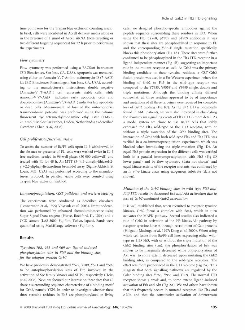
Tyrosines 768, 955 and 969 are ligand-inducedphosphorylation sites in Flt3 and the binding sitesfor the adaptor protein Grb2
We have previously demonstrated Y572, Y589, Y591 and Y599
to be autophosphorylation sites of Flt3 involved in the
activation of Src family kinases and SHP2, respectively (Heiss
et al, 2006). Next, we focused our interest on three sites that all
share a surrounding sequence characteristic of a binding motif
for Grb2, namely YXN. In order to investigate whether these
three tyrosine residues in Flt3 are phosphorylated in living
cells, we designed phospho-specific antibodies against the
peptide sequence surrounding these residues in Flt3. When
using the Flt3 pY768, pY955 and pY969 antibodies it was
shown that these sites are phosphorylated in response to FL
and the corresponding Y-to-F single mutation specifically
blocks this phosphorylation (Fig 1A). These sites were further
confirmed to be phosphorylated in the Flt3 ITD receptor in a
ligand-independent manner (Fig 1B), suggesting an important
role in the mutant receptor as well. As Grb2 was the primary
binding candidate to these tyrosine residues, a GST-Grb2
fusion protein was used in a Far Western experiment where the
binding of Grb2 to Flt3 in the wild-type receptor was
compared to the Y768F, Y955F and Y969F single, double and
triple mutations. Although the binding affinity differed
somewhat, all three residues appeared to be of importance
and mutations of all three tyrosines were required for complete
loss of Grb2 binding (Fig 1C). As the Flt3 ITD is commonly
found in AML patients, we were also interested in elucidating
the downstream signalling events of Flt3 ITD in more detail. As
a model system we chose to use Ba/F3 cells that stably
expressed the Flt3 wild-type or the ITD receptor, with or
without a triple mutation of the Grb2 binding sites. The
interaction of Grb2 with both wild-type Flt3 and Flt3 ITD was
verified in a co-immunoprecipitation experiment, which was
blocked when introducing the triple mutation (Fig 1D). An
equal Flt3 protein expression in the different cells was verified
both in a parallel immunoprecipitation with Flt3 (Fig 1D
lower panel) and by flow cytometry (data not shown) and
equal kinase activity of the receptor mutants was confirmed by
an in vitro kinase assay using exogenous substrate (data not
shown).
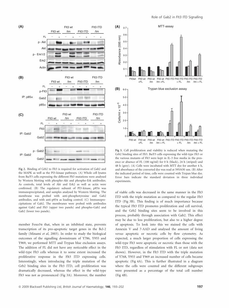
Mutation of the Grb2 binding sites in wild-type Flt3 andFlt3 ITD results in decreased Erk and Akt activation due toloss of Grb2-mediated Gab2 association
It is well established that, when recruited to receptor tyrosine
kinases, Grb2 forms a complex with Sos1, which in turn
activates the MAPK pathway. Several studies also indicated a
role of Grb2 in activation of the PI3-kinase/Akt pathway by
receptor tyrosine kinases through recruitment of Gab proteins
(Holgado-Madruga et al, 1997; Kong et al, 2000). When using
whole cell lysate from Ba/F3 cell lines expressing either wild-
type or ITD Flt3, with or without the triple mutation of the
Grb2 binding sites (tm), the phosphorylation of Erk was
shown to be marginally decreased while phosphorylation of
Akt was, to some extent, decreased upon mutating the Grb2
binding sites, as compared to the wild-type receptors. The
effect was more pronounced in the ITD receptor (Fig 2A). This
suggests that both signalling pathways are regulated by the
Grb2 binding sites Y768, Y955 and Y969. The normal ITD
receptor shows a weak and, to some extent, ligand-induced
activation of Erk and Akt (Fig 2A). We and others have shown
that this frequently occurs in mutated receptors like Flt3 and
c-Kit, and that the constitutive activation of downstream
Role of Gab2 in Flt3 ITD Signalling
ª 2009 Blackwell Publishing Ltd, British Journal of Haematology, 146, 193–202 195
signalling can be further enhanced by ligand stimulation (data
not shown). As Flt3 lacks a direct binding motif for the p85
subunit of PI3-kinase, a possible mediator of PI3-kinase
activation is Gab2. We immunoprecipitated p85a in the
different Ba/F3 cell lines and analysed the phosphorylation
levels of the Flt3 receptor in the absence or presence of FL.
Although the protein levels of p85a were equal in all samples,
wild-type Flt3 and p85a co-immunoprecipated in a ligand-
dependent manner. In contrast, Flt3 triple mutant showed a
decreased interaction with p85a. Probing the filter with an
antibody against Gab2 demonstrated that Gab2 association was
also dependent on the three phosphorylation sites (Fig 2B).
This suggests that Flt3 activates PI3-kinase in a Gab2-
dependent fashion, via the Grb2 binding sites on Flt3. An
interaction between Flt3 and Gab2 in Ba/F3 cells was further
confirmed by co-immunoprecipitation (Fig 2C, upper two
panels) and when investigating the levels of phosphorylated
Gab2, we could conclude that the activation of Gab2
depends on the Grb2 binding sites (Fig 2C, lower two
panels). A constitutive interaction between Grb2 and Gab2
has been shown in previous studies (Zhang & Broxmeyer,
2000; Sun et al, 2008).
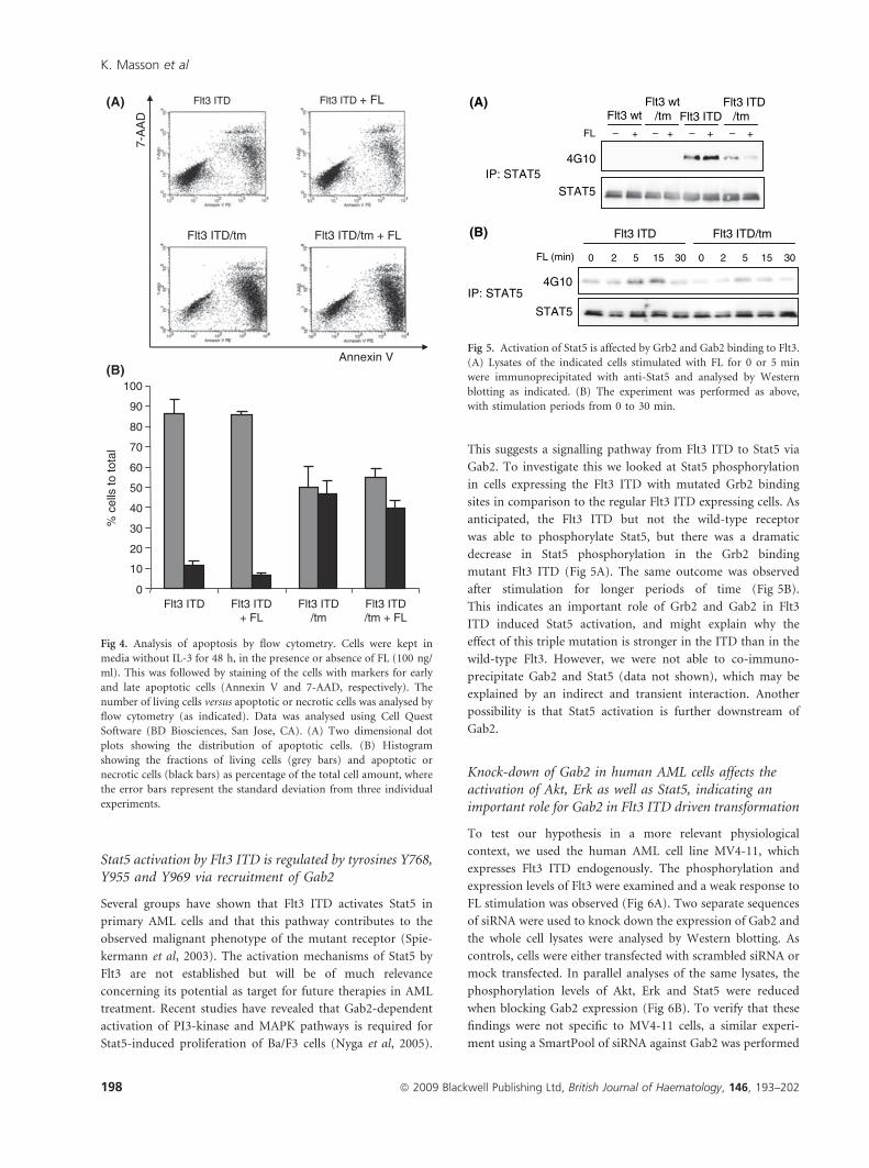
Flt3 ITD-mediated proliferation and survival of Ba/F3is dependent on intact Grb2 binding sites in Flt3
The Flt3 ITD receptor has been studied extensively during
recent years but the details of its transforming capacities are
not fully understood. Signalling through the ITD receptor
inhibits apoptosis by phosphorylating the Forkhead family
Flt3 ITDFL ++
Flt3 wt
pY955-Flt3
Flt3
Flt3 ITDFL – – ++++– – ++
Flt3 wt
pY955-Flt3
Flt3
Flt3
pY768-Flt3
FL – – ++
Flt3 wt Y768F
4G10
Flt3 wt Y969F
Flt3
pY969-Flt3
4G10
Flt3 wt Y969F
Flt3
pY969-Flt3
4G10
Flt3
pY955-Flt3
FL – ++
Flt3 wt Y955F
4G10
Flt3
pY955-Flt3
FL – ++ FL – ++FL – ++
FL – ++FL – ++
Flt3 wt Y955F
4G10
Flt3 ITDFlt3 wt
pY969-Flt3
Flt3
Flt3 ITDFL
Flt3 wt
pY768-Flt3
Flt3
GST-Grb2
4G10
Flt3
FL + + + + + + + +
Flt3 wt Y768F Y955F Y969FY768/955F
Y768/969F
Y955/969F
Y768/955/969F
IP: Flt3
GST-Grb2
4G10
Flt3
FL + + + + + + + +– – –
– – – –
– – – – –
Flt3 wt Y768F Y955F Y969FY768/955F
Y768/969F
Y955/969F
Y768/955/969F
IP: Flt3
IP: Grb2
IP: Flt3
IP: Flt3
IP: Flt3
FL + + + +
Flt3 wtFlt3 wt
/tmFlt3 ITD
/tmFlt3 ITD
Flt3
Grb2
Flt3
FL + + + +
Flt3 wtFlt3 wt
/tmFlt3 ITD
/tmFlt3 ITD
Flt3
Grb2
Flt3
(A)
(II) (III)(I)
(II) (III)(I)
(B)
(C)
(D)
Fig 1. Identification of Y768, Y955 and Y969 as phosphorylation and Grb2 binding sites in COS-1 and Ba/F3 cells. (A) COS-1 cells, transiently
transfected with either the wild-type Flt3 receptor or the respective Y-to-F mutant, were stimulated with FL for 5 min and lysed. Lysates were
immunoprecipitated with an Flt3 antibody and analysed by Western blotting. Membranes were probed with phosphospecific antibodies and after
stripping, re-probed with 4G10 and anti-Flt3. (B) Lysates from Ba/F3 cells with stable expression of either the wild-type Flt3 or the Flt3 ITD receptor
were processed as above. (C) Ba/F3 cells stably expressing either wild-type Flt3, or the various Y-to-F mutants of Flt3 were stimulated with FL for
5 min. Cell lysates were immunoprecipitated with anti-Flt3 and analysed by Far Western blotting, using a GST-fusion protein of Grb2 SH2 domain.
(D) Grb2 (upper two panels) or Flt3 (lower panel) was immunoprecipitated from cell lysates of the Ba/F3 cells mentioned above and samples were
analysed by Western blotting with the indicated antibodies. The Y768F/Y955F/Y969F mutation is denoted tm (triple mutation).
K. Masson et al
196 ª 2009 Blackwell Publishing Ltd, British Journal of Haematology, 146, 193–202
member Foxo3a that, when in an inhibited state, prevents
transcription of its pro-apoptotic target genes in the Bcl-2
family (Minami et al, 2003). In order to study the biological
outcomes of the signalling downstream of Y786, Y955 and
Y969, we performed MTT and Trypan blue exclusion assays.
The addition of FL did not have any noticeable effect in the
wild-type Flt3 cells whereas it to some extent increased the
proliferative response in the Flt3 ITD expressing cells.
Interestingly, when introducing the triple mutation of the
Grb2 binding sites in the Flt3 ITD, cell proliferation was
dramatically decreased, whereas the effect in the wild-type
Flt3 was not as pronounced (Fig 3A). Moreover, the number
of viable cells was decreased in the same manner in the Flt3
ITD with the triple mutation as compared to the regular Flt3
ITD (Fig 3B). This finding is of much importance because
the typical Flt3 ITD promotes proliferation and cell survival,
and the Grb2 binding sites seem to be involved in this
process, probably through association with Gab2. This effect
may be due to less proliferation, but also to a higher degree
of apoptosis. To look into this we stained the cells with
Annexin V and 7-AAD and analysed the amount of living
versus apoptotic or necrotic cells by flow cytometry. As
expected, a much larger proportion of cells expressing the
wild-type Flt3 were apoptotic or necrotic than those with the
Flt3 ITD, regardless of stimulation with FL or not (data not
shown). However, in the Flt3 ITD with the triple mutation
of Y768, Y955 and Y969 an increased number of cells became
apoptotic (Fig 4A). This is further illustrated in a diagram
where the cells were counted and the different subgroups
were presented as a percentage of the total cell number
(Fig 4B).
FL – – – – + + + +
F l t 3 w t F l t 3 I T D
F l t 3 w t
/ t m
F l t 3 I T D
/ t m
p - A k t
A k t
p - E r k 1 / 2
Er k 2
Actin
FL – – – – + + + +
F l t 3 w t F l t 3 I T D
F l t 3 w t
/ t m
F l t 3 I T D
/ t m
p-Flt3
p85a
Ga b 2
F l t 3 w t F l t 3 w t
/ t m F l t 3 I T D
/ t m F l t 3 I T D
FL + + + + – – – –
F l t 3
Ga b 2
p - G a b 2
Ga b 2
I P : p 85a
I P : G a b 2
I P : G a b 2
(A)
(B)
(C)
Fig 2. Binding of Grb2 to Flt3 is required for activation of Gab2 and
the MAPK as well as the PI3-kinase pathways. (A) Whole cell lysates
from Ba/F3 cells expressing the different Flt3 mutations were analysed
by Western blotting with phospho-Akt and phospho-Erk antibodies.
As controls, total levels of Akt and Erk2 as well as actin were
confirmed. (B) The regulatory subunit of PI3-kinase, p85a was
immunoprecipitated, and samples analysed by Western blotting. The
membrane was probed with anti-phosphotyrosine and Gab2
antibodies, and with anti-p85a as loading control. (C) Immunopre-
cipitations of Gab2. The membranes were probed with antibodies
against Gab2 and Flt3 (upper two panels) and phospho-Gab2 and
Gab2 (lower two panels).
0
0·1
0·2
0·3
0·4
0·5
0·6
0
1·0
2·0
3·0
4·0
5·0
6·0
0·7
Flt3wt Flt3 wt +FL
Flt3 wt /tm
Flt3 wt/tm +FL
Flt3 ITD + FL
Flt3 ITD/tm
Flt3 ITD /tm + FL
Flt3 ITD
Flt3 wt Flt3 wt +FL
Flt3 wt /tm
Flt3 wt /tm + FL
Flt3 ITD + FL
Flt3 ITD/tm
Flt3 ITD /tm + FL
Flt3 ITD
Abs
orba
nce
(595
nm
) C
ells
/ml ×
10–5
MTT-assay
Trypan blue exclusion assay
(A)
(B)
Fig 3. Cell proliferation and viability is reduced when mutating the
Grb2 binding sites of Flt3. Ba/F3 cells expressing the wild-type Flt3 or
the various mutants of Flt3 were kept in IL-3 free media in the pres-
ence or absence of FL (100 ng/ml) for 0 h (black), 24 h (striped) and
48 h (grey). (A) Cells were incubated with MTT dye for another 4 h,
and absorbance of the converted dye was read at 595/630 nm. (B) After
the indicated period of time, cells were counted with Trypan blue dye.
Error bars indicate the standard deviation in three individual
experiments.
Role of Gab2 in Flt3 ITD Signalling
ª 2009 Blackwell Publishing Ltd, British Journal of Haematology, 146, 193–202 197
Stat5 activation by Flt3 ITD is regulated by tyrosines Y768,Y955 and Y969 via recruitment of Gab2
Several groups have shown that Flt3 ITD activates Stat5 in
primary AML cells and that this pathway contributes to the
observed malignant phenotype of the mutant receptor (Spie-
kermann et al, 2003). The activation mechanisms of Stat5 by
Flt3 are not established but will be of much relevance
concerning its potential as target for future therapies in AML
treatment. Recent studies have revealed that Gab2-dependent
activation of PI3-kinase and MAPK pathways is required for
Stat5-induced proliferation of Ba/F3 cells (Nyga et al, 2005).
This suggests a signalling pathway from Flt3 ITD to Stat5 via
Gab2. To investigate this we looked at Stat5 phosphorylation
in cells expressing the Flt3 ITD with mutated Grb2 binding
sites in comparison to the regular Flt3 ITD expressing cells. As
anticipated, the Flt3 ITD but not the wild-type receptor
was able to phosphorylate Stat5, but there was a dramatic
decrease in Stat5 phosphorylation in the Grb2 binding
mutant Flt3 ITD (Fig 5A). The same outcome was observed
after stimulation for longer periods of time (Fig 5B).
This indicates an important role of Grb2 and Gab2 in Flt3
ITD induced Stat5 activation, and might explain why the
effect of this triple mutation is stronger in the ITD than in the
wild-type Flt3. However, we were not able to co-immuno-
precipitate Gab2 and Stat5 (data not shown), which may be
explained by an indirect and transient interaction. Another
possibility is that Stat5 activation is further downstream of
Gab2.
Knock-down of Gab2 in human AML cells affects theactivation of Akt, Erk as well as Stat5, indicating animportant role for Gab2 in Flt3 ITD driven transformation
To test our hypothesis in a more relevant physiological
context, we used the human AML cell line MV4-11, which
expresses Flt3 ITD endogenously. The phosphorylation and
expression levels of Flt3 were examined and a weak response to
FL stimulation was observed (Fig 6A). Two separate sequences
of siRNA were used to knock down the expression of Gab2 and
the whole cell lysates were analysed by Western blotting. As
controls, cells were either transfected with scrambled siRNA or
mock transfected. In parallel analyses of the same lysates, the
phosphorylation levels of Akt, Erk and Stat5 were reduced
when blocking Gab2 expression (Fig 6B). To verify that these
findings were not specific to MV4-11 cells, a similar experi-
ment using a SmartPool of siRNA against Gab2 was performed
Annexin V
7-A
AD
% c
ells
to to
tal
0
30
20
10
40
50
60
70
80
90
100
Flt3 ITD Flt3 ITD + FL
Flt3 ITD /tm
Flt3 ITD /tm + FL
Flt3 ITD Flt3 ITD + FL
Flt3 ITD/tm + FL Flt3 ITD/tm
(A)
(B)
Fig 4. Analysis of apoptosis by flow cytometry. Cells were kept in
media without IL-3 for 48 h, in the presence or absence of FL (100 ng/
ml). This was followed by staining of the cells with markers for early
and late apoptotic cells (Annexin V and 7-AAD, respectively). The
number of living cells versus apoptotic or necrotic cells was analysed by
flow cytometry (as indicated). Data was analysed using Cell Quest
Software (BD Biosciences, San Jose, CA). (A) Two dimensional dot
plots showing the distribution of apoptotic cells. (B) Histogram
showing the fractions of living cells (grey bars) and apoptotic or
necrotic cells (black bars) as percentage of the total cell amount, where
the error bars represent the standard deviation from three individual
experiments.
4G10
STAT5
Flt3 wtFlt3 wt
/tm Flt3 ITDFlt3 ITD
/tmFL + + + +––– –
4G10
STAT5
Flt3 ITD
FL (min) 0 2
Flt3 ITD/tm
5 15 30 0 2 5 15 30
IP: STAT5
IP: STAT5
(A)
(B)
Fig 5. Activation of Stat5 is affected by Grb2 and Gab2 binding to Flt3.
(A) Lysates of the indicated cells stimulated with FL for 0 or 5 min
were immunoprecipitated with anti-Stat5 and analysed by Western
blotting as indicated. (B) The experiment was performed as above,
with stimulation periods from 0 to 30 min.
K. Masson et al
198 ª 2009 Blackwell Publishing Ltd, British Journal of Haematology, 146, 193–202

in Ba/F3 cells. The results were equivalent in terms of the effect
on Erk and Akt phosphorylation (data not shown), although a
more pronounced decrease of Stat5 phosphorylation was seen
as a result of silencing Gab2 (Fig 6C). A Trypan blue cell
counting assay indicated that the MV4-11 cells transfected with
Gab2 siRNA showed significantly less proliferation and
survival than the control cells (Fig 6D).
Discussion
It is well recognised that Flt3 ITD receptor signalling differs
from that of wild-type Flt3, both in terms of ligand indepen-
dency and in the ability to activate Stat5. Previous studies have
shown that Flt3 ITD has transforming potential in haemato-
poietic cells, and the ITD receptor induced a strong activation
of Stat5 but not Stat3 in primary AML blasts (Spiekermann
et al, 2003; Mizuki et al, 2000).
In this study, we identified the three tyrosines 768, 955 and
969 as ligand-induced phosphorylation sites in Flt3 by the use
of phospho-specific antibodies. As all three sites contain the
adaptor protein Grb2 binding YXN motif, we verified a direct
interaction with Grb2, which in turn was found to recruit the
scaffolding protein Gab2 to the receptor. This feature has also
been shown with some other tyrosine kinases, such as tyrosine
177 of Bcr-Abl, which recruits Gab2 via a Grb2/Gab2 complex
where this interaction is required for full activation of the PI3-
kinase and MAPK pathways as well as for optimal proliferation
and migration in Ba/F3 cells (Sattler et al, 2002). Gab2 protein
is over activated in several malignancies and GAB2 has been
identified as co amplified in 70% of all patients with the MLL
4G 1 0
F l t 3
0 2 5 1 5 F L ( m i n )
4G 1 0
S T A T 5
Ga b 2
S c r a
m b l
e d
S i G
ab 2 -
1
M o c
k
S c r a
m b l
e d
S i G
ab 2 -
1
M o c
k
4G 1 0
S T A T 5
Ga b 2
S c r a
m b l
e d
S i G
ab 2
M o c
k
S i G
ab 2 -
2
S i G
ab 2 -
2
p - E r k 1 / 2
A ct i n
p - A k t
S c r a
m b l
e d
S i G
ab 2 -
1
M o c
k
S i G
ab 2 -
2
I P : Flt3
I P : G a b 2
I P : S T A T5
I P : G a b 2
I P : S T A T5
Ga b 2
Cel
ls/m
l × 1
0–5
Mock Scram SiGab2-1 SiGab2-2 0
5
10
15
20
25
30
I P : G a b 2
(A)
(B) (I)
(II)
(III)
(C) (D)
Fig 6. Silencing of Gab2 in the Flt3 ITD expressing human AML cell line MV4-11. (A) Expression and activation of Flt3 ITD was verified by
immunoprecipitation of Flt3 after ligand stimulation for the indicated periods of time. (B) Silencing of Gab2 was achieved by transfecting the cells
with two separate sequences of Accell siRNA, and control cells were either transfected with non-targeting siRNA (scrambled) or mock (Accell Delivery
medium only). Cells were kept in culture for 72 h after transfection. Whole cell lysates as well as immunoprecipitates for Gab2 and Stat5 were
analysed in parallel by Western blotting. Membranes were probed with antibodies against Gab2 (I), 4G10 and Stat5 (II), phospho-Erk1/2, phospho-
Akt and b-actin (III). (C) A similar experiment was performed using Ba/F3 cells, but silencing of Gab2 was achieved by transfecting the cells with
Gab2 SmartPool siRNA by electroporation, and control cells were either transfected with non-targeting siRNA or mock-transfected. Cells were kept in
culture for 48 h after transfection and starved for 4 h before lysis, whereafter the samples were immunoprecipitated with Gab2 and Stat5 antibodies
and analysed for Gab2 levels and Stat5 activation (D) After transfection, MV4-11 cells from the same experiment as in (B) were kept in culture for 0
(dark grey bars) or 24 h (light grey bars) and counted for Trypan blue dye exclusion. The figures are presented in a diagram where the error bars
indicate the standard deviation from three individual experiments.
Role of Gab2 in Flt3 ITD Signalling
ª 2009 Blackwell Publishing Ltd, British Journal of Haematology, 146, 193–202 199

gene which is a target of chromosome 11q amplification in
AML/MDS (Zatkova et al, 2006). This indicates that Gab2
plays an important role in AML signalling pathways and if
regulated by Flt3, might contribute to the poor prognosis
observed in AML patients who also carry the Flt3 ITD
mutation.
By looking at Erk and Akt phosphorylation, we could
conclude that both the MAPK and PI3-kinase signalling
pathways were affected by reduced Grb2/Gab2 interaction with
Flt3. Serine 623 of Gab2 is a phosphorylation target of Erk that
further affects the binding of Shp-2 to Gab2. A Gab2 S623A
mutant showed not only decreased Erk phosphorylation but
also reduced Shp-2 recruitment, which further leads to a
sustained Stat5 activity and cell proliferation (Arnaud et al,
2004). When the three tyrosine residues to phenylalanine were
mutated in Flt3 ITD, the phenotype of the Ba/F3 cells
resembled more that of the wild-type Flt3 in terms of
proliferation and survival. Remarkably, Stat5 phosphorylation
was also decreased in these cells, leading us to hypothesise that
the Stat5 pathway is activated by Flt3 ITD via Gab2. Stat5 has
previously been shown to associate with Gab2 (Brockdorff
et al, 2001), and this process is of importance in Stat5-induced
cell proliferation and survival via the PI3-kinase and MAPK
signalling pathways (Nyga et al, 2005). Interestingly, the
authors also found that this interaction was independent of
JAK and it is believed that formation of the Gab2/Stat5/p85
complex requires Gab2 phosphorylation but so far, no
consensus sequences for binding of Stat5 have been identified
within Gab2. One might therefore consider the interaction to
be indirect through some other signalling molecule. This
remains to be shown. Moreover, a Stat3 binding motif was
recently identified in Gab2 and proved to be required for
erythropoietin-independent growth of Friend virus-infected
erythroid progenitor cells (Ni et al, 2007). Taken together, the
data provided here add to the conclusion that Stat5 is a
downstream target of Gab2.
Ba/F3 cells expressing the Grb2 binding Y-to-F triple
mutation have an increased apoptotic response upon IL-3
withdrawal, and the effect was more prominent in the Flt3 ITD
than in the wild-type Flt3. This observation is most likely due
to decreased Stat5 activity. Wild-type Flt3 signalling has been
associated with anti apoptosis through the activation of Bad.
However, dephosphorylation of Bad is insufficient to induce
apoptosis where the downregulation of Bcl-x is required,
which is restored by Stat5 signalling, hence only seen in the
Flt3 ITD expressing cells (Minami et al, 2003). Other studies in
Ba/F3 cells have shown that Stat5 might promote cell survival
not only as a transcription factor by regulating expression of
Bcl-x, but as a signalling intermediate in PI3-kinase signalling
(Santos et al, 2001) and that PI3-kinase and Stat5 co-operate
to promote IL-3 mediated suppression of apoptosis (Rosa
Santos et al, 2000). Persistent Stat5 phosphorylation was
detected in primary cells from patients with AML or chronic
myeloid leukaemia and a major fraction of Stat5 molecules
turned out to be cytoplasmic in the presence of growth factors.
These results suggest that a large part of the oncogenic activity
of Stat5 involves a cytoplasmic signalling complex between
Stat5, Gab2 and PI3-kinase (Harir et al, 2007). This is also in
line with our findings and, when knocking down Gab2
expression in AML cells, the Akt phosphorylation as well as
the Stat5 phosphorylation was lowered. These data support the
idea of a Flt3 ITD signalling complex which eventually leads to
the activation of Stat5 target genes and contributes to the
transforming phenotype.
The mechanism by which Flt3 ITD signalling leads to
phosphorylation of Stat5 is not fully characterised. It has been
suggested that Flt3 ITD-meditated Stat5 activation is directly
mediated by Flt3 itself (Choudhary et al, 2007). However,
those experiments were mainly performed in mouse embry-
onal fibroblasts known to express low levels of Gab2 [(Liu
et al, 2001); data not shown] leaving the possibility that other
tyrosine kinases might mediate phosphorylation of Stat5, at
least in part. One such candidate is a member of the Src family
kinases. The erythropoietin receptor associates with Lyn in
haematopoietic cells, which in turn has the ability to induce
phosphorylation of Stat5 (Chin et al, 1998). When mutating
the two Src binding sites Y589 and Y591 in Flt3 ITD, the Stat5
phosphorylation was decreased, indicating that Src family
kinases or some other protein biding to these residues, might
mediate the activation of Stat5 in an indirect manner
(Hayakawa & Naoe, 2006; Rocnik et al, 2006). Future studies
will elucidate the details of this mechanism.
In conclusion, the data provided here supports the idea of
Gab2 as an important mediator in Flt3 ITD signalling.
Tyrosines 768, 955 and 969 of Flt3 are phosphorylated and
shown to be Grb2 binding sites of importance for cell
proliferation and survival, most likely as a result of Gab2
association. Normally, the Flt3 ITD receptor promotes
increased survival and growth as well as strong activation of
Stat5, which is a potent contributor to the malignant
phenotype, and these characteristics were lost when mutating
the Grb2 binding sites in Ba/F3 cells. Moreover, after silencing
Gab2 expression in both human AML cells and in Flt3 ITD
expressing Ba/F3 cells, the activation of MAPK and PI3-kinase
pathways as well as the phosphorylation of Stat5 was lowered,
suggesting an important role for Gab2 in Flt3 ITD-driven
transformation. We hypothesise that Gab2 regulates the
activation of Stat5 in Flt3 ITD receptor signalling, possibly
through other signal transduction intermediates, and that
tyrosines 768, 955 and 969 of Flt3 are of much importance in
this process. Our findings contribute to the overall under-
standing of Flt3 signalling, both in terms of the wild-type
receptor and the oncogenic Flt3 ITD receptor in AML, and
introduce a signal transduction pathway as a possible future
therapeutic target.
Acknowledgements
This work was supported by grants from Swedish Cancer
Society, Swedish Children’s Cancer Foundation, Swedish
K. Masson et al
200 ª 2009 Blackwell Publishing Ltd, British Journal of Haematology, 146, 193–202

Research Council, Malmo University Hospital Cancer Fund,
Malmo University Hospital General Funds, The Wallenberg
Foundation and the Royal Physiographical Society, Lund.
References
Arnaud, M., Crouin, C., Deon, C., Loyaux, D. & Bertoglio, J. (2004)
Phosphorylation of Grb2-associated binder 2 on serine 623 by ERK
MAPK regulates its association with the phosphatase SHP-2 and
decreases STAT5 activation. Journal of Immunology, 173, 3962–3971.
Brockdorff, J.L., Gu, H., Mustelin, T., Kaltoft, K., Geisler, C., Ropke, C.
& Ødum, N. (2001) Gab2 is phosphorylated on tyrosine upon
interleukin-2/interleukin-15 stimulation in mycosis-fungoides-
derived tumor T cells and associates inducibly with SHP-2 and
Stat5a. Experimental and Clinical Immunogenetics, 18, 86–95.
Bunting, K.D. (2007) STAT5 signaling in normal and pathologic
hematopoiesis. Frontiers in Bioscience, 12, 2807–2820.
Chillon, M.C., Fernandez, C., Garcıa-Sanz, R., Balanzategui, A.,
Ramos, F., Fernandez-Calvo, J., Gonzalez, M. & Miguel, J.F. (2004)
FLT3-activating mutations are associated with poor prognostic
features in AML at diagnosis but they are not an independent
prognostic factor. The Hematology Journal, 5, 239–246.
Chin, H., Arai, A., Wakao, H., Kamiyama, R., Miyasaka, N. & Miura,
O. (1998) Lyn physically associates with the erythropoietin receptor
and may play a role in activation of the Stat5 pathway. Blood, 91,
3734–3745.
Choudhary, C., Schwable, J., Brandts, C., Tickenbrock, L., Sargin, B.,
Kindler, T., Fischer, T., Berdel, W.E., Muller-Tidow, C. & Serve, H.
(2005) AML-associated Flt3 kinase domain mutations show signal
transduction differences compared with Flt3 ITD mutations. Blood,
106, 265–273.
Choudhary, C., Brandts, C., Schwable, J., Tickenbrock, L., Sargin, B.,
Ueker, A., Bohmer, F.D., Berdel, W.E., Muller-Tidow, C. & Serve, H.
(2007) Activation mechanisms of STAT5 by oncogenic Flt3-ITD.
Blood, 110, 370–374.
Dumon, S., Santos, S.C., Debierre-Grockiego, F., Gouilleux-Gruart, V.,
Cocault, L., Boucheron, C., Mollat, P., Gisselbrecht, S. & Gouilleux,
F. (1999) IL-3 dependent regulation of Bcl-xL gene expression by
STAT5 in a bone marrow derived cell line. Oncogene, 18, 4191–4199.
Gilliland, D.G. & Griffin, J.D. (2002) The roles of FLT3 in hemato-
poiesis and leukemia. Blood, 100, 1532–1542.
Griffith, J., Black, J., Faerman, C., Swenson, L., Wynn, M., Lu, F.,
Lippke, J. & Saxena, K. (2004) The structural basis for autoinhibi-
tion of FLT3 by the juxtamembrane domain. Molecular Cell, 13,
169–178.
Harir, N., Pecquet, C., Kerenyi, M., Sonneck, K., Kovacic, B., Nyga, R.,
Brevet, M., Dhennin, I., Gouilleux-Gruart, V., Beug, H., Valent, P.,
Lassoued, K., Moriggl, R. & Gouilleux, F. (2007) Constitutive acti-
vation of Stat5 promotes its cytoplasmic localization and association
with PI3-kinase in myeloid leukemias. Blood, 109, 1678–1686.
Hayakawa, F. & Naoe, T. (2006) SFK-STAT pathway: an alternative
and important way to malignancies. Annals of the New York Acad-
emy of Sciences, 1086, 213–222.
Hayakawa, F., Towatari, M., Kiyoi, H., Tanimoto, M., Kitamura, T.,
Saito, H. & Naoe, T. (2000) Tandem-duplicated Flt3 constitutively
activates STAT5 and MAP kinase and introduces autonomous cell
growth in IL-3-dependent cell lines. Oncogene, 19, 624–631.
Heiss, E., Masson, K., Sundberg, C., Pedersen, M., Sun, J., Bengtsson,
S. & Ronnstrand, L. (2006) Identification of Y589 and Y599 in the
juxtamembrane domain of Flt3 as ligand-induced autophosphory-
lation sites involved in binding of Src family kinases and the protein
tyrosine phosphatase SHP2. Blood, 108, 1542–1550.
Holgado-Madruga, M., Moscatello, D.K., Emlet, D.R., Dieterich, R. &
Wong, A.J. (1997) Grb2-associated binder-1 mediates phosphati-
dylinositol 3-kinase activation and the promotion of cell survival by
nerve growth factor. Proceedings of the National Academy of Sciences
of the United States of America, 94, 12419–12424.
Khan, R., Schmidt-Mende, J., Karimi, M., Gogvadze, V., Hassan, M.,
Ekstrom, T.J., Zhivotovsky, B. & Hellstrom-Lindberg, E. (2008)
Hypomethylation and apoptosis in 5-azacytidine-treated myeloid
cells. Experimental Hematology, 36, 149–157.
Kiyoi, H., Ohno, R., Ueda, R., Saito, H. & Naoe, T. (2002) Mechanism
of constitutive activation of FLT3 with internal tandem duplication
in the juxtamembrane domain. Oncogene, 21, 2555–2563.
Kong, M., Mounier, C., Wu, J. & Posner, B.I. (2000) Epidermal growth
factor-induced phosphatidylinositol 3-kinase activation and DNA
synthesis. Identification of Grb2-associated binder 2 as the major
mediator in rat hepatocytes. Journal of Biological Chemistry, 275,
36035–36042.
Lennartsson, J., Blume-Jensen, P., Hermanson, M., Ponten, E., Carl-
berg, M. & Ronnstrand, L. (1999) Phosphorylation of Shc by Src
family kinases is necessary for stem cell factor receptor/c-kit medi-
ated activation of the Ras/MAP kinase pathway and c-fos induction.
Oncogene, 18, 5546–5553.
Liu, Y., Jenkins, B., Shin, J.L. & Rohrschneider, L.R. (2001) Scaffolding
protein Gab2 mediates differentiation signaling downstream of Fms
receptor tyrosine kinase. Molecular and Cellular Biology, 21, 3047–
3056.
Maroc, N., Rottapel, R., Rosnet, O., Marchetto, S., Lavezzi, C., Man-
noni, P., Birnbaum, D. & Dubreuil, P. (1993) Biochemical charac-
terization and analysis of the transforming potential of the FLT3/
FLK2 receptor tyrosine kinase. Oncogene, 8, 909–918.
Minami, Y., Yamamoto, K., Kiyoi, H., Ueda, R., Saito, H. & Naoe, T.
(2003) Different antiapoptotic pathways between wild-type and
mutated FLT3: insights into therapeutic targets in leukemia. Blood,
102, 2969–2975.
Mizuki, M., Fenski, R., Halfter, H., Matsumura, I., Schmidt, R., Muller,
C., Gruning, W., Kratz-Albers, K., Serve, S., Steur, C., Buchner, T.,
Kienast, J., Kanakura, Y., Berdel, W.E. & Serve, H. (2000) Flt3
mutations from patients with acute myeloid leukemia induce
transformation of 32D cells mediated by the Ras and STAT5 path-
ways. Blood, 96, 3907–3914.
Moriggl, R., Topham, D.J., Teglund, S., Sexl, V., McKay, C., Wang,
D., Hoffmeyer, A., van Deursen, J., Sangster, M.Y., Bunting, K.D.,
Grosveld, G.C. & Ihle, J.N. (1999) Stat5 is required for IL-2-
induced cell cycle progression of peripheral T cells. Immunity, 10,
249–259.
Ni, S., Zhao, C., Feng, G.S., Paulson, R.F. & Correll, P.H. (2007) A
novel Stat3 binding motif in Gab2 mediates transformation of
primary hematopoietic cells by the Stk/Ron receptor tyrosine kinase
in response to Friend virus infection. Molecular and Cellular Biology,
27, 3708–3715.
Nosaka, T., Kawashima, T., Misawa, K., Ikuta, K., Mui, A.L. &
Kitamura, T. (1999) STAT5 as a molecular regulator of proliferation,
differentiation and apoptosis in hematopoietic cells. EMBO Journal,
18, 4754–4765.
Nyga, R., Pecquet, C., Harir, N., Gu, H., Dhennin-Duthille, I., Regnier,
A., Gouilleux-Gruart, V., Lassoued, K. & Gouilleux, F. (2005)
Role of Gab2 in Flt3 ITD Signalling
ª 2009 Blackwell Publishing Ltd, British Journal of Haematology, 146, 193–202 201
Activated STAT5 proteins induce activation of the PI 3-kinase/Akt
and Ras/MAPK pathways via the Gab2 scaffolding adapter. Bio-
chemical Journal, 390, 359–366.
Rocnik, J.L., Okabe, R., Yu, J.C., Lee, B.H., Giese, N., Schenkein, D.P.
& Gilliland, D.G. (2006) Roles of tyrosine 589 and 591 in STAT5
activation and transformation mediated by FLT3-ITD. Blood, 108,
1339–1345.
Rosa Santos, S.C., Dumon, S., Mayeux, P., Gisselbrecht, S. & Gouil-
leux, F. (2000) Cooperation between STAT5 and phosphatidylino-
sitol 3-kinase in the IL-3-dependent survival of a bone marrow
derived cell line. Oncogene, 19, 1164–1172.
Santos, S.C., Lacronique, V., Bouchaert, I., Monni, R., Bernard, O.,
Gisselbrecht, S. & Gouilleux, F. (2001) Constitutively active STAT5
variants induce growth and survival of hematopoietic cells through a
PI 3-kinase/Akt dependent pathway. Oncogene, 20, 2080–2090.
Sattler, M., Mohi, M.G., Pride, Y.B., Quinnan, L.R., Malouf, N.A.,
Podar, K., Gesbert, F., Iwasaki, H., Li, S., Van Etten, R.A., Gu, H.,
Griffin, J.D. & Neel, B.G. (2002) Critical role for Gab2 in transfor-
mation by BCR/ABL. Cancer Cell, 1, 479–492.
Songyang, Z., Shoelson, S.E., McGlade, J., Olivier, P., Pawson, T.,
Bustelo, X.R., Barbacid, M., Sabe, H., Hanafusa, H., Yi, T., Ren, R.,
Baltimore, D., Ratnofsky, S., Feldman, R.A. & Cantley, L.C. (1994)
Specific motifs recognized by the SH2 domains of Csk, 3BP2, fps/fes,
GRB-2, HCP, SHC, Syk, and Vav. Molecular and Cellular Biology, 14,
2777–2785.
Spiekermann, K., Bagrintseva, K., Schwab, R., Schmieja, K. & Hidde-
mann, W. (2003) Overexpression and constitutive activation of
FLT3 induces STAT5 activation in primary acute myeloid leukemia
blast cells. Clinical Cancer Research, 9, 2140–2150.
Stirewalt, D.L. & Radich, J.P. (2003) The role of FLT3 in haemato-
poietic malignancies. Nature Reviews. Cancer, 3, 650–665.
Sun, J., Pedersen, M., Bengtsson, S. & Ronnstrand, L. (2007) Grb2
mediates negative regulation of stem cell factor receptor/c-Kit sig-
naling by recruitment of Cbl. Experimental Cell Research, 313, 3935–
3942.
Sun, J., Pedersen, M. & Ronnstrand, L. (2008) Gab2 is involved in
differential phosphoinositide 3-kinase signaling by two splice forms
of c-Kit. Journal of Biological Chemistry, 283, 27444–27451.
Voytyuk, O., Lennartsson, J., Mogi, A., Caruana, G., Courtneidge, S.,
Ashman, L.K. & Ronnstrand, L. (2003) Src family kinases are
involved in the differential signaling from two splice forms of c-Kit.
Journal of Biological Chemistry, 278, 9159–9166.
Yanada, M., Matsuo, K., Suzuki, T., Kiyoi, H. & Naoe, T. (2005)
Prognostic significance of FLT3 internal tandem duplication and
tyrosine kinase domain mutations for acute myeloid leukemia: a
meta-analysis. Leukemia, 19, 1345–1349.
Zatkova, A., Schoch, C., Speleman, F., Poppe, B., Mannhalter, C.,
Fonatsch, C. & Wimmer, K. (2006) GAB2 is a novel target of 11q
amplification in AML/MDS. Genes, Chromosomes and Cancer, 45,
798–807.
Zhang, S. & Broxmeyer, H.E. (1999) p85 subunit of PI3 kinase does
not bind to human Flt3 receptor, but associates with SHP2, SHIP,
and a tyrosine-phosphorylated 100-kDa protein in Flt3 ligand-
stimulated hematopoietic cells. Biochemical and Biophysical Research
Communications, 254, 440–445.
Zhang, S. & Broxmeyer, H.E. (2000) Flt3 ligand induces tyrosine
phosphorylation of gab1 and gab2 and their association with shp-2,
grb2, and PI3 kinase. Biochemical and Biophysical Research Com-
munications, 277, 195–199.
Zhang, S., Mantel, C. & Broxmeyer, H.E. (1999) Flt3 signaling involves
tyrosyl-phosphorylation of SHP-2 and SHIP and their association
with Grb2 and Shc in Baf3/Flt3 cells. Journal of Leukocyte Biology, 65,
372–380.
Zhang, Y., Diaz-Flores, E., Li, G., Wang, Z., Kang, Z., Haviernikova, E.,
Rowe, S., Qu, C.K., Tse, W., Shannon, K.M. & Bunting, K.D. (2007)
Abnormal hematopoiesis in Gab2 mutant mice. Blood, 110, 116–124.
K. Masson et al
202 ª 2009 Blackwell Publishing Ltd, British Journal of Haematology, 146, 193–202


![Optimization of Imidazo[4,5- b ]pyridine-Based Kinase Inhibitors: Identification of a Dual FLT3/Aurora Kinase Inhibitor as an Orally Bioavailable Preclinical Development Candidate](https://static.fdokumen.com/doc/165x107/6345ee68596bdb97a909280b/optimization-of-imidazo45-b-pyridine-based-kinase-inhibitors-identification.jpg)