Stat5 Is Required for IL-2-Induced Cell Cycle Progression of Peripheral T Cells
11
Immunity, Vol. 10, 249–259, February, 1999, Copyright 1999 by Cell Press Stat5 Is Required for IL-2-Induced Cell Cycle Progression of Peripheral T Cells tyrosine phosphorylation and activation. Our initial inter- est in the Stat5 proteins was based on their activation by cytokines that affect the myeloid lineages including Richard Moriggl, 1,2,8 David J. Topham, 5,8 Stephan Teglund, 2,8 Veronika Sexl, 3 Catriona McKay, 1,2 Demin Wang, 2 erythropoietin (Epo), thrombopoietin (Tpo), interleukin-3 Angelika Hoffmeyer, 1,2 Jan van Deursen, 4 (IL-3), and granulocyte-macrophage colony-stimulating Mark Y. Sangster, 5 Kevin D. Bunting, 2 factor (GM-CSF). However, the Stat5 proteins are also Gerard C. Grosveld, 4 and James N. Ihle 1,2,7 activated by a variety of cytokines that affect lymphoid 1 Howard Hughes Medical Institute lineages. For example, IL-7, which is required for the 2 Department of Biochemistry early amplification and survival of lymphoid lineage pro- 3 Department of Tumor Cell Biology genitors, activates the Stat5 proteins (Lin et al., 1995), 4 Department of Genetics as do a number of cytokines affecting various lymphoid 5 Department of Immunology functions, including IL-2, IL-4, IL-9, IL-13, and IL-15 (Fujii St. Jude Children’s Research Hospital et al., 1995; Gaffen et al., 1995; Gilmour et al., 1995; Hou Memphis, Tennessee 38105 et al., 1995; Wakao et al., 1995). The role for activation 6 Department of Biochemistry of the Stat5 proteins by the IL-2 receptor has been exam- University of Tennessee Medical School, Memphis ined by introducing the IL-2 receptor chains into immor- Memphis, Tennessee 38063 talized, myeloid lineage cells (Zamorano et al., 1998). Under these conditions, Stat5 was found to not be re- quired for cell cycle progression. More recently, transgenic Summary mice have been derived with IL-2 receptor b chain mu- tants that suggested that the region required for Stat5 Many cytokines activate two highly homologous Stat activation was not essential for most peripheral T cell proteins, 5a and 5b. Mice deficient in both genes lack functions (Fujii et al., 1998). all growth hormone and prolactin functions but retain We have previously described the derivation and char- functions associated with cytokines such as erythro- acterization of mice that lack Stat5a, Stat5b, or both poietin. Here, we demonstrate that, while lymphoid genes (Teglund et al., 1998). Viable mice were obtained development is normal, Stat5a/b mutant peripheral T in each case, including mutant mice that lacked both cells are profoundly deficient in proliferation and fail Stat5 proteins. The most evident phenotypes were asso- to undergo cell cycle progression or to express genes ciated with the loss of growth hormone and prolactin controlling cell cycle progression. In addition, the mice functions. Indeed, most of the phenotypic changes seen lack NK cells, develop splenomegaly, and have T cells in prolactin receptor (Ormandy et al., 1997)- and growth with an activated phenotype, phenotypes seen in IL-2 hormone receptor (Zhou et al., 1997)-deficient mice were receptor b chain-deficient mice. These phenotypes evident in the Stat5a/b mutant mice. Conversely, the are not seen in mice lacking Stat5a or Stat5b alone. hematopoietic lineages were largely unaffected, sug- The results demonstrate that the Stat5 proteins, re- gesting that in the responses to cytokines such as Epo, dundantly, are essential mediators of IL-2 signaling in the activation of the Stat5 proteins is either not important T cells. or is redundant to other signaling pathways. The initial characterization of the lymphoid lineages also indicated the lack of any alterations in the normal Introduction production of T or B lineage cells. However, as de- scribed in this manuscript, analysis of mature peripheral Cytokines regulate a variety of physiological responses T cells demonstrated a profound inability to proliferate through their interaction with receptors of the cytokine in response to T cell receptor ligation in the presence receptor superfamily. These receptors couple ligand or absence of IL-2. The T cell defects are only revealed binding to the transphosphorylation and activation of when both Stat5a and Stat5b are disrupted, indicating one or more of the receptor-associated Janus protein that the two Stat5 proteins function interchangeably in tyrosine kinases (Jaks) (Ihle, 1995). Following their acti- T cells. In this regard, the conclusions derived from vation, the Jaks phosphorylate the receptors and a vari- Stat5a/b-deficient mice concerning the roles of the Stat5 ety of signaling proteins that are recruited to the receptor proteins in peripheral T cells are substantially different complex. Common substrates of Jak tyrosine phosphor- from those derived from studies of mice deficient in only ylation are one or more members of the signal transduc- Stat5a (Nakajima et al., 1997a) or Stat5b (Imada et al., ers and activators of transcription (Stats) (Darnell et al., 1998), which failed to take into consideration the high 1994, 1997). Among the seven mammalian family mem- degree of homology of the two Stat5 proteins. The inabil- bers, the two highly related Stat5 gene products, Stat5a ity to enter the cell cycle and proliferate is associated and Stat5b, have been of particular interest because with a loss in the ability to express cyclin D2 and D3 as of the broad spectrum of cytokines that induce their well as cdk6. Taken together, the phenotype of periph- eral T cells from Stat5a/b-deficient mice resembles that of IL-2 receptor b chain-deficient mice, revealing an 7 To whom correspondence should be addressed (e-mail: james. unexpected, crucial role for Stat5 in IL-2 signaling in [email protected]. 8 These authors contributed equally to this work. peripheral T cells.
-
Upload
independent -
Category
Documents
-
view
0 -
download
0
Transcript of Stat5 Is Required for IL-2-Induced Cell Cycle Progression of Peripheral T Cells

Immunity, Vol. 10, 249–259, February, 1999, Copyright 1999 by Cell Press
Stat5 Is Required for IL-2-Induced Cell CycleProgression of Peripheral T Cells
tyrosine phosphorylation and activation. Our initial inter-est in the Stat5 proteins was based on their activationby cytokines that affect the myeloid lineages including
Richard Moriggl,1,2,8 David J. Topham,5,8
Stephan Teglund,2,8 Veronika Sexl,3
Catriona McKay,1,2 Demin Wang,2
erythropoietin (Epo), thrombopoietin (Tpo), interleukin-3Angelika Hoffmeyer,1,2 Jan van Deursen,4
(IL-3), and granulocyte-macrophage colony-stimulatingMark Y. Sangster,5 Kevin D. Bunting,2
factor (GM-CSF). However, the Stat5 proteins are alsoGerard C. Grosveld,4 and James N. Ihle1,2,7
activated by a variety of cytokines that affect lymphoid1Howard Hughes Medical Institutelineages. For example, IL-7, which is required for the2Department of Biochemistryearly amplification and survival of lymphoid lineage pro-3Department of Tumor Cell Biologygenitors, activates the Stat5 proteins (Lin et al., 1995),4Department of Geneticsas do a number of cytokines affecting various lymphoid5Department of Immunologyfunctions, including IL-2, IL-4, IL-9, IL-13, and IL-15 (FujiiSt. Jude Children’s Research Hospitalet al., 1995; Gaffen et al., 1995; Gilmour et al., 1995; HouMemphis, Tennessee 38105et al., 1995; Wakao et al., 1995). The role for activation6Department of Biochemistryof the Stat5 proteins by the IL-2 receptor has been exam-University of Tennessee Medical School, Memphisined by introducing the IL-2 receptor chains into immor-Memphis, Tennessee 38063talized, myeloid lineage cells (Zamorano et al., 1998).Under these conditions, Stat5 was found to not be re-quired for cell cycle progression. More recently, transgenic
Summary mice have been derived with IL-2 receptor b chain mu-tants that suggested that the region required for Stat5
Many cytokines activate two highly homologous Stat activation was not essential for most peripheral T cellproteins, 5a and 5b. Mice deficient in both genes lack functions (Fujii et al., 1998).all growth hormone and prolactin functions but retain We have previously described the derivation and char-functions associated with cytokines such as erythro- acterization of mice that lack Stat5a, Stat5b, or bothpoietin. Here, we demonstrate that, while lymphoid genes (Teglund et al., 1998). Viable mice were obtaineddevelopment is normal, Stat5a/b mutant peripheral T in each case, including mutant mice that lacked bothcells are profoundly deficient in proliferation and fail Stat5 proteins. The most evident phenotypes were asso-to undergo cell cycle progression or to express genes ciated with the loss of growth hormone and prolactincontrolling cell cycle progression. In addition, the mice functions. Indeed, most of the phenotypic changes seenlack NK cells, develop splenomegaly, and have T cells in prolactin receptor (Ormandy et al., 1997)- and growthwith an activated phenotype, phenotypes seen in IL-2 hormone receptor (Zhou et al., 1997)-deficient mice werereceptor b chain-deficient mice. These phenotypes evident in the Stat5a/b mutant mice. Conversely, theare not seen in mice lacking Stat5a or Stat5b alone. hematopoietic lineages were largely unaffected, sug-The results demonstrate that the Stat5 proteins, re- gesting that in the responses to cytokines such as Epo,dundantly, are essential mediators of IL-2 signaling in the activation of the Stat5 proteins is either not importantT cells. or is redundant to other signaling pathways.
The initial characterization of the lymphoid lineagesalso indicated the lack of any alterations in the normalIntroductionproduction of T or B lineage cells. However, as de-scribed in this manuscript, analysis of mature peripheralCytokines regulate a variety of physiological responsesT cells demonstrated a profound inability to proliferatethrough their interaction with receptors of the cytokinein response to T cell receptor ligation in the presencereceptor superfamily. These receptors couple ligandor absence of IL-2. The T cell defects are only revealedbinding to the transphosphorylation and activation ofwhen both Stat5a and Stat5b are disrupted, indicatingone or more of the receptor-associated Janus proteinthat the two Stat5 proteins function interchangeably intyrosine kinases (Jaks) (Ihle, 1995). Following their acti-T cells. In this regard, the conclusions derived fromvation, the Jaks phosphorylate the receptors and a vari-Stat5a/b-deficient mice concerning the roles of the Stat5ety of signaling proteins that are recruited to the receptorproteins in peripheral T cells are substantially differentcomplex. Common substrates of Jak tyrosine phosphor-from those derived from studies of mice deficient in onlyylation are one or more members of the signal transduc-Stat5a (Nakajima et al., 1997a) or Stat5b (Imada et al.,ers and activators of transcription (Stats) (Darnell et al.,1998), which failed to take into consideration the high1994, 1997). Among the seven mammalian family mem-degree of homology of the two Stat5 proteins. The inabil-bers, the two highly related Stat5 gene products, Stat5aity to enter the cell cycle and proliferate is associatedand Stat5b, have been of particular interest becausewith a loss in the ability to express cyclin D2 and D3 asof the broad spectrum of cytokines that induce theirwell as cdk6. Taken together, the phenotype of periph-eral T cells from Stat5a/b-deficient mice resembles thatof IL-2 receptor b chain-deficient mice, revealing an7 To whom correspondence should be addressed (e-mail: james.unexpected, crucial role for Stat5 in IL-2 signaling [email protected].
8 These authors contributed equally to this work. peripheral T cells.

Immunity250
Figure 1. Flow Cytometric Analysis of Wild-Type and STAT5a/b Mutant Mice
(A) Thymic and splenic lymphocytes were obtained from individual 5-week-old Stat5a/b wild-type and mutant mice and stained with FITC-conjugated RM-4-4 MAb to CD4 and PE-conjugated 53-6.7.2 MAb to CD8.(B) Spleen cells were also stained with FITC-conjugated 11–26c.2a MAb to mouse IgD and PE-conjugated goat anti-mouse IgM. The stainedcells were then analyzed on a Becton-Dickinson FACScan in two-color mode using CellQuest software.(C) Splenic T cells from 6-week-old Stat5a/b wild-type and mutant mice were stained with the PE-conjugated anti-CD4 (RM-4-4) or anti-CD8(53-6.7.2) and counterstained with MAb FITC-IM7 to CD44 and biotinylated MAb MEL-14 to CD62L. The biotinylated antibody was thendeveloped with streptavidin-red-670. The cells were analyzed in three-color mode. A gate was set on CD41 and CD81 cells and the expressionof the activation markers assessed. T cells with an activated/memory phenotype are CD44high and CD62Llow and naive T cells are CD44low andCD62Lhigh.(D) Spleen cells were stained with PE-conjugated 2B4 MAb for NK cells and FITC-conjugated RB6-8C5 MAb to the mouse granulocyte/neutrophil marker Ly6G/Gr-1.
Results and the distribution of single- and double-positive Tcells were also comparable to normal mice. In the pe-riphery, the numbers of B cells were unaffected andNormal Development of the Lymphoidboth CD4 and CD8 single-positive T cells were present.Lineages in Stat5A/B Mutant Mice
However, in the Stat5a/b mutant mice there were de-To assess the contributions of Stat5a, Stat5b, or thetectable changes in the T cell populations relative totwo proteins in a redundant way to normal lymphoidcontrols or to Stat5a or Stat5b mutant mice. For exam-development, a number of parameters were examinedple, there was a decrease in CD8 single-positive cellsin 3- to 5-week-old mice. With regard to gross anatomyrelative to CD4 single-positive cells (Figure 1A) with timeof lymphoid organs, there were no apparent defects inand, also with time, a decrease in the total number ofany of the mutants. The size and cellularity of the thymiperipheral T cells. We also detected an increase overand spleens from young mice were comparable amongtime in the expression of the activation antigen, CD44,all the mutant mice and similar to those observed inon CD41 and CD81 lymphocytes, and a decrease incontrol wild-type mice. However, as noted below, a frac-the percentage of T cells expressing L-selectin (CD62L)tion of the Stat5a/b mutant mice develop splenomegaly
with age. Serum immunoglobulin levels were normal, (Figure 1C). In addition, peripheral T cells were largerand had the morphology of activated T cells (data notboth with regard to the concentrations and representa-
tion of the various isotypes (data not shown). As indi- shown). There was also an absence of NK cells (Figure1D) and, consistent with this, there was no detectablecated in Figures 1A and 1B, the phenotypic properties

T Cell Proliferation Requires Active Stat5251
and, unexpectedly, expression was primarily localizedto the medullary regions of the thymus. This pattern ofexpression was also seen in the Stat5a and Stat5b mu-tant mice (data not shown). However, CIS expressionwas not detected in the thymus of a Stat5a/b mutantmouse (Figure 2). Thus, either Stat5a or Stat5b is re-quired for the expression of CIS during T cell differentia-tion in the thymus, but, importantly, no additional path-ways exist that are capable of inducing CIS expressionin medullary thymocytes.
Inability of Peripheral T Cells from Stat5-DeficientMice to Proliferate in Response to Anti-CD3and IL-2To assess the functionality of peripheral T cells, prolifer-ation assays and cell cycle analysis were employed.Splenic lymphocytes were stimulated with anti-CD3 atvarious concentrations or with a limiting amount of anti-CD3 in the presence of varying concentrations of IL-2.As illustrated in Figure 3A, increasing concentrations ofanti-CD3 induced the proliferation of splenic lympho-cytes from wild-type mice as well as with lymphocytesfrom Stat5a- or Stat5b-deficient mice. However, therewas a dramatically reduced proliferative response withcells from mice lacking both Stat5a and Stat5b at allof the anti-CD3 concentrations examined. In addition,splenic lymphocytes from Stat5a/b, but not Stat5a orStat5b, mutant mice failed to significantly respond toincreasing concentrations of IL-2 in the presence of anti-
Figure 2. Expression of CIS in the Thymus Is Dependent upon the CD3 (Figure 3B). Importantly, increasing the concentra-Stat5 Proteins
tions of IL-2 to superphysiological levels (1000 units/ml),Thymi from wild-type (A) and Stat5a/b mutant (B) mice were fixed
which would eliminate the required upregulation of theand embedded as described in Experimental Procedures. In situIL-2 receptor a chain, also failed to induce a proliferativehybridization was carried out essentially as described (Angerer andresponse with cells from Stat5a/b mutant mice (Willer-Angerer, 1992) using an [a-32P] UTP labeled antisense RNA probe
transcribed from a plasmid containing a fragment (216 bp) of the ford et al., 1995). Similar results were obtained withmurine cDNA for CIS. Tissue sections were counterstained with FACS purified T cells from spleen or lymph nodes (datatoluidine blue and a dark-field image obtained. The medullary (M) not shown). Consistent with the thymidine incorporationand cortical (C) regions of the thymi are indicated.
data, cell cycle analysis of Thy1.2 purified T cells demon-strated an inability of Stat5a/b-deficient cells to enterthe cell cycle and accumulate S/G2 phase cells followingin vitro NK cytotoxic activity in the presence or absence
of IL-12 or IL-15 (data not shown). Importantly, this phe- stimulation with anti-CD3 and anti-CD28 in the presenceof IL-2 (Figure 3C).notype is similar to that observed in mice lacking the
IL-2 receptor a chain (Willerford et al., 1995) or the IL-2 In addition to the response to anti-CD3 and IL-2, therewas no proliferation in the response of Stat5a/b mutant Treceptor b chain (Suzuki et al., 1995). The results were
consistent with the hypothesis that the Stat5 proteins cells to ConA or to a combination of PMA and ionomycin(data not shown). There was also a comparable reduc-are not required for the normal differentiation of the
lymphoid lineages but may be required for peripheral T tion in the response of Stat5a/b mutant cells to a combi-nation of anti-CD3, anti-CD28, and increasing concen-cell functions.trations of IL-2 (data not shown). The results suggestthat the Stat5 proteins are essential for T cell prolifera-Stat5a, or Stat5b, Is Required for CIS
Expression in Thymocytes tion although Stat5a and Stat5b can fulfill this require-ment in a completely redundant manner.The absence of any effect on lymphoid numbers or thy-
mocyte differentiation might suggest that another Stat We also examined the proliferative response of Bcells. In these experiments, spleen cells were stimulatedprotein, or another signaling pathway, could function
redundantly to the Stat5 proteins in thymocyte develop- with either LPS or anti-IgM, with or without IL-4, andthymidine incorporation was assessed after 3 days inment. We explored this possibility by examining the ex-
pression of a Stat5 regulated gene in the thymus. Pre- culture. In contrast to the results obtained with T cells,there were no detectable differences between any ofvious studies have shown that the cytokine-induced
SH2-containing gene (CIS) is transcriptionally regulated the mutant and wild-type control mice (data not shown).Therefore, the Stat5 proteins are not required for B cellby the Stat5 proteins (Yoshimura et al., 1995). As illus-
trated in Figure 2, CIS expression was readily detected differentiation or function in a nonredundant manner toother signaling pathways.by in situ hybridization in the thymus of wild-type mice

Immunity252
Figure 3. Peripheral T Cells from Stat5a/b-Deficient Mice Fail to Proliferate followingStimulation
(A) Splenic lymphocytes from wild-type mice(closed triangle), Stat5b-deficient mice (closedcircle), Stat5a-deficient mice (closed square),or mice deficient in both Stat5a and Stat5b(open triangle) were stimulated with increas-ing concentrations of anti-CD3.(B) The samples were as in (A) and the cellswere stimulated with increasing concentra-tions of IL-2 in the presence of anti-CD3 (0.2mg/ml).(C) Thy1.2 positive cells were purified byFACS from splenic lymphocytes and were.98% Thy1 positive. The cells were culturedwith plate-bound anti-CD3 and anti-CD28and IL-2 (500 units/ml) or in media alone. Thecells were incubated with propidium iodideat 48 hr or at 0 hr and examined by FACS forDNA content.
Peripheral T Cell Defects Are Rescued by to the distribution seen in cultures of lymphocytes fromwild-type mice (data not shown). The differences be-Introducing Stat5 with Retroviral Vectors
The defect in peripheral T cells we detected was much tween the percentage of GFP positive lymphocytes fromcultures of wild-type or Stat5a/b-deficient peripheral Tmore severe than suggested from studies of Stat5a-
deficient mice (Nakajima et al., 1997a), Stat5b-deficient cells are consistent with the hypothesis that only withrestoration of Stat5 can the peripheral T cells frommice (Imada et al., 1998), or mutant IL-2 receptor b chain
transgenic mice (Fujii et al., 1998), and suggested a more Stat5a/b-deficient mice proliferate in culture. Consistentwith this hypothesis, Stat5a/b-deficient lymphocytescentral role for the Stat5 proteins in T cell proliferation.
Alternatively, the absence of Stat5a and Stat5b could expressing the Stat5a-GFP virus proliferated in re-sponse to anti-CD3 and IL-2 comparable to cultures ofalter T cell differentiation. Another possibility was that
during the targeted disruption of the Stat5 genes other lymphocytes from wild-type mice (Figure 4B). Therefore,a transcriptionally active Stat5 dimer is essential for thegenes were affected that are responsible for the T cell
phenotype. To distinguish among these possibilities, we proliferative response of peripheral T cells.assessed the ability to rescue T cell function by introduc-ing Stat5 into lymphocytes with a retroviral vector. T Cell Receptor Activation of CD25 and Cytokine
Production Are Unaffected by the AbsenceSplenic lymphocytes from wild-type or mutant micewere incubated for 2 days in the presence of anti-CD3 of Stat5 Proteins
The activation of peripheral T cells and cell cycle pro-and IL-2 with a cell line producing a retroviral vectorexpressing wild-type Stat5a. As a control, lymphocytes gression requires signals derived from both the T cell
receptor and the IL-2 receptor. Engagement of the Twere also incubated with a Stat5a that was inactivatedby mutation of the tyrosine (Y693) required for phospho- cell receptor is required for the increased expression of
the IL-2 receptor a chain (CD25) and the formation of atyrosine-mediated dimer formation. Both viruses con-tained the green fluorescent protein (GFP) gene as a high-affinity IL-2 receptor. As illustrated in Figure 5A,
the ability of anti-CD3 to induce the expression of themarker. Following initial activation and infection, thecells were incubated for 7–10 days and analyzed for cell IL-2 receptor a chain on CD41 or on CD81 (data not
shown) lymphocytes was comparable between wild-markers and proliferation (Figure 4). After 7–10 days,cultures of lymphocytes from Stat5a/b-deficient mice type and Stat5a/b mutant lymphocytes. In addition, the
expression of the IL-2 receptor b chain and the commonexposed to the control virus, or the mutant-Stat5a(Y693F)-expressing virus contained few cells (data not shown). gc chain were not altered in any of the mutant mice (data
not shown). The induction of Fas ligand expression,However, the cultures exposed to the Stat5a-expressingvector had increased cell numbers that were similar to which relies on IL-2 signaling (Refaeli et al., 1998), was
also not altered in lymphocytes from Stat5a/b mutantthe numbers seen in cultures of wild-type lymphocytes.Staining of lymphocytes from wild-type mice for the mice (data not shown).
Engagement of the T cell receptor on CD41 T cellsexpression of GFP indicated that relatively few of thecells were infected (Figure 4A, left panel). In contrast, activates signaling pathways that control the production
of cytokines. As shown in Figure 5B, peripheral lympho-approximately 82% of the lymphocytes from culturesof Stat5a/b-deficient peripheral T cells expressed GFP cytes from both wild-type and Stat5a/b mutant mice
were capable of producing cytokines in response to(Figure 4A). Among the cells, approximately 90% ex-pressed CD8, while 10% were CD4 positive, comparable anti-CD3. However, the Stat5a/b-deficient lymphocytes

T Cell Proliferation Requires Active Stat5253
Figure 4. Rescue of Peripheral T Cell Func-tion by the Retroviral Transduction of Stat5a
Splenic lymphocytes from wild-type mice orStat5a/b-deficient mice were incubated withan MSCV based retrovirus capable of ex-pressing green fluorescent protein (GFP) andStat5a or with a control virus expressing onlyGFP for 2 days in the presence of anti-CD3(2.5 mg/ml) and IL-2 (500 units/ml). The cellswere subsequently expanded for 5 days inthe presence of IL-2. (A) Cells from culturesof Stat5a/b-deficient lymphocytes, incubatedwith the Stat5a/GFP virus, were analyzed forexpression of Thy-1 and GFP. Lymphocytesfrom cultures of wild-type peripheral T cellsare shown on the left while the lymphocytesfrom Stat5a/b-deficient peripheral T cells areshown on the right. Approximately 82% ofThy 1.2 positive cells from cultures of periph-eral T cells from Stat5a/b-deficient mice ex-pressed GFP. (B) Viable cells from the cul-tures were collected and proliferation inresponse to anti-CD3 (2.5 mg/ml) in the pres-ence of IL-2 (500 units/ml) was examined by3H-thymidine incorporation. The open bars in-dicate the responses in the absence of anti-CD3 and IL-2 while the closed bars indicatethe response in the presence of anti-CD3 andIL-2. The viral constructs utilized and the ge-notype of the cells are indicated at thebottom.
consistently produced less IL-2 and more IFN-g than Stat5 Proteins Are Required for the Expressionof Genes that Regulate the Cell Cyclecontrol lymphocytes. Comparable results were obtained
by RT-PCR and by analysis of cytokine-producing cells The activation of peripheral T cells is associated withchanges in the levels of a number of proteins that arewith an ELISPOT assay (data not shown). The change
in cytokine production reflects the predominance of involved in cell cycle progression. For example, the acti-vation of peripheral T cells results in the loss of the cellCD62L2/CD441 T cells in the mutant mice; these T cells
primarily produce IFN-g (Kanegane et al., 1996). To- cycle kinase inhibitor, p27, a response that is attributedto IL-2 signaling and hypothesized to be critical for entrygether, the results demonstrate that the signal trans-
duction through the T cell receptor is intact in the into the cell cycle (Nourse et al., 1994). As illustrated inFigure 6A, activation of lymphocytes from either wild-Stat5a/b-deficient mice.
Previous studies have suggested that Stat5a contrib- type or Stat5a/b-deficient mice resulted in a comparableloss of p27. Therefore, p27 turnover is not regulated byutes to IL-2-mediated regulation of CD25 in activated T
cells (Nakajima et al., 1997a). To confirm this observa- Stat5 but may be regulated by another IL-2 receptor-dependent signaling pathway or through the T cell re-tion, we examined the ability of IL-2 to sustain CD25
expression on activated T cells from our various mutant ceptor.The activation of peripheral T cells is also associatedstrains. Unexpectedly, there was no defect in mainte-
nance of CD25 expression on lymphocytes from Stat5a- with the induction of the expression of cyclin D2, cyclinD3, and Cdk6 (Ajchenbaum et al., 1993; Lucas et al.,deficient mice (Figure 5C) nor was there a defect on lym-
phocytes from Stat5b-deficient mice (data not shown). 1995), which are required for G1 cell cycle progression.As shown in Figure 6A, cyclin D2, cyclin D3, cyclin A,However, the expresssion of CD25 on lymphocytes from
Stat5a/b-deficient mice was not maintained in the pres- and Cdk6 were induced in splenic T cells from wild-type mice by treatment with anti-CD3 in the presenceence of IL-2 consistent with a role for the Stat5 proteins,
redundantly, in CD25 expression at this stage of lympho- or absence of exogenously added IL-2. In addition, thelevels of cyclin E increased following stimulation of wild-cyte differentiation.
The expression of CD25 also occurs during early type cells. Similarly, the ability to induce the expressionof cyclin D2, cyclin D3, cyclin A, and Cdk6 was unalteredlymphoid differentiation, during a phase of development
in which expansion of the lymphoid progenitors is de- in peripheral lymphocytes from Stat5a or Stat5b mutantmice (Figure 6B). However, the induction of these pro-pendent upon IL-7. The ability of IL-7 to activate Stat5
suggested the possibility that at this stage of lympho- teins did not occur in lymphocytes from Stat5a/b mutantmice following stimulation with anti-CD3 alone or in thecyte development, CD25 expression may also require
Stat5. However, normal numbers of CD251 double-neg- presence of exogenously added IL-2 (Figure 6A). In addi-tion, the increases in cyclin E levels seen in wild-typeative thymocytes were present in thymi from Stat5a/b-
deficient mice (data not shown). cells were not observed in the Stat5a/b-deficient cells.

Immunity254
Figure 5. CD25 Expression and Cytokine Pro-duction Are Not Altered but IL-2 SustainedExpression of CD25 Is Lost in Stat5a/b-Defi-cient Mice
(A) Splenic lymphocytes from control (top) orStat5a/b-deficient mice (bottom) were incu-bated overnight in the presence or absenceof anti-CD3 (2 mg/ml). The cells were subse-quently examined by FACS for the expressionof CD4 and CD25.(B) Lymph node lymphocytes were obtainedfrom either control or Stat5a/b-deficient miceand were incubated in either media alone orwith anti-CD3 (4 mg/ml) for 48 hr. The super-natants were subsequently collected and thelevels of cytokines were determined by ELISAassays.(C) Cells from Stat5a/b wild-type [5AB(1/1)],Stat5a mutant [5A(-/-)], and Stat5a/b mutantmice [5AB(-/-)] were cultured for 48 hr withplate-bound anti-CD3 (10 mg/ml) for 24 hr,washed, and then cultured an additional 48hr in new plates with fresh medium alone(control) or supplemented with 500 U/ml re-combinant human IL-2. CD25 expression onCD41 spleen cells was analyzed by FACSafter staining with FITC-PC61 MAb.
In contrast, the expression of cdk4 and cdk2 were not Ter119 positive cells, a marker for early erythroid lineagecells (Figure 7E). Consistent with the histological data,dependent upon Stat5.colony assays revealed a dramatic increase in Epo re-sponsive colonies.Stat5a/b Mutant Mice Develop
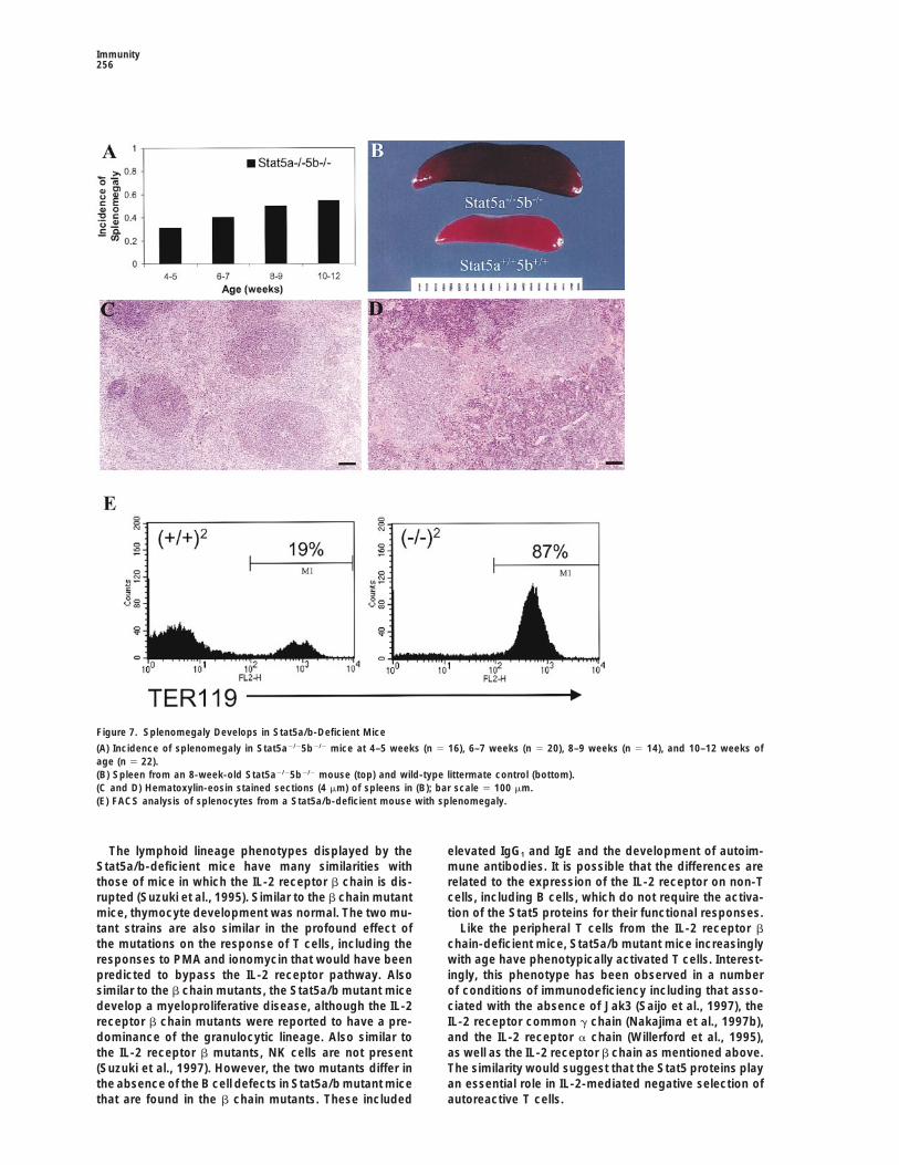
Splenomegaly with AgeThe Stat5a/b mutant mice, but not the Stat5a or Stat5b Discussionmutant mice, were also observed to develop spleno-megaly with age. As indicated in Figure 7A, approxi- The results demonstrate an unexpected and remarkably
specific requirement for the Stat5 proteins in cell cyclemately one fourth of the mice had spleens that were4- to 10-fold larger than normal at 4–5 weeks of age and progression of peripheral T cells in response to T cell
receptor ligation and IL-2 receptor activation. Moreover,this increased to approximately half of the mice at 10–12weeks of age. Histologically, the structure of the en- the lack of any phenotype in the Stat5a or Stat5b mutant
T cells demonstrate that either protein can function re-larged spleens was detectably altered as illustrated bya comparison of a spleen from a wild-type animal (C) dundantly in supporting peripheral T cell proliferation.
A role for the Stat5 proteins in T cell proliferation wasand a spleen from a Stat5a/b mutant animal with spleno-megaly (D). The altered size and morphology was char- unexpected based on the existing literature. In particu-
lar, the activation of Stat5 by the IL-2 receptor wasacterized as a hyperplasia of the red pulp with extensiveextramedullary hematopoiesis. This increase included shown to require the carboxy-terminal sequences of the
b chain, a region that is not required for a proliferativethe granulocytic and megakaryocytic lineages, but theerythroid elements were particularly numerous. To fur- signal in a myeloid cell line (Fujii et al., 1995). More
recently, it was concluded that Stat5 was involved inther examine the lineages of cells contributing to thesplenomegaly, FACS analysis and colony assays were IL-2-mediated protection from apoptosis but was not
involved in cell cycle progression (Zamorano et al.,done. Remarkably, the expanded cell population lackedany of the markers examined including T cell lineage 1998). However, both studies involved reconstitution of
the IL-2 receptor in immortalized myeloid cell lines and,markers (CD3, CD4, CD8, and Thy1) or B cell lineagemarkers (B220, IgM, and IgD). Myeloid lineage markers as previously demonstrated by the phenotype of Stat5-
deficient mice (Teglund et al., 1998), myeloid cells do(Gr1, Nk1, pan Nk marker, CD11b, and CD34) were alsonot increased, but there was a dramatic increase in not require Stat5 for proliferation.

T Cell Proliferation Requires Active Stat5255
the phenotype was due to secondary effects of the ge-netic manipulations. That these possibilities are not rele-vant is demonstrated by the ability to rescue peripheralT cell function by introducing Stat5a with retroviral con-structs. As illustrated in the studies presented here, res-cue of peripheral T cell proliferation occurred with thewild-type protein but not with a mutant that lacks thecritical tyrosine for phosphorylation and dimer forma-tion. This approach is currently being used to explorethe role of dimer-dimer formation or serine phosphoryla-tion for Stat5 function in T cells.
A previous study (Nakajima et al., 1997a) concludedthat Stat5a played a minor role in IL-2-mediated IL-2receptor a chain induction and that this function indi-rectly affected IL-2-induced proliferation. More recently,the same group described relatively minor alterationsin NK cell functions in Stat5b-deficient mice (Imada etal, 1998). Our results and conclusions are significantlydifferent from those reported. Specifically, we have notfound any evidence for a unique role of Stat5a relativeto Stat5b in peripheral T cell function, including eitherIL-2 regulation of CD25 or NK cell function. The reasonfor this difference is not known but may be due to apartial effect of the disruption of the Stat5a gene on thelevel of expression of the adjacent Stat5b gene in theNakajima et al. (1997a) studies. Indeed, in the Nakajimaet al. (1997a) studies there was a decrease of Stat5blevels in some mice; however, the authors concludedthat this did not correlate with phenotype. This criticalissue was not addressed in the studies of Stat5b-defi-cient mice. Irrespective, the studies with our inde-Figure 6. Lack of Induction of Expression of Cell Cycle Relatedpendently derived mutant strains demonstrate that theGenes in T Cells from Stat5a/b-Deficient Miceconclusion that Stat5a and Stat5b have unique, nonre-(A) Splenic lymphocytes were obtained from control or Stat5a/b-dundant functions in T cells is incorrect.deficient mice and were cultured in media, media containing anti-
The most significant difference between the NakajimaCD3 (2.5 mg/ml), media containing IL-2 (100 units/ml), or both anti-CD3 and IL-2 as indicated. Following 18 hr of culture, the cells were et al. (1997a) studies and our studies is the conclusioncollected and the levels of the various proteins assessed by Western that the effects of Stat5, either individually or redun-blotting. (B) The samples were treated as in (A) but the splenocytes dantly, on IL-2 receptor a chain expression can explainwere obtained from Stat5a- or Stat5b-deficient mice.
the decreased proliferation of peripheral T cells. As dem-onstrated here, the profound inability of peripheral Tcells from Stat5a/b-deficient mice to proliferate cannot
A potential role for Stat5 activation by the IL-2 receptor be rescued by concentrations of IL-2 that would bypasshas also been examined by deriving transgenic lines of the requirement for a chain expression. Whether themice expressing mutant receptors on the IL-2 receptor decreased proliferation reported by Nakajima et al.b chain-deficient background (Fujii et al., 1998). Trans- (1997a) is due to a decrease in a chain expression or isgenic lines lacking the membrane distal region of the due to a decrease in the levels of Stat5 to a point thatreceptor, which has been implicated in Stat5 activation it becomes limiting is an unanswered question.as well as the activation of other signaling pathways, Recent studies have also examined the role of Stat3have a subtle phenotype that includes a lack of NK cells, activation in T cell functions through the generation ofgd T cells, and a reduced proliferation at low concentra- mice that lack Stat3 in the lymphoid lineages (Takedations of IL-2. Based on the phenotype of the Stat5a/b- et al., 1998). Consistent with the results presented, wedeficient mice, it must be concluded that some degree have observed little, if any, activation of Stat3 in re-of activation of Stat5 proteins was occurring in the mice sponse to IL-2 although Stat3 is strongly activated byexpressing the truncated receptor either through the IL-6. It should also be noted that the levels of Stat3mutated b chain or through another receptor-associated are only slightly reduced in Stat5a/Stat5b-deficient miceprotein. In this regard, it should be mentioned that the (Teglund et al., 1998). The low proliferative response oflack of Stat5 activation in Jak3-deficient T cells pre- peripheral T cells to IL-6, in combination with anti-CD3,cludes a contribution to Stat5 activation by the T cell has precluded us from determining whether this re-receptor complex (our unpublished data). sponse is also affected in Stat5a/Stat5b-deficient mice.
Because of the unexpected nature of the defect, we Irrespective, Takeda et al. (1998) concluded that Stat3considered the possibility that the absence of one or was only required for prevention of apoptosis throughboth Stat5 proteins might influence differentiation rather a Bcl-2 independent pathway. Therefore, it is likely thatthan peripheral T cell function directly. Even more indi- Stat3 functions are quite distinct from those of the Stat5
proteins in peripheral T cells.rectly, as with any gene disruption, it was possible that
Immunity256
Figure 7. Splenomegaly Develops in Stat5a/b-Deficient Mice
(A) Incidence of splenomegaly in Stat5a2/25b2/2 mice at 4–5 weeks (n 5 16), 6–7 weeks (n 5 20), 8–9 weeks (n 5 14), and 10–12 weeks ofage (n 5 22).(B) Spleen from an 8-week-old Stat5a2/25b2/2 mouse (top) and wild-type littermate control (bottom).(C and D) Hematoxylin-eosin stained sections (4 mm) of spleens in (B); bar scale 5 100 mm.(E) FACS analysis of splenocytes from a Stat5a/b-deficient mouse with splenomegaly.
The lymphoid lineage phenotypes displayed by the elevated IgG1 and IgE and the development of autoim-mune antibodies. It is possible that the differences areStat5a/b-deficient mice have many similarities with
those of mice in which the IL-2 receptor b chain is dis- related to the expression of the IL-2 receptor on non-Tcells, including B cells, which do not require the activa-rupted (Suzuki et al., 1995). Similar to the b chain mutant
mice, thymocyte development was normal. The two mu- tion of the Stat5 proteins for their functional responses.Like the peripheral T cells from the IL-2 receptor btant strains are also similar in the profound effect of
the mutations on the response of T cells, including the chain-deficient mice, Stat5a/b mutant mice increasinglywith age have phenotypically activated T cells. Interest-responses to PMA and ionomycin that would have been
predicted to bypass the IL-2 receptor pathway. Also ingly, this phenotype has been observed in a numberof conditions of immunodeficiency including that asso-similar to the b chain mutants, the Stat5a/b mutant mice
develop a myeloproliferative disease, although the IL-2 ciated with the absence of Jak3 (Saijo et al., 1997), theIL-2 receptor common g chain (Nakajima et al., 1997b),receptor b chain mutants were reported to have a pre-
dominance of the granulocytic lineage. Also similar to and the IL-2 receptor a chain (Willerford et al., 1995),as well as the IL-2 receptor b chain as mentioned above.the IL-2 receptor b mutants, NK cells are not present
(Suzuki et al., 1997). However, the two mutants differ in The similarity would suggest that the Stat5 proteins playan essential role in IL-2-mediated negative selection ofthe absence of the B cell defects in Stat5a/b mutant mice
that are found in the b chain mutants. These included autoreactive T cells.

T Cell Proliferation Requires Active Stat5257
In Situ HybridizationOur results demonstrate that genes that have beenFor in situ hybridization, thymi were fixed with 4% paraformaldehydeimplicated in cell cycle progression are regulated by the(Sigma), and 6 mm sections were prepared from frozen tissue sam-Stat5 proteins in the response to IL-2. Previous studiesples. In situ hybridization was carried out essentially as described
demonstrated that cyclin D2 and D3, but not D1, were (Angerer and Angerer, 1992) using [g-33P]UTP-labeled antisenseinduced following T cell activation (Ajchenbaum et al., RNA probes transcribed from a plasmid containing murine cDNA
fragments of Cis (216 bp). Tissue sections were counterstained using1993). The ability of cyclosporin A to block this inductiontoluidine blue (Sigma).suggested a role for IL-2 signaling. The possiblity that
cyclin D2 and/or D3 are regulated in T cells by the Stat5Protein Analysisproteins is supported by the observation that prolactin,Harvested cells were washed twice with PBS and lysed in Tween-which strongly induces Stat5 activation, induces cyclin20 buffer containing 50 mM Hepes buffer (pH 7.5), 0.1% Tween-20,D2 and D3 expression in the rat pre-T-lymphoma cell150 mM NaCl, 1 mM EDTA, 20 mM b-glycerophosphate, 0.1 mM
line, Nb2 (Hosokawa et al., 1994). Moreover, the promot- sodium orthovanadate, 1 mM sodium fluoride, 10 mg/ml each aproti-ers for both cyclin D2 and D3 contain Stat5 binding sites nin and leupeptin (both from Sigma), and 1 mM PMSF. After brief(our unpublished data). Therefore, it will be important sonication, lysates were cleared of nuclear debris by centrifugation
at 14,000 g for 15 min. Protein concentrations were determinedto further determine whether D2 and D3 are immediateusing the BCA-kit as recommended by the manufacturer (Pierce).early genes in T cells and therefore potentially directlyTo assess expression levels of cyclin D2 and cyclin D3, 200 mgregulated by the Stat5 proteins.total protein/sample was electrophoretically resolved on a 12.5%
The results also suggest that the Stat5 proteins may polyacrylamide gel containing SDS and transferred onto NitroPurebe involved in the regulation of the Cdk6 gene. Previous membranes (MicronSeparationsInc.). Membranes were either probedstudies have shown that Cdk6 is present as the major with a rat monoclonal antibody directed against cyclin D2 (34B1–3)
or directed against cyclin D3 (18B6–10) followed by rabbit anti-ratcyclin D2 and D3 associated kinase in T lymphocytesIgG (Vallance et al., 1994). Sites of antibody binding were detectedand that its increased accumulation is one of the earliestusing protein A-conjugated horse-radish peroxidase (EY Labora-detectable events after mitogen stimulation (Lucas ettories) with the ECL chemiluminescent detection kit (Amersham). To
al., 1995). However, the observation that the increased assess expression levels of p27Kip1, cyclin E, cyclin A, and the cyclinCdk6 expression was not inhibited by cyclosporin A or dependent kinases cdk4, cdk6, and cdk2, 100 mg total protein/FK506 suggested that the increase was independent of sample was electrophoretically resolved on a 12% polyacrylamide
gel containing SDS and transferred onto NitroPure membranes. TheIL-2 signaling. In contrast, the regulation of p27 levelsmembranes were either probed with affinity-purified antiserum di-in lymphocytes from Stat5a/b mutant mice was normal.rected against the full-length mouse p27 (Kato et al., 1994) or affinity-Regulation of p27 levels occurs both at the protein levelpurified antibodies against cyclins or cyclin-dependent kinases
and at the transcriptional level. In both cases, the ability (Santa Cruz). Sites of antibody binding were detected using proteinof cyclosporin A to block the downregulation has impli- A-conjugated horse-radish peroxidase as described above.cated a role for IL-2 signaling events. Our data wouldsuggest that if IL-2 signaling is involved in p27 regula- Flow Cytometrytion, it involves a Stat5 independent pathway. Thymus and spleen obtained from individual mice were made into
In summary, the derivation of Stat5a/b-deficient mice single cell suspensions in PBS supplemented with 1% bovine serumalbumin (PBS/BSA). Nonspecific staining was minimized by blockinghas revealed an unexpected but essential role for Stat5with 10% normal mouse serum in PBS/BSA. The cells were thenin the regulation of peripheral T cell activation by IL-2.stained with cocktails of MAb’s in PBS/BSA conjugated to fluores-With this information, efforts can be now focused oncein isothyocyanate (FITC), phycoerythrin (PE), or biotin for 30 min
the mechanisms by which Stat5 regulates cell cycle at 48C. The cells were washed with PBS/BSA, and the biotinylatedprogression of activated T cells. The results also suggest antibodies were developed with streptavidin conjugated to red 670that drugs that target Stat5 function may have utility in (Life Technologies, Gaithersburg, MD). All MAb’s with the exceptions
of anti-CD25 and anti-IgM were purchased from Pharmingen (Sancontrolling peripheral T cell expansion and function.Diego, CA) and can be referenced in their current catalog. The FITC-anti-CD25 MAb clone PC61 was purchased from Accurate Chemicaland Scientific Corp. (Westbury, NY) and the anti-mouse IgM fromExperimental ProceduresSouthern Biotechnology Associates, Inc. (Birmingham, AL). The cellsurface expression of the markers was analyzed in a Becton-Dickin-T Cell Isolation, Culture, and Activation Conditionsson FACScan in two- or three color mode using CellQuest software.Splenic T cells were isolated from 8- to 12-week-old wild-type or
Stat5a/b-deficient mice without splenomegaly. In brief, fresh iso-lated organs were meshed and passed through a cell strainer (70 Proliferation Responses
Single cell suspensions of spleen cells in SMEM (Life Technologies,mm; Becton Dickinson) to separate fibrous tissue. The splenic cellsolution was kept in T cell culture media. The media consisted of Gaithersburg, MD) supplemented with 10% fetal bovine serum (At-
lanta Biologicals, Norcross, GA), L-glutamine, penicillin, streptomy-RPMI 1640, 10% fetal bovine serum (HyghClone), 10 mM Hepes (pH7.0), 2 mM L-glutamine, (13) nonessential amino acids, 1 mM sodium cin, gentamycin (all from Life Technologies), sodium bicarbonate
(Sigma, St. Louis, MO), essential and nonessential amino acids (Lifepyruvate, and 50 mM 2-mercaptoethanol. The T cells were centri-fuged and a red blood cell lysis was performed for 5 min using a Technologies), and 2-mercaptoethanol (Sigma) were plated in 96-
well round-bottom plates (Corning, Corning, NY) at a density of 1 3lysis buffer (pH 7.3) containing 150 mM NH4Cl, 1 mM KHCO3, and0.1 mM Na2EDTA at 106 cells/ml. Lysis was stopped by adding an 105 per well in 100 ml. Stimuli were added as indicated at a range
of concentrations to assess dose dependency. The stimuli wereexcess (43) of T cell media and centrifugation, followed by threesubsequent wash and centrifugation steps with T cell culture media. anti-CD3 (145.2C11), anti-CD28 (37.51) (both antibodies from Phar-
mingen, San Diego, CA), recombinant human IL-2 (Chiron, Em-The splenic cells were resuspended at 106 cells/ml culture media.T cell receptor activation after CD3 ligation was induced by addition eryville, CA), and recombinant murine IL-4 (R&D Systems, Minneap-
olis, MN). Proliferation was assessed by measuring radioactivityof the monoclonal antibody 145.2C11 (Pharmingen) into the culturemedia (1 mg/ml culture media) if not detailed differently. Recombi- using a Matrix 9600 beta counter (Packard, Meriden CT) 18 hr after
adding 1 mCi/well 3H-thymidine (Amersham Life Sciences, Arlingtonnant human IL-2 (Boehringer Mannheim) was added at 100 units/ml medium if not stated otherwise. Heights, IL).

Immunity258
Retroviral Constructs Hou, J., Schindler, U., Henzel, W.J., Wong, S.C., and McKnight,S.L. (1995). Identification and purification of human Stat proteinsA bicistronic retroviral vector, using the murine stem cell virus
(MSCV) long terminal repeats, was utilized for expression of Stat5a activated in response to interleukin-2. Immunity 2, 321–329.or Stat5a(Y693F). The methods utilized for preparation of the con- Ihle, J.N. (1995). Cytokine receptor signalling. Nature 377, 591–594.structs and methods for obtaining high-titered viral stocks have Imada, K., Bloom, E.T., Nakajima, H., Horvath-Arcidiacono, J.A.,been described in detail (Persons et al., 1997, 1998). Splenic cells Udy, G.B., Davey, H.W., Leonard, W.J. (1998). Stat5b is essentialwere isolated as above and cocultured on irradiated (1500 rads) for natural killer cell-mediated proliferation and cytolytic activity. J.ecotropic producer cell lines for 48 hr in the presence of 3 mg/ Exp. Med. 188, 2067–2074.ml anti-CD3, 500 units/ml of IL-2, and 6 mg/ml polybrene (Sigma).
Kanegane, H., Kasahara, Y., Niida, Y., Yachie, A., Sughii, S., Takatsu,Semiconfluent cultures were divided 1:4 and subcultured with 500K., Taniguchi, N., and Miyawaki, T. (1996). Expression of L-selectinunits/ml of IL-2. For proliferation assays, cells were washed three(CD62L) discriminates Th1- and Th2-like cytokine-producing mem-times in T cell media and cultured at 2 3 105 per well in 100 ml withory CD41 T cells. Immunology 87, 186–190.media alone or anti-CD3 and IL-2 as above. After 24 hr, 3H-thymidineKato, J.Y., Matsuoka, M., Strom, D.K., and Sherr, C.J. (1994). Regula-was added for an additional 24 hr and the incorporation determinedtion of cyclin D-dependent kinase 4 (cdk4) by cdk4-activating kinase.as above.Mol. Cell Biol. 14, 2713–2721.
Lin, J.-X., Migone, T.-S., Tsang, M., Friedmann, M., Weatherbee,AcknowledgmentsJ.A., Zhou, L., Yamauchi, A., Bloom, E.T., Mietz, J., John, S., et al.(1995). The role of shared receptor motifs and common stat proteinsThe authors would like to thank Lynda Snyder, Kristen Rothammer,in the generation of cytokine pleiotropy and redundancy by IL-2,and Suzette Wingo for their excellent technical assistance. WeIL-4, IL-7, IL-13, and IL-15. Immunity 2, 331–339.would also like to thank Charles Sherr for providing monoclonal
antibodies against cyclin D2 and D3, antisera against p27 and Cdk2 Lucas, J.J., Szepesi, A., Modiano, J.F., Domenico, J., and Gelfand,and baculovirus produced p27, the cyclins D2, D3, E, and A, and E.W. (1995). Regulation of synthesis and activity of the PLSTIREthe kinases Cdk2, Cdk4, and Cdk6 that were used for controls. We protein (cyclin-dependent kinase 6 (cdk6)), a major cyclin D-associ-would also like to thank Richard Ashmun, Richard Cross, Sam Lucas, ated cdk4 homologue in normal human T lymphocytes. J. Immunol.Mahnaz Paktinat, and Ed Wingfield for FACS analysis. V. S. was a 154, 6275–6284.fellow supported by the Austrian Fond zur Forderung der Wissen- Nakajima, H., Liu, X.W., Wynshaw-Boris, A., Rosenthal, L.A., Imada,schaftlichen Forschung (FWF). This work was supported in part by K., Finbloom, D.S., Hennighausen, L., and Leonard, W.J. (1997a).Cancer Center CORE Grant CA21765, by grant RO1 DK42932 to An indirect effect of Stat5a in IL-2-induced proliferation: a criticalJ. N. I., by grant P01 HL53749, and by the American Lebanese Syrian role for Stat5a in IL-2-mediated IL-2 receptor alpha chain induction.Associated Charities (ALSAC). Immunity 7, 691–701.
Nakajima, H., Shores, E.W., Noguchi, M., and Leonard, W.J. (1997b).Received December 8, 1998; revised December 16, 1998. The common cytokine receptor gamma chain plays an essential
role in regulating lymphoid homeostasis. J. Exp. Med. 185, 189–195.References Nourse, J., Firpo, E., Flanagan, W.M., Coats, S., Polyak, K., Lee,
M.H., Massague, J., Crabtree, G.R., and Roberts, J.M. (1994). In-Ajchenbaum, F., Ando, K., DeCaprio, J.A., and Griffin, J.D. (1993). terleukin-2-mediated elimination of the p27Kip1 cyclin-dependentIndependent regulation of human D-type cyclin gene expression kinase inhibitor prevented by rapamycin. Nature 372, 570–573.during G1 phase in primary human T lymphocytes. J. Biol. Chem.
Ormandy, C.J., Camus, A., Barra, J., Damotte, D., Lucas, B., Buteau,268, 4113–4119.H., Edery, M., Brousse, N., Babinet, C., Binart, N., et al. (1997). Null
Angerer, L.M. and Angerer, R.C. (1992). In situ hybridization to cellu- mutation of the prolactin receptor gene produces multiple reproduc-lar RNA with radiolabelled RNA probes. In In Situ Hybridization: A tive defects in the mouse. Genes Dev. 11, 167–178.Practical Approach. D.G. Wilkinson, ed. (Oxford: Oxford University
Persons, D.A., Allay, J.A., Allay, E.R., Smeyne, R.J., Ashmun, R.A.,Press), pp. 15–32.
Sorrentino, B.P., and Nienhuis, A.W. (1997). Retroviral-mediatedDarnell, J.E., Jr. (1997). STATs and gene regulation. Science 277, transfer of the green fluorescent protein gene into murine hemato-1630–1635. poietic cells facilitates scoring and selection of transduced progeni-Darnell, J.E., Jr., Kerr, I.M., and Stark, G.R. (1994). Jak-STAT path- tors in vitro and identification of genetically modified cells in vivo.ways and transcriptional activation in response to IFNs and other Blood 90, 1777–1786.extracellular signaling proteins. Science 264, 1415–1421. Persons, D.A., Mehaffey, M.G., Kaleko, M., Nienhuis, A.W., andFujii, H., Nakagawa, Y., Schindler, U., Kawahara, A., Mori, H., Gouil- Vanin, E.F. (1998). An improved method for generating retroviral
producer clones for vectors lacking a selectable marker gene. Bloodleux, F., Groner, B., Ihle, J.N., Minami, Y., Miyazaki, T., et al. (1995).Cells, Mol. Dis. 24, 167–182.Activation of Stat5 by interleukin 2 requires a carboxyl-terminal re-
gion of the interleukin 2 receptor b chain but is not essential for Refaeli, Y., Parijs, L., London, C.A., Tschopp, J., and Abbas, A.K.the proliferative signal transmission. Proc. Natl. Acad. Sci. USA 92, (1998). Biochemical mechanisms of IL-2-regulated Fas-mediated T5482–5486. cell apoptosis. Immunity 8, 615–623.Fujii, H., Ogasawara, K., Otsuka, H., Suzuki, M., Yamamura, K.-I., Saijo, K., Park, S.Y., Ishida, Y., Arase, H., and Saito, T. (1997). CrucialYokochi, T., Miyawaki, T., Suzuki, H., Mak, T.W., Taki, S., et al. (1998). role of Jak3 in negative selection of self-reactive T cells. J. Exp.Functional dissection of the cytoplasmic subregions of the IL-2 re- Med. 185, 351–356.ceptor bc chain in primary lymphocyte populations. EMBO J. 17, Suzuki, H., Kundig, T.M., Furlonger, C., Wakeham, A., Timms, E.,6551–6557. Matsuyama, T., Schmits, R., Simard, J.J.L., Ohashi, P.S., Griesser,Gaffen, S.L., Lai, S.Y., Xu, W., Gouilleux, F., Groner, B., Goldsmith, H., et al. (1995). Deregulated T cell activation and autoimmunity inM.A., and Greene, W.C. (1995). Signaling through the interleukin 2 mice lacking interleukin-2 receptor b. Science 268, 1472–1476.receptor b chain activates a STAT-5-like DNA-binding activity. Proc. Suzuki, H., Duncan, G.S., Takimoto, H., and Mak, T.W. (1997). Abnor-Natl. Acad. Sci. USA 92, 7192–7196. mal development of intestinal intraepithelial lymphocytes and pe-Gilmour, K.C., Pine, R., and Reich, N.C. (1995). Interleukin 2 activates ripheral natural killer cells in mice lacking the IL-2 receptor betaSTAT5 transcription factor (mammary gland factor) and specific chain. J. Exp. Med. 185, 499–505.gene expression in T lymphocytes. Proc. Natl Acad. Sci. USA 92, Takeda, K., Kaisho, T., Yoshida, N., Takeda, J., Kishimoto, T., and10772–10776. Akira, S. (1998). Stat3 activation is responsible for IL-6-DependentHosokawa, Y., Onga, T., and Nakashima, K. (1994). Induction of T cell proliferation through preventing apoptosis: Generation andD2 and D3 cyclin-encoding genes during promotion of the G1/S characterization of T cell-specific Stat3-deficient mice. J. Immunol.
161, 4652–4660.transition by prolactin in rat Nb2 cells. Gene 147, 249–252.
T Cell Proliferation Requires Active Stat5259
Teglund, S., McKay, C., Schuetz, E., van Deursen, J., Stravopodis,D., Wang, D., Brown, M., Bodner, S., Grosveld, G., and Ihle, J.N.(1998). Stat5a and Stat5b proteins have essential and non-essential,or redundant, roles in cytokine responses. Cell 93, 841–850.
Vallance, S.J., Lee, H.M., Roussel, M.F., Shurtleff, S.A., Kato, J.Y.,Strom, D.K., and Sherr, C.J. (1994). Monoclonal antibodies to mam-malian D-type G1 cyclins. Hybridoma 13, 37–44.
Wakao, H., Harada, N., Kitamura, T., Mui, A.L.F., and Miyajima, A.(1995). Interleukin 2 and erthropoietin activate STAT5/MGF via dis-tinct pathways. EMBO J. 14, 2527–2535.
Willerford, D.M., Chen, J., Ferry, J.A., Davidson, L., Ma, A., and Alt,F.W. (1995). Interleukin-2 receptor a chain regulates the size andcontent of the peripheral lymphoid compartment. Immunity 3,521–530.
Yoshimura, A., Ohkubo, T., Kiguchi, T., Jenkins, N.A., Gilbert, D.J.,Copeland, N.G., Hara, T., and Miyajima, A. (1995). A novel cytokine-inducible gene CIS encodes an SH2-containing protein that binds totyrosine-phosphorylated interleukin-3 and erythropoietin receptors.EMBO J. 14, 2816–2826.
Zamorano, J., Wang, H.Y., Wang, R., Shi, Y., Longmore, G.D., andKeegan, A.D. (1998). Regulation of cell growth by IL-2: role of STAT5in protection from apoptosis but not in cell cycle progression. J.Immunol. 160, 3502–3512.
Zhou, Y., Xu, B.C., Maheshwari, H.G., He. L., Reed. M., Lozykowski,M., Okada, S., Cataldo, L., Coschigamo, K., Wagner, T.E., et al.(1997). A mammalian model for Laron syndrome produced by tar-geted disruption of the mouse growth hormone receptor/bindingprotein gene (the Laron mouse). Proc. Natl. Acad. Sci. USA 94,13215–13220.