Stat5 tetramer formation is associated with leukemogenesis.
13
A R T I C L E Stat5 tetramer formation is associated with leukemogenesis Richard Moriggl, 1, * Veronika Sexl, 2 Lukas Kenner, 1 Christopher Duntsch, 3 Katharina Stangl, 1 Sebastien Gingras, 3 Angelika Hoffmeyer, 3 Anton Bauer, 4 Roland Piekorz, 5 Demin Wang, 6 Kevin D. Bunting, 7 Erwin F. Wagner, 1 Karoline Sonneck, 8 Peter Valent, 8 James N. Ihle, 3 and Hartmut Beug 1 1 Institute of Molecular Pathology, Dr. Bohr-Gasse 7, A-1030 Vienna, Austria 2 Institute of Pharmacology, Medical University of Vienna, A-1090 Vienna, Austria 3 Department of Biochemistry, St. Jude Children’s Research Institute, Howard Hughes Medical Institute, Memphis, Tennessee 38105 4 IMC Krems, University of Applied Sciences, A-3500 Krems, Austria 5 Institute of Biochemistry and Molecular Biology II, Heinrich-Heine-University, D-40225 Du ¨ sseldorf, Germany 6 Blood Research Institute, The Blood Center of Southeastern Wisconsin and Department of Microbiology and Molecular Genetics, Medical College of Wisconsin, Milwaukee, Wisconsin 53226 7 Division of Hematology/Oncology, Center for Stem Cell and Regenerative Medicine, Case Western Reserve University, Cleveland, Ohio 44106-7284 8 Department of Internal Medicine I, Division of Hematology and Hemostaseology, Medical University of Vienna, A-1097 Vienna, Austria *Correspondence: [email protected] Summary Activation of Stat5 is frequently found in leukemias. To study the mechanism and role of Stat5 activation, we introduced a constitutively activated Stat5a mutant, cS5 F , into murine bone marrow (BM) cells. BM transplantation with cS5 F -transfected cells caused development of multilineage leukemias in lethally irradiated wild-type or nonirradiated Rag2 / mice. The leukemic cells showed strongly enhanced levels of cS5 F tetramers but unchanged cS5 F dimer levels in a DNA binding assay. Moreover, Stat5a mutants engineered to form only dimers, but not tetramers, failed to induce leukemias. In addition, Stat5 tetramers were found to accumulate in excess compared to dimers in various human leukemias. These data suggest that Stat5 tetramers are associated with leukemogenesis. Introduction Most signal transduction events are controlled by gene ex- pression or require kinase cascades and adaptor molecules to Gene transcription through the Jak-Stat pathway mediates cyto- phosphorylate and activate nuclear transcription factors (Bri- kine and growth factor functions, with important consequences vanlou and Darnell, 2002). In contrast, Stat proteins are highly for cellular proliferation, differentiation, and survival (Levy and and constitutively expressed, facilitating their rapid activation. Gilliland, 2000; O’Shea et al., 2002). Persistent Stat1/3/5 activa- Cancer cells often become cytokine- and growth factor-inde- tion is frequently found in cancer. Stat1 activation is a negative pendent through autocrine factor synthesis or through persis- regulator of proliferation, whereas Stat3/5 activation is associ- tent activation of key signaling molecules, like deregulated tyro- sine kinase or tyrosine phosphatases (Brivanlou and Darnell, ated with cancer progression, defining Stat3/5 as a molecular target for therapeutic intervention (Bromberg, 2002; Buettner et 2002; O’Shea et al., 2002). This can cause persistent Stat activa- tion. A well-known example of Stat5 activation is signaling al., 2002). Stat5a and Stat5b double-deficient knockout mice (Stat5 / ) have multiple hematopoietic defects. Stat5 / cells through the Bcr-Abl tyrosine kinase in chronic myelogenous leukemia (CML; Buettner et al., 2002). Other genetic changes display impaired cytokine-mediated proliferation and survival of hematopoietic progenitors, and T, NK, erythroid, and myeloid leading to Stat5 activation in leukemias include Flt-3 or c-Kit mutations, which recruit Stat5 to the respective receptors and cells (Bunting et al., 2002; Kieslinger et al., 2000; Moriggl et al., 1999a; Teglund et al., 1998). The responsible Stat5-dependent thereby lead to persistent Stat5 activation (Mizuki et al., 2003; Taketani et al., 2004). In addition, Stat5 activation is found in cytokine pathways are pleiotropic (O’Shea et al., 2002). SIGNIFICANCE Stats, particularly Stat3 and Stat5, are gaining increasing attention as essential players in the formation of leukemias and solid tumors. Stats are rarely mutated but frequently overexpressed and hyperactivated, often by alterations in upstream signaling pathways. We provide evidence that a Stat5a mutant, which forms enhanced levels of stable tetramers but otherwise behaves like wt Stat5, caused multilineage leukemias. Furthermore, our studies show that tetramerization is essential for leukemia development in mice. In addition, human leukemic samples displayed strong Stat5 tetramer formation. Our results suggest that disruption of Stat5 tetramer formation could represent a therapeutic concept to treat leukemia. CANCER CELL : JANUARY 2005 · VOL. 7 · COPYRIGHT © 2005 ELSEVIER INC. DOI 10.1016/j.ccr.2004.12.010 87
-
Upload
hybridbioscience -
Category
Documents
-
view
1 -
download
0
Transcript of Stat5 tetramer formation is associated with leukemogenesis.

A R T I C L E
Stat5 tetramer formation is associated with leukemogenesis
Richard Moriggl,1,* Veronika Sexl,2 Lukas Kenner,1 Christopher Duntsch,3 Katharina Stangl,1
Sebastien Gingras,3 Angelika Hoffmeyer,3 Anton Bauer,4 Roland Piekorz,5
Demin Wang,6 Kevin D. Bunting,7 Erwin F. Wagner,1 Karoline Sonneck,8
Peter Valent,8 James N. Ihle,3 and Hartmut Beug1
1Institute of Molecular Pathology, Dr. Bohr-Gasse 7, A-1030 Vienna, Austria2 Institute of Pharmacology, Medical University of Vienna, A-1090 Vienna, Austria3 Department of Biochemistry, St. Jude Children’s Research Institute, Howard Hughes Medical Institute, Memphis,
Tennessee 381054 IMC Krems, University of Applied Sciences, A-3500 Krems, Austria5 Institute of Biochemistry and Molecular Biology II, Heinrich-Heine-University, D-40225 Dusseldorf, Germany6 Blood Research Institute, The Blood Center of Southeastern Wisconsin and Department of Microbiology and Molecular
Genetics, Medical College of Wisconsin, Milwaukee, Wisconsin 532267 Division of Hematology/Oncology, Center for Stem Cell and Regenerative Medicine, Case Western Reserve University,
Cleveland, Ohio 44106-72848 Department of Internal Medicine I, Division of Hematology and Hemostaseology, Medical University of Vienna, A-1097
Vienna, Austria*Correspondence: [email protected]
Summary
Activation of Stat5 is frequently found in leukemias. To study the mechanism and role of Stat5 activation, we introduceda constitutively activated Stat5a mutant, cS5F, into murine bone marrow (BM) cells. BM transplantation with cS5F-transfectedcells caused development of multilineage leukemias in lethally irradiated wild-type or nonirradiated Rag2�/� mice. Theleukemic cells showed strongly enhanced levels of cS5F tetramers but unchanged cS5F dimer levels in a DNA bindingassay. Moreover, Stat5a mutants engineered to form only dimers, but not tetramers, failed to induce leukemias. In addition,Stat5 tetramers were found to accumulate in excess compared to dimers in various human leukemias. These data suggestthat Stat5 tetramers are associated with leukemogenesis.
Introduction Most signal transduction events are controlled by gene ex-pression or require kinase cascades and adaptor molecules to
Gene transcription through the Jak-Stat pathway mediates cyto- phosphorylate and activate nuclear transcription factors (Bri-kine and growth factor functions, with important consequences vanlou and Darnell, 2002). In contrast, Stat proteins are highlyfor cellular proliferation, differentiation, and survival (Levy and and constitutively expressed, facilitating their rapid activation.Gilliland, 2000; O’Shea et al., 2002). Persistent Stat1/3/5 activa- Cancer cells often become cytokine- and growth factor-inde-tion is frequently found in cancer. Stat1 activation is a negative pendent through autocrine factor synthesis or through persis-regulator of proliferation, whereas Stat3/5 activation is associ- tent activation of key signaling molecules, like deregulated tyro-
sine kinase or tyrosine phosphatases (Brivanlou and Darnell,ated with cancer progression, defining Stat3/5 as a moleculartarget for therapeutic intervention (Bromberg, 2002; Buettner et 2002; O’Shea et al., 2002). This can cause persistent Stat activa-
tion. A well-known example of Stat5 activation is signalingal., 2002). Stat5a and Stat5b double-deficient knockout mice(Stat5�/�) have multiple hematopoietic defects. Stat5�/� cells through the Bcr-Abl tyrosine kinase in chronic myelogenous
leukemia (CML; Buettner et al., 2002). Other genetic changesdisplay impaired cytokine-mediated proliferation and survival ofhematopoietic progenitors, and T, NK, erythroid, and myeloid leading to Stat5 activation in leukemias include Flt-3 or c-Kit
mutations, which recruit Stat5 to the respective receptors andcells (Bunting et al., 2002; Kieslinger et al., 2000; Moriggl et al.,1999a; Teglund et al., 1998). The responsible Stat5-dependent thereby lead to persistent Stat5 activation (Mizuki et al., 2003;
Taketani et al., 2004). In addition, Stat5 activation is found incytokine pathways are pleiotropic (O’Shea et al., 2002).
S I G N I F I C A N C E
Stats, particularly Stat3 and Stat5, are gaining increasing attention as essential players in the formation of leukemias and solid tumors.Stats are rarely mutated but frequently overexpressed and hyperactivated, often by alterations in upstream signaling pathways. Weprovide evidence that a Stat5a mutant, which forms enhanced levels of stable tetramers but otherwise behaves like wt Stat5, causedmultilineage leukemias. Furthermore, our studies show that tetramerization is essential for leukemia development in mice. In addition,human leukemic samples displayed strong Stat5 tetramer formation. Our results suggest that disruption of Stat5 tetramer formationcould represent a therapeutic concept to treat leukemia.
CANCER CELL : JANUARY 2005 · VOL. 7 · COPYRIGHT © 2005 ELSEVIER INC. DOI 10.1016/j .ccr.2004.12.010 87

A R T I C L E
carcinomas of the breast, prostate, ovary, head, and neck(Bromberg, 2002; Buettner et al., 2002).
Recently, several mouse models showing transgenic Stat5overexpression have been established. Stat5 activation wasfound to be a proliferation- and/or survival-promoting eventin neoplastic cells (Iavnilovitch et al., 2002; Kelly et al., 2003;Tsuruyama et al., 2002). In other studies, Stat5 function wasanalyzed through the use of a constitutively active (ca) Stat5mutant, which carried two engineered point mutations (H299→Rand S711→F, called caStat5a1*6 and referred here to cS5RF;Onishi et al., 1998). Expression of the cS5RF mutant in variouscell lines showed its heterodimerization to endogenous Stat5(Lee et al., 2001; Nosaka et al., 1999; Onishi et al., 1998; Santoset al., 2001; Schwaller et al., 2000).
Stat5 proteins contain several domains with different func-tional properties (Figure 1A). The N-terminal tetramer formationdomain (TD) is of major interest for this study. Stat N termini haveregulatory functions such as receptor association, phosphatasebinding, and nuclear translocation. They modulate oligomeriza-tion, tyrosine dephosphorylation, and nuclear accumulation(Meyer et al., 2004; Ota et al., 2004; Vinkemeier et al., 1996,1998; Xu et al., 1996). Stat tetramerization is thought to increasespecificity among Stat family members. Stat3 tetramerizationwas shown to occur on the �2-macroglobulin promoter, wheremultiple transcription factors assemble to build up an efficienttranscription enhanceosome (Lerner et al., 2003). Similarly, tet-ramerization has been observed for Stat4 in the IFN-� transcrip-tional regulatory region (Xu et al., 1996). Tetramer formation ofStat5 was analyzed on various transcriptional regulatory regions(John et al., 1999; Kim and Leonard, 2002; Meyer et al., 1997;Soldaini et al., 2000; Verdier et al., 1998). Stat tetramers resultin more stable DNA binding complexes than dimers throughincreased contact points with DNA, which also allow bindingto weak affinity sites (Lerner et al., 2003; Soldaini et al., 2000).Stabilized tetramers increase binding site occupancy to athreshold required for transcriptional activity. The greater degreeof flexibility in DNA sequence by Stat tetramer recognition wassuggested to widen target gene spectra (John et al., 1999;Meyer et al., 1997).
We studied the role of persistent Stat5a activation using acS5F mutant, which closely mimicked wt Stat5 function. Ourdata show that enhanced Stat5 tetramers are present in humanleukemias, and that they are the relevant Stat5 DNA binding Figure 1. Biochemical and functional characterization of constitutively ac-complexes to induce leukemia. tive Stat5a mutants
A: Schematic diagram of Stat5a and mutant derivatives. Amino acid posi-Results tions of different functional domains and point mutations are indicated.
Gray circles highlight the caStat5 mutants.B: DNA binding activity (EMSA; �-casein RE), P-Y-Stat5 (WB), and subsequentCharacterization of a constitutively activetotal Stat5 analysis by Western blotting. The cS5F and cS5RF mutants bound
Stat5a mutant to DNA due to persistent tyrosine-phosphorylation in absence of cytokineThe introduction of the original cS5RF mutant into wt BM induced activation. Upper panel: 293T cells were transfected with the Stat constructs
indicated, together with the EpoR, and stimulated with Epo (50 U/ml) formyeloid hyperplasia (Schwaller et al., 2000). We failed, however,30 min or left untreated. Lower panel: Stat5�/� T cell lymphoma lines express-to complement Stat5�/� hematopoietic phenotypes with cS5RF.ing the Stat5 proteins indicated were stimulated with IL-4 (50 U/ml) for 30Thus, it remained unclear to what extent cS5RF functionally re- min or left untreated.
sembles wt Stat5. To dissect which point mutation of the origi- C: Rescue of Stat5�/� primary T cells in vitro. Splenic T cells from Stat5�/�
mice cannot proliferate in vitro in response to �-CD3 (1 �g/ml) and IL-2 (500nally described cS5RF molecule can both substitute for wt Stat5aU/ml), but they regained proliferation capacity upon retroviral rescue with(S5) function and induce hyperplasia, we constructed the re-S5, S5A, or cS5F. Proliferating T cells (Thy1.2�) become increasingly GFP�
spective single Stat5 point mutants (Figure 1A). We termed themafter 3, 7, and 10 culture days. Shown are representative examples from 3
S5R and cS5F, carrying the single point mutations H299→R or individual experiments with similar results.S711→F. The cS5F mutant converts Stat5a to a constitutivelyactive mutant, and as a control for cS5F, we replaced S711→Afor a closer structural resemblance to cS5F and termed it S5A.
88 CANCER CELL : JANUARY 2005

A R T I C L E
Two different cell systems were employed to functionally com- infiltrated the portal liver fields of diseased cS5F-transplantedmice.pare Stat5a mutants to wt Stat5a. First, respective retroviral
vectors were transiently transfected into 293T cells, together One report showed that the cS5RF mutant caused myeloidhyperproliferation in the presence of Stat5a (Schwaller et al.,with the erythropoietin receptor (EpoR). Secondly, the same
constructs were transduced into a transformed Stat5�/� cy- 2000), but when Stat5�/� BM was transduced with this mutantand transplanted, no disease occurred (R.M., unpublished data).tokine-independent T cell lymphoma line and sorted for high
GFP expression. Transfected 293T cells and mutant-expressing This raised the question of whether the cS5F mutant can causeleukemia independent of an endogenous Stat5 heterodimeriza-T cell lines (expressing the endogenous IL-4 receptor) were
stimulated with Epo or IL-4. In both cell systems, bandshift tion partner. Thus, Stat5�/� BM was transduced with cS5F andtransplanted into lethally irradiated wt mice. Although leukemiaassays and Western blotting revealed that cS5F and cS5RF were
persistently activated in absence of exogenous cytokine stimu- onset was delayed (6 months), the evolving leukemia was com-parable to that induced by transplantation of cS5F-transducedlation, in contrast to S5, S5R, and S5A (Figure 1B). DNA binding
specificity of Stat5a and mutant derivatives was verified on wt BM. These results define cS5F as oncogenic, independentof wt Stat5 as a heterodimerization partner.�-casein (binding of Stat5/6), IG� light chain-� (Stat6), and
SIEm67 binding sites (Stat1/3/4; Figure 1B and data not shown).Next, we asked whether the mutants could complement FACS surface marker analysis manifests development
of a multilineage leukemiaperipheral T cell proliferation like Stat5a in Stat5�/� T cells. Wehave shown that Stat5�/� T cells fail to proliferate upon IL-2 or Leukemic cells from cS5F-transplanted animals and empty GFP-
vector-transplanted controls were subjected to lineage markerIL-4 stimulation independent of T cell receptor activation, butproliferation can be regained upon retroviral transduction of S5 analysis by FACS, testing for GFP� cells from blood, spleen,
and BM (Figure 3). FACS was either done by testing cells forinto Stat5�/� T cells (Moriggl et al., 1999a, 1999b; Wang et al.,2000). Interestingly, S5, cS5F, and S5A were able to rescue T cell the lineage marker plus GFP, or by gating (see black histogram
plots in Figures 3A–3C) and analyzing GFP� cells for lineageproliferation, while S5R or cS5RF failed to do so (Figure 1C). Inparticular, the mutant cS5RF was unable to rescue Stat5�/� marker expression. The erythroid marker Ter119, the myeloid
markers Gr-1/Mac-1, the B and T lymphocyte markers CD19T cells, probably due to the S5R mutation (Figure 1C). The ex-pression level of the Stat5a protein variants was moderate and and Thy-1.2, and combinations of CD31, CD34, and Sca-1 with
c-Kit, typical for immature multipotent cells or HSCs, were ana-�3 enhanced in primary T cells. cS5F and Stat5a-transducedStat5�/� T cells maintained a physiological IL-2 dose response lyzed (Figure 3). The majority of GFP� BM cells were Gr-1 and
Mac-1 double positive myeloid cells, and �20% of GFP� BMfor proliferation (data not shown).cells were of B or T lymphoid origin (Figure 3B). Most notably,we obtained cS5F-leukemia-specific fractions of GFP� BM cells,Transplantation systems define cS5F
as a powerful oncogene which expressed marker combinations typical for primitivemultipotent cells, suggesting that cS5F induced the HSCs orTo analyze the leukemogenesis of the caStat5 mutants, BM cells
were infected with respective retroviral constructs, analyzed for immature multipotent progenitors to give rise to multilineageleukemia. These cS5F-GFP� BM cells from diseased mice wereinfection efficiency by FACS (�5%), and injected into lethally
irradiated wt or nonirradiated Rag2�/� mice. Mice were fully expanded in vitro and retained an immature HSC surface markerphenotype (F. Gouilleux and R.M., unpublished data). GFP�reconstituted at 4 weeks posttransplantation. Mice grafted with
cells carrying the cS5F mutation developed leukemia after 4 cells showing FACS surface markers similar to GFP� cells werenot enriched, pointing to a cell-autonomous advantage of cS5F-weeks, while no disease was seen in S5-transplanted controls.
A highly elevated WBC, blast cell morphology in the BM, spleno- transduced cells. Furthermore, the leukemia latency in nonirra-diated Rag2�/� mice or upon transplantation of Stat5�/� BMmegaly, lymphadenopathy, and massive liver infiltration mani-
fested leukemia by hematopoietic blasts. Histopathology re- (Figure 2) could indicate that cS5F also integrated into HSC/multipotent progenitors. The majority of leukemic cells in thevealed similar alterations in all cS5F-transplanted mice, but with
varying latency depending on the transplant systems. The cS5F- blood and the spleen represented T (Thy1.2), B cells (CD19),and erythroblasts (Ter119, Figures 3B and 3C). FACS analysisinduced leukemia first evolved as early as 4 weeks posttrans-
plant in lethally irradiated wt mice, which did not survive 8 weeks of leukemic mouse blood (without red blood cell lysis) revealeda high fraction of erythroid GFP�/Ter119� cells or 3 elevatedposttransplantation. In contrast, transplantation of S5, S5R, S5A,
or the GFP vector did not induce leukemia up to 10 months. GFP� platelet/megakaryocyte numbers (data not shown). Fi-nally, livers from diseased mice also contained abnormally highcS5F transplantation induced leukemias within one month in
all (n � 8 mice) lethally irradiated (1 Gy) wt mice, but not in levels of multiple hematopoietic cell types, as verified by FACSnonirradiated Rag2�/� mice (6 month latency, �70% mice af- analysis (data not shown). We conclude that cS5F-transplantedfected). Sublethal � irradiation (0.7 Gy) of wt mice again pro- mice develop multilineage leukemia originating from primitivelonged cS5F disease onset in all mice analyzed (n � 6 mice, stem cell-like progenitors.data not shown). A second cS5F mutant (including a C-terminalFLAG, termed cS5F-FLAG) was similarly leukemogenic when Persistent Stat5 activation causes enhanced Stat5
tetramer formationcompared to cS5F. Lymphoid and myeloid blasts densely infil-trated the BM of diseased mice (Figure 2A). Moreover, mice All Stat5a mutants maintained DNA binding specificity and were
supershifted by antibodies against N- or C-terminal epitopes ofdeveloped splenomegaly (�10 enlargement), with disruptedsplenic architecture (Figure 2A). Liver sections showed massive Stat5a (Figure 4A). To rule out that slight structural changes in
mutant dimers would alter DNA binding specificity, we per-infiltration by leukemic cells. Lymphoid and myeloid cells (Figure2B and immunophenotyping by FACS, data not shown) densely formed DNA binding site selection assays (Selex), but did not
CANCER CELL : JANUARY 2005 89

A R T I C L E
Figure 2. Histopathology and myeloid or T cellmarker analysis
A: BM from wt or Stat5�/� was retrovirally trans-duced with Stat5a or cS5F and transplanted intolethally irradiated wt mice (wt BM: S5, n � 8 miceor cS5F, n � 8 mice; Stat5�/� BM: S5, n � 4 mice[not shown] or cS5F, n � 4 mice). In addition,nonirradiated Rag2�/� mice were employed (forStat5a, n � 4 mice, data not shown; or cS5F, n �
4 mice). The cS5F-transplanted animal groups de-veloped leukemia with distinct latency. S5 trans-planted mice do not develop leukemia andhave a normal histology. Mice transplanted withcS5F-transduced cells developed leukemia withelevated numbers of lymphocytes, myeloid lin-eage cells, and immature precursors in PB. Thespleen germinal center architecture is disruptedin cS5F-grafted mice, and splenomegaly devel-oped. The cS5F-transduced BM is filled with blast-like cells. In addition, the portal tracts as wellas parts of cS5F liver parenchyma are diffuselyinfiltrated by hematopoietic blasts. Note the sig-nificant delay (4 longer) in onset of diseasewhen cS5F is transplanted into Rag2�/� mice ortransduced into Stat5�/� BM.B: Diseased livers were examined for the pres-ence of hematopoietic cells using immunostain-ing for �-CD3 (T cell surface marker, brown stain-ing, upper) or esterase (myeloid marker, redstaining, lower). The cS5F-transplanted miceshow highly positive immunostaining in contrastto controls. Four mice were analyzed from eachtransplant group with similar histology.
observe significant DNA consensus site differences between Epo to activate Stat5a or mutant derivatives. First, we verifiedDNA binding activity with the �-casein dimer site. Stat5a andwt and mutant Stat5 proteins (see Figure 4B and Supplemental
Table S1). Importantly, several Stat5 target genes contain multi- mutant Stat5a extracts were added to the DNA binding reactionsin rising amounts as indicated in Figure 4C. Second, the CIS-ple high-affinity Stat5 binding sites to one another in their tran-
scriptional regulatory region, and we give sequence examples CD and IL-2R� tetramer elements only allowed the formationof a slow migrating Stat5a tetramer complex (see below),of the Stat5 response elements in the Supplemental Data (if
spaced less than 70 bp, they are given in sequence context). We whereas the tetramer elements CIS-AB and �-casein-2 al-lowed both tetramer and dimer binding (Figure 4D). Specificityselected three Stat5 tetramer elements from natural promoters,
which had 2 closely spaced (3 to 11 base pairs) response of the observed complexes was verified by supershift analysis(Figure 4D). Next, the adjusted mutant extracts were loadedelements (CIS-AB, CIS-CD, CD25), and used a �-casein-2x
high-affinity Stat5 tetramer element to analyze mutant tetramer onto continuously running native PAGE gels. Cold probes wereadded after achieving a saturated binding reaction, displacingformation compared to wt Stat5a (see Supplemental Data).
Transiently transfected EpoR-293T cells were stimulated with dimers within minutes. Tetramers, however, required more than
90 CANCER CELL : JANUARY 2005

A R T I C L E
Figure 3. FACS analysis of leukemic cS5F-cells de-fines a multilineage leukemia
Cells from BM (A and B), spleen (B), and PB (C)from cS5F and control transplanted animals(empty GFP vector, GFP) were subjected toFACS analysis, using markers and combinationstypical for erythroid (Ter119/CD117) myeloid,(GR-1/Mac-1) T (Thy-1.2), and B cells (CD19) andmultipotent cells (CD117, CD31, CD34, Sca-1).Cells were analyzed after gating for GFP� cells,showing GFP fluorescence intensity distributions(black histograms), and percentages of GFP�
cells were gate-selected as indicated in the his-togram plots (A–C). Alternatively, a live gate wasapplied to analyze cells double-positive for GFPand the marker chosen (B and C: lymphoid mark-ers in BM, spleen, or PB). Here, GFP� empty vec-tor control cells contained 14-fold reducednumbers of GFP� cells and showed FACS sur-face marker patterns distinct from those of thecS5F-transduced leukemic cells. Four cS5F-trans-planted mice were analyzed at 5 weeks post-transplant with �15 elevated WBC, versus twoGFP-transplanted mice with normal WBC. The rel-ative FACS marker expression in the four differentcS5F-transplanted mice was similar.
30 min for partial displacement. All mutant extracts displayed for controls. Peripheral T cells lacking Stat5 failed to enter thecell cycle and died in presence of cytokines. We focused ona similar DNA binding activity compared to Stat5a extracts on
the CIS-AB site (Figure 4E). The CIS-AB site cannot be regarded Stat5 target genes important for cell-cycle progression or sur-vival in T cells, like c-Myc, Bcl-xL, or D-type cyclins (Lord et al.,as a loading control of Stat5-tetramers. Only the dimer activity
adjustment allows for comparison of different extracts on tetra- 2000; Moriggl et al., 1999a, 1999b). Moreover, we hybridizedfor the expression of the cytokine oncostatin M (Osm), the IL-mer sites (Figures 4C and 4E). The mutant cS5F proteins showed
more stable tetramer formation and affinity on the CIS-CD, 2R� chain, and members of the Socs gene family (CIS, Socs-1,Socs-3) as important regulators for cytokine signaling and�-casein-2, and IL-2R� sites, as compared to Stat5a. Differ-
ences in tetramerization were most striking on the �-casein-2 known Stat5-regulated mRNAs. Thus, we performed Northernblot analysis of mRNAs isolated from Stat5�/� T cells rescuedsite, on which Stat5a did not form a stable tetramer (Figure 4E).by Stat5a, cS5F, or S5A upon IL-2 deprivation and IL-2 restimula-tion, in comparison to wt T cells. An aliquot of the stimulatedcS5F-rescued Stat5�/� T cells retain Stat5 target genecells was used for an EMSA to control for the starvation andspecificity in response to IL-2restimulation with IL-2 (Figure 5A). cS5F-rescued T cells hadWe used the Stat5�/� T cell rescue assay to determine theactivated Stat5a proteins even after 18 hr of IL-2 deprivation.regulation of IL-2 regulated mRNA expression in Stat5-deficientThe control cells lost Stat5a activation and downregulated cyclinT cells, complemented by mutant Stat5 (Figure 1C). Primary
GFP� T cell cultures were established, and mRNAs were iso- D2, cyclin D3, and c-Myc mRNAs upon IL-2 withdrawal (Figure5). cS5F-rescued T cells showed impaired downregulation oflated from S5-, cS5F-, S5A-complemented Stat5�/� or wt T cells
CANCER CELL : JANUARY 2005 91

A R T I C L E
Figure 5. Analysis of cytokine-dependent mRNA expression of comple-mented Stat5�/� T cells
A: Stat5�/� T cells were rescued by transduction with Stat5a, cS5F, or S5A,and wt T cells served as a control. Cells were taken after 2 weeks growingcultures (g.c.), and before starvation, they were washed 3 in T cell mediawithout IL-2. Cell aliquots were taken after 4 or 18 hr of IL-2 starvation (�),after which IL-2 was added back for 2 or 8 hr to restimulate Stat5a signaling.Extracts were isolated and introduced to EMSA assays.B: Total RNA was isolated, followed by Northern blot using probes specific forStat5 target genes or gapdh as loading control. Two individual experimentsyielded similar results.
mRNAs for c-Myc, Bcl-xL, cyclin D2, cyclin D3, IL-2R�, Osm,Figure 4. The cS5F mutant exhibits unchanged dimers, but enhanced tetra-CIS, or Socs-1 upon IL-2 deprivation (Figure 5B). Cyclin D3mer formationmRNA upregulation occurs only after 8 hr of IL-2 readdition inA: Stat5a containing extracts from 293T cells stimulated with Epo (50 U/ml;
30 min) were subjected to EMSA using the �-casein site. Three different wt T cells (data not shown), whereas S5A- or cS5F-rescuedantisera for supershift analysis were used (Stat5 epitope: 1 � N20, N-terminal; Stat5�/� T cells displayed enhanced cyclin D3 levels even after2 � C17, C-terminal; 3 � �-Stat5a, C-terminal; Wang et al., 2000). 2 hr of IL-2 readdition. Differences of magnitude in mRNA induc-B: Selex analysis revealed a closely related consensus sequence recognized
tion are seen in the regulation of CIS, which is less activatedby Stat5a or cS5RF (see Supplemental Table S1).C: Stat5a or cS5F were stimulated with Epo and equal activities were loaded upon IL-2 restimulation in cS5F-rescued T cells than in the threeto estimate equal dimer activity before analyzing the extracts in tetramer control groups. Socs-1 mRNA induction was transiently en-assays. hanced in cS5F-, S5A-, or S5-rescued T cells, but Socs-1 andD: Dimer or tetramer formation assays with Stat5a extracts on various Stat5
Socs-3 activation were not significantly expressed in wt T cells.response elements, allowing dimer (�-casein) or tetramer binding (CIS-AB,We conclude that target gene specificity was retained whenCIS-CD, �-casein2, IL-2R�). The tetramer elements show either dimer and
tetramer formation (white arrowheads) or tetramer formation only (black cS5F was introduced to Stat5�/� T cells. In general, S5- or S5A-arrowheads). C17 antisera were used for supershift analysis. rescued T cells displayed a stronger mRNA upregulation afterE: Tetramer formation of the indicated Stat5a derivatives was analyzed with293T cell extracts stimulated with Epo using four different tetramer DNAbinding elements (see Supplemental Data). Stat5a and mutant derivativesbound to the CIS-AB site similarly. Tetramers were more stable toward com-petition by cold DNA as compared to dimers. The absence of cold DNA(0) represents the saturated binding reaction. This was followed by a 100 stability toward competition by cold DNA on the CIS-CD, �-casein-2, andcold DNA competition for the following times: 1, 5, 10, 20, or 30 min. Note IL-2R� elements. S5 and the control mutant S5A failed to do so. Representa-that cS5F and cS5RF form tetramers with enhanced DNA binding activity and tive blots of two individual experiments are shown.
92 CANCER CELL : JANUARY 2005

A R T I C L E
IL-2 restimulation. As a consequence, cS5F-rescued T cells didnot undergo a G1 cell cycle arrest upon IL-2 deprivation (R.M.,unpublished data).
Leukemic Stat5a proteins from patient samples displayenhanced tetramer formationTo demonstrate relevance for human leukemias, we screenedsamples of primary cells obtained from 58 patients with leuke-mias and 6 controls (BM or peripheral blood, PB), and analyzedStat5-tetramers. Stat5 activation was detectable in roughly 25%of the leukemic samples independent of the type of leukemia,but not in normal human BM or PB. Nine different leukemiasamples were analyzed in detail (Figures 6A and 6B). As a con-trol, we used human erythroblasts (Ebls; Leberbauer et al., 2005)stimulated with Epo (100 u/ml). Control and leukemic extractswere adjusted to equal dimer DNA binding activity (data notshown). We verified Stat5 specificity by supershift analysis withN-terminal antisera for integrity of the tetramerization domain(Figure 6C). The N-terminal rabbit polyclonal antisera results inpartial supershift and DNA binding inhibition, as can be seenupon comparison with the C-terminal antisera supershift (Figure6C, right). Some patient samples contain various shorter Stat5proteins as recognized by a faster migration in the DNA bindingassays (Figures 6B and 6C). The normalized extracts were thentested for tetramers using the four tetramer binding DNA ele-ments as described above. Results from the exclusive tetramerDNA binding sites CIS-CD and IL-2R� are shown in Figure 6D.No Stat5 tetramer formation was observed in Ebls, even when10 more Ebls extract was loaded (see Supplemental Data).However, we found strong Stat5 tetramers and stability undercompetition kinetics independent of the leukemia type (CML,
Figure 6. Human leukemia samples display strong Stat5 tetramerizationAML, and ALL; Figure 6D). Supershifts with N- or C-terminal
A: Primary human leukemic blast cells were obtained from BM or PB of fiveantisera were carried out (see Supplemental Data). Stat5 tetra-patients with AML, two patients with pre-pre-B or Bcr-Abl� ALL, and two
mer formation was enhanced in the 9 leukemic patient samples patients with CML in blast or accelerated phase (all patients’ first diagnosisinvestigated. Antibody supershift reactions were performed to and untreated). Informed consent was obtained prior to PB donation or
BM puncture.further characterize the stoichiometry of dimers versus tetra-B: Human Ebls were cultivated and stimulated with Epo (100 U/ml) for 30mers in patient extracts. cS5F-transfected 293T cell extractsmin. Extracts from Ebls and leukemic patient samples were loaded on the
(see above) served as controls (Figure 6E), since Ebls extracts�-casein dimer site. Note the different migration of DNA binding complexes.
did not form tetramers (Figure 6D). The quantities of dimer and C: Specificity of the complexes was verified with an N- (N20) or C-terminalStat5 antisera (C17).tetramer DNA binding response elements were equalized andD: Tetramer competition was carried out as described in Figure 4D. The Epo-incubated with extracts with and without N-terminal antiserastimulated Ebls extracts did not form tetramers, in contrast to the three(Figure 6E). Stat5 tetramer formation resulted in stronger andleukemia patient samples on the CIS-CD and IL-2R� tetramer response ele-
larger DNA binding complexes than dimer DNA binding com- ments. Specificity of the slow migrating tetramer complexes was verified onplexes. Although there were strong differences between the tetramer DNA binding sites using N20- and C17-terminal supershift analysis
(see Supplemental Data).individual patient samples, the above analysis allows the conclu-E: Stoichiometry of dimer versus tetramer Stat5 DNA binding complexes wassion that a fraction of human leukemic patient extracts displayanalyzed, and specificity was controlled by N-terminal Stat5 antisera (N20)
persistent Stat5 dimer and accelerated Stat5 tetramer activity, of four individual leukemia extracts on the CIS-CD and IL-2R� tetramer DNAindependent of the leukemia cell type. binding response elements. cS5F extracts from 293T cells served as a control.
Stat5a tetramer formation is essential for inductionof multilineage leukemiasWe performed a genetic approach to address the questions of 293T cells, together with the EpoR, led to an Epo-induciblewhether Stat5a tetramers are essential for normal Stat5 function complex when extracts were analyzed using the �-casein site.and whether cS5F tetramers are required for multilineage leuke- Surprisingly, deletion of the N-terminal domain of wt Stat5amia onset. Two Stat mutants were described as defective in (S5�N) rendered S5�N constitutively active due to tyrosinetetramerization: (1) N-terminal deletion of a stable domain of 136 phosphorylation (Figures 7B and 7C). Thus, the Stat5a N termi-amino acids and (2) a point mutation of a conserved tryptophane nus has a regulatory role in preventing Stat5 activation in ab-
sence of cytokines. The W37→A mutation was only constitutivelyresidue (W37) in the polar interface mutated to alanine (John etal., 1999; Vinkemeier et al., 1998). Both mutations abolish Stat5a active in combination with the cS5F mutation (Figures 7A–7C).
We then addressed the ability of the mutants to dimerizetetramerization (data not shown). Hence, they were introducedinto cS5F (Figure 7A). Transient transfection of the mutants into or tetramerize, using the CIS-AB tetramer and dimer, and the
CANCER CELL : JANUARY 2005 93
A R T I C L E
2 and the CD25 tetramer DNA binding elements (data notshown).
Next, we performed BM transplantations with the tetrameri-zation-deficient mutants (Figure 7A) and corresponding controls(Figure 1A), including the cS5F-FLAG mutant (see above). BothcS5F and cS5F-FLAG engrafted mice developed multilineageleukemia with comparable latency and a highly elevated WBC(Figures 8A and 8B). In contrast, mice engrafted with BM cellsexpressing the above-described tetramerization-deficient mu-tants stayed disease-free for more then 10 months posttrans-plantation. All transplanted mice were analyzed by routine WBCand FACS analysis up to 10 months after injection (Figure 8B anddata not shown). While the cS5F and cS5F-FLAG-transplantedanimals developed typical multilineage leukemia (see Figures 2and 3), histological analysis of mice engrafted with the threetetramer-deficient caStat5 mutants failed to reveal major histo-logical abnormalities, despite the fact that these Stat5 proteinswere persistently active (Figure 7). These experiments provethat enhanced Stat5a tetramer formation is essential for leuke-mogenesis.
The cS5F-transduced leukemic BM cells promotefactor-independent colony growthOne characteristic feature of leukemic cells is that their cytokinerequirement for survival and proliferation in vitro is decreased.BM was isolated six weeks after transplantation, and cells ana-lyzed for colony formation in response to various factor combi-nations (IL-3, IL-6, oncostatin M and/or SCF, see ExperimentalProcedures). No significant differences in colony formation wasseen in cS5F-transduced BM cells compared to respectiveStat5a- or wt BM cells, when exposed to high or limiting (10-fold reduced) cytokine levels (data not shown). Similarly, IL-3-dependent colony formation was comparable (Figure 8C, leftpanel), emphasizing that the cS5F mutant transduced BM cellswere still responsive to cytokines. However, cS5F-transduced,but not S5 or control BM cells, gave rise to factor-independent
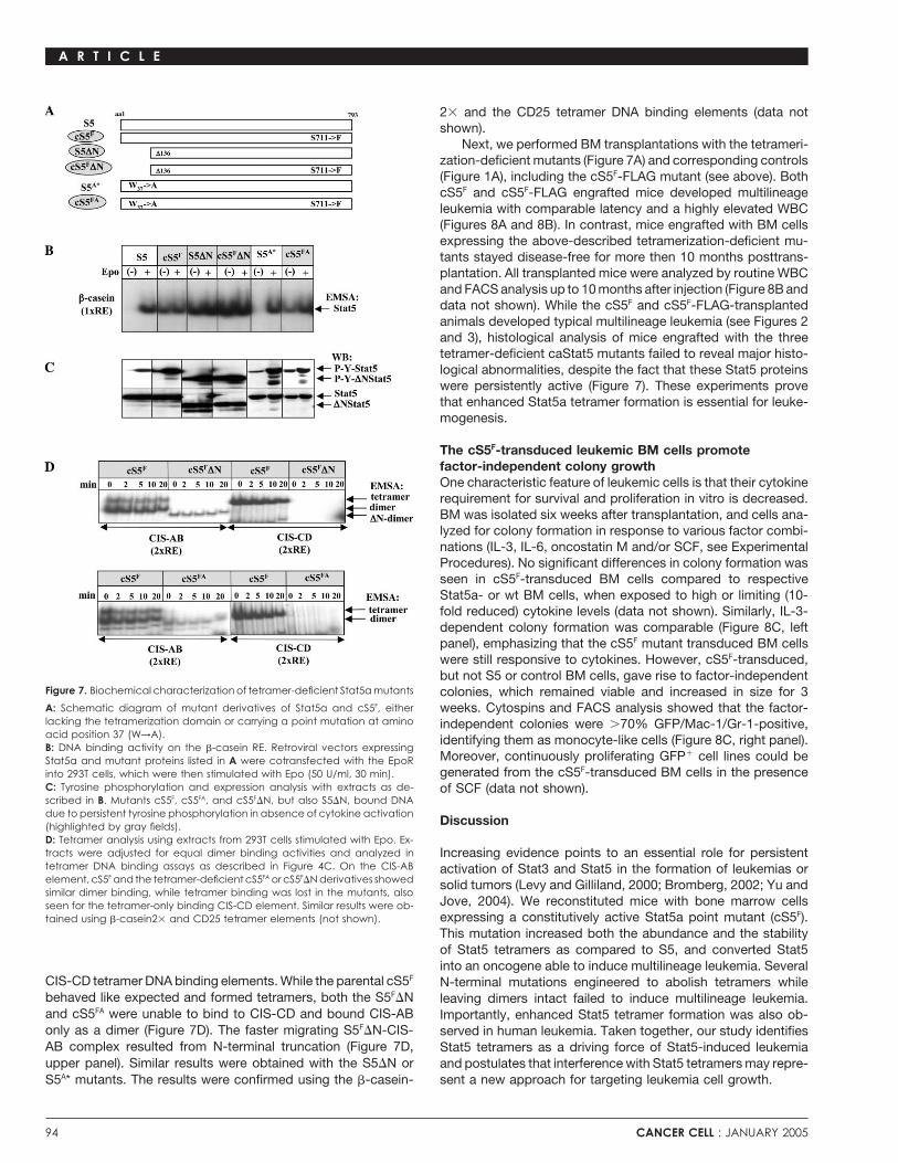
Figure 7. Biochemical characterization of tetramer-deficient Stat5a mutants colonies, which remained viable and increased in size for 3A: Schematic diagram of mutant derivatives of Stat5a and cS5F, either weeks. Cytospins and FACS analysis showed that the factor-lacking the tetramerization domain or carrying a point mutation at amino independent colonies were 70% GFP/Mac-1/Gr-1-positive,acid position 37 (W→A). identifying them as monocyte-like cells (Figure 8C, right panel).B: DNA binding activity on the �-casein RE. Retroviral vectors expressing
Moreover, continuously proliferating GFP� cell lines could beStat5a and mutant proteins listed in A were cotransfected with the EpoRgenerated from the cS5F-transduced BM cells in the presenceinto 293T cells, which were then stimulated with Epo (50 U/ml, 30 min).
C: Tyrosine phosphorylation and expression analysis with extracts as de- of SCF (data not shown).scribed in B. Mutants cS5F, cS5FA, and cS5F�N, but also S5�N, bound DNAdue to persistent tyrosine phosphorylation in absence of cytokine activation
Discussion(highlighted by gray fields).D: Tetramer analysis using extracts from 293T cells stimulated with Epo. Ex-tracts were adjusted for equal dimer binding activities and analyzed in Increasing evidence points to an essential role for persistenttetramer DNA binding assays as described in Figure 4C. On the CIS-AB activation of Stat3 and Stat5 in the formation of leukemias orelement, cS5F and the tetramer-deficient cS5FA or cS5F�N derivatives showed solid tumors (Levy and Gilliland, 2000; Bromberg, 2002; Yu andsimilar dimer binding, while tetramer binding was lost in the mutants, also
Jove, 2004). We reconstituted mice with bone marrow cellsseen for the tetramer-only binding CIS-CD element. Similar results were ob-expressing a constitutively active Stat5a point mutant (cS5F).tained using �-casein2 and CD25 tetramer elements (not shown).
This mutation increased both the abundance and the stabilityof Stat5 tetramers as compared to S5, and converted Stat5into an oncogene able to induce multilineage leukemia. Several
CIS-CD tetramer DNA binding elements. While the parental cS5F N-terminal mutations engineered to abolish tetramers whilebehaved like expected and formed tetramers, both the S5F�N leaving dimers intact failed to induce multilineage leukemia.and cS5FA were unable to bind to CIS-CD and bound CIS-AB Importantly, enhanced Stat5 tetramer formation was also ob-only as a dimer (Figure 7D). The faster migrating S5F�N-CIS- served in human leukemia. Taken together, our study identifiesAB complex resulted from N-terminal truncation (Figure 7D, Stat5 tetramers as a driving force of Stat5-induced leukemiaupper panel). Similar results were obtained with the S5�N or and postulates that interference with Stat5 tetramers may repre-
sent a new approach for targeting leukemia cell growth.S5A* mutants. The results were confirmed using the �-casein-
94 CANCER CELL : JANUARY 2005

A R T I C L E
Figure 8. Leukemia depends on a functional cS5F
tetramer supporting factor-independent colo-nies
A: Kaplan-Meier plot of Stat5a or mutant deriva-tives used in this survival study (n 4 for eachgroup). Only cS5F- or cS5F-FLAG-transplantedmice (n 8 for each group) died within �7 weeksposttransplantation, while mice transplantedwith Stat5a or the tetramer-deficient mutants re-mained disease-free for up to 10 months aftertransplantation.B: Elevated WBC counts are seen in cS5F-graftedmice, but not in wt Stat5a or tetramer-deficientStat5a mutant transplanted mice (see A).C: BM from wt mice or Stat5a- (2 mice analyzed)or cS5F-transplanted mice (4 mice analyzed)were isolated 5 weeks after transplantation,seeded into methylcellulose, and analyzed forIL-3-dependent or factor-independent colonyformation. Inset: May-Grunwald-Giemsa-stainedcytospin from isolated, factor-independent col-onies, containing GFP/Mac-1/Gr-1 triple-positivecells with a monocyte-like morphology. Typicaldata from three individual experiments yieldingidentical results are shown.D: Schematic model for the functional Stat5complexes in hematopoietic cells. Stat5 mono-mers (black ellipse) can form a dimer (left) ortetramer (middle and right) on DNA (gray area).Stat5 tetramer formation is essential for periph-eral T cell proliferation and multilineage leukemiadevelopment, but tetramerization is dispensablefor myeloid mast or erythroid cell proliferation(K.D.B. and R.M., manuscript in preparation).Other transcription or cofactors like the GR dockto Stat5 to enhance transcription, but they mightuse Stat5 tetramers as a signaling platform in leu-kemias (right).
Enhanced Stat5 tetramer formation is essential Moreover, Stat5a mutations at the N terminus designed to abol-ish tetramer formation also failed to induce leukemias or tofor multilineage leukemia
The single point mutation (S711→F) in cS5F rendered Stat5 consti- rescue Stat5�/� T cell proliferation, despite the persistent activa-tion of S5�N proteins. These observations indicate that thetutively active and allowed the formation of stable tetramers on
a variety of binding sites. S5 and the S5A control mutant (S711→A) enhanced tetramer formation of cS5F is the essential featureresponsible for leukemogenesis.lacked constitutive activity and formed tetramers to a lower
extent on a restricted number of DNA elements (Figure 4E and Stat tetramers have more contact points with DNA thandimers, and they form more stable DNA complexes with en-data not shown). While S5 and S5A rescued proliferation of
Stat5�/� T cells, they failed to induce multilineage leukemia. hanced binding to “weak” binding sites (Soldaini et al., 2000;
CANCER CELL : JANUARY 2005 95

A R T I C L E
Zhang and Darnell, 2001). Tetramer formation may therefore cS5RF-expressing Ba/F3 cells induced apoptosis (Nosaka et al.,increase binding site occupancy on weak sites to a threshold 1999). These responses are unexpected and hard to explain,required for transcriptional activity which, together with the since wt Stat5 promotes cytokine-dependent survival and cell-greater degree of flexibility in DNA sequence tetramer recogni- cycle progression (Bunting et al., 2002; Kieslinger et al., 2000;tion, was suggested to widen target gene spectra (John et al., Moriggl et al., 1999b). cS5F causes the same events in a cyto-1999; Meyer et al., 1997). Thus, tetramerization increases speci- kine-independent fashion (data not shown). Other studies de-ficity and selectivity among Stat family members, as has been scribed a role for cS5RF in �-� T cell generation (Lee et al., 2001),described for the CD25 enhancer (Meyer et al., 1997). Putative despite the presence of �-� T cells in Stat5�/� mice (Moriggl etStat5 tetramer target genes include D-type cyclins, Bcl-xL, Osm, al., 1999b). We therefore speculate that the cS5RF mutant hasCD25, CIS, Socs-2, als, and igf-1, all exhibiting two to four high- additional, cell type-specific functions as a heterodimer partneraffinity Stat5 binding sites in their transcriptional regulatory units of endogenous wt Stat5.(see Supplemental Table S2). Stat tetramers have a different
Another important question is whether cS5F and S5 differprotein surface accessible to different proteins compared to
in their DNA binding specificity. DNA binding site selection re-dimers (Vinkemeier et al., 1998). This may allow selective recruit-vealed that both use essentially the same consensus DNA bind-ment of transcription factors or coactivators by Stat5 tetramers.ing site. Analysis of 10 Stat5 target genes for IL-2-dependentThe binding of activated Stat5 to the tetramer elements in theactivation in Stat5�/� T cells confirmed that cS5F and S5 inducetranscriptional regulatory regions of spi2.1, �-casein, igf-1, orthe same target genes. Moreover, we did not observe any sig-als led to recruitment of the GR in absence of GR-DNA bindingnificant upregulation of c-fos mRNA expression, a known target(see model Figure 8D; Stoecklin et al., 1997; Tronche et al.,gene of Stat3 in T cells, or mRNA expression of Socs-2, a2004).gene predominantly controlled via Stat5 in hepatocytes (dataTwo recent reports show that the N-terminal domain of Stat4not shown). The most striking difference was the kinetic of targethas functions in oligomerization, dephosphorylation, and nu-gene mRNA downregulation after IL-2 withdrawal. Loss of geneclear accumulation (Meyer et al., 2004; Ota et al., 2004). These
observations raise the issue of whether other functions of Stat5 activation occurred more slowly in cS5F-transduced Stat5�/�
besides tetramer formation are affected in the S5�N, S5F�N, T cells. This observation is consistent with cell cycle analysisS5A*, or S5FA* mutants. However, the deletion of the Stat4 N performed in cS5F-transduced Stat5�/� T cells. A significantterminus resulted in a lack of cytokine inducibility, and the muta- fraction of cS5F-transduced Stat5�/� T cells remained in thetion of the conserved W37 residue to alanine led to misfolding S-G2-M phases of the cell cycle upon cytokine starvation, simi-of Stat1 or Stat4 (Meyer et al., 2004; Ota et al., 2004). This was lar to growing culture conditions. In contrast, wt T cells strictlynot the case for Stat5, since both N-terminal Stat5a mutants depended on IL-2 and arrested in G1 or died upon IL-2 depriva-showed normal tyrosine phosphorylation, dimer formation, and tion (R.M., unpublished data).dimer binding to DNA (Figure 7C). Moreover, both S5F�N andS5FA rescued cytokine-dependent cell expansion of primary
What is the target cell of cS5F-inducedStat5�/� mast cells, again indicating that these mutants showmultilineage leukemia?largely normal biological activity (K.D.B. and R.M., unpublishedcS5F-induced multilineage leukemia is characterized by abun-data). Further evidence comes from a recently published reportdant, immature myeloid/erythroid GFP� cells in the BM andon the generation of Stat5Null mice (Cui et al., 2004). These micemassive infiltrates of myeloid and lymphoid cells in peripheraldisplay an aggravated phenotype compared to the originallyorgans. This suggests that HSC or primitive multipotent progeni-described hypomorphic Stat5�/� mice (Cui et al., 2004; Teglund
et al., 1998). The hypomorphic Stat5�/� express N-terminally tor cells are the target cells of cS5F for leukemia induction.truncated Stat5 proteins to those we have characterized here The fact that cS5F induces leukemia in Stat5�/� BM with �6with the S5�N mutant. Apparently, the S5�N mutant is able to prolonged latency supports this idea, since Stat5�/� HSC dopartially compensate the complete loss of Stat5a and Stat5b not respond efficiently to cytokines and thus, the retrovirus canin a cell lineage-specific manner (see model, Figure 8D). not efficiently transduce Stat5�/� HSC (Bunting et al., 2002). In
addition, Stat5�/� HSC have �10 reduced capacity to repopu-cS5F induces multilineage leukemia in the absence late the hematopoietic system under competitive engraftmentof endogenous Stat5 proteins conditions (Bunting et al., 2002). Importantly, enforced hemato-We also show that cS5F induces multilineage leukemia in the poiesis as experimentally mimicked by lethal � irradiation isabsence of wt Stat5a, i.e., in Stat5�/� BM cells. This strongly
beside the presence of endogenous Stat5 an additional drivingsupports our concept that the essential function of cS5F in leuke-
force for cS5F induced multilineage leukemias. We did not ob-mogenesis is enhanced Stat5 tetramer formation on promoterserve any latency difference when we used Stat5�/� versus wtelements where wt Stat5 forms predominantly dimers. cS5RF
bone marrow (data not shown), but we observed increasedmutant phenotypes may also be interpreted as a consequencelatency when sublethal instead of lethal � irradiation was cho-of heterodimers or heterotetramers formed between mutant andsen. Lethal � irradiation followed by transplantation mimickedwt Stat5 proteins in cells containing wt Stat5. The cS5RF mutantenforced hematopoiesis, and genetic changes led to cytokineshowed constitutive activation and enhanced tetramers similarrelease and Stat5 activation that might accelerate leukemogene-to cS5F (Figures 1 and 4). cS5RF caused myeloid hyperprolifera-sis. The �6 shorter disease onset in lethally irradiated wt micetion upon transplantation of wt BM (Schwaller et al., 2000), butas compared to nonirradiated Rag2�/� mice supports the aboveit failed to induce both Stat5�/� T cell proliferation and leukemiahypothesis for leukemia promoting factors under enforced he-in Stat5�/� bone marrow. The cS5RF mutant also promoted IL-
3-independent Ba/F3 cell proliferation, while IL-3 readdition to matopoiesis, and overall, cS5F is a potent protooncogene.
96 CANCER CELL : JANUARY 2005

A R T I C L E
Teglund et al., 1998). BM was harvested from both hind limbs of eitherAre Stat5 tetramers targets for selective interventionStat5�/� or wt males. Freshly isolated BM cells were preactivated for 48 hrin human cancer?in medium containing IL-3 (25 ng/ml), IL-6 (50 ng/ml), and SCF (200 ng/ml),Hyperactivation of Stat5 is a frequent event in various leukemiasand consequently cocultured on irradiated (1.5 Gy) semiconfluent ecotropic(AML, ALL, CML, and HTLV-1). Stat5 activation was also shownproducer cell lines for 48 hr in the presence of 6 �g/ml polybrene. Lethally
to be important for head and neck, prostate, and breast carcino- irradiated wt female mice (1 Gy) or nonirradiated Rag2�/� female recipientsmas (Nevalainen et al., 2004; Ren et al., 2002; Xi et al., 2003a, (C57B/L6) were reconstituted with the transduced BM by tail vein injection2003b). Our analysis of human leukemic patient samples and the (4 106 cells). Transplanted mice were checked for disease onset, upon
which they were analyzed. Primary human leukemic blast cells were isolatedmurine transplant experiments suggests a pathophysiologicallyfrom BM or PB using Ficoll and washed in ice-cold PBS supplemented withrelevant signaling function for Stat5 tetramers in leukemia. Tetra-complete protease inhibitors (Boehringer) and phosphatase blockers (1 mMmeric Stat5 may rarely be the consequence of mutations inNaF, 1 mM Na3VO4, 10 mM �-glycerophosphate).human leukemia, but it could result from a persistent Stat5
activation caused by overexpressed or mutated constitutivelyCytokine stimulation, Western blotting, and DNA binding assays
active receptor tyrosine kinases (i.g. c-Kit and Flt3; Stirewalt Naive splenic T cells were stimulated with �-CD3 (145.2C11; Pharmingen)and Radich, 2003). Furthermore, Stat5 tetramer complexes from and IL-2 (500 U/ml; R&D Systems) and expanded in presence of IL-2. Cellleukemic extracts migrated slower then activated Stat5a tetra- purity was determined by FACS. At the indicated times, the cells weremers from 293T cells or Epo-stimulated Ebls, which did not collected, added to excess ice-cold PBS, and processed to lysis. 293T cells
were transfected by calcium phosphate precipitation with the murine pXM-form significant Stat5 tetramer complexes (compare Figures 4D,EpoR expression vector (4 �g/transfection) and/or Stat5a (4 �g/transfection)4E, 6D, and 6E and Supplemental Data). Probably due to theor mutant derivatives in the retroviral pMSCV-IRES-EGFP vector. Sampleuse of N-terminal antisera, Stat5 tetramer complexes showedpreparation, electrophoresis, and transfers were as described (Moriggl etan unusual behavior in supershift experiments, which had to beal., 1999b). Stat5 Y-phosphorylation was detected by �-P-Y-Stat5ab (#71-
used to probe for the integrity of the tetramerization domain of6900; Zymed), and reprobed with �-Stat5ab (aa 451–649; BD Transduction
Stat5 and to detect different C-terminal splice variants of Stat5 Laboratories). Bandshift assays were performed for Stat5 dimers or for tetra-in leukemia (Figures 6B and 6C). The reasons for the observed mers (Vinkemeier et al., 1996). 20 �g of extracts were analyzed using blunt-incomplete Stat5 tetramer supershifts are currently unclear. ended annealed oligonucleotides. C17 and N20 supershift reagents were
purchased from Santa Cruz.Unfortunately, the N-terminal antisera employed are the onlysource specific to the native N terminus of Stat5 (we noted
Northern blot analysisbatch variations in quality). Thus, improved antisera able toRNA was isolated as described (Moriggl et al., 1999b) and murine DNArecognize distinct variants of Stat5 DNA binding complexes areprobes for cyclin D2 (1.2 kb EcoRI), D3 (1.7 kb EcoRI), c-Myc (1 kb SacII/required for future human cancer research.XbaI), Bcl-xL (0.2 kb BamHI), Osm (0.6 kb of Acc. D31942: nt26–588), CD25
Our current work describes the expression of N-terminal (0.5 kb of Acc. M30856: nt451–980), CIS (0.9 kb EcoRI/XbaI), Socs-1 (EcoRI/variants of Stat5 in vivo, and such proteins can still interact XbaI), and Socs-3 (0.8 kb EcoRI) genes were used, with gapdh (1 kb PstI)with endogenous Stat5 or other transcription factor complexes as the loading control.(K.D.B., R.M., and M. Kerenyi, unpublished data). It is also
Generation of retroviral packaging cell lines and descriptiontempting to speculate that leukemic Stat5 tetramer complexesof Stat5�/� T cell linesrecruit tetramer-specific Stat5-protein binding partners, whichThe Stat5a-pMSCV-IRES-GFP vector was used, and all mutants were PCR-enhance Stat5 tetramerization. One candidate protein is themutagenized and sequenced (Moriggl et al., 1999b). Ecotropic, replicationglucocorticoid receptor (GR), which docks to Stat5 as a cofactorincompetent, retroviral producer cell lines were based on the gpE�86 sys-independent of its own DNA binding ability (Figure 8D; Lernertem. GFP� FACS-sorted mass populations or single clones gave identical
et al., 2003; Tronche et al., 2004). In addition, several other leukemia onset. Producer cell lines had high viral titers, in the range of �106
transcription factors, chromatin regulators, or cofactors were particles/ml. The MuMoMTV T cell lymphoma cell line was obtained aftershown in the past to use DNA-bound Stat5 as a signaling plat- transformation with the murine moloney leukemia virus isolated from a T cellform, as illustrated in Figure 8D. lymphoma arising 6 month post infection from Stat5�/� mice. Retroviral
constructs were transduced and sorted 2 for stable GFP expressionOur data from the murine transplant models indicate that(98%).the specific disruption of Stat5 tetramers may be an innovative
approach to inhibit growth of neoplastic cells. Whether the sameMouse PB hematology, FACS, and histologyholds true for human leukemias is presently unknown. It may beTissues were fixed in 4% formaldehyde/PBS, paraffin-embedded, sectioned,an attractive concept, since human leukemia samples containedand stained with hematoxylin-eosin. BM analysis was performed with paraffinsignificant Stat5 tetramer levels. Moreover, we found that incu-and acrylate-embedded specimens. Esterase and �-CD3 staining (DAKO)
bation of primary CML cells with the Bcr/Abl tyrosine kinase were performed. Blood was obtained from the tail vein, and WBCs wereinhibitor STI571 (clinically used to target leukemia cell growth) determined using an automated cell counter (SYSMEX K-1000; TAO Medicalresults in downregulation of Stat5 activity, and a parallel de- Electronics). PB smears were stained with a HEMA3 xanthene/thiazine dyecrease in Stat5 tetramer formation (R.M., unpublished data). In set (Fisher Scientific). Cell suspensions were preincubated with CD16/CD32
antibodies to prevent nonspecific Fc receptor binding. Thereafter, 5 105this regard, it is also noteworthy that peptides from the Stat3cells were stained with antibodies conjugated with fluorescent markersor Stat5 tetramerization domain were found to inhibit breast(Pharmingen) and analyzed by FACS (Becton Dickinson Calibur, Cell Questcancer cell growth (Primiano et al., 2003).Software).
Experimental proceduresIn vitro transformation assaysBM was harvested from mice, washed 2 in PBS, and plated in methylcellu-Human patient samples, animals, primary cell isolation,lose (M3231, StemCell Technology) at a density of 1 105 cells/ml. Cytokinescultivation, and retroviral infectionwere used individually or in combinations: IL-3 (25 ng/ml), IL-6 (50 ng/ml),Splenic T or BM cells were isolated from 6- to 12-week-old wt mice with
the same genetic background as the Stat5�/� mice (Moriggl et al., 1999b; oncostatin M (20 ng/ml), and SCF (200 ng/ml).
CANCER CELL : JANUARY 2005 97

A R T I C L E
Lord, J.D., McIntosh, B.C., Greenberg, P.D., and Nelson, B.H. (2000). TheSupplemental dataIL-2 receptor promotes lymphocyte proliferation and induction of the c-myc,Supplemental data for this article can be found at http://www.cancercell.org/bcl-2, and bcl-x genes through the trans-activation domain of Stat5. J.cgi/content/full/7/1/87/DC1/.Immunol. 164, 2533–2541.
Acknowledgments Meyer, W.K., Reichenbach, P., Schindler, U., Soldaini, E., and Nabholz, M.(1997). Interaction of STAT5 dimers on two low affinity binding sites mediates
We thank C. Leberbauer for Ebls, A. Sommer and S. Schauer for excellent interleukin 2 (IL-2) stimulation of IL-2 receptor alpha gene transcription. J.technical support, and F. Gouilleux for critical reading. R.M. was supported Biol. Chem. 272, 31821–31828.by a Marie Curie HIF and by FWF-SFB 006.
Meyer, T., Hendry, L., Begitt, A., John, S., and Vinkemeier, U. (2004). Asingle residue modulates tyrosine dephosphorylation, oligomerization, andnuclear accumulation of stat transcription factors. J. Biol. Chem. 279, 18998–19007.
Received: July 13, 2004Mizuki, M., Schwable, J., Steur, C., Choudhary, C., Agrawal, S., Sargin,Revised: October 25, 2004B., Steffen, B., Matsumura, I., Kanakura, Y., Bohmer, F.D., et al. (2003).Accepted: December 8, 2004Suppression of myeloid transcription factors and induction of STAT responsePublished: January 17, 2005genes by AML-specific Flt3 mutations. Blood 101, 3164–3173.
References Moriggl, R., Sexl, V., Piekorz, R., Topham, D., and Ihle, J.N. (1999a). Stat5activation is uniquely associated with cytokine signaling in peripheral T cells.Immunity 11, 225–230.Brivanlou, A.H., and Darnell, J.E., Jr. (2002). Signal transduction and the
control of gene expression. Science 295, 813–818. Moriggl, R., Topham, D.J., Teglund, S., Sexl, V., McKay, C., Wang, D.,Hoffmeyer, A., van Deursen, J., Sangster, M.Y., Bunting, K.D., et al. (1999b).Bromberg, J. (2002). Stat proteins and oncogenesis. J. Clin. Invest. 109,Stat5 is required for IL-2-induced cell cycle progression of peripheral T cells.1139–1142.Immunity 10, 249–259.
Buettner, R., Mora, L.B., and Jove, R. (2002). Activated STAT signaling inNevalainen, M.T., Xie, J., Torhorst, J., Bubendorf, L., Haas, P., Kononen, J.,human tumors provides novel molecular targets for therapeutic intervention.Sauter, G., and Rui, H. (2004). Signal transducer and activator of transcrip-Clin. Cancer Res. 8, 945–954.tion-5 activation and breast cancer prognosis. J. Clin. Oncol. 22, 2053–2060.
Bunting, K.D., Bradley, H.L., Hawley, T.S., Moriggl, R., Sorrentino, B.P., andNosaka, T., Kawashima, T., Misawa, K., Ikuta, K., Mui, A.L., and Kitamura,Ihle, J.N. (2002). Reduced lymphomyeloid repopulating activity from adultT. (1999). STAT5 as a molecular regulator of proliferation, differentiation andbone marrow and fetal liver of mice lacking expression of STAT5. Blood 99,apoptosis in hematopoietic cells. EMBO J. 18, 4754–4765.479–487.
O’Shea, J.J., Gadina, M., and Schreiber, R.D. (2002). Cytokine signaling inCui, Y., Riedlinger, G., Miyoshi, K., Tang, W., Li, C., Deng, C.X., Robinson,2002: New surprises in the Jak/Stat pathway. Cell 109 (Suppl), S121–S131.G.W., and Hennighausen, L. (2004). Inactivation of Stat5 in mouse mammary
epithelium during pregnancy reveals distinct functions in cell proliferation, Onishi, M., Nosaka, T., Misawa, K., Mui, A.L., Gorman, D., McMahon, M.,survival, and differentiation. Mol. Cell. Biol. 24, 8037–8047. Miyajima, A., and Kitamura, T. (1998). Identification and characterization of
a constitutively active STAT5 mutant that promotes cell proliferation. Mol.Iavnilovitch, E., Groner, B., and Barash, I. (2002). Overexpression and forcedCell. Biol. 18, 3871–3879.activation of stat5 in mammary gland of transgenic mice promotes cellular
proliferation, enhances differentiation, and delays postlactational apoptosis. Ota, N., Brett, T.J., Murphy, T.L., Fremont, D.H., and Murphy, K.M. (2004).Mol. Cancer Res. 1, 32–47. N-domain-dependent nonphosphorylated STAT4 dimers required for cyto-
kine-driven activation. Nat. Immunol. 5, 208–215.John, S., Vinkemeier, U., Soldaini, E., Darnell, J.E., Jr., and Leonard, W.J.(1999). The significance of tetramerization in promoter recruitment by Stat5. Primiano, T., Baig, M., Maliyekkel, A., Chang, B.D., Fellars, S., Sadhu, J.,Mol. Cell. Biol. 19, 1910–1918. Axenovich, S.A., Holzmayer, T.A., and Roninson, I.B. (2003). Identification
of potential anticancer drug targets through the selection of growth-inhibitoryKelly, J.A., Spolski, R., Kovanen, P.E., Suzuki, T., Bollenbacher, J., Pise-genetic suppressor elements. Cancer Cell 4, 41–53.Masison, C.A., Radonovich, M.F., Lee, S., Jenkins, N.A., Copeland, N.G.,
et al. (2003). Stat5 synergizes with T cell receptor/antigen stimulation in the Ren, S., Cai, H.R., Li, M., and Furth, P.A. (2002). Loss of Stat5a delaysdevelopment of lymphoblastic lymphoma. J. Exp. Med. 198, 79–89. mammary cancer progression in a mouse model. Oncogene 21, 4335–4339.
Kieslinger, M., Woldman, I., Moriggl, R., Hofmann, J., Marine, J.C., Ihle, J.N., Santos, S.C., Lacronique, V., Bouchaert, I., Monni, R., Bernard, O., Gissel-Beug, H., and Decker, T. (2000). Antiapoptotic activity of Stat5 required brecht, S., and Gouilleux, F. (2001). Constitutively active STAT5 variantsduring terminal stages of myeloid differentiation. Genes Dev. 14, 232–244. induce growth and survival of hematopoietic cells through a PI 3-kinase/Akt
dependent pathway. Oncogene 20, 2080–2090.Kim, H.P., and Leonard, W.J. (2002). The basis for TCR-mediated regulationof the IL-2 receptor alpha chain gene: Role of widely separated regulatory Schwaller, J., Parganas, E., Wang, D., Cain, D., Aster, J.C., Williams, I.R.,elements. EMBO J. 21, 3051–3059. Lee, C.K., Gerthner, R., Kitamura, T., Frantsve, J., et al. (2000). Stat5 is
essential for the myelo- and lymphoproliferative disease induced by TEL/Leberbauer, C., Boulme, F., Unfried, G., Huber, J., Beug, H., and Mullner,
JAK2. Mol. Cell 6, 693–704.E.W. (2005). Different steroids co-regulate long-term expansion versus termi-nal differentiation in primary human erythroid progenitors. Blood, in press. Soldaini, E., John, S., Moro, S., Bollenbacher, J., Schindler, U., and Leonard,
W.J. (2000). DNA binding site selection of dimeric and tetrameric Stat5Lee, H.C., Ye, S.K., Honjo, T., and Ikuta, K. (2001). Induction of germline proteins reveals a large repertoire of divergent tetrameric Stat5a bindingtranscription in the human TCR gamma locus by STAT5. J. Immunol. 167, sites. Mol. Cell. Biol. 20, 389–401.320–326.
Stirewalt, D.L., and Radich, J.P. (2003). The role of FLT3 in haematopoieticLerner, L., Henriksen, M.A., Zhang, X., and Darnell, J.E., Jr. (2003). STAT3- malignancies. Nat. Rev. Cancer 3, 650–665.dependent enhanceosome assembly and disassembly: Synergy with GR forfull transcriptional increase of the alpha 2-macroglobulin gene. Genes Dev. Stoecklin, E., Wissler, M., Moriggl, R., and Groner, B. (1997). Specific DNA17, 2564–2577. binding of Stat5, but not of glucocorticoid receptor, is required for their
functional cooperation in the regulation of gene transcription. Mol. Cell. Biol.Levy, D.E., and Gilliland, D.G. (2000). Divergent roles of STAT1 and STAT5 17, 6708–6716.in malignancy as revealed by gene disruptions in mice. Oncogene 19, 2505–2510. Taketani, T., Taki, T., Sugita, K., Furuichi, Y., Ishii, E., Hanada, R., Tsuchida,
98 CANCER CELL : JANUARY 2005

A R T I C L E
M., Ida, K., and Hayashi, Y. (2004). FLT3 mutations in the activation loop of Vinkemeier, U., Moarefi, I., Darnell, J.E., Jr., and Kuriyan, J. (1998). Structuretyrosine kinase domain are frequently found in infant ALL with MLL rearrange- of the amino-terminal protein interaction domain of STAT-4. Science 279,ments and pediatric ALL with hyperdiploidy. Blood 103, 1085–1088. 1048–1052.
Teglund, S., McKay, C., Schuetz, E., van Deursen, J.M., Stravopodis, D., Wang, D., Moriggl, R., Stravopodis, D., Carpino, N., Marine, J.C., Teglund,Wang, D., Brown, M., Bodner, S., Grosveld, G., and Ihle, J.N. (1998). Stat5a S., Feng, J., and Ihle, J.N. (2000). A small amphipathic alpha-helical regionand Stat5b proteins have essential and nonessential, or redundant, roles in is required for transcriptional activities and proteasome-dependent turnovercytokine responses. Cell 93, 841–850. of the tyrosine-phosphorylated Stat5. EMBO J. 19, 392–399.
Tronche, F., Opherk, C., Moriggl, R., Kellendonk, C., Reimann, A., Schwake, Xi, S., Zhang, Q., Dyer, K.F., Lerner, E.C., Smithgall, T.E., Gooding, W.E.,L., Reichardt, H.M., Stangl, K., Gau, D., Hoeflich, A., et al. (2004). Glucocorti- Kamens, J., and Grandis, J.R. (2003a). Src kinases mediate STAT growthcoid receptor function in hepatocytes is essential to promote postnatal body pathways in squamous cell carcinoma of the head and neck. J. Biol. Chem.growth. Genes Dev. 18, 492–497.
278, 31574–31583.Tsuruyama, T., Nakamura, T., Jin, G., Ozeki, M., Yamada, Y., and Hiai, H.
Xi, S., Zhang, Q., Gooding, W.E., Smithgall, T.E., and Grandis, J.R. (2003b).(2002). Constitutive activation of Stat5a by retrovirus integration in earlyConstitutive activation of Stat5b contributes to carcinogenesis in vivo. Can-pre-B lymphomas of SL/Kh strain mice. Proc. Natl. Acad. Sci. USA 99,cer Res. 63, 6763–6771.8253–8258.
Xu, X., Sun, Y.L., and Hoey, T. (1996). Cooperative DNA binding and se-Verdier, F., Rabionet, R., Gouilleux, F., Beisenherz-Huss, C., Varlet, P.,quence-selective recognition conferred by the STAT amino-terminal domain.Muller, O., Mayeux, P., Lacombe, C., Gisselbrecht, S., and Chretien, S.Science 273, 794–797.(1998). A sequence of the CIS gene promoter interacts preferentially with
two associated STAT5A dimers: A distinct biochemical difference betweenYu, H., and Jove, R. (2004). The STATs of cancer–new molecular targetsSTAT5A and STAT5B. Mol. Cell. Biol. 18, 5852–5860.come of age. Nat. Rev. Cancer 4, 97–105.
Vinkemeier, U., Cohen, S.L., Moarefi, I., Chait, B.T., Kuriyan, J., and Darnell,Zhang, X., and Darnell, J.E., Jr. (2001). Functional importance of Stat3 tetra-J.E., Jr. (1996). DNA binding of in vitro activated Stat1 alpha, Stat1 betamerization in activation of the alpha 2-macroglobulin gene. J. Biol. Chem.and truncated Stat1: Interaction between NH2-terminal domains stabilizes
binding of two dimers to tandem DNA sites. EMBO J. 15, 5616–5626. 276, 33576–33581.
CANCER CELL : JANUARY 2005 99