Inhibitors of Stat5 protein signalling
6
Inhibitors of Stat5 protein signalling Abbarna A. Cumaraswamy, a Aleksandra Todic, a Diana Resetca, a Mark D. Minden b and Patrick T. Gunning * a Received 7th July 2011, Accepted 25th August 2011 DOI: 10.1039/c1md00175b Evidence shows that signal transducer and activator of transcription 5 (Stat5) protein, a member of the STAT family of signalling proteins, plays a pivotal role in the progression of many human cancers including acute myeloid leukemias and prostate cancer. This mini-review outlines progress made towards identifying agents capable of silencing aberrant Stat5 signalling. Introduction Signal transducer and activator of transcription 5 (Stat5) protein, a member of the STAT family of signalling proteins, plays a crucial transcriptional role in numerous human cancers and has become a target for therapeutic intervention. Due to growth factor receptor mutations, amplified expression or misdirected kinase activity in the cytoplasm, constitutively activated Stat5 is present in a diverse number of human cancers, including those of the breast, prostate, liver, skin, blood, head and neck. 1 The STAT family of proteins is comprised of seven members including Stat1, Stat2, Stat3, Stat4, Stat5A and 5B, as well as Stat6. These proteins perform dual roles as both cytosolic sig- nalling proteins and as nuclear transcription factors that mediate the expression of specific sets of genes. In particular, expression of genes involved in proliferation (Bcl-xl, c-Myc, pim-1), apoptosis (JAB), cell differentiation (p21), 2 and inflammation is mediated. In cancer cells, STAT proteins are routinely hyper- activated through either somatic mutation or dysregulated expression of upstream signalling pathways. As a result, several members of the STAT family, including Stat5, can act as onco- genes by facilitating the aberrant expression of key proteins associated with the cancer phenotype. This article will review the progress made towards the development of Stat5-targeting molecular therapeutics. Stat5 proteins are activated/phosphorylated by kinases asso- ciated with transmembrane receptors, including IL-2, GM-CSF, erythropoietin, IL-5, IL-7, thrombopoietin, prolactin, and growth hormone receptors. 1 Extracellular ligand binding to these target receptors induces receptor dimerization and intracellular activation of receptor-associated kinases, such as JAK. JAK- mediated phosphorylation of key tyrosine (Y) residues on the cytoplasmic receptor domain creates docking sites for non- phosphorylated Stat5 proteins. Stat5 binds to the receptor through its phosphotyrosine binding, Src Homology 2 (SH2) domain. Once recruited, Stat5 is phosphorylated by receptor associated JAK kinases, such as Jak2, which phosphorylate Stat5 proteins at specific Y residues in the C-terminus (Y694 for Stat5a and Y699 for Stat5b). Phosphorylated Stat5 protein dissociates from the receptor and forms either homo- or hetero- Stat5–StatX dimers, through reciprocal SH2 domain-pY interactions. Acti- vated Stat5 translocates to the nucleus and binds to Stat5 response elements and induces target gene transcription. In normal cells, Stat5 proteins are transiently activated by extracellular cytokine or ligand-receptor engagement and rapidly deactivated by a number of intracellular mechanisms, including cytosolic and nuclear phosphatases. For example, deactivation of Stat5 can occur directly via phosphatases such as protein inhibitors of aberrant STAT proteins (PIAS) or protein tyrosine phosphatases such as SHP-2, or indirectly by down-regulation of cytokine signalling with SOCS1, SOCS3 and CIS proteins. 1,3 However, in cancer cells Stat5 is constitutively phosphorylated, leading to elevated expression levels of its target genes, such as the anti-apoptotic proteins Bcl-xl, Bcl-2, Myc and MCL. Aber- rant Stat5 activity has been associated with hyperactivated upstream cytosolic tyrosine kinases including TEL-Jak2 and Bcr- Abl, mutated FMS-like tyrosine kinase 3 (FLT3), as well as overactive receptor tyrosine kinases, such as SRC and EGFR. 4 Medicinal chemists have attempted to silence aberrant Stat5 activity through a number of direct and indirect molecular approaches. These therapeutic efforts have included the targeting of upstream effector proteins or the direct targeting of Stat5 protein. This review will explore the progress made towards developing effective inhibitors of Stat5 function in human cancers. Indirect inhibitors of Stat5 signalling Stat5’s crucial role in cellular signalling and gene transcription is mediated by an intricate cascade of protein–protein interactions. Consequently, there are several junctures at which inhibitors may disrupt the Stat5 signalling pathway. Notably, most inhibitors of Stat5 function have not directly targeted Stat5 protein complexation events, but rather focussed on inhibiting upstream a Department of Chemistry, University of Toronto, 3359 Mississauga Road North, Mississauga, ON, L5L 1C6, Canada. E-mail: patrick.gunning@ utoronto.ca; Tel: +905-828-5354 b Princess Margaret Hospital, Ontario Cancer Institute, 610 University Avenue, Toronto, ON, M5G 2M9, Canada. E-mail: mark.minden@uhn. on.ca; Fax: +416-946-6546; Tel: +416.946.4501 22 | Med. Chem. Commun., 2012, 3, 22–27 This journal is ª The Royal Society of Chemistry 2012 Dynamic Article Links C < MedChemComm Cite this: Med. Chem. Commun., 2012, 3, 22 www.rsc.org/medchemcomm REVIEW Downloaded on 12 July 2012 Published on 03 October 2011 on http://pubs.rsc.org | doi:10.1039/C1MD00175B View Online / Journal Homepage / Table of Contents for this issue
-
Upload
independent -
Category
Documents
-
view
0 -
download
0
Transcript of Inhibitors of Stat5 protein signalling
Dynamic Article LinksC<MedChemComm
Cite this: Med. Chem. Commun., 2012, 3, 22
www.rsc.org/medchemcomm REVIEW
Dow
nloa
ded
on 1
2 Ju
ly 2
012
Publ
ishe
d on
03
Oct
ober
201
1 on
http
://pu
bs.r
sc.o
rg |
doi:1
0.10
39/C
1MD
0017
5BView Online / Journal Homepage / Table of Contents for this issue
Inhibitors of Stat5 protein signalling
Abbarna A. Cumaraswamy,a Aleksandra Todic,a Diana Resetca,a Mark D. Mindenb and Patrick T. Gunning*a
Received 7th July 2011, Accepted 25th August 2011
DOI: 10.1039/c1md00175b
Evidence shows that signal transducer and activator of transcription 5 (Stat5) protein, a member of the
STAT family of signalling proteins, plays a pivotal role in the progression of many human cancers
including acute myeloid leukemias and prostate cancer. This mini-review outlines progress made
towards identifying agents capable of silencing aberrant Stat5 signalling.
Introduction
Signal transducer and activator of transcription 5 (Stat5) protein,
a member of the STAT family of signalling proteins, plays
a crucial transcriptional role in numerous human cancers and has
become a target for therapeutic intervention. Due to growth
factor receptor mutations, amplified expression or misdirected
kinase activity in the cytoplasm, constitutively activated Stat5 is
present in a diverse number of human cancers, including those of
the breast, prostate, liver, skin, blood, head and neck.1 The
STAT family of proteins is comprised of seven members
including Stat1, Stat2, Stat3, Stat4, Stat5A and 5B, as well as
Stat6. These proteins perform dual roles as both cytosolic sig-
nalling proteins and as nuclear transcription factors that mediate
the expression of specific sets of genes. In particular, expression
of genes involved in proliferation (Bcl-xl, c-Myc, pim-1),
apoptosis (JAB), cell differentiation (p21),2 and inflammation is
mediated. In cancer cells, STAT proteins are routinely hyper-
activated through either somatic mutation or dysregulated
expression of upstream signalling pathways. As a result, several
members of the STAT family, including Stat5, can act as onco-
genes by facilitating the aberrant expression of key proteins
associated with the cancer phenotype. This article will review the
progress made towards the development of Stat5-targeting
molecular therapeutics.
Stat5 proteins are activated/phosphorylated by kinases asso-
ciated with transmembrane receptors, including IL-2, GM-CSF,
erythropoietin, IL-5, IL-7, thrombopoietin, prolactin, and
growth hormone receptors.1 Extracellular ligand binding to these
target receptors induces receptor dimerization and intracellular
activation of receptor-associated kinases, such as JAK. JAK-
mediated phosphorylation of key tyrosine (Y) residues on the
cytoplasmic receptor domain creates docking sites for non-
phosphorylated Stat5 proteins. Stat5 binds to the receptor
aDepartment of Chemistry, University of Toronto, 3359 Mississauga RoadNorth, Mississauga, ON, L5L 1C6, Canada. E-mail: [email protected]; Tel: +905-828-5354bPrincess Margaret Hospital, Ontario Cancer Institute, 610 UniversityAvenue, Toronto, ON, M5G 2M9, Canada. E-mail: [email protected]; Fax: +416-946-6546; Tel: +416.946.4501
22 | Med. Chem. Commun., 2012, 3, 22–27
through its phosphotyrosine binding, Src Homology 2 (SH2)
domain. Once recruited, Stat5 is phosphorylated by receptor
associated JAK kinases, such as Jak2, which phosphorylate Stat5
proteins at specific Y residues in the C-terminus (Y694 for Stat5a
and Y699 for Stat5b). Phosphorylated Stat5 protein dissociates
from the receptor and forms either homo- or hetero- Stat5–StatX
dimers, through reciprocal SH2 domain-pY interactions. Acti-
vated Stat5 translocates to the nucleus and binds to Stat5
response elements and induces target gene transcription.
In normal cells, Stat5 proteins are transiently activated by
extracellular cytokine or ligand-receptor engagement and rapidly
deactivated by a number of intracellular mechanisms, including
cytosolic and nuclear phosphatases. For example, deactivation
of Stat5 can occur directly via phosphatases such as protein
inhibitors of aberrant STAT proteins (PIAS) or protein tyrosine
phosphatases such as SHP-2, or indirectly by down-regulation of
cytokine signalling with SOCS1, SOCS3 and CIS proteins.1,3
However, in cancer cells Stat5 is constitutively phosphorylated,
leading to elevated expression levels of its target genes, such as
the anti-apoptotic proteins Bcl-xl, Bcl-2, Myc and MCL. Aber-
rant Stat5 activity has been associated with hyperactivated
upstream cytosolic tyrosine kinases including TEL-Jak2 and Bcr-
Abl, mutated FMS-like tyrosine kinase 3 (FLT3), as well as
overactive receptor tyrosine kinases, such as SRC and EGFR.4
Medicinal chemists have attempted to silence aberrant Stat5
activity through a number of direct and indirect molecular
approaches. These therapeutic efforts have included the targeting
of upstream effector proteins or the direct targeting of Stat5
protein. This review will explore the progress made towards
developing effective inhibitors of Stat5 function in human
cancers.
Indirect inhibitors of Stat5 signalling
Stat5’s crucial role in cellular signalling and gene transcription is
mediated by an intricate cascade of protein–protein interactions.
Consequently, there are several junctures at which inhibitors may
disrupt the Stat5 signalling pathway. Notably, most inhibitors of
Stat5 function have not directly targeted Stat5 protein
complexation events, but rather focussed on inhibiting upstream
This journal is ª The Royal Society of Chemistry 2012
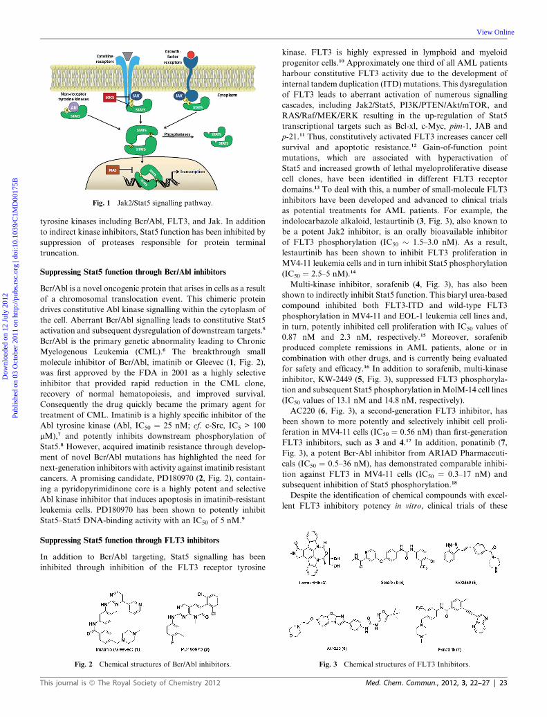
Fig. 1 Jak2/Stat5 signalling pathway.
Dow
nloa
ded
on 1
2 Ju
ly 2
012
Publ
ishe
d on
03
Oct
ober
201
1 on
http
://pu
bs.r
sc.o
rg |
doi:1
0.10
39/C
1MD
0017
5B
View Online
tyrosine kinases including Bcr/Abl, FLT3, and Jak. In addition
to indirect kinase inhibitors, Stat5 function has been inhibited by
suppression of proteases responsible for protein terminal
truncation.
Suppressing Stat5 function through Bcr/Abl inhibitors
Bcr/Abl is a novel oncogenic protein that arises in cells as a result
of a chromosomal translocation event. This chimeric protein
drives constitutive Abl kinase signalling within the cytoplasm of
the cell. Aberrant Bcr/Abl signalling leads to constitutive Stat5
activation and subsequent dysregulation of downstream targets.5
Bcr/Abl is the primary genetic abnormality leading to Chronic
Myelogenous Leukemia (CML).6 The breakthrough small
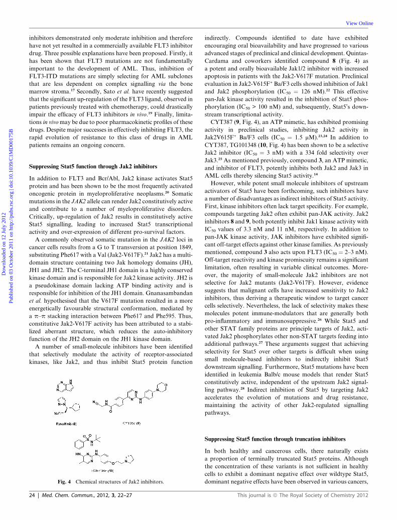
molecule inhibitor of Bcr/Abl, imatinib or Gleevec (1, Fig. 2),
was first approved by the FDA in 2001 as a highly selective
inhibitor that provided rapid reduction in the CML clone,
recovery of normal hematopoiesis, and improved survival.
Consequently the drug quickly became the primary agent for
treatment of CML. Imatinib is a highly specific inhibitor of the
Abl tyrosine kinase (Abl, IC50 ¼ 25 nM; cf. c-Src, IC5 > 100
mM),7 and potently inhibits downstream phosphorylation of
Stat5.8 However, acquired imatinib resistance through develop-
ment of novel Bcr/Abl mutations has highlighted the need for
next-generation inhibitors with activity against imatinib resistant
cancers. A promising candidate, PD180970 (2, Fig. 2), contain-
ing a pyridopyrimidinone core is a highly potent and selective
Abl kinase inhibitor that induces apoptosis in imatinib-resistant
leukemia cells. PD180970 has been shown to potently inhibit
Stat5–Stat5 DNA-binding activity with an IC50 of 5 nM.9
Suppressing Stat5 function through FLT3 inhibitors
In addition to Bcr/Abl targeting, Stat5 signalling has been
inhibited through inhibition of the FLT3 receptor tyrosine
Fig. 2 Chemical structures of Bcr/Abl inhibitors.
This journal is ª The Royal Society of Chemistry 2012
kinase. FLT3 is highly expressed in lymphoid and myeloid
progenitor cells.10 Approximately one third of all AML patients
harbour constitutive FLT3 activity due to the development of
internal tandem duplication (ITD) mutations. This dysregulation
of FLT3 leads to aberrant activation of numerous signalling
cascades, including Jak2/Stat5, PI3K/PTEN/Akt/mTOR, and
RAS/Raf/MEK/ERK resulting in the up-regulation of Stat5
transcriptional targets such as Bcl-xl, c-Myc, pim-1, JAB and
p-21.11 Thus, constitutively activated FLT3 increases cancer cell
survival and apoptotic resistance.12 Gain-of-function point
mutations, which are associated with hyperactivation of
Stat5 and increased growth of lethal myeloproliferative disease
cell clones, have been identified in different FLT3 receptor
domains.13 To deal with this, a number of small-molecule FLT3
inhibitors have been developed and advanced to clinical trials
as potential treatments for AML patients. For example, the
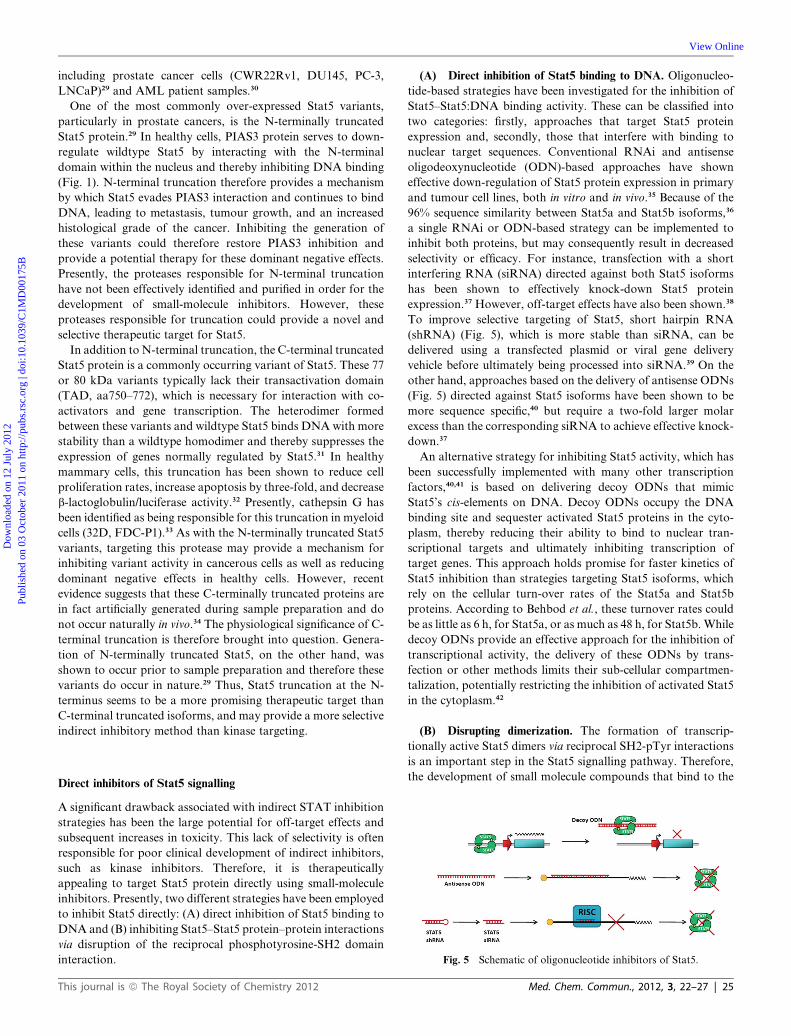
indolocarbazole alkaloid, lestaurtinib (3, Fig. 3), also known to
be a potent Jak2 inhibitor, is an orally bioavailable inhibitor
of FLT3 phosphorylation (IC50 � 1.5–3.0 nM). As a result,
lestaurtinib has been shown to inhibit FLT3 proliferation in
MV4-11 leukemia cells and in turn inhibit Stat5 phosphorylation
(IC50 ¼ 2.5–5 nM).14
Multi-kinase inhibitor, sorafenib (4, Fig. 3), has also been
shown to indirectly inhibit Stat5 function. This biaryl urea-based
compound inhibited both FLT3-ITD and wild-type FLT3
phosphorylation in MV4-11 and EOL-1 leukemia cell lines and,
in turn, potently inhibited cell proliferation with IC50 values of
0.87 nM and 2.3 nM, respectively.15 Moreover, sorafenib
produced complete remissions in AML patients, alone or in
combination with other drugs, and is currently being evaluated
for safety and efficacy.16 In addition to sorafenib, multi-kinase
inhibitor, KW-2449 (5, Fig. 3), suppressed FLT3 phosphoryla-
tion and subsequent Stat5 phosphorylation inMolM-14 cell lines
(IC50 values of 13.1 nM and 14.8 nM, respectively).
AC220 (6, Fig. 3), a second-generation FLT3 inhibitor, has
been shown to more potently and selectively inhibit cell proli-
feration in MV4-11 cells (IC50 ¼ 0.56 nM) than first-generation
FLT3 inhibitors, such as 3 and 4.17 In addition, ponatinib (7,
Fig. 3), a potent Bcr-Abl inhibitor from ARIAD Pharmaceuti-
cals (IC50 ¼ 0.5–36 nM), has demonstrated comparable inhibi-
tion against FLT3 in MV4-11 cells (IC50 ¼ 0.3–17 nM) and
subsequent inhibition of Stat5 phosphorylation.18
Despite the identification of chemical compounds with excel-
lent FLT3 inhibitory potency in vitro, clinical trials of these
Fig. 3 Chemical structures of FLT3 Inhibitors.
Med. Chem. Commun., 2012, 3, 22–27 | 23
Dow
nloa
ded
on 1
2 Ju
ly 2
012
Publ
ishe
d on
03
Oct
ober
201
1 on
http
://pu
bs.r
sc.o
rg |
doi:1
0.10
39/C
1MD
0017
5B
View Online
inhibitors demonstrated only moderate inhibition and therefore
have not yet resulted in a commercially available FLT3 inhibitor
drug. Three possible explanations have been proposed. Firstly, it
has been shown that FLT3 mutations are not fundamentally
important to the development of AML. Thus, inhibition of
FLT3-ITD mutations are simply selecting for AML subclones
that are less dependent on complex signalling via the bone
marrow stroma.17 Secondly, Sato et al. have recently suggested
that the significant up-regulation of the FLT3 ligand, observed in
patients previously treated with chemotherapy, could drastically
impair the efficacy of FLT3 inhibitors in vivo.19 Finally, limita-
tions in vivomay be due to poor pharmacokinetic profiles of these
drugs. Despite major successes in effectively inhibiting FLT3, the
rapid evolution of resistance to this class of drugs in AML
patients remains an ongoing concern.
Suppressing Stat5 function through Jak2 inhibitors
In addition to FLT3 and Bcr/Abl, Jak2 kinase activates Stat5
protein and has been shown to be the most frequently activated
oncogenic protein in myeloproliferative neoplasms.20 Somatic
mutations in the JAK2 allele can render Jak2 constitutively active
and contribute to a number of myeloproliferative disorders.
Critically, up-regulation of Jak2 results in constitutively active
Stat5 signalling, leading to increased Stat5 transcriptional
activity and over-expression of different pro-survival factors.
A commonly observed somatic mutation in the JAK2 loci in
cancer cells results from a G to T transversion at position 1849,
substituting Phe617 with a Val (Jak2-V617F).21 Jak2 has a multi-
domain structure containing two Jak homology domains (JH),
JH1 and JH2. The C-terminal JH1 domain is a highly conserved
kinase domain and is responsible for Jak2 kinase activity. JH2 is
a pseudokinase domain lacking ATP binding activity and is
responsible for inhibition of the JH1 domain. Gnanasambandan
et al. hypothesised that the V617F mutation resulted in a more
energetically favourable structural conformation, mediated by
a p–p stacking interaction between Phe617 and Phe595. Thus,
constitutive Jak2-V617F activity has been attributed to a stabi-
lized aberrant structure, which reduces the auto-inhibitory
function of the JH2 domain on the JH1 kinase domain.
A number of small-molecule inhibitors have been identified
that selectively modulate the activity of receptor-associated
kinases, like Jak2, and thus inhibit Stat5 protein function
Fig. 4 Chemical structures of Jak2 inhibitors.
24 | Med. Chem. Commun., 2012, 3, 22–27
indirectly. Compounds identified to date have exhibited
encouraging oral bioavailability and have progressed to various
advanced stages of preclinical and clinical development. Quintas-
Cardama and coworkers identified compound 8 (Fig. 4) as
a potent and orally bioavailable Jak1/2 inhibitor with increased
apoptosis in patients with the Jak2-V617F mutation. Preclinical
evaluation in Jak2-V615F+ Ba/F3 cells showed inhibition of Jak1
and Jak2 phosphorylation (IC50 ¼ 126 nM).22 This effective
pan-Jak kinase activity resulted in the inhibition of Stat5 phos-
phorylation (IC50 > 100 nM) and, subsequently, Stat5’s down-
stream transcriptional activity.
CYT387 (9, Fig. 4), an ATP mimetic, has exhibited promising
activity in preclinical studies, inhibiting Jak2 activity in
Jak2V615F+ Ba/F3 cells (IC50 ¼ 1.5 mM).23,24 In addition to
CYT387, TG101348 (10, Fig. 4) has been shown to be a selective
Jak2 inhibitor (IC50 ¼ 3 nM) with a 334 fold selectivity over
Jak3.25 As mentioned previously, compound 3, an ATP mimetic,
and inhibitor of FLT3, potently inhibits both Jak2 and Jak3 in
AML cells thereby silencing Stat5 activity.14
However, while potent small molecule inhibitors of upstream
activators of Stat5 have been forthcoming, such inhibitors have
a number of disadvantages as indirect inhibitors of Stat5 activity.
First, kinase inhibitors often lack target specificity. For example,
compounds targeting Jak2 often exhibit pan-JAK activity. Jak2
inhibitors 8 and 9, both potently inhibit Jak1 kinase activity with
IC50 values of 3.3 nM and 11 nM, respectively. In addition to
pan-JAK kinase activity, JAK inhibitors have exhibited signifi-
cant off-target effects against other kinase families. As previously
mentioned, compound 3 also acts upon FLT3 (IC50 ¼ 2–3 nM).
Off-target reactivity and kinase promiscuity remains a significant
limitation, often resulting in variable clinical outcomes. More-
over, the majority of small-molecule Jak2 inhibitors are not
selective for Jak2 mutants (Jak2-V617F). However, evidence
suggests that malignant cells have increased sensitivity to Jak2
inhibitors, thus deriving a therapeutic window to target cancer
cells selectively. Nevertheless, the lack of selectivity makes these
molecules potent immune-modulators that are generally both
pro-inflammatory and immunosuppressive.26 While Stat5 and
other STAT family proteins are principle targets of Jak2, acti-
vated Jak2 phosphorylates other non-STAT targets feeding into
additional pathways.27 These arguments suggest that achieving
selectivity for Stat5 over other targets is difficult when using
small molecule-based inhibitors to indirectly inhibit Stat5
downstream signalling. Furthermore, Stat5 mutations have been
identified in leukemia Balb/c mouse models that render Stat5
constitutively active, independent of the upstream Jak2 signal-
ling pathway.28 Indirect inhibition of Stat5 by targeting Jak2
accelerates the evolution of mutations and drug resistance,
maintaining the activity of other Jak2-regulated signalling
pathways.
Suppressing Stat5 function through truncation inhibitors
In both healthy and cancerous cells, there naturally exists
a proportion of terminally truncated Stat5 proteins. Although
the concentration of these variants is not sufficient in healthy
cells to exhibit a dominant negative effect over wildtype Stat5,
dominant negative effects have been observed in various cancers,
This journal is ª The Royal Society of Chemistry 2012
Dow
nloa
ded
on 1
2 Ju
ly 2
012
Publ
ishe
d on
03
Oct
ober
201
1 on
http
://pu
bs.r
sc.o
rg |
doi:1
0.10
39/C
1MD
0017
5B
View Online
including prostate cancer cells (CWR22Rv1, DU145, PC-3,
LNCaP)29 and AML patient samples.30
One of the most commonly over-expressed Stat5 variants,
particularly in prostate cancers, is the N-terminally truncated
Stat5 protein.29 In healthy cells, PIAS3 protein serves to down-
regulate wildtype Stat5 by interacting with the N-terminal
domain within the nucleus and thereby inhibiting DNA binding
(Fig. 1). N-terminal truncation therefore provides a mechanism
by which Stat5 evades PIAS3 interaction and continues to bind
DNA, leading to metastasis, tumour growth, and an increased
histological grade of the cancer. Inhibiting the generation of
these variants could therefore restore PIAS3 inhibition and
provide a potential therapy for these dominant negative effects.
Presently, the proteases responsible for N-terminal truncation
have not been effectively identified and purified in order for the
development of small-molecule inhibitors. However, these
proteases responsible for truncation could provide a novel and
selective therapeutic target for Stat5.
In addition to N-terminal truncation, the C-terminal truncated
Stat5 protein is a commonly occurring variant of Stat5. These 77
or 80 kDa variants typically lack their transactivation domain
(TAD, aa750–772), which is necessary for interaction with co-
activators and gene transcription. The heterodimer formed
between these variants and wildtype Stat5 binds DNA with more
stability than a wildtype homodimer and thereby suppresses the
expression of genes normally regulated by Stat5.31 In healthy
mammary cells, this truncation has been shown to reduce cell
proliferation rates, increase apoptosis by three-fold, and decrease
b-lactoglobulin/luciferase activity.32 Presently, cathepsin G has
been identified as being responsible for this truncation in myeloid
cells (32D, FDC-P1).33 As with the N-terminally truncated Stat5
variants, targeting this protease may provide a mechanism for
inhibiting variant activity in cancerous cells as well as reducing
dominant negative effects in healthy cells. However, recent
evidence suggests that these C-terminally truncated proteins are
in fact artificially generated during sample preparation and do
not occur naturally in vivo.34 The physiological significance of C-
terminal truncation is therefore brought into question. Genera-
tion of N-terminally truncated Stat5, on the other hand, was
shown to occur prior to sample preparation and therefore these
variants do occur in nature.29 Thus, Stat5 truncation at the N-
terminus seems to be a more promising therapeutic target than
C-terminal truncated isoforms, and may provide a more selective
indirect inhibitory method than kinase targeting.
Fig. 5 Schematic of oligonucleotide inhibitors of Stat5.
Direct inhibitors of Stat5 signalling
A significant drawback associated with indirect STAT inhibition
strategies has been the large potential for off-target effects and
subsequent increases in toxicity. This lack of selectivity is often
responsible for poor clinical development of indirect inhibitors,
such as kinase inhibitors. Therefore, it is therapeutically
appealing to target Stat5 protein directly using small-molecule
inhibitors. Presently, two different strategies have been employed
to inhibit Stat5 directly: (A) direct inhibition of Stat5 binding to
DNA and (B) inhibiting Stat5–Stat5 protein–protein interactions
via disruption of the reciprocal phosphotyrosine-SH2 domain
interaction.
This journal is ª The Royal Society of Chemistry 2012
(A) Direct inhibition of Stat5 binding to DNA. Oligonucleo-
tide-based strategies have been investigated for the inhibition of
Stat5–Stat5:DNA binding activity. These can be classified into
two categories: firstly, approaches that target Stat5 protein
expression and, secondly, those that interfere with binding to
nuclear target sequences. Conventional RNAi and antisense
oligodeoxynucleotide (ODN)-based approaches have shown
effective down-regulation of Stat5 protein expression in primary
and tumour cell lines, both in vitro and in vivo.35 Because of the
96% sequence similarity between Stat5a and Stat5b isoforms,36
a single RNAi or ODN-based strategy can be implemented to
inhibit both proteins, but may consequently result in decreased
selectivity or efficacy. For instance, transfection with a short
interfering RNA (siRNA) directed against both Stat5 isoforms
has been shown to effectively knock-down Stat5 protein
expression.37 However, off-target effects have also been shown.38
To improve selective targeting of Stat5, short hairpin RNA
(shRNA) (Fig. 5), which is more stable than siRNA, can be
delivered using a transfected plasmid or viral gene delivery
vehicle before ultimately being processed into siRNA.39 On the
other hand, approaches based on the delivery of antisense ODNs
(Fig. 5) directed against Stat5 isoforms have been shown to be
more sequence specific,40 but require a two-fold larger molar
excess than the corresponding siRNA to achieve effective knock-
down.37
An alternative strategy for inhibiting Stat5 activity, which has
been successfully implemented with many other transcription
factors,40,41 is based on delivering decoy ODNs that mimic
Stat5’s cis-elements on DNA. Decoy ODNs occupy the DNA
binding site and sequester activated Stat5 proteins in the cyto-
plasm, thereby reducing their ability to bind to nuclear tran-
scriptional targets and ultimately inhibiting transcription of
target genes. This approach holds promise for faster kinetics of
Stat5 inhibition than strategies targeting Stat5 isoforms, which
rely on the cellular turn-over rates of the Stat5a and Stat5b
proteins. According to Behbod et al., these turnover rates could
be as little as 6 h, for Stat5a, or as much as 48 h, for Stat5b. While
decoy ODNs provide an effective approach for the inhibition of
transcriptional activity, the delivery of these ODNs by trans-
fection or other methods limits their sub-cellular compartmen-
talization, potentially restricting the inhibition of activated Stat5
in the cytoplasm.42
(B) Disrupting dimerization. The formation of transcrip-
tionally active Stat5 dimers via reciprocal SH2-pTyr interactions
is an important step in the Stat5 signalling pathway. Therefore,
the development of small molecule compounds that bind to the
Med. Chem. Commun., 2012, 3, 22–27 | 25
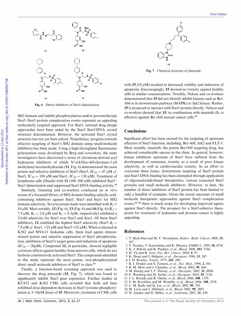
Fig. 6 Direct inhibitors of Stat5 dimerization.
Fig. 7 Chemical structure of pimozide.
Dow
nloa
ded
on 1
2 Ju
ly 2
012
Publ
ishe
d on
03
Oct
ober
201
1 on
http
://pu
bs.r
sc.o
rg |
doi:1
0.10
39/C
1MD
0017
5B
View Online
SH2 domain and inhibit phosphorylation and/or prevent/disrupt
Stat5–Stat5 protein complexation events represent an appealing
molecularly targeted approach. For Stat3, rational drug design
approaches have been aided by the Stat3–Stat3:DNA crystal
structure determination. However, the activated Stat5 crystal
structure has not yet been solved. Nonetheless, progress towards
effective targeting of Stat5’s SH2 domain using small-molecule
inhibitors has been made. Using a high-throughput fluorescence
polarization assay developed by Berg and coworkers, the same
investigators have discovered a series of chromone-derived acyl
hydrazone inhibitors of which N0-((4-Oxo-4H-chromen-3-yl)
methylene) nicotinohydrazide (11, Fig. 6) demonstrated the most
potent and selective inhibition of Stat5 (Stat5, IC50 ¼ 47 mM cf.
Stat3, IC50 ¼ 180 mM and Stat1, IC50 ¼ 130 mM). Treatment of
lymphoma cells (Daudi) with 11 (100–200 mM) inhibited Stat5–
Stat5 dimerization and suppressed Stat5 DNA binding activity.43
Similarly, Gunning and co-workers conducted an in vitro
screen of a focussed library of SH2 domain binding salicylic acid-
containing inhibitors against Stat5, Stat3 and Stat1 for SH2
domain selectivity. Several potent leads were identified with Ki <
10 mM.Most notably, 12 (Fig. 6), 13 (Fig. 6) and 14 (Fig. 6) (Ki¼7.9 mM, Ki ¼ 2.8 mM and Ki ¼ 8.3mM, respectively) exhibited a
3-fold selectivity for Stat5 over Stat3 and Stat1. Of these Stat5
inhibitors, 12 exhibited the highest Stat5 selectivity (Stat5 Ki ¼7.9 mM cf. Stat1, >25 mM and Stat3 >25 mM). When evaluated in
K562 and MV4-11 leukemia cells, these lead agents demon-
strated potent and selective suppression of Stat5 phosphoryla-
tion, inhibition of Stat5’s target genes and induction of apoptosis
(IC50 � 20mM). Compound 12, in particular, showed negligible
cytotoxic effects against healthy bone marrow cells, which do not
harbour constitutively activated Stat5. The compounds identified
in this study represent the most potent, non-phosphorylated
direct small molecule inhibitors of Stat5 to date.44
Finally, a function-based screening approach was used to
discover the drug pimozide (15, Fig. 7), which was found to
significantly inhibit Stat5 gene expression. Further studies in
KU812 and K562 CML cells revealed that both cell lines
exhibited dose-dependent decreases in Stat5 tyrosine phosphory-
lation at 3–10mM doses of 15. Moreover, treatment of CML cells
26 | Med. Chem. Commun., 2012, 3, 22–27
with 15 (10 mM) resulted in decreased viability and induction of
apoptosis. Encouragingly, 15 showed no toxicity against healthy
cells at similar concentrations. Notably, Nelson and co-workers
demonstrated that 15 did not directly inhibit kinases such as Bcr-
Abl or its downstream pathway (MAPK) or Jak2 kinase. Rather,
15 is proposed to interact with Stat5 protein directly. Nelson and
co-workers showed that 15, in combination with imatinib (1), is
effective against Bcr-Abl mutant cancer cells.49
Conclusions
Significant effort has been exerted for the targeting of upstream
effectors of Stat5 function, including, Bcr-Abl, Jak2 and FLT-3.
Most notably, imatinib, the potent Bcr/Abl targeting drug, has
achieved considerable success in the clinic. In general, however,
kinase inhibitors upstream of Stat5 have suffered from the
development of resistance, toxicity as a result of poor kinase
selectivity, as well as cardiovascular toxicity. In an effort to
overcome these issues, downstream targeting of Stat5 protein
and Stat5-DNA binding has been attempted through application
of oligonucleotide-based therapeutics, various truncated Stat5
proteins and small molecule inhibitors. However, to date, the
number of direct inhibitors of Stat5 protein has been limited to
only a handful of examples. Given the recent successes of small
molecule therapeutic approaches against Stat3 complexation
events,45–48 there is much scope for developing improved agents
against Stat5 directly. The prospect for a Stat5-selective thera-
peutic for treatment of leukemias and prostate cancer is highly
appealing.
References
1 T. Shyh-Han and M. T. Nevaleinen, Endocr. Relat. Cancer, 2008, 15,367.
2 T. Nosaka, T. Kawashima and K. Misawa, EMBO J., 1999, 18, 4754.3 K. J. Peltola and K. Paukku, et al., Blood, 2004, 103, 3744.4 H. Yu and R. Jove, Nat. Rev. Cancer, 2004, 4, 97.5 K. Shuai and J. Halpern, et al., Oncogene, 1996, 13, 247.6 J. D. Rowley, Nature, 1973, 243, 290.7 B. J. Druker and S. Tamura, et al., Nat. Med., 1996, 2, 561.8 B. M. Mow and J. Chandra, et al., Blood, 2002, 99, 664.9 M. Huang and J. F. Dorsey, et al., Oncogene, 2002, 21, 8804.10 E. Weisberg and M. Sattler, et al., Oncogene, 2010, 29, 5120.11 J. L. Rocnik and R. Okabe, et al., Blood, 2006, 108, 1339.12 S. M. Kornblau and M. Womble, et al., Blood, 2006, 108, 2358.13 L. M. Kelly and Q. Liu, et al., Blood, 2002, 99, 310.14 M. Levis and J. Allebach, et al., Blood, 2002, 99, 3885.15 D. Auclair and D. Miller, et al., Leukemia, 2007, 21, 439.
This journal is ª The Royal Society of Chemistry 2012
Dow
nloa
ded
on 1
2 Ju
ly 2
012
Publ
ishe
d on
03
Oct
ober
201
1 on
http
://pu
bs.r
sc.o
rg |
doi:1
0.10
39/C
1MD
0017
5B
View Online
16 A. T. Fathi, S. Grant and J. E. Karp, Cancer Treat. Rev., 2010, 36,142.
17 Q. Chao and K. G. Sprankle, et al., J. Med. Chem., 2009, 52, 7808.18 T. Sato and X. Yang, et al., Blood, 2011, 117, 3286.19 J. M. Gozgit and M. J. Wong, et al., Blood, 2011, 10, 1.20 A. Pardanani and A. M. Vannucchi, et al., Leukemia, 2011, 25, 218.21 K. Gnanasambandan and A. Magis, et al., Biochemistry, 2010, 49,
9972.22 A. Quintas-Cardama and K. Vaddi, et al., Blood, 2010, 115, 3109.23 A. Pardanani and T. Lasho, et al., Leukemia, 2009, 23, 1441.24 F. P. Santos and H. M. Kantarjian, et al., Blood, 2010, 115, 1131.25 G. Wernig and M. G. Kharas, et al., Cancer Cell, 2008, 13, 311.26 T. Ikezoe and J. Yang, et al., Int. J. Cancer, 2011, 128, 2317.27 C. Carter-Su, L. Rui andM. R. Stofega,Recent Prog Horm Res, 2000,
55, 293.28 R. L. Ilaria and R. G. Hawley, et al., Blood, 1999, 93, 4154.29 A. Dagvadorj and S. Tan, et al., Int. J. Biochem. Cell Biol., 2010, 42,
2037.30 Z. Xia and N. J. Sheila, et al., Leuk. Res., 2001, 25, 473.31 D. Wang and D. Stravopodis, et al., Mol. Cell Biol., 1996, 16, 6141.32 E. Iavnilovitch and T. Eilon, et al., Mol. Reprod. Dev., 2006, 73, 841.33 B. Schuster and L. Hendry, et al., Biochem. J., 2007, 404, 81.
This journal is ª The Royal Society of Chemistry 2012
34 H. L. Ramos, J. J. O’Shea andW. T. Watford, Biochem. J., 2007, 404,e1.
35 F. Behbod and Z. S. Nagy, et al., J Immunol, 2003, 171, 3919.36 S. Cao and C. Wang, et al., Neurosci. Lett., 2011, 487, 228.37 X. Liu and G. W. Robinson, et al., Proc. Natl. Acad. Sci. U. S. A.,
1995, 92, 8831.38 C. A. Sledz and B. R. Williams, Biochem. Soc. Trans., 2004, 32, 952.39 S. K. Klosek and K. Nakashiro, et al., Oncol Rep, 2008, 20, 873.40 X. Wang and J. Zeng, et al., DNA Cell Biol., 2011, 30, 71.41 J. Shen and R. Li, et al., In Vivo, 2009, 23, 237.42 D. W. Kim and J. H. Kim, et al., Biomaterials, 2011, 32, 2593.43 J. Muller and T. Berg, et al., ChemBioChem, 2008, 9, 723.44 B. D. Page and P. T. Gunning, et al., J Med Chem, 2011, DOI: jm-
2011-00720n, submitted.45 M. Avadisian, S. Haftchenary and P. T. Gunning, Anticancer Drugs,
2010, 22, 115.46 S. Fletcher and J. A. Drewry, et al., Biochem. Cell Biol., 2009, 87, 825.47 S. Fletcher, J. Turkson and P. T. Gunning, ChemMedChem, 2008, 3,
1159.48 B. D. Page, D. P. Ball and P. T. Gunning, Expert Opin. Ther. Pat.,
2011, 21, 65.49 E. A. Nelson and S. R. Walker, et al., Blood, 2011, 117, 3421.
Med. Chem. Commun., 2012, 3, 22–27 | 27