Recommendations for the prevention of secondary Haemophilus influenzae type b (Hib) disease
Upload
independentCategory
view
0download
0
BioMed CentralBMC Infectious Diseases
ss
Open AcceResearch articleAntimicrobial activity of innate immune molecules against Streptococcus pneumoniae, Moraxella catarrhalis and nontypeable Haemophilus influenzaeHaa-Yung Lee1, Ali Andalibi1,3, Paul Webster1,2,3, Sung-Kyun Moon1,6, Karen Teufert1, Sung-Ho Kang1,7, Jian-Dong Li1,3, Mitsuyoshi Nagura1,8, Tomas Ganz5 and David J Lim*1,4Address: 1The Gonda Department of Cell and Molecular Biology, House Ear Institute, Los Angeles, California 90057, USA, 2Ahmanson Center for Advanced Electron Microscopy, House Ear Institute, Los Angeles, California 90057, USA, 3Department of Otolaryngology, University of Southern California, Los Angeles, California 90033, USA, 4Department of Cell and Neurobiology, University of Southern California, Los Angeles, California 90033, USA, 5Department of Medicine, University of California, Los Angeles, California 90024, USA, 6Departments of Otolaryngology, Ajou University School of Medicine, Suwon, Korea, 7Department of Otolaryngology and Head & Neck Surgery, School of Medicine, KonKuk University Hospital, Chungju, Korea and 8Department of Otorhinolaryngology Hamamatsu University School of Medicine, Hamamatsu, Japan
Email: Haa-Yung Lee - [email protected]; Ali Andalibi - [email protected]; Paul Webster - [email protected]; Sung-Kyun Moon - [email protected]; Karen Teufert - [email protected]; Sung-Ho Kang - [email protected]; Jian-Dong Li - [email protected]; Mitsuyoshi Nagura - [email protected]; Tomas Ganz - [email protected]; David J Lim* - [email protected]
* Corresponding author
AbstractBackground: Despite its direct connection to the nasopharynx which harbors otitis mediapathogens as part of its normal flora, the middle ear cavity is kept free of these bacteria by as yetunknown mechanisms. Respiratory mucosal epithelia, including those of the middle ear andeustachian tube, secrete antimicrobial effectors including lysozyme, lactoferrin and β defensins-1and -2. To elucidate the role of these innate immune molecules in the normal defense andmaintenance of sterility of respiratory mucosa such as that of the middle ear, we assessed theireffect on the respiratory pathogens nontypeable Haemophilus influenzae (NTHi) 12, Moraxellacatarrhalis 035E, and Streptococcus pneumoniae 3, and 6B.
Methods: Two assay methods, the radial assay and the liquid broth assay, were employed fortesting the antimicrobial activity of the molecules. This was done in order to minimize thepossibility that the observed effects were artifacts of any single assay system employed. Also,transmission electron microscopy (TEM) was employed to evaluate the effect of antimicrobialinnate immune molecules on OM pathogens. For the statistical analysis of the data, Student's t-testwas performed.
Results: Results of the radial diffusion assay showed that β defensin-2 was active against all fourOM pathogens tested, while treatment with β defensin-1 appeared to only affect M. catarrhalis. Theradial assay results also showed that lysozyme was quite effective against S. pneumoniae 3 and 6Band was partially bacteriostatic/bactericidal against M. catarrhalis. Lysozyme however, appeared notto affect the growth of NTHi. Thus, lysozyme seems to have a more pronounced impact on thegrowth of the Gram-positive S. pneumoniae as compared to that of Gram-negative pathogens.Lactoferrin on the other hand, enhanced the growth of the bacteria tested. The results of the radial
Published: 05 May 2004
BMC Infectious Diseases 2004, 4:12
Received: 14 November 2003Accepted: 05 May 2004
This article is available from: http://www.biomedcentral.com/1471-2334/4/12
© 2004 Lee et al; licensee BioMed Central Ltd. This is an Open Access article: verbatim copying and redistribution of this article are permitted in all media for any purpose, provided this notice is preserved along with the article's original URL.
Page 1 of 12(page number not for citation purposes)

BMC Infectious Diseases 2004, 4 http://www.biomedcentral.com/1471-2334/4/12
assays were confirmed using liquid broth assays for antimicrobial activity, and showed thatlysozyme and β defensin-2 could act synergistically against S. pneumoniae 6B. Moreover, in the liquidbroth assay, β defensin-1 showed a modest inhibitory effect on the growth of S. pneumoniae 6B. Asassessed by ultrastructural analysis, lysozyme and β defensin-2, and to a much lesser extent, βdefensin-1, appeared to be able to cause damage to the bacterial membranes.
Conclusions: Here we report that lysozyme and the β defensins can inhibit the growth of clinicalisolates of otitis media pathogens – namely NTHi strain 12, S. pneumoniae strains 3 and 6B and M.catarrhalis strain 035E – and cause ultrastructural damage to these pathogens. Moreover, wedemonstrate that lysozyme and β defensin-2 can act synergistically against S. pneumoniae. Thesefindings are consistent with the concept that secreted antimicrobial peptides and othercomponents of innate immunity constitute the first line of defense protecting host mucosalsurfaces, including the tubotympanal (eustachian tube and middle ear cavity) mucosa, againstpathogens.
BackgroundOtitis media (OM) is the most prevalent mucosal infec-tious disease affecting young children, second only to thecommon cold [1]. OM is also the major cause of conduc-tive hearing loss among this group and the leading indica-tion for antibiotic therapy [1]. OM results in 31 millionannual visits to physicians' offices and is estimated tohave a yearly cost exceeding $5 billion.
Most cases of OM are caused by three major pathogens: S.pneumoniae (30–40%), nontypeable Haemophilus influen-zae (NTHi, 30%) and M. catarrhalis (20%) [1,2]. Theseorganisms are also pathogens for sinusitis and chronicobstructive pulmonary disease (COPD) [3]. In the pastthree decades, there has been a dramatic worldwideincrease in antibiotic resistance in respiratory pathogens[4,5]. This has resulted in a reduction of the number ofeffective antibiotics for OM and has begun to pose a majorpublic health threat. There is, thus, an urgent need todevelop new and innovative non-antibiotic approaches toprevent and manage this disease. To this end, it is imper-ative to understand the mechanisms that defend thetubotympanum (the middle ear and eustachian tube)against invading pathogens and determine if the defensemechanisms can be augmented to prevent or treat OM.
The pathogenesis of OM is multi-factorial with risk factorssuch as delayed development of the adaptive immune sys-tem, complement system abnormality, as well as otherenvironmental and genetic factors playing an importantrole [6-10]. Although anatomic immaturity of the eus-tachian tube and delayed development of systemic immu-nity are considered to be the most important factorsunderlying OM susceptibility among young children,other factors may be of greater importance before theadaptive immune system is fully matured [1,8]. For exam-ple, in a study of children with recurrent acute otitis media(rAOM), Prellner and colleagues showed that only 1/3 oftwo-year-old children with rAOM had significantly lower
levels of antibodies against the pneumococcal capsulartype 19F. This suggests that factors other than circulatingantibody titers affect susceptibility to rAOM [8]. Onegroup of factors that may be of particular relevance to OMsusceptibility consists of the innate immune molecules,including the defensins, lysozyme and lactoferrin [11-16],as well as the pathogen recognition receptors such as thetoll-like receptors (TLR) [3,17]. Many studies have dem-onstrated the presence of lysozyme and lactoferrin inchronic middle ear effusion [18-21] and recent work hasshown that TLR-2, TLR-4, the defensins, lysozyme andlactoferrin are expressed, albeit at different levels, in nor-mal middle ear epithelial cells, as well as in those cellsfrom patients with chronic otitis media with effusion(OME) [3,17,22-24]. The exact role however, that thesemolecules play in normal mucosal defense and mainte-nance of tympanic cavity sterility as well as in the OM sus-ceptibility has not yet been clearly elucidated.
Here we report that lysozyme and the β defensins caninhibit the growth of clinical isolates of otitis media path-ogens, namely, NTHi 12, M. catarrhalis 035E, and S. pneu-moniae 3, and 6B and cause ultrastructural damage tothese pathogens. Moreover, we demonstrate that lys-ozyme and β defensin-2 can act synergistically against S.pneumoniae.
MethodsBacteriaAll bacteria used in this study were clinical isolates.
NTHi and M. catarrhalisStocks of NTHi 12 (obtained from Dr. Steve Barenkamp,St. Louis University, School of Medicine) and M. catarrha-lis 035E (obtained from Dr. Eric Hansen, University ofTexas, Southwestern Medical Center) were maintained at-80°C. The bacteria were plated on chocolate agar andincubated overnight at 37°C in 5% CO2. A single colonywas used to inoculate 10 ml of brain heart infusion (BHI,
Page 2 of 12(page number not for citation purposes)

BMC Infectious Diseases 2004, 4 http://www.biomedcentral.com/1471-2334/4/12
Becton Dickinson, Cockeysville, MD), supplemented withhemin (10 µg/ml, Sigma, St. Louis, MO) and nicotina-mide adenine dinucleotide (NAD, 10 µg/ml, Sigma, St.Louis, MO), and allowed to grow overnight. In the morn-ing, 1/10 volume of the overnight culture was transferredto fresh medium and incubated for 3 hours. The subcul-ture was then washed twice with 10 mM sodium phos-phate and the optical density (O.D.) at 620 nm wasdetermined.
S. pneumoniaeFor the S. pneumoniae 3 and 6B (obtained from AmecricanType Culture Collection, ATCC), a 50 µl aliquot of the fro-zen stock was thawed on ice, added to 10 ml of Todd-Hewitt broth (THB, Becton Dickinson, Cockeysville, MD)and cultured overnight. One tenth of the volume of theovernight culture was then transferred to fresh mediumand incubated for 3 hours. The subculture was thenwashed twice with 10 mM sodium phosphate and theO.D. at 620 nm was determined.
Testing of antimicrobial activityRadial assayThe radial assay method of Lehrer and coworkers wasused, with minor modifications [39]. The subculturedbacteria (4 × 10⁶ CFU/10 ml underlay gel for NTHi and M.catarrhalis and 106 CFU/10 ml underlay gel for S. pneumo-niae) were mixed with melted underlay agarose mediumat 42°C (0.1X culture broth – BHI for NTHi and M.catarrhalis and THB for S. pneumoniae- 10 mM sodiumphosphate, 0.8% low electroendosmosis (EEO)-type aga-rose) and poured into 8 cm × 8 cm square petri plates. Thegel was allowed to solidify in the petri plates and wellswere punched out with a cork borer (4 mm, VWR Scien-tific, West Chester, PA). The antimicrobial peptides andproteins, human milk lysozyme (Sigma, St. Louis, MO),human milk apo-lactoferrin (0.03% iron content, Sigma,St. Louis, MO), human β defensin-1 and human βdefensin-2 (Peptides International, Louisville, KY), weredissolved in 0.01% acetic acid/0.1% bovine serum albu-min (Sigma, St. Louis, MO) and 4 µl was added to eachwell. The plates were then incubated for 3 hours at 37°Cin the 5% CO2 incubator. They were next overlaid with theoverlay agarose medium (0.5X culture broth, 10 mMsodium phosphate, 0.8% low EEO-type agarose) and cov-ered. The plates were then allowed to incubate overnightat 37°C in a 5% CO2. The diameter of the zones of inhi-bition produced by each of the antimicrobial moleculesagainst the OM pathogens (NTHi, M. catarrhalis and S.pneumoniae) was then measured in three separate experi-ments and the mean and standard deviation were calcu-lated. To analyze the salt dependency of the activity ofantimicrobial molecules against OM pathogens, NaCl wasadded to the underlay gel solution to obtain a final NaClconcentration of 100 mM.
Antibacterial liquid broth assayFor the antibacterial liquid broth assays, 1/10 of the vol-ume of each overnight culture was transferred to freshmedium and allowed to grow to mid-log phase. The bac-teria were then exposed to various concentrations of lys-ozyme, β defensin-1, β defensin-2 or a combination of thelysozyme and β defensin-2 molecules for 3 h at 37°C. Cellviability was measured by plating several dilutions andcounting the number of colonies after and overnight incu-bation. The assay was done in triplicate for each concen-tration of test molecules.
Statistical analysisStudent's t-test was used to perform statistical analysis ofthe data.
Electron microscopyTransmission electron microscopy (TEM) was employedto evaluate the effect of antimicrobial innate immunemolecules on OM pathogens. The bacterial culturestreated with antimicrobial proteins or peptides for 30minutes (β defensin-1 treatment was for 3 hours) weremixed with an equal volume of 5% buffered glutaralde-hyde (pH 7.4) and centrifuged (5000 × g, for 20 minutes).The bacterial pellets were left in the fixative at 4°C for 2hr, post-fixed in 2% osmium tetroxide, dehydrated ingraded ethanol and embedded in Eponate 12 (Ted Pella,Inc. Redding, CA). Thin sections (70 µm) were mountedon coated specimen grids, contrasted with uranyl acetateand lead citrate and examined in a transmission electronmicroscope (CM120 BioTwin, FEI-Philips, Hillsboro, OR)operating at 80 kV.
ResultsAntimicrobial activity of innate immune molecules against OM pathogensWe tested the bacteriostatic/bactericidal effects of theinnate immune molecules on four clinically relevant otitismedia pathogens: NTHi 12, M. catarrhalis 035E and S.pneumoniae 3 and 6B. Two assay methods, the radial assayand the liquid broth assay, were employed for testing theantimicrobial activity of the molecules. This was done inorder to minimize the possibility that the observed effectswere artifacts of any single assay system employed.
Radial assayThe results of the radial assay indicated that the treatmentof the bacteria with lysozyme resulted in a visible inhibi-tion of the growth of M. catarrhalis and S. pneumoniae 3and 6B (Figure 1A and Table 1). This effect was seen atconcentrations of 12.5 mg/ml (50 µg of the protein perwell) and higher. There appeared to be a dose-dependentincrease in the effect of lysozyme on the growth of S. pneu-moniae and M. catarrhalis, although the dose dependenceof the effect was not as apparent with the latter pathogen.
Page 3 of 12(page number not for citation purposes)

BMC Infectious Diseases 2004, 4 http://www.biomedcentral.com/1471-2334/4/12
Lysozyme appeared not to affect the growth of NTHi (Fig-ure 1A and Table 1). Thus, lysozyme appears to have amore pronounced impact on the growth of the Gram-pos-itive S. pneumoniae compared to the Gram-negative path-ogens (Figure 1A and Table 1).
It should be noted that the concentration of lysozymedecreases as it diffuses radially into the three-dimensionalgel matrix. Moreover, the concentration of lysozymeranges from 1.1 mg/ml in serous middle ear effusions to3.71 mg/ml in mucoid in middle ear effusions [20]. Thus,once dilution as a result of diffusion is taken into accountthe amounts used in our assays are likely to give nearphysiologic levels of this protein in the gel matrix.
Apo-lactoferrin treatment did not inhibit bacterialgrowth, and appeared to result in an enhancement of thegrowth of all bacteria tested at 40 µg dosage (Figure 1Band Table 1).
Treatment with β defensin-1 was effective only against M.catarrhalis and only at the high dose (10 µg). In contrast,β defensin-2, at 4 µg and 10 µg, significantly inhibited thegrowth of all four bacterial types (Figure 1B and Table 1).The 40 µg dose of β defensin-2 was not tested because theeffects were already observable at the lower doses. Like-wise, because β defensin-1 showed an effect against M.catarrhalis at 10 µg, a higher dose was not tested. M.catarrhalis 035E was most susceptible to β defensin-2, fol-lowed by NTHi 12, S. pneumoniae 3 and S. pneumoniae 6B.
Radial inhibition assay results demonstrating the effect of innate immune molecules on otitis media pathogensFigure 1Radial inhibition assay results demonstrating the effect of innate immune molecules on otitis media patho-gens. Representative radial inhibition assay for the measurement of the activity of innate immune molecules against NTHi 12, M. catarrhalis 035E (M cat), S. pneumoniae 3 (Sp 3) and S. pneumoniae 6B (Sp 6B). Panel A shows the results of testing three doses (50 µg, 100 µg and 200 µg) of human lysozyme (Lz) against the bacteria. Panel B shows the results of testing the lactof-errin (Lf) and the defensins against the bacteria. Three doses (4 µg, 10 µg and 40 µg) of human lactoferrin (Lf) were tested, as were two doses (4 µg and 10 µg) each of human β defensin-1 (hBD-1) and β defensin-2 (hBD-2). Each dose was delivered in a total volume of 4 µl. The control well received only solvent (0.01% acetic acid). The diameter of the inhibition zone was meas-ured and averaged in three separate experiments.
Page 4 of 12(page number not for citation purposes)
BMC Infectious Diseases 2004, 4 http://www.biomedcentral.com/1471-2334/4/12
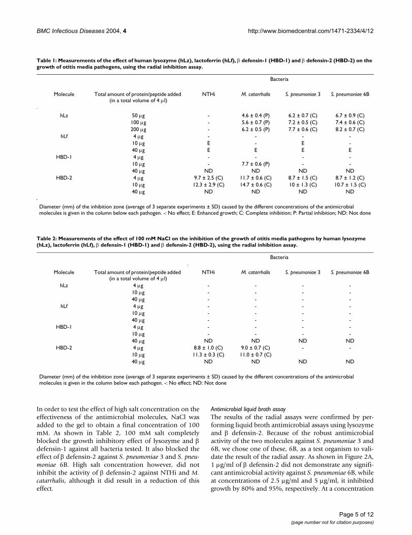
In order to test the effect of high salt concentration on theeffectiveness of the antimicrobial molecules, NaCl wasadded to the gel to obtain a final concentration of 100mM. As shown in Table 2, 100 mM salt completelyblocked the growth inhibitory effect of lysozyme and βdefensin-1 against all bacteria tested. It also blocked theeffect of β defensin-2 against S. pneumoniae 3 and S. pneu-moniae 6B. High salt concentration however, did notinhibit the activity of β defensin-2 against NTHi and M.catarrhalis, although it did result in a reduction of thiseffect.
Antimicrobial liquid broth assayThe results of the radial assays were confirmed by per-forming liquid broth antimicrobial assays using lysozymeand β defensin-2. Because of the robust antimicrobialactivity of the two molecules against S. pneumoniae 3 and6B, we chose one of these, 6B, as a test organism to vali-date the result of the radial assay. As shown in Figure 2A,1 µg/ml of β defensin-2 did not demonstrate any signifi-cant antimicrobial activity against S. pneumoniae 6B, whileat concentrations of 2.5 µg/ml and 5 µg/ml, it inhibitedgrowth by 80% and 95%, respectively. At a concentration
Table 1: Measurements of the effect of human lysozyme (hLz), lactoferrin (hLf), β defensin-1 (HBD-1) and β defensin-2 (HBD-2) on the growth of otitis media pathogens, using the radial inhibition assay.
Bacteria
Molecule Total amount of protein/peptide added (in a total volume of 4 µl)
NTHi M. catarrhalis S. pneumoniae 3 S. pneumoniae 6B
hLz 50 µg - 4.6 ± 0.4 (P) 6.2 ± 0.7 (C) 6.7 ± 0.9 (C)100 µg - 5.6 ± 0.7 (P) 7.2 ± 0.5 (C) 7.4 ± 0.6 (C)200 µg - 6.2 ± 0.5 (P) 7.7 ± 0.6 (C) 8.2 ± 0.7 (C)
hLf 4 µg - - - -10 µg E - E -40 µg E E E E
HBD-1 4 µg - - - -10 µg - 7.7 ± 0.6 (P) - -40 µg ND ND ND ND
HBD-2 4 µg 9.7 ± 2.5 (C) 11.7 ± 0.6 (C) 8.7 ± 1.5 (C) 8.7 ± 1.2 (C)10 µg 12.3 ± 2.9 (C) 14.7 ± 0.6 (C) 10 ± 1.3 (C) 10.7 ± 1.5 (C)40 µg ND ND ND ND
Diameter (mm) of the inhibition zone (average of 3 separate experiments ± SD) caused by the different concentrations of the antimicrobial molecules is given in the column below each pathogen. -: No effect; E: Enhanced growth; C: Complete inhibition; P: Partial inhibition; ND: Not done
Table 2: Measurements of the effect of 100 mM NaCl on the inhibition of the growth of otitis media pathogens by human lysozyme (hLz), lactoferrin (hLf), β defensin-1 (HBD-1) and β defensin-2 (HBD-2), using the radial inhibition assay.
Bacteria
Molecule Total amount of protein/peptide added (in a total volume of 4 µl)
NTHi M. catarrhalis S. pneumoniae 3 S. pneumoniae 6B
hLz 4 µg - - - -10 µg - - - -40 µg - - - -
hLf 4 µg - - - -10 µg - - - -40 µg - - - -
HBD-1 4 µg - - - -10 µg - - - -40 µg ND ND ND ND
HBD-2 4 µg 8.8 ± 1.0 (C) 9.0 ± 0.7 (C) - -10 µg 11.3 ± 0.3 (C) 11.0 ± 0.7 (C)40 µg ND ND ND ND
Diameter (mm) of the inhibition zone (average of 3 separate experiments ± SD) caused by the different concentrations of the antimicrobial molecules is given in the column below each pathogen. -: No effect; ND: Not done
Page 5 of 12(page number not for citation purposes)

BMC Infectious Diseases 2004, 4 http://www.biomedcentral.com/1471-2334/4/12
of 10 µg/ml, β defensin-2 completely inhibited the growthof the bacteria. Beta defensin-1, on the other hand, didnot have a significant growth inhibitory effect at concen-trations of 1 and 5 µg/ml, but inhibited the growth of S.pneumoniae 6B by 25% at a concentration of 10 µg/ml.Lysozyme also had a significant impact on bacterialgrowth and at a concentration of 1 mg/ml resulted in a70% inhibition of growth (Figure 2A). As shown in Figure2B, a combination of β defensin-2 (1 µg/ml or 2.5 µg/ml)and lysozyme (1 mg/ml) appeared to have a significantsynergistic effect and resulted in 95–100% inhibition ofbacterial growth.
The compatibility of the results obtained by the two assaymethods suggests that the radial assay, which is simpler toperform than the liquid broth assay, is indeed a suitablemethodology for studying the antimicrobial activity ofinnate immune molecules.
Ultrastructural changes in OM pathogens after treatment with lysozyme and β defensin-2Ultrastructural analysis of NTHi 12, S. pneumoniae 3, S.pneumoniae 6B and M. catarrhalis treated in solution with
the antimicrobial molecules revealed that changesoccurred in the bacteria following exposure to β defensin-2 or lysozyme. The morphology of the untreated NTHi isshown in Figure 3A. Figures 3B and 3C show themorphology of NTHi treated with 10 µg/ml β defensin-2for 30 minute, and 1 mg/ml of human milk lysozyme for30 minute, respectively. It should be noted that lysozyme,at concentrations lower than 1 mg/ml, had no observableeffect on the ultrastructure of the bacteria (data notshown). In the presence of β defensin-2 or lysozyme,NTHi show blebbing of the membranes with extrusion ofthe cytoplasmic contents into the blebs (Figures 3B and3C). The percentage of profiles showing membrane "bleb-bing" was estimated by examination of approximately100 bacterial profiles using systematic random samplingmethods [25]. With β defensin-2 treatment, at least 30%of the bacteria showed signs of membrane damage, whilewith lysozyme only 5% of the bacteria showed blebbing.NTHi, incubated in liquid broth alone for 30 min, showedblebbing in approximately 5% of the cases. This low inci-dence of blebbing in the untreated bacteria suggests thatthe observed changes in treated bacteria were not fixation
Liquid broth assay results demonstrating the susceptibility of S. pneumoniae to lysozyme and the β defensinsFigure 2Liquid broth assay results demonstrating the susceptibility of S. pneumoniae to lysozyme and the β defensins. Antibacterial liquid broth assays for the measurement of the activity of lysozyme (Lz) and β defensins-1 and -2 (HBD1 and HBD2), alone (A) and in combination (B) (Lz+HBD2), against S. pneumoniae 6B (Sp 6B). The combination of Lz and HBD1 was not tested. The combination of lysozyme (1000 µg/ml) and β defensin-2 (1 µg/ml or 2.5 µg/ml) was significantly more effective than lysozyme (1000 µg/ml), or β defensin-2 (1 µg/ml or 2.5 µg/ml) alone. (P = 0.008 and 0.005, respectively, for the difference in the effect of the Lz-HBD2 combinations versus Lz (1 mg/ml) alone. P= 0.0007 and 0.03 for the difference in the effect of the Lz-HBD2 combinations versus 2.5 µg/ml of β defensin-2 alone). Four concentrations (1 µg/ml, 2.5 µg/ml, 5 µg/ml and 10 µg) of human β defensin-2 (HBD-2) and one concentration (1 mg/ml) of lysozyme (Lz) were tested against the bacteria – either alone, or in combination.
Page 6 of 12(page number not for citation purposes)

BMC Infectious Diseases 2004, 4 http://www.biomedcentral.com/1471-2334/4/12
Ultrastructural changes in OM pathogens treated with innate immune moleculesFigure 3Ultrastructural changes in OM pathogens treated with innate immune molecules. Panels A, B and C are electron micrographs showing the experimental results with NTHi. Untreated NTHi are shown in panel A. Bacteria were treated for 30 minutes with β defensin-2 (10 µg/ml) (panel B), or with 1 mg/ml human milk lysozyme (panel C). Bar = 0.5 micron. Panels D, E and F show results of experiments with S. pneumoniae 3. Untreated bacteria are shown in panel D. Bacteria were treated for 30 minutes with human β defensin-2 (10 µg/ml) (panel E), or with 1 mg/ml human milk lysozyme (panel F). Bar = 0.5 micron. Panels G, H and I show the results of experiments S. pneumoniae 6B. Untreated bacteria are shown in panel G. Bacteria treated for 30 minutes with human β defensin-2 (10 µg/ml) are shown in panel H and those treated for 3 hours with human β defensin-1 (10 µg/ml) are shown in panel I. Bar = 0.5 micron. Panels J, K and L show experiments results with M. catarrhalis. Untreated bacteria are shown in panel J. Results of a 30-minute incubation with β defensin-2 are shown in panel K and those of a 30 minute treatment of the bacteria with lysozyme are shown in panel L. Bar = 0.5 micron.
Page 7 of 12(page number not for citation purposes)

BMC Infectious Diseases 2004, 4 http://www.biomedcentral.com/1471-2334/4/12
artifacts. Also, the difference in the percentage of damagedbacteria could account for the absence of lysozyme'sgrowth inhibitory effect on NTHi in the radial assay.
Treatment of S. pneumoniae 3 with β defensin-2 or lys-ozyme also resulted in damage to the bacteria, althoughthe former molecule appeared to be much more potent.The untreated bacteria are shown in Figure 3D, while Fig-ures 3E and 3F show the effect of a 30 minute treatmentwith β defensin-2 and lysozyme, respectively. As seen inFigure 3E, treatment with β defensin-2 results in the dis-ruption of the cell membrane and the lysis of the bacteria.Although lysozyme had a similar effect, fewer bacteriaappear to be lysed, consistent with the results of the radialassay.
Human β defensin-2 showed activity against S. pneumo-niae 6B as well. A 30 minute treatment with β defensin-2,resulted in damage to the cell membrane and condensa-tion of the cytoplasmic material (Figure 3H). Attempts toevaluate the ultrastructural effect of lysozyme on S. pneu-moniae 6B were unsuccessful because the bacteriaappeared to have been fully lysed by lysozyme. Treatmentof S. pneumoniae 6B with 10 µg of human β defesnsin-1 for3 hours had a relatively small effect on the ultrastructureof the bacteria (Figure 3I). Based on the enumeration oflysed S. pneumoniae 6B in a random sampling of multipleelectron micrographs, less than 6% of the bacteriaappeared to have been lysed by this treatment, consistentwith the modest reduction in growth observed in the solu-tion assay (data not shown).
The effect of β defensin-2 or lysozyme on M. catarrhaliswas also tested. Untreated M. catarrhalis are shown in Fig-ure 3J. As shown in Figure 3K, a 30 minute incubation ofM. catarrhalis with β defensin-2 resulted in substantialdamage to the cells. Results of a 30 minute treatment ofthe bacteria with lysozyme are shown in Figure 3L andsuggest that this treatment can also result in the formationof blebs. Samples from later time points, however,showed no evidence of blebs and, as expected, containedmore lysed bacteria (data not shown).
DiscussionRespiratory mucosal epithelia, including the epitheliumof the middle ear, paranasal sinuses and lung, serve as aneffective barrier against pathogen invasion and help tomaintain the sterility of these sites. Innate immune mole-cules produced by the epithelial cells provide the hostwith a constitutive or immediately-inducible defense sys-tem that is capable of effectively dealing with the contin-uous attacks of a variety of pathogens at the mucosalepithelial surfaces. Under normal conditions, the middleear of humans and laboratory animals remains sterile,although OM pathogens, which are part of the normal
nasopharyngeal flora, can reach the middle ear via theeustachian tube [26]. Furthermore, non-inflamed tubaland middle ear mucosa have been shown to contain rela-tively few immunocytes [7,27]. These findings suggestthat the components of the innate immune system may beimportant in the defense of the tubotympanum (middleear and eustachian tube). Furthermore, as the develop-ment of adaptive immunity occurs in late childhood,innate immune molecules are likely to play an especiallyimportant role in protecting infants and young childrenagainst OM pathogens. Consistent with the hypothesisthat innate immune molecules protect the tubotympa-num during the neonatal and early postnatal periods,Park and Lim [15] showed that the number of secretorycells expressing lysozyme was similar in the epitheliumand glandular structures of the eustachian tube of bothadult and early post-natal animals. Innate immune mole-cules may, however, also be important for keeping thetubotympanum sterile later in life, by serving as a first lineof defense even after the adaptive immune system hasfully matured.
As little is known about the antimicrobial activity ofinnate immune molecules against respiratory pathogens,in the present study we analyzed the effect of lysozyme,lactoferrin, β defensin-1 and β defensin-2 on the growthof known OM pathogens. The bacteria used in the presentstudy were chosen on the basis of their clinical relevancein OM and other respiratory infections. NTHi 12 and M.catarrhalis 035E are middle ear effusion clinical isolatesthat have been used extensively in vaccine studies [28],while S. pneumoniae 6B and 3 are prevalent OM pathogensand have been isolated from middle ear effusions in asmany as 18.7% and 8.7%, respectively, of children in theUS [29].
The results of our studies suggest that lysozyme, βdefensin-1 and β defensin-2 have activity against theabove pathogens. Moreover, lysozyme and β defensin-2appeared to have a profound synergistic effect against S.pneumoniae 6B. These results are consistent with our pre-vious observations and suggest that alone, or incombination, innate immune molecules can be effectiveagainst several clinically relevant OM pathogens [13].
Of the four molecules tested in the radial inhibition assay,β defensin-2 displayed the highest potency and was effec-tive against all pathogens tested. These results were con-firmed using the liquid broth assay and additionallyshowed that β defensin-2 had a potent, dose-dependenteffect on the growth of S. pneumoniae, even at concentra-tions as low as 2.5 µg/ml. Previous studies have shown theLD90 of β defensin-2 (the dose that achieves 90% reduc-tion of colony-forming units) to be between 4 and 10 µg/ml for Gram negative bacteria and as high as 100 µg/ml
Page 8 of 12(page number not for citation purposes)

BMC Infectious Diseases 2004, 4 http://www.biomedcentral.com/1471-2334/4/12
for Gram positive bacteria [30,31]. Other studies haveshown the minimal inhibitory concentrations of βdefensin-2 for E. coli to be between 4 and 62 µg/ml [32].β defensin-2 levels in the saliva and gingival crevicularfluid have been shown to be approximately 150 ng/mland 75–175 ng/ml, respectively [33]. The normal plasmaconcentration of β defensin-2 has been shown to be in the30 pg/ml range and its epidermal concentration is esti-mated to be between 3.5–16 µM [31,32]. It should benoted, however, that local production of β defensin-2 andits secretion into the periciliary fluid underneath themucous blanket is likely to result in higher concentrationsof this molecule on mucosal surfaces protected by themucociliary system.
β defensin-2 is produced by epithelial cells following con-tact with microorganisms, or exposure to cytokines suchas TNF-α and IL-1 [34,35]. It has been shown to exhibitpotent antimicrobial activity against Gram-negative bacte-ria and Candida, but was less effective against Gram-pos-itive Staphylococcus aureus [36,37]. We recently showedthat β defensin-2 could inhibit the growth of NTHi [13].Our observations were later confirmed by Starner and col-leagues [38] who showed that β defensins-2 and 3 couldkill NTHi. β defensin-2 functions as a NF-kB target gene inthe middle ear epithelium and blocking NF-kB activationinhibits the up-regulation of human β defensin-2 inresponse to IL-1α stimulation or bacterial infection [24].Although the effect of β defensin-2 on NTHi was consist-ent with our previous results [13] and those obtained byStarner and coworkers [38], to our knowledge, its effect onM. catarrhalis and S. pneumoniae has not been previouslyreported.
In contrast to β defensin-2, β defensin-1 had a relativelyminor impact on the growth of the respiratory pathogens.β defensin-1 is not upregulated by inflammatory stimuli,is predominantly expressed in the distal tubules of thekidney and in the digestive and reproductive tracts andserves as a defense molecule in the absence of inflamma-tion [34,39-41]. Consistent with previous observations ofthe antimicrobial activities of this molecule against Gram-negative bacteria [36], β defensin-1 did show some activ-ity against M. catarrhalis in the radial diffusion assay. Inthe liquid broth assay however, it also showed activityagainst S. pneumoniae and caused a 25% inhibition of thegrowth of this bacterium. When the damage to the βdefensin-1 treated S. pneumoniae was assessed by electronmicroscopy, it was seen that only a small fraction of thebacteria (less than 6%) were affected by the treatment.Such a modest effect is unlikely to be observable in theradial diffusion assay, although, as our results demon-strate, it is detectable in the liquid broth assay. Regardless,the results of both assays indicate that relatively high con-centrations of β defensin-1 may be required for its antimi-
crobial effects and are consistent with the idea that thismolecule may be acting in concert with other componentsof the innate immune system in order to be most effective.As expected, defensin molecules showed salt sensitivityand were inhibited by 100 mM salt, as previously reported[42]. But, as the defensins have been shown to display aconcentration-dependent resistance to salt inactivation[43], we believe that they will be active in vivo where theirhigh local concentration in the periciliary fluid space willbe sufficient to overcome the salt effect despite the pres-ence of physiological levels of NaCl.
Lysozyme is an important component of innate immunityagainst pathogens at mucosal surfaces, including the mid-dle ear, where it is produced by the secretory cells of theepithelium, as well as by neutrophils and macrophages[12,19,44-46]. In studies by Liu and coworkers [20] theyshowed that chronic mucoid middle ear effusions con-tained on average 3.71 mg/ml of lysozyme, while chronicserous middle ear effusions contained 1.1 mg/ml of thisprotein. Moreover, their results suggested that the major-ity of the lysozyme produced did not come from recruitedmacrophages and neutrophils, but from local sourcessuch as epithelial cells. The presence of lysozyme in serouscells of the tubal glands and the secretory epithelial cells[18] also suggests an important role for this molecule inthe defense of the tubotympanum against pathogens [21].
Lysozyme is a muramidase that cleaves the glycan back-bone by catalyzing the hydrolysis of 1–4-glycosidic bondsbetween N-acetylmuraminic acid and N-acetyl-D-glu-cosamine, which are constituents of the cell walls of mostbacteria. The amount of lysozyme used in the liquid broth(1 mg/ml) was not outside the physiological range as evi-denced by the fact that concentration of lysozyme inhuman milk is nearly 0.9 mg/ml [47]. In the radial diffu-sion assay however, higher concentrations of lysozyme(12.5, 25 and 50 mg/ml) were required in order to see aneffect. This is because, the radial assay, in contrast to theliquid broth assay, is based on the diffusion of molecules,thus translating into a lower concentration of lysozymeper unit volume in the gel.
The results of the radial diffusion assay showed that lys-ozyme had activity against both S. pneumoniae serotypes,as well as against M. catarrhalis. Lysozyme however, wasnot shown to cause any detectable inhibition in thegrowth of NTHi, although lysozyme-treated NTHi didshow changes at the ultrastructural level. This apparentdisparity may be due to the fact that only a small fraction(5%) of lysozyme-treated NTHi showed any sign of dam-age. Thus, with 95% of the cells still undamaged, it isunlikely that any inhibition of growth would have beendetectable in the radial assay. Consistent with the observa-tions of Chun and Hancock [48], lysozyme also appeared
Page 9 of 12(page number not for citation purposes)

BMC Infectious Diseases 2004, 4 http://www.biomedcentral.com/1471-2334/4/12
to display salt sensitivity and its effect was inhibited by100 mM salt. As with the defensins, we believe that highlocal concentrations of lysozyme in the periciliary fluidspace may be able to overcome the inhibitory effect ofphysiological levels of NaCl.
The results of the solution assay corroborated our radialdiffusion assay data and demonstrated that physiologicalconcentrations of lysozyme do indeed inhibit the growthof S. pneumoniae 6B. Moreover, our results showed thatlysozyme and β defensin-2 act synergistically against thispathogen.
Unlike the results obtained with lysozyme and the βdefensins, treatment of the four respiratory pathogenswith lactoferrin appeared to have a growth enhancingeffect using the current culture conditions. All four bacte-ria tested had a positive growth response to lactoferrin,with NTHi 12 and S. pneumoniae 3 showing a higher sen-sitivity than the other two. This result, however, is not sur-prising, as M. catarrhalis has been shown to be able to uselactoferrin as an iron source for growth in vitro and S.pneumoniae has been shown to specifically recognize andbind human lactoferrin [49]. Moreover, other bacteriahave also been shown to be able to use lactoferrin forgrowth. For example, when grown under iron starvation,Neisseria meningitidis expresses receptors for transferrinand lactoferrin in the outer membrane [50].
Lactoferrin is a proteolytic, iron-binding antimicrobialglycoprotein found in the milk and exocrine secretions ofmammals and is released from neutrophilic granules dur-ing inflammation [51,52]. It has been suggested that thepresence of lactoferrin (and lysozyme) in human milkmay be at least one reason why babies raised on breastmilk are more resistant to OM than their formula-fedcounterparts [10,53]. Although in the present study wedid not observe any antibacterial activity by lactoferrinwhen tested alone, in our previous studies we showed thatin the solution assay this molecule could enhance theeffect of lysozyme and defensins against respiratory path-ogens [13]. Consistent with our observations, Plaut andcolleagues have shown that although the proteolyticactivity of lactoferrin causes Hap-positive NTHi strains tolose their ability to adhere to Chang epithelial cells, itdoes not by itself affect the viability of the bacteria [54].Moreover, by using apo-lactoferrin with a 0.03% iron con-tent, we ruled out the possibility that the growth-enhanc-ing effect of lactoferrin may be due to its saturation withiron and thus its activity as an iron source for the bacteria.
Although it is currently not known if an in vivo deficiencyof innate immune molecules is a risk factor for OM, ourpreliminary results with mice deficient in surfactant pro-tein D (SP-D), suggest that it is (Lee et al., unpublished
data). Surfactant proteins A (SP-A) and D are members ofthe collectin family of innate immune molecules and havebeen reported to bind lipopolysaccharide (LPS), opsonizemicroorganisms, enhance the clearance of lung pathogensand inhibit the growth of Gram-negative bacteria [55]. Aspart of our future studies, we plan to expand our in vivostudies by studying OM susceptibility in SP-A knockoutsas well in knockouts of other innate immune molecules.
ConclusionsOur results demonstrate that innate immune moleculessuch as lysozyme, β defensin-1 and β defensin-2 canreduce the viability of major OM pathogens, perhaps bydisrupting their membrane integrity. Our results alsoshow that β defensin-2 and lysozyme can act synergisti-cally against S. pneumoniae 6B, an important OM patho-gen. The current findings appear to be consistent withearlier observations of the effect of these molecules onbacterial integrity [48,56,57]. We, therefore, suggest thatthe innate immune molecules such as lysozyme and βdefensins-1 and -2 provide the first line of defense againstrespiratory pathogens in the tubotympanum and may beuseful for treating otitis media as an adjunct to antibiotics.
Competing interestsNone declared.
Authors' contributionsHYL performed all antimicrobial assays and performeddata analysis
AA oversaw study design and data analysis, drafted themanuscript and performed statistical analysis
PW performed EM studies
SKM did Q-PCR on middle ear specimens
KT collected middle ear specimens and helped with Q-PCR
SHK helped to perform antimicrobial assays
JDL provided expert advice with regards to the pathogensused in the study
MN collected middle ear specimens
TG provided expert advice regarding the defensins andantimicrobial assays
DJL is recipient of NIDCD R01 DC05025, which sup-ported this work and the studies were conducted in hislaboratory, with him as the principal investigator
Page 10 of 12(page number not for citation purposes)

BMC Infectious Diseases 2004, 4 http://www.biomedcentral.com/1471-2334/4/12
AcknowledgmentsWe would like to thank Drs. Xing Xing Gu (NIH, NIDCD) and Howard Faden (SUNY, Buffalo) of for helpful discussions. This work was supported in part by a grant from the NIDCD (R01 DC05025) to DJL.
References1. Bluestone C, Klein J: Otits media in infants and children. 2nd edi-
tion. Philadelphia: W.B. Saunders Company; 1995. 2. Maxson S, Yamauchi T: Acute otitis media. Pediatr Rev 1996,
17(6):191-195. quiz 1963. Shuto T, Imasato A, Jono H, Sakai A, Xu H, Watanabe T, Rixter DD,
Kai H, Andalibi A, Linthicum F, et al.: Glucocorticoids synergisti-cally enhance nontypeable Haemophilus influenzae-inducedToll-like receptor 2 expression via a negative cross-talk withp38 MAP kinase. J Biol Chem 2002, 277(19):17263-17270.
4. Dagan R: Treatment of acute otitis media – challenges in theera of antibiotic resistance. Vaccine 2000, 19 Suppl 1:S9-S16.
5. Rosenfeld R, Bluestone C: Evidence-Based Otitis Media. SaintLouis: BC Decker Inc 1999.
6. Bakaletz L: Otits Media. In: Polymicrobial Diseases Edited by: BrogdenK, Guthmiller J. Washington, D.C.: ASM Press; 2002.
7. Ichimiya I, Kawauchi H, Mogi G: Analysis of immunocompetentcells in the middle ear mucosa. Arch Otolaryngol Head Neck Surg1990, 116(3):324-330.
8. Prellner K, Kalm O, Pedersen FK: Pneumococcal antibodies andcomplement during and after periods of recurrent otitis. IntJ Pediatr Otorhinolaryngol 1984, 7(1):39-49.
9. Ryan AF: Immune-mediated otitis media in an animal model.Ann Otol Rhinol Laryngol Suppl 1988, 132:24-27.
10. Teele DW, Klein JO, Rosner B: Epidemiology of otitis mediaduring the first seven years of life in children in greater Bos-ton: a prospective, cohort study. J Infect Dis 1989, 160(1):83-94.
11. Boe R, Silvola J, Yang J, Moens U, McCray PB Jr, Stenfors LE, SeljfelidR: Human beta-defensin-1 mRNA is transcribed in tympanicmembrane and adjacent auditory canal epithelium. InfectImmun 1999, 67(9):4843-4846.
12. Lim DJ: Functional morphology of the mucosa of the middleear and Eustachian tube. Ann Otol Rhinol Laryngol 1976, 85(2Suppl 25 Pt 2):36-43.
13. Lim DJ, Chun YM, Lee HY, Moon SK, Chang KH, Li JD, Andalibi A:Cell biology of tubotympanum in relation to pathogenesis ofotitis media – a review. Vaccine 2000, 19 Suppl 1:S17-25.
14. Nonomura N, Giebink GS, Zelterman D, Harada T, Juhn SK: Middleear fluid lysozyme source in experimental pneumococcalotitis media. Ann Otol Rhinol Laryngol 1991, 100(7):593-596.
15. Park K, Lim DJ: Development of secretory elements in murinetubotympanum: lysozyme and lactoferrinimmunohistochemistry. Ann Otol Rhinol Laryngol 1993,102(5):385-395.
16. Suzuki T, Somekawa Y, Yamanaka N, Niida Y, Kataura A: Lactofer-rin in middle ear effusion. Auris Nasus Larynx 1985, 12 Suppl1:S154-155.
17. Imasato A, Desbois-Mouthon C, Han J, Kai H, Cato AC, Akira S, LiJD: Inhibition of p38 MAPK by glucocorticoids via inductionof MAPK phosphatase-1 enhances nontypeable Haemo-philus influenzae-induced expression of toll-like receptor 2. JBiol Chem 2002, 277(49):47444-47450.
18. Hanamure Y, Lim DJ: Normal distribution of lysozyme- andlactoferrin-secreting cells in the chinchilla tubotympanum.Am J Otolaryngol 1986, 7(6):410-425.
19. Lim DJ, Liu YS, Birck H: Secretory lysozyme of the human mid-dle ear mucosa: immunocytochemical localization. Ann OtolRhinol Laryngol 1976, 85(1 Pt 1):50-60.
20. Liu YS, Lim DJ, Lang RW, Birck HG: Chronic middle ear effusions.Immunochemical and bacteriological investigations. ArchOtolaryngol 1975, 101(5):278-286.
21. Stenfors LE: Non-specific and specific immunity to bacterialinvasion of the middle ear cavity. Int J Pediatr Otorhinolaryngol1999, 49 Suppl 1:S223-226.
22. Chung MH, Choi JY, Lee WS, Kim HN, Yoon JH: Compositionaldifference in middle ear effusion: mucous versus serous.Laryngoscope 2002, 112(1):152-155.
23. Harada T, Juhn SK, Adams GL: Lysozyme levels in middle eareffusion and serum in otitis media. Arch Otolaryngol Head NeckSurg 1990, 116(1):54-56.
24. Moon SK, Lee HY, Li JD, Nagura M, Kang SH, Chun YM, LinthicumFH, Ganz T, Andalibi A, Lim DJ: Activation of a Src-dependentRaf-MEKl/2-ERK signaling pathway is required for IL-1 alpha-induced upregulation of beta-defensin 2 in human middle earepithelial cells. Biochim Biophys Acta 2002, 1590(1–3):41-51.
25. Watt SA, Kular G, Fleming IN, Downes CP, Lucocq JM: Subcellularlocalization of phosphatidylinositol 4,5-bisphosphate usingthe pleckstrin homology domain of phospholipase C deltal.Biochem J 2002, 363(Pt 3):657-666.
26. Lim DJ, DeMaria TF: Immunobarriers of the tubotympanum.Acta Otolaryngol 1987, 103(5–6):355-362.
27. Mogi G, Maeda S, Watanabe N: The development of mucosalimmunity in guinea pig middle ears. Int J Pediatr Otorhinolaryngol1980, 1(4):331-349.
28. Hu WG, Chen J, Battey JF, Gu XX: Enhancement of clearance ofbacteria from murine lungs by immunization with detoxifiedlipooligosaccharide from Moraxella catarrhalis conjugatedto proteins. Infect Immun 2000, 68(9):4980-4985.
29. Hausdorff WP, Yothers G, Dagan R, Kilpi T, Pelton SI, Cohen R,Jacobs MR, Kaplan SL, Levy C, Lopez EL, et al.: Multinational studyof pneumococcal serotypes causing acute otitis media inchildren. Pediatr Infect Dis J 2002, 21(11):1008-1016.
30. Harder J, Bartels J, Christophers E, Schroder JM: A peptide antibi-otic from human skin. Nature 1997, 387(6636):861.
31. Hiratsuka T, Nakazato M, Date Y, Ashitani J, Minematsu T, Chino N,Matsukura S: Identification of human beta-defensin-2 in respi-ratory tract and plasma and its increase in bacterialpneumonia. Biochem Biophys Res Commun 1998, 249(3):943-947.
32. Liu AY, Destoumieux D, Wong AV, Park CH, Valore EV, Liu L, GanzT: Human beta- defensin-2 production in keratinocytes isregulated by interleukin-1, bacteria, and the state ofdifferentiation. J Invest Dermatol 2002, 118(2):275-281.
33. Mathews M, Jia HP, Guthmiller JM, Losh G, Graham S, Johnson GK,Tack BF, McCray PB Jr: Production of beta-defensin antimicro-bial peptides by the oral mucosa and salivary glands. InfectImmun 1999, 67(6):2740-2745.
34. Lehrer RI, Ganz T: Antimicrobial peptides in mammalian andinsect host defence. Curr Opin Immunol 1999, 11(1):23-27.
35. McNamara N, Van R, Tuchin OS, Fleiszig SM: Ocular surface epi-thelia express mRNA for human beta defensin-2. Exp Eye Res1999, 69(5):483-490.
36. Ganz T, Lehrer RI: Antimicrobial peptides of vertebrates. CurrOpin Immunol 1998, 10(1):41-44.
37. Schroder JM, Harder J: Human beta-defensin-2. Int J Biochem CellBiol 1999, 31(6):645-651.
38. Starner TD, Swords WE, Apicella MA, McCray PB Jr: Susceptibilityof nontypeable Haemophilus influenzae to human beta-defensins is influenced by lipooligosaccharide acylation. InfectImmun 2002, 70(9):5287-5289.
39. Lehrer RI, Rosenman M, Harwig SS, Jackson R, Eisenhauer P: Ultra-sensitive assays for endogenous antimicrobial polypeptides. JImmunol Methods 1991, 137(2):167-173.
40. O'Neil DA, Porter EM, Elewaut D, Anderson GM, Eckmann L, GanzT, Kagnoff MF: Expression and regulation of the human beta-defensins hBD-1 and hBD-2 in intestinal epithelium. J Immunol1999, 163(12):6718-6724.
41. Schnapp D, Reid CJ, Harris A: Localization of expression ofhuman beta defensin-1 in the pancreas and kidney. J Pathol1998, 186(1):99-103.
42. Bals R, Goldman MJ, Wilson JM: Mouse beta-defensin 1 is a salt-sensitive antimicrobial peptide present in epithelia of thelung and urogenital tract. Infect Immun 1998, 66(3):1225-1232.
43. Singh PK, Jia HP, Wiles K, Hesselberth J, Liu L, Conway BA, Green-berg EP, Valore EV, Welsh MJ, Ganz T, et al.: Production of beta-defensins by human airway epithelia. Proc Natl Acad Sci USA1998, 95(25):14961-14966.
44. Dohrman A, Tsuda T, Escudier E, Cardone M, Jany B, Gum J, Kim Y,Basbaum C: Distribution of lysozyme and mucin (MUC2 andMUC3) mRNA in human bronchus. Exp Lung Res 1994,20(4):367-380.
45. Klockars M, Osserman EF: Localization of lysozyme in normalrat tissues by an immunoperoxidase method. J HistochemCytochem 1974, 22(3):139-146.
46. Klockars M, Reitamo S: Tissue distribution of lysozyme in man.J Histochem Cytochem 1975, 23(12):932-940.
Page 11 of 12(page number not for citation purposes)

BMC Infectious Diseases 2004, 4 http://www.biomedcentral.com/1471-2334/4/12
Publish with BioMed Central and every scientist can read your work free of charge
"BioMed Central will be the most significant development for disseminating the results of biomedical research in our lifetime."
Sir Paul Nurse, Cancer Research UK
Your research papers will be:
available free of charge to the entire biomedical community
peer reviewed and published immediately upon acceptance
cited in PubMed and archived on PubMed Central
yours — you keep the copyright
Submit your manuscript here:http://www.biomedcentral.com/info/publishing_adv.asp
BioMedcentral
47. Montagne P, Cuilliere ML, Mole C, Bene MC, Faure G: Changes inlactoferrin and lysozyme levels in human milk during thefirst twelve weeks of lactation. Adv Exp Med Biol 2001,501:241-247.
48. Chun W, Hancock RE: Action of lysozyme and nisin mixturesagainst lactic acid bacteria. Int J Food Microbiol 2000, 60(1):25-32.
49. Aebi C, Stone B, Beucher M, Cope LD, Maciver I, Thomas SE,McCracken GH Jr, Sparling PF, Hansen EJ: Expression of the CopBouter membrane protein by Moraxella catarrhalis is regu-lated by iron and affects iron acquisition from transferrin andlactoferrin. Infect Immun 1996, 64(6):2024-2030.
50. Bonnah RA, Yu RH, Wong H, Schryvers AB: Biochemical andimmunological properties of lactoferrin binding proteinsfrom Moraxella (Branhamella) catarrhalis. Microb Pathog 1998,24(2):89-100.
51. Ellison RT 3rd: The effects of lactoferrin on gram-negativebacteria. Adv Exp Med Biol 1994, 357:71-90.
52. Qiu J, Hendrixson DR, Baker EN, Murphy TF, St Geme JW 3rd, PlautAG: Human milk lactoferrin inactivates two putative coloni-zation factors expressed by Haemophilus influenzae. Proc NatlAcad Sci USA 1998, 95(21):12641-12646.
53. Hanson LA, Korotkova M, Haversen L, Mattsby-Baltzer I, Hahn-ZoricM, Silfverdal SA, Strandvik B, Telemo E: Breast-feeding, a complexsupport system for the offspring. Pediatr Int 2002, 44(4):347-352.
54. Plaut AG, Qiu J, St Geme JW 3rd: Human lactoferrin proteolyticactivity: analysis of the cleaved region in the IgA protease ofHaemophilus influenzae. Vaccine 2000, 19 Suppl 1:S148-152.
55. Wu H, Kuzmenko A, Wan S, Schaffer L, Weiss A, Fisher JH, Kim KS,McCormack FX: Surfactant proteins A and D inhibit thegrowth of Gram-negative bacteria by increasing membranepermeability. J Clin Invest 2003, 111(10):1589-1602.
56. Shimoda M, Ohki K, Shimamoto Y, Kohashi O: Morphology ofdefensin-treated Staphylococcus aureus. Infect Immun 1995,63(8):2886-2891.
57. Singh PK, Tack BF, McCray PB Jr, Welsh MJ: Synergistic and addi-tive killing by antimicrobial factors found in human airwaysurface liquid. Am J Physiol Lung Cell Mol Physiol 2000,279(5):L799-805.
Pre-publication historyThe pre-publication history for this paper can be accessedhere:
http://www.biomedcentral.com/1471-2334/4/12/prepub
Page 12 of 12(page number not for citation purposes)
Copyright © 2022 FDOKUMEN